Article

The Role of Intraligand Charge Transfer Processes in Iridium(III) Complexes with Morpholine-Decorated 4′-Phenyl-2,2′:6′,2″-terpyridine

Institute of Chemistry, University of Silesia, 9 Szkolna Str., 40-006 Katowice, Poland
*
Authors to whom correspondence should be addressed.
Molecules 2024, 29(13), 3074; https://doi.org/10.3390/molecules29133074
Submission received: 3 June 2024 / Revised: 24 June 2024 / Accepted: 25 June 2024 / Published: 27 June 2024
(This article belongs to the Special Issue Photochemical Studies of Metal Complexes)

Abstract

:
To investigate the impact of the electron-donating morpholinyl (morph) group on the ground- and excited-state properties of two different types of Ir(III) complexes, [IrCl3(R-C6H4-terpy-κ3N)] and [Ir(R-C6H4-terpy-κ3N)2](PF6)3, the compounds [IrCl3(morph-C6H4-terpy-κ3N)] (1A), 4[Ir(morph-C6H4-terpy-κ3N)2](PF6)3 (2A), [IrCl3(Ph-terpy-κ3N)] (1B) and [Ir(Ph-terpy-κ3N)2](PF6)3 (2B) were obtained. Their photophysical properties were comprehensively investigated with the aid of static and time-resolved spectroscopic methods accompanied by theoretical DFT/TD-DFT calculations. In the case of bis-terpyridyl iridium(III) complexes, the attachment of the morpholinyl group induced dramatic changes in the absorption and emission characteristics, manifested by the appearance of a new, very strong visible absorption tailing up to 600 nm, and a significant bathochromic shift in the emission of 2A relative to the model chromophore. The emission features of 2A and 2B were found to originate from the triplet excited states of different natures: intraligand charge transfer (3ILCT) for 2A and intraligand with a small admixture of metal-to-ligand charge transfer (3IL–3MLCT) for 2B. The optical properties of the mono-terpyridyl iridium(III) complexes were less significantly impacted by the morpholinyl substituent. Based on UV–Vis absorption spectra, emission wavelengths and lifetimes in different environments, transient absorption studies, and theoretical calculations, it was demonstrated that the visible absorption and emission features of 1A are governed by singlet and triplet excited states of a mixed MLLCT-ILCT nature, with a dominant contribution of the first component, that is, metal-ligand-to-ligand charge transfer (MLLCT). The involvement of ILCT transitions was reflected by an enhancement of the molar extinction coefficients of the absorption bands of 1A in the range of 350–550 nm, and a small red shift in its emission relative to the model chromophore.

1. Introduction

4′-subsituted 2,2′:6′,2″-terpyridines (R-terpys) are among the most important building blocks in coordination chemistry [1,2,3,4,5,6]. When combined with numerous transition metal ions, they provide opportunities to obtain mono- and polynuclear coordination compounds suitable for applications in optoelectronics [7,8,9,10,11,12], photocatalysis [1,13,14,15,16,17,18], chemotherapy, and photodynamic therapy [19,20,21,22,23,24,25,26,27]. Thanks to the highly convenient Krönhke methodology [28], the functional properties of R-terpys and their transition metal complexes can be widely tuned, allowing for further progress in the improvement of phosphorescent materials and potent chemotherapeutic drugs.
Recently, significant effort has been dedicated to understanding the impact of substituents attached via aryl linkers to the central pyridine ring of the terpy backbone. The crucial role of the remote substituent of R-C6H4-terpys in controlling the ground- and excited-state properties of [ReCl(CO)3(R-C6H4-terpy-κ2N)] was confirmed by Fernández-Terán [14,29] and our research groups [30,31,32,33,34,35,36]. The optical properties of [ReCl(CO)3(R-C6H4-terpy-κ2N)] were found to systematically vary with increasing electron-donating abilities of the appended groups (–CN < –CF3 < –Br < –H < –OMe), as manifested by a hypsochromic shift of the metal-ligand-to-ligand charge transfer (1MLLCT) absorption and 3MLLCT emission bands, increased 3MLLCT lifetimes, and ΔGS-T from –CN to –OMe. The introduction of the stronger electron-donating group –NMe2 resulted in a switch of the excited state from 3MLLCT (with –CN, –CF3, –Br, and –OMe groups) to intraligand charge transfer 3ILCT (–NMe2), which was reflected in a dramatically enhanced visible light absorption band, prolonged excited-state lifetime in solution, red shift of the emission upon cooling, and improved photocatalytic properties of [ReCl(CO)3(Me2N-C6H4-terpy-κ2N)] in hydrogen evaluation experiments [14]. As demonstrated by our research group [36], the photoinduced processes in [ReCl(CO)3(R-C6H4-terpy-κ2N)] with remote electron-donating N-methyl-piperazinyl and (2-cyanoethyl)methylamine groups were additionally affected by the polarity environment. Increased solvent polarity favored the population of the 3ILCT excited state, leading to much more extended triplet excited-state lifetimes in polar solvents. Conversely to those of [ReCl(CO)3(R-C6H4-terpy-κ2N)], the optical properties of the Re(I) carbonyl complexes with meridonally co-ordinated 4′-subsituted 2,2′:6′,2″-terpyridines [ReCl(CO)2(Me2N-C6H4-terpy-κ3N)] were governed by MLLCT excited states, independent of the electron-donating abilities of the pendant group of R-C6H4-terpy. The more electron-rich {Re(CO)2}+ moiety in [ReX(CO)2(R-C6H4-terpy-κ3N)] systems was found to hinder access to ILCT excited states [14].
In the current work, we investigated the effect of the electron-donating morpholinyl (morph) group on ground- and excited-state properties of two different types of Ir(III) complexes: [IrCl3(R-C6H4-terpy-κ3N)] and [Ir(R-C6H4-terpy-κ3N)2](PF6)3 (Scheme 1).
The photophysical properties of [IrCl3(morph-C6H4-terpy-κ3N)] (1A) and [Ir(morph-C6H4-terpy-κ3N)2](PF6)3 (2A) were comprehensively explored using static and time-resolved spectroscopic methods, accompanied by theoretical DFT/TD-DFT calculations, and analyzed in comparison to the photobehavior of the corresponding parent chromophores [IrCl3(Ph-terpy-κ3N)] (1B) and [Ir(Ph-terpy-κ3N)2](PF6)3 (2B), which was partially discussed in [37,38]. We demonstrated that the morpholinyl remote group has a significantly larger impact on the photophysical behavior of the bis-terpyridyl Ir(III) complex.

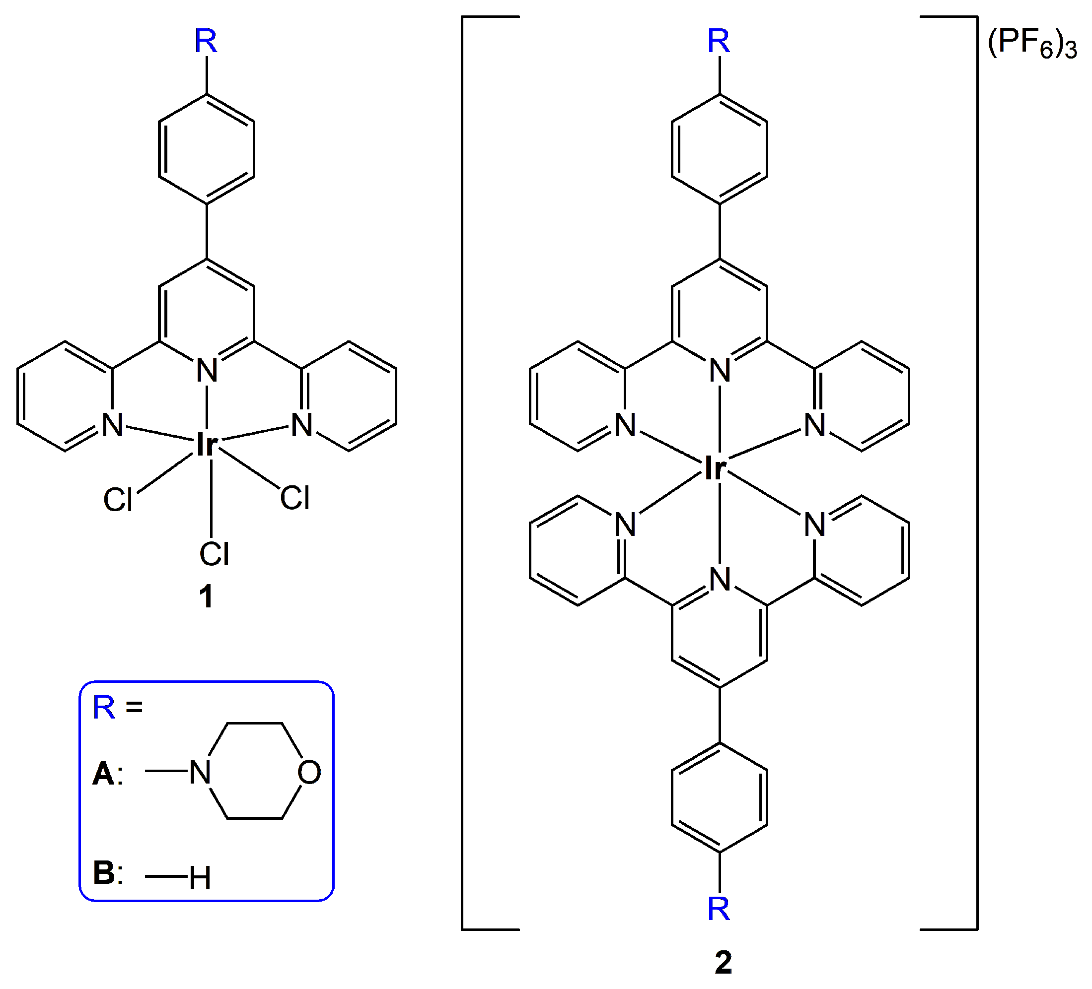
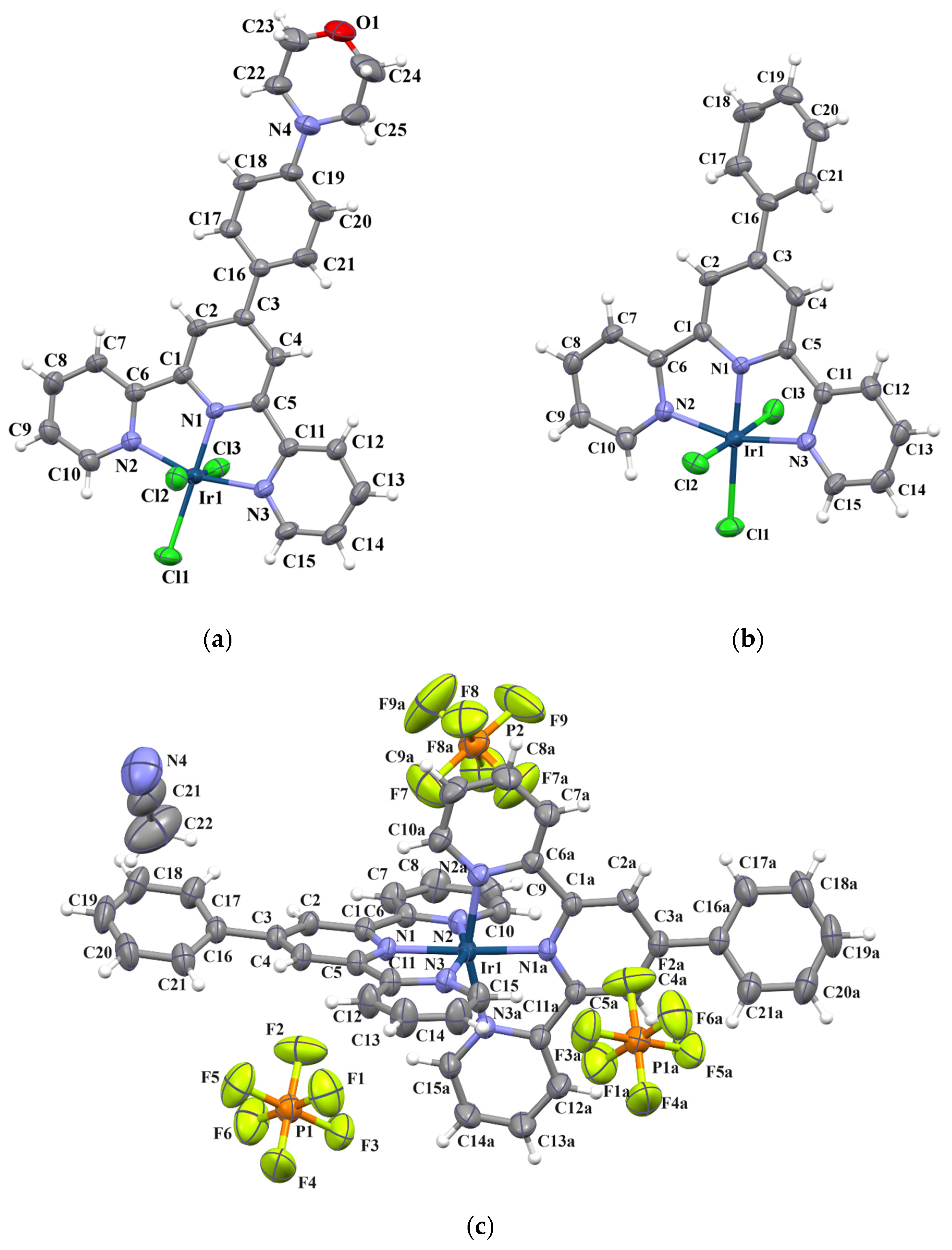
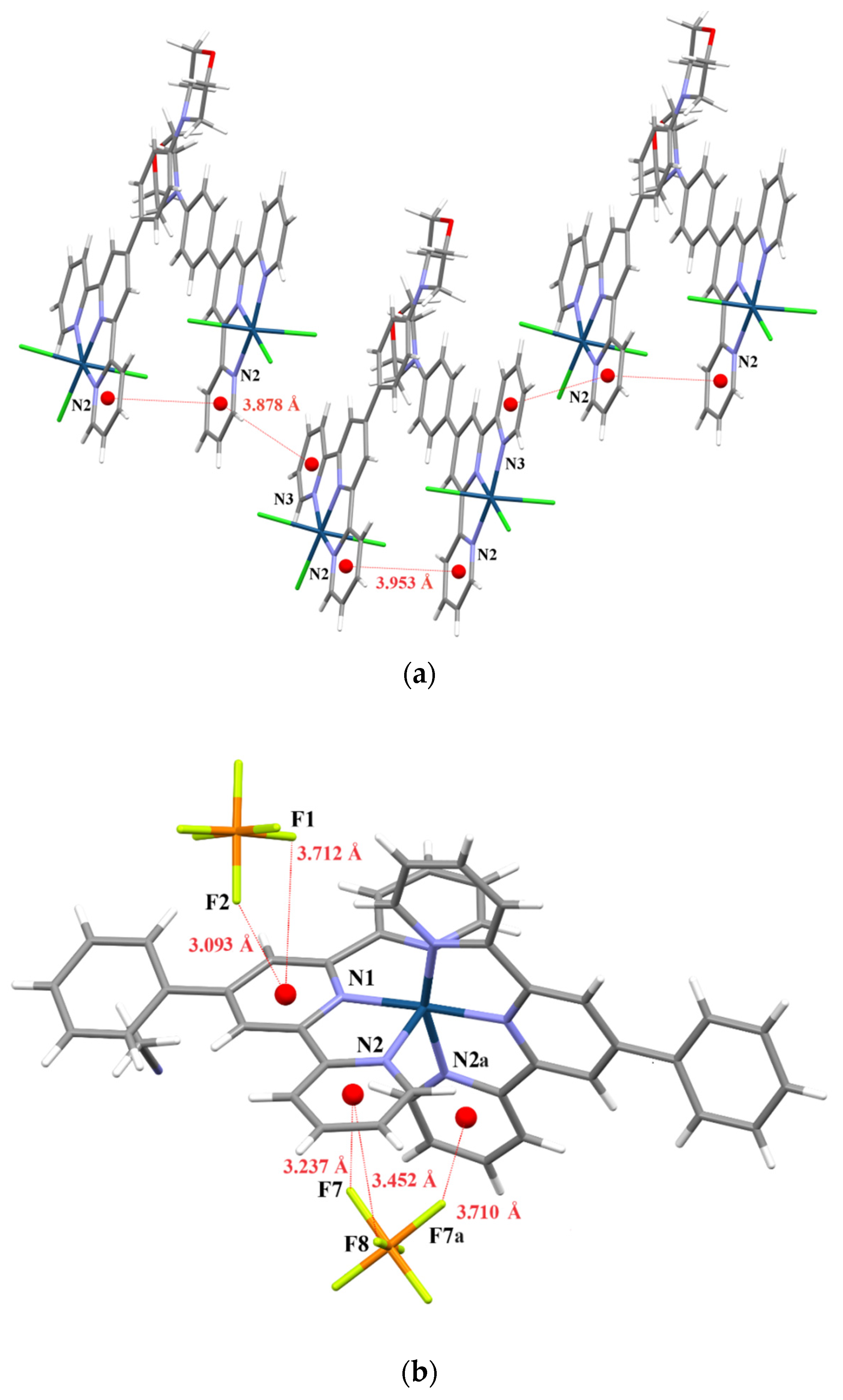
2. Results and Discussion

The complexes [IrCl3(R-C6H4-terpy-κ3N)] (1A and 1B) and [Ir(R-C6H4-terpy-κ3N)2](PF6)3 (2A and 2B) were prepared using conventional synthetic procedures [39,40,41]. Specifically, the iridium(III) chloride salt was heated with one equivalent of the appropriate R-C6H4-terpy ligand in methoxyethanol to synthesize 1A and 1B. The corresponding [IrCl3(R-C6H4-terpy-κ3N)] complex was then reacted with a slight excess of R-C6H4-terpy in ethylene glycol to obtain 2A and 2B. The identity and composition of 12 was confirmed by elemental analysis, 1H and 13C NMR spectroscopies (Figure 1 and Figures S1–S4), and the FT-IR technique (Figure S5).
As demonstrated in Figure 1, the proton signals of the central pyridine ring in the terpyridine backbone (represented by the singlet) were noticeably shifted upfield in the morpholinyl-substituted Ir(III) systems, indicating that the appended electron-donating morpholine group affects the electron density in the distal part of the ligand molecule. The difference in chemical shifts, from 9.11 ppm for 1B to 9.04 ppm for 1A and from 9.62 ppm for 2B to 9.49 for 2A, shows that this effect was stronger for the cationic Ir(III) complexes.
The FT-IR spectra of all the investigated Ir(III) complexes displayed characteristic bands in the region 1615–1525 cm−1, attributable to ν(C=N)terpy and ν(C=C)terpy stretches. The intense bands occurring at ~840 cm−1 and ~555 cm−1 in the FT-IR spectra of 2A and 2B are indicative of PF6 ions (Figure S5) [42].
Additionally, the molecular structures of 1A, 1B, and 2B were unambiguously determined by X-ray analysis. The full structural data of these systems are provided in the Supplementary Materials (Tables S1–S4 and Figure S6). Perspective views of the molecular structures of 1A, 1B, and 2B, with atom numbering, are depicted in Figure 2, while the most relevant bond lengths and angles are summarized in Table 1.
In all reported complexes, the Ir(III) ion was located in a distorted octahedral environment. The coordination sphere of 1A and 1B was defined by three chloride ions and three nitrogen atoms of the tridendate-coordinated R-C6H4-terpy (κ3N) ligand. Due to the geometrical constraints imposed by the planar terpy framework, both nitrogen and chlorine donor atoms adopted meridional arrangements. The crystal structure of 2B consisted of the complex cations [Ir(Ph-terpy-κ3N)2]3+, PF6 counteranions in a 1:3 molar ratio, and solvent (MeCN) molecules. The metal center of [Ir(Ph-terpy-κ3N)2]3+ was octahedrally surrounded by six nitrogen atoms from two Ph-terpy-κ3N ligands, coordinated in a mer-fashion.
Typical of the terpy-κ3N coordination mode [43], the Ir–N bond lengths involving peripheral pyridine rings in structures 1A, 1B, and 2B were longer than the Ir–Ncentral pyridine distances (Table 1). The shortening of the Ir–Ncentral pyridine bond length was rationalized by a more efficient overlap of the t2g metal orbitals with the π* orbitals of the central pyridine relative to the peripheral pyridyl groups. Consequently, the chloride ligand trans-located to the central pyridine ring of R-C6H4-terpy in molecules 1A and 1B exhibited an elongated bond distance in relation to the other Ir–Cl ones (Table 1).
In all the studied Ir(III) complexes, a considerable angular distortion from the octahedral geometry was reflected in the N–Ir–N bite angles due to the formation of five-membered metallocycles upon the chelating coordination of R-C6H4-terpy. The N–Ir–N bite angles varied from 79.84(12) to 80.99(16)° (Table 1). The terpy framework in structures 1A, 1B, and 2B showed good planarity, with the dihedral angles between the mean planes of the central pyridine and terminal aromatic rings ranging from 1.26° to 7.92°. The plane of the phenyl ring of 1A maintained near co-planarity (1.92°) with the central pyridine plane, while in the model compounds, it was inclined to the central pyridine by 20.48° in 1B, and 23.46° in 2B.
The arrangement of molecules [IrCl3(R-C6H4-terpy-κ3N)] in the crystal structures of 1A and 1B was governed by π•••π stacking interactions and weak hydrogen bonds (C–H•••Cl and C–H•••O). The crystal packing analysis of 2B revealed interactions between the cations [Ir(Ph-terpy-κ3N)2]3+ and anions PF6, facilitated by weak hydrogen bonds (C–H•••F) and P–F•••π contacts (Figure 3 and Tables S2 and S4). The shortest Ir•••F distances were 5.542 Å and 5.940 Å.

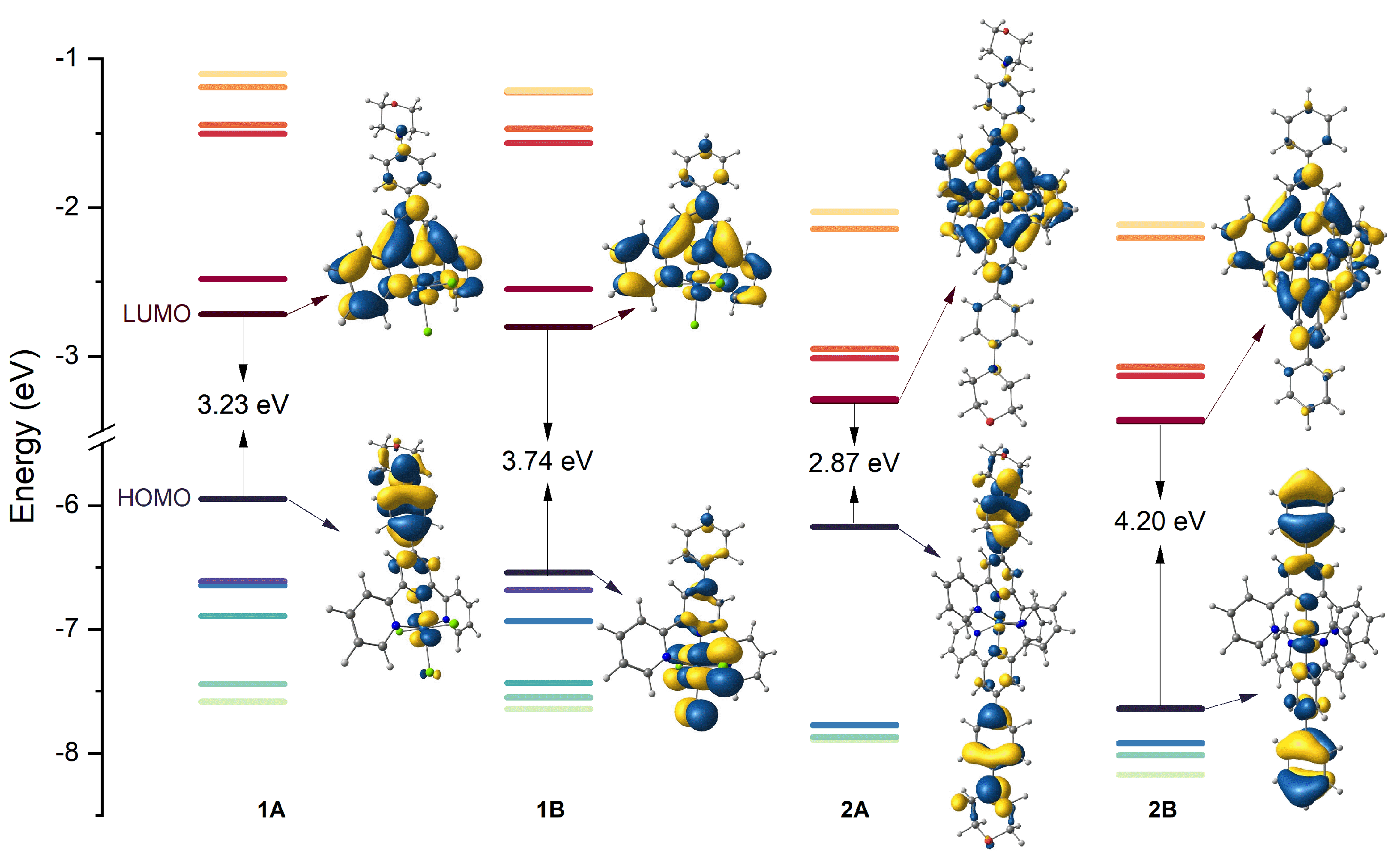
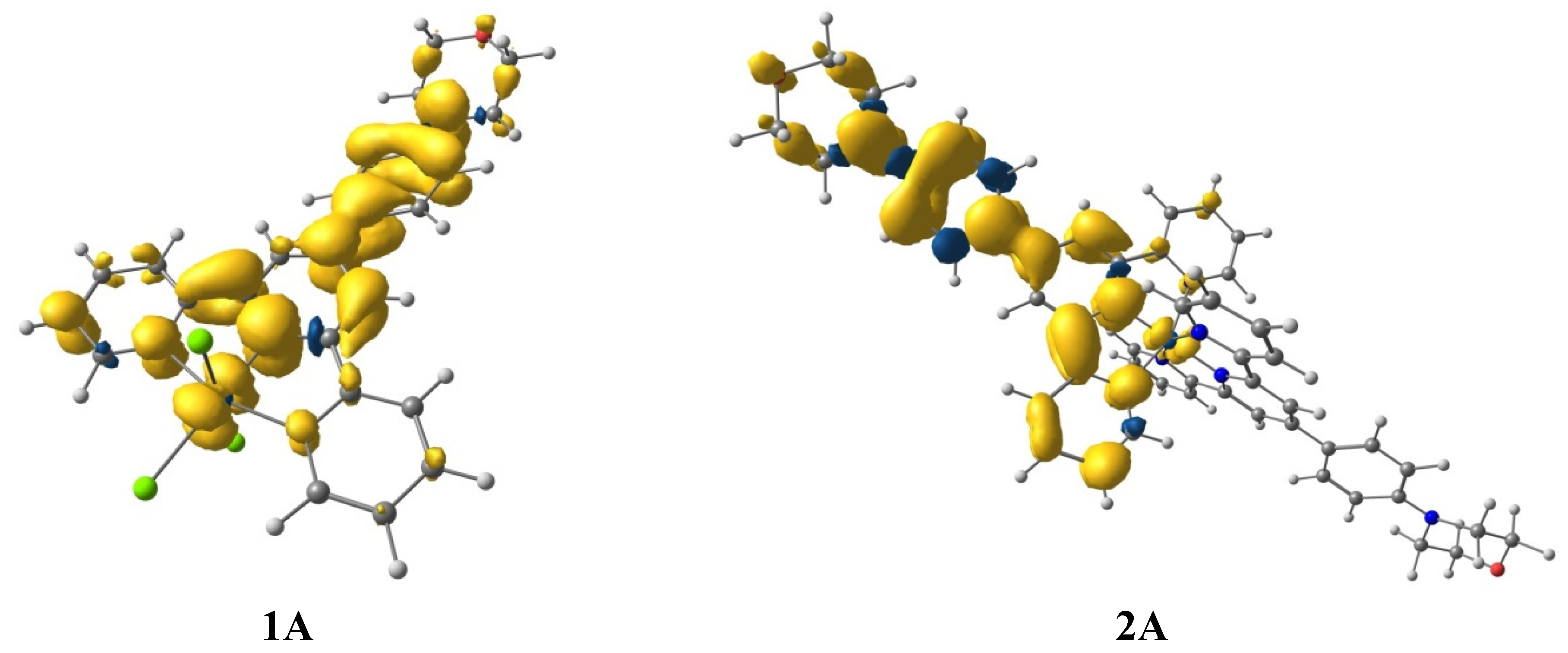
3. Ground-State Molecular Orbitals

To better understand the impact of the electron-donating morpholinyl group on the photophysical behavior of the [IrCl3(R-C6H4-terpy-κ3N)] (1A, 1B) and [Ir(R-C6H4-terpy-κ3N)2](PF6)3 (2A, 2B) systems, the energies and electron distributions of the highest occupied (HOMO) and lowest unoccupied (LUMO) molecular orbitals are briefly presented prior to the discussion of the optical properties of 1A2A and 1B2B. DFT calculations were performed using Gaussian-16 software [44] at the TD-DFT/PCM/PBE0/SDD/def2-TZVP level [45] (Figure 4, Tables S5–S7, Figure S7).
As shown in Figure 4, the attachment of the morpholinyl group to Ph-terpy led to the destabilization of both the HOMO and LUMO orbitals, with more pronounced energy variations in the case of the HOMO level. In the pairs 1A1B and 2A2B, the HOMO energy increased by ~0.60 eV and ~1.47 eV upon going from the unsubstituted to the morpholinyl-decorated system, respectively. The destabilization of the LUMO level due to the morpholine incorporation was ~0.08 eV and ~0.14 eV for pairs 1A1B and 2A2B. Consequently, the Ir(III) complexes with the morph-C6H4-terpy ligand (1A and 2A) exhibited significantly reduced HOMO–LUMO gaps relative to their reference complexes (1B and 2B). The HOMO–LUMO energy gap decreased in the order of 2B (4.20 eV) > 1B (3.74 eV) > 1A (3.23 eV) > 2A (2.87 eV).
Notably, the replacement of three electron-donating halide ions in [IrCl3(R-C6H4-terpy-κ3N)] by the π-accepting R-C6H4-terpyκ3N ligand in [Ir(R-C6H4-terpy-κ3N)2]3+resulted in the stabilization of the HOMO and LUMO levels of the bis-terpyridyl systems. In the pairs 1A2A and 1B2B, the HOMO energy decreased by ~0.22 eV and ~1.1 eV, respectively. In turn, the LUMO energy of 1A and 1B dropped by ~0.58 eV and ~0.64 eV compared to 2A and 2B, respectively.
As expected, the morpholine substituent also altered the nature of the HOMO orbital. Unlike the HOMO of 1B, which predominantly comprised the orbitals of the Ir(III) center (61.8 %) and chloride ions (27.4 %), the HOMO of 1A was a combination of morpholine (32.1%), phenylene (48.1%), and terpy (9.5%) orbitals. Compared to 1B, the metal character of the HOMO of 1A decreased from 61.8 % to 8.5 %. The highest occupied molecular orbitals of 2A and 2B resided almost exclusively on the R-C6H4-terpy ligand. For morpholinyl-substituted complex (2A), it comprised the morpholine group (35.9 %) and phenylene linker (46.5%). The metal contributions in the HOMO of 2A and 2B were 2.1% and 12.5%, respectively. The lowest unoccupied molecular orbital of all the studied Ir(III) complexes was predominately constituted by π* orbitals of the terpy backbone.

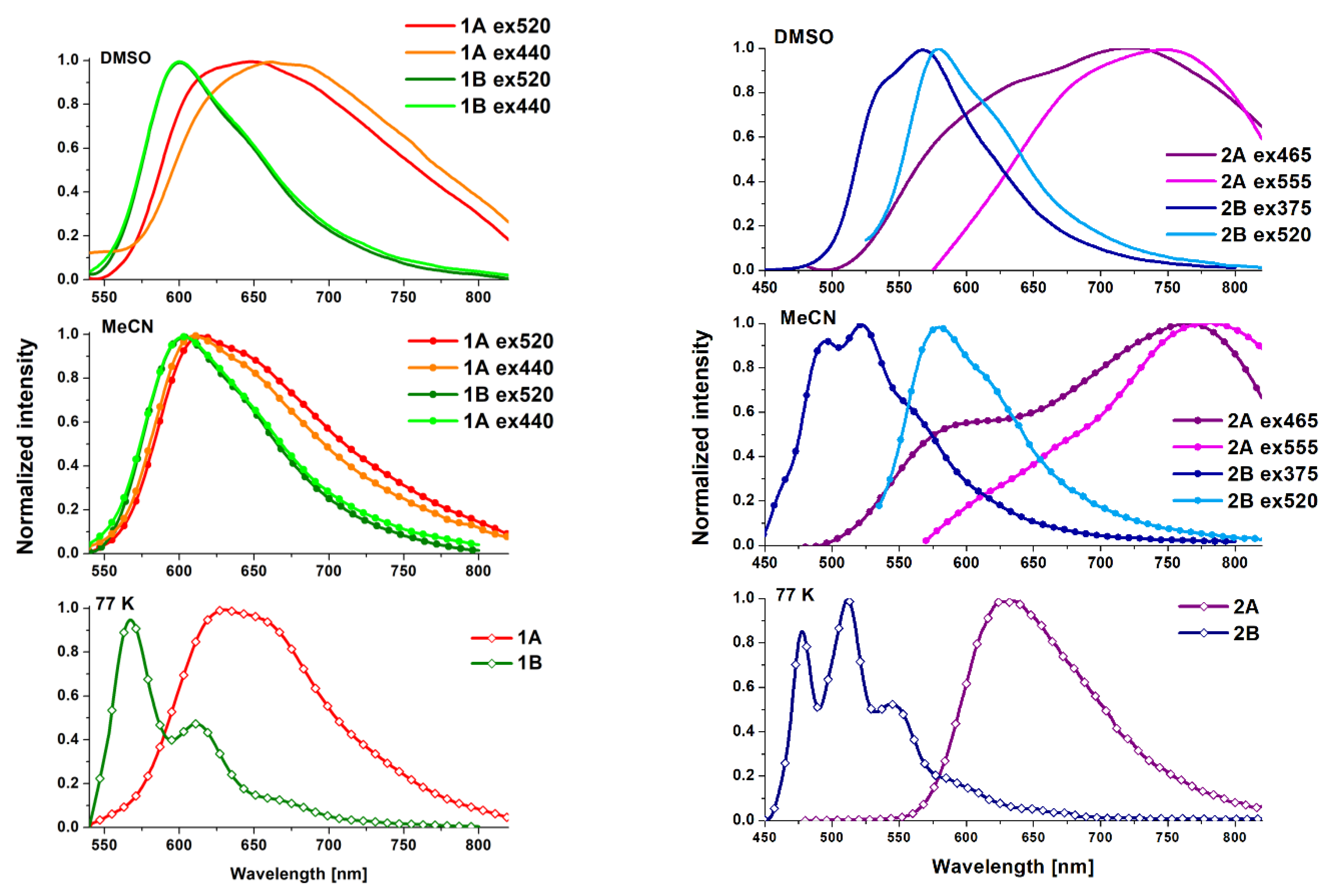
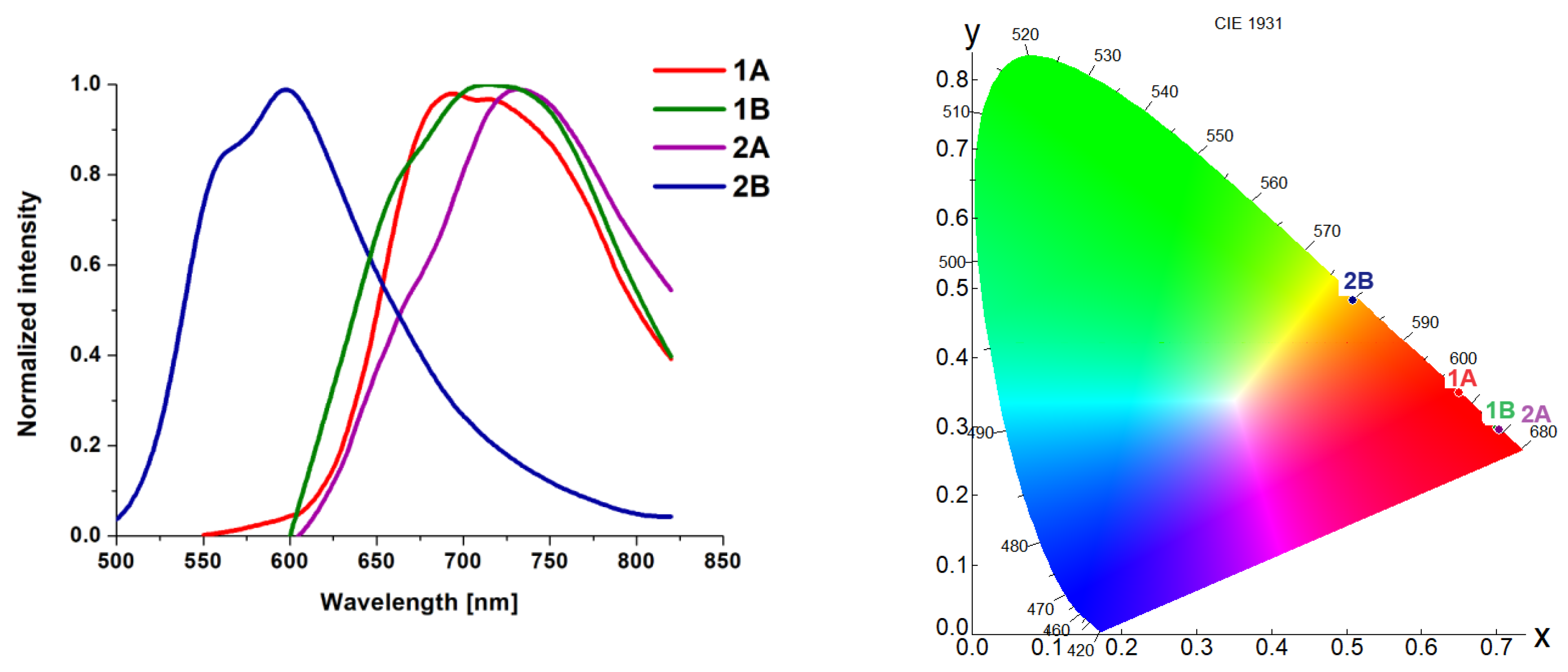
4. Photophysical Properties–Experimental and Theoretical Insights

The electronic absorption properties of [IrCl3(R-C6H4-terpy-κ3N)] and [Ir(R-C6H4-terpy-κ3N)2](PF6)3 were studied in MeCN and DMSO (Figure 5 and Figure S8). The complexes 1 and 2 showed insufficient solubility in less polar solvents. The absorption band maxima and molar extinction coefficients of 12 are summarized in Table S8.
As demonstrated in Figure 5, the model complex 1B exhibited intense high-energy absorption bands in the range of 225–350 nm, which were attributed to π–π* transitions of the Ph-terpy ligand, and noticeably weaker absorptions with maxima above 350 nm, which were tentatively assigned to charge-transfer transitions 1MLLCT with a possible admixture of spin-forbidden singlet-triplet 3MLLCT due to the high spin-orbit coupling constant of iridium [46,47,48]. In contrast, the complex 2B principally absorbed wavelengths below 400 nm. As previously established for related systems [37,41,49,50,51,52], these UV bands of 2B were predominantly attributed to 1IL transitions within the coordinated Ph-terpy ligands. The very low magnitudes of molar extinction coefficients for the absorptions of 2B in the visible region (Table S8 and Figure S8) are consistent with spin-forbidden singlet-triplet 3MLCT transitions [49,53].
Within the series [IrCl3(R-C6H4-terpy-κ3N)], the attachment of the electron-donating morpholinyl group to the phenyl ring of 2,2′:6′,2″-terpyridine evoked a minor effect on the absorption energies but led to a significant enhancement of the molar extinction coefficients of absorptions bands in the range of 350–550 nm (Table S8 and Figure 5). In contrast, dramatic changes in the absorption characteristics were observed for 2A in relation to 2B. The morpholinyl-substituted complex (2A) was deeply red and displayed a very strong absorption tailing up to 600 nm, which was absent in the UV–Vis spectrum of its parent chromophore 2B, which was pale yellow in solution. The molar extinction coefficient of this band exceeded 4 × 10⁴ M−1·cm−1 in solution.
The character of electronic transitions underlying the absorption features of [IrCl3(R-C6H4-terpy-κ3N)] and [Ir(R-C6H4-terpy-κ3N)2](PF6)3 was also investigated theoretically at the DFT/PCM/PBE0/SDD/def2-TZVP level (Figure 6). The calculations confirmed that the changes in the absorption characteristics of the morpholinyl-substituted Ir(III) complexes (1A and 2A) relative to the parent chromophores (1B and 2B) were due to the involvement of 1ILCT transitions, occurring from the electron-donating morpholinyl group to the π-acceptor terpy unit. The low-energy absorptions (above 350 nm) of 1A mainly originated from the excitations HOMO→LUMO, HOMO→L + 1, H–1→LUMO, H–1→L + 1, H-2→LUMO, and H–2→L + 1. Based on the percentage composition of the molecular orbitals, the transitions HOMO→LUMO and HOMO→L + 1 can be designated as 1ILCT/1IL, while the other ones correspond to the 1MLLCT excitations. The low-energy absorptions of 1B only comprised 1MLLCT transitions (Table S6). Some discrepancies between the experimental and theoretical electronic spectra of 1A and 1B in the low energy part may be rationalized by the possible involvement of spin-forbidden singlet-triplet transitions, which are evidenced by the TD-DFT calculations [46]. The strong visible absorption of 2A was dominated by the transitions HOMO→LUMO and HOMO–1→LUMO + 1 of prevailing ILCT nature. Both HOMO and HOMO-1 comprised the morpholine group and phenylene linker, while LUMO and LUMO + 1 resided predominately on the terpy backbone.
Prior to the investigations of the excited-state properties of 12, their stability and photostability in solution were confirmed by UV–Vis spectroscopy, as demonstrated in Figures S9 and S10. The photoluminescence properties of the Ir(III) complexes were explored in MeCN and DMSO solutions at room temperature (RT), in a solid state, and in a frozen matrix of EtOH/MeOH (4:1 v/v) at 77 K, as presented in Figure 7 and Table 2. Some additional photophysical data of 12 are provided in the Supplementary Materials (Figures S11–S18).
For all the studied complexes, their lifetimes were in the microsecond or sub-microsecond domain (Table 2), and the photoluminescence intensities and lifetimes were sensitive to oxygen quenching, supporting the idea that the observed emission originates from a triplet excited state (Figures S11 and S12).
The complexes 1A and 1B in deaerated MeCN and DMSO solutions showed unstructured emission bands. Compared to the model chromophore 1B, the phosphorescence band of 1A was noticeably broader and appeared at a lower energy, with the emission maximum red-shifted by 50 nm for the DMSO solution and 10 nm for MeCN upon excitation at 520 nm. A bathochromic shift in the emission maximum of 1A was accompanied by a slight decrease in the excited-state lifetime relative to 1B (Table 2).
In contrast to 1B, which showed excitation-independent emission, the energy of the emission of 1A was affected by excitation wavelengths, becoming noticeably red-shifted upon wavelength excitations in the range of 400–525 (Figure S13). A difference between 1A and 1B was also noticed when the frozen-state emissions were considered. Upon cooling to 77 K, the emission band of 1A remained structureless and appeared almost in the same range as the RT emission band, while the frozen-state emission band of 1B exhibited well-resolved vibronic progressions and appeared at a higher energy in relation to that in solution, as shown in Figure 7. All these findings indicate that the complex 1B emits from a triplet excited state of 3MLLCT character with a minor contribution of 3π→π*terpy transitions (3IL), while the emissive triplet excited state of the morpholinyl-substituted Ir(III) analog (1A) has a mixed nature 3MLLCT–3ILCT. By analogy to previous findings [14,29,30,31,32,33,34,35,36], the contribution of 3ILCT in the triplet excited state of 1A manifested in a bathochromic shift of the emission in solution at RT and frozen matrix EtOH/MeOH relative to the parent chromophore. Transition metal complexes functionalized with electron-rich groups, which emit from the 3MLLCT excited state, are expected to show a hypsochromic shift compared to the unsubstituted model compounds [14,29].
For the bis-terpyridyl iridium(III) complexes, the morpholinyl group induced dramatic changes in the emission characteristics (Figure 7 and Figures S14–S17). In solution at RT, the model chromophore 2B displayed a green emission. Its emission band exhibited a weak vibronic structure, which became well resolved upon cooling to 77 K. The frozen-state emission band of 2B appeared almost in the same range as that in MeCN at RT upon excitations of ≤375 nm, but it was slightly blue-shifted in relation to its emission in DMSO (Figure 7 and Figure S14). The differences in the emission energies and spectral profiles of 2B in MeCN and DMSO can be rationalized by the fact that the emission of [Ir(R-terpy-κ3N)2](PF6)3 systems in solution may occur from the triplet excited state of the cation [Ir(R-terpy-κ3N)2]3+ and ion-pair adduct [Ir(R-terpy-κ3N)2]3+·PF6 [41,50,54,55,56,57]. While the emission of 2B in MeCN upon excitations of ≤375 nm can be attributed to the phosphorescence of [Ir(R-terpy-κ3N)2]3+, the ion-pair formation quenches the phosphorescence of [Ir(R-terpy-κ3N)2]3+ in DMSO. The observed emission of 2B in DMSO originated mainly from the ion-pair adduct [Ir(R-terpy-κ3N)2]3+·PF6. In acetonitrile solution, the emission from the ion-pair adduct occurred using longer excitation wavelengths (Figure S14), as previously reported for the related complex [Ir(terpy-κ3N)2](PF6)3 [57]. It is worth noting that there were also two alternative explanations of the photoluminescence behavior of [Ir(R-terpy-κ3N)2]3+ cations in previous reports. The emission properties of such systems are attributed to the triplet ligand-centered (3IL) or mixed 3IL–3MLCT excited states [39]. Regarding the first assignment, the lack of the strong vibronic progression of the emission band at RT, typical of 3IL emission, is rationalized by the thermal distribution of conformers with different torsion angles between the terpy core and aryl pendant group, leading to a less well-resolved emission band [39]. The transient absorption findings, discussed in the next section, indicate that the emission of 2B is not of pure of 3IL phosphorescence, but rather occurs from the triplet excited state of the mixed 3IL–3MLCT character.
The emission spectrum of the morpholinyl-substituted complex (2A) was dominated by the structureless band in the NIR region, with the maximum at 760 nm in MeCN and 750 nm in DMSO, red-shifted by ~240 and 180 nm relative to the model complex 2B, respectively. A bathochromic shift in the emission of 2A was accompanied by a noticeable decrease in the excited-state lifetime relative to 2B (Table 2). The shoulder/band at higher energies of 2A can be assigned the reabsorption or emission from the 3MLLCT excited state (Figure S14). The frozen-state emission of 2A remained non-structured and occurred in a higher energy region (630 nm) compared to its room-temperature emission. The photoluminescence characteristics of 2A are typical of the emission occurring from 3ILCT excited state [58,59,60,61]. It is worth noting that NIR emitters are of high significance due to their potential applications in biomedical imaging [62,63,64].
The photoluminescence properties of 12 were also investigated in the solid state. As demonstrated in Figure 8, the solid-state triplet emission band of 2B occurred in a higher energy region (orange-yellow) and was characterized by a noticeably prolonged lifetime (~10 μs) compared to other studied complexes, showing emission in the red wavelength range, with lifetimes in the sub-microsecond domain (Table 2).
To theoretically investigate the excited-state properties of complexes 12, their geometries were optimized in triplet states (T1) and triplet energies were calculated from the energy difference between the ground singlet and triplet excited states Δ E T 1 S 0 . The theoretically determined triplet emissions, 694 nm for 1A, 590 nm for 1B, 832 nm for 2A, and 540 nm for 2B, are in satisfactory agreement with the experimental values (Table 2 and Table S7). Regarding the spin density surface plots (Figure 9 and Figure S7), the emission in the complexes 1A, 1B, 2A, and 2B occurred due to the triplet excited state of an MLCT-ILCT, MLLCT, ILCT, and IL-MLCT nature, respectively.

5. Femtosecond Transient Absorption Spectra

To further understand the triplet excited state characteristics of the studied complexes, femtosecond transient absorption (fs-TA) measurements were recorded in deaerated DMSO (1A and 1B) and MeCN (2A and 2B) solutions upon excitation at 355 nm. TA solutions were prepared at concentrations of 50–250 μM to provide an absorbance of ~0.5 in 2 mm path length quartz cells at the excitation wavelength. The results of the fs-TA measurements and global fitting analyses are summarized in Figure 10 and Figures S19–S23. The photostability of the Ir(III) complexes during the TA experiments was verified by comparing their UV–Vis spectra recorded before and after irradiation (Figure S24).
The complexes 1A and 2A exhibited distinct transient absorption (TA) features compared to their parent chromophores (1B and 2B), indicating that the introduction of the electron-donating morpholinyl group significantly affected the nature of the lowest triplet excited state of [IrCl3(R-C6H4-terpy-κ3N)] and [Ir(R-C6H4-terpy-κ3N)2](PF6)3. Specifically, 1A and 2A displayed ground-state bleaching (GSB) in wavelength regions corresponding to their visible charge-transfer absorptions, along with excited-state absorption bands (ESA) in the ranges of 375–415 nm and 522–670 nm for 1A, and 375–425 nm and 540–670 nm for 2A. In contrast, the TA spectra of the parent chromophores displayed only positive bands across the UV and visible regions, indicating that 1B and 2B have a stronger triplet excited-state absorption than the ground-state absorption [65,66,67,68]. For all the studied Ir(III) complexes, the TA signals appeared promptly after photoexcitation at 355 nm. After vibrational cooling, solvation, and geometrical relaxation within the triplet manifold, these signals persisted up to the end of the delay stage (7 ns). Ultrafast intersystem crossing occurs on a time scale shorter than the instrument response and is not detected with our experimental setup [48].
The comparison of the TA spectral profiles of 2A and 2B (Figure S23) indicated the involvement of different triplet excited states in their transient absorptions. For complex 2A, the TA spectra comprised two ESA bands separated by a strong GSB signal, with two well-defined isosbestic points at 425 and 540 nm. The decay of both ESA bands and the recovery of the GSB occurred on the same time scale. The ESA band in the UV range was attributed to the terpyridyl anion radical [69,70], associated with the 3ILCT excited state, represented by the ESA with a maximum at 645 nm. The triplet excited state was of a predominantly 3ILCT nature.
The fs-TA data of 2B indicated a mixed 3IL-3MLCT nature. With reference to the TA findings for [Ir(terpy-κ3N)2](PF6)3 [51], the higher energy TA signal suggests a 3ILterpy triplet excited state, while the lower energy one can be assigned to the 3MLCT transient absorption. The presence of two different transient species undergoing interconversion was supported by the isosbestic point at 515 nm in the TA spectrum of 2B. Additionally, the lower energy ESA band of 2A was similar to the low energy ESA band of the model chromophore 1B, characterized by the lowest triplet excited state of a 3MLLCT nature (Figure S23).
The comparison of the TA spectral profiles of 1A and 1B (Figure S23) demonstrated that the morpholinyl group induces smaller variations in the TA spectral features of [IrCl3(R-C6H4-terpy-κ3N)]. The presence of a GSB band, the broadening of the visible ESA band, and different decay kinetics relative to 1B suggest that the TA spectral features of 1A correspond to a triplet excited state of a mixed 3MLLCT-3ILCT nature, with a dominant contribution of the first component.

6. Conclusions

The work presents comprehensive studies of [IrCl3(morph-C6H4-terpy-κ3N)] (1A), [Ir(morph-C6H4-terpy-κ3N)2](PF6)3 (2A), [IrCl3(Ph-terpy-κ3N)] (1B), and [Ir(Ph-terpy-κ3N)2](PF6)3 (2B), which were designed to explore the impact of the electron-donating morpholinyl (morph) group on the ground- and excited-state properties of two different types of Ir(III) complexes: [IrCl3(R-C6H4-terpy-κ3N)] and [Ir(R-C6H4-terpy-κ3N)2](PF6)3. We demonstrated that the attachment of morpholine leads to a change in the nature of the singlet and triplet excited states of bis-terpyridyl iridium(III) complexes, switching from intraligand with a small admixture of metal-to-ligand charge transfer (IL–MLCT) for 2B to intraligand charge transfer (ILCT) in 2A. Consequently, the compounds 2A and 2B exhibited completely different absorption and emission features. The deeply red complex 2A displayed very strong absorption tailing up to 600 nm, which was absent in the UV–Vis spectrum of its parent chromophore 2B, which was pale yellow in solution and predominately absorbed wavelengths below 400 nm. The room-temperature emission of 2A was red-shifted by ~200 nm and showed a decreased lifetime relative to the model complex 2B. The optical properties of the mono-terpyridyl iridium(III) complexes were less impacted by the morpholinyl substituent. Only an absorption enhancement in the range of 350–550 nm and a small red shift in the emission were observed for 1A compared to the model chromophore 1B. Bases on the static and time-resolved spectroscopic findings, accompanied with theoretical DFT/TD-DFT calculations, the character of the singlet and triplet excited states of 1A was assigned as a mixed MLLCT-ILCT. The structure–property relationships discussed herein are of high importance for controlling the photophysical characteristics of [IrCl3(R-C6H4-terpy-κ3N)] and [Ir(R-C6H4-terpy-κ3N)2](PF6)3, and making further progress in the development of Ir-based luminophores.

7. Experimental

7.1. Materials and Methods

Commercially available iridium(III) chloride salt hydrate, ammonium hexafluorophosphate, 2-acetylpyridine, 4-(4-morpholinyl)benzaldehyde and benzaldehyde were used without further purification. The solvents for the syntheses and spectroscopic measurements were reagent and HPLC grade, respectively. Ligands morph-C6H4-terpy and Ph-terpy were prepared through base-mediated Kröhnke condensation from 2-acetylpyridine and two equivalents of the appropriate aldehyde (4-(4-morpholinyl)benzaldehyde for morph-C6H4-terpy and benzaldehyde for Ph-terpy, as described previously [31,71]. Elemental analyses were performed using a Vario EL Cube (Elementar) for the C, H, and N content. NMR spectra were recorded on a Bruker Avance 500 NMR spectrometer in DMSO-d6. IR spectra were acquired using a Nicolet iS5 FTIR spectrophotometer (4000–400 cm−1) using the KBr pellet method. X-ray diffraction data were collected at room temperature using a Gemini A Ultra diffractometer (Oxford Diffraction) with MoKα radiation (λ = 0.71073 Å), and crystallographic data for 1A, 1B, and 2B were deposited with the Cambridge Crystallographic Data Center (CCDC 2359006-2359008). The UV–Vis absorption spectra were recorded on an Evolution 220 (ThermoScientific) UV–Vis spectrometer. The emission and excitation spectra along with the time-resolved TCSPC measurements at room temperature and in 77 K frozen matrix (4:1 v/v) were measured on an FLS-980 fluorescence spectrophotometer (Edinburgh Instruments). The fs TA spectra were acquired using a pump-probe transient absorption spectroscopy system (Ultrafast Systems, Helios). Theoretical calculations were performed using the GAUSSIAN-16 (Rev. C.01) program package [44] at the DFT or TD-DFT level with the PBE0 [72,73,74] functional. The basis sets used were Stuttgart Relativistic Small Core ECP with the corresponding pseudopotentials [45,75] for iridium (obtained from Basis Set Exchange Database [76]) and def2-TZVP for other elements [45,77]. A more extended experimental description is provided in the Supplementary Materials.

7.2. Preparation of Ir(III) Complexes

[IrCl3(R-C6H4-terpy-κ3N)]: A mixture of IrCl3·xH2O (0.30 g; 1 mmol) and the appropriate R-terpy ligand (1 mmol) in degassed methoxyethanol was placed in a 25 mL Teflon-lined hydrothermal synthesis autoclave reactor and heated at 120 °C for 24 h. After that, the autoclave was gradually cooled to room temperature (24 h). The resulting reddish-brown crystalline precipitate was collected by filtration; washed with acetonitrile, chloroform, and diethyl ether; and dried in air.
[IrCl3(morph-C6H4-terpy-κ3N)] (1A): Yield: 0.48 g, 70%. Anal. calc. for C25H22Cl3IrN4O (693.04 g/mol): C 43.33 H 3.20, N 8.08% found: C 43.50; H 3.03; N 8.43%. 1H NMR (500 MHz, DMSO-d6) 1H NMR (500 MHz, DMSO-d6) δ = 9.22 (dd, J = 5.8, 1.6 Hz, 1H), 9.04 (s, 1H), 8.92 (d, J = 8.1 Hz, 1H), 8.29 (td, J = 7.8, 1.6 Hz, 1H), 8.17 (d, J = 9.0 Hz, 1H), 7.96 (ddd, J = 7.6, 5.5, 1.4 Hz, 1H), 7.20 (d, J = 9.1 Hz, 1H), 3.81–3.79 (m, 2H), 3.38–3.36 (m, 2H) ppm. 13C NMR (126 MHz, DMSO-d6) δ = 159.93, 157.44, 153.45, 151.20, 140.56, 129.62, 128.63, 125.62, 124.64, 119.59, 114.87 ppm. 31P NMR (202 MHz, DMSO-d6) δ = −133.56–(−154.64) (m, PF6) ppm. IR (KBr, cm−1) intensity: s—strong, m—medium, w—weak: 3449 (w), 3060 (w), 2952 (w), 2844 (w), 1596 (s), 1529 (m), 1474 (w), 1446 (w), 1414 (m), 1385 (w), 1305 (w), 1265 (w), 1230 (s), 1220 (s), 1119 (m), 1051 (w), 930 (m), 883(w), 814 (w), 791 (m), 762 (w), 725 (w), 568 (w), 523 (w), 454 (w).
[IrCl3(Ph-terpy-κ3N)] (1B): Yield: 0.110 g, 55%. Anal. calc. for C21H15Cl3IrN3 (607.94 g/mol): C 41.49, H 2.49, N 6.91% found: C 40.27; H 2.78; N 6.70%. 1H NMR (500 MHz, DMSO-d6) δ = 9.22 (dd, J = 5.6, 1.6 Hz, 1H), 9.11 (s, 1H), 8.92 (d, J = 8.1 Hz, 1H), 8.31 (td, J = 7.8, 1.6 Hz, 1H), 8.21 (dd, J = 7.2, 1.4 Hz, 1H), 7.98 (ddd, J = 7.2, 5.6, 1.4 Hz, 1H), 7.71 (t, J = 7.7 Hz, 1H), 7.61 (t, J = 7.4 Hz, 1H) ppm. 13C NMR (126 MHz, DMSO-d6) δ = 159.68, 159.07, 157.84, 155.95, 153.49, 153.38, 142.67, 140.73, 135.16, 130.32, 129.91, 129.72, 128.84, 128.81, 128.76, 127.46, 125.85, 123.03, 122,11, 121.56 ppm. 31P NMR (202 MHz, DMSO-d6) δ = −133.55–(−154.63) (m, PF6) ppm. IR (KBr, cm−1) intensity: vs—very strong; s—strong, m—medium, w—weak: 3441 (m), 3047 (m), 1605 (s), 1547 (w), 1475 (m), 1451 (w), 1413 (s), 1304 (w), 1246(w), 1233 (m), 1161 (m), 1103 (w), 1051 (w), 1030 (w), 890 (m), 790 (s), 768 (s), 729 (w), 691 (m), 655 (w), 646 (w), 626 (w), 505 (w), 479 (m), 455 (w).
[Ir(R-C6H4-terpy-κ3N)2](PF6)3: A mixture of [IrCl3(R-C6H4-terpy-κ3N)] (0.1 mmol) and the appropriate R-C6H4-terpy ligand (0.125 mmol) in ethylene glycol (8 mL) was heated at 180 °C (oil bath) in an argon atmosphere overnight. Subsequently, a clear solution was cooled to room temperature, and a saturated aqueous solution of (NH4)2PF6 was added dropwise, leading to a reddish-brown (2A) and yellow-orange (2B) precipitate. After stirring for 8 h, the obtained precipitate was filtered, washed with water and diethyl ether, and dried in air. The crude products of 2A and 2B were purified by crystallization from the dichloromethane–methanol mixture.
[Ir(morph-C6H4-terpy-κ3N)2](PF6)3 (2A): Yield: 0.064 g, 45%. Anal. calc. for C50H44F18IrN8O2P3·CH2Cl2 (1500.98 g/mol): C 40.81, H 3.09, N 7.47% found: C 41.14; H 2.69; N 7.41%. 1H NMR (500 MHz, DMSO-d6) δ = 9.49 (s, 1H), 9.21 (d, J = 8.4 Hz, 1H), 8.45 (d, J = 9.0 Hz, 1H), 8.35 (td, J = 7.9, 1.5 Hz, 1H), 7.95 (dd, J = 5.7, 1.5 Hz, 1H), 7.55 (ddd, J = 7.5, 5.7, 1.4 Hz, 1H), 7.31 (d, J = 9.1 Hz, 1H), 3.84 (t, J = 4.9 Hz, 2H), 3.47 (t, J = 4.9 Hz, 2H) ppm. 13C NMR (126 MHz, DMSO-d6) δ = 159.04, 154.45, 153.93, 153.86, 153.61, 142.77, 129.98, 129.77, 127.41, 123.70, 121.61, 118.55, 114.61, 66.41, 47.27 ppm. IR (KBr, cm−1) intensity: vs—very strong; s—strong, m—medium, w—weak: 3422 (m), 3086 (w), 2853 (w), 1592 (s), 1527 (w), 1478 (m), 1465 (m), 1449 (m), 1419 (m), 1361 (s), 1298 (w), 1232 (s), 1213 (s), 1114 (m), 1048 (m), 1032 (m), 928 (m), 839 (vs), 785 (m), 755 (w), 645 (w), 558 (s).
[Ir(Ph-terpy-κ3N)2](PF6)3 (2B): Yield: 0.081 g, 65%. Anal. calc. for C42H30F18IrN6P3·CH2Cl2 (1330.77 g/mol): C 38.81, H 2.42, N 6.32% found: C 39.19; H 2.13; N 6.35%. 1H NMR (500 MHz, DMSO-d6 δ = 9.62 (s, 2H), 9.23 (dd, J = 8.2, 0.7 Hz, 2H), 8.46 (d, J = 7.2 Hz, 2H), 8.38 (td, J = 7.9, 1.4 Hz, 2H), 7.96 (dd, J = 5.7, 0.8 Hz, 2H), 7.85 (t, J = 7.6 Hz, 2H), 7.77 (t, J = 7.4 Hz, 1H), 7.58 (ddd, J = 7.5, 5.7, 1.4 Hz, 2H) ppm. 13C NMR (126 MHz, DMSO-d6) δ = 158.69, 154.96, 154.46, 153.79, 142.97, 135.20, 132.40, 130.08, 129.96, 128.80, 127.69, 123.99, 118.55 ppm. IR (KBr, cm−1) intensity: vs—very strong; s—strong, m—medium, w—weak: 3789 (w), 3661 (w), 3432 (m), 3126 (m), 2250 (w), 1610 (s), 1556 (m), 1479 (s), 1454 (w), 1421 (s), 1244 (m), 1170 (m), 1103 (w), 1061 (w), 1034 (w), 1018(w), 977 (w), 896 (w), 838 (vs), 789 (w), 768 (s), 728 (w), 692 (m), 644(w), 558 (s), 498 (w).

Supplementary Materials

Crystallographic data for 1A, 1B and 2B were deposited with the Cambridge Crystallographic Data Center, CCDC 2359006-2359008. Copies of this information may be obtained free of charge from the Director, CCDC, 12 Union Road, Cambridge CB2 1EZ, UK (Fax: +44 1223 336033; e-mail: [email protected] or www.ccdc.cam.ac.uk). The following supporting information can be downloaded at: https://www.mdpi.com/article/10.3390/molecules29133074/s1, Figures S1–S4: 1H and 13C NMR spectra; Figure S5: IR spectra; Table S1: Crystal data and structure refinement; Table S2: Short intra- and intermolecular contacts; Table S3: Short π•••π ring interactions; Table S4: P—F••• π-ring interactions for 2B; Figure S6: View of 3D supramolecular structure of 1B resulting from weak π•••π interactions; Table S5: Experimental and theoretical bond lengths and angles; Table S6: Assignment of electron excitations computed at TD-DFT/PCM/PBE0/SDD/def2TZVP level to acetonitrile UV–Vis absorptions; Table S7: Triplet energies; Figure S7: Spin density surface plots; Table S8: The absorption maxima and molar extinction coefficient values; Figure S8: The absorption spectra of 2B recorded at 2.5 × 10⁻⁵ and 5 × 10⁻⁴ mol/dm3 concentrations; Figure S9: The photostability in DMSO and MeCN solutions; Figure S10: The kinetic stability; Figures S11 and S12: The emission spectra and decay curves in aerated and deaerated solutions: Figures S13 and S14: Excitation-dependent emission; Figures S15–S17: Absorbance, excitation, and emission steady-state spectra of 12 with the corresponding decay curves; Figure S18: Decay curves of 12 in solid state; Figures S19–S23: Summary of the TA analysis; Figure S24: The photodamage tests. References [31,44,45,75,76,77,78,79,80,81,82] has been cited the in the Supplementary Materials.

Author Contributions

The manuscript was prepared through the contributions of all authors. B.M. and J.P.-G. conceptualized, designed, and supervised the research. M.P. carried out the synthesis of the organic ligands and NMR analysis. B.M. and J.P.-G. performed the synthesis of the Ir(III) complexes and X-ray single crystal analysis. J.P.-G., A.K. (Aleksandra Kwiecień) and A.K. (Anna Kryczka) performed the steady-state spectroscopic measurements, while K.C. computed the theoretical calculations. J.P.-G. performed the time-resolved spectroscopic measurements. J.P.-G., A.K. (Aleksandra Kwiecień) and K.C. prepared the figures in the paper and Supplementary Materials files. B.M. and J.P.-G. wrote the manuscript with contributions from all authors. All authors have read and agreed to the published version of the manuscript.

Funding

This work was financed by the funds granted under the Research Excellence Initiative of the University of Silesia in Katowice.

Institutional Review Board Statement

Not applicable.

Informed Consent Statement

Not applicable.

Data Availability Statement

Data are contained within the article and Supplementary Materials.

Acknowledgments

The calculations were carried out in the Wroclaw Centre for Networking and Supercomputing (http://www.wcss.wroc.pl, accessed on 1 June 2024). The authors would like to thank inż Mariola Siwy for the elemental analysis.

Conflicts of Interest

The authors declare no conflicts of interest.

References

  1. Wei, C.; He, Y.; Shi, X.; Song, Z. Terpyridine-Metal Complexes: Applications in Catalysis and Supramolecular Chemistry. Coord. Chem. Rev. 2019, 385, 1–19. [Google Scholar] [CrossRef]
  2. Rupp, M.T.; Shevchenko, N.; Hanan, G.S.; Kurth, D.G. Enhancing the Photophysical Properties of Ru(II) Complexes by Specific Design of Tridentate Ligands. Coord. Chem. Rev. 2021, 446, 214127. [Google Scholar] [CrossRef]
  3. Panicker, R.R.; Sivaramakrishna, A. Remarkably Flexible 2,2′:6′,2″-Terpyridines and Their Group 8–10 Transition Metal Complexes—Chemistry and Applications. Coord. Chem. Rev. 2022, 459, 214426. [Google Scholar] [CrossRef]
  4. Momeni, B.Z.; Abd-El-Aziz, A.S. Recent Advances in the Design and Applications of Platinum-Based Supramolecular Architectures and Macromolecules. Coord. Chem. Rev. 2023, 486, 215113. [Google Scholar] [CrossRef]
  5. Palion-Gazda, J.; Choroba, K.; Maroń, A.M.; Malicka, E.; Machura, B. Structural and Photophysical Trends in Rhenium(I) Carbonyl Complexes with 2,2′:6′,2″-Terpyridines. Molecules 2024, 29, 1631. [Google Scholar] [CrossRef] [PubMed]
  6. Momeni, B.Z.; Davarzani, N.; Janczak, J.; Ma, N.; Abd-El-Aziz, A.S. Progress in Design and Applications of Supramolecular Assembly of 2,2′:6′,2″-Terpyridine-Based First Row d-Block Elements. Coord. Chem. Rev. 2024, 506, 215619. [Google Scholar] [CrossRef]
  7. Breivogel, A.; Kreitner, C.; Heinze, K. Redox and Photochemistry of Bis(Terpyridine)Ruthenium(II) Amino Acids and Their Amide Conjugates—From Understanding to Applications. Eur. J. Inorg. Chem. 2014, 2014, 5468–5490. [Google Scholar] [CrossRef]
  8. Saccone, D.; Magistris, C.; Barbero, N.; Quagliotto, P.; Barolo, C.; Viscardi, G. Terpyridine and Quaterpyridine Complexes as Sensitizers for Photovoltaic Applications. Materials 2016, 9, 137. [Google Scholar] [CrossRef]
  9. Kreitner, C.; Mengel, A.K.C.; Lee, T.K.; Cho, W.; Char, K.; Kang, Y.S.; Heinze, K. Strongly Coupled Cyclometalated Ruthenium Triarylamine Chromophores as Sensitizers for DSSCs. Chem. Eur. J. 2016, 22, 8915–8928. [Google Scholar] [CrossRef]
  10. Mills, I.N.; Porras, J.A.; Bernhard, S. Judicious Design of Cationic, Cyclometalated Ir(III) Complexes for Photochemical Energy Conversion and Optoelectronics. Acc. Chem. Res. 2018, 51, 352–364. [Google Scholar] [CrossRef] [PubMed]
  11. Liu, P.; Shi, G.; Chen, X. Terpyridine-Containing π-Conjugated Polymers for Light-Emitting and Photovoltaic Materials. Front. Chem. 2020, 8, 592055. [Google Scholar] [CrossRef] [PubMed]
  12. Laschuk, N.O.; Ebralidze, I.I.; Easton, E.B.; Zenkina, O.V. Osmium- and Cobalt-Terpyridine-Based Electrochromic Devices for “Smart” Signage Application: The Effect of Lighting on Color Perception. Adv. Electron. Mater. 2021, 7, 2100460. [Google Scholar] [CrossRef]
  13. Genoni, A.; Chirdon, D.N.; Boniolo, M.; Sartorel, A.; Bernhard, S.; Bonchio, M. Tuning Iridium Photocatalysts and Light Irradiation for Enhanced CO2 Reduction. ACS Catal. 2017, 7, 154–160. [Google Scholar] [CrossRef]
  14. Fernández-Terán, R.; Sévery, L. Living Long and Prosperous: Productive Intraligand Charge-Transfer States from a Rhenium(I) Terpyridine Photosensitizer with Enhanced Light Absorption. Inorg. Chem. 2021, 60, 1334–1343. [Google Scholar] [CrossRef] [PubMed]
  15. Wu, A.; Tang, Y.; Li, X.; Zhang, B.; Zhou, A.; Qiao, Z.; Tong, L. A Photofunctional Platform of Bis-Terpyridine Ruthenium Complex-Linked Coordination Polymers with Structural Diversity. J. Mater. Chem. A 2022, 10, 25063–25069. [Google Scholar] [CrossRef]
  16. Ranjan Jena, S.; Mandal, T.; Choudhury, J. Metal-Terpyridine Assembled Functional Materials for Electrochromic, Catalytic and Environmental Applications. Chem. Rec. 2022, 22, e202200165. [Google Scholar] [CrossRef]
  17. Saha, S.; Doughty, T.; Banerjee, D.; Patel, S.K.; Mallick, D.; Iyer, E.S.S.; Roy, S.; Mitra, R. Electrocatalytic Reduction of CO2 to CO by a Series of Organometallic Re(I)-Tpy Complexes. Dalton Trans. 2023, 52, 15394–15411. [Google Scholar] [CrossRef]
  18. Jacques, A.; Devaux, A.; Rubay, C.; Pennetreau, F.; Desmecht, A.; Robeyns, K.; Hermans, S.; Elias, B. Polypyridine Iridium(III) and Ruthenium(II) Complexes for Homogeneous and Graphene-Supported Photoredox Catalysis. ChemCatChem 2023, 15, e202201672. [Google Scholar] [CrossRef]
  19. Eryazici, I.; Moorefield, C.N.; Newkome, G.R. Square-Planar Pd(II), Pt(II), and Au(III) Terpyridine Complexes: Their Syntheses, Physical Properties, Supramolecular Constructs, and Biomedical Activities. Chem. Rev. 2008, 108, 1834–1895. [Google Scholar] [CrossRef]
  20. Grau, J.; Caubet, A.; Roubeau, O.; Montpeyó, D.; Lorenzo, J.; Gamez, P. Time-Dependent Cytotoxic Properties of Terpyridine-Based Copper Complexes. ChemBioChem 2020, 21, 2348–2355. [Google Scholar] [CrossRef]
  21. Musiol, R.; Malecki, P.; Pacholczyk, M.; Mularski, J. Terpyridines as Promising Antitumor Agents: An Overview of Their Discovery and Development. Expert Opin. Drug Discov. 2022, 17, 259–271. [Google Scholar] [CrossRef] [PubMed]
  22. Abhijnakrishna, R.; Magesh, K.; Ayushi, A.; Velmathi, S. Advances in the Biological Studies of Metal-Terpyridine Complexes: An Overview from 2012 to 2022. Coord. Chem. Rev. 2023, 496, 215380. [Google Scholar] [CrossRef]
  23. Li, J.; Wang, Z.; Chen, Z.; Xue, X.; Lin, K.; Chen, H.; Pan, L.; Yuan, Y.; Ma, Z. Silver Complexes with Substituted Terpyridines as Promising Anticancer Metallodrugs and Their Crystal Structure, Photoluminescence, and DNA Interactions. Dalton Trans. 2023, 52, 9607–9621. [Google Scholar] [CrossRef] [PubMed]
  24. Gil-Moles, M.; Concepción Gimeno, M. The Therapeutic Potential in Cancer of Terpyridine-Based Metal Complexes Featuring Group 11 Elements. ChemMedChem 2024, 19, e202300645. [Google Scholar] [CrossRef] [PubMed]
  25. Mandal, A.A.; Singh, V.; Saha, S.; Peters, S.; Sadhukhan, T.; Kushwaha, R.; Yadav, A.K.; Mandal, A.; Upadhyay, A.; Bera, A.; et al. Green Light-Triggered Photocatalytic Anticancer Activity of Terpyridine-Based Ru(II) Photocatalysts. Inorg. Chem. 2024, 63, 7493–7503. [Google Scholar] [CrossRef] [PubMed]
  26. Smoleński, P.; Śliwińska-Hill, U.; Kwiecień, A.; Wolińska, J.; Poradowski, D. Design, Synthesis, and Anti-Cancer Evaluation of Novel Water-Soluble Copper(I) Complexes Bearing Terpyridine and PTA Ligands. Molecules 2024, 29, 945. [Google Scholar] [CrossRef] [PubMed]
  27. Choroba, K.; Machura, B.; Erfurt, K.; Casimiro, A.R.; Cordeiro, S.; Baptista, P.V.; Fernandes, A.R. Copper(II) Complexes with 2,2′:6′,2″-Terpyridine Derivatives Displaying Dimeric Dichloro−μ–Bridged Crystal Structure: Biological Activities from 2D and 3D Tumor Spheroids to In Vivo Models. J. Med. Chem. 2024, 67, 5813–5836. [Google Scholar] [CrossRef] [PubMed]
  28. Kröhnke, F. The Specific Synthesis of Pyridines and Oligopyridines. Synthesis 1976, 1976, 1–24. [Google Scholar] [CrossRef]
  29. Fernández-Terán, R.J.; Sucre-Rosales, E.; Echevarria, L.; Hernández, F.E. Dissecting Conjugation and Electronic Effects on the Linear and Non-Linear Optical Properties of Rhenium(I) Carbonyl Complexes. Phys. Chem. Chem. Phys. 2022, 24, 28069–28079. [Google Scholar] [CrossRef]
  30. Klemens, T.; Świtlicka-Olszewska, A.; Machura, B.; Grucela, M.; Janeczek, H.; Schab-Balcerzak, E.; Szlapa, A.; Kula, S.; Krompiec, S.; Smolarek, K.; et al. Synthesis, Photophysical Properties and Application in Organic Light Emitting Devices of Rhenium(I) Carbonyls Incorporating Functionalized 2,2′:6′,2″-Terpyridines. RSC Adv. 2016, 6, 56335–56352. [Google Scholar] [CrossRef]
  31. Klemens, T.; Świtlicka, A.; Szlapa-Kula, A.; Krompiec, S.; Lodowski, P.; Chrobok, A.; Godlewska, M.; Kotowicz, S.; Siwy, M.; Bednarczyk, K.; et al. Experimental and Computational Exploration of Photophysical and Electroluminescent Properties of Modified 2,2′:6′,2″-Terpyridine, 2,6-Di(Thiazol-2-yl)Pyridine and 2,6-Di(Pyrazin-2-yl)Pyridine Ligands and Their Re(I) Complexes. Appl. Organomet. Chem. 2018, 32, e4611. [Google Scholar] [CrossRef]
  32. Klemens, T.; Świtlicka, A.; Machura, B.; Kula, S.; Krompiec, S.; Łaba, K.; Korzec, M.; Siwy, M.; Janeczek, H.; Schab-Balcerzak, E.; et al. A Family of Solution Processable Ligands and Their Re(I) Complexes towards Light Emitting Applications. Dyes Pigment. 2019, 163, 86–101. [Google Scholar] [CrossRef]
  33. Klemens, T.; Świtlicka, A.; Szlapa-Kula, A.; Łapok, Ł.; Obłoza, M.; Siwy, M.; Szalkowski, M.; Maćkowski, S.; Libera, M.; Schab-Balcerzak, E.; et al. Tuning Optical Properties of Re(I) Carbonyl Complexes by Modifying Push–Pull Ligands Structure. Organometallics 2019, 38, 4206–4223. [Google Scholar] [CrossRef]
  34. Maroń, A.M.; Szlapa-Kula, A.; Matussek, M.; Kruszynski, R.; Siwy, M.; Janeczek, H.; Grzelak, J.; Maćkowski, S.; Schab-Balcerzak, E.; Machura, B. Photoluminescence Enhancement of Re(I) Carbonyl Complexes Bearing D–A and D–π–A Ligands. Dalton Trans. 2020, 49, 4441–4453. [Google Scholar] [CrossRef] [PubMed]
  35. Choroba, K.; Maroń, A.; Świtlicka, A.; Szłapa-Kula, A.; Siwy, M.; Grzelak, J.; Maćkowski, S.; Pedzinski, T.; Schab-Balcerzak, E.; Machura, B. Carbazole Effect on Ground- and Excited-State Properties of Rhenium(I) Carbonyl Complexes with Extended Terpy-like Ligands. Dalton Trans. 2021, 50, 3943–3958. [Google Scholar] [CrossRef] [PubMed]
  36. Palion-Gazda, J.; Szłapa-Kula, A.; Penkala, M.; Erfurt, K.; Machura, B. Photoinduced Processes in Rhenium(I) Terpyridine Complexes Bearing Remote Amine Groups: New Insights from Transient Absorption Spectroscopy. Molecules 2022, 27, 7147. [Google Scholar] [CrossRef] [PubMed]
  37. Liu, B.; Monro, S.; Li, Z.; Jabed, M.A.; Ramirez, D.; Cameron, C.G.; Colón, K.; Roque, J.I.; Kilina, S.; Tian, J.; et al. New Class of Homoleptic and Heteroleptic Bis(Terpyridine) Iridium(III) Complexes with Strong Photodynamic Therapy Effects. ACS Appl. Bio Mater. 2019, 2, 2964–2977. [Google Scholar] [CrossRef] [PubMed]
  38. Moura, N.M.M.; Castro, K.A.D.F.; Biazzotto, J.C.; Prandini, J.A.; Lodeiro, C.; Faustino, M.A.F.; Simões, M.M.Q.; da Silva, R.S.; Neves, M.G.P.M.S. Ruthenium and Iridium Complexes Bearing Porphyrin Moieties: PDT Efficacy against Resistant Melanoma Cells. Dyes Pigment. 2022, 205, 110501. [Google Scholar] [CrossRef]
  39. Williams, J.A.G.; Wilkinson, A.J.; Whittle, V.L. Light-Emitting Iridium Complexes with Tridentate Ligands. Dalton Trans. 2008, 2008, 2081–2099. [Google Scholar] [CrossRef]
  40. Qin, Q.-P.; Meng, T.; Tan, M.-X.; Liu, Y.-C.; Luo, X.-J.; Zou, B.-Q.; Liang, H. Synthesis and in Vitro Biological Evaluation of Three 4′-(4-Methoxyphenyl)-2,2′:6′,2″-Terpyridine Iridium(III) Complexes as New Telomerase Inhibitors. Eur. J. Med. Chem. 2018, 143, 1387–1395. [Google Scholar] [CrossRef]
  41. Ma, Y.; Shen, L.; She, P.; Hou, Y.; Yu, Y.; Zhao, J.; Liu, S.; Zhao, Q. Constructing Multi-Stimuli-Responsive Luminescent Materials through Outer Sphere Electron Transfer in Ion Pairs. Adv. Opt. Mater. 2019, 7, 1801657. [Google Scholar] [CrossRef]
  42. Heyns, A.M.; van Schalkwyk, G.J. A Study of the Infrared and Raman Spectra of Ammonium Hexafluorophosphate NH4PF6 over a Wide Range of Temperatures. Spectrochim. Acta Part Mol. Spectrosc. 1973, 29, 1163–1175. [Google Scholar] [CrossRef]
  43. Groom, C.R.; Bruno, I.J.; Lightfoot, M.P.; Ward, S.C. The Cambridge Structural Database. Acta Crystallogr. Sect. B Struct. Sci. Cryst. Eng. Mater. 2016, 72, 171–179. [Google Scholar] [CrossRef]
  44. Frisch, M.J.; Trucks, G.W.; Schlegel, H.B.; Scuseria, G.E.; Robb, M.A.; Cheeseman, J.R.; Scalmani, G.; Barone, V.; Petersson, G.A.; Nakatsuji, H.; et al. Gaussian 16 Rev. C.01 2016. Available online: https://gaussian.com/gaussian16/ (accessed on 1 June 2024).
  45. Andrae, D.; Häußermann, U.; Dolg, M.; Stoll, H.; Preuß, H. Energy-Adjustedab Initio Pseudopotentials for the Second and Third Row Transition Elements. Theor. Chim. Acta 1990, 77, 123–141. [Google Scholar] [CrossRef]
  46. Tang, K.-C.; Liu, K.L.; Chen, I.-C. Rapid Intersystem Crossing in Highly Phosphorescent Iridium Complexes. Chem. Phys. Lett. 2004, 386, 437–441. [Google Scholar] [CrossRef]
  47. Yoshikawa, N.; Yamabe, S.; Kanehisa, N.; Inoue, T.; Takashima, H.; Tsukahara, K. Detailed Description of the Metal-to-Ligand Charge-Transfer State in Monoterpyridine IrIII Complexes. Eur. J. Inorg. Chem. 2009, 2009, 2067–2073. [Google Scholar] [CrossRef]
  48. Kuang, Z.; Wang, X.; Wang, Z.; He, G.; Guo, Q.; He, L.; Xia, A. Phosphorescent Cationic Iridium(III) Complexes with 1,3,4-Oxadiazole Cyclometalating Ligands: Solvent-Dependent Excited-State Dynamics. Chin. J. Chem. Phys. 2017, 30, 259–267. [Google Scholar] [CrossRef]
  49. Lo, K.K.-W.; Chung, C.-K.; Ng, D.C.-M.; Zhu, N. Syntheses, Characterisation and Photophysical Studies of Novel Biological Labelling Reagents Derived from Luminescent Iridium(III) Terpyridine Complexes. New J. Chem. 2002, 26, 81–88. [Google Scholar] [CrossRef]
  50. Ito, W.; Hattori, S.; Kondo, M.; Sakagami, H.; Kobayashi, O.; Ishimoto, T.; Shinozaki, K. Dual Emission from an Iridium(III) Complex/Counter Anion Ion Pair. Dalton Trans. 2021, 50, 1887–1894. [Google Scholar] [CrossRef] [PubMed]
  51. Collin, J.-P.; Dixon, I.M.; Sauvage, J.-P.; Williams, J.A.G.; Barigelletti, F.; Flamigni, L. Synthesis and Photophysical Properties of Iridium(III) Bisterpyridine and Its Homologues:  A Family of Complexes with a Long-Lived Excited State. J. Am. Chem. Soc. 1999, 121, 5009–5016. [Google Scholar] [CrossRef]
  52. Arm, K.J.; Leslie, W.; Williams, J.A.G. Synthesis and pH-Sensitive Luminescence of Bis-Terpyridyl Iridium(III) Complexes Incorporating Pendent Pyridyl Groups. Inorganica Chim. Acta 2006, 359, 1222–1232. [Google Scholar] [CrossRef]
  53. Otaif, H.Y.; Adams, S.J.; Horton, P.N.; Coles, S.J.; Beames, J.M.; Pope, S.J.A. Bis-Cyclometalated Iridium(III) Complexes with Terpyridine Analogues: Syntheses, Structures, Spectroscopy and Computational Studies. RSC Adv. 2021, 11, 39718–39727. [Google Scholar] [CrossRef] [PubMed]
  54. Ricciardi, L.; Mastropietro, T.F.; Ghedini, M.; La Deda, M.; Szerb, E.I. Ionic-Pair Effect on the Phosphorescence of Ionic Iridium(III) Complexes. J. Organomet. Chem. 2014, 772–773, 307–313. [Google Scholar] [CrossRef]
  55. Guo, S.; Huang, T.; Liu, S.; Zhang, K.Y.; Yang, H.; Han, J.; Zhao, Q.; Huang, W. Luminescent Ion Pairs with Tunable Emission Colors for Light-Emitting Devices and Electrochromic Switches. Chem. Sci. 2016, 8, 348–360. [Google Scholar] [CrossRef] [PubMed]
  56. Ilic, S.; Cairnie, D.R.; Bridgewater, C.M.; Morris, A.J. Investigation into Dual Emission of a Cyclometalated Iridium Complex: The Role of Ion-Pairing. J. Photochem. Photobiol. 2021, 8, 100084. [Google Scholar] [CrossRef]
  57. Earley, J.; Zieleniewska, A.; Ripberger, H.; Lazorski, M.; Mast, Z.; Sayre, H.; Knowles, R.; McCusker, J.; Scholes, G.; Reid, O.; et al. Dipole Moment and Charge Reorganization in Photoredox Catalysts. ChemRxiv 2021. [CrossRef]
  58. Leslie, W.; Batsanov, A.S.; Howard, J.A.K.; Williams, J.A.G. Cross-Couplings in the Elaboration of Luminescent Bis-Terpyridyl Iridium Complexes: The Effect of Extended or Inhibited Conjugation on Emission. Dalton Trans. 2004, 2004, 623–631. [Google Scholar] [CrossRef]
  59. Leslie, W.; Poole, R.A.; Murray, P.R.; Yellowlees, L.J.; Beeby, A.; Williams, J.A.G. Near Infra-Red Luminescence from Bis-Terpyridyl Iridium(III) Complexes Incorporating Electron-Rich Pendants. Polyhedron 2004, 23, 2769–2777. [Google Scholar] [CrossRef]
  60. Flamigni, L.; Ventura, B.; Barigelletti, F.; Baranoff, E.; Collin, J.-P.; Sauvage, J.-P. Luminescent Iridium(III)-Terpyridine Complexes—Interplay of Ligand Centred and Charge Transfer States. Eur. J. Inorg. Chem. 2005, 2005, 1312–1318. [Google Scholar] [CrossRef]
  61. Goldstein, D.C.; Cheng, Y.Y.; Schmidt, T.W.; Bhadbhade, M.; Thordarson, P. Photophysical Properties of a New Series of Water Soluble Iridium Bisterpyridine Complexes Functionalised at the 4′ Position. Dalton Trans. 2011, 40, 2053–2061. [Google Scholar] [CrossRef]
  62. Xiang, H.; Cheng, J.; Ma, X.; Zhou, X.; Chruma, J.J. Near-Infrared Phosphorescence: Materials and Applications. Chem. Soc. Rev. 2013, 42, 6128–6185. [Google Scholar] [CrossRef] [PubMed]
  63. Zhang, Y.; Qiao, J. Near-Infrared Emitting Iridium Complexes: Molecular Design, Photophysical Properties, and Related Applications. iScience 2021, 24, 102858. [Google Scholar] [CrossRef] [PubMed]
  64. Chelushkin, P.S.; Shakirova, J.R.; Kritchenkov, I.S.; Baigildin, V.A.; Tunik, S.P. Phosphorescent NIR Emitters for Biomedicine: Applications, Advances and Challenges. Dalton Trans. 2022, 51, 1257–1280. [Google Scholar] [CrossRef] [PubMed]
  65. Liu, B.; Lystrom, L.; Brown, S.L.; Hobbie, E.K.; Kilina, S.; Sun, W. Impact of Benzannulation Site at the Diimine (N^N) Ligand on the Excited-State Properties and Reverse Saturable Absorption of Biscyclometalated Iridium(III) Complexes. Inorg. Chem. 2019, 58, 5483–5493. [Google Scholar] [CrossRef] [PubMed]
  66. Lu, T.; Lu, C.; Cui, P.; Kilina, S.; Sun, W. Impacts of Extending the π-Conjugation of the 2,2′-Biquinoline Ligand on the Photophysics and Reverse Saturable Absorption of Heteroleptic Cationic Iridium(III) Complexes. J. Mater. Chem. C 2021, 9, 15932–15941. [Google Scholar] [CrossRef]
  67. Lu, J.; Pan, Q.; Zhu, S.; Liu, R.; Zhu, H. Ligand-Mediated Photophysics Adjustability in Bis-Tridentate Ir(III) Complexes and Their Application in Efficient Optical Limiting Materials. Inorg. Chem. 2021, 60, 12835–12846. [Google Scholar] [CrossRef] [PubMed]
  68. Li, G.; Jiang, Z.; Tang, M.; Jiang, X.; Tu, H.; Zhu, S.; Liu, R.; Zhu, H. Synthesis, Photophysics and Tunable Reverse Saturable Absorption of Bis-Tridentate Iridium(III) Complexes via Modification on Diimine Ligand. Molecules 2023, 28, 566. [Google Scholar] [CrossRef] [PubMed]
  69. Han, X.; Wu, L.-Z.; Si, G.; Pan, J.; Yang, Q.-Z.; Zhang, L.-P.; Tung, C.-H. Switching between Ligand-to-Ligand Charge-Transfer, Intraligand Charge-Transfer, and Metal-to-Ligand Charge-Transfer Excited States in Platinum(II) Terpyridyl Acetylide Complexes Induced by pH Change and Metal Ions. Chem. Eur. J. 2007, 13, 1231–1239. [Google Scholar] [CrossRef] [PubMed]
  70. Choroba, K.; Kotowicz, S.; Maroń, A.; Świtlicka, A.; Szłapa-Kula, A.; Siwy, M.; Grzelak, J.; Sulowska, K.; Maćkowski, S.; Schab-Balcerzak, E.; et al. Ground- and Excited-State Properties of Re(I) Carbonyl Complexes—Effect of Triimine Ligand Core and Appended Heteroaromatic Groups. Dyes Pigment. 2021, 192, 109472. [Google Scholar] [CrossRef]
  71. Palion-Gazda, J.; Machura, B.; Klemens, T.; Szlapa-Kula, A.; Krompiec, S.; Siwy, M.; Janeczek, H.; Schab-Balcerzak, E.; Grzelak, J.; Maćkowski, S. Structure-Dependent and Environment-Responsive Optical Properties of the Trisheterocyclic Systems with Electron Donating Amino Groups. Dyes Pigment. 2019, 166, 283–300. [Google Scholar] [CrossRef]
  72. Perdew, J.P.; Burke, K.; Ernzerhof, M. Generalized Gradient Approximation Made Simple. Phys. Rev. Lett. 1996, 77, 3865–3868. [Google Scholar] [CrossRef] [PubMed]
  73. Adamo, C.; Barone, V. Toward Reliable Density Functional Methods without Adjustable Parameters: The PBE0 Model. J. Chem. Phys. 1999, 110, 6158–6170. [Google Scholar] [CrossRef]
  74. Ernzerhof, M.; Scuseria, G.E. Assessment of the Perdew–Burke–Ernzerhof Exchange-Correlation Functional. J. Chem. Phys. 1999, 110, 5029–5036. [Google Scholar] [CrossRef]
  75. Martin, J.M.L.; Sundermann, A. Correlation Consistent Valence Basis Sets for Use with the Stuttgart–Dresden–Bonn Relativistic Effective Core Potentials: The Atoms Ga–Kr and In–Xe. J. Chem. Phys. 2001, 114, 3408–3420. [Google Scholar] [CrossRef]
  76. Pritchard, B.P.; Altarawy, D.; Didier, B.; Gibson, T.D.; Windus, T.L. New Basis Set Exchange: An Open, Up-to-Date Resource for the Molecular Sciences Community. J. Chem. Inf. Model. 2019, 59, 4814–4820. [Google Scholar] [CrossRef] [PubMed]
  77. Weigend, F.; Ahlrichs, R. Balanced Basis Sets of Split Valence, Triple Zeta Valence and Quadruple Zeta Valence Quality for H to Rn: Design and Assessment of Accuracy. Phys. Chem. Chem. Phys. 2005, 7, 3297–3305. [Google Scholar] [CrossRef]
  78. CrysAlis PRO 2011. Available online: https://rigaku.com/ja/products/crystallography/x-ray-diffraction/crysalispro (accessed on 1 June 2024).
  79. Sheldrick, G.M. Crystal Structure Refinement with SHELXL. Acta Crystallogr. Sect. C Struct. Chem. 2015, 71, 3–8. [Google Scholar] [CrossRef] [PubMed]
  80. Szlapa-Kula, A.; Małecka, M.; Maroń, A.M.; Janeczek, H.; Siwy, M.; Schab-Balcerzak, E.; Szalkowski, M.; Maćkowski, S.; Pedzinski, T.; Erfurt, K.; et al. In-Depth Studies of Ground- and Excited-State Properties of Re(I) Carbonyl Complexes Bearing 2,2′:6′,2″-Terpyridine and 2,6-Bis(Pyrazin-2-Yl)Pyridine Coupled with π-Conjugated Aryl Chromophores. Inorg. Chem. 2021, 60, 18726–18738. [Google Scholar] [CrossRef]
  81. Małecka, M.; Szlapa-Kula, A.; Maroń, A.M.; Ledwon, P.; Siwy, M.; Schab-Balcerzak, E.; Sulowska, K.; Maćkowski, S.; Erfurt, K.; Machura, B. Impact of the Anthryl Linking Mode on the Photophysics and Excited-State Dynamics of Re(I) Complexes [ReCl(CO)3(4′-An-Terpy-κ2N)]. Inorg. Chem. 2022, 61, 15070–15084. [Google Scholar] [CrossRef]
  82. van Wilderen, L.J.G.W.; Lincoln, C.N.; van Thor, J.J. Modelling Multi-Pulse Population Dynamics from Ultrafast Spectroscopy. PLoS ONE 2011, 6, e17373. [Google Scholar] [CrossRef]
Scheme 1. Chemical structures of investigated Ir(III) compounds.
Molecules 29 03074 sch001
Figure 1. Proton signals of Ir(III) complexes.
Molecules 29 03074 g001
Figure 2. Molecular structures of 1A (a), 1B (b), and 2B (c), with the atom numbering and thermal ellipsoids set at 50% probability for non-hydrogen atoms. (Symmetry code for 2B: (a) = 2 − x, y, 3/2 − z).
Molecules 29 03074 g002
Figure 3. View of the supramolecular chain of [IrCl3(morph-C6H4-terpy-κ3N)]n resulting from weak π•••π interactions indicated by red dashed line (a); C–H•••F and P–F•••π contacts between the cations [Ir(Ph-terpy-κ3N)2]3+ and anions PF6 in the structure 2B (b) (see also Figure S6 and Tables S2–S4).
Molecules 29 03074 g003
Figure 4. The partial molecular orbital energy level diagram with the plots of electron density distributions in the HOMO and LUMO of 1A2A and 1B2B.
Molecules 29 03074 g004
Figure 5. UV–Vis spectra of Ir(III) complexes recorded in MeCN and DMSO solutions.
Molecules 29 03074 g005
Figure 6. Experimental absorption spectra (black line) of 1A and 2A alongside vertical lines presenting singlet–singlet transitions with corresponding oscillator strengths, computed at the TD-DFT/PCM/PBE0/SDD/def2-TZVP level with the use of the PCM model at polarities corresponding to MeCN (a); electron density distributions plots of frontier low energy transitions (b). The data for parent compounds 1B and 2B are provided in Table S6 in the Supplementary Materials.
Molecules 29 03074 g006
Figure 7. Normalized emission spectra of pairs 1A1B and 2A2B in DMSO and MeCN solutions at RT, and MeOH/EtOH (1:4 v:v) rigid matrix at 77 K.
Molecules 29 03074 g007
Figure 8. Normalized emission spectra of pairs 1A1B and 2A2B in the solid state alongside the CIE chromaticity diagram.
Molecules 29 03074 g008
Figure 9. Spin density surface plots for 1A and 2A. For parent chromophores, spin density surface plots are provided in the Supplementary Materials.
Molecules 29 03074 g009
Figure 10. The results of fs-TA measurements and global fitting analyses: (a) 2D time–wavelength plots; (b) fs-TA spectra at selected time delays (ps); (c) decay-associated spectra DASi with appropriate time constant ti (see also Figures S19–S22).
Molecules 29 03074 g010
Table 1. Experimental bond lengths [Å] and angles [°] for 1A, 1B, and 2B.
1A1B2B
Bond length [Å]
Ir(1)–Cl(1)2.372(1)2.375(2)
Ir(1)–Cl(2)2.361(2)2.347(2)
Ir(1)–Cl(3)2.363(1)2.362(2)
Ir(1)–N(1)1.935(4)1.938(5)1.974(3)
Ir(1)–N(2)2.046(4)2.040(5)2.051(3)
Ir(1)–N(3)2.028(4)2.045(5)2.056(3)
Bond angle [°]
Cl(1)–Ir(1)–Cl(2)92.55(60)90.03(60)
Cl(1)–Ir(1)–Cl(3)91.40(60)91.29 (60)
Cl(2)–Ir(1)–Cl(3)175.67(50)178.43(60)
Cl(1)–Ir(1)–N(1)179.39(13)177.15(14)
Cl(1)–Ir(1)–N(2)98.69(12)99.80(15)
Cl(1)–Ir(1)–N(3)99.39(12)99.00(14)
Cl(2)–Ir(1)–N(1)86.93(12)92.80(14)
Cl(2)–Ir(1)–N(2)91.46(12)90.56(14)
Cl(2)–Ir(1)–N(3)87.92(12)88.66(15)
Cl(3)–Ir(1)–N(1)89.13(12)85.88(14)
Cl(3)–Ir(1)–N(2)89.69(12)88.37(14)
Cl(3)–Ir(1)–N(3)89.70(13)91.98(15)
N(1)–Ir(1)–N(1a) 177.47(16)
N(1)–Ir(1)–N(2)80.99(16)80.53(19)79.82 (11)
N(1)–Ir(1)–N(2a) 98.43(11)
N(1)–Ir(1)–N(3)80.93(16)80.73(19)79.90(11)
N(1)–Ir(1)–N(3a) 101.88(11)
N(2)–Ir(1)–N(2a) 93.62(17)
N(2)–Ir(1)–N(3)161.92(16)161.18(19)159.70(11)
N(2)–Ir(1)–N(3a) 90.27(12)
N(3)–Ir(1)–N(3a) 92.97(17)
Table 2. Summary of luminescence properties of 1A1B and 2A2B.
MediumCompoundλex a
[nm]
λem b
[nm]
τav c
[μs]
Φ d
[%]
Compoundλex
[nm]
λem
[nm]
τav
[μs]
Φ
[%]
77 K1A52063910.901B52056512.43
DMSO440/520670/6501.223.74440/5206001.7537.80
MeCN440/520611/6141.451.63440/5206041.965.86
solid5307100.214.385257450.212.48
77 K2A46563290.452B37051240.08
DMSO465/555720/7500.107.40375/520567/5784.464.22
MeCN465/555765/7800.070.16375/520522/5806.5010.18
solid5007350.58<0.0550060010.304.99
a λex—excitation wavelength; b λem—emission wavelength; c τav—average lifetime of luminescence; d Φ–quantum yield.
Disclaimer/Publisher’s Note: The statements, opinions and data contained in all publications are solely those of the individual author(s) and contributor(s) and not of MDPI and/or the editor(s). MDPI and/or the editor(s) disclaim responsibility for any injury to people or property resulting from any ideas, methods, instructions or products referred to in the content.

