Article

Bioassay-Guided Isolation and Identification of Xanthine Oxidase Inhibitory Constituents from the Fruits of Chaenomeles speciosa (Sweet) Nakai

by
Kui Li
,
Ruoling Xu
,
Mengting Kuang
,
Wei Ma
and
Ning Li
*
Anhui Key Laboratory of Bioactivity of Natural Products, School of Pharmacy, Anhui Medical University, Hefei 230032, China
*
Author to whom correspondence should be addressed.
These authors contributed equally to this work.
Molecules 2024, 29(18), 4468; https://doi.org/10.3390/molecules29184468
Submission received: 31 August 2024 / Revised: 18 September 2024 / Accepted: 18 September 2024 / Published: 20 September 2024

Abstract

:
Chaenomeles speciosa (Sweet) Nakai (C. speciosa) is a traditional Chinese herbal medicine that possesses not only abundant nutritional value but also significant medicinal properties. The extracts of C. speciosa fruits effectively reduce urate levels, but the specific chemical constituents responsible for this effect in C. speciosa fruits are still unknown. Therefore, this study aims to investigate and analyze the structure–activity relationships of these constituents to better understand their ability to lower uric acid. Activity-guided fractionation and purification processes were used to isolate compounds with xanthine oxidase (XO) inhibitory activity from C. speciosa fruits, resulting in three extracts: petroleum ether, ethyl acetate, and n-butanol. The ethyl acetate and n-butanol fractions showed strong activity and underwent further separation and purification using chromatographic techniques. Twenty-four compounds were isolated and identified, with nine showing potent activity, including chlorogenic acid, methyl chlorogenate, butyl chlorogenate, ethyl chlorogenate, cryptochlorogenic acid methyl ester, caffeic acid, p-coumaric acid, benzoic acid and protocatechuic acid. The docking analysis showed that these compounds interacted with amino acid residues in the active site of XO through hydrogen bonding and hydrophobic interactions. These findings suggest that these compounds help reduce uric acid in C. speciosa, supporting further investigation into their mechanism of action.

1. Introduction

Xanthine oxidase (XO) is an enzyme that catalyzes hypoxanthine to produce xanthine, which is further converted into uric acid. Additionally, XO can directly convert xanthine into uric acid while releasing superoxide [1,2], and uric acid, as the end product of purine metabolism in the human body, which accumulates in blood, can lead to hyperuricemia [3]. The molybdopterin (Mo-pt) domain of XO is an important catalytic center, where xanthine oxidation occurs [4]. Due to its significant regulatory effect on uric acid production, inhibiting XO activity has become an effective approach for controlling both uric acid generation and free radical release, thereby treating hyperuricemia [5]. Hence, it is important to investigate XO inhibitors that are effective in preventing hyperuricemia.
XO inhibitors have been predominantly employed as an anti-hyperuricemia therapy for gout treatment [6]. The main drugs used clinically for hyperuricemia treatment include allopurinol (AP), febuxostat, and other medications. However, these drugs are associated with notable side effects. Allopurinol has been reported to frequently cause renal damage [7], while febuxostat often leads to adverse cardiovascular events [8]. Therefore, it is imperative to identify drugs that can effectively treat hyperuricemia without toxic side effects. Chinese herbal medicines (CHMs) represent evidence-based therapies with desirable efficacy and acceptable safety profiles, thus serving as valuable sources for discovering safe agents that lower serum urate levels in the management of hyperuricemia-related diseases [7].
The plant Chaenomeles speciosa (Sweet) Nakai (C. speciosa), which belongs to the genus Chaenomeles in the Rosaceae family, is a native temperate plant extensively cultivated in Asia and Europe. It has been widely utilized in the medicine and functional food industries [9]. Pharmacological investigations have revealed that this plant exhibits a diverse range of attributes, including anti-inflammatory and analgesic activities, antioxidant activities, antihyperglycemic and antihyperlipidemic activities, gastrointestinal protective activities, as well as antitumor and immunomodulatory activities [10]. Our research group has previously conducted a study on the urate-lowering effect of C. speciosa [11], and it has been confirmed that the C. speciosa extract exhibited significant inhibitory effects on XO and demonstrated promising therapeutic potential for hyperuricemia in rats. However, further exploration is required to elucidate the specific components of C. speciosa extract and their underlying molecular mechanisms. Therefore, in this study, we further revealed the active parts and components of C. speciosa with urate-lowering effects, and analyzed the material basis of its therapeutic effect.

2. Results

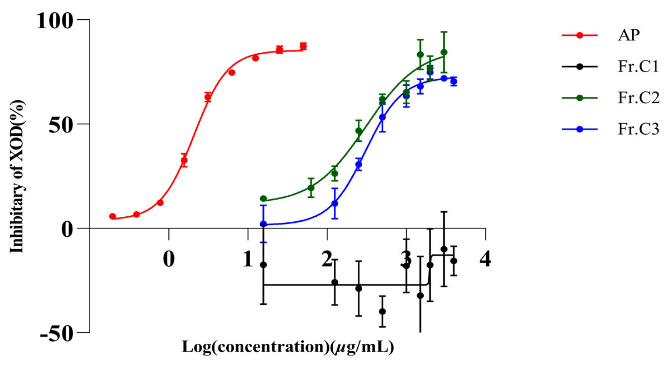
2.1. In Vitro Inhibition of XO by AP and Three Extracts of C. speciosa

The inhibitory effects of the petroleum ether fraction (Fr.C1), ethyl acetate fraction (Fr.C2), n-butanol fraction (Fr.C3) and AP on XO activity are illustrated in Figure 1. Among these fractions, only Fr.C1 did not exhibit any inhibition towards XO, while the remaining two fractions demonstrated dose-dependent inhibition towards XO. The IC50 values for Fr.C2, Fr.C3, and AP were determined to be 341.80 µg/mL, 321.10 µg/mL and 2.08 µg/mL (Table 1), respectively. The findings indicate that ethyl acetate fraction and n-butanol fraction exhibited superior inhibitory effects on XO in comparison to the petroleum ether fraction.

2.2. Monomer Compounds of C. speciosa and Its Inhibitory Effect on XO

The known compounds (124) were identified by comparing their experimental NMR spectral data with the corresponding data reported in the literature. These compounds were characterized as methyl chlorogenate (1) [12], butyl chlorogenate (2) [13], ethyl chlorogenate (3) [14], chlorogenic acid (4) [15], cryptochlorogenic acid methyl ester (5) [14], caffeic acid (6) [16], p-coumaric acid (7) [17], benzoic acid (8) [18], protocatechuic acid (9) [19], ethylmethyl malate isomers (10a/10b) [20], butylmethyl malate isomers (11a/11b) [21], bibutyl malate (12) [22], butyl 2-hydroxysuccinate (13) [23], dimethylmalate (14) [24], methylmalate (15) [25], oleanolic acid (16) [26], ursolic acid (17) [27], β-sitosterol (18) [28], betulinic acid (19) [29], fupenzic acid (20) [30], 5-hydroxymethylfurfural (21) [31], vomifoliol (22) [32], 2, 2′-oxybis (1, 4-di-tert-butylbenzen) (23) [33], and 1, 2, 4-benzenetriol (24) [34] (Figure 2). It is noteworthy that compounds 115, 1824 were isolated and identified from C. speciosa for the first time.

2.3. Inhibition of XO by Monomeric Compounds in C. speciosa

Table 2 showed the concentrations at which compounds 124 inhibited half of the XO activity, and the values are expressed as IC50. Figure 3 illustrated the inhibitory effects of compounds 19 on XO activity. Figure 3a showed that compounds 16 exhibited inhibitory activity against XO. Notably, these compounds contained a coffee acyl group, and the data indicated a dose-dependent inhibition of XO by this compound class. When the concentration was lower than 200 µM, there was basically no inhibition, and when the concentration was around 1000 µM, the inhibition rate reached almost 100%. Compared to compound 6, there was not a significant difference in the XO inhibiting effect of compounds 15. Therefore, we supposed that the decisive role in coffee’s acyl group lies in the chlorogenic acid ester compounds’ rate of XO inhibition. Figure 3b illustrated the in vitro inhibition of XO by compounds 6 and 7. At the same concentration, the inhibition rate of compound 6 on XO was higher than that of compound 7. An analysis of its molecular structure revealed that compound 6 had one more phenolic hydroxyl group than compound 7. Therefore, we speculated that the hydroxyl group on the benzene ring of this class of compounds could increase the inhibition of XO. Figure 3c illustrated the inhibition of XXO by benzoic acid compounds. These compounds also inhibited XO in a dose-dependent manner, but compound 8 showed a high inhibition at a concentration of less than 200 µM, while compound 9 achieved the same inhibition at a concentration of about 1000 µM. An observation of its molecular structure revealed that compound 9 had two more phenolic hydroxyl groups than compound 8. Thus, we speculated that replacing benzoic acid compounds’ hydroxyl groups on the benzene ring will lower its inhibitory effect on XO.

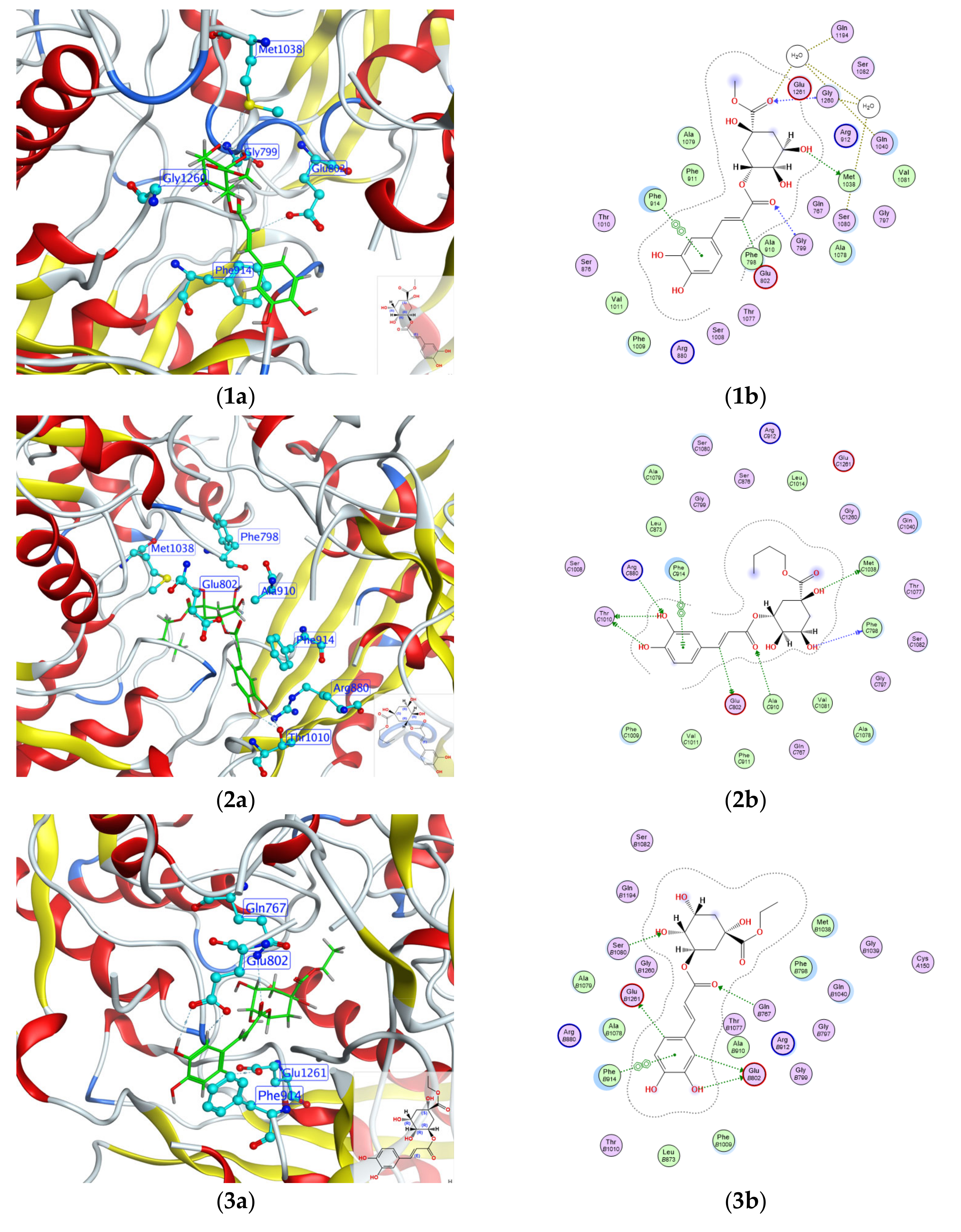
2.4. Molecular Docking Analysis

Molecular docking has been frequently utilized for the analysis of ligand–protein binding modes. In this study, we focused on the highest inhibition of XO and conducted a conformational analysis of molecular docking to determine the binding constants. Figure 4 illustrates the docking results of compounds 19 with XO (PDBID: 1FIQ). The molybdopterin (Mo-pt) domain is the functional site of XO in which the oxidation of xanthine to uric acid occurs; Arg880, Phe1009, Phe914, Glu802, Asn768, Thr1010, Val1011, Leu873, and Glu1216 are its critical amino acids [35]. Compound 1 showed interaction with Glu802, Met1038, Gly799, Gly1260 and Phe914 of the XO 1FIQ protein, with bonding distances of 3.48 Å, 3.28 Å, 3.12 Å, 3.40 Å, 3.37 Å, respectively. Compound 2 exhibited interactions with Thr1010, Glu802, Met1038, Phe798, Arg880, Ala910, and Phe914, with bonding distances of 3.09 Å, 3.41 Å, 3.31 Å, 2.90 Å, 3.34 Å, 3.30 Å, and 3.63 Å, respectively. Compound 3 demonstrated interaction with Glu1261, Glu802, Gln767, and Phe914, with bonding distances of 3.33 Å, 3.43 Å, 3.28 Å, and 3.43 Å, respectively. Compound 4 displayed interactions with Glu802, Met1038, Gly799, Phe798, and Phe914, with bonding distances of 3.31 Å, 3.05 Å, 3.07 Å, 3.31 Å, and 3.30 Å, respectively. Compound 5 showed interactions with Glu802, Met1038, Gly799, Gly1260, and Phe914, with bonding distances of 3.35 Å, 3.89 Å, 3.39 Å, 3.48 Å, and 3.30 Å, respectively. Compound 6 exhibited interactions with Glu802, Glu1261, Gly799, and Phe914, with bonding distances of 3.23 Å, 2.71 Å, 3.17 Å, and 3.39 Å, respectively. Compound 7 demonstrated interactions with Glu802, Glu1261, Gly799, and Phe914, with bonding distances of 3.28 Å, 2.71 Å, 3.46 Å, and 3.46 Å, respectively. Compound 8 displayed interactions with Arg880, Thr1010, and Phe914, with bonding distances of 3.13 Å, 3.03 Å, and 3.46 Å, respectively. Compound 9 showed interactions with Glu802, Thr1010, and Phe914, with bonding distances of 3.14 Å, 2.88 Å, and 3.61 Å, respectively. AP showed interactions with Glu802, Arg880, Thr1010, and Phe914, with bonding distances of 3.21 Å, 3.11 Å, and 2.97 Å, respectively. The binding energies for compound 19 were found to be −8.77, −7.76, −8.67, −8.45, −9.21, −5.90, −5.68, −5.08, −5.44 kcal/mol, respectively. The binding energy of ap to XO was −4.18 kcal/mol. The results demonstrated that compounds 19 and AP effectively occupied the functional site of XO and interacted with the key amino acids in the Mo-pt domain, and compounds 19 had a good binding affinity to XO compared with the AP. This indicates that the ligand molecules exhibit a greater affinity for the protein active site and therefore hold a promising place as effective inhibitors for XO.

3. Discussion

XO is one form of xanthine oxidoreductase (XOR), with a molecular weight of about 300 kd. Native XO contains two subunits with a molecular weight of approximately 150 kd [1], and its molybdopterin (Mo-Pt) domain is an important catalytic center when xanthine and hypoxanthine are oxidated [36]. XO catalyzes the oxidative hydroxylation of hypoxanthine to xanthine and xanthine to uric acid, the final two steps in purine catabolism in humans [37]. Uric acid deposition directly triggers the development of gout, which has been increasingly prevalent over recent years. The prevalence of hyperuricemia is approximately 20% in America and even higher in Korea, reaching up to 26.6% [38]. In China, with a quarter of the world’s population, the incidence of gout stands at about 16.6% [39] Hence, it is imperative to discover XO inhibitors in order to suppress the synthesis of uric acid and decrease the prevalence of hyperuricemia.
Currently, XO inhibitors such as allopurinol and febuxostat are primarily employed for the clinical management of hyperuricemia [40]. However, these medications have been associated with cardiovascular diseases, including heart failure, ischemic heart disease, hypertension, and cardiomyopathy [41]. Therefore, there is an urgent need to find XO inhibitors with fewer side effects for the treatment of hyperuricemia.
Thousands of years ago, extensive research was conducted on the use of food and herb medicines for treating gout and hyperuricemia. Cordyceps militaris, Poria cocos, and G. applanatum [42] were among the examples studied. Based on our previous reports [11], C. speciosa, a traditional Chinese medicinal material used as both medicine and food with minimal side effects, has been confirmed to possess uric acid-lowering properties. Therefore, we believe that the active components can be extracted and isolated from C. speciosa, which may have the effect of reducing uric acid, and provide some theoretical basis for the development of safe and effective drugs for the treatment of hyperuricemia.
This study aims to identify chemical compositions from C. speciosa fruits that can effectively inhibit XO activity by conducting in vitro enzyme activity screening on extracts obtained from this plant material. It was observed that the petroleum ether fraction had no inhibition of XO, while the ethyl acetate fraction and n-butanol fraction exhibited significant inhibitory effects on XO (Figure 1); Therefore, we focused primarily on isolating small molecules from the ethyl acetate and n-butanol fractions. A total of 24 compounds were isolated from these two fractions, and subsequent in vitro enzyme activity tests revealed that compounds 19 all demonstrated inhibitory effects on XO activity. The compounds 19 were structurally identified as chlorogenic acid and its esters, caffeic acid, and benzoic acid derivatives. Henceforth, it is reasonable to assume that chlorogenic esters, caffeic acids, and benzoic acids are the main urate-lowering chemicals present in C. speciosa. This study demonstrated that 16 (Figure 3a) exhibited a dose-dependent effect on XO, effectively inhibiting XO activity at concentrations up to 1000 µM. Furthermore, altering the quininate ester groups did not significantly impact its inhibitory effect on XO. Hence, this study suggests that caffeic acid group plays a pivotal role in XO inhibition. Figure 3b compares the effect of caffeic acid on XO with p-coumaric acid (7), which lacks a hydroxyl group on a benzene ring compared to caffeic acid, and the inhibition results show that caffeic acid exhibits better inhibition at low concentrations. Therefore, we suggest that the hydroxyl group on the benzene ring of these compounds inhibits XO. Noriyoshi Masuoka and Isao Kubo [43] suggested that XO inhibitors should possess alkyl chains and specific hydroxy group arrangements in the phenol portion. Yuan-Ching Chang et al. [44] proposed that C6-C3 phenylpropanoids are effective XO inhibitors, particularly caffeic acid and caffeic acid phenethyl ester, which exhibit significant XO inhibitory activity. They also indicated that the hydroxyl arrangement on the benzene ring and carboxyl esterification on the long chain greatly influence XO activity. Urszula Gawlik-Dziki et al. [45] discovered the noncompetitive inhibition of XO by chlorogenic acid. Yin Wan et al. [3] found that dicaffeoylquinic acids (diCQAs) have a stronger XO inhibitory effect compared to monocaffeoylquinic acids (monoCQAs). It is worth noting that 15 are all structures esterified with both caffeic acid groups and quininate ester groups, both of which have been reported to possess XO inhibitory activity. Among them, caffeic acid primarily exerts competitive inhibition on XO, while chlorogenic acid exhibits a non-competitive inhibitory effect on XO [46]. These views support the accuracy of the experimental results. Figure 3c compares the inhibition of XO by benzoic acid and protocatechuic acid, and the results show that the inhibition of benzoic acid is better than that of protocatechuic acid at low concentrations. By analyzing the structure of this class of compounds, we found that compound 9 has two more phenolic hydroxyl groups than compound 8. Therefore, we thought that the hydroxyl group on the benzene ring of this class of compounds would reduce the inhibition of XO. Falodun et al. [47] reported the XO inhibitory properties of benzoic acid (8), while Jun Li et al. [48] found protocatechuic acid (9) to possess similar effects on XO inhibition. Another investigation [42] revealed that 2, 4-dihydroxybenzoic acid methyl ester (DA) also exhibits an inhibitory effect on XO, but its inhibition rate at the same concentration is lower than that of protocatechuic acid. These experimental results also confirmed the correctness of our experimental results. Studies have shown that phenolic acids have antioxidant activity [49], and it has been reported that chlorogenic acid (4), caffeic acid (6), p-coumaric acid (7), and protocatechuic acid (9) have high antioxidant activity [50,51]. In addition, XO catalyzes the production of hypoxanthine from xanthine mainly at Mo-pt domain, which in turn catalyzes the production of uric acid from hypoxanthine. Therefore, we hypothesized that phenolic acids such as compounds 4, 6, 7, 9 might exert their urate-lowering effects by inhibiting the REDOX reaction of XO at the active site.
To gain insights into the atomic-level binding modes of 19, their 3D structures were simulated using Molecular Operating Environment (MOE) software and subsequently subjected to molecular docking studies with XO (PDB ID:1FIQ) [52]. The binding energies for compound 19 were found to be −8.77, −7.76, −8.67, −8.45, −9.21, −5.90, −5.68, −5.08, −5.44 kcal/mol, respectively. The present study showed that compounds 19 displayed good interaction with the amino acid residues at the active site, as indicated by their binding energies. In addition, it was also shown that compounds 19 could interact with key amino acid residues in the Mo-pt domain, such as Phe914 and Glu802. It can be concluded that compounds 19 can act in the active center of XO and inhibit XO activity through hydrogen bonding and π-π conjugation.
Taken together, it is reasonable to suggest that chlorogenic acid and its esters, caffeic acid, and benzoic acid compounds are the main urate-lowering chemicals in C. speciosa fruits. These compounds could be used as potential drugs for the prevention and treatment of hyperuricemia in clinic.

4. Materials and Methods

4.1. Instrumentation and General Experimental Techniques

The 1D and 2D NMR spectra were recorded on an Avance III-600 spectrometer (Bruker, Billerica, MA, USA), with TMS serving as an internal standard. Chemical shifts (δ) are expressed in ppm. Sephadex LH-20 gel (25–100 µm) was sourced from Pharmacia Fine Chemical located in Uppsala, Sweden. C18 silica gel (50 µm) was obtained from YMC (Kyoto, Japan). Silica gel 60 F254 aluminum sheets (Merck, Darmstadt, Germany) were utilized for thin-layer chromatography (TLC). Spots on the TLC plate were detected by being exposed to UV light (254 nm) and by spraying with p-anisaldehyde reagent. Column chromatography silica gel (200–300 mesh) was purchased from Shanghai Haohong Biomedical Technology in Shanghai, China. HPLC separations were performed using a Thermo Scientific UltiMate3000 liquid chromatography system (Waltham, MA, USA). A Thermo Hypersil ODS column (ODS, 250 × 10 mm, 5 µm; Thermo, Waltham, MA, USA) was employed. Xanthine (X7375), XO (4376-5UN), and AP (A8003) were supplied by Sigma-Aldrich LLC, a company based in St. Louis, MO, USA. MOE 2012 software was acquired by the Department of Medicinal Chemistry at Anhui Medical University (Hefei, China).

4.2. Plant Materials

The fruits of C. speciosa were furnished by Anhui Xiehecheng Chinese Herb Limited Corporation based in Bozhou, China in September 2021. Prof. Kai-Jin Wang of Anhui University provided authentication. A voucher specimen (20210930) was placed at the School of Pharmacy, Anhui Medical University.

4.3. Extraction, Isolation and Structure Identification

The plant materials of C. speciosa fruits (10 kg) were successively extracted in 95%, 85%, and 75% ethanol (at a ratio of 1:10 w/v) at a temperature of 60 °C for 3 h. The resulting filtrate was collected and subsequently mixed before being concentrated using a rotary evaporator (operating at a temperature of 60 °C and a speed of 45 rpm), yielding approximately 2904 g of crude residue. This residue was then resuspended in water and subjected to extraction with petroleum ether, ethyl acetate, and n-butyl alcohol to obtain three fractions: petroleum ether fraction (Fr.C1, 132.3 g), ethyl acetate fraction (Fr.C2, 532.5 g) and n-butanol fraction (Fr.C3, 926.2 g).

4.4. Inhibitory Assays of XO by Different Extracts of C. speciosa Fruits In Vitro

According to the previously determined method, the optical lighting method is measured by measuring the generation of uric acid of the elasticine [11]. Mixed solutions are prepared in the phosphate buffer (0.01 M, pH 7.5), which contains XO (0.01 units/mL of constant concentration; 1 unit converts 1.0 µmol xanthine to 25 °C and PH 7.5). Add the different concentrations of each extract of C. speciosa fruits and incubate for 15 min at 25 °C. Start the response by adding a lot of substrates (9 mM), and the reaction is terminated after 15 min of incubation by adding hydrochloric acid (50 µL, concentration: 1 N). Vortex and centrifuge the hybrid solution, and use the upper liquid to measure the value of the optical light at 290 nm. A positive drug control (AP) was included for comparative analysis. The inhibition ratio of XO was calculated by using the following formula:
I n h i b i t i o n   r a t i o % = 1 S S 0 B B 0 × 100
The experiments performed on the sample in the presence and absence of XO are, respectively, labeled as S and S0. The enzyme activity when there is no sample is denoted as B, and B0 represents the control of B when neither the sample nor XO is present.

4.5. Extraction and Separation of Monomer Compound

The ethyl acetate extract (Fr.C2, 532.5 g) was subjected to column chromatography on silica gel (200–300 mesh) using gradients of petroleum ether (PE)/CH2Cl2 (from 1:1 to 0:1, v/v) and CH2Cl2/MeOH (from 100:1 to 0:1, v/v), resulting in the isolation of seven fractions (Fr.C2.1–Fr.C2.7), which were monitored by thin-layer chromatography. Nine sub-fractions (Fr.C2.2.1–Fr.C2.2.9) were obtained from the partition of Fr.C2.2 (41.9 g) through silica gel column chromatography, eluted with a CH2Cl2/MeOH gradient ranging from 100:1 to 1:1. The sample Fr.C2.2.8 (26.2 g) was fractionated using a Sephadex LH-20 column, eluted with a gradient of MeOH/H2O (ranging from 10:90 to 100:0), resulting in the isolation of four subfractions (Fr.C2.2.8.1–Fr.C2.2.8.4). Compounds 10a/10b (6732 mg) and 15 (700 mg) and were obtained from Fr.C2.2.8.3 through silica gel column chromatography, using CH2Cl2/MeOH as the eluent in a ratio of 100:1. Fr.C2.9 (99 g) was separated by silica gel column chromatography (200–300 mesh), employing CH2Cl2/MeOH gradients ranging from 100:1 to 0:1, v/v, yielding nine fractions (Fr.C2.9.1–Fr.C2.9.9), which were monitored by TLC. Compound 16 (88 mg) was isolated from FrC2.9.3 via silica gel column chromatography using CH2Cl2/MeOH as the eluent in a ratio of 40:1. FrC2.9.4 underwent purification on a Sephadex LH-20 column using MeOH as the solvent and further purified through preparative TLC, utilizing CH2Cl2/MeOH at a ratio of 60:1, leading to the isolation of 17 (20 mg). Fr.C2.5 (101.2 g) was subjected to MCI column elution with MeOH/H2O (from 10:90 to 100:0) to yield five subfractions (Fr.C2.5.1 to Fr.C2.5.5). Fr.C2.5.1 (53.1 g) was separated on a Sephadex LH-20 column eluted with MeOH/H2O (from 10% to 100%) and purified by preparative TLC (CH2Cl2/MeOH, 40:1) to afford 14 (13.1 mg). Fr.C2.5.2 (9.1 g) was separated by an Sephadex LH-20, eluted with isocratic of MeOH/H2O (from 10% to 100%, v/v) to afford 8 fractions (Fr.C2.5.2.1–Fr.C2.5.2.8) monitored by TLC. Fr.C2.5.2.1 (2.0 g) was separated by Sephadex LH-20, column eluted with MeOH/H2O (from 10% to 100%) and purified by silica gel CC with CH2Cl2/MeOH (30:1) to produce 11a/11b (273.2 mg), 12 (17.1 mg), 13 (219.3 mg). Fr.C2.5.2.4 was separated by Sephadex LH-20 column, eluted with MeOH/H2O (from 10% to 100%) to yield 21 (62 mg). Fr.C2.5.2.5 was isolated by Sephadex LH-20, eluted with MeOH/H2O (from 10% to 100%) and purified by silica gel CC, eluted with CH2Cl2/MeOH (30:1) to afford 22 (16 mg). Fr.C2.5.3 was subjected to a Sephadex LH-20 column (MeOH) and purified by preparative TLC (CH2Cl2/MeOH, 50:1) to yield 18 (7 mg), 19 (114 mg) and 23 (139 mg). The n-BuOH extract (Fr.C3, 926.6 g) was separated to silica gel (200–300 mesh) CC with CH2Cl2/MeOH gradients (from 100:1 to 0:1, v/v) to afford 6 fractions (Fr.C3.1–Fr.C3.6). Seven fractions (Fr.C3.4.1–Fr.C3.4.7) were obtained from the Fr.C3.4 (384.6 g) partition by silica gel CC, eluted with CH2Cl2/MeOH (from 40:1 to 1:1). Compounds 6 (18 mg) and 7 (9 mg) were isolated from the Fr.C3.4.3 partition by silica gel column chromatography and eluted with CH2Cl2/MeOH (30:1). Fr.C3.4.4 was isolated by C18, eluted with MeOH/H2O (from 10% to 100%) and purified by HPLC (45%, MeOH/H2O) to afford 8 (27 mg) and 9 (20 mg). Compound 20 (38 mg) was isolated from the Fr.C3.4.5 partition by silica gel column chromatography and eluted with CH2Cl2/MeOH (40:1). Fr.C3.4.6 (40 g) was separated by an Sephadex LH-20 eluted with isocratic of MeOH/H2O (from 10% to 100%, v/v) to yield 2 (75 mg) and 24 (28 mg). Fr.C3.4.6 was isolated by ODS, eluted with MeOH/H2O (from 10% to 100%) to afford 1 (12 mg) and 4 (20 mg). Fr.C3.4.7 was purified using semipreparative HPLC (MeOH/H2O, 60:40, v/v, 2.5 mL/min) to yield 5 (6 mg) and 3 (3 mg).

4.6. Molecular Docking

To ensure the accuracy of our target selection, we considered several criteria to guarantee the precision of our results. First, we limited our selection to PDB structures that contained a predefined ligand which had been reported before. This is of great importance as it makes sure that our results can be compared to previous studies and are consistent with the existing literature. Second, the docking protocol was validated by docking with the internal ligands extracted from the obtained PDB IDs of the target candidates. The protocol with the best docking pose of the internal ligand, as reported in the respective PDB, was chosen. The crystal structures of the candidate protein targets for XO were retrieved from the RCSB Protein Data Bank (http://www.pdb.org/, PDBID: 1FIQ;) [52]. There was no solvent present, and all heteroatoms and water molecules were eliminated.
The structures of compounds 19 and AP were drawn and then saved in sdf format and imported into the MOE software database. The structures of compounds 19 and AP were washed to obtain their 3D conformation using the wash function under the computer panel. Amino acids (Arg880, Phe1009, Phe914, Glu802, Asn768, Thr1010, Val1011, Leu873, and Glu1216) in the active central site (Mo-pt domain) were selected in the target protein (1FIQ), and docking targets were created using the set create function under the select panel. The interactions between XO inhibitors and target candidates were assessed by docking which was carried out by utilizing selected parameters (Placement: Triangle Matcher, Refinement: Rigid Receptor, Poses: 30). Finally, the S score was employed as a basis to select the top five targets, calculated by the default scoring built-in function in MOE is the S value, which is a scoring number that evaluates the affinity of the ligand with the receptor [53]. For further analysis, 2D protein–ligand interaction images were obtained using the ligand interactions option in MOE. The energies of docking complexes and H-bonding length were incorporated in the ligand interaction analysis. Then, the best five targets were imported into the STRING 12.0 database and analyzed for co-expression.

5. Conclusions

In this study, 24 compounds were isolated from the active ethyl acetate and n-butanol fractions of C. speciosa fruits extract, and their XO inhibitory activity was assessed by measurement. Among these compounds, methyl chlorogenate, butyl chlorogenate, ethyl chlorogenate, chlorogenic acid, cryptochlorogenic acid methyl ester, caffeic acid, p-coumaric acid, benzoic acid and protocatechuic acid exhibited superior XO inhibitory effects. The molecular docking analysis suggested that 19 inhibit XO activity by binding to the active catalytic site, thereby preventing substrate entry and inducing conformational changes in XO. However, the specific binding sites between the protein and xo remain unclear and require further investigation. Overall, this study investigated the active substances in C. speciosa responsible for reducing uric acid levels, providing a theoretical foundation for drug development and functional food products aimed at preventing hyperuricemia.

Supplementary Materials

The following supporting information can be downloaded at: https://www.mdpi.com/article/10.3390/molecules29184468/s1, Figures S1–S48 shows 1D NMR of compounds 124; Table S1: Molecular docking parameters.

Author Contributions

Conceptualization, N.L. and K.L.; methodology, K.L., W.M. and R.X.; validation, N.L. and K.L.; formal analysis, K.L.; investigation, N.L. and K.L.; resources, N.L.; data curation, K.L. and R.X.; writing—original draft preparation, K.L. and R.X.; writing—review and editing, K.L., M.K. and N.L.; funding acquisition, N.L. All authors have read and agreed to the published version of the manuscript.

Funding

This research received no external funding.

Institutional Review Board Statement

Not applicable.

Informed Consent Statement

Not applicable.

Data Availability Statement

Data are contained within the article and Supplementary Materials.

Conflicts of Interest

The authors declare no conflicts of interest.

References

  1. Sun, Z.; Wu, K.; Ullah, I.; Zhu, H. Recent advances in xanthine oxidase inhibitors. Mini Rev. Med. Chem. 2024, 24, 1177–1186. [Google Scholar] [CrossRef] [PubMed]
  2. Pauff, J.M.; Hille, R. Inhibition Studies of Bovine Xanthine Oxidase by Luteolin, Silibinin, Quercetin, and Curcumin. J. Nat. Prod. 2009, 72, 725–731. [Google Scholar] [CrossRef] [PubMed]
  3. Wan, Y.; Qian, J.; Li, Y.; Shen, Y.; Chen, Y.; Fu, G.; Xie, M. Inhibitory mechanism of xanthine oxidase activity by caffeoylquinic acids in vitro. Int. J. Biol. Macromol. 2021, 184, 843–856. [Google Scholar] [CrossRef] [PubMed]
  4. Cao, W.; Fang, Y.; Wu, T.; Liang, F.; Cheng, Y.; Salah, M.; Pan, S.; Xu, X. Insights from multispectral and molecular docking investigation on the xanthine oxidase inhibition by 1,4-dicaffeoylquinic acid. J. Mol. Struct. 2020, 1219, 128475. [Google Scholar] [CrossRef]
  5. Zhang, G.; Zhu, M.; Liao, Y.; Gong, D.; Hu, X. Action mechanisms of two key xanthine oxidase inhibitors in tea polyphenols and their combined effect with allopurinol. J. Sci. Food Agric. 2022, 102, 7195–7208. [Google Scholar] [CrossRef]
  6. Ojha, R.; Singh, J.; Ojha, A.; Singh, H.; Sharma, S.; Nepali, K. An updated patent review: Xanthine oxidase inhibitors for the treatment of hyperuricemia and gout (2011–2015). Expert. Opin. Ther. Pat. 2016, 27, 311–345. [Google Scholar] [CrossRef]
  7. Guo, X.; Gao, Y.; Yang, Y.; Zhu, Q.; Guan, H.; He, X.; Zhang, C.; Wang, Y.; Xu, G.; Zou, S.; et al. Amelioration effects of α-viniferin on hyperuricemia and hyperuricemia-induced kidney injury in mice. Phytomedicine 2023, 116, 154868–154882. [Google Scholar] [CrossRef]
  8. White, W.; Saag, K.; Becker, M.; Borer, J.; Gorelick, P.; Whelton, A.; Hunt, B.; Castillo, M.; Gunawardhana, L.; Investigators, C. Cardiovascular safety of febuxostat or allopurinol in patients with gout. N. Engl. J. Med. 2018, 378, 1200–1210. [Google Scholar] [CrossRef]
  9. Cao, P.; Huang, Y.; Zong, M.; Xu, Z. De novo assembly and comparative analysis of the complete mitochondrial genome of Chaenomeles speciosa (Sweet) Nakai revealed the existence of two structural isomers. Genes 2023, 14, 526–541. [Google Scholar] [CrossRef]
  10. Xu, R.; Kuang, M.; Li, N. Phytochemistry and pharmacology of plants in the genus Chaenomeles. Arch. Pharm. Res. 2023, 46, 825–854. [Google Scholar] [CrossRef]
  11. Xu, R.; Deng, P.; Ma, Y.; Li, K.; Ren, F.; Li, N. Anti-hyperuricemic effects of extracts from Chaenomeles speciosa (Sweet) Nakai Fruits on hyperuricemic rats. Metabolites 2024, 14, 117–128. [Google Scholar] [CrossRef]
  12. Zeller, W. Synthesis of 1-O-methylchlorogenic acid: Reassignment of structure for MCGA3 isolated from bamboo (Phyllostachys edulis) leaves. J. Agric. Food Chem. 2014, 62, 1860–1865. [Google Scholar] [CrossRef] [PubMed]
  13. Kirmizibekmez, H.; Bassarello, C.; Piacente, S.; Celep, E.; Atay, I.; Mercanoglu, G.; Yesilada, E. Phenolic compounds from hypericum calycinum and their antioxidant activity. Nat. Prod. Commun. 2009, 4, 531–534. [Google Scholar] [CrossRef] [PubMed]
  14. Zhu, X.; Dong, X.; Wang, W.; Ju, P.; Luo, S. Phenolic compounds from Viburnum cylindricum. Helv. Chim. Acta 2005, 88, 339–342. [Google Scholar] [CrossRef]
  15. Tong, Y.; Li, G.; Shi, X.; Wang, L.; Zhou, J.; Chu, M.; Wang, Z.; Abd El-Aty, A.M.; Dang, J. Protection against myocardial ischemia/reperfusion injury in mice by 3-caffeoylquinic acid isomers isolated from Saxifraga tangutica. RSC Adv. 2024, 14, 6642–6655. [Google Scholar] [CrossRef] [PubMed]
  16. Chen, Y.; Chang, F.; Lin, Y.; Wang, L.; Chen, J.; Wu, Y.; Wu, M. Identification of phenolic antioxidants from Sword Brake fern (Pteris ensiformis Burm.). Food Chem. 2007, 105, 48–56. [Google Scholar] [CrossRef]
  17. Rho, T.; Yoon, K. Chemical constituents of nelumbo nucifera seeds. Nat. Prod. Sci. 2017, 23, 253–257. [Google Scholar] [CrossRef]
  18. Lü, S.; Zhao, S.; Zhao, M.; Guo, Y.; Li, G.; Yang, B.; Wang, Q.; Kuang, H. Systematic screening and characterization of prototype constituents and metabolites of triterpenoid saponins of Caulopphyllum robustum Maxim using UPLC-LTQ Orbitrap MS after oral administration in rats. J. Pharm. Biomed. Anal. 2019, 168, 75–82. [Google Scholar] [CrossRef]
  19. Wang, W.; Yang, C.R.; Zhang, Y.J. Phenolic constituents from the fruits of Amomum tsaoko (Zingiberaceae). Acta Botanica Yunnanica 2009, 31, 284–288. [Google Scholar] [CrossRef]
  20. Dias, C.; Dias, M.; Borges, C.; Almoster Ferreira, M.A.; Paulo, A.; Nascimento, J. Structural elucidation of natural 2-hydroxy di- and tricarboxylic acids and esters, phenylpropanoid esters and a flavonoid from Autonoë madeirensis using gas chromatographic/electron ionization, electrospray ionization and tandem mass spectrometric techniques. J. Mass. Spectrom. 2003, 38, 1240–1244. [Google Scholar] [CrossRef]
  21. Samoylenko, V.; Zhao, J.; Dunbar, D.; Khan, I.; Rushing, J.; Muhammad, I. New constituents from Noni (Morinda citrifolia) fruit juice. J. Agric. Food Chem. 2006, 54, 6398–6402. [Google Scholar] [CrossRef] [PubMed]
  22. Cho, D.; Song, S.; Jang, D. A method for preparation of unnatural (R)-malic acid derivatives with phenylsilanes. Synth. Commun. 2003, 33, 515–519. [Google Scholar] [CrossRef]
  23. Akihisa, T.; Tochizawa, S.; Takahashi, N.; Yamamoto, A.; Zhang, J.; Kikuchi, T.; Fukatsu, M.; Tokuda, H.; Suzuki, N. Melanogenesis-inhibitory saccharide fatty acid esters and other constituents of the fruits of Morinda citrifolia (Noni). Chem. Biodivers. 2012, 9, 1172–1187. [Google Scholar] [CrossRef] [PubMed]
  24. Xing, R.; Wu, L.; Fei, Z.; Wu, P. Palladium phthalocyaninesulfonate functionalized mesoporous polymer: A highly efficient photocatalyst for degradation of 4-chlorophenol under visible light irradiation. J. Mol. Catal. A Chem. 2013, 371, 15–20. [Google Scholar] [CrossRef]
  25. Houston, T.; Wilkinson, B.; Blanchfield, J. Boric acid catalyzed chemoselective esterification of α-hydroxycarboxylic acids. Org. Lett. 2004, 6, 679–681. [Google Scholar] [CrossRef] [PubMed]
  26. Chen, J.; Ge, Z.; Liao, X.; Xue, J.; Wu, L.; Liang, L. α-Glucosidase inhibitory phytochemical components of chinese endemic plant whitfordiodendron filipes var. tomentosum. Plants 2024, 13, 692–703. [Google Scholar] [CrossRef]
  27. Silva, M.; Vieira, I.; Mendes, F.; Albuquerque, I.; dos Santos, R.; Silva, F.; Morais, S. Variation of ursolic acid content in eight Ocimum species from northeastern Brazil. Molecules 2008, 13, 2482–2487. [Google Scholar] [CrossRef]
  28. Koizumi, N.; Fujimoto, Y.; Takeshita, T.; Ikekawa, N. Carbon-13 nuclear magnetic resonance of 24-substituted steroids. Chem. Pharm. Bull. 1979, 27, 38–42. [Google Scholar] [CrossRef]
  29. Chatterjee, P.; Pezzuto, J.; Kouzi, S. Glucosidation of betulinic acid by Cunninghamell species. J. Nat. Prod. 1999, 62, 761–763. [Google Scholar] [CrossRef]
  30. Zhang, Q.; Li, Q.; Wang, J.; Jang, W.; Hai, P.; Jia, C.; Ren, L.; Wu, X.; Kang, C.; Yang, J.; et al. Triterpenoids in sorbus pohuashanensis suspension cell treated with yeast extract. China J. Chin. Mater. Med. 2024, 49, 130–140. [Google Scholar] [CrossRef]
  31. Lee, S.; Kim, B.; Cho, S.; Shin, K. Phytochemical constituents from the fruits of Acanthopanax sessiliflorus. Arch. Pharm. Res. 2002, 25, 280–284. [Google Scholar] [CrossRef] [PubMed]
  32. Andersson, R.; Lundgren, L. Monoaryl and cyclohexenone glycosides from needles of Pinus sylvestris. Phytochemistry 1988, 27, 559–562. [Google Scholar] [CrossRef]
  33. Sun, B.; Shen, H.; Wu, H.; Yao, L.; Cheng, Z. Isolation and identification of chemical constituents from Peronia verruculata. Chin. Pharm. J. 2014, 25, 1019–1021. [Google Scholar] [CrossRef]
  34. Huang, Y.; Zeng, G.; Yang, Y.; Zhao, Y. Chemical constituents from euphorbia royleana. J. Yunnan Univ. Nat. Sci. Ed. 2021, 30, 205–208. [Google Scholar]
  35. Ghallab, D.; Shawky, E.; Metwally, A.; Celik, I.; Ibrahim, R.; Mohyeldin, M. Integrated in silico—In vitro strategy for the discovery of potential xanthine oxidase inhibitors from Egyptian propolis and their synergistic effect with allopurinol and febuxostat. RSC Adv. 2022, 12, 2843–2872. [Google Scholar] [CrossRef]
  36. Song, J.; Chen, M.; Meng, F.; Chen, J.; Wang, Z.; Zhang, Y.; Cui, J.; Wang, J.; Shi, D. Studies on the interaction mechanism between xanthine oxidase and osmundacetone: Molecular docking, multi-spectroscopy and dynamical simulation. Spectrochim. Acta A Mol. Biomol. Spectrosc. 2023, 299, 122861. [Google Scholar] [CrossRef]
  37. Hille, R. Xanthine oxidase-a personal history. Molecules 2023, 28, 1921. [Google Scholar] [CrossRef]
  38. Wang, R.; Halimulati, M.; Huang, X.; Ma, Y.; Li, L.; Zhang, Z. Sulforaphane-driven reprogramming of gut microbiome and metabolome ameliorates the progression of hyperuricemia. J. Adv. Res. 2023, 52, 19–28. [Google Scholar] [CrossRef]
  39. Chen, J.; Xu, L.; Jiang, L.; Wu, Y.; Wei, L.; Wu, X.; Xiao, S.; Liu, Y.; Gao, C.; Cai, J.; et al. Sonneratia apetala seed oil attenuates potassium oxonate/hypoxanthine-induced hyperuricemia and renal injury in mice. Food Funct. 2021, 12, 9416–9431. [Google Scholar] [CrossRef]
  40. Becker, M.; Schumacher, H.; Wortmann, R.; MacDonald, P.; Eustace, D.; Palo, W.; Streit, J.; Joseph-Ridge, N. Febuxostat compared with allopurinol in patients with hyperuricemia and gout. N. Engl. J. Med. 2005, 353, 2450–2461. [Google Scholar] [CrossRef]
  41. Bai, Y.; Wu, B.; Gou, L.; Fang, Z.; Xu, T.; Zhang, T.; Li, Y. Cardiovascular safety evaluation of febuxostat and allopurinol: Findings from the FDA adverse event reporting system. J. Clin. Med. 2023, 12, 6089–6101. [Google Scholar] [CrossRef] [PubMed]
  42. Yong, T.; Liang, D.; Xiao, C.; Huang, L.; Chen, S.; Xie, Y.; Gao, X.; Wu, Q.; Hu, H.; Li, X.; et al. Hypouricemic effect of 2,4-dihydroxybenzoic acid methyl ester in hyperuricemic mice through inhibiting XOD and down-regulating URAT1. Biomed. Pharmacother. 2022, 153, 113303–113313. [Google Scholar] [CrossRef] [PubMed]
  43. Masuoka, N.; Kubo, I. Characterization of the xanthine oxidase inhibitory activity of alk(en)yl phenols and related compounds. Phytochemistry 2018, 155, 100–106. [Google Scholar] [CrossRef] [PubMed]
  44. Chang, Y.; Lee, F.; Chen, C.; Huang, S.; Tsai, S.; Huang, S.; Lin, C. Structure-activity relationship of C6-C3 phenylpropanoids on xanthine oxidase-inhibiting and free radical-scavenging activities. Free Radic. Biol. Med. 2007, 43, 1541–1551. [Google Scholar] [CrossRef] [PubMed]
  45. Gawlik-Dziki, U.; Dziki, D.; Świeca, M.; Nowak, R. Mechanism of action and interactions between xanthine oxidase inhibitors derived from natural sources of chlorogenic and ferulic acids. Food Chem. 2017, 225, 138–145. [Google Scholar] [CrossRef]
  46. Wang, S.; Chen, C.; Huang, S.; Yu, S.; Lai, Z.; Huang, S.; Lin, C. Hydrophilic ester-bearing chlorogenic acid binds to a novel domain to inhibit xanthine oxidase. Planta Med. 2009, 75, 1237–1240. [Google Scholar] [CrossRef]
  47. Falodun, A.; Ali, S.; Quadir, I.M.; Choudhary, I.M.I. Phytochemical and biological investigation of chloroform and ethylacetate fractions of Euphorbia heterophylla leaf (Euphorbiaceae). J. Med. Plant Res. 2008, 2, 365–369. [Google Scholar]
  48. Li, J.; Gong, Y.; Li, J.; Fan, L. In vitro inhibitory effects of polyphenols from Tartary buckwheat on xanthine oxidase: Identification, inhibitory activity, and action mechanism. Food Chem. 2022, 379, 132100–132109. [Google Scholar] [CrossRef]
  49. Palafox-Carlos, H.; Gil-Chavez, J.; Sotelo-Mundo, R.R.; Namiesnik, J.; Gorinstein, S.; Gonzalez-Aguilar, G.A. Antioxidant interactions between major phenolic compounds found in ‘Ataulfo’ mango pulp: Chlorogenic, gallic, protocatechuic and vanillic acids. Molecules 2012, 17, 12657–12664. [Google Scholar] [CrossRef]
  50. Shao, Y.; Hu, Z.; Yu, Y.; Mou, R.; Zhu, Z.; Beta, T. Phenolic acids, anthocyanins, proanthocyanidins, antioxidant activity, minerals and their correlations in non-pigmented, red, and black rice. Food Chem. 2018, 239, 733–741. [Google Scholar] [CrossRef]
  51. Foss, K.; Przybylowicz, K.E.; Sawicki, T. Antioxidant Activity and Profile of Phenolic Compounds in Selected Herbal Plants. Plant Foods Hum. Nutr. 2022, 77, 383–389. [Google Scholar] [CrossRef] [PubMed]
  52. Li, X.; Jin, W.; Zhang, W.; Zheng, G. The inhibitory kinetics and mechanism of quercetin-3-O-rhamnoside and chlorogenic acid derived from Smilax china L. ethyl acetate fraction on xanthine oxidase. Int. J. Biol. Macromol. 2022, 213, 447–455. [Google Scholar] [CrossRef] [PubMed]
  53. Karhana, S.; Dabral, S.; Garg, A.; Bano, A.; Agarwal, N.; Khan, M.A. Network pharmacology and molecular docking analysis on potential molecular targets and mechanism of action of BRAF inhibitors for application in wound healing. J. Cell Biochem. 2023, 124, 1023–1039. [Google Scholar] [CrossRef] [PubMed]
Figure 1. Four C. speciosa fruit fractions, petroleum ether fraction (Fr.C1), ethyl acetate fraction (Fr.C2) and n-butanol fraction (Fr.C3), and AP inhibited XO in vitro. Values are expressed as means = SD.
Molecules 29 04468 g001
Figure 2. Structure of compounds 124 isolated from C. speciosa.
Molecules 29 04468 g002
Figure 3. (ac) Inhibition of XO by compounds 19 in vitro. Values are expressed as means = SD.
Molecules 29 04468 g003
Figure 4. Molecular docking images of compounds 19 and AP with XO. (1a10a) The binding orientation of compounds 19 and AP inside the binding site of XO; (1b10b) The 2D ligand–protein interaction of compounds 19 and AP with residues of XO. (11) Legend for the 2D images.
Molecules 29 04468 g004aMolecules 29 04468 g004bMolecules 29 04468 g004c
Table 1. Inhibitory effects of C. speciosa fruit extracts and AP on XO in vitro.
ExtractIC50 (µg/mL)
Fr.C1nc
Fr.C2341.80
Fr.C3321.10
AP2.08
nc: not calculated. IC50: inhibition of XO 50% activity (concentration in µg/mL required for a 50% reduction in XO activity).
Table 2. Inhibitory effects of compounds (19) on XO in vitro.
NO.CompoundClassificationIC50 (µg/mL)
1methyl chlorogenatepenylpropanoids156.5
2butyl chlorogenatepenylpropanoids163.6
3ethyl chlorogenatepenylpropanoids172.2
4chlorogenic acidpenylpropanoids105.4
5cryptochlorogenic acid methyl esterpenylpropanoids220.9
6caffeic acidhydroxycinnamic acid43.6
7p-coumaric acidhydroxycinnamic acid57.6
8benzoic acidaromatic carboxylic acids12.7
9protocatechuic acidaromatic carboxylic acids100.27
IC50: inhibition of XO 50% activity (concentration in µg/mL required for a 50% reduction in XO activity).
Disclaimer/Publisher’s Note: The statements, opinions and data contained in all publications are solely those of the individual author(s) and contributor(s) and not of MDPI and/or the editor(s). MDPI and/or the editor(s) disclaim responsibility for any injury to people or property resulting from any ideas, methods, instructions or products referred to in the content.

