

Elaboration of Functionalized Iron Oxide Nanoparticles by Microwave-Assisted Co-Precipitation: A New One-Step Method in Water
 , and
, and 
Abstract
:1. Introduction
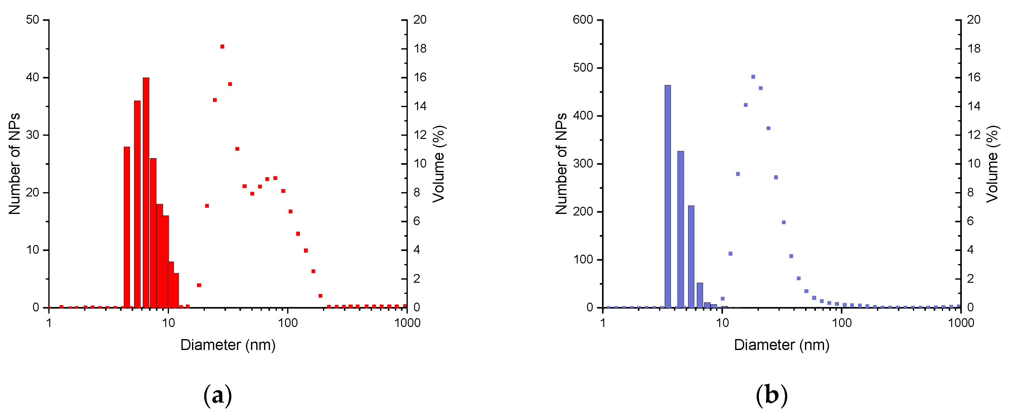
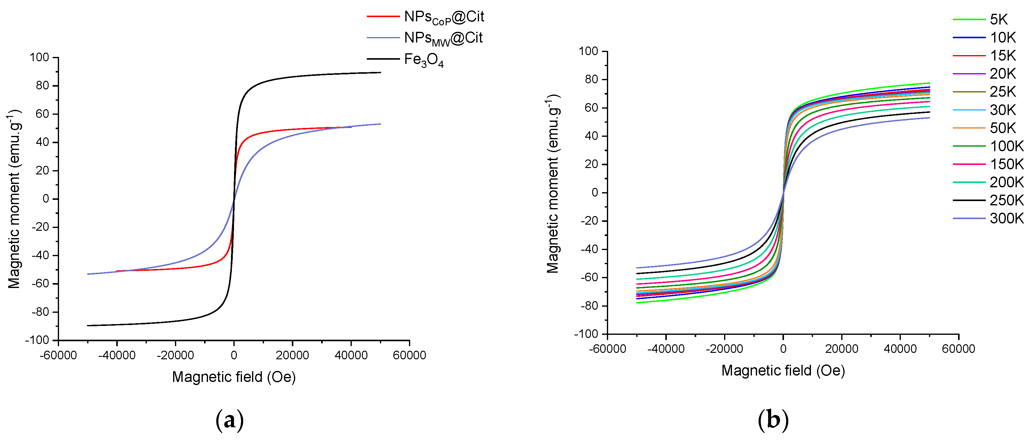
2. Results
3. Discussion
4. Materials and Methods
4.1. Synthesis of Nanoparticles by Co-Precipitation
4.2. Synthesis of Nanoparticles by Microwave-Assisted Co-Precipitation
4.3. Characterizations of Nanoparticles
4.3.1. Transmission Electron Microscopy
4.3.2. Dynamic Light Scattering
4.3.3. X-ray Diffraction
4.3.4. Fourier Transform Infrared Spectroscopy
4.3.5. Thermogravimetric Analysis
4.3.6. Magnetic Measurement
5. Conclusions
Author Contributions
Funding
Institutional Review Board Statement
Informed Consent Statement
Data Availability Statement
Acknowledgments
Conflicts of Interest
Appendix A

References
- Ortega-Villar, R.; Lizárraga-Mendiola, L.; Coronel-Olivares, C.; López-León, L.D.; Bigurra-Alzati, C.A.; Vázquez-Rodríguez, G.A. Effect of Photocatalytic Fe2O3 Nanoparticles on Urban Runoff Pollutant Removal by Permeable Concrete. J. Environ. Manag. 2019, 242, 487–495. [Google Scholar] [CrossRef] [PubMed]
- Tang, Z.-D.; Sun, X.-M.; Huang, T.-T.; Liu, J.; Shi, B.; Yao, H.; Zhang, Y.-M.; Wei, T.-B.; Lin, Q. Pillar[n]arenes-Based Materials for Detection and Separation of Pesticides. Chin. Chem. Lett. 2023, 34, 107698. [Google Scholar] [CrossRef]
- Li, Y.-J.; Huang, T.-T.; Liu, J.; Xie, Y.-Q.; Shi, B.; Zhang, Y.-M.; Yao, H.; Wei, T.-B.; Lin, Q. Detection of Lead(II) in Living Cells by Inducing the Transformation of a Supramolecular System into Quantum Dots. ACS Sustain. Chem. Eng. 2022, 10, 7907–7915. [Google Scholar] [CrossRef]
- Peng, J.A.; Zhang, W.; Chen, L.; Wu, T.; Zheng, M.; Dong, H.; Hu, H.; Xiao, Y.; Liu, Y.; Liang, Y. Versatile Route to Metal Oxide Nanoparticles Impregnated in Carbon Matrix for Electrochemical Energy Storage. Chem. Eng. J. 2021, 404, 126461. [Google Scholar] [CrossRef]
- Liu, L.; Corma, A. Metal Catalysts for Heterogeneous Catalysis: From Single Atoms to Nanoclusters and Nanoparticles. Chem. Rev. 2018, 118, 4981–5079. [Google Scholar] [CrossRef]
- Teja, A.S.; Koh, P.-Y. Synthesis, Properties, and Applications of Magnetic Iron Oxide Nanoparticles. Prog. Cryst. Growth Charact. Mater. 2009, 55, 22–45. [Google Scholar] [CrossRef]
- Al-Thani, A.N.; Jan, A.G.; Abbas, M.; Geetha, M.; Sadasivuni, K.K. Nanoparticles in Cancer Theragnostic and Drug Delivery: A Comprehensive Review. Life Sci. 2024, 352, 122899. [Google Scholar] [CrossRef]
- Aparicio-Blanco, J.; Pucci, C.; De Pasquale, D.; Marino, A.; Debellis, D.; Ciofani, G. Development and Characterization of Lipid Nanocapsules Loaded with Iron Oxide Nanoparticles for Magnetic Targeting to the Blood–Brain Barrier. Drug Deliv. Transl. Res. 2024. [Google Scholar] [CrossRef]
- Peréz, D.L.; Puentes, I.; Romero, G.A.M.; Gaona, I.M.S.; Vargas, C.A.P.; Rincón, R.J. Synthesis of Superparamagnetic Iron Oxide Nanoparticles Coated with Polyethylene Glycol as Potential Drug Carriers for Cancer Treatment. J. Nanopart Res. 2024, 26, 2. [Google Scholar] [CrossRef]
- Laurent, S.; Dutz, S.; Häfeli, U.O.; Mahmoudi, M. Magnetic Fluid Hyperthermia: Focus on Superparamagnetic Iron Oxide Nanoparticles. Adv. Colloid. Interface Sci. 2011, 166, 8–23. [Google Scholar] [CrossRef]
- Xu, H.; Aguilar, Z.P.; Yang, L.; Kuang, M.; Duan, H.; Xiong, Y.; Wei, H.; Wang, A. Antibody Conjugated Magnetic Iron Oxide Nanoparticles for Cancer Cell Separation in Fresh Whole Blood. Biomaterials 2011, 32, 9758–9765. [Google Scholar] [CrossRef] [PubMed]
- Xu, X.; Dong, P.-L.; Chai, Y.-Y.; Yang, R.; Ma, Z.-H.; Lu, C.-C. Doping Engineering of Iron Oxide Nanoparticles towards High Performance and Biocompatible T1-Weighted MRI Contrast Agents. Rare Met. 2024, 43, 298–308. [Google Scholar] [CrossRef]
- Sun, S.; Zeng, H. Size-Controlled Synthesis of Magnetite Nanoparticles. J. Am. Chem. Soc. 2002, 124, 8204–8205. [Google Scholar] [CrossRef] [PubMed]
- Shavel, A.; Liz-Marzán, L.M. Shape Control of Iron Oxide Nanoparticles. Phys. Chem. Chem. Phys. 2009, 11, 3762. [Google Scholar] [CrossRef]
- Mornet, S.; Vasseur, S.; Grasset, F.; Duguet, E. Magnetic Nanoparticle Design for Medical Diagnosis and Therapy. J. Mater. Chem. 2004, 14, 2161. [Google Scholar] [CrossRef]
- García-Acevedo, P.; González-Gómez, M.A.; Arnosa-Prieto, Á.; De Castro-Alves, L.; Piñeiro, Y.; Rivas, J. Role of Dipolar Interactions on the Determination of the Effective Magnetic Anisotropy in Iron Oxide Nanoparticles. Adv. Sci. 2023, 10, 2203397. [Google Scholar] [CrossRef]
- Sanna Angotzi, M.; Mameli, V.; Zákutná, D.; Rusta, N.; Cannas, C. On the Thermal and Hydrothermal Stability of Spinel Iron Oxide Nanoparticles as Single and Core-Shell Hard-Soft Phases. J. Alloys Compd. 2023, 940, 168909. [Google Scholar] [CrossRef]
- Kalidass, J.; Anandan, S.; Sivasankar, T. Rapid Ultrasound Driven Self-Assembled Nanoarchitectonics Iron Oxide-Reduced Graphene Oxide as an Anode for Li-Ion Battery and Its Electrochemical Performance Studies. J. Energy Storage 2023, 74, 109361. [Google Scholar] [CrossRef]
- Kessler, V.G.; Seisenbaeva, G.A. Molecular Mechanisms of the Metal Oxide Sol-Gel Process and Their Application in Approaches to Thermodynamically Challenging Complex Oxide Materials. J. Sol-Gel Sci. Technol. 2023, 107, 190–200. [Google Scholar] [CrossRef]
- Cheah, P.; Cowan, T.; Zhang, R.; Fatemi-Ardekani, A.; Liu, Y.; Zheng, J.; Han, F.; Li, Y.; Cao, D.; Zhao, Y. Continuous Growth Phenomenon for Direct Synthesis of Monodisperse Water-Soluble Iron Oxide Nanoparticles with Extraordinarily High Relaxivity. Nanoscale 2020, 12, 9272–9283. [Google Scholar] [CrossRef]
- Vassallo, M.; Martella, D.; Barrera, G.; Celegato, F.; Coïsson, M.; Ferrero, R.; Olivetti, E.S.; Troia, A.; Sözeri, H.; Parmeggiani, C.; et al. Improvement of Hyperthermia Properties of Iron Oxide Nanoparticles by Surface Coating. ACS Omega 2023, 8, 2143–2154. [Google Scholar] [CrossRef] [PubMed]
- Crippa, F.; Rodriguez-Lorenzo, L.; Hua, X.; Goris, B.; Bals, S.; Garitaonandia, J.S.; Balog, S.; Burnand, D.; Hirt, A.M.; Haeni, L.; et al. Phase Transformation of Superparamagnetic Iron Oxide Nanoparticles via Thermal Annealing: Implications for Hyperthermia Applications. ACS Appl. Nano Mater. 2019, 2, 4462–4470. [Google Scholar] [CrossRef]
- Baaziz, W.; Pichon, B.P.; Fleutot, S.; Liu, Y.; Lefevre, C.; Greneche, J.-M.; Toumi, M.; Mhiri, T.; Begin-Colin, S. Magnetic Iron Oxide Nanoparticles: Reproducible Tuning of the Size and Nanosized-Dependent Composition, Defects, and Spin Canting. J. Phys. Chem. C 2014, 118, 3795–3810. [Google Scholar] [CrossRef]
- Goroncy, C.; Saloga, P.E.J.; Gruner, M.; Schmudde, M.; Vonnemann, J.; Otero, E.; Haag, R.; Graf, C. Influence of Organic Ligands on the Surface Oxidation State and Magnetic Properties of Iron Oxide Particles. Z. Für Phys. Chem. 2018, 232, 819–844. [Google Scholar] [CrossRef]
- Masur, S.; Zingsem, B.; Marzi, T.; Meckenstock, R.; Farle, M. Characterization of the Oleic Acid/Iron Oxide Nanoparticle Interface by Magnetic Resonance. J. Magn. Magn. Mater. 2016, 415, 8–12. [Google Scholar] [CrossRef]
- Karouta, N.; Simos, Y.V.; Basina, G.; Spyrou, K.; Subrati, M.; Chatzikonstantinou, A.V.; Hammami, M.A.; Tzitzios, V.; Alhassan, S.M.; Al Wahedi, Y.; et al. Highly Hydrophilic Oleylamine-Modified Superparamagnetic Iron Oxide Nanoparticles for Biomedical Applications. ACS Appl. Nano Mater. 2023, 6, 2770–2783. [Google Scholar] [CrossRef]
- Kottana, R.K.; Maurizi, L.; Schnoor, B.; Morris, K.; Webb, J.A.; Massiah, M.A.; Millot, N.; Papa, A. Anti-Platelet Effect Induced by Iron Oxide Nanoparticles: Correlation with Conformational Change in Fibrinogen. Small 2021, 17, 2004945. [Google Scholar] [CrossRef]
- Pimpha, N.; Woramongkolchai, N.; Sunintaboon, P.; Saengkrit, N. Recyclable Iron Oxide Loaded Poly (Methyl Methacrylate) Core/Polyethyleneimine Shell Nanoparticle as Antimicrobial Nanomaterial for Zoonotic Pathogen Controls. J. Clust. Sci. 2022, 33, 567–577. [Google Scholar] [CrossRef]
- Segers, F.M.E.; Ruder, A.V.; Westra, M.M.; Lammers, T.; Dadfar, S.M.; Roemhild, K.; Lam, T.S.; Kooi, M.E.; Cleutjens, K.B.J.M.; Verheyen, F.K.; et al. Magnetic Resonance Imaging Contrast-Enhancement with Superparamagnetic Iron Oxide Nanoparticles Amplifies Macrophage Foam Cell Apoptosis in Human and Murine Atherosclerosis. Cardiovasc. Res. 2023, 118, 3346–3359. [Google Scholar] [CrossRef]
- Girardet, T.; Kessler, M.; Migot, S.; Aranda, L.; Diliberto, S.; Suire, S.; Ferté, T.; Hupont, S.; Cleymand, F.; Fleutot, S. One Step Superparamagnetic Iron Oxide Nanoparticles by a Microwave Process: Optimization of Microwave Parameters with an Experimental Design. Part. Part. Syst. Charact. 2024, 2024, 2300226. [Google Scholar] [CrossRef]
- Fiume, M.M.; Heldreth, B.A.; Bergfeld, W.F.; Belsito, D.V.; Hill, R.A.; Klaassen, C.D.; Liebler, D.C.; Marks, J.G.; Shank, R.C.; Slaga, T.J.; et al. Safety Assessment of Citric Acid, Inorganic Citrate Salts, and Alkyl Citrate Esters as Used in Cosmetics. Int. J. Toxicol. 2014, 33 (Suppl. S2), 16S–46S. [Google Scholar] [CrossRef] [PubMed]
- Krebs, H.A. The History of the Tricarboxylic Acid Cycle. Perspect. Biol. Med. 1970, 14, 154–172. [Google Scholar] [CrossRef] [PubMed]
- LaMer, V.K.; Dinegar, R.H. Theory, Production and Mechanism of Formation of Monodispersed Hydrosols. J. Am. Chem. Soc. 1950, 72, 4847–4854. [Google Scholar] [CrossRef]
- Zhao, B.; Shao, G.; Fan, B.; Zhao, W.; Xie, Y.; Zhang, R. Synthesis of Flower-like CuS Hollow Microspheres Based on Nanoflakes Self-Assembly and Their Microwave Absorption Properties. J. Mater. Chem. A 2015, 3, 10345–10352. [Google Scholar] [CrossRef]
- Liao, X.; Zhu, J.; Zhong, W.; Chen, H.-Y. Synthesis of Amorphous Fe2O3 Nanoparticles by Microwave Irradiation. Mater. Lett. 2001, 50, 341–346. [Google Scholar] [CrossRef]
- Schanche, J.-S. Microwave Synthesis Solutions from Personal Chemistry. Mol. Divers. 2003, 7, 291–298. [Google Scholar] [CrossRef]
- Liang, Y.-J.; Zhang, L.; Chen, M.; Fan, L.; Liao, W.; Xun, Y.; Fu, L.; Liu, J.; Liu, F.; Yang, A. Anisotropic Shaped Fe3O4 Nanoparticles: Microwave-Assisted Thermal Decomposition Synthesis and Their Electromagnetic Properties. AIP Adv. 2020, 10, 085208. [Google Scholar] [CrossRef]
- Liang, Y.; Zhang, Y.; Guo, Z.; Xie, J.; Bai, T.; Zou, J.; Gu, N. Ultrafast Preparation of Monodisperse Fe3O4 Nanoparticles by Microwave-Assisted Thermal Decomposition. Chem. A Eur. J. 2016, 22, 11807–11815. [Google Scholar] [CrossRef]
- Liu, Z.; Miao, F.; Hua, W.; Zhao, F. Fe3O4 Nanoparticles: Microwave-Assisted Synthesis and Mechanism. Mater. Lett. 2012, 67, 358–361. [Google Scholar] [CrossRef]
- Girardet, T. Development of microwave synthesis of superparamagnetic iron oxide nanoparticles for biomedical applications. Ph.D. Thesis, Université de Lorraine, Nancy, France, 2022. [Google Scholar]
- Bragg, W.H. The Structure of Magnetite and the Spinels. Nature 1915, 95, 561. [Google Scholar] [CrossRef]
- Roushenas, P.; Yusop, Z.; Majidnia, Z.; Nasrollahpour, R. Desalination Water Treat. 2016, 57, 5837–5841. 57.
- Bruvera, I.J.; Mendoza Zélis, P.; Pilar Calatayud, M.; Goya, G.F.; Sánchez, F.H. Determination of the Blocking Temperature of Magnetic Nanoparticles: The Good, the Bad, and the Ugly. J. Appl. Phys. 2015, 118, 184304. [Google Scholar] [CrossRef]
- Abràmoff, M.D.; Magalhães, P.J.; Ram, S. Image Processing with ImageJ. J. Biophotonics Int. 2004, 11, 36–42. [Google Scholar]
- Lim, J.; Yeap, S.P.; Che, H.X.; Low, S.C. Characterization of Magnetic Nanoparticle by Dynamic Light Scattering. Nanoscale Res. Lett. 2013, 8, 381. [Google Scholar] [CrossRef] [PubMed]


| Synthesis | Conditions | T (°C) | Time | Solvent | Size of NPs (nm) | Size Distribution | Advantages | Disadvantages |
|---|---|---|---|---|---|---|---|---|
| Hydrothermal | Simple | >200 | Hours | Water | <1000 | Narrow | Easy control of the size and shape | Synthesis with high pressure and temperature |
| Sonochemistry | Simple | 30–50 | Several minutes to hours | Water | <100 | Narrow | Control of the size | Complete mechanism still unknown |
| Sol–gel process | Complex | 20–100 | Hours | Water | 6–15 | Good | Control of the size and shape | Low wear resistance |
| Thermal decomposition | Complex | 200–400 | Hours | Organic solvent | <20 | Narrow | Total control of the size and shape | Synthesis at high temperature and with organic solvent |
| Co-precipitation | Simple | 20–100 | Minutes to few hours | Water | <20 | Good to large | Easy, quickly and with a huge yield | No control of the shape/size. Formation of aggregation |
| Microwave process | Simple | 30–120 | Minutes | Water | <20 | Narrow | Easy, quickly, control of the size | Low yield (due to the volume of the reactor) |
Disclaimer/Publisher’s Note: The statements, opinions and data contained in all publications are solely those of the individual author(s) and contributor(s) and not of MDPI and/or the editor(s). MDPI and/or the editor(s) disclaim responsibility for any injury to people or property resulting from any ideas, methods, instructions or products referred to in the content. |
© 2024 by the authors. Licensee MDPI, Basel, Switzerland. This article is an open access article distributed under the terms and conditions of the Creative Commons Attribution (CC BY) license (https://creativecommons.org/licenses/by/4.0/).
Share and Cite
Girardet, T.; Cherraj, A.; Venturini, P.; Martinez, H.; Dupin, J.-C.; Cleymand, F.; Fleutot, S. Elaboration of Functionalized Iron Oxide Nanoparticles by Microwave-Assisted Co-Precipitation: A New One-Step Method in Water. Molecules 2024, 29, 4484. https://doi.org/10.3390/molecules29184484
Girardet T, Cherraj A, Venturini P, Martinez H, Dupin J-C, Cleymand F, Fleutot S. Elaboration of Functionalized Iron Oxide Nanoparticles by Microwave-Assisted Co-Precipitation: A New One-Step Method in Water. Molecules. 2024; 29(18):4484. https://doi.org/10.3390/molecules29184484
Chicago/Turabian StyleGirardet, Thomas, Amel Cherraj, Pierre Venturini, Hervé Martinez, Jean-Charles Dupin, Franck Cleymand, and Solenne Fleutot. 2024. "Elaboration of Functionalized Iron Oxide Nanoparticles by Microwave-Assisted Co-Precipitation: A New One-Step Method in Water" Molecules 29, no. 18: 4484. https://doi.org/10.3390/molecules29184484