
A Review on the Design of Carbon-Based Nanomaterials as MRI Contrast Agents


Abstract
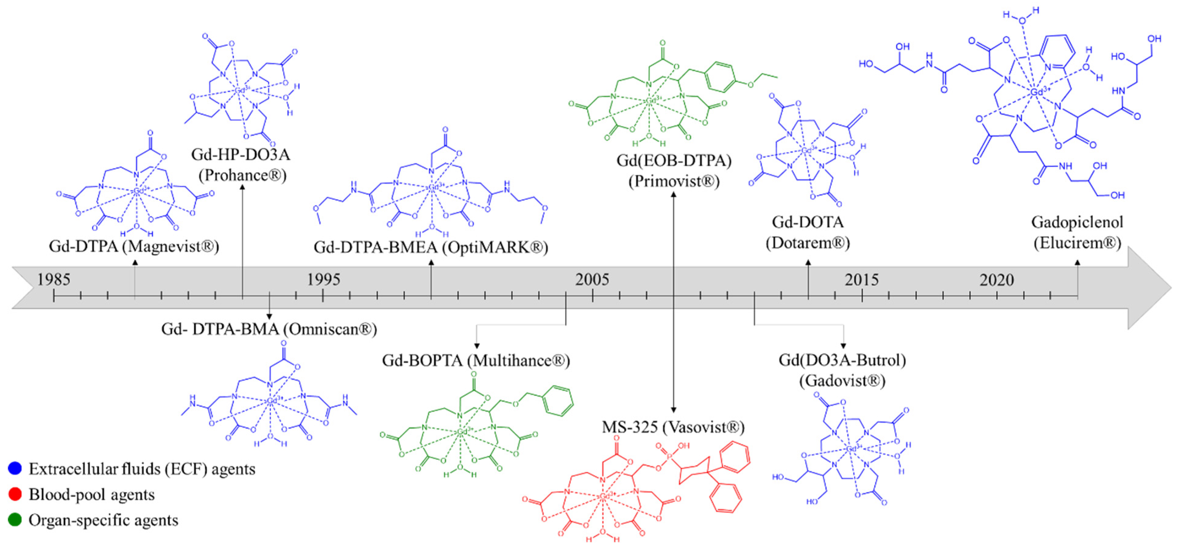
1. Introduction
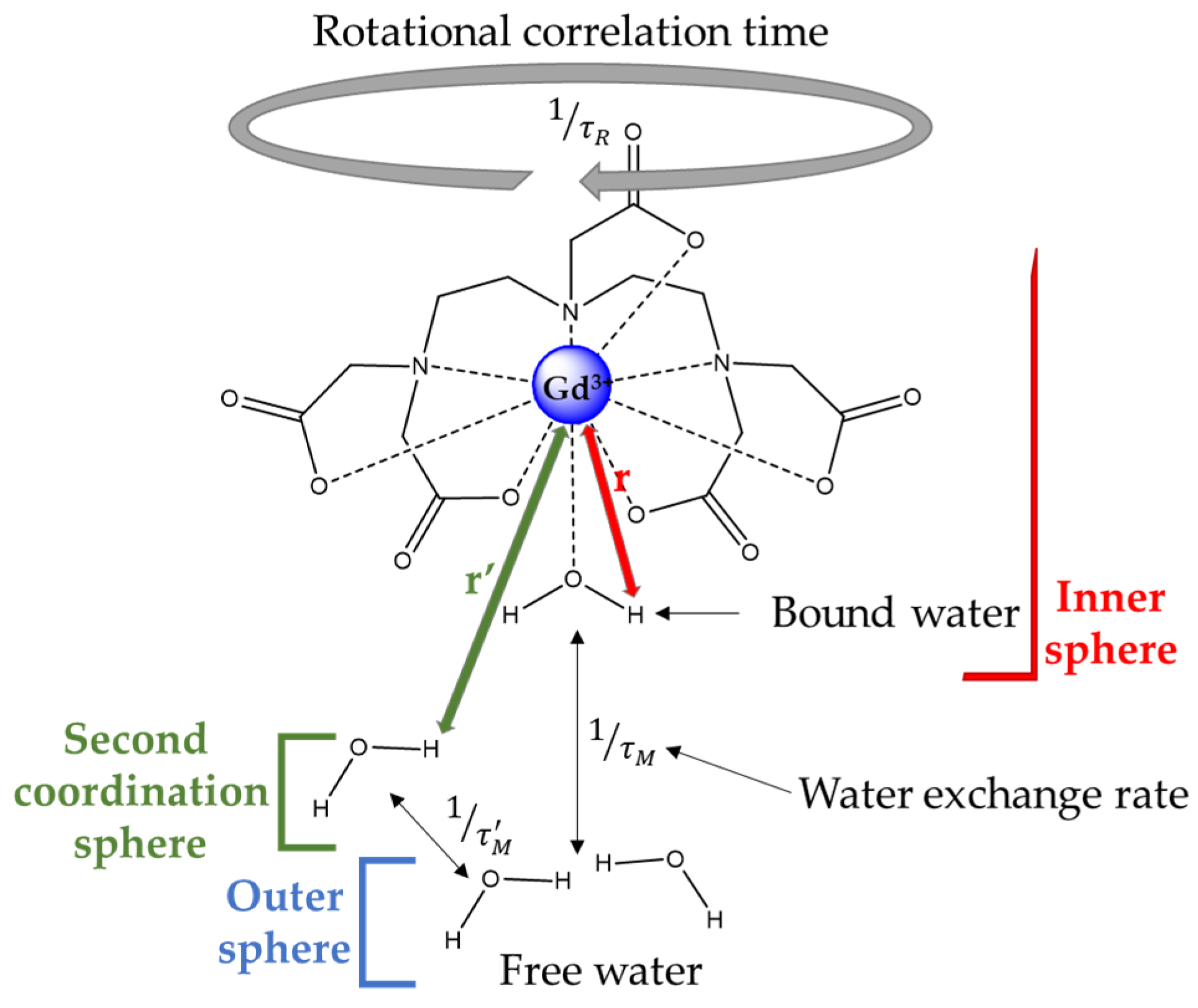
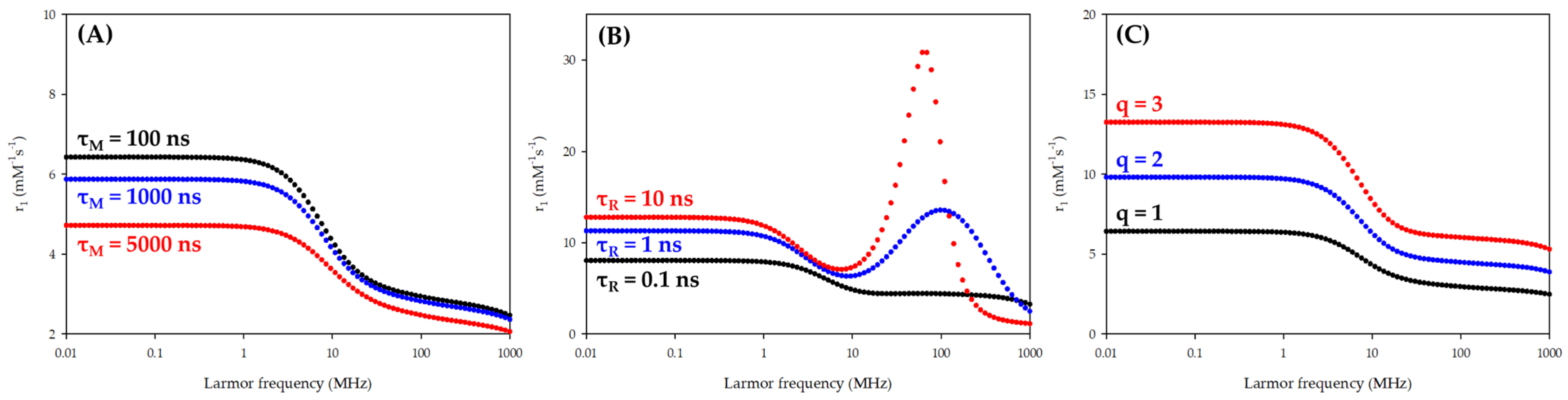
2. Paramagnetic Relaxation Theory
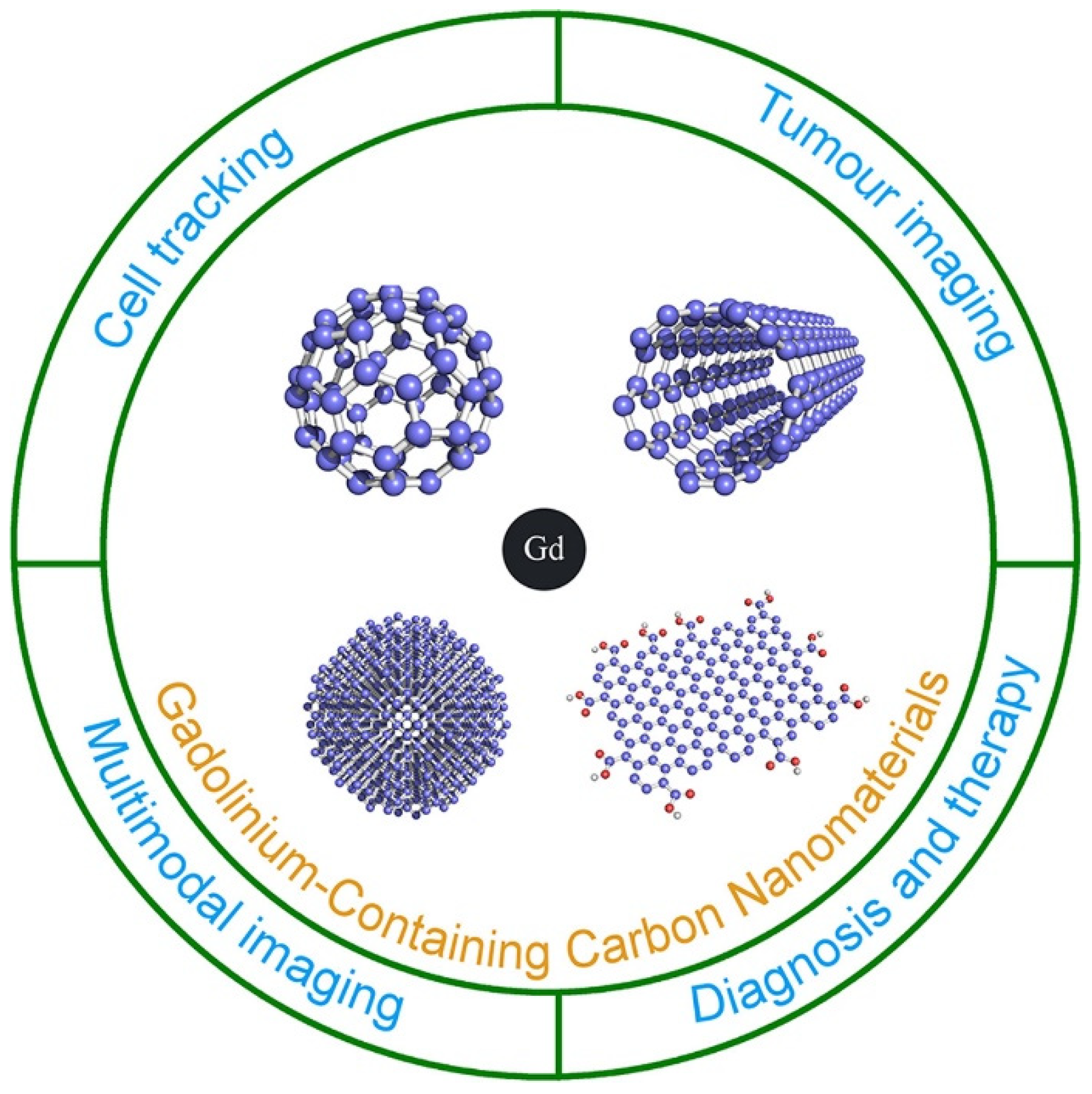
3. Paramagnetic Carbon-Based Nanomaterials
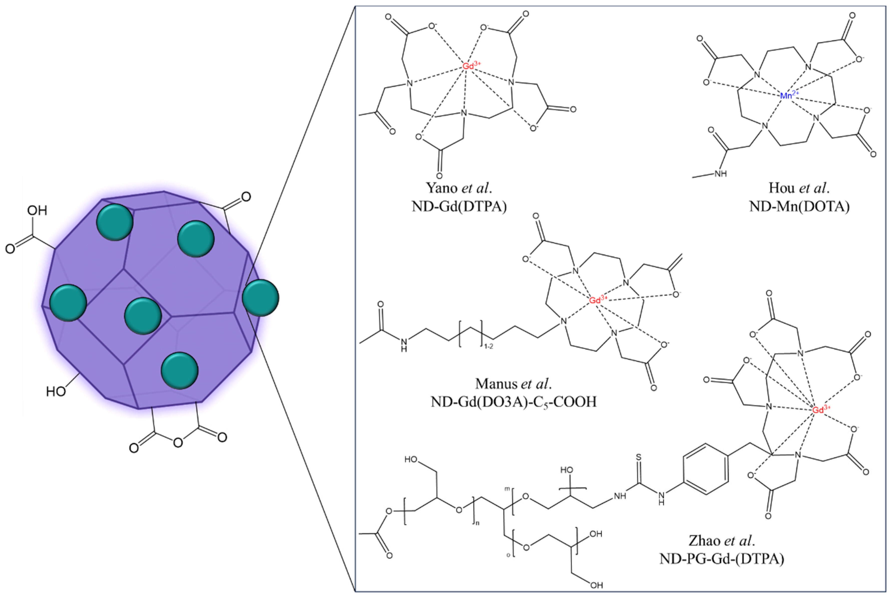
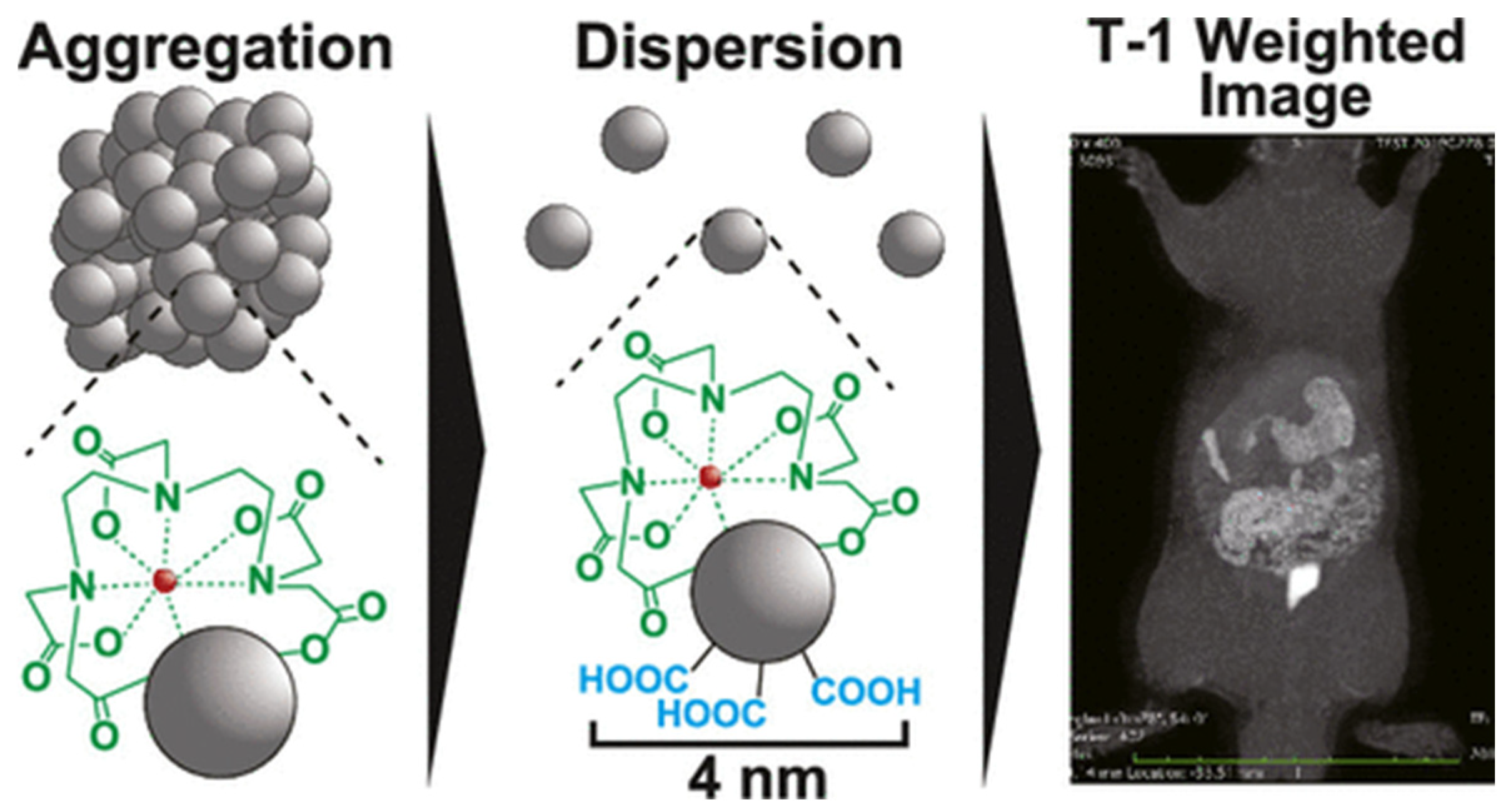
3.1. Nanoparticles of Diamond (NDs)

| Material | Systems | Size (TEM) (nm) | r1para (s−1 per mM Paramagnetic Center Mn/Gd) in Water or Buffer | Enhancement (%) | Magnetic Field (Tesla) | Ref. |
|---|---|---|---|---|---|---|
| Nanodiamond | DND-C6-Gd(DO3A) q = 2 | 128 nm (DLS) | 58.8 s−1 mM−1 Gd-DO3A: 5.4 s−1 mM−1 | 988% | 1.4 T/37 °C | [26] |
| DND-C5-Gd(DO3A) q = 2 | 75 nm (DLS) | 11.1 s−1 mM−1 Gd-DO3A-C5-COOH: 6.4 s−1 mM−1 11.5 s−1 mM−1 Gd-DO3A-C5-COOH: 4.8 s−1 mM−1 | 73% 139% | 1.4 T/37 °C 7 T/37 °C | [31] | |
| DND-Gd(III) | 7 nm (DLS)a | 33.4 s−1 mM−1 Gd-BOPTA: 4.8 s−1 mM−1 | 596% | 8 T/37 °C | [35] | |
| DND-PG-Gd(DTPA) (PG: polyglycerol) q = 1 | 18 nm | 19.4 s−1 mM−1 Gd-DTPA: 3.7 s−1 mM−1 16.7 s−1 mM−1 Gd-DTPA: 3.5 s−1 mM−1 8.2 s−1 mM−1 Gd-DTPA: 3.4 s−1 mM−1 | 424% 377% 141% | 1.5 T 3 T 7 T | [30] | |
| ND-Gd(III)/PVP | 45–70 nm (DLS)a | 15.9 s−1 mM−1 ND-Gd(III): 33.4 s−1 mM−1 | −52% | 8 T/37 °C | [36] | |
| DND-Gd(DTPA) q = 1 | 4.2 nm 630 nm (DLS) | u.d. | _ | 1.5 T | [28] | |
| DND-Mn(EDTA) | 65 nm (DLS) | 22.7 s−1 mM−1 Mn-EDTA: 1.7 s−1 mM−1 | 1235% | 7 T | [29] | |
| DND-Mn(II) | 4.5 nm | 19.6 s−1 mM−1 DND: 2.1 s−1 mM−1 | 833% | 8 T/37 °C | [37,38] | |
| DND-Mn(II)-PVP | 4.5 nm | 17.6 s−1 mM−1 DND: 2.1 s−1 mM−1 | 738% | 7 T | [29] | |
| DND (air oxidized) | 3–4 nm | 11.3 s−1 mM−1 DND: 1.7 s−1 mM−1 | 558% | 7 T | [45] | |
| Mn(II)-doped@HPHT-ND | 150 nm 1 µm (DLS) | 0.11 s−1 (g/mL)−1 ND: 0.02 s−1 (g/mL)−1 | 450% | 7 T | [40] | |
| Fe-doped@HPHT-ND (T2 agent) | 100 nm | r2Fe@ND = 0.95 s−1 (mg/mL)−1 r2ND = 0.14 s−1 (mg/mL)−1 | 555% | 7 T | [41] | |
| ND-RE(TTA)/DOX (RE: rare-earths = Eu3+, Gd3+) (TTA: 2-thenoyltrifluoroacetone complexes) | 4.2 nm | r1 = 1.1 s−1(mg/mL)−1 | _ | 3 T | [39] | |
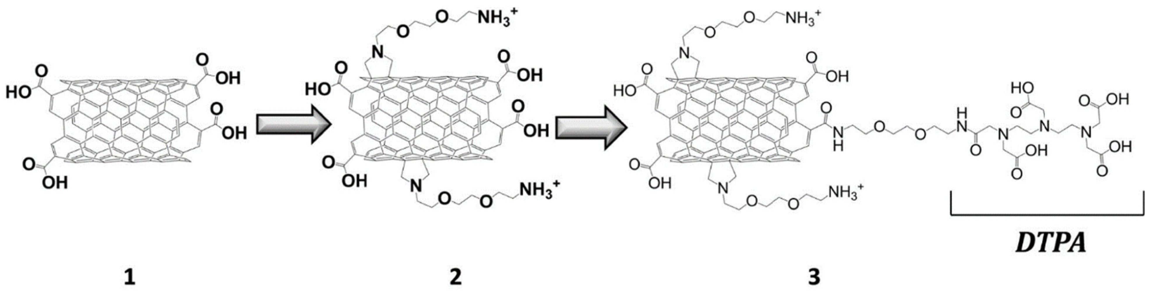
| Carbon nanotube | MWCT-Gd(DTPA) | 20–30 nm (in diameter size) 0.5–2 µm (in length size) | 6.61 s−1 mM−1 Gd-DTPA: 2.1 s−1 mM−1 | 314% | 7 T | [46] |
| MWCT-Gd2O3 | 10–20 nm (in diameter size) (SEM) | 18.9 s−1 mM−1 Gd2O3 NPs: 9.9 s−1 mM−1 | 91% | 9.4 T/25 °C | [47] | |
| Gd(III)-doped-US-SWCT | 20–80 nm (in length size) | 170 s−1 mM−1 Gd-DTPA: 4 s−1 mM−1 | 4150% | 1.41 T/40 °C | [48] | |
| Gd(III)-doped-US-SWCT/PLGA | 20–80 nm (in length size) | r2 = 578 s−1 mM−1 | _ | 7 T/25 °C | [49] | |
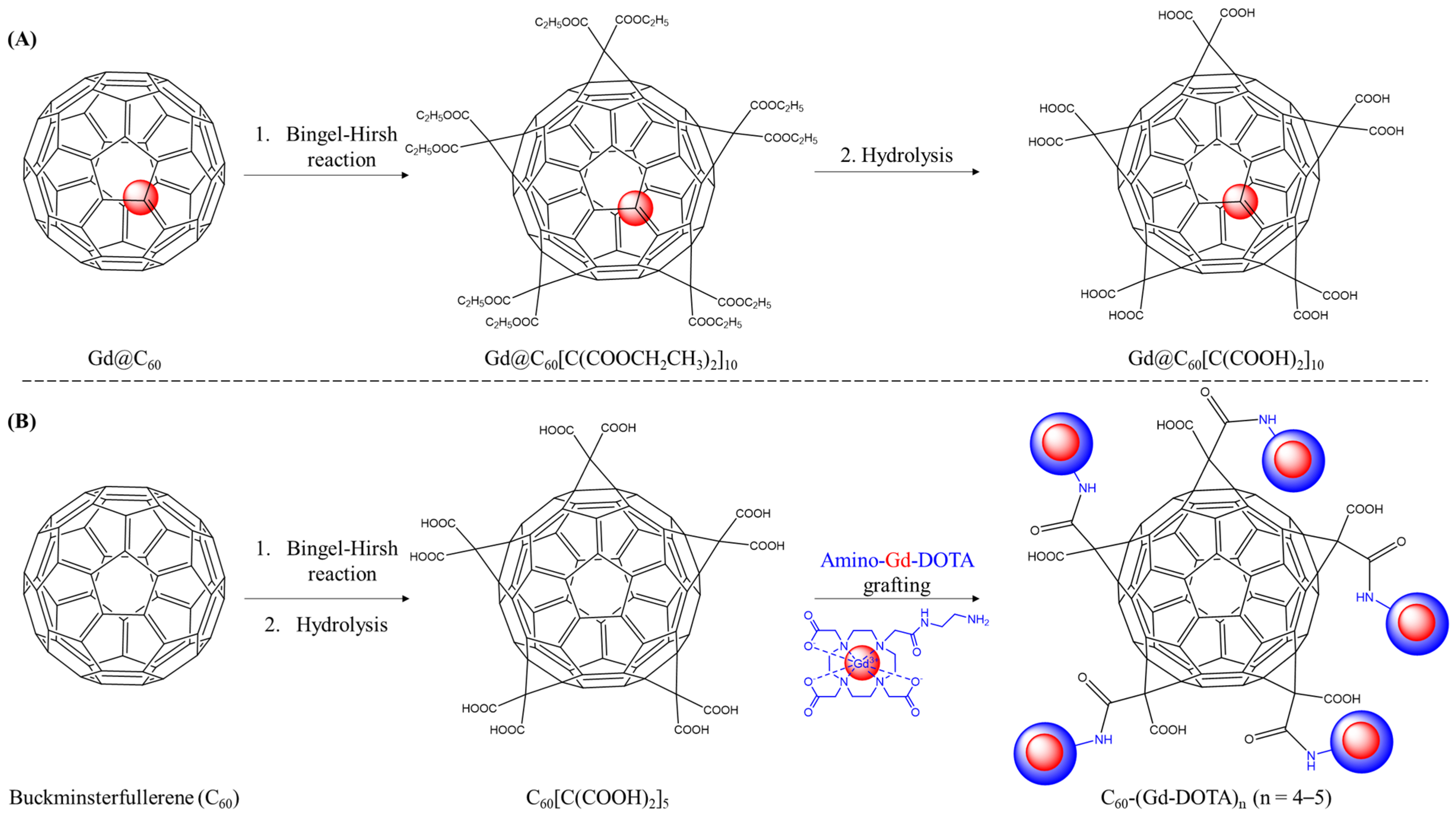
| C2n-like fullerene | Gd3+@C60[C(COOH)2]10 (endofullerene) | 10 nm (DLS) | 4.6 s−1 mM−1 | _ | 0.5 T/40 °C | [50] |
| Gd3+@C60[C(COOH)2]10 (endofullerene) | u.d. | 10.4 s−1 mM−1 Gd3+@C60(OH)x: 38.5 s−1 mM−1 | −73% | 0.5 T/25 °C | [51] | |
| Gd3+@C60(OH)x (endofullerene) | u.d. | 38.5 s−1 mM−1 Gd3+@C60[C(COOH)2]10: 10.4 s−1 mM−1 | 270% | 0.5 T/25 °C | [51] | |
| Gd3+@C82[OH]-FA/FITC (endofullerene) | 127 nm (DLS) | 20.2 s−1 mM−1 Gd-DTPA: 4.5 s−1 mM−1 | 348% | 0.5 T/37 °C | [52] | |
| C60-PEG-Gd(DTPA) | u.d. | 5.1 s−1 mM−1 Gd-DTPA: 5.3 s−1 mM−1 | −4% | 7 T/25 °C | [53] | |
| C60-Gd(DOTA) | 8.9 nm (DLS)b | 49.7 s−1 mM−1 Gd-DOTA: 5.4 s−1 mM−1 29.2 s−1 mM−1 Gd-DOTA: 3.2 s−1 mM−1 | 820% 812% | 0.5 T 1.5 T | [54] | |
| Gd3N@C80[DiPEG350Da(OH)x] (endofullerene) | 75 nm (DLS) | 75.7 s−1 mM−1 79.0 s−1 mM−1 22.7 s−1 mM−1 | _ | 0.35 T/25 °C 2.4 T/25 °C 9.4 T/25 °C | [55] | |
| Gd3N@C80[DiPEG5kDa(OH)x] (endofullerene) | 37 nm (DLS) | 46.3 s−1 mM−1 | _ | 2.4 T/25 °C | [54] | |
| Gd3N@C80-ZD2peptide (endofullerene) | 2.8 nm | 74.6 s−1 mM−1 Gd3N@C80: 57.1 s−1 mM−1 24.8 s−1 mM−1 Gd3N@C80: 24.7 s−1 mM−1 | 30% 0.4% | 1.5 T 7 T | [56] | |
Gd3N@C80-DOX-RNPs (endofullerene) | 146 nm (DLS) pH = 7.4 | 17.8 s−1 mM−1 5.7 s−1 mM−1 | 212% | 7 T/pH = 6.6 7 T/pH = 7.4 | [57] | |
| C60-Gd2(DOTA)2 | 171 nm (DLS) | 18.2 s−1 mM−1 Magnevist: 4.7 s−1 mM−1 | 287% | 4.7 T/20 °C | [58] | |
| C60-Mn(APTSPP) | 76 nm | 19.2 s−1 mM−1 Mn(APTSPP): 11.3 s−1 mM−1 12.2 s−1 mM−1 Mn(APTSPP): 8.2 s−1 mM−1 | 70% 49% | 0.5 T/37 °C 3 T/37 °C | [59] | |
| Graphene oxide | GO-Gd(III) | 36–44 nm (AFM) | 78–85 s−1 mM−1 | _ | 1.41 T/37 °C | [60] |
| GO-Gd(DTPA) q = 1 | 76 nm | 28–32 s−1 mM−1 | _ | 0.5 T/37 °C 3 T/37 °C | ||
| GO-Gd(DO3A) q = 2 | 36–44 nm (AFM) < 50 nm (SEM) 20–50 nm (AFM)c | 49–63 s−1 mM−1 | _ | 1.41 T/37 °C 1.5 T 11.7 T | ||
| rGO-Gd(DTPA) q = 1 | 16.8 s−1 mM−1 Gd-DTPA: 4 s−1 mM−1 | 321% | ||||
| GO-PEG-Gd(DOTA) | 14.2 s−1 mM−1 Gd-DOTA: 4.5 s−1 mM−1 | 215% | ||||
| G-Mn(DPDP) | ±100 nm | 92 s−1 mM−1 Mn-DPDP: 2.8 s−1 mM−1 | 3185% | 0.5 T | [61] |
3.2. Carbon Nanotubes (CNTs)
3.3. Buckminsterfullerene (C2n)
3.4. Graphene and Graphene Oxide Nanosheet (GO)
4. Conclusions
Author Contributions
Funding
Institutional Review Board Statement
Informed Consent Statement
Data Availability Statement
Acknowledgments
Conflicts of Interest
References
- Wahsner, J.; Gale, E.M.; Rodríguez-Rodríguez, A.; Caravan, P. Chemistry of MRI Contrast Agents: Current Challenges and New Frontiers. Chem. Rev. 2019, 119, 957–1057. [Google Scholar] [CrossRef] [PubMed]
- Iyad, N.; Ahmad, S.M.; Alkhatib, S.G.; Hjouj, M. Gadolinium Contrast Agents- Challenges and Opportunities of a Multidisciplinary Approach: Literature Review. Eur. J. Radiol. Open 2023, 11, 100503–100513. [Google Scholar] [CrossRef] [PubMed]
- Hazelton, J.M.; Chiu, M.K.; Abujudeh, H.H. Nephrogenic Systemic Fibrosis: A Review of History, Pathophysiology, and Current Guidelines. Curr Radiol Rep 2019, 7, 5. [Google Scholar] [CrossRef]
- Malikova, H. Nephrogenic Systemic Fibrosis: The End of the Story? Quant. Imaging Med. Surg. 2019, 9, 1470–1474. [Google Scholar] [CrossRef] [PubMed]
- Minton, L.E.; Pandit, R.; Porter, K.K. Contrast-Enhanced MRI: History and Current Recommendations. Appl. Radiol. 2021, 50, 15–19. [Google Scholar] [CrossRef]
- Le Fur, M.; Caravan, P. The Biological Fate of Gadolinium-Based MRI Contrast Agents: A Call to Action for Bioinorganic Chemists. Metallomics 2019, 11, 240–254. [Google Scholar] [CrossRef]
- Robic, C.; Port, M.; Rousseaux, O.; Louguet, S.; Fretellier, N.; Catoen, S.; Factor, C.; Le Greneur, S.; Medina, C.; Bourrinet, P.; et al. Physicochemical and Pharmacokinetic Profiles of Gadopiclenol: A New Macrocyclic Gadolinium Chelate With High T1 Relaxivity. Invest Radiol 2019, 54, 475–484. [Google Scholar] [CrossRef]
- Botta, M.; Carniato, F.; Esteban-Gómez, D.; Platas-Iglesias, C.; Tei, L. Mn(II) Compounds as an Alternative to Gd-Based MRI Probes. Future Med. Chem. 2019, 11, 1461–1483. [Google Scholar] [CrossRef]
- Henoumont, C.; Devreux, M.; Laurent, S. Mn-Based MRI Contrast Agents: An Overview. Molecules 2023, 28, 7275. [Google Scholar] [CrossRef]
- Bianchi, A.; Gobbo, O.L.; Dufort, S.; Sancey, L.; Lux, F.; Tillement, O.; Coll, J.; Crémillieux, Y. Orotracheal Manganese-enhanced MRI (MEMRI): An Effective Approach for Lung Tumor Detection. NMR Biomed. 2017, 30, e3790. [Google Scholar] [CrossRef]
- Yuan, D.; Ellis, C.M.; Davis, J.J. Mesoporous Silica Nanoparticles in Bioimaging. Materials 2020, 13, 3795. [Google Scholar] [CrossRef] [PubMed]
- Carniato, F.; Tei, L.; Botta, M. Gd-Based Mesoporous Silica Nanoparticles as MRI Probes. Eur J Inorg Chem 2018, 2018, 4936–4954. [Google Scholar] [CrossRef]
- Marasini, R.; Thanh Nguyen, T.D.; Aryal, S. Integration of Gadolinium in Nanostructure for Contrast Enhanced-magnetic Resonance Imaging. WIREs Nanomed. Nanobiotechnol. 2020, 12, e1580. [Google Scholar] [CrossRef] [PubMed]
- Gan, S.; Lin, Y.; Feng, Y.; Shui, L.; Li, H.; Zhou, G. Magnetic Polymeric Nanoassemblies for Magnetic Resonance Imaging-Combined Cancer Theranostics. IJN 2018, 13, 4263–4281. [Google Scholar] [CrossRef] [PubMed]
- Kotha, R.; Kara, D.D.; Roychowdhury, R.; Tanvi, K.; Rathnanand, M. Polymersomes Based Versatile Nanoplatforms for Controlled Drug Delivery and Imaging. Adv. Pharm. Bull. 2023, 13, 218–232. [Google Scholar] [CrossRef] [PubMed]
- Puente-Santiago, A.R.; Rodríguez-Padrón, D. (Eds.) Surface-Modified Nanobiomaterials for Electrochemical and Biomedicine Applications; Topics in Current Chemistry Collections; Springer International Publishing: Cham, Switzerland, 2020; ISBN 978-3-030-55501-6. [Google Scholar]
- Fernández-Barahona, I.; Muñoz-Hernando, M.; Ruiz-Cabello, J.; Herranz, F.; Pellico, J. Iron Oxide Nanoparticles: An Alternative for Positive Contrast in Magnetic Resonance Imaging. Inorganics 2020, 8, 28. [Google Scholar] [CrossRef]
- Rodríguez-Galván, A.; Rivera, M.; García-López, P.; Medina, L.A.; Basiuk, V.A. Gadolinium-containing Carbon Nanomaterials for Magnetic Resonance Imaging: Trends and Challenges. J Cell. Mol. Medi 2020, 24, 3779–3794. [Google Scholar] [CrossRef] [PubMed]
- Garifo, S.; Stanicki, D.; Ayata, G.; Muller, R.N.; Laurent, S. Nanodiamonds as Nanomaterial for Biomedical Field. Front. Mater. Sci. 2021, 15, 334–351. [Google Scholar] [CrossRef]
- Bondon, N.; Raehm, L.; Charnay, C.; Boukherroub, R.; Durand, J.-O. Nanodiamonds for Bioapplications, Recent Developments. J. Mater. Chem. B 2020, 8, 10878–10896. [Google Scholar] [CrossRef]
- Tegafaw, T.; Liu, S.; Ahmad, M.Y.; Ali Al Saidi, A.K.; Zhao, D.; Liu, Y.; Yue, H.; Nam, S.-W.; Chang, Y.; Lee, G.H. Production, Surface Modification, Physicochemical Properties, Biocompatibility, and Bioimaging Applications of Nanodiamonds. RSC Adv. 2023, 13, 32381–32397. [Google Scholar] [CrossRef]
- Jung, H.-S.; Neuman, K.C. Surface Modification of Fluorescent Nanodiamonds for Biological Applications. Nanomaterials 2021, 11, 153. [Google Scholar] [CrossRef] [PubMed]
- Nanodiamonds: Advanced Material Analysis, Properties and Applications; Arnault, J.-C., Ed.; Micro & Nano Technologies Series; Elsevier: Amsterdam, Netherlands, 2017; ISBN 978-0-323-43029-6. [Google Scholar]
- Pan, F.; Khan, M.; Ragab, A.H.; Javed, E.; Alsalmah, H.A.; Khan, I.; Lei, T.; Hussain, A.; Mohamed, A.; Zada, A.; et al. Recent Advances in the Structure and Biomedical Applications of Nanodiamonds and Their Future Perspectives. Mater. Des. 2023, 233, 112179. [Google Scholar] [CrossRef]
- Mermoux, M.; Chang, S.; Girard, H.A.; Arnault, J.-C. Raman Spectroscopy Study of Detonation Nanodiamond. Diam. Relat. Mater. 2018, 87, 248–260. [Google Scholar] [CrossRef]
- Manus, L.M.; Mastarone, D.J.; Waters, E.A.; Zhang, X.-Q.; Schultz-Sikma, E.A.; MacRenaris, K.W.; Ho, D.; Meade, T.J. Gd(III)-Nanodiamond Conjugates for MRI Contrast Enhancement. Nano Lett. 2010, 10, 484–489. [Google Scholar] [CrossRef] [PubMed]
- Rammohan, N.; MacRenaris, K.W.; Moore, L.K.; Parigi, G.; Mastarone, D.J.; Manus, L.M.; Lilley, L.M.; Preslar, A.T.; Waters, E.A.; Filicko, A.; et al. Nanodiamond–Gadolinium(III) Aggregates for Tracking Cancer Growth In Vivo at High Field. Nano Lett. 2016, 16, 7551–7564. [Google Scholar] [CrossRef] [PubMed]
- Yano, K.; Matsumoto, T.; Okamoto, Y.; Bito, K.; Kurokawa, N.; Hasebe, T.; Hotta, A. Gadolinium-Complexed Carboxylated Nanodiamond Particles for Magnetic Resonance Imaging of the Lymphatic System. ACS Appl. Nano Mater. 2021, 4, 1702–1711. [Google Scholar] [CrossRef]
- Hou, W.; Toh, T.B.; Abdullah, L.N.; Yvonne, T.W.Z.; Lee, K.J.; Guenther, I.; Chow, E.K.-H. Nanodiamond–Manganese Dual Mode MRI Contrast Agents for Enhanced Liver Tumor Detection. Nanomed. Nanotechnol. Biol. Med. 2017, 13, 783–793. [Google Scholar] [CrossRef] [PubMed]
- Zhao, L.; Shiino, A.; Qin, H.; Kimura, T.; Komatsu, N. Synthesis, Characterization, and Magnetic Resonance Evaluation of Polyglycerol-Functionalized Detonation Nanodiamond Conjugated with Gadolinium(III) Complex. J. Nanosci. Nanotechnol. 2015, 15, 1076–1082. [Google Scholar] [CrossRef] [PubMed]
- Perevedentseva, E.V. Nanodiamonds for Biomedical Applications – Features of Interaction with Blood Components and Behavior in the Circulatory System. J. Biomed. Photonics Eng. 2022, 8, 040506. [Google Scholar] [CrossRef]
- Nakamura, T.; Ohana, T.; Yabuno, H.; Kasai, R.; Suzuki, T.; Hasebe, T. Simple Fabrication of Gd(III)-DTPA-Nanodiamond Particles by Chemical Modification for Use as Magnetic Resonance Imaging (MRI) Contrast Agent. Appl. Phys. Express 2013, 6, 015001. [Google Scholar] [CrossRef]
- Osipov, V.Y.; Boukhvalov, D.W.; Takai, K. Isolated Spin-7/2 Species of Gadolinium(III) Chelate Complexes on the Surface of 5-Nm Diamond Particles. Nanomaterials 2023, 13, 1995. [Google Scholar] [CrossRef] [PubMed]
- Panich, A.M.; Shames, A.I.; Sergeev, N.A.; Osipov, V.Y.; Alexenskiy, A.E.; Vul’, A.Y. Magnetic Resonance Study of Gadolinium-Grafted Nanodiamonds. J. Phys. Chem. C 2016, 120, 19804–19811. [Google Scholar] [CrossRef]
- Panich, A.M.; Salti, M.; Goren, S.D.; Yudina, E.B.; Aleksenskii, A.E.; Vul’, A.Y.; Shames, A.I. Gd(III)-Grafted Detonation Nanodiamonds for MRI Contrast Enhancement. J. Phys. Chem. C 2019, 123, 2627–2631. [Google Scholar] [CrossRef]
- Panich, A.M.; Salti, M.; Prager, O.; Swissa, E.; Kulvelis, Y.V.; Yudina, E.B.; Aleksenskii, A.E.; Goren, S.D.; Vul’, A.Y.; Shames, A.I. PVP-coated Gd-grafted Nanodiamonds as a Novel and Potentially Safer Contrast Agent for in Vivo MRI. Magn. Reson. Med. 2021, 86, 935–942. [Google Scholar] [CrossRef] [PubMed]
- Panich, A.M.; Shames, A.I.; Aleksenskii, A.E.; Yudina, E.B.; Vul’, A.Y. Manganese-Grafted Detonation Nanodiamond, a Novel Potential MRI Contrast Agent. Diam. Relat. Mater. 2021, 119, 108590. [Google Scholar] [CrossRef]
- Panich, A.M.; Salti, M.; Aleksenskii, A.E.; Kulvelis, Y.V.; Chizhikova, A.; Vul’, A.Y.; Shames, A.I. Suspensions of Manganese-Grafted Nanodiamonds: Preparation, NMR, and MRI Study. Diam. Relat. Mater. 2023, 131, 109591. [Google Scholar] [CrossRef]
- Qin, S.-R.; Zhao, Q.; Cheng, Z.-G.; Zhang, D.-X.; Zhang, K.-K.; Su, L.-X.; Fan, H.-J.; Wang, Y.-H.; Shan, C.-X. Rare Earth-Functionalized Nanodiamonds for Dual-Modal Imaging and Drug Delivery. Diam. Relat. Mater. 2019, 91, 173–182. [Google Scholar] [CrossRef]
- Lin, B.-R.; Chen, C.-H.; Kunuku, S.; Chen, T.-Y.; Hsiao, T.-Y.; Niu, H.; Lee, C.-P. Fe Doped Magnetic Nanodiamonds Made by Ion Implantation as Contrast Agent for MRI. Sci. Rep. 2018, 8, 7058. [Google Scholar] [CrossRef] [PubMed]
- Kunuku, S.; Lin, B.-R.; Chen, C.-H.; Chang, C.-H.; Chen, T.-Y.; Hsiao, T.-Y.; Yu, H.-K.; Chang, Y.-J.; Liao, L.-C.; Chen, F.-H.; et al. Nanodiamonds Doped with Manganese for Applications in Magnetic Resonance Imaging. ACS Omega 2023, 8, 4398–4409. [Google Scholar] [CrossRef]
- Waddington, D.E.J.; Sarracanie, M.; Zhang, H.; Salameh, N.; Glenn, D.R.; Rej, E.; Gaebel, T.; Boele, T.; Walsworth, R.L.; Reilly, D.J.; et al. Nanodiamond-Enhanced MRI via in Situ Hyperpolarization. Nat. Commun. 2017, 8, 15118. [Google Scholar] [CrossRef]
- Waddington, D.E.J.; Boele, T.; Rej, E.; McCamey, D.R.; King, N.J.C.; Gaebel, T.; Reilly, D.J. Phase-Encoded Hyperpolarized Nanodiamond for Magnetic Resonance Imaging. Sci. Rep. 2019, 9, 5950. [Google Scholar] [CrossRef] [PubMed]
- Sękowska, A.; Majchrowicz, D.; Sabisz, A.; Ficek, M.; Bułło-Piontecka, B.; Kosowska, M.; Jing, L.; Bogdanowicz, R.; Szczerska, M. Nanodiamond Phantoms Mimicking Human Liver: Perspective to Calibration of T1 Relaxation Time in Magnetic Resonance Imaging. Sci. Rep. 2020, 10, 6446. [Google Scholar] [CrossRef] [PubMed]
- Lazovic, J.; Goering, E.; Wild, A.; Schützendübe, P.; Shiva, A.; Löffler, J.; Winter, G.; Sitti, M. Nanodiamond-Enhanced Magnetic Resonance Imaging. Adv. Mater. 2023, 36, 2310109. [Google Scholar] [CrossRef] [PubMed]
- Servant, A.; Jacobs, I.; Bussy, C.; Fabbro, C.; da Ros, T.; Pach, E.; Ballesteros, B.; Prato, M.; Nicolay, K.; Kostarelos, K. Gadolinium-Functionalised Multi-Walled Carbon Nanotubes as a T1 Contrast Agent for MRI Cell Labelling and Tracking. Carbon 2016, 97, 126–133. [Google Scholar] [CrossRef]
- Paul, R.; Chatterjee, D.; Das Ghosh, L.; Narayanswamy, V.; Singh, M.P.; Agarwal, M.; Ghosh, D.; Radhakrishna, M.; Tiwary, C.S.; Provazník, I.; et al. Synthesis, Characterization and In-Vitro Studies of CNT/Gd2O3 Hybrid Structure. Carbon Trends 2023, 11, 100272. [Google Scholar] [CrossRef]
- Sitharaman, B.; Kissell, K.R.; Hartman, K.B.; Tran, L.A.; Baikalov, A.; Rusakova, I.; Sun, Y.; Khant, H.A.; Ludtke, S.J.; Chiu, W.; et al. Superparamagnetic Gadonanotubes Are High-Performance MRI Contrast Agents. Chem. Commun. 2005, 31, 3915–3917. [Google Scholar] [CrossRef] [PubMed]
- Sitharaman, B.; Van Der Zande, M.; Ananta, J.S.; Shi, X.; Veltien, A.; Walboomers, X.F.; Wilson, L.J.; Mikos, A.G.; Heerschap, A.; Jansen, J.A. Magnetic Resonance Imaging Studies on Gadonanotube-Reinforced Biodegradable Polymer Nanocomposites. J. Biomed. Mater. Res. 2010, 93A, 1454–1462. [Google Scholar] [CrossRef] [PubMed]
- Bolskar, R.D.; Benedetto, A.F.; Husebo, L.O.; Price, R.E.; Jackson, E.F.; Wallace, S.; Wilson, L.J.; Alford, J.M. First Soluble M@C60 Derivatives Provide Enhanced Access to Metallofullerenes and Permit in Vivo Evaluation of Gd@C60[C(COOH)2]10 as a MRI Contrast Agent. J. Am. Chem. Soc. 2003, 125, 5471–5478. [Google Scholar] [CrossRef] [PubMed]
- Tóth, É.; Bolskar, R.D.; Borel, A.; González, G.; Helm, L.; Merbach, A.E.; Sitharaman, B.; Wilson, L.J. Water-Soluble Gadofullerenes: Toward High-Relaxivity, pH-Responsive MRI Contrast Agents. J. Am. Chem. Soc. 2005, 127, 799–805. [Google Scholar] [CrossRef]
- Zheng, J.; Liu, Q.; Zhen, M.; Jiang, F.; Shu, C.; Jin, C.; Yang, Y.; Alhadlaq, H.A.; Wang, C. Multifunctional Imaging Probe Based on Gadofulleride Nanoplatform. Nanoscale 2012, 4, 3669. [Google Scholar] [CrossRef]
- Liu, J.; Ohta, S.; Sonoda, A.; Yamada, M.; Yamamoto, M.; Nitta, N.; Murata, K.; Tabata, Y. Preparation of PEG-Conjugated Fullerene Containing Gd3+ Ions for Photodynamic Therapy. J. Control. Release 2007, 117, 104–110. [Google Scholar] [CrossRef] [PubMed]
- Wang, L.; Zhu, X.; Tang, X.; Wu, C.; Zhou, Z.; Sun, C.; Deng, S.-L.; Ai, H.; Gao, J. A Multiple Gadolinium Complex Decorated Fullerene as a Highly Sensitive T 1 Contrast Agent. Chem. Commun. 2015, 51, 4390–4393. [Google Scholar] [CrossRef] [PubMed]
- Zhang, J.; Fatouros, P.P.; Shu, C.; Reid, J.; Owens, L.S.; Cai, T.; Gibson, H.W.; Long, G.L.; Corwin, F.D.; Chen, Z.-J.; et al. High Relaxivity Trimetallic Nitride (Gd3N) Metallofullerene MRI Contrast Agents with Optimized Functionality. Bioconjugate Chem. 2010, 21, 610–615. [Google Scholar] [CrossRef] [PubMed]
- Han, Z.; Wu, X.; Roelle, S.; Chen, C.; Schiemann, W.P.; Lu, Z.-R. Targeted Gadofullerene for Sensitive Magnetic Resonance Imaging and Risk-Stratification of Breast Cancer. Nat. Commun. 2017, 8, 692. [Google Scholar] [CrossRef] [PubMed]
- Wang, S.; Zhou, Z.; Wang, Z.; Liu, Y.; Jacobson, O.; Shen, Z.; Fu, X.; Chen, Z.; Chen, X. Gadolinium Metallofullerene-Based Activatable Contrast Agent for Tumor Signal Amplification and Monitoring of Drug Release. Small 2019, 15, 1900691. [Google Scholar] [CrossRef] [PubMed]
- Kim, J.B.-K.; Mackeyev, Y.; Raghuram, S.; Cho, S.H.; Krishnan, S. Synthesis and Characterization of Gadolinium-Decorated [60]Fullerene for Tumor Imaging and Radiation Sensitization. Int. J. Radiat. Biol. 2021, 97, 1129–1139. [Google Scholar] [CrossRef] [PubMed]
- Zou, T.; Zhen, M.; Chen, D.; Li, R.; Guan, M.; Shu, C.; Han, H.; Wang, C. The Positive Influence of Fullerene Derivatives Bonded to Manganese(III) Porphyrins on Water Proton Relaxation. Dalton Trans. 2015, 44, 9114–9119. [Google Scholar] [CrossRef] [PubMed]
- Hung, A.H.; Duch, M.C.; Parigi, G.; Rotz, M.W.; Manus, L.M.; Mastarone, D.J.; Dam, K.T.; Gits, C.C.; MacRenaris, K.W.; Luchinat, C.; et al. Mechanisms of Gadographene-Mediated Proton Spin Relaxation. J. Phys. Chem. C 2013, 117, 16263–16273. [Google Scholar] [CrossRef]
- Kanakia, S.; Toussaint, J.; Hoang, D.M.; Mullick Chowdhury, S.; Lee, S.; Shroyer, K.R.; Moore, W.; Wadghiri, Y.Z.; Sitharaman, B. Towards An Advanced Graphene-Based Magnetic Resonance Imaging Contrast Agent: Sub-Acute Toxicity and Efficacy Studies in Small Animals. Sci Rep 2015, 5, 17182. [Google Scholar] [CrossRef]
- Chawda, N.; Basu, M.; Majumdar, D.; Poddar, R.; Mahapatra, S.K.; Banerjee, I. Engineering of Gadolinium-Decorated Graphene Oxide Nanosheets for Multimodal Bioimaging and Drug Delivery. ACS Omega 2019, 4, 12470–12479. [Google Scholar] [CrossRef]
- Zhang, M.; Liu, X.; Huang, J.; Wang, L.; Shen, H.; Luo, Y.; Li, Z.; Zhang, H.; Deng, Z.; Zhang, Z. Ultrasmall Graphene Oxide Based T1 MRI Contrast Agent for in Vitro and in Vivo Labeling of Human Mesenchymal Stem Cells. Nanomed. Nanotechnol. Biol. Med. 2018, 14, 2475–2483. [Google Scholar] [CrossRef] [PubMed]
- Panich, A.M. Can Detonation Nanodiamonds Serve as MRI Phantoms? Magn. Reson. Mater. Phys. 2022, 35, 345–347. [Google Scholar] [CrossRef] [PubMed]
- Herlem, G.; Picaud, F.; Girardet, C.; Micheau, O. Carbon Nanotubes. In Nanocarriers for Drug Delivery; Elsevier: Amsterdam, The Netherlands, 2019; pp. 469–529. ISBN 978-0-12-814033-8. [Google Scholar]
- Prato, M.; Kostarelos, K.; Bianco, A. Functionalized Carbon Nanotubes in Drug Design and Discovery. Acc. Chem. Res. 2008, 41, 60–68. [Google Scholar] [CrossRef] [PubMed]
- Hernández-Rivera, M.; Zaibaq, N.G.; Wilson, L.J. Toward Carbon Nanotube-Based Imaging Agents for the Clinic. Biomaterials 2016, 101, 229–240. [Google Scholar] [CrossRef] [PubMed]
- Tang, L.; Xiao, Q.; Mei, Y.; He, S.; Zhang, Z.; Wang, R.; Wang, W. Insights on Functionalized Carbon Nanotubes for Cancer Theranostics. J. Nanobiotechnol. 2021, 19, 423. [Google Scholar] [CrossRef] [PubMed]
- Sethi, R.; Mackeyev, Y.; Wilson, L.J. The Gadonanotubes Revisited: A New Frontier in MRI Contrast Agent Design. Inorganica Chim. Acta 2012, 393, 165–172. [Google Scholar] [CrossRef]
- Ananta, J.S.; Matson, M.L.; Tang, A.M.; Mandal, T.; Lin, S.; Wong, K.; Wong, S.T.; Wilson, L.J. Single-Walled Carbon Nanotube Materials as T2-Weighted MRI Contrast Agents. J. Phys. Chem. C 2009, 113, 19369–19372. [Google Scholar] [CrossRef]
- Pochkaeva, E.I.; Podolsky, N.E.; Zakusilo, D.N.; Petrov, A.V.; Charykov, N.A.; Vlasov, T.D.; Penkova, A.V.; Vasina, L.V.; Murin, I.V.; Sharoyko, V.V.; et al. Fullerene Derivatives with Amino Acids, Peptides and Proteins: From Synthesis to Biomedical Application. Prog. Solid State Chem. 2020, 57, 100255. [Google Scholar] [CrossRef]
- Gulati, S.; Mansi; Vijayan, S.; Kumar, S.; Agarwal, V.; Harikumar, B.; Varma, R.S. Magnetic Nanocarriers Adorned on Graphene: Promising Contrast Enhancing Agents with State-of-the-Art Performance in Magnetic Resonance Imaging (MRI) and Theranostics. Mater. Adv. 2022, 3, 2971–2989. [Google Scholar] [CrossRef]
- Sitharaman, B.; Bolskar, R.D.; Rusakova, I.; Wilson, L.J. Gd@C60[C(COOH)2]10 and Gd@C60(OH)x: Nanoscale Aggregation Studies of Two Metallofullerene MRI Contrast Agents in Aqueous Solution. Nano Lett. 2004, 4, 2373–2378. [Google Scholar] [CrossRef]
- Grebowski, J.; Litwinienko, G. Metallofullerenols in Biomedical Applications. Eur. J. Med. Chem. 2022, 238, 114481. [Google Scholar] [CrossRef] [PubMed]
- Pathak, Y.; Thassu, D. (Eds.) Fullerene-Based Nanostructures: A Novel High-Performance Platform Technology for Magnetic Resonance Imaging (MRI). In Drug Delivery Nanoparticles Formulation and Characterization; Informa Health Care: New York, NY, USA, 2016; pp. 330–348. ISBN 978-1-4200-7805-3. [Google Scholar]
- Bolskar, R.D. Gadolinium Endohedral Metallofullerene-Based MRI Contrast Agents. In Medicinal Chemistry and Pharmacological Potential of Fullerenes and Carbon Nanotubes; Cataldo, F., Da Ros, T., Eds.; Carbon Materials: Chemistry and Physics; Springer: Dordrecht, The Netherlands, 2008; Volume 1, pp. 157–180. ISBN 978-1-4020-6844-7. [Google Scholar]
- Sun, X.; Liu, Z.; Welsher, K.; Robinson, J.T.; Goodwin, A.; Zaric, S.; Dai, H. Nano-Graphene Oxide for Cellular Imaging and Drug Delivery. Nano Res. 2008, 1, 203–212. [Google Scholar] [CrossRef] [PubMed]


Disclaimer/Publisher’s Note: The statements, opinions and data contained in all publications are solely those of the individual author(s) and contributor(s) and not of MDPI and/or the editor(s). MDPI and/or the editor(s) disclaim responsibility for any injury to people or property resulting from any ideas, methods, instructions or products referred to in the content. |
© 2024 by the authors. Licensee MDPI, Basel, Switzerland. This article is an open access article distributed under the terms and conditions of the Creative Commons Attribution (CC BY) license (https://creativecommons.org/licenses/by/4.0/).
Share and Cite
Garifo, S.; Vangijzegem, T.; Stanicki, D.; Laurent, S. A Review on the Design of Carbon-Based Nanomaterials as MRI Contrast Agents. Molecules 2024, 29, 1639. https://doi.org/10.3390/molecules29071639
Garifo S, Vangijzegem T, Stanicki D, Laurent S. A Review on the Design of Carbon-Based Nanomaterials as MRI Contrast Agents. Molecules. 2024; 29(7):1639. https://doi.org/10.3390/molecules29071639
Chicago/Turabian StyleGarifo, Sarah, Thomas Vangijzegem, Dimitri Stanicki, and Sophie Laurent. 2024. "A Review on the Design of Carbon-Based Nanomaterials as MRI Contrast Agents" Molecules 29, no. 7: 1639. https://doi.org/10.3390/molecules29071639
APA StyleGarifo, S., Vangijzegem, T., Stanicki, D., & Laurent, S. (2024). A Review on the Design of Carbon-Based Nanomaterials as MRI Contrast Agents. Molecules, 29(7), 1639. https://doi.org/10.3390/molecules29071639