Article

New Transferrin Receptor-Targeted Peptide–Doxorubicin Conjugates: Synthesis and In Vitro Antitumor Activity

1
Hebei Province Key Laboratory of Research and Development of Traditional Chinese Medicine, Institute of Chinese Mateia Medica, Chengde Medical University, Chengde 067000, China
2
School of Basic Medical Sciences, Chengde Medical University, Chengde 067000, China
*
Author to whom correspondence should be addressed.
Molecules 2024, 29(8), 1758; https://doi.org/10.3390/molecules29081758
Submission received: 10 March 2024 / Revised: 1 April 2024 / Accepted: 8 April 2024 / Published: 12 April 2024
(This article belongs to the Special Issue The Development of Peptides and Peptide-Modified Delivery Systems)

Abstract

:
Poor selectivity to tumor cells is a major drawback in the clinical application of the antitumor drug doxorubicin (DOX). Peptide–drug conjugates (PDCs) constructed by modifying antitumor drugs with peptide ligands that have high affinity to certain overexpressed receptors in tumor cells are increasingly assessed for their possibility of tumor-selective drug delivery. However, peptide ligands composed of natural L-configuration amino acids have the defects of easy enzymatic degradation and insufficient biological stability. In this study, two new PDCs (LT7-SS-DOX and DT7-SS-DOX) were designed and synthesized by conjugating a transferrin receptor (TfR) peptide ligand LT7 (HAIYPRH) and its retro-inverso analog DT7 (hrpyiah), respectively, with DOX via a disulfide bond linker. Both conjugates exhibited targeted antiproliferative effects on TfR overexpressed tumor cells and little toxicity to TfR low-expressed normal cells compared with free DOX. Moreover, the DT7-SS-DOX conjugate possessed higher serum stability, more sustained reduction-triggered drug release characteristics, and stronger in vitro antiproliferative activity as compared to LT7-SS-DOX. In conclusion, the coupling of antitumor drugs with the DT7 peptide ligand can be used as a promising strategy for the further development of stable and efficient PDCs with the potential to facilitate TfR-targeted drug delivery.

1. Introduction

The clinical application of most antitumor agents has been greatly restricted due to severe toxic and side effects resulting from the inherently poor selectivity to tumor cells [1]. So far, diversified strategies have been developed to improve the specificity of these agents to tumor cells. One of the most effective approaches to target the tumor site is constructing peptide–drug conjugates (PDCs) by coupling antitumor drugs with tumor-homing peptide ligands that have high affinity to the specific receptors overexpressed on tumor cells via a cleavable linker [2,3]. In recent years, peptide ligands have been increasingly used for conjugation with drugs as a kind of “magic bullet” due to their advantages of easier preparation and structural modification, higher tissue permeability, and lower immunogenicity compared with protein ligands such as monoclonal antibodies [4,5].
Transferrin receptor (TfR) is an important transmembrane glycoprotein that plays a critical role in cellular uptake of iron by interacting with its natural ligand transferrin (Tf) [6]. Because of the rapid proliferation of tumor cells and increased demand for iron, TfR has been proven to be overexpressed in various kinds of tumor cells, including brain, liver, and lung cancers, whereas it is low-expressed in normal cells [7,8], which makes it an excellent target for tumor-targeted drug delivery systems (DDSs). By conjugating drugs or modifying nano-drug carriers with TfR peptide ligands, it is expected to improve the selective antitumor effect of drugs on TfR overexpressed tumor cells [9,10,11].
Doxorubicin (DOX) is a widely used chemotherapeutical drug that kills tumor cells by inhibiting DNA topoisomerase II [12]. It has been verified that DOX is highly effective on various malignant tumors, including brain, liver, and lung cancers [13,14], which makes it one of the most commonly used models of antineoplastic drugs for constructing tumor-targeted DDS [15,16]. LT7 (HAIYPRH) is a TfR homing peptide screened from the 7-mer phage display library, and its binding site for TfR is distinct from that of Tf [17], which can help to avoid competitive inhibition by endogenous Tf [18]. The N-terminus L-cysteine (Cys)-modified analog of LT7, namely Cys-LT7 (CHAIYPRH), has been extensively used to construct TfR-targeted DDSs [19,20,21]. However, like most peptide ligands composed of natural L-configuration amino acids, LT7 is also susceptible to proteolytic enzymes, leading to poor biostability and insufficient targeting potential in vivo. Recently, an analog of LT7 which was designed by using the retro-inverso strategy, namely DT7 (hrpyiah), has been proven to possess higher TfR affinity and serum stability in comparison with that of LT7 [22]. Meanwhile, studies showed that the DT7 peptide with a D-cysteine on C-terminus (DT7-Cys, hrpyiahc)-modified nanoparticles could be used as effective vectors for TfR-targeted drug delivery [23,24,25]. Nevertheless, the application of both Cys-LT7 and DT7-Cys as the drug carrier for constructing PDCs is rarely reported now.
In this study, Cys-LT7 and DT7-Cys were, respectively, connected with DOX via N-succinimidyl 3-(2-pyridyldithio) propionate (SPDP) as the cross-linker to afford two new PDCs (LT7-SS-DOX and DT7-SS-DOX) containing an intramolecular disulfide bond. The in vitro serum stability, reduction-responsive drug release profile, and TfR-targeted antitumor activity of the two peptide–DOX conjugates were evaluated.

2. Results and Discussion

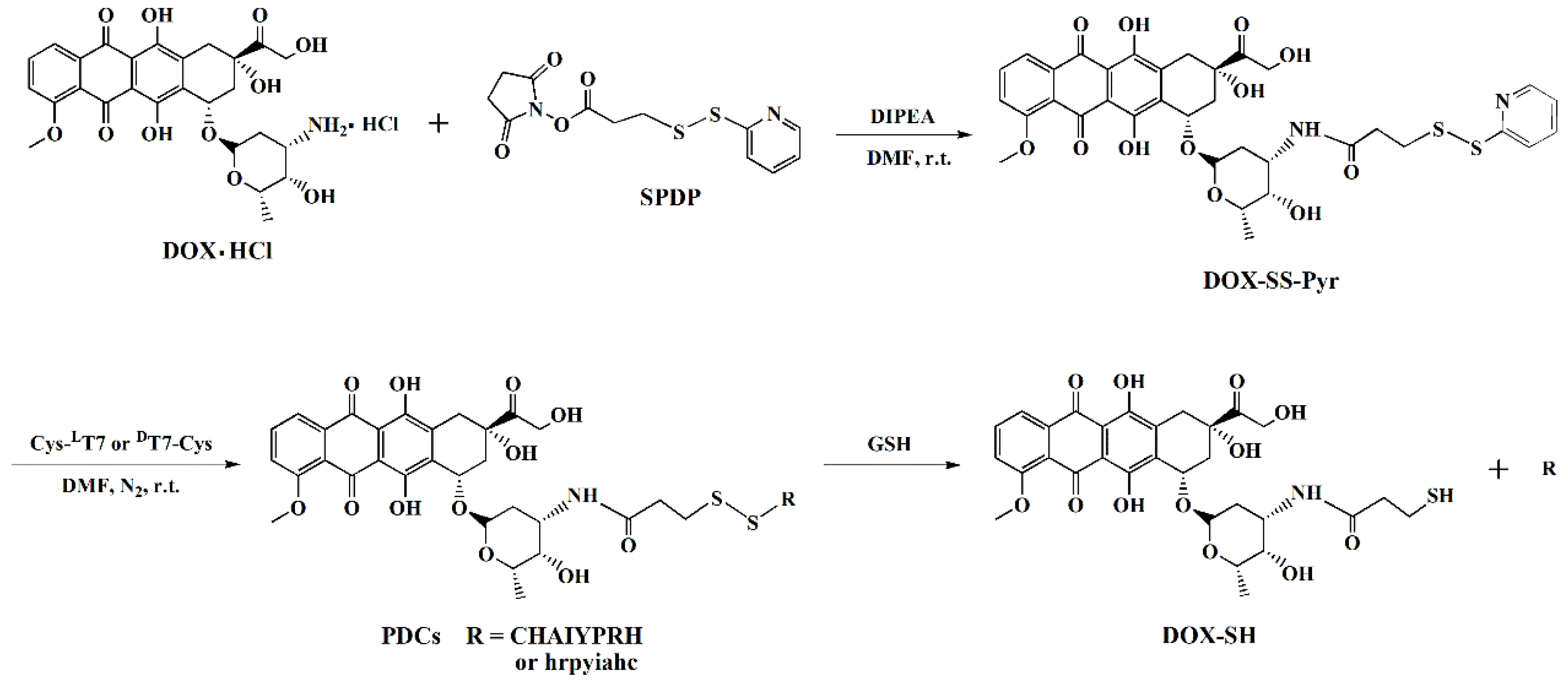
2.1. Synthesis of PDCs

The LT7-SS-DOX and DT7-SS-DOX conjugates were synthesized in two steps, as shown in Scheme 1. First, the reaction between DOX and SPDP afforded DOX-SS-Pyr as a red solid (34.8 mg, 84.9% yield) with 95.3% purity; ESI MS (m/z) [C35H36N2O12S2]: calculated, 740.2; found, 741.1 [M + H]+; the 1H NMR spectrum (400 MHz, DMSO-d6) data were consistent with the literature [26] (Figures S1–S3). High-performance liquid chromatography (HPLC) analysis, ESI MS, and 1H NMR indicated that DOX-SS-Pyr was successfully synthesized. Then, DOX-SS-Pyr reacted with peptide Cys-LT7 or DT7-Cys via a disulfide bond exchange reaction to generate the crude PDC, which was purified by semi-preparative HPLC. The HPLC purity of the purified LT7-SS-DOX conjugate (3.42 mg, 31.5% yield) was 97.3%; ESI MS (m/z) [C74H96N16O22S2]: calculated, 1624.6; found, 813.3 [M + 2H]2+ (Figures S4 and S5). The HPLC purity of the purified DT7-SS-DOX conjugate (3.15 mg, 43.9% yield) was 97.4%; ESI MS (m/z) [C74H96N16O22S2]: calculated, 1624.6; found, 813.4 [M + 2H]2+ (Figures S6 and S7).

2.2. Serum Stability and Drug Release of PDCs

To verify whether the characteristics of DT7-SS-DOX constructed with DT7 as the ligand can be improved in comparison with those of LT7-SS-DOX under different in vitro mimetic physiological conditions and tumor microenvironment, the stability and reduction-responsive drug release behavior were investigated by co-incubating the PDCs with mouse serum and different concentrations of glutathione (GSH), respectively.

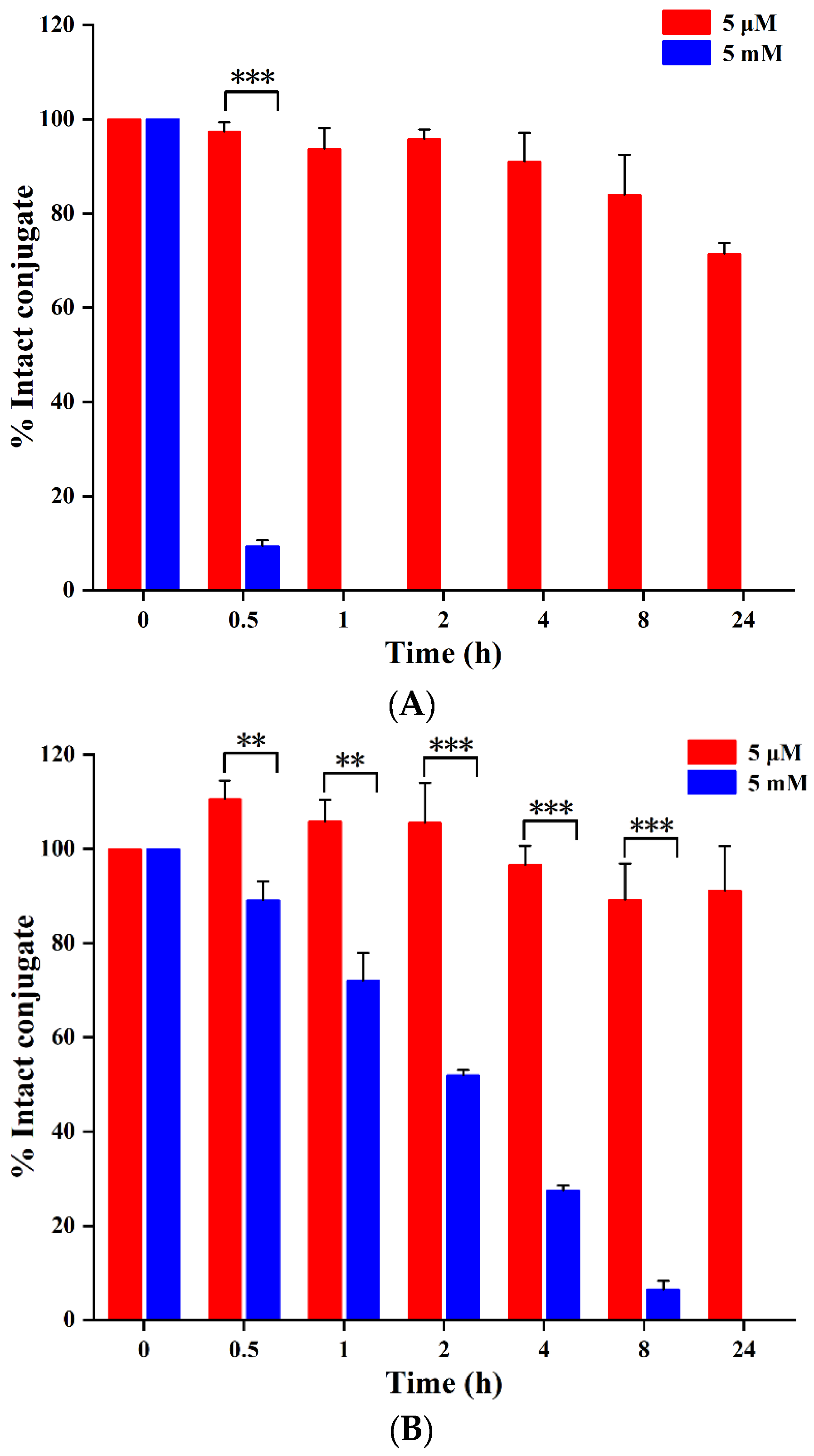
2.2.1. Serum Stability

With the extension of incubation time, both of the conjugates that remained intact in mouse serum decreased continuously (Figure 1). LT7-SS-DOX was rapidly degraded within 0.5 h and was almost entirely degraded at 1 h. Whereas the degradation of DT7-SS-DOX (t1/2 = 8.58 ± 0.85 h) was significantly slower than that of LT7-SS-DOX (t1/2 = 0.37 ± 0.02 h) (p < 0.001). Namely, DT7-SS-DOX was much more stable in mouse serum than LT7-SS-DOX, indicating that the serum stability of the PDC constructed with DT7 as the ligand can be prominently elevated.

2.2.2. Reduction-Triggered Drug Release

As shown in Figure 2A, LT7-SS-DOX was completely degraded in the presence of 5 mM GSH within 1 h. In contrast, the degradation of this conjugate in 5 μM GSH was much slower because 93.7% of the conjugate remained undegraded at 1 h, and 71.4% of the intact conjugate was detected when incubated for 24 h. As for DT7-SS-DOX (Figure 2B), when incubated in 5 mM GSH, the conjugate that remained intact gradually decreased within 24 h. Overall, 72.1% of DT7-SS-DOX remained undegraded at 1 h, and no intact conjugate was detected at 24 h. Nevertheless, this conjugate was more stable in the presence of 5 μM GSH because 91.2% of the conjugate still remained intact when incubated for 24 h.
It is noteworthy that the degradations of DT7-SS-DOX in both 5 mM and 5 μM GSH were slower than that of LT7-SS-DOX. In 5 mM GSH mimicking the tumor cell reductive microenvironment (2–10 mM GSH) [27], the DT7-SS-DOX conjugate showed 50% degradation in approximately 2 h (t1/2 = 1.93 ± 0.13 h), which was almost 8 times longer than that of LT7-SS-DOX (t1/2 = 0.26 ± 0.01 h), demonstrating that DT7-SS-DOX had a sustained drug release manner which might help to exert the antitumor effect for a longer time. When incubated in 5 μM GSH, DT7-SS-DOX was more stable than LT7-SS-DOX during the incubation period of 24 h (Figure 2A,B), which might result in higher stability, less drug release, and toxicity of DT7-SS-DOX in normal physiological condition.

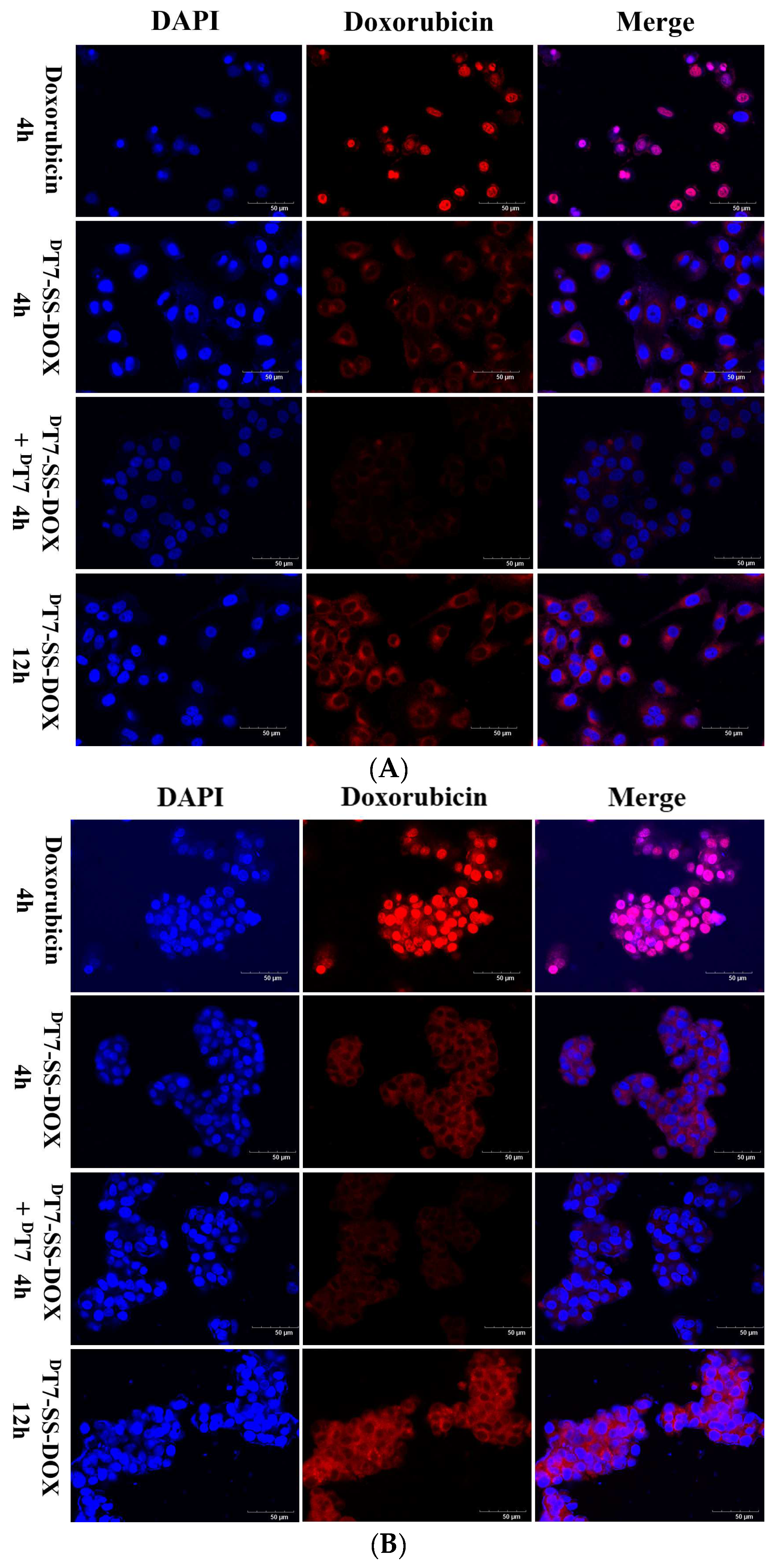
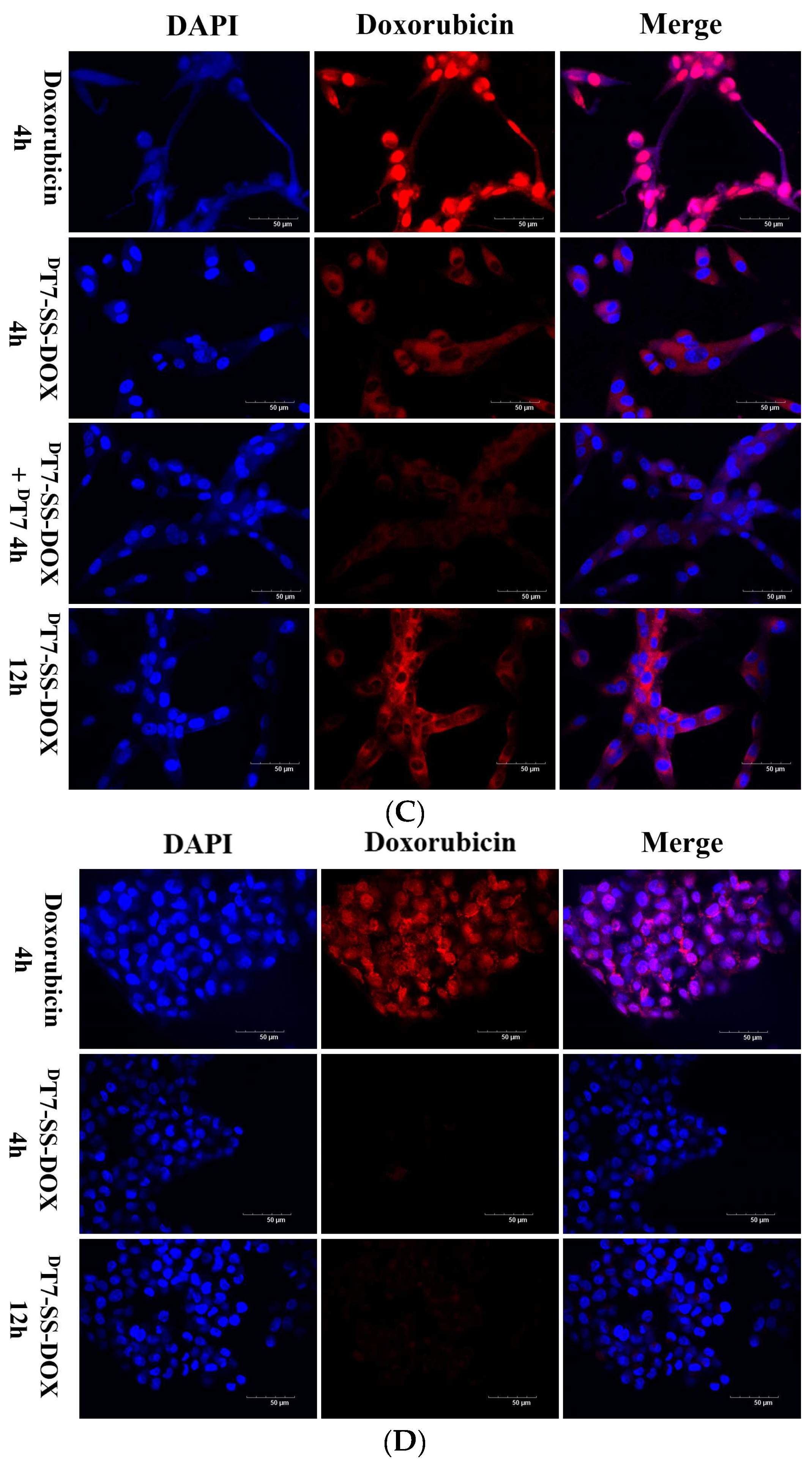
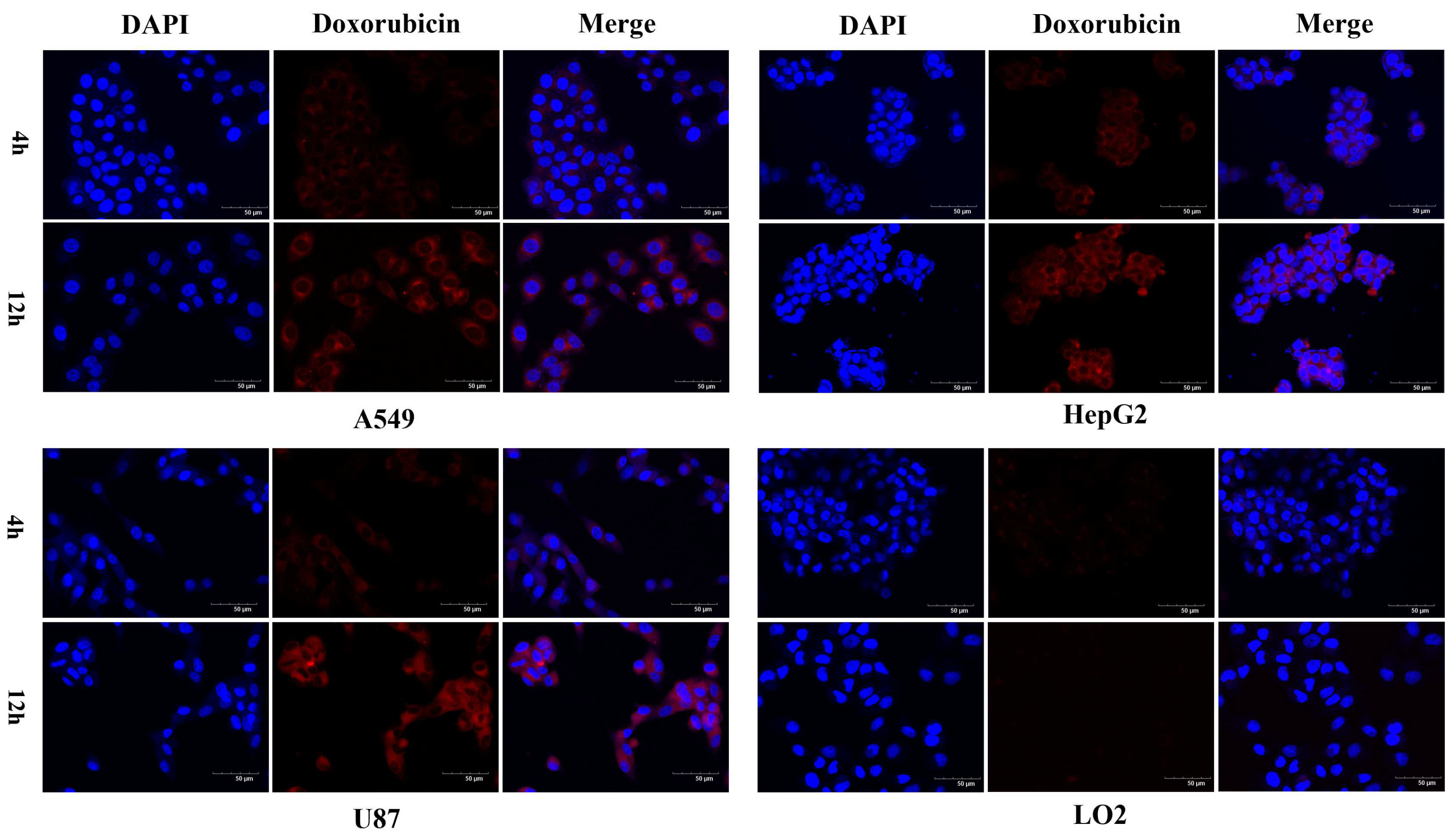
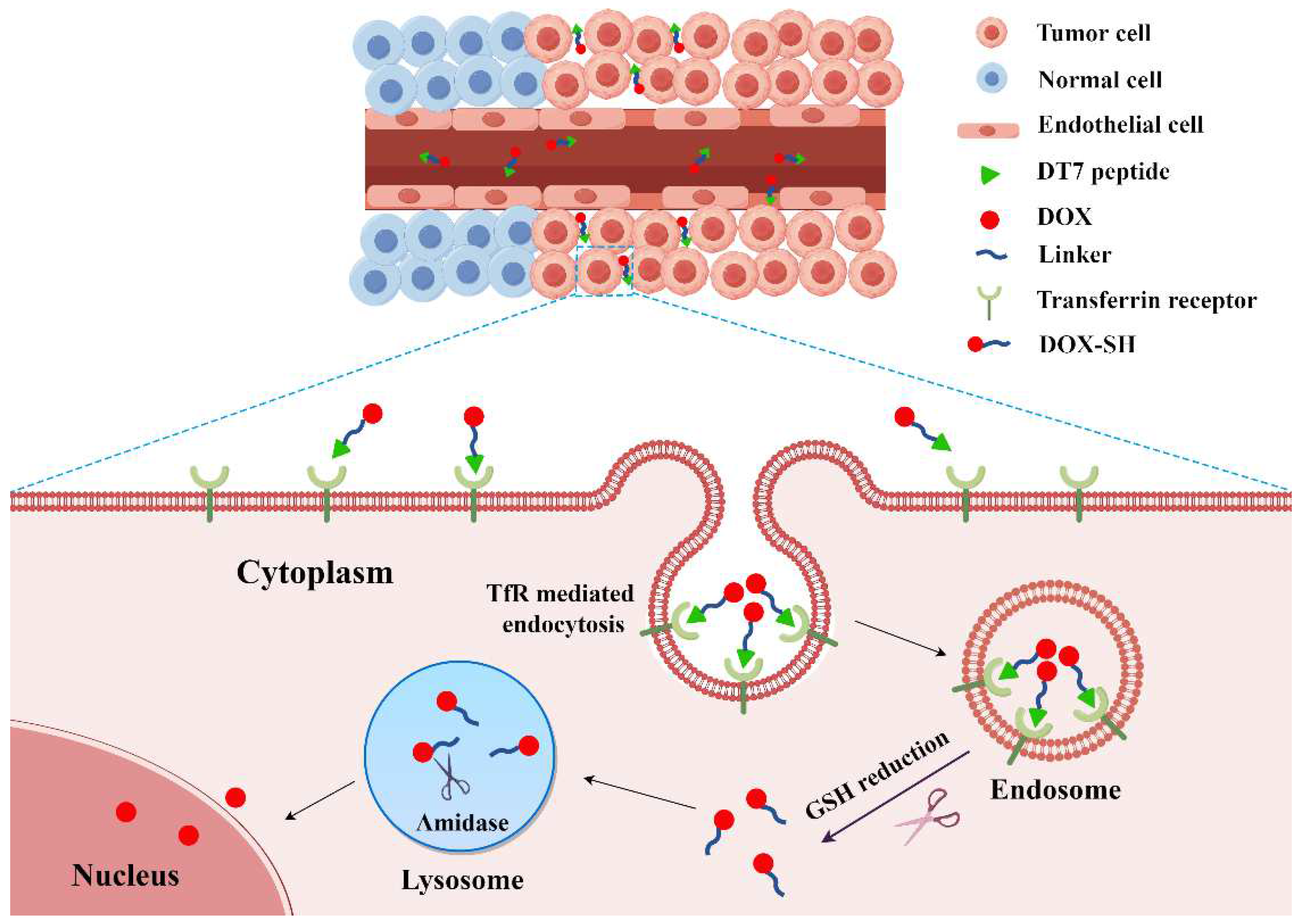
2.3. Confocal Microscopy Analysis of Cellular Uptake

TfR overexpressed human A549, HepG2, U87 tumor cell lines [22,28], and TfR low-expressed human LO2 normal liver cell line [29] were used to evaluate the in vitro tumor-targeted cellular uptake of the conjugates by using laser confocal scanning microscopy.
The cell nuclei stained with DAPI were shown as blue fluorescence, and DOX was detected with red fluorescence. As exhibited in Figure 3A–D, the red fluorescence located in the cell nuclei of A549, HepG2, U87, and LO2 cells after treatment with free DOX for 4 h, indicating that free DOX could enter into both tumor and normal cells due to its poor selectivity to tumor cells. As for cells treated with the conjugates for 4 and 12 h (Figure 3 and Figure 4), the red fluorescence was mainly distributed in the cytoplasm of A549, HepG2, and U87 tumor cells but can barely be found in LO2 cells, indicating that the conjugates can selectively enter into tumor cells, and free DOX was not released from the conjugates. Namely, the results demonstrated that the selectivity of DOX to TfR overexpressed tumor cells was effectively improved by conjugating with the TfR affinity peptides. Moreover, the cellular uptake pathway of the conjugates might be different from that of free DOX.
In a previous study, it was found that LT7-, DT7-, and Tf-modified liposomes (LT7-LIP, DT7-LIP, and Tf-LIP) could effectively enter into HepG2 cells without pre-incubation of the corresponding TfR-targeting ligands, indicating that the three ligands maintained binding affinity to TfR after conjugation with liposomes. However, the pre-incubation of HepG2 cells with the TfR affinity peptide (LT7 or DT7) significantly reduced the cellular uptake of both LT7-LIP and DT7-LIP but did not decrease the uptake of Tf-LIP. These results demonstrated that the binding site of DT7 on the TfR might be the same as that of LT7 but was different from that of Tf [22]. Based on these findings, we speculated that the cellular uptake of our PDCs might also be related to the binding of the peptide ligands to TfR. To investigate the cellular uptake pathway of the conjugates, a TfR competitive inhibition assay was utilized. The results revealed that the fluorescence intensity of DT7-SS-DOX in A549, HepG2, and U87 tumor cells pre-treated with the TfR affinity peptide was lower than that of the corresponding type of tumor cells without pre-incubation of the TfR affinity peptide (Figure 3A–C), indicating that pre-occupation of the binding site of TfR overexpressed on the surface of tumor cells with the TfR affinity peptide clearly reduced the uptake of DT7-SS-DOX by cells, suggesting that the conjugate might enter into cells via a TfR-mediated endocytosis pathway.
When incubated with drugs for 4 h, the fluorescence intensity in the same kind of tumor cells ranking from the highest to the lowest was free DOX, DT7-SS-DOX, and LT7-SS-DOX (Figure 3A–C and Figure 4). This phenomenon is probably due to the fast passive diffusion of free DOX into cells without a drug release process, whereas the entry of the conjugates into cells via TfR-mediated endocytosis was a relatively slow process. Meanwhile, the binding affinity of DT7 to TfR is higher than that of LT7, so DT7-SS-DOX could bring more drugs into tumor cells via the TfR-mediated endocytosis pathway during the same incubation period. In addition, the fluorescence intensity of both LT7-SS-DOX and DT7-SS-DOX in tumor cells increased with the prolongation of incubation time (Figure 3A–C and Figure 4, 4 h versus 12 h), demonstrating that the TfR-mediated endocytosis of PDCs and the subsequent cleavage of the disulfide bond by GSH to generate DOX-SH (Scheme 1) was a gradually cumulative process which was time-dependent.

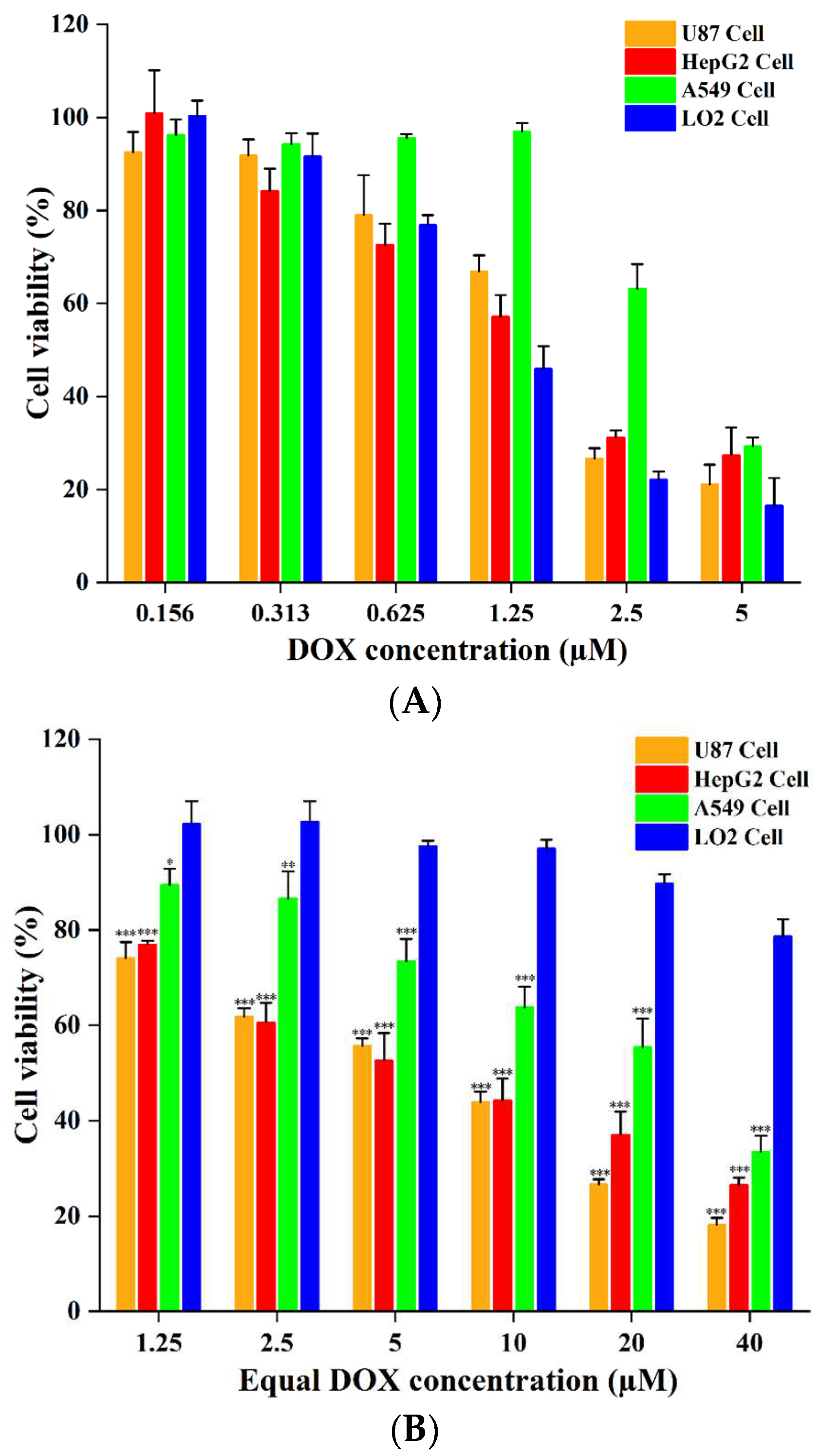
2.4. In Vitro Cytotoxicity

The in vitro cytotoxicities of free DOX, LT7-SS-DOX, and DT7-SS-DOX against U87, HepG2, A549, and LO2 cells were investigated by CCK-8 assay. As revealed in Figure 5 and Figure S8, the cell viability of each kind of cell line after incubation with free DOX, LT7-SS-DOX, and DT7-SS-DOX decreased in a dose-dependent manner. The antiproliferative activity of drugs against tumor cells ranking from the highest to the lowest was free DOX, DT7-SS-DOX, and LT7-SS-DOX, but the toxicities of the two conjugates to LO2 normal cells were much lower than that of free DOX, which was consistent with the aforementioned results of cellular uptake. DOX showed strong in vitro cytotoxicity against all four kinds of cells without tumor selectivity (Figure 5A), with IC50 values of 1.65 ± 0.20 µM (U87), 1.58 ± 0.19 µM (HepG2), 3.55 ± 0.21 µM (A549), and 1.29 ± 0.18 µM (LO2), respectively (Table 1). DT7-SS-DOX exhibited good in vitro antiproliferative activity against the three tumor cell lines (Figure 5B), with IC50 values of 5.70 ± 0.22 µM (U87), 7.01 ± 1.64 µM (HepG2), and 20.61 ± 4.81 µM (A549), respectively (Table 1). The proliferation inhibitory activity of LT7-SS-DOX was the weakest among the three drugs because the cell viabilities of U87, HepG2, and A549 cells after incubation with LT7-SS-DOX (equal DOX concentration of 20 µM) for 48 h were 95.1%, 73.1%, and 83.2%, respectively. Even at an equal DOX concentration of 40 µM, the cell viability of the three types of tumor cells after exposure to this conjugate for 48 h were 41.0%, 61.5%, and 67.2%, respectively (Figure S8). Because of the inconspicuous antiproliferative activity of LT7-SS-DOX at an equal DOX concentration of 20 µM, drugs below this concentration were not used.
The reason for the stronger antitumor activity of free DOX is possible because this drug can quickly enter into tumor cells via passive diffusion and immediately kill tumor cells in vitro. However, it is supposed that PDCs constructed by forming an amide bond via the amino of DOX may not exert cell proliferation inhibitory activity until free DOX is released. One of the PDCs like this, named cRGD-SS-DOX, released DOX-SH rather than free DOX after co-incubation with B16 mouse melanoma cells for 3 h. Furthermore, the in vitro cytotoxicity of cRGD-SS-DOX against B16 cells was found to be 20.6 times lower than that of free DOX [30]. Similarly, after taken up by tumor cells, we speculated that the peptide–doxorubicin conjugates in our study can exhibit antiproliferative activity only if the disulfide bond is cleaved by GSH to generate DOX-SH, and the subsequent cleavage of the amide bond of DOX-SH by amidases present in lysosomes to release free drug finally occurs, as illustrated in Figure 6. Nevertheless, cellular uptake results of the above PDCs showed that the red fluorescence was still mainly located in the cytoplasm of tumor cells even at an incubation time of 12 h, indicating that the release of free DOX was a slow and time-dependent process, which made the antiproliferative activity of PDCs weaker than that of free DOX. In addition, the stronger antiproliferative activity of DT7-SS-DOX in comparison with that of LT7-SS-DOX was probably related to the higher affinity of DT7-SS-DOX to TfR as well as its sustained drug release behavior in tumor cells.
It is worth mentioning that although the in vitro antiproliferative activity of DT7-SS-DOX against U87, HepG2, and A549 tumor cells was 3.4, 4.4, and 5.8 times, respectively, less potent than that of free DOX, its toxicity to LO2 normal cells (IC50 value > 100 µM) was significantly reduced compared with that of free DOX (1.29 ± 0.18 µM) (Table 1), indicating that this conjugate had an excellent selectivity to TfR overexpressed tumor cells due to the targeting effect of the DT7 peptide ligand, which is of significant importance considering the clinical application of DOX is severely restricted by its poor tumorous selectivity. Moreover, given that insufficient stability is the major drawback of PDCs constructed with L-peptide ligands, the remarkedly enhanced serum stability of the DT7-SS-DOX conjugate is crucial to ensure its integrity in blood circulation and maintain the targeting effect of the peptide ligand on TfR after reaching the tumor site, which might facilitate the accumulation of drugs in target tumor cells, as depicted in Figure 6.

3. Materials and Methods

3.1. Materials

Doxorubicin hydrochloride (DOX·HCl, 98%) was purchased from Meilun Biotechnology Co., Ltd. (Dalian, China); Cys-LT7 (97.9%) was synthesized by our group as previously described [31]; DT7-Cys (99.2%) was provided by Chinapeptides BioTech Co., Ltd. (Shanghai, China); SPDP (96%) was obtained from J&K Scientific Co., Ltd. (Beijing, China); GSH (98%) was purchased from Sigma-Aldrich (Wuxi, China); CCK-8 was obtained from APExBIO Technology (Shanghai, China); 4′, 6-diamidino-2-phenylindole (DAPI) solution was purchased from Solarbio Life Sciences Co., Ltd. (Beijing, China); mouse serum was provided by Sbjbio Tech Co., Ltd. (Nanjing, China). All other chemicals were of analytical or chromatographic grade.
Human HepG2 hepatocellular carcinoma cells and human U87 glioblastoma cells were purchased from Procell Life Science & Technology Co., Ltd. (Wuhan, China), human A549 lung carcinoma cells were obtained from the School of Basic Medical Sciences, Chengde Medical University, human LO2 normal hepatic cells were purchased from Jennio Biotech Co., Ltd. (Guangzhou, China). The cells were cultured in DMEM (HepG2), MEM containing 1% non-essential amino acids (U87), and RPMI-1640 (A549 and LO2), supplemented with 10% fetal bovine serum (FBS) and 1% penicillin/streptomycin at 37 °C in a humidified atmosphere with 5% CO2.

3.2. Synthesis of Peptide–DOX Conjugates

3.2.1. Synthesis of DOX-SS-Pyr

DOX-SS-Pyr was synthesized according to the published procedure [32] with minor modifications. Briefly, N, N-Diisopropylethylamine (DIPEA, 19 μL, 109.1 μmol) was added dropwise to a stirred solution of DOX·HCl (30.6 mg, 52.8 μmol) and SPDP (20.1 mg, 64.3 μmol) in 2.2 mL of anhydrous N, N-Dimethylformamide (DMF), and the mixture was stirred at room temperature for 2 h. The process of the reaction was monitored by analytical HPLC. Then, the reaction solution was treated with deionized water, and the resulting precipitate was collected by centrifugation and dried under vacuum to give DOX-SS-Pyr as a red solid, which was characterized by HPLC, ESI MS, and 1H NMR. HPLC analyses in this study were all performed on a ZORBAX Eclipse XDB-C18 reversed-phase column (250 mm × 4.6 mm, 5 μm; Agilent, Santa Clara, CA, USA) connected to an Agilent 1200 HPLC system running with a linear gradient of 5–95% acetonitrile/water with 0.1% trifluoroacetic acid at 1 mL/min in 20 min. The monitoring wavelength was 254 nm.

3.2.2. Synthesis of Peptide–DOX Conjugates

The PDC was synthesized via a disulfide bond exchange reaction between the free sulfydryl in the structure of peptide Cys-LT7 or DT7-Cys and the 2-pyridyldithio group in the DOX-SS-Pyr. Briefly, DOX-SS-Pyr (5.0 mg, 6.8 μmol) and Cys-LT7 peptide (6.5 mg, 6.5 μmol) were dissolved in 0.7 mL of anhydrous DMF, and the mixture was stirred under a nitrogenous atmosphere at room temperature for 1 h. For the synthesis of DT7-SS-DOX, DOX-SS-Pyr (3.9 mg, 5.3 μmol), and DT7-Cys peptide (4.3 mg, 4.3 μmol) were dissolved in 0.5 mL of anhydrous DMF, and the mixture was stirred under a nitrogenous atmosphere at room temperature for 6 h. The process of the reaction was monitored by analytical HPLC. Upon completion of the reaction, the resulting LT7-SS-DOX or DT7-SS-DOX conjugate was purified by semi-preparative HPLC conducted on an Ultimate XB-C18 reversed-phase column (250 mm × 10 mm, 5 μm; Welch, Shanghai, China). The mobile phases consisted of solvent A (0.1% formic acid in water) and solvent B (0.1% formic acid in acetonitrile). An isocratic elution of 21% eluent B over 20 min and a linear gradient of 5–70% eluent B over 70 min were applied for LT7-SS-DOX and DT7-SS-DOX purification, respectively. The flow rate was 2 mL/min, and peaks were detected at 215 nm. Fractions were collected, evaporated, and lyophilized as a red powder, which was characterized by analytical HPLC and ESI MS.

3.3. In Vitro Stability of Conjugates

The in vitro stability experiment of the conjugates was conducted by co-incubation with mouse serum, as previously described [33], with minor modifications. An 80 μL of LT7-SS-DOX or DT7-SS-DOX stock solution (1 mM) was diluted by adding ultra-pure water (520 μL) and mouse serum (200 μL). The mixture was incubated at 37 °C, aliquots (100 μL) were taken at scheduled time intervals, and then methanol (300 μL) was added to terminate the incubation. After centrifugation at 10,000 rpm for 10 min, the intact PDC that remained in the supernatant was analyzed by HPLC.

3.4. In Vitro Drug Release of Conjugates

A stock solution of LT7-SS-DOX or DT7-SS-DOX was diluted by adding 5 mM or 5 μM GSH dissolved in acetonitrile-PBS (3:7, v/v) to a final volume of 800 μL with 100 μM conjugate, and the mixture was incubated at 37 °C. Aliquots (100 μL) were taken from the incubation solution at scheduled time intervals and diluted with acetonitrile–PBS (3:7, v/v, 200 μL). After filtration, the intact conjugate that remained in the sample was analyzed by HPLC.

3.5. Confocal Microscopy Studies

A549, HepG2, U87, or LO2 cells were seeded on glass coverslips (5 × 10⁴ cells/well) in 24-well cell culture plates and incubated at 37 °C for 24 h. Then, cells were treated with the peptide–DOX conjugates (equivalent DOX concentration of 10 μM) for 4 and 12 h, respectively. Cells treated with free DOX (10 μM) for 4 h were used as the positive control. After the drugs in each well were discarded, cells were washed three times with PBS and fixed with 4% paraformaldehyde for 12 h. Cell nuclei were stained with DAPI for 8 min. The fluorescent images of cells were observed by using a confocal laser scanning microscope (Olympas FV3000).
For the TfR competitive inhibition assay, A549, HepG2, or U87 cells seeded on glass coverslips in 24-well cell culture plates were incubated at 37 °C overnight and then pre-treated with serum-free cultural medium containing the TfR affinity peptide (100 μM) at 37 °C for 8 h. After removal of the peptide solution, cells were washed three times with PBS and incubated with the DT7-SS-DOX conjugate (equivalent DOX concentration of 10 μM) for 4 h. Cell fixation, staining, and cellular uptake analysis by confocal microscopy were conducted as aforementioned methods.

3.6. In Vitro Cytotoxicity Assay

A549, HepG2, U87, and LO2 cells were each seeded in 96-well cell culture plates at a density of 6 × 10³ cells per well and incubated at 37 °C for 24 h. Then, cells were treated with serial concentrations of free DOX or the peptide–DOX conjugates for 48 h. After incubation, CCK-8 (10 μL) was added to each well and incubated for an additional 2 h. The absorbance of each well was measured at 450 nm with a reference wavelength of 630 nm by using a Multiskan GO microplate reader (Thermo Fisher Scientific, Waltham, MA, USA). Data for each cell line treated with free DOX or PDCs were obtained from three independent experiments.

3.7. Statistical Analysis

Data were presented as the mean ± SD. All statistical analyses were performed using SPSS 26. The comparison of parameters among the two groups was made by the unpaired Student’s t-test. Group differences were determined by one-way analysis of variance (ANOVA) followed by Fisher’s LSD post hoc. The differences were considered to be significant at p < 0.05.

4. Conclusions

In this present study, two new TfR affinity peptide–doxorubicin conjugates were designed and synthesized. Compared with LT7-SS-DOX, the DT7-SS-DOX conjugate had the advantages of much higher in vitro serum stability, more sustained reduction-responsive drug release behavior, and stronger tumor cell proliferation inhibitory activity, indicating that DT7 can be used as an excellent peptide ligand for constructing stable TfR targeted PDCs. In view of that the in vitro antiproliferative activity of DT7-SS-DOX against tumor cells is less potent than that of free DOX, PDCs with an acid-triggered cleavable linkage such as hydrazone or ester bond between DT7 and DOX need to be investigated in the future so as to release free DOX more efficiently within tumor cells and further improve the antitumor efficacy.

Supplementary Materials

The following supporting information can be downloaded at https://www.mdpi.com/article/10.3390/molecules29081758/s1. Figures S1–S3: HPLC chromatogram, ESI MS, and 1H NMR spectrum of DOX-SS-Pyr; Figures S4–S5: HPLC chromatogram and ESI MS of the LT7-SS-DOX conjugate; Figures S6–S7: HPLC chromatogram and ESI MS of the DT7-SS-DOX conjugate; Figure S8: In vitro cytotoxicity of LT7-SS-DOX against cells.

Author Contributions

Conceptualization, S.L. and J.Y.; formal analysis, J.Y. and S.L.; investigation, J.Y. and X.M.; resources, X.Y.; data curation, J.Y. and S.L.; writing—original draft preparation, J.Y.; writing—review and editing, G.Z. and S.L.; funding acquisition, S.L. All authors have read and agreed to the published version of the manuscript.

Funding

This study was funded by the Hebei Natural Science Foundation (H2021406058); Science Research Project of Hebei Education Department (ZD2021002).

Institutional Review Board Statement

Not applicable.

Informed Consent Statement

Not applicable.

Data Availability Statement

Data are contained within the article and Supplementary Materials.

Conflicts of Interest

The authors declare no conflicts of interest.

References

  1. Fang, R.H.; Gao, W.; Zhang, L. Targeting drugs to tumours using cell membrane-coated nanoparticles. Nat. Rev. Clin. Oncol. 2023, 20, 33–48. [Google Scholar] [CrossRef] [PubMed]
  2. Zhu, Y.; Tang, K.; Lv, J. Peptide-drug conjugate-based novel molecular drug delivery system in cancer. Trends Pharmacol. Sci. 2021, 42, 857–869. [Google Scholar] [CrossRef] [PubMed]
  3. Alas, M.; Saghaeidehkordi, A.; Kaur, K. Peptide-Drug Conjugates with Different Linkers for Cancer Therapy. J. Med. Chem. 2021, 64, 216–232. [Google Scholar] [CrossRef] [PubMed]
  4. Wang, M.; Liu, J.; Xia, M.; Yin, L.; Zhang, L.; Liu, X.; Cheng, Y. Peptide-drug conjugates: A new paradigm for targeted cancer therapy. Eur. J. Med. Chem. 2024, 265, 116119. [Google Scholar] [CrossRef] [PubMed]
  5. Wu, M.; Huang, W.; Yang, N.; Liu, Y. Learn from antibody-drug conjugates, consideration in the future construction of peptide-drug conjugates for cancer therapy. Exp. Hematol. Oncol. 2022, 11, 93. [Google Scholar] [CrossRef]
  6. Mojarad-Jabali, S.; Mahdinloo, S.; Farshbaf, M.; Sarfraz, M.; Fatahi, Y.; Atyabi, F.; Valizadeh, H. Transferrin receptor-mediated liposomal drug delivery: Recent trends in targeted therapy of cancer. Expert Opin. Drug Deliv. 2022, 19, 685–705. [Google Scholar] [CrossRef] [PubMed]
  7. Kang, Z.; Zeng, C.; Tian, L.; Wang, T.; Yang, S.; Cheng, Q.; Zhang, J.; Meng, Q.; Zhang, C.; Meng, Z. Transferrin receptor targeting segment T7 containing peptide gene delivery vectors for efficient transfection of brain tumor cells. Drug Deliv. 2022, 29, 2375–2385. [Google Scholar] [CrossRef]
  8. Falvo, E.; Damiani, V.; Conti, G.; Boschi, F.; Messana, K.; Giacomini, P.; Milella, M.; De Laurenzi, V.; Morea, V.; Sala, G.; et al. High activity and low toxicity of a novel CD71-targeting nanotherapeutic named The-0504 on preclinical models of several human aggressive tumors. J. Exp. Clin. Cancer Res. 2021, 40, 63. [Google Scholar] [CrossRef]
  9. Zhang, Q.; Yu, Q.; Luan, W.; Li, T.; Xiao, Y.; Zhang, L.; Li, Y.; Rong, R.; Ren, C. LWJ-M30, a conjugate of DM1 and B6, for the targeted therapy of colorectal cancer with improved therapeutic effects. RSC Adv. 2023, 13, 10840–10846. [Google Scholar] [CrossRef]
  10. Mojarad-Jabali, S.; Farshbaf, M.; Hemmati, S.; Sarfraz, M.; Motasadizadeh, H.; Mojarrad, J.S.; Atyabi, F.; Zakeri-Milani, P.; Valizadeh, H. Comparison of three synthetic transferrin mimetic small peptides to promote the blood-brain barrier penetration of vincristine liposomes for improved glioma targeted therapy. Int. J. Pharm. 2022, 613, 121395. [Google Scholar] [CrossRef]
  11. Sun, P.; Xiao, Y.; Di, Q.; Ma, W.; Ma, X.; Wang, Q.; Chen, W. Transferrin Receptor-Targeted PEG-PLA Polymeric Micelles for Chemotherapy Against Glioblastoma Multiforme. Int. J. Nanomed. 2020, 15, 6673–6688. [Google Scholar] [CrossRef] [PubMed]
  12. Elfadadny, A.; Ragab, R.F.; Hamada, R.; Al Jaouni, S.K.; Fu, J.; Mousa, S.A.; El-Far, A.H. Natural bioactive compounds-doxorubicin combinations targeting topoisomerase II-alpha: Anticancer efficacy and safety. Toxicol. Appl. Pharmacol. 2023, 461, 116405. [Google Scholar] [CrossRef] [PubMed]
  13. Paskeh, M.D.A.; Saebfar, H.; Mahabady, M.K.; Orouei, S.; Hushmandi, K.; Entezari, M.; Hashemi, M.; Aref, A.R.; Hamblin, M.R.; Ang, H.L.; et al. Overcoming doxorubicin resistance in cancer: siRNA-loaded nanoarchitectures for cancer gene therapy. Life Sci. 2022, 298, 120463. [Google Scholar] [CrossRef] [PubMed]
  14. Radu, E.R.; Semenescu, A.; Voicu, S.I. Recent Advances in Stimuli-Responsive Doxorubicin Delivery Systems for Liver Cancer Therapy. Polymers 2022, 14, 5249. [Google Scholar] [CrossRef] [PubMed]
  15. Almajidi, Y.Q.; Kadhim, M.M.; Alsaikhan, F.; Jalil, A.T.; Sayyid, N.H.; Ramírez-Coronel, A.A.; Jawhar, Z.H.; Gupta, J.; Nabavi, N.; Yu, W.; et al. Doxorubicin-loaded micelles in tumor cell-specific chemotherapy. Environ. Res. 2023, 227, 115722. [Google Scholar] [CrossRef] [PubMed]
  16. Zeng, W.; Luo, Y.; Gan, D.; Zhang, Y.; Deng, H.; Liu, G. Advances in Doxorubicin-based nano-drug delivery system in triple negative breast cancer. Front. Bioeng. Biotechnol. 2023, 11, 1271420. [Google Scholar] [CrossRef] [PubMed]
  17. Lee, J.H.; Engler, J.A.; Collawn, J.F.; Moore, B.A. Receptor mediated uptake of peptides that bind the human transferrin receptor. Eur. J. Biochem. 2001, 268, 2004–2012. [Google Scholar] [CrossRef] [PubMed]
  18. He, G.; Lin, W. Peptide-Functionalized Nanoparticles-Encapsulated Cyclin-Dependent Kinases Inhibitor Seliciclib in Transferrin Receptor Overexpressed Cancer Cells. Nanomaterials 2021, 11, 772. [Google Scholar] [CrossRef]
  19. Piantino, M.; Louis, F.; Shigemoto-Mogami, Y.; Kitamura, K.; Sato, K.; Yamaguchi, T.; Kawabata, K.; Yamamoto, S.; Iwasaki, S.; Hirabayashi, H.; et al. Brain microvascular endothelial cells derived from human induced pluripotent stem cells as in vitro model for assessing blood-brain barrier transferrin receptor-mediated transcytosis. Mater. Today Bio 2022, 14, 100232. [Google Scholar] [CrossRef]
  20. Riaz, M.K.; Zhang, X.; Wong, K.; Chen, H.; Liu, Q.; Chen, X.; Zhang, G.; Lu, A.; Yang, Z. Pulmonary delivery of transferrin receptors targeting peptide surface-functionalized liposomes augments the chemotherapeutic effect of quercetin in lung cancer therapy. Int. J. Nanomed. 2019, 14, 2879–2902. [Google Scholar] [CrossRef]
  21. Zhang, Y.; Zhai, M.; Chen, Z.; Han, X.; Yu, F.; Li, Z.; Xie, X.; Han, C.; Yu, L.; Yang, Y.; et al. Dual-modified liposome codelivery of doxorubicin and vincristine improve targeting and therapeutic efficacy of glioma. Drug Deliv. 2017, 24, 1045–1055. [Google Scholar] [CrossRef] [PubMed]
  22. Tang, J.; Wang, Q.; Yu, Q.; Qiu, Y.; Mei, L.; Wan, D.; Wang, X.; Li, M.; He, Q. A stabilized retro-inverso peptide ligand of transferrin receptor for enhanced liposome-based hepatocellular carcinoma-targeted drug delivery. Acta Biomater. 2019, 83, 379–389. [Google Scholar] [CrossRef] [PubMed]
  23. Yu, M.; Su, D.; Yang, Y.; Qin, L.; Hu, C.; Liu, R.; Zhou, Y.; Yang, C.; Yang, X.; Wang, G.; et al. D-T7 Peptide-Modified PEGylated Bilirubin Nanoparticles Loaded with Cediranib and Paclitaxel for Antiangiogenesis and Chemotherapy of Glioma. ACS Appl. Mater. Interfaces 2019, 11, 176–186. [Google Scholar] [CrossRef] [PubMed]
  24. Hou, Q.; Zhu, L.; Wang, L.; Liu, X.; Xiao, F.; Xie, Y.; Zheng, W.; Jiang, X. Screening on-chip fabricated nanoparticles for penetrating the blood-brain barrier. Nanoscale 2022, 14, 3234–3241. [Google Scholar] [CrossRef] [PubMed]
  25. Zhou, Y.; Guan, L.; Li, W.; Jia, R.; Jia, L.; Zhang, Y.; Wen, X.; Meng, S.; Ma, D.; Zhang, N.; et al. DT7 peptide-modified lecithin nanoparticles co-loaded with γ-secretase inhibitor and dexamethasone efficiently inhibit T-cell acute lymphoblastic leukemia and reduce gastrointestinal toxicity. Cancer Lett. 2022, 533, 215608. [Google Scholar] [CrossRef] [PubMed]
  26. Song, Q.; Chuan, X.; Chen, B.; He, B.; Zhang, H.; Dai, W.; Wang, X.; Zhang, Q. A smart tumor targeting peptide-drug conjugate, pHLIP-SS-DOX, synthesis and cellular uptake on MCF-7 and MCF-7/Adr cells. Drug Deliv. 2016, 23, 1734–1746. [Google Scholar] [CrossRef] [PubMed]
  27. Bao, Y.; Guo, Y.; Zhuang, X.; Li, D.; Cheng, B.; Tan, S.; Zhang, Z. D-α-tocopherol polyethylene glycol succinate-based redox-sensitive paclitaxel prodrug for overcoming multidrug resistance in cancer cells. Mol. Pharm. 2014, 11, 3196–3209. [Google Scholar] [CrossRef] [PubMed]
  28. Wei, L.; Guo, X.; Yang, T.; Yu, M.; Chen, D.; Wang, J. Brain tumor-targeted therapy by systemic delivery of siRNA with Transferrin receptor-mediated core-shell nanoparticles. Int. J. Pharm. 2016, 510, 394–405. [Google Scholar] [CrossRef] [PubMed]
  29. Zheng, Q.; Xiong, Y.; Su, Z.; Zhang, Q.; Dai, X.; Li, L.; Xiao, X.; Huang, Y. Expression of curcin-transferrin receptor binding peptide fusion protein and its anti-tumor activity. Protein Expr. Purif. 2013, 89, 181–188. [Google Scholar] [CrossRef]
  30. Liang, Y.; Li, S.; Wang, X.; Zhang, Y.; Sun, Y.; Wang, Y.; Wang, X.; He, B.; Dai, W.; Zhang, H.; et al. A comparative study of the antitumor efficacy of peptide-doxorubicin conjugates with different linkers. J. Control Release 2018, 275, 129–141. [Google Scholar] [CrossRef]
  31. Yu, J.; Li, S.; Zhao, G. Preparation of transferrin receptor binding peptide derivative. Chem. Bioeng. 2024, 41, 33–35. [Google Scholar]
  32. Feng, C.; Rui, M.; Shen, H.; Xin, Y.; Zhang, J.; Li, J.; Yue, L.; Lai, W.; Xu, X. Tumor-specific delivery of doxorubicin through conjugation of pH-responsive peptide for overcoming drug resistance in cancer. Int. J. Pharm. 2017, 528, 322–333. [Google Scholar] [CrossRef] [PubMed]
  33. Park, S.E.; El-Sayed, N.S.; Shamloo, K.; Lohan, S.; Kumar, S.; Sajid, M.I.; Tiwari, R.K. Targeted Delivery of Cabazitaxel Using Cyclic Cell-Penetrating Peptide and Biomarkers of Extracellular Matrix for Prostate and Breast Cancer Therapy. Bioconjug Chem. 2021, 32, 1898–1914. [Google Scholar] [CrossRef] [PubMed]
Scheme 1. Synthetic route and reduction-triggered drug release of PDCs.
Molecules 29 01758 sch001
Figure 1. Stability analysis of PDCs in mouse serum.
Molecules 29 01758 g001
Figure 2. Reduction-triggered degradation of LT7-SS-DOX (A) and DT7-SS-DOX (B) by GSH. Data were expressed as mean ± SD (n = 3), ** p < 0.01 and *** p < 0.001.
Molecules 29 01758 g002
Figure 3. Confocal microscopy images of free DOX (10 µM, 4 h) and DT7-SS-DOX (equivalent DOX concentration of 10 µM, 4 and 12 h) in A549 (A), HepG2 (B), U87 (C), and LO2 (D) cells. For the TfR competitive inhibition assay, cells were pre-incubated with the TfR affinity peptide (100 μM) for 8 h and then treated with DT7-SS-DOX for 4 h. Blue represents the nuclei stained with DAPI, and red represents the fluorescence of DOX. Scale bars: 50 µm.
Molecules 29 01758 g003aMolecules 29 01758 g003b
Figure 4. Confocal microscopy images of LT7-SS-DOX (equivalent DOX concentration of 10 µM, 4 and 12 h) in A549, HepG2, U87, and LO2 cells. Blue represents the nuclei stained with DAPI, and red represents the fluorescence of DOX. Scale bars: 50 µm.
Molecules 29 01758 g004
Figure 5. In vitro cytotoxicity of free DOX (A) and DT7-SS-DOX (B) against cells. Data were presented as mean ± SD (n = 3), * p < 0.05; ** p < 0.01 and *** p < 0.001 versus LO2 cells group.
Molecules 29 01758 g005
Figure 6. Schematic illustration of the stability in blood circulation, cellular uptake pathway, and drug release behavior of DT7-SS-DOX in TfR overexpressed tumor cells (Created by Figdraw).
Molecules 29 01758 g006
Table 1. IC50 of DT7-SS-DOX and free DOX on the four cell lines. Data were presented as mean ± SD (n = 3).
CompoundIC50 (µM)
U87HepG2A549LO2
DT7-SS-DOX5.70 ± 0.227.01 ± 1.6420.61 ± 4.81>100
DOX1.65 ± 0.201.58 ± 0.193.55 ± 0.211.29 ± 0.18
Disclaimer/Publisher’s Note: The statements, opinions and data contained in all publications are solely those of the individual author(s) and contributor(s) and not of MDPI and/or the editor(s). MDPI and/or the editor(s) disclaim responsibility for any injury to people or property resulting from any ideas, methods, instructions or products referred to in the content.

