Abstract
Non-communicable diseases (NCDs) represent a global health challenge, constituting a major cause of mortality and disease burden in the 21st century. Addressing the prevention and management of NCDs is crucial for improving global public health, emphasizing the need for comprehensive strategies, early interventions, and innovative therapeutic approaches to mitigate their far-reaching consequences. Marine organisms, mainly algae, produce diverse marine natural products with significant therapeutic potential. Harnessing the largely untapped potential of algae could revolutionize drug development and contribute to combating NCDs, marking a crucial step toward natural and targeted therapeutic approaches. This review examines bioactive extracts, compounds, and commercial products derived from macro- and microalgae, exploring their protective properties against oxidative stress, inflammation, cardiovascular, gastrointestinal, metabolic diseases, and cancer across in vitro, cell-based, in vivo, and clinical studies. Most research focuses on macroalgae, demonstrating antioxidant, anti-inflammatory, cardioprotective, gut health modulation, metabolic health promotion, and anti-cancer effects. Microalgae products also exhibit anti-inflammatory, cardioprotective, and anti-cancer properties. Although studies mainly investigated extracts and fractions, isolated compounds from algae have also been explored. Notably, polysaccharides, phlorotannins, carotenoids, and terpenes emerge as prominent compounds, collectively representing 42.4% of the investigated compounds.
1. Introduction
Over the course of four billion years and since the first life form, marine life has evolved considerably. As a result, this primordial ecosystem [1,2], which is essential to life on Earth, has presented a high level of biodiversity throughout time, offering an abundance of potential resources that are unique to this environment, thus acting as a valuable repository of new bioactive compounds, presenting promising opportunities for the discovery of drugs with unparalleled chemical novelty [3,4]. The exploitation of marine natural products (MNPs) is relatively recent (1950s) [4], whereas the scientific community’s interest in their hidden potential is constantly rising, and, to date, over 30,000 MNPs have been uncovered. Yet, these sources, including algae, are still considered to be largely untapped [1,5].
Algae, which can be multicellular (seaweed or macroalgae), unicellular, or colonial (microalgae, including cyanobacteria) organisms, are extraordinary reservoirs of biodiversity, with more than 30,000 identified species. However, even the most conservative estimations state this number as being at least as high as the undiscovered part [6]. Macroalgae can be classified into three phyla, based on their pigmentation: Chlorophyta (green algae) such as Ulva and Codium; Rhodophyta (red algae) such as Chondrus and Pyropia; and Ochrophyta (brown algae) such as Ecklonia and Saccharina [7]. Microalgae can be subdivided into Cyanophyta (blue-green prokaryotic algae such as Oscillatoria), Chlorophyta (green eukaryotic algae such as Chlorella), Rhodophyta (red eukaryotic algae such as Porphyridium), Chrysophyta (golden eukaryotic diatoms as Phaeodactylum), and Pyrrophyta (brown eukaryotic dinoflagellates such as Ceratium) [8,9].
These organisms are mostly photoautotrophic and use carbon dioxide from the atmosphere or the marine environment as a carbon source and sunlight as an energy source through photosynthesis, producing oxygen and being considered sustainable feedstocks [10,11]. Additionally, algae have an interesting nutritional profile, being rich in essential nutrients, such as proteins, vitamins, minerals, carbohydrates (including fibers), or lipids (with a focus on mono- and polyunsaturated fatty acids), depending on species or cultivation methods, among other factors [11,12]. MNPs from algae include peptides, lectins, carotenoids (e.g., fucoxanthin and β-carotene), polysaccharides, enzymes, vitamins, fatty acids, phenolic compounds (e.g., flavonoids), and phytosterols [10,12]. These MNPs have been found to possess numerous bioactivities, including antimicrobial, neuroprotective, cytotoxic, anti-aging, aggregative, anti-diabetic, vasoconstricting, anti-fungal, anti-tumoral, hypocholesterolemia, antioxidant, anti-inflammatory, immunosuppressive, anti-fouling, and antiviral properties, which can help in the mitigation of many human health-related issues, including non-communicable diseases [10,12,13].
Non-communicable diseases (NCDs) are non-infectious chronic pathologies regarded as one of the significant health challenges in the 21st century, with some describing it as being this century’s epidemic. NCDs are the primary cause of mortality (accounting for 74% of all fatalities and for over 80% of premature deaths) and disease burden globally. NCDs can exacerbate the occurrence of other illnesses, leading to a further decline in the quality of life for those affected and resulting in preventable long-term incapacity among patients. The main NCDs are cardiovascular diseases (such as heart attacks and strokes), cancers, chronic respiratory diseases, and diabetes [14,15]. Obesity and overweight are metabolic risk factors that significantly enhance the likelihood of developing non-communicable diseases. These conditions can lead to gut dysbiosis, which has been well documented as a contributing factor to NCDs [16,17,18]. Oxidative stress and chronic inflammation also represent cornerstones in the development and progression of NCDs and are often targets for drug development [19,20].
A significant number of conventional therapeutic medications (e.g., orlistat, epirubicin, and acarbose) used to treat or manage NCDs exhibit considerable adverse effects (e.g., hepatotoxicity, cardiotoxicity, and gastrointestinal, neurologic, and renal disturbances) due to their low selectivity [21,22]. As a result, there is a growing interest in finding new, more natural, and targeted therapeutic approaches. Therefore, various novel leads for pharmaceuticals have been investigated, including those based on natural products [4,23]. According to the World Health Organization, natural products are the main source of therapeutic agents worldwide [1]. Although most of these known compounds come from land-based sources like plants and bacteria, there is a largely unexplored collection of marine natural products. Algae, in particular, hold great potential as a source of unique bioactive compounds with structures that could be valuable for drug research [4]. As a result, they can induce health benefits through multiple biological mechanisms, such as antioxidant, anti-inflammatory, cardioprotective, gut-health balancing, anti-adipogenic, and anti-cancerous activities, among others [4,24,25,26,27]. Consequently, algae possess the capability to offer valuable perspectives on distinctive chemical architectures for the purpose of drug research to aid in the treatment of non-communicable diseases [4].
This review aims to provide a comprehensive analysis of the potential pharmacological and biological uses of marine algae and/or their compounds by compiling existing knowledge and research findings. The primary goal of this review is to examine and assess the therapeutic potential of marine algae species, specifically their impact on human health and their capacity to generate novel pharmacological and health-promoting compounds for the treatment of human diseases and the enhancement of human well-being, aiming at six main properties or bioactivities: (1) antioxidant properties; (2) anti-inflammatory effects; (3) cardioprotective activity; (4) gastrointestinal health modulation; (5) metabolic-health-promoting activity; and (6) anti-cancer activity. This review will conclude by discussing future directions, identifying research gaps, and addressing issues associated with the use of algal products for drug development.
2. Methodology
This systematic review was performed according to the recommendations of the Preferred Reporting Items for Systematic Reviews and Meta-Analyses (PRISMA) statement.
The literature search was performed on the 25 September 2023 in two databases: Web of Science (https://www.webofscience.com/, accessed on 25 September 2023) and Wiley Online Library (https://onlinelibrary.wiley.com/, accessed on 25 September 2023). The used search string was as follows: (alga* OR seaweed* OR macroalg* OR “microalga*” OR “cyanobac*”) AND (“Marine”) AND (“bioactivit*” OR “Antioxidant*” OR “oxidati*” OR “inflammat*” OR “anti-inflammat*”) NOT (review). As filters, in Web of Science, were selected “Web of Science Core Collection—Open Access” and “Enriched References NOT Open Access”, whilst in Wiley Online Library the selected filter was “Journals”. In both databases, the filter “Last 5 years” was applied. References were organized in EndNoteTM version 21, and duplicates were removed using the same program. The initial screening was performed based on information available in the titles and abstracts of the papers.
All available studies that assessed the anti-inflammatory and antioxidant effect of marine algae on human health, both in vitro and in vivo studies, were included in our review. The inclusion criteria were articles that were published in the previous 5 years, in English, and assessed the anti-inflammatory and antioxidant effects of macroalgal, microalgal, or cyanobacterial extracts (or isolated compounds extracted from the abovementioned biomass sources) in diseases related to human health. The exclusion criteria were review articles, conference abstracts, studies performed with freshwater species, studies that had exclusively in chemico results or in vitro studies that did not display results as half-maximal inhibitory concentration (IC50 values), and studies on bioactivities not targeting human diseases (e.g., focusing on animal and plant health). The present review also eliminated research papers that utilized biomass sources other than algal biomass or involved interactions with compounds or products not of algal origin.
A total of 2927 studies were screened in the initial electronic search; 65 duplicated studies were found as well as 22 conference papers, which were both disregarded. After screening the titles and abstracts, 538 studies were considered suitable for retrieval, due to fulfilling the inclusion criteria. However, 77 papers could not be retrieved by EndNote (n = 461). After reviewing full-text articles, 296 were excluded for the following reasons: studies regarding bioactivities not relevant to the scope of this review (animal/plant, immunomodulatory, neurological, bone-related, or eye health) (n = 166), studies on food formulation and/or packaging including algae biomass, which did not contain relevant information for this review (n = 14), studies on biomasses other than marine algae or marine algae-derived compounds (n = 68), and studies presenting only in chemico results (n = 48). After this process, 165 studies were considered eligible for the review. Of the studies included in this review, a total of 72 had cell-based results and 32 studies were included by presenting in vitro results. Additionally, 61 in vivo studies showed the effects of marine algae compounds with several health-related bioactivities on humans, kittens, dogs, rats, mice, and zebrafish. When studies included more than one bioactivity, the study was only included in the section where the results were more significant. If several treatment models were tested, only the results from the higher model were included in the review, to avoid repetition. A flow diagram of the article selection process is shown in Figure 1.
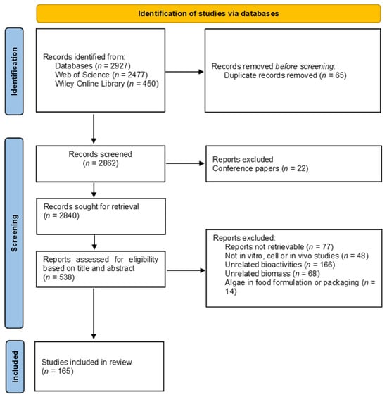
Figure 1.
Flow diagram of the systematic review study selection process. Adapted from reference [28].
3. Health-Related Bioactivities
3.1. Antioxidant Properties
3.1.1. Antioxidants and Their Role in Oxidative Stress and Disease Development
Free radicals, also known as oxidizing agents, are essential but unstable metabolic by-products of various normal cellular processes (e.g., mitochondrial respiration, inflammatory responses, and cellular signaling); however, their production can be increased due to exogenous stimuli (e.g., radiation, chemical agents, lifestyle factors, infections, and inflammation). The main oxidizing species are reactive oxygen species (ROS) such as hydroxyl radical (•OH), super oxide anion (O2−), hydrogen peroxide (H2O2), and singlet oxygen (O2) and reactive nitrogen species (RNS) like nitroxyl anion (NO−), nitrosonium cation (NO+), and various nitric oxide (•NO)-derived compounds, which are produced by the inducible nitric oxide synthase (iNOS) [29,30].
Antioxidants play a crucial role in preventing and repairing damages caused by ROS and RNS, by either stabilizing or eliminating them and preventing the oxidation of other molecules. Under normal circumstances, cellular endogenous antioxidant systems, which consist of enzymes (e.g., superoxide dismutase (SOD), catalase (CAT), and glutathione peroxidase (GPx)), and non-enzymatic biomolecules (such as bilirubin, ascorbate, glutathione, and albumin), are enough to safeguard against these reactive species and maintain a proper balance between oxidants and antioxidants. However, if there is an excessive production of ROS, the ability of cells to produce sufficient antioxidants may be compromised, resulting in inadequate protection for the organism. Under such conditions, the body can also resort to exogenous antioxidants sources such as food or nutraceuticals in an effort to restore equilibrium [30]. Nevertheless, if there is a persistent disturbance in the equilibrium between oxidants and antioxidants, it can result in the development of oxidative stress. Long-term oxidative stress can cause a chain reaction that plays a crucial role in cell damage and potential cell death (apoptosis), affecting various components like membranes, lipids, nucleic acids, and proteins [31]. Biomarkers for oxidative stress-induced damage include mutagenic and cytotoxic degradation products like malondialdehyde (MDA), resulting from lipid peroxidation, and deoxyguanosine (8-OHdG), resulting from DNA oxidation [29]. Oxidative stress can depolarize mitochondrial membranes and promote the release of pro-apoptotic proteins like cytochrome c into the cytosol. Cytochrome c associates with pro-caspase-9 and apoptosis activating factor-1 in the cytosol, activating caspase-9, which activates effector caspases (caspase-3, -6, and -7) to cleave cellular proteins and cause apoptosis. Other mitochondrial ROS-mediated apoptosis pathways involve DNA fragmentation, chromatin condensation, and the activation of the p53 and/or Jun N-terminal kinase (JNK) pathways, promoting intrinsic apoptosis (e.g., by activating pro-apoptotic Bax proteins and inhibiting anti-apoptotic proteins like Bcl-2) [32]. ROS activates apoptosis-related signaling pathways and transcription factors, including phosphoinositide 3-kinase (PI3K)/protein kinase B (Akt), mitogen-activated protein kinase (MAPK), nuclear factor erythroid 2–related factor 2 (Nrf2)/Kelch-like-ECH-associated protein 1 (Keap1), and nuclear factor kappa-B (NF-κB). When the Nrf2/Heme oxygenase-1 (HO-1) pathway is activated, Nrf2’s phosphorylation (mainly through kinases like MAPKs and PI3K) dissociates it from the Nrf2-Keap1 complex, allowing it to translocate to the nucleus and promote the expression of antioxidant response element (ARE) genes, controlling the expression of several enzymatic antioxidants like heme oxygenase 1 and increasing the cellular defense against oxidative stress [33]. If not properly regulated, oxidative stress can not only lead to the development of acute pathologies and premature aging, but also to chronic and degenerative conditions such as cancer and inflammatory, cardiovascular, gastrointestinal, and metabolic disorders [29]. Given the link between oxidative stress and these non-communicable diseases, antioxidant therapy or supplementation shows great potential in maintaining a healthy redox balance, postponing aging, and preventing or mitigating various health-related issues [30].
3.1.2. Potential Health Benefits of Algal Antioxidants
A total of 13 cell experiments (Table 1) and 11 in vivo experiments (Table 2) fulfilled the inclusion criteria in this review regarding antioxidant properties of algae. All of them concern macroalgae or pure compounds of not-detailed algal origin. Many algal compounds exert their antioxidant effect by increasing the activity of endogenous antioxidant enzymes like SOD, CAT, and GPx [34], decreasing ROS production [35,36], and/or decreasing the expression of iNOS, leading to a decreased production of NO [37]. In skeletal myoblasts, algal compounds (phloroglucinol and Indole-6-carboxaldehyde) exerted antioxidant effects by regulating oxidative stress-induced apoptosis, mainly by decreasing mitochondrial dysfunction and modulating apoptosis-regulatory factors (e.g., caspases, Bcl-2, Bax, and cytochrome c) [38,39]. Some algal compounds (e.g., from Ecklonia cava and Sargassum thunbergii) were also able to decrease oxidative stress responses by modulating the signaling pathways, mainly by downregulating AMP-activated protein kinase (AMPK) and NF-κB, and upregulating Nrf2/ARE/HO-1/AKT pathways [38,39,40].
When studying skin conditions, matrix metalloproteinases (MMPs) emerged as pivotal players, due to their role in the degradation of the different components of the extracellular matrix. In the presence of oxidative stress (e.g., due to radiation exposure), the control of MMPs is disturbed, causing the breakdown of collagen and the extracellular matrix, resulting in visible skin aging signs like wrinkles and loss of elasticity, besides also contributing to the development of various skin disorders [41]. The use of algal compounds (6,6′-bieckol; bromophenol bis (2,3,6-tribromo-4,5-dihydroxybenzyl) ether; 3-Bromo-4,5-dihydroxybenzaldehyde; bromophenols; and phloroglucinol) seems to be a successful strategy to counteract oxidative stress-induced skin damage [42,43,44,45] and enhance overall skin health by maintaining the integrity of the matrix and decreasing the deregulation of MMPs [46]. The skin protective effect from extracts from Fucus spiralis [47] (in HaCaT cells) can potentially be attributed to its phlorotannin content, whilst the Sargassum thunbergii extract had 14 phenolic compounds in its composition [48].

Table 1.
Cell experiments regarding algal extracts/compounds with antioxidant activities.
Table 1.
Cell experiments regarding algal extracts/compounds with antioxidant activities.
| Complication | Algae Type | Algae Species | Algal Extract or Compound | Cell Line | Oxidative Stress Induced by | Concentrations Tested | Outcomes and Mechanism | References |
|---|---|---|---|---|---|---|---|---|
| n.d. | Macroalgae | Ulva pertusa | Ulvan | RAW 264.7 | H2O2 | 200 µg/mL | ↑ antioxidant activity (↑ CAT and SOD); ↑ expression of antioxidant genes (↑ GST, CAT, MnSOD, and GPx mRNA expression) | [34] |
| Undaria pinnatifida | Phlorotannin extract | RAW 264.7 | H2O2 | 10, 20, and 40 µg/mL | ↑ cell survival; ↓ NO production and iNOS protein expression | [37] | ||
| Liver | Macroalgae | Nizamuddinia zanardinii | Fucoidan | HepG2 | H2O2 | 0.1, 0.2, 0.5, and 0.7 µg/mL | Protective effect on H2O2-induced cytotoxicity; ↓ intracellular H2O2-induced ROS production; ↓ H2O2-induced damages | [49] |
| Pyropia haitanensis | Floridoside | L-02 | n.d. | 200 µmol/L | No cytotoxic effect; ↑ SOD and GSH-Px activity; activation of HO-1 expression via upregulation on Nrf2/ARE and p38/ERK MAPK-Nrf2 pathway | [40] | ||
| Lungs | Macroalgae | Gelidiella acerosa | Ethyl acetate extract | A549 | H2O2 | 1.5 mg/mL | ↑ SOD and peroxidase activity | [50] |
| Skeletal muscle | Macroalgae | Ecklonia cava | Phloroglucinol | C2C12 | H2O2 | 10 and 20 µg/mL | ↓ cell toxicity (↓ H2O2-induced cell death); ↓ apoptosis (↓ DNA fragmentation, nuclear fragmentation, and chromatin condensation); ↓ mitochondrial dysfunction; regulation of apoptosis regulatory factors (↑ cytochrome c in the mitochondria, ↑ Bcl-2 expression, and ↑ caspase-3); ↓ ROS H2O2-induced accumulation; upregulation of Nrf2/HO-1 signaling pathway | [38] |
| Macroalgae | Sargassum thunbergii | Indole-6-carboxaldehyde | C2C12 | H2O2 | 400 µM | ↓ cell toxicity (↓ H2O2-induced cell death); ↓ ROS overproduction; ↓ DNA damage; ↓ apoptosis; ↓ mitochondrial dysfunction; regulation of apoptosis regulatory factors (cytochrome c, Bax, Bcl-2, and caspase-3 and -9); downregulation of AMPK signaling pathway | [39] | |
| Skin | Macroalgae | Ecklonia cava | 6,6′-bieckol | HaCaT | UVB radiation | 50 and 100 µM | ↑ cell survival; antioxidant effect (↑ antioxidant enzymes); downregulation of matrix metalloproteinases (MMPs) through MAPK and NF-κB pathways | [46] |
| Fucus spiralis | Ethyl acetate, water, and ethanol extracts | HaCaT | UVB radiation or H2O2 | 1000 µg/mL | ↓ ROS production | [47] | ||
| Symphyocladia latiuscula | Bromophenol bis (2,3,6-tribromo-4,5-dihydroxybenzyl) ether (BTDE) | HaCaT; HUVEC | H2O2 | 5 and 10 µM | ↑ cell survival (↓ apoptosis); reverse oxidative damage induced by H2O2 (↓ ROS generation, ↓ MDA level, ↓ GSSG/GSH, and ↑ SOD activity); upregulation of Nrf2 and decrease in Keap1 expression; activation of AKT signaling pathway | [42] | ||
| n.d. | n.d. | 3-Bromo-4,5-dihydroxybenzaldehyde | HaCaT | H2O2 or UV-B radiation | 30 µM | Protective effect against oxidative stress (↑ cell viability) possibly regulated by ERK and Akt pathways, inducing HO-1 and Nrf2 expression | [43] | |
| Bromophenols | HaCaT | H2O2 | 10 µM | ↑ cell survival (↓ apoptosis); ↓ oxidative cell damage (↓ ROS generation); increased expression of antioxidant proteins (TrxR1 and HO-1) | [44] | |||
| Phloroglucinol | HaCaT | H2O2 | 50 µM | Protected cells from H2O2-induced cytotoxicity (↑ cell viability); upregulation of Nrf2/HO-1 signaling pathway; ↓ oxidative stress (↓ ROS generation and DNA damage); ↓ apoptosis (↓ mitochondrial dysfunction); modulation of apoptosis regulatory genes (↑ Bcl-2, ↑ PARP, ↓ Bax, and ↑ caspase-3 and -9 expression); ↓ release of mitochondrial cytochrome c into the cytoplasm | [45] |
AMPK: AMP-activated protein kinase; ARE: Antioxidant response element; Bax: Bcl-2-associated X protein; Bcl-2: B-cell lymphoma 2; DNA: Deoxyribonucleic acid; GPx: Glutathione peroxidase; GSH: Glutathione (reduced); GSSG: Glutathione disulfide; GST: Glutathione S-transferase; H2O2: Hydrogen peroxide; HO-1: Heme oxygenase-1; iNOS: Inducible nitric oxide synthase; MDA: Malondialdehyde; MMPs: Matrix metalloproteinases; n.d.: No data available; NF-κB: Nuclear factor kappa B; Nrf2: Nuclear factor (erythroid-derived 2)-like 2; NO: Nitric oxide; PARP: Poly(ADP-ribose) polymerase; ROS: Reactive oxygen species; SOD: Superoxide dismutase; ↑: Increase; ↓: Decrease.
Most in vivo experiments (Table 2) were conducted on zebrafish embryos, a well-established model system, including as a preclinical screening model. Results showed that extracts and isolated compounds from Fucus virsoides [35], Gracilaria lemaneiformis [36] (oligosaccharide), Hizikia fusiforme [51] (polysaccharide), Padina boryana [52], Pyropia yezoensis [53] (polyphenol), Sargassum fulvellum [54] (polysaccharide), and Undaria pinnatifida sporophylls [55] (polysaccharide) were successful in decreasing oxidative stress by reducing lipid peroxidation and ROS production, leading to decreased heart-beating disorders and increased survival rates. The liver and kidneys are particularly susceptible to the deleterious effects of ROS/RNS due to their elevated metabolic and mitochondrial activity. Hence, the monitoring of hepatic enzymes (e.g., aspartate aminotransferase, alanine aminotransferase, and alkaline phosphatase) and kidney function markers (e.g., urea and creatinine) in the serum is a common approach to assess potential liver injury and kidney impairment [56,57]. The methanol extract from Halamphora sp. exhibited a notable ability to reduce the concentration of liver enzymes in the bloodstream, indicating a hepatoprotective effect against oxidative stress-induced injuries [58]. This was further confirmed by improved hepatocyte histology, which might be associated with the high fatty acid (mainly palmitic and palmitoleic acid) content of the extract. Improved renal function markers were observed, as well as improved renal histology, suggesting a renal protective effect from the abovementioned extract and polysaccharide extract of Ulva lactuca [59]. Phenolic compounds extracted from Sargassum thunbergii [48], such as benzene and its derivatives (protocatechuic acid, difucol, gallic acid, and 4-hydroxybenzoic acid), cinnamic acids and their derivatives (p-Coumaric acid), flavonoids (isoquercitrin, quercitrin, isorhamnetin, and catechin), and phlorotannins (bifuhalol, pentafuhalol A, 7-hydroxyeckol, deshydroxypentafuhalol, trifuhalol A), along with a sulfated polysaccharide from Ecklonia maxima [60], were found to reduce the production of reactive oxygen species (ROS) and repair skin damage. Therefore, the identified algal compounds and extracts seem to have antioxidant properties [35,36,51,52,53,54,55], being able not only to restore a healthy balance between oxidants and antioxidants, but also aid in the regulation and mitigation of oxidative stress-induced damages in specific organs, such as the skin [48,60], liver, and kidney [58,59].
Table 2.
In vivo experiments regarding algal extracts/compounds with antioxidant activities.
Table 2.
In vivo experiments regarding algal extracts/compounds with antioxidant activities.
| Complication | Algae Type | Algae Species | Algal Extraction or Compound | Route of Administration | Dosage | Experimental Period | Animal Model (Age) | Oxidative Stress Induced by | n/Group | Outcomes and Mechanism | References |
|---|---|---|---|---|---|---|---|---|---|---|---|
| n.d. | Macroalgae | Fucus virsoides | Less polar fractions | Incubation with embryo media | 7.5, 15, and 30 µg/mL | 4 d | Zebrafish embryos | H2O2 | 30 | Decreased heartbeat frequency; ↓ ROS formation | [35] |
| Gracilaria lemaneiformis | Agaro-oligosaccharides prepared from the agar | Incubation with embryo media | 25 and 50 µg/mL | 3 d | Zebrafish embryos | H2O2 | n.d. | Increased survival rate (↓ cell death); ↓ heart-beating disorder; ↓ ROS production; ↓ lipid peroxidation | [36] | ||
| Hizikia fusiforme | Fucoidan | Incubation with embryo media | 25 and 50 µg/mL | 2 d | Zebrafish embryos | H2O2 | 15 | Increased survival rate (↓ cell death); ↓ heart-beating disorder; ↓ ROS production; ↓ lipid peroxidation | [51] | ||
| Padina boryana | Ethanol precipitation | Incubation with embryo media | 50 and 100 µg/mL | 3 d | Zebrafish embryos (7–9 hpf) | H2O2 | n.d. | Increased survival rate (↓ cell death); improved heart-beating rates; ↓ intracellular ROS; ↓ lipid peroxidation | [52] | ||
| Pyropia yezoensis | Polyphenols and protein-rich extracts | Incubation with embryo media | 12.5, 25, and 50 µg/mL | 1 d | Zebrafish embryos (7–9 hpf) | AAPH | 15 | Decreased cell death; ↓ ROS production; ↓ lipid peroxidation production | [53] | ||
| Sargassum fulvellum | Polysaccharides | Incubation with embryo media | 50 and 100 µg/mL | 3 d | Zebrafish embryos (7–9 hpf) | AAPH | 15 | Increased survival rate (↓ cell death); improved heart rate; ↓ intracellular ROS; ↓ lipid peroxidation | [54] | ||
| Undaria pinnatifida sporophylls | Fucoidan | Incubation with embryo media | 125 and 250 µg/mL | 7 d | Zebrafish embryos (8 hpf) | AAPH | 15 | Increased survival rate (↓ cell death); ↓ heartbeat rate; ↓ ROS production; ↓ lipid peroxidation | [55] | ||
| Kidney | Macroalgae | Ulva lactuca | Polysaccharide extract | Intragastric | 50 and 300 mg/kg | 10 w | Kunming mice (8 w) | D-gal and ascorbic acid (subcutaneously) | 9 | Protective effect on kidney injury (↓ atrophy, ↓ serum creatinine and serum cystatin C); ↓ oxidative stress in kidney (↓ MDA, protein carbonyl, and 8-OHdG levels, and ↑ SOD, GSH-Px, and T-AOC); ↓ apoptosis (↓ expression of caspase-3 in kidney) | [59] |
| Liver and Kidney | Macroalgae | Halamphora sp. | Methanol extract (80%) | Gastric gavage | 2 mg/kg/day | 3 w | Wistar albino rats (adults) | Lead acetate (i.p.) | 6 | ↓ lipid peroxidation in liver and kidney (↓ MDA); ↑ protection against oxidative stress in liver and kidneys (↑ GPx, SOD, and CAT); improved serum biochemical parameters (↓ AST, ALT, ALP, and LDH, and ↓ creatine and urea) | [58] |
| Skin | Macroalgae | Ecklonia maxima | Sulfated polysaccharides | Incubation with embryo media | 50 and 100 µg/mL | 3 d | Zebrafish embryos (7–9 hpf) | AAPH | 15 | ↑ survival rate (↓ cell death, ↓ apoptosis); improved heart beating disorder; ↓ oxidative stress (↓ ROS generation and ↓ lipid peroxidation) | [60] |
| UVB-exposure | 10 | ↓ intracellular ROS levels; ↓ cell death; ↓ NO production and lipid peroxidation; improved collagen content and inhibition of MMPs | |||||||||
| Sargassum thunbergii | Phenolic-rich extract | Incubation with embryo media | 1.67 µg/mL | 6 d | Zebrafish embryos (2 dpf) | UVB-exposure | 8 to 10 | Repaired skin damage; ↓ intracellular ROS accumulation | [48] |
8-OHdG: 8-hydroxylated deoxyguanosine; AAPH: 2,2′-azobis (2-amidinopropane) dihydrochloride; ALT: Alanine aminotransferase; ALP: Alkaline phosphatase; AST: Aspartate aminotransferase; GPx: Glutathione peroxidase; H2O2: Hydrogen peroxide; hpf: Hours post fertilization; LDH: Lactate dehydrogenase; MDA: Malondialdehyde; MMPs: Matrix metalloproteinase; n.d.: No data available; NO: Nitric oxide; ROS: Reactive oxygen species; SOD: Superoxide dismutase; T-AOC: Total antioxidant capacity; ↑: Increase; ↓: Decrease.
3.2. Anti-Inflammatory Effects
3.2.1. Inflammation and Its Role in the Onset and Progression of Diseases
Inflammation is a fundamental and intricate biological process that plays a vital role in maintaining the body’s homeostasis. An acute inflammatory response is triggered by either tissue injury or exposure to external stimuli (e.g., viruses or allergens). This response is initiated by various mediators, including cytokines like interleukins (IL) or tumor necrosis factors (TNFs), acute phase proteins (e.g., C-reactive protein), chemokines (e.g., Monocyte Chemoattractant Protein-1), or prostaglandins (PGE). These mediators facilitate the movement of immune cells (neutrophils and macrophages) to the site of inflammation by promoting vasodilation and angiogenesis, which allow for the migration of additional inflammatory cells. Usually, once the episode is resolved, inflammation is no longer needed. However, in some cases, inflammation can persist at low levels without any apparent cause, leading to chronic and uncontrolled inflammatory conditions, which has been linked to the development of various human diseases and disorders [61]. A complex interplay between oxidative stress and inflammation has been established, where the activation of the inflammatory cascade leads to the production of inflammatory mediators, causing oxidative stress, which in turn activates the inflammatory cascades [62].
3.2.2. Mechanisms of Inflammation Modulation
The three most important intracellular inflammatory signaling pathways include the mitogen-activated protein kinase (MAPK), nuclear factor kappa-B (NF-κB), and Janus kinase (JAK)-signal transducer and activator of transcription (STAT) pathways. These pathways regulate pro-inflammatory cytokine production and inflammatory cell recruitment, which contribute to the inflammatory response [63]. The activation of the MAPKs, including Erk1/2, p38 and JNK, leads to the phosphorylation and activation of transcription factors, regulating pro-inflammatory gene expression, which initiates the inflammatory response (e.g., the expression of cytokines, chemokines, and inflammatory mediators). The activation of MAPK pathway is also linked to NF-κB and phosphoinositide 3-kinase (PI3K) pathways, as the MAPK mediates the phosphorylation of IκB kinase (IKK), which undergoes proteasomal degradation. This allows the NF-κB heterodimer (p50/p65) to translocate into the nucleus, bind to DNA, and induce target pro-inflammatory transcription. Meanwhile, the JAK/STAT signaling pathway is mostly activated by ligands (e.g., interleukins), activating the direct translation of an extracellular signal into a transcriptional response and controlling inflammatory gene transcription [63,64]. Several of these pathways are concurrently triggered by inflammatory mediators, regulating the expression of pro-inflammatory genes and ultimately leading to the synthesis of inflammatory mediators. In chronic inflammation, this becomes a positive feedback loop, leading to pathophysiological events [61,65].
Additional molecules that can modulate the inflammatory response are the arachidonic acid cascade-related eicosanoids (prostaglandins, thromboxanes, and leukotrienes). After phospholipases release arachidonic acid from the plasma membrane, cyclooxygenase (COX) or lipoxygenase (LOX) enzymes metabolize it, producing bioactive lipid mediators which act as signaling molecules. While COX-1 and some LOX are involved in normal cellular homeostasis, COX-2 is an inducible enzyme and, together with LOX-5, is upregulated in response to inflammatory stimuli, leading to the overexpressing of pro-inflammatory mediators, further increasing the inflammatory event; it is also overexpressed in pathophysiological events. Therefore, COX and LOX might be attractive therapeutic targets [66]. Nonsteroidal anti-inflammatory drugs (NSAIDs) are widely prescribed medications that reduce inflammation by blocking the COX enzyme. However, NSAIDs like ibuprofen and celecoxib can also have significant adverse side effects on the gastrointestinal, cardiovascular, hepatic, renal, cerebral, and pulmonary systems [21,67].
3.2.3. Algal Applications in Managing Inflammatory Conditions
The anti-inflammatory activity of algae was found in a total of 48 studies—6 in vitro (Table 3), 31 cell experiments (Table 4), and 10 in vivo (Table 5).
The in vitro anti-inflammatory studies that fitted the inclusion criteria for this review were all of macroalgal origin, with most of them focusing on extracts. Purified compounds were only obtained from two species, Gracilaria salicornia and Turbinaria decurrens, the former containing two 2H-chromenyl derivatives [68], two spiro-compounds [69], and a abeo-labdane type diterpenoid [70], whereas the latter accumulated a triterpene compound [71]). In terms of extracts, the anti-inflammatory activity (measured by the inhibition of inflammatory-inducing enzymes—COX and LOX) was most pronounced in Gloeothece sp. [72], Gracilaria salicornia, and Padina tetrastromatica Hauck [73], as these presented the lowest IC50 values compared to the other species. Notably, the anti-inflammatory effects observed in Gloeothece sp. were 10–20 times lower than those observed in the remaining species, showing that values not only vary significantly among species [73], but also according to extraction solvents [72].
Table 3.
In vitro studies regarding algal extracts/compounds with anti-inflammatory activities.
Table 3.
In vitro studies regarding algal extracts/compounds with anti-inflammatory activities.
| Algae Type | Algae Strain | Type of Analyzed Sample (Extract or Pure Compound) | In Vitro Assays Against Pro-Inflammatory Enzymes (IC50 Values in µg/mL Unless Otherwise Stated) | References | ||
|---|---|---|---|---|---|---|
| COX-1 | COX-2 | 5-LOX | ||||
| Macroalgae | Amphiroa fragilíssima (Linnaeus) J.V. Lamouroux | EtOAc-MeOH extracts | 4990 | 5010 | 5020 | [73] |
| Gloeothece sp. | Acetone Ethanol Hexane:isopropanol (3:2) | 120 200 130 | [72] | |||
| Gracilaria canaliculata Sonder | EtOAc-MeOH extracts | 2920 | 2000 | 2010 | [73] | |
| Gracilaria corticata (J. Agardh) J. Agardh | 2990 | 3010 | 3020 | |||
| Gracilaria salicornia | 4′-[10′-[7-hydroxy-2,8-dimethyl-6-(pentyloxy)- 2H-chromen-2-yl]ethyl]-3′,4′-dimethyl-cyclohexanone | 2.46 mM | [68] | |||
| 3′-[10′-(8-hydroxy-5-methoxy-2,6,7-trimethyl-2H-chromen2-yl)ethyl]-3′-methyl-2′-methylene cyclohexyl butyrate | 2.03 mM | |||||
| Gracilaria salicornia | spiro[5.5]undecanes, 3-(hydroxymethyl)-7-(methoxymethyl)-3,11-dimethyl-9-oxospiro[5.5]undec-4-en-10-methylbutanoate | 2.78 mM | [69] | |||
| 4-ethoxy-11,11-dimethyl-7-methylene-8-(propionyloxy)spiro[5.5]undec-2-en-104,106-dihydroxytetrahydro-2H-pyran-10-carboxylate | 1.91 mM | |||||
| Gracilaria salicornia | Methyl-16(13→14)-abeo-7-labdene-(12-oxo) carboxylate | 860 | [70] | |||
| Gracilaria salicornia | EtOAc-MeOH extracts | 1010 | 1020 | 980 | [73] | |
| Halymenia dilatata Zanardini | 3040 | 3000 | 3020 | |||
| Hydropuntia edulis (S.G.Gmelin) Gurgel & Fredericq | 2910 | 3010 | 2980 | |||
| Padina tetrastromatica Hauck | 1230 | 1340 | 1280 | |||
| Palisada pedrochei J.N.Norris | 4040 | 4030 | 4010 | |||
| Portieria hornemannii (Lyngbye) P.C. Silva | 2010 | 1990 | 2030 | |||
| Spyridia filamentosa (Wulfen) Harvey | 3010 | 2980 | 3040 | |||
| Turbinaria decurrens | Decurrencyclic B | 14.0 µM | 3.0 µM | [71] | ||
EtOAc: Ethyl acetate; COX: Cyclooxygenase; LOX: Lipoxygenase; MeOH: Methanol.
Most cell studies evaluated the effect of algal compounds and extracts on general inflammation in macrophage cell models (e.g., RAW 264.7) (n = 20), with compounds being predominantly of macroalgal origin (n = 18) and only two derived from microalgae (Phaeodactylum tricornutum [74] and Tisochrysis lutea [75]). The algal compounds acted through several mechanisms: decreased oxidative stress (by decreasing ROS and NO production or upregulating the Nrf2/HO-1 pathway); a decreased activity of inflammation enzymes (COX, iNOS); decrease in inflammatory transcription factor (NF-κB) levels; a decreased expression and production of pro-inflammatory chemokines and cytokines (such as interleukin (IL)-6, IL-1beta (IL-1β), TNF-alpha (TNF-α), and prostaglandins (PGE)); an increased expression and production of anti-inflammatory cytokines (IL-4 and IL-10); and a decreased upregulation of inflammatory pathways such as MAPK, NF-κB, and JAK/STAT. These findings were transversal regardless of treating general inflammation or specific disorders such as skin diseases (e.g., atopic dermatitis) and inflammatory myopathy. Interestingly, the ethanol extract from a combination of Ecklonia cava and Sargassum horneri [76] exhibited a more effective anti-inflammatory effect used in combination than individual macroalgae extracts, which might be attributed to synergistic effects. Several aspects influence the algal extract’s activity; for example, when evaluating lipid crude extracts from Sargassum ilicifolium, it was concluded that the anti-inflammatory activity is higher in a preventive scenario rather than in a treatment approach [77], whilst when evaluating the ethyl acetate fraction of Himanthalia elongate [78], it was discovered that the anti-inflammatory activity in the digested sample was increased in comparison to crude extracts, possibly due to the breakdown of complex phlorotannin structures.
Cell assays also focused on skin-related disorders, following consumers’ rising aware of skin aging and search for ways to counteract this trend with novel active ingredients [79]. Besides exerting anti-inflammatory effects through the abovementioned general anti-inflammatory mechanisms, some macroalgal compounds additionally had a protective activity of the skin barrier [80], decreased wrinkle formation [81], increased cell proliferation and collagen production in human dermal fibroblasts [82], and downregulated the expression of MMPs [83], contributing to overall skin health.
Table 4.
Cell studies regarding algal extracts/compounds with anti-inflammatory activities.
Table 4.
Cell studies regarding algal extracts/compounds with anti-inflammatory activities.
| Complication | Algae Type | Algae Species | Algal Extraction or Compound | Cell Line | Inflammation Induced by | Concentrations | Outcomes and Mechanism | References |
|---|---|---|---|---|---|---|---|---|
| n.d. | Macroalgae | Caulerpa racemosa | Ethanol, hexane, and ethyl acetate carotenoid fractions | RAW 264.7 | LPS | 25 µM | ↑ AMPK expression; ↓ TNF-α expression; ↓ mTOR expression | [84] |
| Cystoseira amentacea | Ethanol or DMSO extract | RAW 264.7 | LPS | 100 µg/mL | ↓ inflammation (↓ IL-1β, IL-6, COX-2, and iNOS expression) | [85] | ||
| Dictyopteris membranacea | Disulfides | RAW 264.7 | LPS | 15.62–31.25 µM | Anti-inflammatory activity (↓ TNF-α, IL-6, and IL-12 production); ↓ NO expression by downregulating iNOS; downregulation of AKT/MAPK/ERK signaling pathway | [86] | ||
| Ecklonia cava and Sargassum horneri | Ethanol extract | RAW 264.7 | LPS | 62.5 µg/mL | No cytotoxic effect; ↓ NO production; ↓ inflammatory response (↓ IL-1β, IL-6, PGE2, and TNF-α expression); downregulation of iNOS and COX-2; downregulation of NF-κB and MAPK pathways | [76] | ||
| Ecklonia cava | Ethanol extract | HGF-1 | LPS | 50 and 100 µg/mL | ↓ PGE2 production and pro-inflammatory enzyme expression; ↓ pro-inflammatory chemokine gene expressions; ↓ ROS production; downregulation of MAPK signaling pathway | [87] | ||
| Himanthalia elongata | Ethyl acetate fraction of a crude acetone extract | RAW 264.7 | LPS | 100 µg/mL | ↓ NO and O2 production regardless of being submitted to a simulated gastrointestinal digestion or not | [78] | ||
| Laurencia majuscula | Sesquiterpene (C17H25BrO3); chamigrane | RAW 264.7 | LPS | 3.7 µM; 3.6 µM | ↓ NO production and no cytostatic activity | [88] | ||
| Padina boryana | Fucosterol | RAW 264.7 | Particulate Matter/LPS | 12.5, 25, and 50 µg/mL | ↓ NO production; ↓ cytokines production (↓ IL-1β, IL-6, TNF-α, and PGE2); ↓ mRNA expression of IL-1β, IL-6, TNF-α, iNOS, and COX-2; downregulation of MAPK and NF-κB phosphorylation; upregulation of Nrf2/HO-1 pathway | [89] | ||
| Porphyra tenera | Water extract | RAW 264.7 | LPS | 1000 µg/mL | ↓ PGE2 and NO production; ↓ COX-2 and iNOS protein expression; ↓ TNF-α and IL-6 production | [90] | ||
| Porphyra sp. | Polydeoxyribonucleotide | RAW 264.7 | LPS | 200 µg/mL | ↓ NO production; ↓ iNOS expression by reducing phosphorylation of p38 MAPK and ERK | [82] | ||
| Rugulopteryx okamurae | Rugukadiol A and ruguloptone A | RAW 264.7 | LPS | 10 µM | ↓ NO production; ↓ Nos2 and IL-1β expression | [91] | ||
| Rugulopteryx okamurae | Okaspatol C Okamurol A | RAW 264.7 | LPS | 10 µM | Decrease in NO production | [92] | ||
| Sargassum autumnale | Fucoidan fractions | RAW 264.7 | LPS | 50, 100, and 200 µg/mL | ↓ NO production (↑ cell viability); ↓ PGE2 production; ↓ pro-inflammatory cytokines (TNF-α, IL-6, and IL-1β); ↓ expression of inducible inflammatory enzymes (iNOS and COX2); downregulation of NF-κB and MAPK pathways | [93] | ||
| Sargassum horneri | Sargachromenol | RAW 264.7 | LPS | 62.5 µg/mL | ↑ antioxidant activity (↓ NO and intracellular ROS production); activation of Nrf2/HO-1 signaling pathway (upregulation of HO-1 expression); ↓ expression of inflammatory cytokines (IL-1β, IL-6, and TNF-α) through the downregulation of iNOS and COX-2 expression; suppression of activation of NF-κB and MAPK signaling pathways | [94] | ||
| Sargassum ilicifolium | Crude lipid extracts | RAW 264.7 | LPS | 50 µg/mL | ↓ NO production in pre-incubated and co-incubated cell culture models | [77] | ||
| Saccharina japonica | Fucoidan | RAW 264.7 | LPS | 100, 150, and 200 µg/mL | ↓ NO production; ↓ inflammation (↓ iNOS and COX-2 expression and ↓ TNF-α, IL-6, and IL-1β production); downregulation of NF-κB, MAPK, and JAK2-STAT1/3 signaling pathways | [95] | ||
| Sargassum swartzii | Fucoidan fraction | RAW 264.7 | LPS | 100 and 200 µg/mL | ↓ NO production; ↓ inflammation (↓ PGE2, TNF-α, IL-1β, and IL-6 secretion and expression); ↓ iNOS and COX-2 expression; downregulation of NF-κB and MAPK signaling pathways | [96] | ||
| n.d. | Fucoxanthinol | RAW 264.7 | LPS | 10 and 20 µM | Anti-inflammatory activity (↓ iNOS, IL-6, and TNF-α mRNA expression and ↓ IL-1β, TNF-α, IL-6, and Nitrate production) | [97] | ||
| Microalgae | Phaeodactylum tricornutum | Nonyl8-acetoxy-6-methyloctanoate | RAW 264.7 | LPS | 25 μg/mL | ↓ inflammation (↓ NO, PGE2, IL-1β, and IL-6); downregulation of COX-2 and iNOS | [74] | |
| Tisochrysis lutea | Methanol extract | RAW 264.7 | LPS | 100 µg/mL | Protected cells from cytotoxicity (↓ dendritic structures); ↓ PGE2 production and COX-2 protein expression; ↓ IL-6 and ↑ IL-10 expression; ↓ expression of inflammatory genes (Arg1, SOD2, and NLRP3) | [75] | ||
| Myopathy | Macroalgae | Ishige okamurae | Diphlorethohydroxycarmalol | C2C12 | TNF-α | 3.125, 6.25, and 12.5 µg/mL | ↓ NO and ↓ pro-inflammatory cytokines (TNF-α, IL-1β, and IL-6) production; modulation of NF-κB and MAPK signaling pathways | [98] |
| Skin | Macroalgae | Ecklonia cava | Dieckol | HaCaT | Particulate matter | 10 and 30 µM | ↓ PGE2 production; ↓ COX-1 and COX-2 mRNA expression levels; ↓ ROS; ↓ gene expression of enzymes involved in PGE2 synthesis | [99] |
| Halymenia durvillei | Ethyl acetate fraction | HaCaT | UV radiation | 5 µg/mL | ↓ intracellular ROS production; ↓ matrix metalloproteinases; upregulation of mRNA of antioxidant enzymes (SOD, HMOX1, and GSTP1); ↑ procollagen synthesis; activation of Nrf2 pathway | [81] | ||
| Polyopes affinis | Butanol fraction | HaCaT | IFN-γ or TNF-α | 10, 30, and 60 µg/mL | Downregulation of MAPK, STAT1, and NF-κB pathways | [100] | ||
| Polysiphonia morrowii | 3-bromo-4,5-dihydroxybenzaldehyde | HaCaT | IFN-γ or TNF-α | 144 and 288 µM | ↓ inflammatory cytokines (IL-6, IL-8, IL-13, IFN-y, and TNF-α) and chemokine production; downregulation of MAPK and NF-κB signaling pathways; activation of Nrf2/HO-1 signaling; protective activity against deterioration of skin barrier function (preserving skin moisture and tight junction stability) | [80] | ||
| Pyropia yezoensis | Methanol extract | HaCaT | IFN-γ | 40, 200, and 1000 µg/mL | Improvement of atopic dermatitis (↓ mRNA expression and secretion of pro-inflammatory chemokines; inhibition of MAPK activation; downregulation of NF-κB activation) | [101] | ||
| Sargassum confusum | Low-molecular-weight fucoidan | HaCaT | IFN-γ or TNF-α | 15.6, 31.3, and 62.5 µg/mL | ↓ ROS production; ↓ inflammatory cytokines (IL-1β, IL-6, IL-8, IL-13, IFN-y, and TNF-α) and chemokines; downregulation of MAPK and NF-κB signaling pathways; activation of Nrf2/HO-1 signaling | [102] | ||
| Sargassum horneri | (–)-Loliolide | HaCaT | IFN-γ or TNF-α | 15.6, 31.3, and 62.5 µg/mL | ↓ inflammatory cytokines (IL-4, IL-6, IL-13, IFN-y, and TNF-α) and chemokines; downregulation of MAPK and NF-κB signaling pathways; upregulation of Nrf2/HO-1 signaling | [103] | ||
| Sargassum siliquastrum | Low-molecular-weight fucoidan | RAW 264.7 | LPS | 25, 50, and 100 µg/mL | ↓ ROS production; ↓ production of NO and PGE2; ↓ expression of iNOS and COX-2; ↓ inflammatory cytokine expression (IL-1β, IL-6, and TNF-α); downregulation of MAPK and NF-κB signaling pathways; activation of Nrf2/HO-1 signaling; inhibition of the NLRP3 inflammasome protein complex | [104] | ||
| Sargassum horneri | Fucosterol | HDF | IFN-γ or TNF-α | 60 and 120 µM | ↓ ROS production; activation of Nrf2/HO-1 signaling; no effect of cell viability; ↓ mRNA expressions of inflammatory cytokines (IL-6, IL-8, IL-13, IL-33, IL-1β, TNF-α, and IFN-y) and MMPs; downregulation of MAPK and NF-κB signaling pathways | [83] | ||
| Microalgae | Porphyridium cruentum | Sulfated exopolysaccharides | HaCaT | UVA radiation | 12 µg/mL | Protective effect on cells from oxidative damage (↓ ROS formation, ↓ lipid peroxidation, and ↑ intracellular GSH levels); increased wound healing activity | [105] | |
| Phycoerythrin | 10 nM |
AKT: Protein kinase B; AMPK: AMP-activated protein kinase; COX: Cyclooxygenase; DNA: Deoxyribonucleic acid; DMSO: Dimethyl sulfoxide; ERK: Extracellular signal-regulated kinase; GSH: Glutathione; GST: Glutathione S-transferase; HGF-1: Human gingival fibroblast-1; HDF: Human dermal fibroblast; HO-1: Heme oxygenase-1; IFN-γ: Interferon-gamma; IL: Interleukin; iNOS: Inducible nitric oxide synthase; JAK2-STAT1/3: Janus kinase 2-signal transducer and activator of transcription 1/3; LPS: Lipopolysaccharide; MAPK: Mitogen-activated protein kinase; mTOR: Mammalian target of rapamycin; n.d.: No data available; NF-κB: Nuclear factor-kappa B; NLRP3: NOD-like receptor protein 3; NO: Nitric oxide; Nrf2: Nuclear factor erythroid 2–related factor 2; PGE2: Prostaglandin E2; ROS: Reactive oxygen species; SOD: Superoxide dismutase; STAT1: Signal transducer and activator of transcription 1; STAT3: Signal transducer and activator of transcription 3; TNF-α: Tumor necrosis factor-alpha; TXA2: Thromboxane A2; UVA: Ultraviolet A; UVB: Ultraviolet B; ↑: Increase; ↓: Decrease.
In vivo studies (Table 5) followed the same trend as cell experiments by mainly evaluating general inflammation (n = 7 for macroalgal and n = 2 for microalgal studies) and skin disorders (n = 2, 1 study for each algae type). The anti-inflammatory effects were attributed to the decrease in NO, ROS, and IL-1β, thus decreasing LPS-induced cell death. Interestingly, the two studies with human subjects were performed with microalgae, where Phaeodactylum tricornutum supplements [106] showed good tolerance and improved inflammatory status and the modulation of intestinal permeability, and the skin application of Dunaliella salina demonstrated anti-inflammatory and anti-aging effects [107]. Polysaccharides were the only class of isolated compounds that exhibited anti-inflammatory action [108,109,110,111,112].
Table 5.
In vivo studies regarding algal extracts/compounds with anti-inflammatory activities.
Table 5.
In vivo studies regarding algal extracts/compounds with anti-inflammatory activities.
| Complication | Algae Type | Algae Species | Algal Extraction or Compound | Route of Administration | Dosage | Experimental Period | Animal Model (Age) | Inflammation Induced by | n/Group | Outcomes and Mechanism | References |
|---|---|---|---|---|---|---|---|---|---|---|---|
| n.d. | Macroalgae | Codium fragile | Sulfated polysaccharides | n.d. | 50 and 100 µg/mL | 3 d | Zebrafish embryos (7–9 hpf) | LPS (10 µg/mL) | n.d. | ↓ cell death; ↓ NO and ROS generation | [108] |
| Cystoseira crinita (Desf.) Borry | Fucoidan | Intraperitoneally | 25 and 50 mg/kg | 5 h | Wistar Rats | LPS (0.25 mg/kg) | 8 | Decrease in IL-1β production | [109] | ||
| Ecklonia maxima | Ethyl acetate fraction | Incubation with embryo media | 25 and 50 µg/mL | 3 d | Zebrafish embryos (7–9 hpf) | LPS (10 µg/mL) | 15 | Increased survival rate (↓ cell death); improved heart-beating rates; ↓ ROS and NO generation | [113] | ||
| Saccharina japonica | Sulfated polysaccharide | Incubation with embryo media | 50 and 100 µg/mL | 3 d | Zebrafish embryos (8 hpf) | LPS (10 µg/mL) | 15 | ↓ cell death; ↓ NO and ROS generation; protection of phenotypic changes and toxic damages caused by LPS (↓ yolk sack edema, ↓ heart rate, and ↑ survival rate) | [114] | ||
| Saccharina japonica | Fucoidan | Incubation with embryo media | 25 and 50 μg/mL | 3 d | Zebrafish embryos | LPS (10 µg/mL) | n.d. | Increased survival rate (↓ cell death); improved heart-beating rates; ↓ intracellular ROS; ↓ NO generation | [110] | ||
| Sargassum binderi | Polysaccharides | Incubation with embryo media | 25, 50, and 100 µg/mL | 3 d | Zebrafish larvae (7–9 hpf) | LPS (10 µg/mL) | 15 | ↓ LPS-induced cell death; ↓ NO production | [111] | ||
| Sargassum fulvellum | Polysaccharides | Incubation with embryo media | 50 and 100 µg/mL | 3 d | Zebrafish embryos (7–9 hpf) | LPS (10 µg/mL) | 15 | Increased survival rate (↓ cell death); ↓ heartbeat disorder; ↓ ROS; ↓ NO | [112] | ||
| Microalgae | Phaeodactylum tricornutum | Supplements (whole biomass, β-1,3-glucan-rich, or combination thereof) | Oral supplements | 2.3 g biomass powder; 1.8 g of lyophilised supernatant; 2.3 g biomass powder + 1.8 g of lyophilised supernatant | 2 w | Elderly human individuals (67.7 ± 6.5 years) | - | 4 to 5 | No severe reactions, some mild and minimal were reported; decreased inflammatory marker (IL-6); ↑ plasma carotenoids (fucoxanthin); modulation of intestinal permeability (↓ zonulin) | [106] | |
| Tetraselmis sp. | Ethanol extract | Incubation with embryo media | 100 and 200 µg/mL | 7 d | Zebrafish embryos (7–9 hpf) | LPS (10 µg/mL) | 15 | Increased survival rate (↓ cell death); ↓ NO generation | [115] | ||
| Skin | Macroalgae | Sarcodia suiae sp. | Ethyl acetate fraction of ethanol extract | Skin application | 200 µg/day | 18 d | BABL/c mice (8 w) | DNCB (2%) | 6 | ↓ Atopic dermatitis symptoms (↓ inflammation, skin erythema, edema, dryness, and keratinocyte hyperplasia) and ↓ immunoglobulin E upregulation; ↓ swelling of subiliac lymph nodes and spleen; ↑ skin barrier integrity (↑ claudin-1 expression, cell-to-cell connections, and improved dilaggrin deficiency) | [116] |
| Microalgae | Dunaliella salina | Hydrophobic extract | Skin application | 1% | 56 d | Human subjects (aged 35–60, Fitzpatrick skin phototypes II–IV, and with signs of aging) | Intense solar exposure | 25 | Anti-inflammatory activity (↓ skin reactivity to histamine stimulation and red spot count and area); anti-aging effect (↓ wrinkle count and volume) | [107] |
IL: Interleukin; LPS: Lipopolysaccharide; n.d.: No data available; NO: Nitric oxide; ROS: Reactive oxygen species; w: Weeks; ↑: Increase; ↓: Decrease.
3.3. Cardioprotective Activity
3.3.1. Cardiovascular Diseases and Regulation of Blood Pressure and Blood Lipid Levels
Cardiovascular diseases (CVDs) encompass a range of heart and blood vessel problems, which are the primary death cause worldwide [117,118]. The main risk factor for the development of CVDs is the inherent aging process of the cardiovascular system, where the main stress factors are oxidative stress and chronic inflammation, which interact in a positive feedback loop. ROS contributes to the development of myocardial tissue damage, modifies calcium homeostasis and contractile dysfunction, and causes cardiomyocyte hypertrophy, apoptosis, and fibrosis [119]. ROS also promotes the recruitment of inflammatory cells. It increases the expression of adhesion molecules like intercellular and vascular cell adhesion molecule 1 (ICAM-1 and VCAM-1, respectively), enabling lipids accumulation in the inner layer of blood vessels [120]. Therefore, the inflammatory cascade and ROS play an important role in the development, modulation, and progression of atherosclerotic plaque. Together with lipid core growth, a reduction in the thickness of the fibrous cap leads to plaque instability, significantly increasing the risk of rupture and potential acute events such as stroke [121]. Over time, these factors result in a gradual deterioration of physiological functions and to the development of disorders such as hypertension, heart failure, arteriosclerosis, atherosclerosis, and myocardial infarction [118,119].
Aside from the natural aging process (senescence), there are behavioral risk factors that increase the risk of developing CVDs, which may manifest clinically in individuals, such as hypertension, dyslipidemia, hyperglycemia, overweight, and obesity [117]. The angiotensin-converting enzyme (ACE) is a protease that regulates the renin–angiotensin–aldosterone system, which plays a vital role in maintaining circulatory hemodynamics and participating in cardiac aging. ACE converts inactive angiotensin I into angiotensin II (Ang II), a strong vasoconstrictor and salt retention promoter, which controls blood pressure. Therefore, the deregulation of ACE levels can cause hypertension and other cardiovascular issues such as cardiac hypertrophy and cardiomyocyte apoptosis. Furthermore, Ang II, via the AT1 receptor, contributes to heart inflammation by promoting IL-6, IL-1β, and TNF-α production. Hence, in certain cardiovascular disorders, it is advantageous to inhibit the renin–angiotensin–aldosterone system, thereby preventing blood vessel constriction, reducing blood pressure, and suppressing the production of inflammatory cytokines [119,122]. Dyslipidemia, a contributing factor to the development of cardiovascular diseases, primarily arises from elevated levels of total cholesterol (TC), triglycerides (TG), low-density lipoprotein cholesterol (LDL), and reduced levels of high-density lipoprotein (HDL). This condition negatively impacts blood vessel health by promoting the inflammation and oxidative stress damage of the endothelial cells that line the blood vessels, leading to atherosclerosis and the formation of plaque [118].
3.3.2. Algal Compounds and Their Potential for Reducing Cardiovascular Risk
During the literature search, seven in vitro studies with cardioprotective activity fulfilled the inclusion criteria for this review. These studies evaluated the anti-hypertensive activity of macroalgal extracts, fractions, protein hydrolysates, peptides, and isolated compounds (chromenols from Sargassum macrocarpum [123]) by measuring the inhibition of the ACE (Table 6). ACE-inhibitory potential varied among species but also among extraction solvents [123]. When trying to further identify the bioactive compound of Mazzaella japonica, purification led to a loss of hypertensive activity, as the ACE inhibitory activity in each fraction ranged from 1.3 to 6.7% of the total ACE inhibition, suggesting that every fraction had a significant role in the overall ACE inhibitory activity [124]. In contrast, the hydrolysate from Ulva intestinalis exhibited the opposite behavior, where bioactivity was increased in the fraction with a molecular weight below 3 kDa [125], indicating a concentration of the bioactive compound(s) in this particular fraction.
Table 6.
In vitro studies regarding algal extracts/compounds with anti-hypertensive activities.
Only two cell experiments reporting cardioprotective effects fulfilled the inclusion criteria for this review. A water extract from macroalgae Fucus vesiculosus (250 µg/mL) showed promising results for fighting hypercholesterolemia, by reducing cholesterol permeation in Caco-2 cells by almost 50% [129]. The other study focused on the potential of a nonapeptide (EMFGTSSET) extracted from microalgae Isochrysis zhanjiangensis [130], at 100 µM, to improve atherosclerosis in HUVEC cells stimulated by oxidized low-density lipoprotein. Results were promising by showing a reduction in oxidative stress and inflammatory markers (ROS, IL-6, IL-1β, and TNF-α), cell adhesion molecules (ICAM-1 and VCAM-1), apoptosis (less caspase-3 and -9 expression), downregulating inflammatory pathways (e.g., MAPK), and upregulating Nr2/HO-1 pathway.
Several in vivo studies (n = 6) were found in which macro- and microalgal (n = 5 and n = 1, respectively) compounds demonstrated the potential to reduce the risk of CVD through various mechanisms (Table 7). These include regulating blood lipid levels (increasing HLD and decreasing TC, LDL, and TG) [131], reducing plaque formation and/or instability [131], decreasing oxidative stress and inflammatory markers [132], modulating inflammatory pathways [132], improving blood pressure control [133,134], reducing cardiac fibrosis in the ventricle areas [135], ameliorating histological changes in the cardiac tissue [132], and mitigating senescent deterioration [135]. Hydrolysates [133,134], terpenes [132], a mix of a polysaccharide and a carotenoid [135], and isolated polysaccharides [131,135,136] showed favorable outcomes for hypertension [133,134], myocardial inflammation [132], aging [135], carotid atherosclerotic lesions [131], and heart valve calcification [136], respectively.
Table 7.
In vivo studies regarding algal extracts/compounds with cardioprotective activities.
3.4. Gastrointestinal Health Modulation
3.4.1. Implications for Digestive Health and Gut-Related Disorders
Gastrointestinal diseases are prevalent worldwide, resulting in a significant decline in patients’ quality of life, imposing a significant healthcare burden and expenses, and potentially resulting in mortality [137]. Gastrointestinal problems, such as inflammatory bowel diseases and liver disorders, not only have a direct influence on the affected organs but also have broader consequences, such as impairing gastrointestinal health and reducing the overall health and well-being of patients [138].
Inflammatory bowel disease (IBD) is a persistent inflammatory condition affecting the gastrointestinal system, caused by a multifaceted interplay of genetic, immunological, and environmental factors. Crohn’s disease and ulcerative colitis are the two main subtypes of IBD, differing not only in their location but also in the nature of inflammation. Ulcerative colitis predominantly impacts the colon, causing persistent inflammation and the formation of ulcers. In contrast, Crohn’s disease can affect any segment of the digestive system, characterized by sporadic but transmural bowel inflammation. Both disorders exhibit varying symptoms and risks, and presently, there is no cure available for either. However, a common hallmark of both conditions is dysbiosis in the gut microbiota [138]. In fact, alterations in the gut microbial ecology have been linked to several NCDs (e.g., colorectal cancer, atherosclerosis, diabetes, obesity, liver disorders, and osteoporosis), being postulated as the communicable element [139].
The gut microbiota is essential for maintaining a symbiotic relationship with the host, influencing multiple physiological processes such as digestion, nutrient absorption, and immune system regulation. A healthy host’s gut microbiota, mostly Firmicutes and Bacteroides, synthesizes vitamins that the host cannot produce (e.g., vitamin K) and ferments non-digestible dietary fibers to produce short-chain fatty acids (SCFAs), like acetate, propionate, and butyrate, which regulate immune response, inflammation, and gut barrier integrity. Additionally, the gut microbiota breaks down bile acids, which directly regulate intestinal immune cell populations and maintain mucosal barrier immunity. Gut dysbiosis disturbs the delicate balance of microbial ecology (diversity), which can result in inflammation, compromised intestinal barrier function, and modified metabolism, becoming a central factor in the development of several clinical disorders [138,140].
Gut dysbiosis leads to oxidative stress, which can abnormally activate the intestinal immune system and harm the intestinal mucosal barrier by reducing mucous secretion, antimicrobial peptide secretion, and impair tight junctions. The immune response will activate pro-inflammatory signaling pathways such as NF-κB, JAK/STAT, and MAPK, which result in the release of proinflammatory factors and oxidases (e.g., iNOS, COX-2, and NOX), leading to a positive feedback loop of oxidate stress, inflammation, and changes in the composition of the gut microbiota [141].
Liver disorders, such as non-alcoholic fatty liver disease (NAFLD), often coexist with IBD as comorbid conditions, likely due to the connection between the gut and the liver known as the “gut–liver axis” [138,139]. NAFLD is the most prevalent chronic liver disease worldwide and covers a wide spectrum of liver diseases ranging from simple steatosis to non-alcoholic steatohepatitis (NASH), liver fibrosis, and, ultimately, cirrhosis and hepatocellular carcinoma. Hepatic steatosis is caused by the excessive accumulation of fat in the liver, as well as lipogenesis and systemic insulin resistance. NASH develops gradually because of fat buildup, which disrupts metabolism and leads to an excessive production of ROS in the mitochondria. This, in turn, causes lipid peroxidation and reduces the levels of antioxidant enzymes in the liver. Oxidative stress also controls the activation of genes related to lipid metabolism, such as peroxisome proliferator-activated receptor (PPAR) and sterol regulatory element-binding protein (SREBP). The liver’s compromised metabolic function causes an excess buildup of free fatty acids (FFAs) within the liver cells, causing lipotoxicity, inflammation, cellular damage, and cell death. Hepatic damage can be reversed prior to the onset of fibrosis. The progression of these conditions will lead to chronic injury and the development of fibrosis, resulting in liver cirrhosis and, eventually, hepatocellular cancer [138,142].
3.4.2. Algal Compounds and Their Potential for Improving the Impairment of Gastrointestinal Health
The literature search yielded only two cell assay experiments that fulfil the inclusion criteria of this review, both investigating the liver promoting activity of purified compounds (fucoidan and fucoxanthin) derived from macroalgae. The fucoidan (50 µg/mL) derived from Cystoseira compressa showed antisteatotic action by modulating the lipogenesis pathway (PPARy) and decreasing intracellular triglyceride content in FaO cells stimulated by oleate and palmitate [143]. The other macroalgae compound, fucoxanthin (25 and 50 µM), was able to mitigate zearalenone-induced hepatotoxicity by decreasing inflammation (less production of pro-inflammatory cytokines TNF-α, IL-6, and IL-1β) and oxidative stress (by reducing ROS production and upregulating the PI3K/AKT/NRF2 signaling pathways) in hepatocytes (HepG2 cells) [144].
A total of 14 in vivo studies were incorporated in this review to assess the impact of extracts, compounds, or commercial items on liver health (n = 4) or gut health (n = 10), all of macroalgal origin (Table 8). In terms of liver health, the treatment with algal compounds resulted in a reduction in body [145] and liver weight [146], indicating a decrease in fat accumulation and a decrease in liver steatosis [145]. Additionally, hepatic inflammation was reduced through the reduction in cytokine production and the modulation of inflammatory pathways [147]. The treatment also restored hepatic lipid metabolism by modulating genes involved in lipogenesis [147]. Furthermore, it decreased oxidative stress by increasing the profile of antioxidant liver enzymes and ultimately decreased liver damage, as evidenced by the decrease in liver enzymes in the serum and the improvement of the abnormal cellular architecture of liver tissue [146,148]. Hence, the algae-based compounds that were evaluated effectively influenced all the fundamental factors that contribute to the development and progression of NAFLD, suggesting their potential as a source for therapeutic interventions.
An Ulva pertusa extract also effectively alleviated the negative effects of IBD by providing the relief of symptoms such as pain, weight loss, and colon shortening [149,150]. Additionally, several compounds counteracted ulcerative colitis by decreasing the levels of pro-inflammatory cytokines and enzymes, while increasing the levels of anti-inflammatory cytokines and modulating inflammatory pathways [151,152,153,154,155]. Oxidative stress was reduced via enhancing the activity of antioxidant enzymes, lowering the levels of oxidative stress indicators, and regulating the Nrf2/HO-1 pathway [150,152]. These findings were validated through the observation of histological damage in the colon, where a reduction in inflammation and the mitigation of chronic colitis lesions could be seen [151,152,153,155]. An improvement in the abundance and variety of gut bacteria was detected, indicating a reduction in gut dysbiosis [156]. Furthermore, the integrity of the gut’s protective mucosal barrier was restored and intestinal innate immunity was enhanced, which led to the overall improvement of intestinal health [157]. These findings indicate that isolated compounds derived from macroalgae may hold significant promise for the treatment of gut diseases, whereas intestinal health was mainly improved by hydroquinones [158] and polysaccharides [156]; (ulcerative) colitis by terpenes [155], phlorotannins [152,154], and carotenoids [151,152,154]; and hepatic health by polysaccharides [146] and polyphenols [147]. Interestingly, a commercial formulation composed of polysaccharides, phlorotannins, and other polyphenols [145] displayed promising results for NASH and NAFLD, which may indicate that these compounds could have synergistic effects.
Table 8.
In vivo studies regarding algal extracts/compounds with gastrointestinal implications.
Table 8.
In vivo studies regarding algal extracts/compounds with gastrointestinal implications.
| Complication | Algae Type | Algae Species | Algal Extraction or Compound | Route of Administration | Dosage | Experimental Period | Animal Model (Age) | Induced by | n/Group | Outcomes and Mechanism | References |
|---|---|---|---|---|---|---|---|---|---|---|---|
| Hepatic damage | Macroalgae | Gracilaria caudata | Sulfated polysaccharides | Intraperitoneally | 10 mg/kg/day | 5 d | Swiss mice | Nimesulide (intragastric) | 8 to 10 | ↓ liver weight; improved antioxidant parameter in liver (↓ MPO, MDA, and NO3/NO2 levels, ↑ GSH level); ↓ inflammatory markers (↓ IL-1β and TNF-α levels); enhancement of hepatic function markers (↓ AST, ALT, and GGT levels) | [146] |
| Liver fibrosis | Macroalgae | Caulerpa racemosa | Water extracts | Oral in distilled water | 200 mg/kg/mL | 5 w | Wistar Rats | 40% Carbon tetrachloride (CCl4) intraperitoneally | 7 | Enhanced liver enzymes in serum (↓ ALT, AST, ALP, and LDH) and liver metabolite (↓ total bilirubin and direct bilirubin); improved renal and lipid profile (↓ urea; ↓ creatine); increased hepatic antioxidant enzymes (↑ GSH and CAT; ↓ MDA) | [148] |
| Padina pavonia | 6 | ||||||||||
| NAFLD | Macroalgae | Ishige okamurae | Diphlorethohydroxycarmalol (DPHC) | Incubation with embryo media | 40 µM | 3 d | Transgenic zebrafish embryos (Danio rerio) (3 dpf) | Palmitate | 12 to 15 | ↓ lipogenesis (downregulation of lipogenesis-related genes SREBP1c, ChREBP1 α, and FAS); ↓ liver inflammation (↓ IL-1β, TNF-α, and COX-2); regulation of lipid metabolism (stimulation of AMPK and SIRT1 signaling pathway) | [147] |
| NASH and NAFLD | Macroalgae | A. nodosum and F. vesiculosus | Gdue© | Intragastric gavage | 7.5 mg/kg bw | 12 w for NAFLD; 18 w for NASH | Sprague Dawley rats (4–8 w) | Western diet high-fat diet plus 30% fructose in the drinking water | 10 | ↓ body weight gain; ↓ plasma glucose; reduced liver steatosis (↓ liver TG accumulation); ↓ hepatic inflammation; restored hepatic lipid metabolism (downregulation of lipid droplet forming and fatty acid synthase genes); restored physiological levels of protein expression regulating lipid homeostasis | [145] |
| IBD | Macroalgae | Ulva pertusa | Extract | Oral gavage | 50 mg/kg and 100 mg/kg | 4 d | CD1 mice (4 w) | DNBS injected into the rectum | 10 | ↓ body weight loss; ↑ pain threshold; ↓ DNBS-induced hyperalgesia; ↓ DNBS-induced visceral hypersensitivity; ↓ cell adhesion molecules (↓ ICAM-1 and P-selectin); ↓ gut-inflammation (reduced IL-6, IL-17, and IL-23 levels and enhanced serum IL-10 levels); modulation of innate (↓ CD68+ cells) and adaptive (↓ CD4+ and CD8+ cells) immune system; TLR4 and NLRP3 inflammasome modulation | [149] |
| 11 | ↓ body weight loss; ↓ inflammation (reduced the expression levels of NF-κB and restored the expression of Ikb-α); modulation of pro-inflammatory interleukin production (↓ IL-5, IL-9, and IL-13, and ↑ IL-4); modulation of apoptosis (↓ p-53, caspase-3, -8, and -9, and ↓ ↑ Bcl-2); ↓ oxidative stress (↓ MDA levels, ↑ GSH, CAT, SOD, Mn-SOD, and HO-1); modulation of Nrf2/SIRT1 pathway (↑ Nrf2 and SIRT1 levels) | [150] | |||||||||
| Intestinal Health | Macroalgae | Cymopolia barbata | Cymopol | Oral gavage | 0.1 g/kg and 0.4 g/kg | 3 d | C57BL/6J mice (4 w) | 3% DSS in drinking water | 5 | ↓ inflammatory and oxidant response (downregulation of ERK/MAPK and PI3K/AKT pathways) | [158] |
| Laminaria spp. | Enzymolysis seaweed powder | Feed | 20 g/kg of feed | 4 m | Ragdoll kittens (6 m) | 10 | ↑ growth performance (↑ weight gain); improved immune function (↑ IgG and IgA); improved antioxidant parameters (↑ SOD, ↓ MDA); ↓ inflammation (↓ IL-1β, IL-6, and TNF-α, and ↑ IL-10); more microbiota richness and diversity (↑ relative abundance of Bacteroidetes, Lachnospiraceae, Prevotellaceae, and Faecalibacterium); improved gut mucosal barrier function | [157] | |||
| Porphyra yezoensis | Oligoporphyran | Fish meal | 1% | 8 w | Adult zebrafish (1 m) | n.d. | 25 | Positive effect on digestive enzymes (Protease, Lipase, and Amylase) activity; enhanced lipid content of body composition; enhanced intestinal innate immunity (↑ lysozyme); ↓ inflammation in intestines (↑ IL-10); improvement in gut microbial community | [156] | ||
| Colitis | Macroalgae | Laurencia glandulifera | O11,15-Cyclo-14-Bromo-14,15-Dihydrorogiol-3,11-Diol | Intraperitoneally | 0.25 mg/mouse every 48 h | 5 d | C57BL/6 | DSS in drinking water | 3 to 5 | ↓ Inflammation (↓ IL-1β, TNF-α, and IL-6) | [155] |
| Neorogioldio | |||||||||||
| n.d. | Eckol | Oral gavage | 1 mg/kg | 3 w | C57BL/6J (7–8 w) | DSS | 15 | ↓ body weight loss; attenuation of colitis symptom; improvement in colon shortening; ↓ pro-inflammatory cytokines in colon (TNF-α, IL-1β, and IL-6, ↑ IL-10); downregulation of NF-κB and TLR4 in colons; ↓ apoptosis (↓ caspase-9 protein expression); improved gut microbiota dysbiosis; immunoregulatory effect in colitis (recruitment of dendritic cells to the colonic tissue) | [154] | ||
| UC | Macroalgae | Turbinaria ornate | Methanol fraction from ethanol extract | Oral | 15 mg/kg/ | 6 w | C57BL/6J mice (7 w) | DSS | 6 | ↓ inflammatory response (↓ MPO activity, ↓ COX-2, p-STAT-3, and TNF-α expression levels, ↑ IL-10 and FOXP3 expression levels); upregulation of regulatory T cell activity | [153] |
| n.d. | Fucoxanthin | Oral | 50 mg/kg/day and 100 mg/kg/day | 2 w | C57BL/6J (8 w) | DSS in drinking water | 10 | ↓ body weight loss; improved colon shortening; ↓ inflammation in colon tissues (prevention of increase in colonic PGE2 production, ↓ COX-2 expression and ↓ NF-κB activation) | [151] | ||
| n.d. | n.d. | Dieckol | Oral gavage | 5, 10, and 15 mg/kg | 11 d | C57BL/6J mice | DSS (3% in drinking water) | 6 | ↓ body weight loss; ↑ colon length; ↓ oxidative stress mediators (↓ MPO and MDA activity) in colon tissue; ↓ inflammation (↓ COX-2, IL-1β, and TNF-α); NF-κB inhibition and upregulation of Nrf2/HO-1 signaling cascade | [152] |
AKT: Protein kinase B; ALP: Alkaline phosphatase; ALT: Alanine aminotransferase; CAT: Catalase; ChREBP1: Carbohydrate-responsive element-binding protein 1; COX: Cyclooxygenase; d: Days; DSS: Dextran sulfate sodium; ERK: Extracellular signal-regulated kinase; FAS: Fatty acid synthase; GGT: Gamma-glutamyl transferase; GSH: Glutathione; HO-1: Heme oxygenase-1; IBD: Inflammatory bowel disease; Ig: Immunoglobulin; IL: Interleukin; m: Months; MDA: Malondialdehyde; Mn-SOD: Manganese superoxide dismutase; MPO: Myeloperoxidase; NAFLD: Non-alcoholic fatty liver disease; NASH: Non-alcoholic steatohepatitis; NLRP3: NOD-, LRR-, and pyrin domain-containing protein 3; NF-κB: Nuclear factor-kappa B; n.d.: No data available; Nrf2: Nuclear factor erythroid 2-related factor 2; PGE: Prostaglandin E; PI3K: Phosphoinositide 3-kinase; ROS: Reactive oxygen species; SIRT1: Sirtuin 1; SOD: Superoxide dismutase; SREBP1c: Sterol regulatory element-binding protein 1c; TNF: Tumor necrosis factor; TLR: Toll-like receptor; UC: Ulcerative colitis; w: Weeks; ↑: Increase; ↓: Decrease.
3.5. Metabolic Health-Promoting Activity
3.5.1. Obesity, Diabetes, and Metabolic Health
Obesity is a chronic and very frequent condition that is anticipated to afflict 20% of the global population by 2025, with its hallmarks being dysfunctional adipose tissue and chronic low-grade inflammation [16]. Adipocyte enlargement leads to oxidative stress, the increased production of endothelial adhesion molecules, and the release of pro-inflammatory mediators (mainly through the activation of the JNK and NF-κB signaling pathways), that lead to the infiltration and activation of pro-inflammatory immune cells, thereby intensifying local and systemic inflammation through a positive feedback loop [159]. The combination of chronic inflammation and dietary factors, specifically the consumption of a high-fat diet, leads to an imbalance in the gut microbiota. The higher fat consumption increases mitochondrial stress, leading to dysfunctions inside intestinal epithelial cells, deteriorating their ability to maintain anaerobiosis-driven gut homeostasis and shifting gut microflora from obligate to facultative anaerobes. As discussed previously, dysbiosis in the gut microbiota can lead to oxidative stress, inflammation, the buildup of toxic metabolites, the impaired breakdown of SCFAs, and aberrant immune responses. All these factors contribute to the development of diabetes and metabolic syndrome risk factors such as insulin resistance [16,139].
Diabetes Mellitus type 2 is a chronic disease characterized by hyperglycemia, which is frequently a result of the body’s impaired production of insulin from pancreatic beta-cells or the diminished ability of target tissues (such as skeletal muscle, liver, and adipose tissue) to respond effectively to insulin. Insulin resistance typically triggers an excessive release of insulin by beta-cells, which further decreases the insulin sensitivity of tissues, leading to persisting hyperglycemia over time. Obesity-induced insulin resistance is mainly caused by chronic inflammation (mediated by the JNK and NF-κB pathways) and oxidative stress originated in the adipose tissue. These factors hinder the insulin signaling pathway, specifically PI3K/PKB, resulting in impaired glucose uptake (mainly by preventing the translocation of glucose transporter proteins) and glycogen storage. Certain cytokines can additionally induce apoptosis in beta-cells by activating the MAPK and NF-κB pathways [159]. Gut dysbiosis also seems to play a role in causing diabetes and insulin resistance [139]. Insulin resistance causes microvascular damage, mainly in the heart, blood vessels, eyes, kidneys, and nerves, which can lead to hypertension, renal impairment, ischemic heart disease, metabolic cardiac inflammation, and diabetic retinopathy, among many others [160].
Abdominal obesity and insulin resistance, together with dyslipidemia and hypertension, are primary risk factors for the development of other NCDs. They are considered crucial drivers of the current global cardiovascular crisis by creating a favorable environment for the development and aggravation of insulin resistance and inflammation. This sets the stage for the development of more disorders that compromise overall metabolic health. The occurrence of these illnesses, in turn, heightens the likelihood of developing metabolic syndrome [161].
Obesity and diabetes involve dysfunctions in carbohydrate and lipid metabolism; therefore, some therapeutic agents target digestion enzymes to delay or block the absorption of these macronutrients. Inhibiting amylase and glucosidase, which break carbohydrates into glucose monomers, slows glucose absorption and prevents post-prandial hyperglycemia. Other anti-diabetic strategies include inhibiting enzymes involved in insulin signaling or secretion, such as protein tyrosine phosphatase 1B (PTP-1B) and dipeptidyl peptidase-4 (DPP-4). Inhibiting these enzymes improves insulin signaling and sensitivity (PTP-1B) and insulin secretion (DPP-4), which improves glycemic control in diabetes [162]. One approach to managing obesity involves the use of therapeutics that specifically target lipase, the enzyme that breaks down dietary fats in the gastrointestinal tract. By inhibiting the lipase activity, fat digestion and absorption are decreased while also decreasing overall caloric intake [163].
3.5.2. Algal Compounds in Weight Control and Metabolism Regulation
During this literature review, only three obesity-related studies conducted in vitro were identified, all of which focused on the use of seaweed extracts in the Lipase Inhibition Assay. In the work of Kurniawan et al. (2023) [84], three fractions (ethanol, hexane, and ethyl acetate) of Caulerpa racemosa were analyzed, with IC50 values of 45.5, 48.0, and 59.1 µg/mL, respectively. In the study of Nurkolis et al. (2023) [164], the maceration hexane extract of seaweed Caulerpa lentillifera showed an anti-obesity potential via in vitro Lipase Inhibitory Assay which was stronger than that of orlistat, a standard drug used in the management of obesity, (92.1 vs. 95.2 µg/mL). In the study of Catarino et al. (2019) [165], the acetone extract of Fucus vesiculosus presented an IC50 value of 45.9 µg/mL, whereas its ethyl acetate fraction showed a significantly lower value of 19.0 µg/mL.
The in vitro assays (Table 9) regarding anti-diabetic potential (n = 16) showed the inhibition of four different types of enzymes involved in the glucose digestion (α-amylase and glucosidase) and metabolism (DPP-4 and PTP-1B). All studies concerned macroalgae, but whereas most studies investigated these activities of extracts, only in the case of Padina tetrastromatica [166], Hydropuntia edulis [167], and Turbinaria ornate [168] were isolated compounds tested (dolabellanes and dolastanes, sulfated pyruvylated polysaccharide, and turbinafuranones, respectively). All of these compounds exhibited comparable efficacy to the anti-diabetic reference compounds. The dolabellanes and dolastenes demonstrated antidiabetic potential by effectively inhibiting α-amylase and α-glucosidase, comparable to acarbose (with an IC50 value of 120–140 µg/mL). Additionally, the polysaccharide and turbinafurones demonstrated the inhibition of DPP-4 and PTP-1B, comparable to the standard inhibitors diprotin-A (with an IC50 of 4.21 µM) and sodium metavanadate (with an IC50 of 2.52 mM), respectively. The antidiabetic potential of water extracts from Pterocladia capillacea, Sphacelaria rigidula, and Stoechospermum marginatum was mainly attributed to their phenolic content [169]. In the case of Fucus vesiculosus, the bioactive compounds were found to be more concentrated in the ethyl acetate fraction of the acetone extract, as this fraction showed a ten-fold increase in α-amylase inhibition and a five-fold increase in α-glucosidase compared to the crude extract [165]. This is mainly attributed to the fraction’s higher content in phlorotannins, which are known for their anti-diabetic activity. The ethyl acetate fraction of methanol extract from Halimeda tuna displayed promising results in the α-glucosidase inhibitory activity, being mainly attributed to alkaloids, flavonoids, steroids, and phenol hydroquinone [170].
Table 9.
In vitro studies regarding algal extracts/compounds with metabolic benefits.
Only one cell assay was found to fulfil the inclusion criteria for this review regarding a complication of diabetes, diabetic retinopathy [179]. Fucoxanthin (0.1 and 0.5 mg/mL), of not-described origin was incubated with ARPE-19 cells stimulated with high lipid peroxidation 4-hydroxynonenal and high glucose condition, and showed reduced cell viability, cell and DNA damage, morphology changes, and apoptosis. Additionally, fucoxanthin maintained the integrity of the blood–retinal barrier and decreased oxidative stress by decreasing ROS and increasing CAT activity, therefore showing promising results for the mitigation of diabetic retinopathy.
A total of 14 in vivo trials were found, with 13 involving the effect of macroalgal compounds and only 1 involving a compound of microalgal origin (Table 10). Algal compounds were able to modify all the risk variables associated with metabolic syndrome: obesity, insulin resistance, inflammation, and oxidative stress. Obesity was reduced by decreasing weight and fat gain [180,181,182,183] and restoring the gut microbiota (including an increase in the production of SCFAs) [183,184]. Insulin resistance was improved by reducing plasma glucose levels [180,185] and α-amylase and glucosidase activity [186], increasing glucose transport [187], decreasing serum insulin levels [181], and enhancing insulin sensitivity as well as improving beta-cell function [188]. Additionally, the phlorotannin extract of Cystoseira compressa improved the islet size and function; restored necrotic and fibrotic changes; and reduced the number of degenerative cells in islets [186]. Inflammation and oxidative stress [189] were reduced by decreasing the production of pro-inflammatory markers and modulating the inflammatory signaling pathways [182]. Furthermore, there was a mitigation of complications typically associated with obesity and diabetes, such as improvements in cardiovascular health [182,183] (lower blood pressure, reduced cardiac inflammation, decreased collagen deposition in the heart, and the recovered morphology of cardiac tissues), kidney and liver health [181] (decreased levels of liver and kidney parameters in the blood, indicating less damage), and improved dyslipidemia (lower total cholesterol levels in the blood) [181,182,184].
Based on the accumulated evidence, it seems that several extracts, fractions, and isolated compounds (chlorophyll catabolite [190], phlorotannin [187], polyphenol [188], and polysaccharides [182,184]) obtained from algae hold promising potential in reducing the symptoms and complications of diabetes and obesity. Furthermore, they can enhance metabolic health, reducing the risk of developing metabolic syndrome and providing a way out of the harmful cycle of non-communicable diseases.
Table 10.
In vivo studies regarding algal extracts/compounds with metabolic benefits.
Table 10.
In vivo studies regarding algal extracts/compounds with metabolic benefits.
| Algae Type | Algae Species | Algal Extraction or Compound | Route of Administration | Dosage | Experimental Period | Animal Model (Age) | Induced by | n/Group | Outcomes and Mechanism | References |
|---|---|---|---|---|---|---|---|---|---|---|
| Macroalgae | Caulerpa lentillifera | Biomass | Feed | 5% | 16 w | Wistar rats (8–9 w) | High-carbohydrate, HFD | 12 | ↓ body weight gain; ↓ fat gain (↓ retroperitoneal, epididymal, omental, total abdominal, and visceral fat, and adiposity); ↓ systolic blood pressure; ↓ lipids (↓ TC and liver fat vacuole area); modulation of gut bacteria (↓ Firmicutes to Bacteroidetes ratio) | [183] |
| Caulerpa racemosa | Ethyl acetate extract | Oral-feeding tube | 100 and 200 mg/kg body weight | 24 d | Sprague Dawley rats (8 w) | STZ (i.p.) | 6 | ↓ plasma glucose level; ↓ ALT and AST levels (plasma); ↑ Albumin levels | [185] | |
| Cystoseira compressa | Phlorotannin extracts | Oral | 60 mg/kg | 6 w | Wistar albino rats | STZ (i.p.) | 10 | ↓ blood glucose, ↓ α- amylase and ↓ glucosidase activity; ↓ urea; ↓ creatine; ↓ oxidate stress (↑ GSH and CAT; ↓ MDA) | [186] | |
| Dictyota dichotoma | n-butanol and ethyl acetate extracts | n.d. | 100 and 200 mg/kg | 3 d | Rats | Monohydrate (i.p.) | 6 | Hypoglycemic activity (↓ blood glucose level); activation of AMPK pathway | [191] | |
| Gelidium amansii | Pheophorbide A (PhA) | Oral | 10 mg/kg | 3 w | ICR mice (4 w) | STZ injection | 7 | ↓ blood glucose after 30, 60, and 120 min; ↓ postprandial blood glucose levels | [190] | |
| Ishige okamurae | Diphlorethohydroxycarmalol | Injection | 0.3 µg/g body weight | 90 min | Wild-type zebrafish (adults) | Alloxan (2 mg/mL) and glucose (1%) | 3 | ↓ blood glucose levels; ↑ glucose transport (↑ calcium levels in skeletal myotubes and ↑ Glut4 translocation and ↑ phosphorylation of AMPK); regulation of muscle contraction | [187] | |
| Palmaria palmata | Alcalase/Flavourzyme-produced protein hydrolysate | Oral gavage | 100 mg/kg | 180 min | NIH Swiss mice (10–12 w) | Glucose | 8 | Improved glucose tolerance (↓ blood glucose level) | [192] | |
| Polysiphonia japonica | 5-Bromoprotocatechualdehyde | Incubation with embryo media | 50 µM | 35 h | Zebrafish embryos (3 dpf) | Palmitic acid (0.2 mM); stimulation with glucose | 10 to 12 | Protective effect against PA-induced β-cells dysfunction (↑ insulin secreting cells) | [188] | |
| Rhodomela confervoides | 3,4-Dibromo-5-(2-bromo-6-(ethoxymethyl)-3,4- dihydroxybenzyl)benzene-1,2-diol (BPN) | Oral gavage | 20 mg/kg | 12 w | Wistar Rats | Diet induced obesity; STZ (i.p.) | 10 | ↓ blood glucose | [193] | |
| Sargassum pallidum | Fucoidan | Intragastric | 200 mg/(kg/d) | 8 w | C57BLKS/J db/m and db/db mice (7–8 w)- spontaneous diabetic model | - | 6 | ↓ weight gain; ↓ hyperlipidemia (↓ TG and TC); anti-diabetic activity (↑ glucose tolerance, ↓ insulin resistance, and ↑ insulin sensitivity); ↓ oxidative stress on cardiac tissue (↓ MDA in serum and heart and ↑ SOD, CAT, and GSH/GSSG, ↓ lipid peroxidation); counteracted the repression of AMPK/Nrf2/ARE antioxidant signaling axis in cardiac tissue; ↓ hyperglycemia-associated metabolic cardiac inflammation (↓ activation of NF-κB signaling pathway and ↓ mRNA levels of Il-1β, Il-6, and TNF-α) | [182] | |
| Saccharina japonica | Dietary fibers | Oral | 500 mg/kg/day | 9 w | C57BL/6JGpt (4 w) | HFD | 12 | ↓ body weight; ↑ insulin sensitivity; ameliorated dyslipidemia (↓ TG, LDL-c, and FFA and ↓ visceral fat index); alleviated liver (↑ ALT and AST) and renal (↓ creatine and urea) damage; antioxidant effect on liver (↓ MDA, ↑ CAT, GSH, and SOD); anti-inflammatory potential (↓ TNF-α, IL-6, and MCP-1); improved gut microbiota dysbiosis; modulation of SCFA metabolism (increase in SCFAs production in colonic contents) | [184] | |
| Ulva reticulata | Methanol extract | Oral | 250 mg/kg | 31 d | Wistar albino Rats (2–3 m) | STZ injection | 6 | ↓ cholesterol, ALT, TG, and AST; ↓ blood glucose level; ↓ body weight | [181] | |
| Chloroform fraction | Oral | 10 mg/kg | 17 d | 7 | ↓ cholesterol, ALT, TG, and AST; ↓ blood glucose level; ↓ body weight; ↓ serum insulin | |||||
| n.d. | Fucoidan | Intraperitoneal | 100 mg/kg | 6 w | Wistar albino Rats (3 m) | STZ injection | 9 | ↓ blood glucose; ↓ body weight | [180] | |
| Microalgae | Tetraselmis chui | TetraSOD® | Oral | 17 mg/kg body weight | 16 w | Sprague–Dawley (7 w) | Diet-induced obesity by cafeteria diet | 10 | ↓ oxidative stress (↑ Nox) levels and anti-inflammatory markers (↓ IL-10); ↑ antioxidant enzymes in liver (↑ GSH); modulation of genes involved in antioxidant and anti-inflammatory pathways in the liver, mesenteric white adipose tissue, spleen, and thymus. | [189] |
AMPK: AMP-activated protein kinase; AST: Aspartate aminotransferase; Bax: Bcl-2-associated X protein; Bcl: B-cell lymphoma; CAT: Catalase; dpf: Days post fertilization; GSH: Glutathione; GSSG: Glutathione disulfide; HFD: High-fat diet; i.p.: Intraperitoneally; IL: Interleukin; m: MonthsMDA: Malondialdehyde; n.d.: No data available; Nrf2: Nuclear factor erythroid 2-related factor 2; PARP: Poly(ADP-ribose) polymerase; ROS: Reactive oxygen species; SCFA: Short-chain fatty acid; SOD: Superoxide dismutase; STZ: Streptozotocin; TC: Total cholesterol; TG: Triglycerides; TNF: Tumor necrosis factor; w: Weeks; ↑: Increase; ↓: Decrease.
3.6. Anti-Cancer Activity
3.6.1. Cancer Development and Progression
Cancer is an umbrella term for diseases characterized by uncontrolled cell growth, with the potential to spread to different regions of the body through metastasis, being the second cause of mortality worldwide and a major limiting factor for increased life expectancy. The most prevalent types of cancer in men include lung, prostate, colorectal, stomach, and liver cancer. In women, the most common types are breast, colorectal, lung, cervical, and thyroid cancer [194]. Currently, the main therapeutic approaches for cancer are surgery, radiotherapy, immunotherapy, and chemotherapy. However, these approaches usually possess low selectivity and attack both tumoral and healthy cells. Other approaches include photodynamic therapy, hyperthermia, non-traditional therapy with natural bioactive materials, and tumor vaccination [195]. There are six main hallmarks of cancer: uncontrolled proliferation; evading growth suppressors; enabling replicative immortality; invasiveness and metastasis; inducing angiogenesis; and resisting cell death [195].
3.6.2. Oxidative Stress and Inflammation in Cancer Development
Both oxidative stress and chronic inflammation can be considered enabling characteristics as they contribute to DNA damage and aberrant signaling pathways, creating a tumor-promoting microenvironment [195,196]. Cancerous cells have a higher ROS production than healthy cells due to their uncontrolled proliferation, which increases mitochondrial activity and creates a hypoxic environment due to the insufficient blood supply. In cancer cells, mitochondrial-generated and NOx-generated ROS promote pro-tumorigenic signaling and proliferation while, simultaneously, aerobic glycolysis is upregulated. This leads to increased DNA damage, more production of ROS, and the depletion of oxidative defense enzymes. However, paradoxically, cancerous cells have adapted to higher ROS levels and developed strategies to keep ROS in a tolerable and functional range, avoiding cell death [197,198]. This includes the upregulation of the cells’ antioxidant defense system (namely, through the upregulation of Nrf2). Interestingly, ROS can have pro- or anti- tumorigenic functions, depending on ROS levels. If ROS levels are only moderately elevated in cancerous cells, there is an upregulation of the MAPK/ERK and AKT/PIK3/mTOR pathways, which promote cell proliferation and inactivate pro-apoptotic factors, therefore supporting the survival, chronic inflammation, angiogenesis, and metastasis of cancerous cells. Additionally, a moderate increase in ROS in cancer cells seems to be associated with the activation of oncogenes, the inactivation of tumor suppressor genes, the promotion of angiogenesis, and mitochondrial dysfunction. On the other hand, if ROS levels are above the tolerable threshold, they can lead to cell death, through the apoptosis, autophagy, or necroptosis of the cancer cells. Therefore, some therapies rely on either significantly increasing ROS levels in cancerous cells or reducing ROS to normal levels to stop the proliferation of cancerous cells; both mechanisms are under investigation [195,196,197,198]. Cancer cells also have mechanisms of immune evasion, which ROS can modulate, that either has immunostimulatory or immunosuppressive activity. In some ROS-inducing treatments, an adaptive anti-cancer immune response has been seen, where protection against the tumor is mediated via the development of antitumoral immunity. Ideally, both mechanisms (anti-proliferative) and immune stimulatory activities act synergistically, allowing for better patient outcomes [195,196,197,198].
Chronic inflammation, another hallmark of cancer, is related to almost 20% of human cancers by enabling oncogenic mutations and creating a tumor-promoting microenvironment through the activation of inflammatory signaling pathways, such as NF-κB and MAPKs. This promotes angiogenesis, supporting the growth and survival of cancer cells [195]. Additionally, inflammation is responsible for the release of cytokines and, under hypoxic conditions, growth factors such as vascular endothelial growth factor (VEGF), which are vital for the process of angiogenesis, therefore promoting the vascularization and rapid expansion of tumors. Inflammation is also involved in the remodulation of epithelial–mesenchymal tissue, mainly through the upregulation of matrix metalloproteinases (MMPs), which are known to degrade the proteins found in the basal membrane. In homeostasis conditions, this degradation aids in the migration of immune cells; however, in cancer, the dysregulation of cell adhesion mediated by MMP activity is often associated with invasion and metastasis [195,196,197,198]. Although tumorigenesis is usually related to the site of the chronic inflammation (e.g., colitis, inflammatory bowel disease, pancreatitis, and hepatitis, are linked to a greater risk of colon, colorectal, pancreatic, and liver cancers, respectively), if inflammation is systemic, metastasis might occur, and other body parts might be affected. In general, as inflammation is linked to all stages of tumorigenesis, the inhibition of chronic inflammation is regarded as beneficial and several anti-cancer therapeutics have anti-inflammatory effects [50].
3.6.3. Algal Compounds with Anti-Cancer Properties
A total of 26 cell experiments from 23 studies (Table 11) fulfilled the inclusion criteria defined for this current review, with the five main cancer categories being “breast cancer” (n = 5), “colorectal cancer” (n = 3), “liver cancer” (n = 4), “lung carcinoma” (n = 3), and skin-related cancer (n = 2), which coincide with the most prevalent cancer types. Interestingly, anti-cancer activity featured the highest amount of microalgal studies (n = 7) compared to all the other bioactivities described in this review, which confirms it being a research hotspot [199]. In general, the tested macroalgal and microalgal extracts/compounds were successful in targeting one or multiple hallmarks of cancer—by displaying anti-proliferative effects (mainly through the induction of apoptosis) [72,200,201,202,203,204,205,206,207,208,209,210,211,212,213,214], decreasing the inflammatory state [200,211,212,213], migration capacity [215], angiogenesis potential [213], and adhesion properties [213]. When modulating oxidative stress, mechanisms to decrease cancerous cell survival differed between extracts or isolated compounds. The methanol extract of Skeletonema marinoi [205] led to a decrease in NOx-generated ROS, increased antioxidant enzyme production, and decreased DNA damage in leukemia cells. On the other hand, the purified compounds triphlorethol-A [200] and fucoidan [210] increased oxidative stress by reducing antioxidant enzyme production, downregulating antioxidant pathways, and increasing DNA damage in brain glioma and oral cancer cells, respectively. However, both strategies were successful in decreasing cancer cell proliferation and survival, adding to the debate of therapeutic approaches towards ROS.
In addition to their anti-proliferative effects, polysaccharides from two macroalgae (Chondrus armatus [214] and Sargassum pallidum [204]) also exhibited immunotropic activity, potentially enhancing their overall effectiveness.
Some of the tested compounds were also evaluated for their selective cytotoxicity, in which healthy cells were left unharmed. A study investigating the use of fucoxanthin for breast cancer treatment found that combining this compound with other already established therapies increased its selectivity, suggesting significant potential for its use in combination therapy [202]. The subfractions of Tisochrysis lutea dichloromethane extract [216] also displayed high selectivity, maybe due to identified carotenoid-derived metabolites—loliolide and epi-loliolide. Regarding breast cancer, the anti-proliferative and cytotoxic activities (e.g., by decreasing cell viability and inducing morphological changes in cancerous cells) of Chaetomorpha sp. [217] and Nannochloropsis oculate [218] extracts may be linked to the high content of dichloracetic acid, oximes, and L-a-Terpinol [217], and terpenoids, carotenoids, polyphenolic, and fatty acids [218] in their respective extracts.
Table 11.
Cell experiment studies regarding algal extracts/compounds with anti-cancerous activity.
Table 11.
Cell experiment studies regarding algal extracts/compounds with anti-cancerous activity.
| Complication | Algae Type | Algae Species | Algal Extract or Compound | Cell Line | IC50 of Cell Viability | Concentrations Tested | Outcomes and Mechanism | Reference |
|---|---|---|---|---|---|---|---|---|
| Brain glioma | Macroalgae | Ecklonia cava | Triphlorethol-A | U251 | 20 µM | ↑ ROS accumulation; ↑ mitochondrial apoptosis (↑ chromatin condensation, fragmented nuclei, and membrane blebbing); ↓ antioxidant enzymes (SOD, CAT, and GSH); ↑ Bax expression and ↓ Bcl-2; ↑ protein expression of cytochrome c and caspase-3 and -9; downregulation on phosphorylated JAK2/STAT3 and MAPK/ERK1/2 pathways | [200] | |
| Breast Cancer | Macroalgae | Caulerpa racemosa | Crude polyphenolic extract | KAIMR C1 | 168.5 µM | [172] | ||
| Chaetomorpha sp. | Ethanol extract | MDA-MB-231 | 225.2 µg/mL | [217] | ||||
| Sargassum myriocystum | Methanol extract | MCF-7 | 66.8 µg/mL | ↓ cell viability (morphological cell changes indicating apoptosis) | [201] | |||
| Microalgae | Nannochloropsis oculata | Methanol extract | MDA-MB-231 | 200, 400, and 600 µg/mL | ↓ cell viability; morphological changes in cancerous cells | [218] | ||
| n.d. | n.d. | Fucoxanthin | MDA-MB-231; MCF-1; SKBR3 | 10 µM | ↓ cell viability tumoral cell lines; (↑ apoptotic cells, ↓ cell proliferation, and ↑ cell damage) when used in combination with known anti-cancer drugs (cisplatin and doxorubicin) | [202] | ||
| Colon Cancer | Macroalgae | Caulerpa racemosa | Crude polyphenolic extract | HCT-8 | 160.0 µM | [172] | ||
| Laurencia synderiae | Methanolic extract | HT-29 | 70.2 µg/mL | 50–100 µg/mL | ↑ cell death of cancerous cells (↓ cell viability); ↑ apoptosis (↑ chromatin condensation, nuclear fragmentation, and DNA fragmentation) | [203] | ||
| Colorectal cancer | Macroalgae | Ecklonia maxima | Fucoidans | HCT-116 | 0.1–0.5 mg/mL | ↓ cell adhesion; ↓ colony formation; ↓ cancer cell sphere formation; ↓ cancerous cell migration 2D and 3D models | [215] | |
| Ecklonia radiata | ||||||||
| Sargassum elegans | ||||||||
| Esophageal adenocarcinoma | Macroalgae | Chondrus armatus | Carrageenans | FLO1 | 100 and 400 µg/mL | ↓ cell viability of cancerous cells | [214] | |
| Gastric cancer | Microalgae | Gloeothece sp. | Hexane–isopropanol extract | AGS | 23.2 µg/mL | anti-proliferative effect (↑ cell death of cancerous cells); not toxic in non-cancerous cells (HCMEC cell line) up to 100 µg/mL | [72] | |
| Hepatic cancer | Macroalgae | Sargassum pallidum | Polysaccharide fractions | HepG2 | 25, 100, and 400 µg/mL | ↓ cell viability of cancerous cells (↑ apoptosis) | [204] | |
| Leukemia | Microalgae | Skeletonema marinoi | Methanol extract | K562 | 0.75 mg/mL | ↓ cell viability; ↑ cell apoptosis (↑ proapoptotic Bax protein expression and ↓ antiapoptotic protein Bcl-2 expression); ↓ oxidative stress (↓ NO production through NOX2 pathway); restored redox status (↑ SOD, CAT, and GPx); ↓ oxidative DNA damage | [205] | |
| Liver cancer | Macroalgae | Dictyotaciliolata | Methanol or aqueous extracts | HepG2 | 0.05–1 mg/mL | ↓ cell viability by inducing apoptosis (↑ caspase 3 and 9 activity) | [206] | |
| Pyropia Yezoensis | Phycoerythrin | HepG2 | 20 and 30 µg/mL | ↓ cell viability (altered cell membrane integrity; ↑ apoptosis) | [207] | |||
| Microalgae | Tisochrysis lutea | Dichloromethane extract | HepG2 | 85.1 µg/mL | Subfractions displayed high selectivity index (S17 vs. HepG2). | [216] | ||
| Lung carcinoma | Macroalgae | Sargassum pallidum | Polysaccharide fractions | A549 | 25, 100, and 400 µg/mL | ↓ cell viability of cancerous cells (↑ apoptosis) | [204] | |
| Udotea flabellum | Hydrolysated protein | A549 | 300.7 mg/mL | [128] | ||||
| Microalgae | Oscillatoria simplicissima | Sulfated polysaccharides | A549 | 100 µg/mL | ↓ cell viability of cancerous cells | [219] | ||
| Melanoma | Microalgae | Isochrysis galbana | Methanol extract and fractions | A2058 | 100 µg/mL | Antiproliferative effect of cancerous cells (↓ cell viability) | [208] | |
| Nasopharyngeal carcinoma | n.d. | n.d. | Fucoxanthin | C666-1 | 25 µM | Cytotoxic effect by inducing autophagy and apoptosis | [209] | |
| Oral cancer | n.d. | n.d. | Fucoidan | Ca9-22 CAL 27 | 800 and 1200 µg/mL | Selectively cytotoxic to cancer cells but not in non-malignant oral cells; ↑ apoptosis in cancerous cells (↑ activation of caspase-8, -9, and -3); ↑ ROS levels in oral cancer cells (downregulation of antioxidant signaling genes NRF2, TXN, and HMOX1); DNA damage-inducible effects in cancer cells | [210] | |
| Ovarian cancer | Macroalgae | Agarum clathratum | Extract | ES2 and OV90 | 25 µg/mL | ↓ cell viability (induced apoptosis; ↓ phosphorylation of ERk1/2 MAPK) | [211] | |
| Pancreatic cancer | Macroalgae | Ecklonia cava | Dieckol | PANC-1 | 20 μM | ↑ apoptosis (↓ Bcl2 expression and ↑ Bax); ↑ ROS generation in cancerous cells; ↓ antioxidant enzymes (SOD, CAT, and GSH); ↓ cell adhesion; anti-inflammatory activity (↓ TNF-α, IL-6, IL-8, and IL-1β) | [212] | |
| Prostate cancer | Microalgae | Skeletonema marinoi | Methanol extract | DU145 LNCaP | 100 µg/mL | ↓ cancerous cell proliferation; ↑ apoptosis; ↓ cell vascular mimicry; downregulation of inflammation- and angiogenesis-associated genes | [213] | |
| Squamous-cell carcinoma | Macroalgae | Chondrus armatus | Carrageenans | KYSE30 | 100 and 400 µg/mL | ↓ cell viability of cancerous cells | [214] |
Bax: Bcl-2-associated X protein; Bcl: B-cell lymphoma; CAT: Catalase DNA: Deoxyribonucleic acid; EAC: Ehrlich ascites carcinoma; ERK: Extracellular signal-regulated kinase; GPx: Glutathione peroxidase; GSH: Glutathione; IL: Interleukin; JAK: Janus kinase; MAPK: Mitogen-activated protein kinase; NO: Nitric oxide; NOX2: NADPH oxidase 2; n.d.: No data; ROS: Reactive oxygen species; SOD: Superoxide dismutase; STAT: Signal transducer and activator of transcription; TNF: Tumor necrosis factor; ↑: Increase; ↓: Decrease.
Algae-derived compounds and extracts with in vivo anti-cancer activity which fulfilled the inclusion criteria for this review are summarized in Table 12 (n = 4). These results followed the trends identified in the cell experiments, displaying a decrease in tumor proliferation due to a reduced migration capacity [220], angiogenesis capacity [221], inflammation [220,221] (downregulation of NF-κB), increased apoptosis (mainly through the upregulation of the MAPK pathway and an increase in the Bax/Bcl2 ratio) [220,221,222,223], and anti-tumor immunity (including an increase in leukocytes) [222]. Interestingly, dieckol [223], a phlorotannin, decreased cell tumor survival and restored skin tissue injuries while increasing antioxidant enzymes in skin cancer, adding to the debate of ROS as a therapeutic target. In case of Ehrlich ascites carcinoma, the methanol extracts from Jania rubens and Padina pavonica additionally displayed protective effects on kidney and liver [222]. The antiproliferative activity of Ecklonia cava ethanol extract can probably be attributed to its phlorotannin content [220]. Based on these results, it seems that several algae-derived extracts, fractions, and compounds hold promising potential as anti-cancer agents, not only targeting the classical hallmarks of cancer but also demonstrating higher selectivity towards cancerous cells than healthy ones, thereby minimizing undesirable side effects. Another promising novel strategy for the use of these compounds could be their use in combination with other already established chemotherapeutic agents, inducing pronounced anti-neoplastic effects with additional immunotropic effect, for improving outcomes and minimizing relapse episodes [195].
Table 12.
In vivo studies regarding algal extracts/compounds with anti-cancerous activity.
4. Future Directions and Research Gaps
4.1. Emerging Areas of Research in Algal Bioactivities
Algae have gained attention in biotechnology due to the diverse range of biologically active compounds present in algal biomass, making them suitable for many applications in the field of health and medicine, namely, in the nutraceutical industry [12]. Algal compounds have many activities that were not included in the present review, such as anti-microbial [224], anti-viral [225], anti-bacterial [226], neuroprotective [227], osteogenic potential [228], and immunomodulatory activities [229], which might also benefit human health. Additionally, some bioactive compounds act on plant and animal health (such as biological activities in aquaculture) [230,231] and were not explored in the present review either.
Algae have gained popularity in recent years as “functional foods”, in which the incorporation of algal biomass or compounds into food formulations aims to help mitigate nutritional deficiencies of an ever-growing population [232]. The COVID-19 pandemic heightened curiosity regarding natural functional foods, as individuals have actively pursued dietary components that boost their immune system [233].
Apart from their potential role as functional foods or food supplements, as algae remain largely underexploited, the array of undiscovered compounds with health benefits is yet to be elucidated; however, due to their unique structure, algae-based compounds could also serve as inspiration for the chemical synthesis of pharmaceutical drug development [234]. The use of algal compounds in combination with already established drugs (e.g., chemotherapeutic agents) is therefore an emerging and promising research field [195]. Additionally, nanotechnological perspectives are emerging for algal compounds, such as the synthesis of nanoparticles for advanced applications, including drug delivery systems. Algae-derived nanoparticles show potential for enhancing the targeted delivery of therapeutic agents, improving drug stability, and providing sustained release profiles [235].
4.2. Challenges and Limitations in Studying Algae for Health
Although algae consumption by humans dates back 14,000 years, the study of algae is a relatively new area, research on microalgae only emerged in the mid-20th century, and research on their bioactive compounds has only gained significant attention in recent decades [236,237]. In addition, there is a vast diversity of algae species, each with its unique biochemical composition and bioactive potential, which makes it difficult to identify and isolate the specific bioactive compounds relevant to health applications [6].
The lack of a standardization of methods for the cultivation and/or harvesting of macro- or microalgae poses a challenge, as variations in these processes can significantly impact the biochemical composition of algae and therefore affect the reproducibility of research findings. Many factors can influence algal biochemical composition, either biotic (e.g., the establishment of symbiotic relationships with other organisms) [238] or abiotic (e.g., light, temperature, nutrient availability, and carbon source) [239]. In the case of microalgae, the optimization of culture conditions largely depends on the nature of the target compound and also the specific algae species [240]. Additional challenges are limited market demand and consumer awareness, high cost associated with cultivation and processing, significant knowledge gaps, and the possibility of bioaccumulation of contaminants in algae biomass (e.g., heavy metals), which might pose a health threat [241].
After biomass collection, the extraction process poses the next challenge, as methods are not standardized. Among the main factors that affect extract composition and final bioactivity are the drying method of the biomass, the extraction method, the solvent ratio, and the extraction temperature and time [242,243,244]. Extraction parameters must be optimized depending on the targeted compounds and can also contribute to higher cost [245].
As this review showed, most of the research was conducted on algae extracts, rather than isolated bioactive compounds, which are of most interest to the nutraceutical industry, possibly because the isolation and purification of compounds is a long and costly process [246]. Extracts are complex mixtures of compounds, and often the purification of individual bioactives is counterproductive as the bioactivity of the crude extracts may be a result of synergistic activity of compounds and, therefore, higher than that of isolated compounds [247]. Thus, the isolation of bioactive compounds may be a double-edged sword, bringing an additional challenge to the application of algae compounds in practice.
The majority of the cell and in vivo research that met the inclusion criteria for this study focused on macroalgae (n = 116), most of them concerning extracts (n = 34) and fractions (n = 9), which showed antioxidant, anti-inflammatory, cardioprotective, gut health modulation, metabolic health promoter, and anti-cancer activity. Only one study [183] investigated the whole biomass, with metabolic health-enhancing activity. Isolated compounds were classifiable into fourteen categories: polysaccharides, phlorotannins, terpenes, carotenoids, polyphenols, phenols, indole derivates, chlorophyll catabolites, peptides, monoterpenoid lactones, hydroquinoes, disulfides, and fatty alcohol esters (Figure S1, in the Supplementary Materials).
Antioxidant activity was linked to many compound types, including indole derivatives, phenols, phlorotannins, polyphenols, and polysaccharides. Anti-inflammatory properties were observed in compound classes such as carotenoids, disulfides, monoterpenoid lactones, phlorotannins, polyphenols, polysaccharides, and terpenes. Cardioprotective effects were specifically linked to peptides, polysaccharides, and terpenes. Carotenoid, hydroquinones, phlorotannins, polyphenols, polysaccharides, terpenes, and a combination of polysaccharides, phlorotannins, and other polyphenols showed protective benefits on the gut health, while chlorophyll catabolites, phlorotannins, polyphenols, and polysaccharides improved metabolic health. Phlorotannins, phycobiliproteins, and polysaccharides were identified as the isolated compounds responsible for the demonstrated anti-cancer effects. Among the 16 cell and in vivo studies included in this review on microalgae, the majority examined microalgae extracts (n = 11) which exhibited health benefits by displaying anti-inflammatory, cardioprotective, and anti-cancer activities. Only one study [189] examined the impact on metabolic control while using the entire biomass as a supplement. Isolated compounds from microalgal origin were found in three studies, whereas nonyl8-acetoxy-6-methyloctanoate [74], a fatty alcohol ester, and phycoerythrin [105], a phycobiliprotein, displayed anti-inflammatory activities. Sulfated polysaccharides showed both anti-cancer [219] and anti-inflammatory [105] activities. Isolated compounds of not-detailed algae origin were featured in eleven cell and in vivo papers, being classifiable as carotenoids (with metabolic health promoting [179] and anti-cancer activities [202,209]), phenols [45] (with antioxidant effect), phlorotannins (with antioxidant effect [43,44] and gut-health promoting [152] and anti-cancer activity [223]), polysaccharides (with cardioprotective [131] and anti-cancer activity [210]), and a mix of polysaccharide and carotenoid (with cardioprotective activity) [135]. Four groups of chemicals made up 42.4% of the isolated algae-based compounds used in the cell and in vivo studies in the current review. Polysaccharides accounted for 23.6% of the compounds, phlorotannins for 10.4%, and carotenoids and terpenes for 4.2% each, with various bioactivities, as depicted in Figure 2.
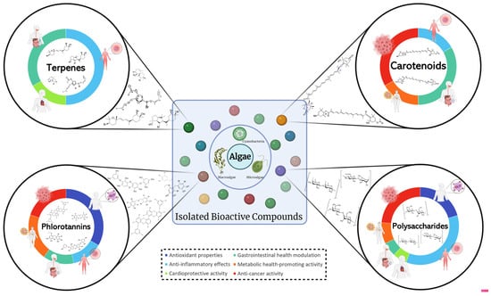
Figure 2.
Overview of the predominant chemical classes (terpenes, carotenoids, polysaccharides, and phlorotannins) of isolated bioactive compounds from algae in cell and in vivo studies found in this review, and their respective human health-related bioactivities (antioxidant properties, anti-inflammatory effects, cardioprotective activity, gastrointestinal health modulation, metabolic health-promoting activity, and anti-cancer activity). Created with BioRender.com (accessed on 4 March 2024).
As shown throughout this review, algae-derived compounds have a great potential to exert an array of health-related bioactivities; however, their effectiveness and applicability depends on several parameters, such as digestibility and bio accessibility, as not all compounds can be metabolized by our organism and digestion might compromise the previously identified bioactivity. This is a special concern for when the whole biomass is used, where the cell wall may prevent compounds from exerting their biological function in the human body [248]. In this review, this concern was only briefly addressed in one research paper [78]. Therefore, additional studies evaluating the effectiveness of compounds are essential to strengthen the understanding of algal compounds for practical use [248].
One of the greatest challenges for the application of algae-derived compounds in human health is their introduction into the consumer market, as, if not traditionally used, these species must undergo the process of novel food approval by regulatory authorities. This is a special concern for microalgae, as from the plethora of microalgae species only a handful of strains are approved for human consumption in the EU. Currently, twenty-four species of seaweed (which did not have to go through the “novel foods” process) are considered as food within the EU in addition to five microalgae species: Aphanizomenon flosaquae, Arthrospira platensis, and three Chlorella strains (Chlorella luteoviridis, Chlorella pyrenoidosa, and Chlorella vulgaris). Only five additional (microalgal) species have been approved as food ingredients and are considered “novel food”: Odontella aurita, Ulkenia sp. (oil extract), Tetraselmis chui, Haematoccocus pluvialis (astaxanthin), and Schizochytrium sp. (oil extract). The application for new algal strains as “novel foods” is a timely and expensive endeavor. Therefore, companies would rather invest their research and development in species of algae that are already approved for human consumption, leaving many species and their compounds commercially unexplored and their activity in academia [249]. This is proven by the limited number of commercial products included in this review (n = 3): Fuco Pets HeartFight® [136], of macroalgal origin, with cardioprotective activity; TetraSOD® [189], from Tetraselmis chui, with metabolic benefits; and Gdue© [145], a brown algae extract and chromium picolinate blend with liver health-promoting ability. However, in February 2024, several algae species (21 macroalgae and 11 microalgae) were added to the EU Novel Food Catalogue, some as food ingredients and others as food supplements [250].
The limited number of microalgae studies in this review may be attributed to several factors: the higher difficulty and cost associated with cultivating microalgae compared to collecting seaweed biomass [241]; microalgae having a shorter research history than seaweeds [236]; the lower number of approved microalgae strains for consumption [249]; and the specific inclusion criteria of the review (limited to marine-origin algae, excluding in chemico studies, only including studies with results in the form of IC50 values in in vitro assays, and focusing on specific algal bioactivities related to human health). These inclusion criteria were chosen according to the pipeline for drug development, whereas preclinical studies with in vitro, ex vivo, and in vivo models, and clinical development with humans (Phase I, II, and III trials) are the most advanced type studies available before launching for the consumer market [251], and therefore include the algae compounds which are closest to practical applicability.
Therefore, further research is needed to address these challenges, improve methodologies, and enhance our understanding of the complex relationship between algae and human health, fostering the development of safe and effective health-promoting products which can enter the consumer market.
5. Conclusions
5.1. Summary of Key Findings on Algal Bioactivities Related to Human Health
This comprehensive review presents an overview of recent research conducted in the past five years on the impact of algae-derived compounds on human health. It provides a summary of the bioactivity and potential therapeutic properties of 92 different strains of algae, including 13 microalgae and 79 macroalgae. However, out of the 50,000 microalgae and approximately 12,000 seaweed species that have been described, many have never been studied and constitute an untapped potential that is yet to be discovered.
Out of the numerous strains discussed in this study, four macroalgal strains demonstrated multiple health-related bioactivities in in vivo models. Notably, three of the macroalgal species are classified as Phaeophiceae (brown algae) and one as chlorophyte (green algae). Ecklonia maxima presented antioxidant [60] and anti-inflammatory activity [113], leading to increased zebrafish larvae’s survival rate and health. Ishige okamurae also presented dual activity by improving both gastrointestinal (by downregulating lipogenesis, decreasing liver inflammation, and regulating lipid metabolism) [147] and metabolic health (by modulating insulin resistance and sensitivity) [187]. Saccharina japonica showed both anti-inflammatory (leading to increased zebrafish larvae’s survival rate and health) [95] and metabolic health-promoting activities (evidenced by a decrease in dyslipidemia, liver, and renal injury, while increasing insulin sensitivity and gut microbiota health) [184]. The polysaccharides of chlorophyte seaweed Ulva lactuca, also known as “sea lettuce”, were shown to possess antioxidant (by decreasing oxidative stress-induced injuries) [59] and anti-cancer activity (through various mechanisms, such as the direct killing of tumorous cells, the inhibition of angiogenesis, and a decrease in inflammation) [221].
These four stains, along with the dozens that are presented in this review, are merely an example of the unexplored potential found in algae. Anti-oxidative, anti-inflammatory, and anti-cancerous activities, along with beneficial effects for gastrointestinal and cardiovascular health, the potential of assisting with diabetes and obesity, and possibly numerous other bioactivities, could place algae in the front line of the future of natural functional ingredients, foods, and additives.
5.2. Implications for Future Research and Applications
Overall, several bioactivities from algae-derived compounds are shown to be effective against oxidative stress and inflammation, both of which are inextricably linked to many diseases that plague modern societies and thus play an important role in health promotion. However, before considering human consumption, the in vivo effectiveness, digestibility, and safety of these extracts must be thoroughly assessed. Other issues described in this review, such as non-standardized extractions, variable biochemical composition of algae, the scarcity of isolated bioactive compounds, and the legislation required to market these products, must be addressed for further practical applications.
While these lines are being written, hundreds of novel compounds, extracts, and whole algal biomass are being investigated as the next ingredient in food, nutraceuticals, cosmetics, and even pharmaceuticals. The big question remains: how many of these bioactive ingredients will reach the end user?
Supplementary Materials
The following supporting information can be downloaded at: https://www.mdpi.com/article/10.3390/molecules29081900/s1, Figure S1: Chemical classification of isolated algal compounds used in cell and in vivo studies.
Author Contributions
Conceptualization, M.S. and L.B.; methodology, M.S.; writing—original draft preparation, M.S.; writing—review and editing, L.B., J.V. and D.A.; supervision, L.B., J.V. and D.A.; project administration, L.B. and D.A.; funding acquisition, L.B., J.V. and D.A. All authors have read and agreed to the published version of the manuscript.
Funding
This study received Portuguese national funds from FCT—Foundation for Science and Technology through projects UIDB/04326/2020 (DOI:10.54499/UIDB/04326/2020), UIDP/04326/2020 (DOI:10.54499/UIDP/04326/2020) and LA/P/0101/2020 (DOI:10.54499/LA/P/0101/2020) as well as doctoral fellowship 2022.12681.BDANA. Additionally, funding was provided by the European Union through the ALGAE4IBD project (grant agreement/EC/H2020/101000501/EU).
Conflicts of Interest
The authors declare no conflicts of interest.
References
- Rotter, A.; Bacu, A.; Barbier, M.; Bertoni, F.; Bones, A.M.; Cancela, M.L.; Carlsson, J.; Carvalho, M.F.; Cegłowska, M.; Dalay, M.C.; et al. A New Network for the Advancement of Marine Biotechnology in Europe and Beyond. Front. Mar. Sci. 2020, 7, 278. [Google Scholar] [CrossRef]
- Johnson, K.; Dalton, G.; Masters, I. Building Industries at Sea: ‘Blue Growth’ and the New Maritime Economy. In Building Industries at Sea: ‘Blue Growth’ and the New Maritime Economy; Johnson, K., Dalton, G., Masters, I., Eds.; River Publishers: Aalborg, Denmark, 2018; pp. 1–516. [Google Scholar]
- van der Westhuyzen, A.E.; Frolova, L.V.; Kornienko, A.; van Otterlo, W.A.L. Chapter Four—The Rigidins: Isolation, Bioactivity, and Total Synthesis—Novel Pyrrolo[2,3-d]Pyrimidine Analogues Using Multicomponent Reactions. In The Alkaloids: Chemistry and Biology; Knölker, H.-J., Ed.; Academic Press: Cambridge, MA, USA, 2018; Volume 79, pp. 191–220. [Google Scholar]
- Lu, W.Y.; Li, H.J.; Li, Q.Y.; Wu, Y.C. Application of marine natural products in drug research. Bioorganic Med. Chem. 2021, 35, 116058. [Google Scholar] [CrossRef] [PubMed]
- Lyu, C.; Chen, T.; Qiang, B.; Liu, N.; Wang, H.; Zhang, L.; Liu, Z. CMNPD: A comprehensive marine natural products database towards facilitating drug discovery from the ocean. Nucleic Acids Res. 2021, 49, D509–D515. [Google Scholar] [CrossRef] [PubMed]
- Barsanti, L.; Gualtieri, P. Algae: Anatomy, Biochemistry, and Biotechnology, 2nd ed.; CRC Press: Boca Raton, FL, USA, 2014. [Google Scholar]
- Pereira, L. Macroalgae. Encyclopedia 2021, 1, 177–188. [Google Scholar] [CrossRef]
- Dini, I. The Potential of Algae in the Nutricosmetic Sector. Molecules 2023, 28, 4032. [Google Scholar] [CrossRef] [PubMed]
- Vieira, M.V.; Pastrana, L.M.; Fucinos, P. Microalgae Encapsulation Systems for Food, Pharmaceutical and Cosmetics Applications. Mar. Drugs 2020, 18, 644. [Google Scholar] [CrossRef] [PubMed]
- Rotter, A.; Barbier, M.; Bertoni, F.; Bones, A.M.; Cancela, M.L.; Carlsson, J.; Carvalho, M.F.; Cegłowska, M.; Chirivella-Martorell, J.; Conk Dalay, M.; et al. The Essentials of Marine Biotechnology. Front. Mar. Sci. 2021, 8, 629629. [Google Scholar] [CrossRef]
- Conde, T.A.; Neves, B.F.; Couto, D.; Melo, T.; Neves, B.; Costa, M.; Silva, J.; Domingues, P.; Domingues, M.R. Microalgae as Sustainable Bio-Factories of Healthy Lipids: Evaluating Fatty Acid Content and Antioxidant Activity. Mar. Drugs 2021, 19, 357. [Google Scholar] [CrossRef] [PubMed]
- Quiterio, E.; Soares, C.; Ferraz, R.; Delerue-Matos, C.; Grosso, C. Marine Health-Promoting Compounds: Recent Trends for Their Characterization and Human Applications. Foods 2021, 10, 3100. [Google Scholar] [CrossRef]
- Silva, M.; Kamberovic, F.; Uota, S.T.; Kovan, I.-M.; Viegas, C.S.B.; Simes, D.C.; Gangadhar, K.N.; Varela, J.; Barreira, L. Microalgae as Potential Sources of Bioactive Compounds for Functional Foods and Pharmaceuticals. Appl. Sci. 2022, 12, 5877. [Google Scholar] [CrossRef]
- Richards, N.C.; Gouda, H.N.; Durham, J.; Rampatige, R.; Rodney, A.; Whittaker, M. Disability, noncommunicable disease and health information. Bull. World Health Organ. 2016, 94, 230–232. [Google Scholar] [CrossRef] [PubMed]
- World Health Organization. Noncommunicable Diseases. Available online: https://www.who.int/news-room/fact-sheets/detail/noncommunicable-diseases (accessed on 6 December 2023).
- Shelton, C.D.; Byndloss, M.X. Gut Epithelial Metabolism as a Key Driver of Intestinal Dysbiosis Associated with Noncommunicable Diseases. Infect. Immun. 2020, 88, e00939-19. [Google Scholar] [CrossRef] [PubMed]
- Hosseinkhani, F.; Heinken, A.; Thiele, I.; Lindenburg, P.W.; Harms, A.C.; Hankemeier, T. The contribution of gut bacterial metabolites in the human immune signaling pathway of non-communicable diseases. Gut Microbes 2021, 13, 1882927. [Google Scholar] [CrossRef] [PubMed]
- Borgoni, S.; Kudryashova, K.S.; Burka, K.; de Magalhaes, J.P. Targeting immune dysfunction in aging. Ageing Res. Rev. 2021, 70, 101410. [Google Scholar] [CrossRef] [PubMed]
- Nediani, C.; Dinu, M. Oxidative Stress and Inflammation as Targets for Novel Preventive and Therapeutic Approaches in Non-Communicable Diseases II. Antioxidants 2022, 11, 824. [Google Scholar] [CrossRef] [PubMed]
- Seyedsadjadi, N.; Grant, R. The Potential Benefit of Monitoring Oxidative Stress and Inflammation in the Prevention of Non-Communicable Diseases (NCDs). Antioxidants 2020, 10, 15. [Google Scholar] [CrossRef] [PubMed]
- Bindu, S.; Mazumder, S.; Bandyopadhyay, U. Non-steroidal anti-inflammatory drugs (NSAIDs) and organ damage: A current perspective. Biochem. Pharmacol. 2020, 180, 114147. [Google Scholar] [CrossRef]
- Jiang, H.; Zuo, J.; Li, B.; Chen, R.; Luo, K.; Xiang, X.; Lu, S.; Huang, C.; Liu, L.; Tang, J.; et al. Drug-induced oxidative stress in cancer treatments: Angel or devil? Redox Biol. 2023, 63, 102754. [Google Scholar] [CrossRef] [PubMed]
- Perera, R.; Herath, K.; Sanjeewa, K.K.A.; Jayawardena, T.U. Recent Reports on Bioactive Compounds from Marine Cyanobacteria in Relation to Human Health Applications. Life 2023, 13, 1411. [Google Scholar] [CrossRef]
- Jimenez-Lopez, C.; Pereira, A.G.; Lourenco-Lopes, C.; Garcia-Oliveira, P.; Cassani, L.; Fraga-Corral, M.; Prieto, M.A.; Simal-Gandara, J. Main bioactive phenolic compounds in marine algae and their mechanisms of action supporting potential health benefits. Food Chem. 2021, 341, 128262. [Google Scholar] [CrossRef]
- Geada, P.; Moreira, C.; Silva, M.; Nunes, R.; Madureira, L.; Rocha, C.M.R.; Pereira, R.N.; Vicente, A.A.; Teixeira, J.A. Algal proteins: Production strategies and nutritional and functional properties. Bioresour. Technol. 2021, 332, 125125. [Google Scholar] [CrossRef] [PubMed]
- Ferdous, U.T.; Yusof, Z.N.B. Medicinal Prospects of Antioxidants From Algal Sources in Cancer Therapy. Front. Pharmacol. 2021, 12, 593116. [Google Scholar] [CrossRef] [PubMed]
- Sun, Z.; Dai, Z.; Zhang, W.; Fan, S.; Liu, H.; Liu, R.; Zhao, T. Antiobesity, Antidiabetic, Antioxidative, and Antihyperlipidemic Activities of Bioactive Seaweed Substances. In Bioactive Seaweeds for Food Applications; Qin, Y., Ed.; Academic Press: Cambridge, MA, USA, 2018; pp. 239–253. [Google Scholar]
- Page, M.J.; McKenzie, J.E.; Bossuyt, P.M.; Boutron, I.; Hoffmann, T.C.; Mulrow, C.D.; Shamseer, L.; Tetzlaff, J.M.; Akl, E.A.; Brennan, S.E.; et al. The PRISMA 2020 statement: An updated guideline for reporting systematic reviews. BMJ 2021, 372, n71. [Google Scholar] [CrossRef] [PubMed]
- Pizzino, G.; Irrera, N.; Cucinotta, M.; Pallio, G.; Mannino, F.; Arcoraci, V.; Squadrito, F.; Altavilla, D.; Bitto, A. Oxidative Stress: Harms and Benefits for Human Health. Oxid. Med. Cell. Longev. 2017, 2017, 8416763. [Google Scholar] [CrossRef] [PubMed]
- Shields, H.J.; Traa, A.; Van Raamsdonk, J.M. Beneficial and Detrimental Effects of Reactive Oxygen Species on Lifespan: A Comprehensive Review of Comparative and Experimental Studies. Front. Cell Dev. Biol. 2021, 9, 628157. [Google Scholar] [CrossRef] [PubMed]
- Redza-Dutordoir, M.; Averill-Bates, D.A. Activation of apoptosis signalling pathways by reactive oxygen species. Biochim. Biophys. Acta 2016, 1863, 2977–2992. [Google Scholar] [CrossRef] [PubMed]
- Dhanasekaran, D.N.; Reddy, E.P. JNK-signaling: A multiplexing hub in programmed cell death. Genes Cancer 2017, 8, 682–694. [Google Scholar] [CrossRef] [PubMed]
- Hammad, M.; Raftari, M.; Cesario, R.; Salma, R.; Godoy, P.; Emami, S.N.; Haghdoost, S. Roles of Oxidative Stress and Nrf2 Signaling in Pathogenic and Non-Pathogenic Cells: A Possible General Mechanism of Resistance to Therapy. Antioxidants 2023, 12, 1371. [Google Scholar] [CrossRef] [PubMed]
- Le, B.; Golokhvast, K.S.; Yang, S.H.; Sun, S. Optimization of Microwave-Assisted Extraction of Polysaccharides from Ulva pertusa and Evaluation of Their Antioxidant Activity. Antioxidants 2019, 8, 129. [Google Scholar] [CrossRef]
- Jerkovic, I.; Cikos, A.M.; Babic, S.; Cizmek, L.; Bojanic, K.; Aladic, K.; Ul’yanovskii, N.V.; Kosyakov, D.S.; Lebedev, A.T.; Coz-Rakovac, R.; et al. Bioprospecting of Less-Polar Constituents from Endemic Brown Macroalga Fucus virsoides J. Agardh from the Adriatic Sea and Targeted Antioxidant Effects In Vitro and In Vivo (Zebrafish Model). Mar. Drugs 2021, 19, 235. [Google Scholar] [CrossRef]
- Wang, L.; Fu, X.; Hyun, J.; Xu, J.; Gao, X.; Jeon, Y.J. In Vitro and In Vivo Protective Effects of Agaro-Oligosaccharides against Hydrogen Peroxide-Stimulated Oxidative Stress. Polymers 2023, 15, 1612. [Google Scholar] [CrossRef] [PubMed]
- Dong, X.; Bai, Y.; Xu, Z.; Shi, Y.; Sun, Y.; Janaswamy, S.; Yu, C.; Qi, H. Phlorotannins from Undaria pinnatifida Sporophyll: Extraction, Antioxidant, and Anti-Inflammatory Activities. Mar. Drugs 2019, 17, 434. [Google Scholar] [CrossRef] [PubMed]
- Park, C.; Cha, H.J.; Hwangbo, H.; Ji, S.Y.; Kim, D.H.; Kim, M.Y.; Bang, E.; Hong, S.H.; Kim, S.O.; Jeong, S.J.; et al. Phloroglucinol Inhibits Oxidative-Stress-Induced Cytotoxicity in C2C12 Murine Myoblasts through Nrf-2-Mediated Activation of HO-1. Int. J. Mol. Sci. 2023, 24, 4637. [Google Scholar] [CrossRef] [PubMed]
- Park, C.; Lee, H.; Park, S.-H.; Hong, S.H.; Song, K.S.; Cha, H.-J.; Kim, G.-Y.; Chang, Y.-C.; Kim, S.; Kim, H.-S.; et al. Indole-6-carboxaldehyde prevents oxidative stress-induced mitochondrial dysfunction, DNA damage and apoptosis in C2C12 skeletal myoblasts by regulating the ROS-AMPK signaling pathway. Mol. Cell. Toxicol. 2020, 16, 455–467. [Google Scholar] [CrossRef]
- Niu, T.; Fu, G.; Zhou, J.; Han, H.; Chen, J.; Wu, W.; Chen, H. Floridoside Exhibits Antioxidant Properties by Activating HO-1 Expression via p38/ERK MAPK Pathway. Mar. Drugs 2020, 18, 105. [Google Scholar] [CrossRef] [PubMed]
- Pittayapruek, P.; Meephansan, J.; Prapapan, O.; Komine, M.; Ohtsuki, M. Role of Matrix Metalloproteinases in Photoaging and Photocarcinogenesis. Int. J. Mol. Sci. 2016, 17, 868. [Google Scholar] [CrossRef] [PubMed]
- Dong, H.; Liu, M.; Wang, L.; Liu, Y.; Lu, X.; Stagos, D.; Lin, X.; Liu, M. Bromophenol Bis (2,3,6-Tribromo-4,5-dihydroxybenzyl) Ether Protects HaCaT Skin Cells from Oxidative Damage via Nrf2-Mediated Pathways. Antioxidants 2021, 10, 1436. [Google Scholar] [CrossRef]
- Ryu, Y.S.; Fernando, P.; Kang, K.A.; Piao, M.J.; Zhen, A.X.; Kang, H.K.; Koh, Y.S.; Hyun, J.W. Marine Compound 3-bromo-4,5-dihydroxybenzaldehyde Protects Skin Cells against Oxidative Damage via the Nrf2/HO-1 Pathway. Mar. Drugs 2019, 17, 234. [Google Scholar] [CrossRef] [PubMed]
- Dong, H.; Wang, L.; Guo, M.; Stagos, D.; Giakountis, A.; Trachana, V.; Lin, X.; Liu, Y.; Liu, M. Antioxidant and Anticancer Activities of Synthesized Methylated and Acetylated Derivatives of Natural Bromophenols. Antioxidants 2022, 11, 786. [Google Scholar] [CrossRef]
- Park, C.; Cha, H.J.; Hong, S.H.; Kim, G.Y.; Kim, S.; Kim, H.S.; Kim, B.W.; Jeon, Y.J.; Choi, Y.H. Protective Effect of Phloroglucinol on Oxidative Stress-Induced DNA Damage and Apoptosis through Activation of the Nrf2/HO-1 Signaling Pathway in HaCaT Human Keratinocytes. Mar. Drugs 2019, 17, 225. [Google Scholar] [CrossRef]
- He, Y.L.; Xiao, Z.; Yang, S.; Zhou, C.; Sun, S.; Hong, P.; Qian, Z.J. A Phlorotanin, 6,6′-bieckol from Ecklonia cava, Against Photoaging by Inhibiting MMP-1, -3 and -9 Expression on UVB-induced HaCaT Keratinocytes. Photochem. Photobiol. 2022, 98, 1131–1139. [Google Scholar] [CrossRef]
- Freitas, R.; Martins, A.; Silva, J.; Alves, C.; Pinteus, S.; Alves, J.; Teodoro, F.; Ribeiro, H.M.; Goncalves, L.; Petrovski, Z.; et al. Highlighting the Biological Potential of the Brown Seaweed Fucus spiralis for Skin Applications. Antioxidants 2020, 9, 611. [Google Scholar] [CrossRef]
- Chen, B.; Chen, H.; Qu, H.; Qiao, K.; Xu, M.; Wu, J.; Su, Y.; Shi, Y.; Liu, Z.; Wang, Q. Photoprotective effects of Sargassum thunbergii on ultraviolet B-induced mouse L929 fibroblasts and zebrafish. BMC Complement. Med. Ther. 2022, 22, 144. [Google Scholar] [CrossRef]
- Alboofetileh, M.; Rezaei, M.; Hamzeh, A.; Tabarsa, M.; Cravotto, G. Cellular antioxidant and emulsifying activities of fucoidan extracted from Nizamuddinia zanardinii using different green extraction methods. J. Food Process. Preserv. 2022, 46, e17238. [Google Scholar] [CrossRef]
- Begum, S.; Hemalatha, S. Gelidiella acerosa Compounds Target NFkappaB Cascade in Lung Adenocarcinoma. Appl. Biochem. Biotechnol. 2022, 194, 1566–1579. [Google Scholar] [CrossRef]
- Wang, L.; Jayawardena, T.U.; Yang, H.W.; Lee, H.G.; Kang, M.C.; Sanjeewa, K.K.A.; Oh, J.Y.; Jeon, Y.J. Isolation, Characterization, and Antioxidant Activity Evaluation of a Fucoidan from an Enzymatic Digest of the Edible Seaweed, Hizikia fusiforme. Antioxidants 2020, 9, 363. [Google Scholar] [CrossRef] [PubMed]
- Jayawardena, T.U.; Wang, L.; Sanjeewa, K.K.A.; Kang, S.I.; Lee, J.S.; Jeon, Y.J. Antioxidant Potential of Sulfated Polysaccharides from Padina boryana; Protective Effect against Oxidative Stress in In Vitro and In Vivo Zebrafish Model. Mar. Drugs 2020, 18, 212. [Google Scholar] [CrossRef] [PubMed]
- Dai, Y.-L.; Kim, G.H.; Kang, M.-C.; Jeon, Y.-J. Protective effects of extracts from six local strains of Pyropia yezoensis against oxidative damage in vitro and in zebrafish model. Algae 2020, 35, 189–200. [Google Scholar] [CrossRef]
- Wang, L.; Oh, J.Y.; Hwang, J.; Ko, J.Y.; Jeon, Y.J.; Ryu, B. In Vitro and In Vivo Antioxidant Activities of Polysaccharides Isolated from Celluclast-Assisted Extract of an Edible Brown Seaweed, Sargassum fulvellum. Antioxidants 2019, 8, 493. [Google Scholar] [CrossRef]
- Oh, J.Y.; Kim, E.A.; Kang, S.I.; Yang, H.W.; Ryu, B.; Wang, L.; Lee, J.S.; Jeon, Y.J. Protective Effects of Fucoidan Isolated from Celluclast-Assisted Extract of Undaria pinnatifida Sporophylls against AAPH-Induced Oxidative Stress In Vitro and In Vivo Zebrafish Model. Molecules 2020, 25, 2361. [Google Scholar] [CrossRef]
- Machado, I.F.; Miranda, R.G.; Dorta, D.J.; Rolo, A.P.; Palmeira, C.M. Targeting Oxidative Stress with Polyphenols to Fight Liver Diseases. Antioxidants 2023, 12, 1212. [Google Scholar] [CrossRef] [PubMed]
- Gyuraszova, M.; Gurecka, R.; Babickova, J.; Tothova, L. Oxidative Stress in the Pathophysiology of Kidney Disease: Implications for Noninvasive Monitoring and Identification of Biomarkers. Oxid. Med. Cell Longev. 2020, 2020, 5478708. [Google Scholar] [CrossRef] [PubMed]
- Guermazi, W.; Boukhris, S.; Annabi-Trabelsi, N.; Rebai, T.; Sellami-Kamoun, A.; Aldahmash, W.; Plavan, G.I.; Harrath, A.H.; Ayadi, H. Hyperhalophilic Diatom Extract Protects against Lead-Induced Oxidative Stress in Rats and Human HepG2 and HEK293 Cells. Pharmaceuticals 2023, 16, 875. [Google Scholar] [CrossRef] [PubMed]
- Yang, Q.; Jiang, Y.; Fu, S.; Shen, Z.; Zong, W.; Xia, Z.; Zhan, Z.; Jiang, X. Protective Effects of Ulva lactuca Polysaccharide Extract on Oxidative Stress and Kidney Injury Induced by D-Galactose in Mice. Mar. Drugs 2021, 19, 539. [Google Scholar] [CrossRef] [PubMed]
- Wang, L.; Jayawardena, T.U.; Yang, H.W.; Lee, H.G.; Jeon, Y.J. The Potential of Sulfated Polysaccharides Isolated from the Brown Seaweed Ecklonia maxima in Cosmetics: Antioxidant, Anti-melanogenesis, and Photoprotective Activities. Antioxidants 2020, 9, 724. [Google Scholar] [CrossRef] [PubMed]
- Furman, D.; Campisi, J.; Verdin, E.; Carrera-Bastos, P.; Targ, S.; Franceschi, C.; Ferrucci, L.; Gilroy, D.W.; Fasano, A.; Miller, G.W.; et al. Chronic inflammation in the etiology of disease across the life span. Nat. Med. 2019, 25, 1822–1832. [Google Scholar] [CrossRef] [PubMed]
- Lugrin, J.; Rosenblatt-Velin, N.; Parapanov, R.; Liaudet, L. The role of oxidative stress during inflammatory processes. Biol. Chem. 2014, 395, 203–230. [Google Scholar] [CrossRef] [PubMed]
- Chen, L.; Deng, H.; Cui, H.; Fang, J.; Zuo, Z.; Deng, J.; Li, Y.; Wang, X.; Zhao, L. Inflammatory responses and inflammation-associated diseases in organs. Oncotarget 2018, 9, 7204–7218. [Google Scholar] [CrossRef] [PubMed]
- Gao, W.; Guo, L.; Yang, Y.; Wang, Y.; Xia, S.; Gong, H.; Zhang, B.K.; Yan, M. Dissecting the Crosstalk Between Nrf2 and NF-kappaB Response Pathways in Drug-Induced Toxicity. Front. Cell Dev. Biol. 2021, 9, 809952. [Google Scholar] [CrossRef]
- Saha, S.; Muller, D.; Clark, A.G. Mechanosensory feedback loops during chronic inflammation. Front. Cell Dev. Biol. 2023, 11, 1225677. [Google Scholar] [CrossRef]
- Wang, B.; Wu, L.; Chen, J.; Dong, L.; Chen, C.; Wen, Z.; Hu, J.; Fleming, I.; Wang, D.W. Metabolism pathways of arachidonic acids: Mechanisms and potential therapeutic targets. Signal Transduct. Target. Ther. 2021, 6, 94. [Google Scholar] [CrossRef] [PubMed]
- Wong, R.S.Y. Role of Nonsteroidal Anti-Inflammatory Drugs (NSAIDs) in Cancer Prevention and Cancer Promotion. Adv. Pharmacol. Sci. 2019, 2019, 3418975. [Google Scholar] [CrossRef] [PubMed]
- Antony, T.; Chakraborty, K. First report of antioxidative 2H-chromenyl derivatives from the intertidal red seaweed Gracilaria salicornia as potential anti-inflammatory agents. Nat. Prod. Res. 2020, 34, 3470–3482. [Google Scholar] [CrossRef] [PubMed]
- Chakraborty, K.; Antony, T. First report of spiro-compounds from marine macroalga Gracilaria salicornia: Prospective natural anti-inflammatory agents attenuate 5-lipoxygenase and cyclooxygenase-2. Nat. Prod. Res. 2021, 35, 770–781. [Google Scholar] [CrossRef] [PubMed]
- Antony, T.; Chakraborty, K. First report of antioxidant abeo-labdane type diterpenoid from intertidal red seaweed Gracilaria salicornia with 5-lipoxygenase inhibitory potential. Nat. Prod. Res. 2020, 34, 1409–1416. [Google Scholar] [CrossRef] [PubMed]
- Thambi, A.; Chakraborty, K. Anti-inflammatory decurrencyclics A-B, two undescribed nor-dammarane triterpenes from triangular sea bell Turbinaria decurrens. Nat. Prod. Res. 2023, 37, 713–724. [Google Scholar] [CrossRef] [PubMed]
- Amaro, H.M.; Barros, R.; Tavares, T.; Almeida, R.; Pinto, I.S.; Malcata, F.X.; Guedes, A.C. Gloeothece sp.—Exploiting a New Source of Antioxidant, Anti-Inflammatory, and Antitumor Agents. Mar. Drugs 2021, 19, 623. [Google Scholar] [CrossRef] [PubMed]
- Antony, T.; Chakraborty, K. Pharmacological Properties of Seaweeds against Progressive Lifestyle Diseases. J. Aquat. Food Prod. Technol. 2019, 28, 1092–1104. [Google Scholar] [CrossRef]
- Samarakoon, K.W.; Ko, J.Y.; Lee, J.H.; Jeon, Y.J. Anti-inflammatory activity of nonyl 8-acetoxy-6-methyloctanoate, isolated from the cultured marine diatom, Phaeodactylum tricornutum: Mediated via suppression of inflammatory mediators in LPS-stimulated RAW 264.7 macrophages. J. Natl. Sci. Found. Sri Lanka 2022, 50, 685–693. [Google Scholar] [CrossRef]
- Bigagli, E.; D’Ambrosio, M.; Cinci, L.; Niccolai, A.; Biondi, N.; Rodolfi, L.; Dos Santos Nascimiento, L.B.; Tredici, M.R.; Luceri, C. A Comparative In Vitro Evaluation of the Anti-Inflammatory Effects of a Tisochrysis lutea Extract and Fucoxanthin. Mar. Drugs 2021, 19, 334. [Google Scholar] [CrossRef]
- Sanjeewa, K.K.A.; Fernando, I.P.S.; Kim, S.-Y.; Kim, W.-S.; Ahn, G.; Jee, Y.; Jeon, Y.-J. Ecklonia cava (Laminariales) and Sargassum horneri (Fucales) synergistically inhibit the lipopolysaccharide-induced inflammation via blocking NF-κB and MAPK pathways. Algae 2019, 34, 45–56. [Google Scholar] [CrossRef]
- Saraswati; Giriwono, P.E.; Iskandriati, D.; Andarwulan, N. Screening of In-Vitro Anti-Inflammatory and Antioxidant Activity of Sargassum ilicifolium Crude Lipid Extracts from Different Coastal Areas in Indonesia. Mar. Drugs 2021, 19, 252. [Google Scholar] [CrossRef] [PubMed]
- Catarino, M.D.; Circuncisao, A.R.; Neves, B.; Marcal, C.; Silva, A.M.S.; Cruz, M.T.; Cardoso, S.M. Impact of Gastrointestinal Digestion on the Anti-Inflammatory Properties of Phlorotannins from Himanthalia elongata. Antioxidants 2022, 11, 1518. [Google Scholar] [CrossRef]
- He, X.; Wan, F.; Su, W.; Xie, W. Research Progress on Skin Aging and Active Ingredients. Molecules 2023, 28, 5556. [Google Scholar] [CrossRef]
- Jayasinghe, A.M.K.; Han, E.J.; Kirindage, K.; Fernando, I.P.S.; Kim, E.A.; Kim, J.; Jung, K.; Kim, K.N.; Heo, S.J.; Ahn, G. 3-Bromo-4,5-dihydroxybenzaldehyde Isolated from Polysiphonia morrowii Suppresses TNF-alpha/IFN-gamma-Stimulated Inflammation and Deterioration of Skin Barrier in HaCaT Keratinocytes. Mar. Drugs 2022, 20, 563. [Google Scholar] [CrossRef] [PubMed]
- Kraokaew, P.; Manohong, P.; Prasertsuksri, P.; Jattujan, P.; Niamnont, N.; Tamtin, M.; Sobhon, P.; Meemon, K. Ethyl Acetate Extract of Marine Algae, Halymenia durvillei, Provides Photoprotection against UV-Exposure in L929 and HaCaT Cells. Mar. Drugs 2022, 20, 707. [Google Scholar] [CrossRef] [PubMed]
- Kim, T.H.; Heo, S.Y.; Han, J.S.; Jung, W.K. Anti-inflammatory effect of polydeoxyribonucleotides (PDRN) extracted from red alga (Porphyra sp.) (Ps-PDRN) in RAW 264.7 macrophages stimulated with Escherichia coli lipopolysaccharides: A comparative study with commercial PDRN. Cell Biochem. Funct. 2023, 41, 889–897. [Google Scholar] [CrossRef]
- Kirindage, K.; Jayasinghe, A.M.K.; Han, E.J.; Jee, Y.; Kim, H.J.; Do, S.G.; Fernando, I.P.S.; Ahn, G. Fucosterol Isolated from Dietary Brown Alga Sargassum horneri Protects TNF-alpha/IFN-gamma-Stimulated Human Dermal Fibroblasts via Regulating Nrf2/HO-1 and NF-kappaB/MAPK Pathways. Antioxidants 2022, 11, 1429. [Google Scholar] [CrossRef]
- Kurniawan, R.; Nurkolis, F.; Taslim, N.A.; Subali, D.; Surya, R.; Gunawan, W.B.; Alisaputra, D.; Mayulu, N.; Salindeho, N.; Kim, B. Carotenoids Composition of Green Algae Caulerpa racemosa and Their Antidiabetic, Anti-Obesity, Antioxidant, and Anti-Inflammatory Properties. Molecules 2023, 28, 3267. [Google Scholar] [CrossRef]
- De La Fuente, G.; Fontana, M.; Asnaghi, V.; Chiantore, M.; Mirata, S.; Salis, A.; Damonte, G.; Scarfi, S. The Remarkable Antioxidant and Anti-Inflammatory Potential of the Extracts of the Brown Alga Cystoseira amentacea var. stricta. Mar. Drugs 2020, 19, 2. [Google Scholar] [CrossRef]
- Daskalaki, M.G.; Bafiti, P.; Kikionis, S.; Laskou, M.; Roussis, V.; Ioannou, E.; Kampranis, S.C.; Tsatsanis, C. Disulfides from the Brown Alga Dictyopteris membranacea Suppress M1 Macrophage Activation by Inducing AKT and Suppressing MAPK/ERK Signaling Pathways. Mar. Drugs 2020, 18, 527. [Google Scholar] [CrossRef]
- Jung, J.I.; Kim, S.; Baek, S.M.; Choi, S.I.; Kim, G.H.; Imm, J.Y. Ecklonia cava Extract Exerts Anti-Inflammatory Effect in Human Gingival Fibroblasts and Chronic Periodontitis Animal Model by Suppression of Pro-Inflammatory Cytokines and Chemokines. Foods 2021, 10, 1656. [Google Scholar] [CrossRef]
- Tammam, M.A.; Daskalaki, M.G.; Tsoureas, N.; Kolliniati, O.; Mahdy, A.; Kampranis, S.C.; Tsatsanis, C.; Roussis, V.; Ioannou, E. Secondary Metabolites with Anti-Inflammatory Activity from Laurencia majuscula Collected in the Red Sea. Mar. Drugs 2023, 21, 79. [Google Scholar] [CrossRef]
- Jayawardena, T.U.; Sanjeewa, K.K.A.; Lee, H.G.; Nagahawatta, D.P.; Yang, H.W.; Kang, M.C.; Jeon, Y.J. Particulate Matter-Induced Inflammation/Oxidative Stress in Macrophages: Fucosterol from Padina boryana as a Potent Protector, Activated via NF-kappaB/MAPK Pathways and Nrf2/HO-1 Involvement. Mar. Drugs 2020, 18, 628. [Google Scholar] [CrossRef] [PubMed]
- Lee, C.W.; Ahn, Y.T.; Zhao, R.; Kim, Y.S.; Park, S.M.; Jung, D.H.; Kim, J.K.; Kim, H.W.; Kim, S.C.; An, W.G. Inhibitory Effects of Porphyra tenera Extract on Oxidation and Inflammatory Responses. Evid. Based Complement. Altern. Med. 2021, 2021, 6650037. [Google Scholar] [CrossRef]
- Cuevas, B.; Arroba, A.I.; de Los Reyes, C.; Gomez-Jaramillo, L.; Gonzalez-Montelongo, M.C.; Zubia, E. Diterpenoids from the Brown Alga Rugulopteryx okamurae and Their Anti-Inflammatory Activity. Mar. Drugs 2021, 19, 677. [Google Scholar] [CrossRef] [PubMed]
- Cuevas, B.; Arroba, A.I.; de Los Reyes, C.; Zubia, E. Rugulopteryx-Derived Spatane, Secospatane, Prenylcubebane and Prenylkelsoane Diterpenoids as Inhibitors of Nitric Oxide Production. Mar. Drugs 2023, 21, 252. [Google Scholar] [CrossRef] [PubMed]
- Liyanage, N.M.; Lee, H.G.; Nagahawatta, D.P.; Jayawardhana, H.; Song, K.M.; Choi, Y.S.; Jeon, Y.J.; Kang, M.C. Fucoidan from Sargassum autumnale Inhibits Potential Inflammatory Responses via NF-kappaB and MAPK Pathway Suppression in Lipopolysaccharide-Induced RAW 264.7 Macrophages. Mar. Drugs 2023, 21, 374. [Google Scholar] [CrossRef]
- Han, E.J.; Jayawardena, T.U.; Jang, J.H.; Fernando, I.P.S.; Jee, Y.; Jeon, Y.J.; Lee, D.S.; Lee, J.M.; Yim, M.J.; Wang, L.; et al. Sargachromenol Purified from Sargassum horneri Inhibits Inflammatory Responses via Activation of Nrf2/HO-1 Signaling in LPS-Stimulated Macrophages. Mar. Drugs 2021, 19, 497. [Google Scholar] [CrossRef]
- Ye, J.; Chen, D.; Ye, Z.; Huang, Y.; Zhang, N.; Lui, E.M.K.; Xue, C.; Xiao, M. Fucoidan Isolated from Saccharina japonica Inhibits LPS-Induced Inflammation in Macrophages via Blocking NF-kappaB, MAPK and JAK-STAT Pathways. Mar. Drugs 2020, 18, 328. [Google Scholar] [CrossRef]
- Jayawardena, T.U.; Sanjeewa, K.K.A.; Nagahawatta, D.P.; Lee, H.G.; Lu, Y.A.; Vaas, A.; Abeytunga, D.T.U.; Nanayakkara, C.M.; Lee, D.S.; Jeon, Y.J. Anti-Inflammatory Effects of Sulfated Polysaccharide from Sargassum Swartzii in Macrophages via Blocking TLR/NF-Kappab Signal Transduction. Mar. Drugs 2020, 18, 601. [Google Scholar] [CrossRef] [PubMed]
- Jin, W.; Yang, L.; Yi, Z.; Fang, H.; Chen, W.; Hong, Z.; Zhang, Y.; Zhang, G.; Li, L. Anti-Inflammatory Effects of Fucoxanthinol in LPS-Induced RAW264.7 Cells through the NAAA-PEA Pathway. Mar. Drugs 2020, 18, 222. [Google Scholar] [CrossRef] [PubMed]
- Kim, S.Y.; Ahn, G.; Kim, H.S.; Je, J.G.; Kim, K.N.; Jeon, Y.J. Diphlorethohydroxycarmalol (DPHC) Isolated from the Brown Alga Ishige okamurae Acts on Inflammatory Myopathy as an Inhibitory Agent of TNF-alpha. Mar. Drugs 2020, 18, 529. [Google Scholar] [CrossRef] [PubMed]
- Ha, J.W.; Song, H.; Hong, S.S.; Boo, Y.C. Marine Alga Ecklonia cava Extract and Dieckol Attenuate Prostaglandin E2 Production in HaCaT Keratinocytes Exposed to Airborne Particulate Matter. Antioxidants 2019, 8, 190. [Google Scholar] [CrossRef] [PubMed]
- Ha, Y.; Lee, W.H.; Kim, J.K.; Jeon, H.K.; Lee, J.; Kim, Y.J. Polyopes affinis Suppressed IFN-gamma- and TNF-alpha-Induced Inflammation in Human Keratinocytes via Down-Regulation of the NF-kappaB and STAT1 Pathways. Molecules 2022, 27, 1836. [Google Scholar] [CrossRef] [PubMed]
- Ha, Y.; Lee, W.H.; Jeong, J.; Park, M.; Ko, J.Y.; Kwon, O.W.; Lee, J.; Kim, Y.J. Pyropia yezoensis Extract Suppresses IFN-Gamma- and TNF-Alpha-Induced Proinflammatory Chemokine Production in HaCaT Cells via the Down-Regulation of NF-kappaB. Nutrients 2020, 12, 1238. [Google Scholar] [CrossRef] [PubMed]
- Jayasinghe, A.M.K.; Kirindage, K.; Fernando, I.P.S.; Han, E.J.; Oh, G.W.; Jung, W.K.; Ahn, G. Fucoidan Isolated from Sargassum confusum Suppresses Inflammatory Responses and Oxidative Stress in TNF-alpha/IFN-gamma- Stimulated HaCaT Keratinocytes by Activating Nrf2/HO-1 Signaling Pathway. Mar. Drugs 2022, 20, 117. [Google Scholar] [CrossRef] [PubMed]
- Han, E.J.; Fernando, I.P.S.; Kim, H.S.; Lee, D.S.; Kim, A.; Je, J.G.; Seo, M.J.; Jee, Y.H.; Jeon, Y.J.; Kim, S.Y.; et al. (-)-Loliolide Isolated from Sargassum horneri Suppressed Oxidative Stress and Inflammation by Activating Nrf2/HO-1 Signaling in IFN-gamma/TNF-alpha-Stimulated HaCaT Keratinocytes. Antioxidants 2021, 10, 856. [Google Scholar] [CrossRef]
- Jayasinghe, A.M.K.; Kirindage, K.; Fernando, I.P.S.; Kim, K.N.; Oh, J.Y.; Ahn, G. The Anti-Inflammatory Effect of Low Molecular Weight Fucoidan from Sargassum siliquastrum in Lipopolysaccharide-Stimulated RAW 264.7 Macrophages via Inhibiting NF-kappaB/MAPK Signaling Pathways. Mar. Drugs 2023, 21, 347. [Google Scholar] [CrossRef]
- Liberti, D.; Imbimbo, P.; Giustino, E.; D’Elia, L.; Silva, M.; Barreira, L.; Monti, D.M. Shedding Light on the Hidden Benefit of Porphyridium cruentum Culture. Antioxidants 2023, 12, 337. [Google Scholar] [CrossRef]
- Stiefvatter, L.; Frick, K.; Lehnert, K.; Vetter, W.; Montoya-Arroyo, A.; Frank, J.; Schmid-Staiger, U.; Bischoff, S.C. Potentially Beneficial Effects on Healthy Aging by Supplementation of the EPA-Rich Microalgae Phaeodactylum tricornutum or Its Supernatant—A Randomized Controlled Pilot Trial in Elderly Individuals. Mar. Drugs 2022, 20, 716. [Google Scholar] [CrossRef] [PubMed]
- Havas, F.; Krispin, S.; Cohen, M.; Loing, E.; Farge, M.; Suere, T.; Attia-Vigneau, J. A Dunaliella salina Extract Counteracts Skin Aging under Intense Solar Irradiation Thanks to Its Antiglycation and Anti-Inflammatory Properties. Mar. Drugs 2022, 20, 104. [Google Scholar] [CrossRef] [PubMed]
- Wang, L.; Je, J.G.; Huang, C.; Oh, J.Y.; Fu, X.; Wang, K.; Ahn, G.; Xu, J.; Gao, X.; Jeon, Y.J. Anti-Inflammatory Effect of Sulfated Polysaccharides Isolated from Codium fragile In Vitro in RAW 264.7 Macrophages and In Vivo in Zebrafish. Mar. Drugs 2022, 20, 391. [Google Scholar] [CrossRef] [PubMed]
- Apostolova, E.; Lukova, P.; Baldzhieva, A.; Delattre, C.; Molinie, R.; Petit, E.; Elboutachfaiti, R.; Nikolova, M.; Iliev, I.; Murdjeva, M.; et al. Structural Characterization and In Vivo Anti-Inflammatory Activity of Fucoidan from Cystoseira crinita (Desf.) Borry. Mar. Drugs 2022, 20, 714. [Google Scholar] [CrossRef] [PubMed]
- Chen, X.; Ni, L.; Fu, X.; Wang, L.; Duan, D.; Huang, L.; Xu, J.; Gao, X. Molecular Mechanism of Anti-Inflammatory Activities of a Novel Sulfated Galactofucan from Saccharina japonica. Mar. Drugs 2021, 19, 430. [Google Scholar] [CrossRef] [PubMed]
- Je, J.G.; Lee, H.G.; Fernando, K.H.N.; Jeon, Y.J.; Ryu, B. Purification and Structural Characterization of Sulfated Polysaccharides Derived from Brown Algae, Sargassum binderi: Inhibitory Mechanism of iNOS and COX-2 Pathway Interaction. Antioxidants 2021, 10, 822. [Google Scholar] [CrossRef] [PubMed]
- Wang, L.; Yang, H.W.; Ahn, G.; Fu, X.; Xu, J.; Gao, X.; Jeon, Y.J. In Vitro and In Vivo Anti-Inflammatory Effects of Sulfated Polysaccharides Isolated from the Edible Brown Seaweed, Sargassum fulvellum. Mar. Drugs 2021, 19, 277. [Google Scholar] [CrossRef] [PubMed]
- Kim, H.S.; Je, J.G.; An, H.; Baek, K.; Lee, J.M.; Yim, M.J.; Ko, S.C.; Kim, J.Y.; Oh, G.W.; Kang, M.C.; et al. Isolation and Characterization of Efficient Active Compounds Using High-Performance Centrifugal Partition Chromatography (CPC) from Anti-Inflammatory Activity Fraction of Ecklonia maxima in South Africa. Mar. Drugs 2022, 20, 471. [Google Scholar] [CrossRef] [PubMed]
- Wang, S.; Ni, L.; Fu, X.; Duan, D.; Xu, J.; Gao, X. A Sulfated Polysaccharide from Saccharina japonica Suppresses LPS-Induced Inflammation Both in a Macrophage Cell Model via Blocking MAPK/NF-kappaB Signal Pathways In Vitro and a Zebrafish Model of Embryos and Larvae In Vivo. Mar. Drugs 2020, 18, 593. [Google Scholar] [CrossRef]
- Kim, E.A.; Kang, N.; Heo, S.Y.; Oh, J.Y.; Lee, S.H.; Cha, S.H.; Kim, W.K.; Heo, S.J. Antioxidant, Antiviral, and Anti-Inflammatory Activities of Lutein-Enriched Extract of Tetraselmis Species. Mar. Drugs 2023, 21, 369. [Google Scholar] [CrossRef]
- Chen, P.C.; Lo, Y.H.; Huang, S.Y.; Liu, H.L.; Yao, Z.K.; Chang, C.I.; Wen, Z.H. The anti-inflammatory properties of ethyl acetate fraction in ethanol extract from Sarcodia suiae sp. alleviates atopic dermatitis-like lesion in mice. Biosci. Biotechnol. Biochem. 2022, 86, 646–654. [Google Scholar] [CrossRef] [PubMed]
- World Health Organization. Cardiovascular Diseases. Available online: https://www.who.int/health-topics/cardiovascular-diseases#tab=tab_1 (accessed on 14 January 2023).
- de Almeida, A.; de Almeida Rezende, M.S.; Dantas, S.H.; de Lima Silva, S.; de Oliveira, J.; de Lourdes Assuncao Araujo de Azevedo, F.; Alves, R.; de Menezes, G.M.S.; Dos Santos, P.F.; Goncalves, T.A.F.; et al. Unveiling the Role of Inflammation and Oxidative Stress on Age-Related Cardiovascular Diseases. Oxid. Med. Cell. Longev. 2020, 2020, 1954398. [Google Scholar] [CrossRef]
- Aimo, A.; Castiglione, V.; Borrelli, C.; Saccaro, L.F.; Franzini, M.; Masi, S.; Emdin, M.; Giannoni, A. Oxidative stress and inflammation in the evolution of heart failure: From pathophysiology to therapeutic strategies. Eur. J. Prev. Cardiol. 2020, 27, 494–510. [Google Scholar] [CrossRef]
- Singh, V.; Kaur, R.; Kumari, P.; Pasricha, C.; Singh, R. ICAM-1 and VCAM-1: Gatekeepers in various inflammatory and cardiovascular disorders. Clin. Chim. Acta 2023, 548, 117487. [Google Scholar] [CrossRef]
- Alfaddagh, A.; Martin, S.S.; Leucker, T.M.; Michos, E.D.; Blaha, M.J.; Lowenstein, C.J.; Jones, S.R.; Toth, P.P. Inflammation and cardiovascular disease: From mechanisms to therapeutics. Am. J. Prev. Cardiol. 2020, 4, 100130. [Google Scholar] [CrossRef] [PubMed]
- Yang, G.; Tan, Z.; Zhou, L.; Yang, M.; Peng, L.; Liu, J.; Cai, J.; Yang, R.; Han, J.; Huang, Y.; et al. Effects of Angiotensin II Receptor Blockers and ACE (Angiotensin-Converting Enzyme) Inhibitors on Virus Infection, Inflammatory Status, and Clinical Outcomes in Patients With COVID-19 and Hypertension: A Single-Center Retrospective Study. Hypertension 2020, 76, 51–58. [Google Scholar] [CrossRef]
- Ko, S.C.; Kim, J.Y.; Lee, J.M.; Yim, M.J.; Kim, H.S.; Oh, G.W.; Kim, C.H.; Kang, N.; Heo, S.J.; Baek, K.; et al. Angiotensin I-Converting Enzyme (ACE) Inhibition and Molecular Docking Study of Meroterpenoids Isolated from Brown Alga, Sargassum macrocarpum. Int. J. Mol. Sci. 2023, 24, 11065. [Google Scholar] [CrossRef]
- Kumagai, Y.; Kitade, Y.; Kobayashi, M.; Watanabe, K.; Kurita, H.; Takeda, H.; Yasui, H.; Kishimura, H. Identification of ACE inhibitory peptides from red alga Mazzaella japonica. Eur. Food Res. Technol. 2020, 246, 2225–2231. [Google Scholar] [CrossRef]
- Sun, S.; Xu, X.; Sun, X.; Zhang, X.; Chen, X.; Xu, N. Preparation and Identification of ACE Inhibitory Peptides from the Marine Macroalga Ulva intestinalis. Mar. Drugs 2019, 17, 179. [Google Scholar] [CrossRef]
- Windarto, S.; Lee, M.C.; Nursyam, H.; Hsu, J.L. First Report of Screening of Novel Angiotensin-I Converting Enzyme Inhibitory Peptides Derived from the Red Alga Acrochaetium sp. Mar. Biotechnol. 2022, 24, 882–894. [Google Scholar] [CrossRef]
- Caijiao, C.; Leshan, H.; Mengke, Y.; Lei, S.; Miansong, Z.; Yaping, S.; Changheng, L.; Xinfeng, B.; Xue, L.; Xin, L.; et al. Comparative Studies on Antioxidant, Angiotensin-Converting Enzyme Inhibitory and Anticoagulant Activities of the Methanol Extracts from Two Brown Algae (Sargassum horneri and Sargassum thunbergii). Russ. J. Mar. Biol. 2021, 47, 380–387. [Google Scholar] [CrossRef]
- Mousaie, M.; Khodadadi, M.; Tadayoni, M. Hydrolysate protein from brown macroalgae (Sargassum ilicifolium): Antioxidant, antitumor, antibacterial, and ACE inhibitory activities. J. Food Process. Preserv. 2022, 46, e17020. [Google Scholar] [CrossRef]
- Andre, R.; Guedes, L.; Melo, R.; Ascensao, L.; Pacheco, R.; Vaz, P.D.; Serralheiro, M.L. Effect of Food Preparations on In Vitro Bioactivities and Chemical Components of Fucus vesiculosus. Foods 2020, 9, 955. [Google Scholar] [CrossRef]
- Pei, Y.; Lui, Y.; Cai, S.; Zhou, C.; Hong, P.; Qian, Z.J. A Novel Peptide Isolated from Microalgae Isochrysis zhanjiangensis Exhibits Anti-apoptosis and Anti-inflammation in Ox-LDL Induced HUVEC to Improve Atherosclerosis. Plant Foods Hum. Nutr. 2022, 77, 181–189. [Google Scholar] [CrossRef] [PubMed]
- Cheng, Y.; Pan, X.; Wang, J.; Li, X.; Yang, S.; Yin, R.; Ma, A.; Zhu, X. Fucoidan Inhibits NLRP3 Inflammasome Activation by Enhancing p62/SQSTM1-Dependent Selective Autophagy to Alleviate Atherosclerosis. Oxid. Med. Cell. Longev. 2020, 2020, 3186306. [Google Scholar] [CrossRef] [PubMed]
- Bhardwaj, M.; Sali, V.K.; Mani, S.; Vasanthi, H.R. Neophytadiene from Turbinaria ornata Suppresses LPS-Induced Inflammatory Response in RAW 264.7 Macrophages and Sprague Dawley Rats. Inflammation 2020, 43, 937–950. [Google Scholar] [CrossRef] [PubMed]
- Su, Y.-J.; Liao, H.-J.; Yang, J.-I. Purification and Identification of an ACE-Inhibitory Peptide from Gracilaria tenuistipitata Protein Hydrolysates. Processes 2022, 10, 1128. [Google Scholar] [CrossRef]
- Barkia, I.; Al-Haj, L.; Abdul Hamid, A.; Zakaria, M.; Saari, N.; Zadjali, F. Indigenous marine diatoms as novel sources of bioactive peptides with antihypertensive and antioxidant properties. Int. J. Food Sci. Technol. 2018, 54, 1514–1522. [Google Scholar] [CrossRef]
- Chang, P.M.; Li, K.L.; Lin, Y.C. Fucoidan(-)Fucoxanthin Ameliorated Cardiac Function via IRS1/GRB2/SOS1, GSK3beta/CREB Pathways and Metabolic Pathways in Senescent Mice. Mar. Drugs 2019, 17, 69. [Google Scholar] [CrossRef]
- Chiang, Y.F.; Tsai, C.H.; Chen, H.Y.; Wang, K.L.; Chang, H.Y.; Huang, Y.J.; Hong, Y.H.; Ali, M.; Shieh, T.M.; Huang, T.C.; et al. Protective Effects of Fucoxanthin on Hydrogen Peroxide-Induced Calcification of Heart Valve Interstitial Cells. Mar. Drugs 2021, 19, 307. [Google Scholar] [CrossRef]
- Peery, A.F.; Crockett, S.D.; Murphy, C.C.; Jensen, E.T.; Kim, H.P.; Egberg, M.D.; Lund, J.L.; Moon, A.M.; Pate, V.; Barnes, E.L.; et al. Burden and Cost of Gastrointestinal, Liver, and Pancreatic Diseases in the United States: Update 2021. Gastroenterology 2022, 162, 621–644. [Google Scholar] [CrossRef] [PubMed]
- Eilam, Y.; Khattib, H.; Pintel, N.; Avni, D. Microalgae—Sustainable Source for Alternative Proteins and Functional Ingredients Promoting Gut and Liver Health. Glob. Chall. 2023, 7, 2200177. [Google Scholar] [CrossRef] [PubMed]
- Kumar, A.; Sakhare, K.; Bhattacharya, D.; Chattopadhyay, R.; Parikh, P.; Narayan, K.P.; Mukherjee, A. Communication in non-communicable diseases (NCDs) and role of immunomodulatory nutraceuticals in their management. Front. Nutr. 2022, 9, 966152. [Google Scholar] [CrossRef]
- Huang, Z.; Li, Y.; Park, H.; Ho, M.; Bhardwaj, K.; Sugimura, N.; Lee, H.W.; Meng, H.; Ebert, M.P.; Chao, K.; et al. Unveiling and harnessing the human gut microbiome in the rising burden of non-communicable diseases during urbanization. Gut Microbes 2023, 15, 2237645. [Google Scholar] [CrossRef] [PubMed]
- Li, L.; Peng, P.; Ding, N.; Jia, W.; Huang, C.; Tang, Y. Oxidative Stress, Inflammation, Gut Dysbiosis: What Can Polyphenols Do in Inflammatory Bowel Disease? Antioxidants 2023, 12, 967. [Google Scholar] [CrossRef] [PubMed]
- Arroyave-Ospina, J.C.; Wu, Z.; Geng, Y.; Moshage, H. Role of Oxidative Stress in the Pathogenesis of Non-Alcoholic Fatty Liver Disease: Implications for Prevention and Therapy. Antioxidants 2021, 10, 174. [Google Scholar] [CrossRef] [PubMed]
- El Rashed, Z.; Lupidi, G.; Grasselli, E.; Canesi, L.; Khalifeh, H.; Demori, I. Antioxidant and Antisteatotic Activities of Fucoidan Fractions from Marine and Terrestrial Sources. Molecules 2021, 26, 4467. [Google Scholar] [CrossRef] [PubMed]
- Ben Ammar, R.; Zahra, H.A.; Abu Zahra, A.M.; Alfwuaires, M.; Abdulaziz Alamer, S.; Metwally, A.M.; Althnaian, T.A.; Al-Ramadan, S.Y. Protective Effect of Fucoxanthin on Zearalenone-Induced Hepatic Damage through Nrf2 Mediated by PI3K/AKT Signaling. Mar. Drugs 2023, 21, 391. [Google Scholar] [CrossRef]
- Gabbia, D.; Roverso, M.; Zanotto, I.; Colognesi, M.; Sayaf, K.; Sarcognato, S.; Arcidiacono, D.; Zaramella, A.; Realdon, S.; Ferri, N.; et al. A Nutraceutical Formulation Containing Brown Algae Reduces Hepatic Lipid Accumulation by Modulating Lipid Metabolism and Inflammation in Experimental Models of NAFLD and NASH. Mar. Drugs 2022, 20, 572. [Google Scholar] [CrossRef]
- Júnior, G.J.D.; Lemos, S.I.A.; de Brito, T.V.; Pereira, C.M.C.; da Cruz Júnior, J.S.; dos Santos Ferreira, J.; da Rocha Rodrigues, L.; do Nascimento Lima, J.V.; da Silva Monteiro, C.E.; Franco, A.X.; et al. Macromolecule extracted from Gracilaria caudata reduces inflammation and restores hepatic function in nimesulide-induced hepatic damage. J. Appl. Phycol. 2020, 32, 1511–1520. [Google Scholar] [CrossRef]
- Cha, S.H.; Hwang, Y.; Heo, S.J.; Jun, H.S. Diphlorethohydroxycarmalol Attenuates Palmitate-Induced Hepatic Lipogenesis and Inflammation. Mar. Drugs 2020, 18, 475. [Google Scholar] [CrossRef] [PubMed]
- Azam, M.; Hira, K.; Qureshi, S.A.; Khatoon, N.; Ara, J.; Ehteshamul-Haque, S. Ameliorative Effect of Marine Macroalgae on Carbon Tetrachloride-Induced Hepatic Fibrosis and Associated Complications in Rats. Turk. J. Pharm. Sci. 2022, 19, 116–124. [Google Scholar] [CrossRef] [PubMed]
- Ardizzone, A.; Mannino, D.; Capra, A.P.; Repici, A.; Filippone, A.; Esposito, E.; Campolo, M. New Insights into the Mechanism of Ulva pertusa on Colitis in Mice: Modulation of the Pain and Immune System. Mar. Drugs 2023, 21, 298. [Google Scholar] [CrossRef] [PubMed]
- Ardizzone, A.; Filippone, A.; Mannino, D.; Scuderi, S.A.; Casili, G.; Lanza, M.; Cucinotta, L.; Campolo, M.; Esposito, E. Ulva pertusa, a Marine Green Alga, Attenuates DNBS-Induced Colitis Damage via NF-kappaB/Nrf2/SIRT1 Signaling Pathways. J. Clin. Med. 2022, 11, 4301. [Google Scholar] [CrossRef] [PubMed]
- Yang, Y.P.; Tong, Q.Y.; Zheng, S.H.; Zhou, M.D.; Zeng, Y.M.; Zhou, T.T. Anti-inflammatory effect of fucoxanthin on dextran sulfate sodium-induced colitis in mice. Nat. Prod. Res. 2020, 34, 1791–1795. [Google Scholar] [CrossRef] [PubMed]
- Zhu, X.; Sun, Y.; Zhang, Y.; Su, X.; Luo, C.; Alarifi, S.; Yang, H. Dieckol alleviates dextran sulfate sodium-induced colitis via inhibition of inflammatory pathway and activation of Nrf2/HO-1 signaling pathway. Environ. Toxicol. 2021, 36, 782–788. [Google Scholar] [CrossRef] [PubMed]
- Kim, N.H.; Lee, S.M.; Kim, Y.N.; Jeon, Y.J.; Heo, J.D.; Jeong, E.J.; Rho, J.R. Standardized Fraction of Turbinaria ornata Alleviates Dextran Sulfate Sodium-Induced Chronic Colitis in C57BL/6 Mice via Upregulation of FOXP3+ Regulatory T Cells. Biomolecules 2020, 10, 1463. [Google Scholar] [CrossRef] [PubMed]
- Zhu, Y.; Guo, J.; Hu, X.; Liu, J.; Li, S.; Wang, J. Eckol protects against acute experimental colitis in mice: Possible involvement of Reg3g. J. Funct. Foods 2020, 73, 104088. [Google Scholar] [CrossRef]
- Daskalaki, M.G.; Vyrla, D.; Harizani, M.; Doxaki, C.; Eliopoulos, A.G.; Roussis, V.; Ioannou, E.; Tsatsanis, C.; Kampranis, S.C. Neorogioltriol and Related Diterpenes from the Red Alga Laurencia Inhibit Inflammatory Bowel Disease in Mice by Suppressing M1 and Promoting M2-Like Macrophage Responses. Mar. Drugs 2019, 17, 97. [Google Scholar] [CrossRef]
- Wang, X.; Wei, M.; Wang, J.; Liu, J.; Zhang, Q. Effects of oligo-porphyran on immunological parameters related to immunoregulation and growth in RAW264.7 macrophages and zebrafish model. Aquac. Int. 2022, 31, 759–775. [Google Scholar] [CrossRef]
- Zhang, M.; Mo, R.; Li, M.; Qu, Y.; Wang, H.; Liu, T.; Liu, P.; Wu, Y. Comparison of the Effects of Enzymolysis Seaweed Powder and Saccharomyces boulardii on Intestinal Health and Microbiota Composition in Kittens. Metabolites 2023, 13, 637. [Google Scholar] [CrossRef] [PubMed]
- Bousquet, M.S.; Ratnayake, R.; Pope, J.L.; Chen, Q.Y.; Zhu, F.; Chen, S.; Carney, T.J.; Gharaibeh, R.Z.; Jobin, C.; Paul, V.J.; et al. Seaweed natural products modify the host inflammatory response via Nrf2 signaling and alter colon microbiota composition and gene expression. Free Radic. Biol. Med. 2020, 146, 306–323. [Google Scholar] [CrossRef] [PubMed]
- Zatterale, F.; Longo, M.; Naderi, J.; Raciti, G.A.; Desiderio, A.; Miele, C.; Beguinot, F. Chronic Adipose Tissue Inflammation Linking Obesity to Insulin Resistance and Type 2 Diabetes. Front. Physiol. 2019, 10, 1607. [Google Scholar] [CrossRef] [PubMed]
- Regufe, V.M.G.; Pinto, C.; Perez, P. Metabolic syndrome in type 2 diabetic patients: A review of current evidence. Porto Biomed. J. 2020, 5, e101. [Google Scholar] [CrossRef] [PubMed]
- Noubiap, J.J.; Nansseu, J.R.; Lontchi-Yimagou, E.; Nkeck, J.R.; Nyaga, U.F.; Ngouo, A.T.; Tounouga, D.N.; Tianyi, F.L.; Foka, A.J.; Ndoadoumgue, A.L.; et al. Geographic distribution of metabolic syndrome and its components in the general adult population: A meta-analysis of global data from 28 million individuals. Diabetes Res. Clin. Pract. 2022, 188, 109924. [Google Scholar] [CrossRef] [PubMed]
- Zhao, B.T.; Le, D.D.; Nguyen, P.H.; Ali, M.Y.; Choi, J.S.; Min, B.S.; Shin, H.M.; Rhee, H.I.; Woo, M.H. PTP1B, alpha-glucosidase, and DPP-IV inhibitory effects for chromene derivatives from the leaves of Smilax china L. Chem. Biol. Interact. 2016, 253, 27–37. [Google Scholar] [CrossRef] [PubMed]
- Liu, T.T.; Liu, X.T.; Chen, Q.X.; Shi, Y. Lipase Inhibitors for Obesity: A Review. Biomed. Pharmacother. 2020, 128, 110314. [Google Scholar] [CrossRef] [PubMed]
- Nurkolis, F.; Taslim, N.A.; Qhabibi, F.R.; Kang, S.; Moon, M.; Choi, J.; Choi, M.; Park, M.N.; Mayulu, N.; Kim, B. Ulvophyte Green Algae Caulerpa lentillifera: Metabolites Profile and Antioxidant, Anticancer, Anti-Obesity, and In Vitro Cytotoxicity Properties. Molecules 2023, 28, 1365. [Google Scholar] [CrossRef]
- Catarino, M.D.; Silva, A.M.S.; Mateus, N.; Cardoso, S.M. Optimization of Phlorotannins Extraction from Fucus vesiculosus and Evaluation of Their Potential to Prevent Metabolic Disorders. Mar. Drugs 2019, 17, 162. [Google Scholar] [CrossRef]
- Antony, T.; Chakraborty, K.; Joy, M. Antioxidative dolabellanes and dolastanes from brown seaweed Padina tetrastromatica as dual inhibitors of starch digestive enzymes. Nat. Prod. Res. 2021, 35, 614–626. [Google Scholar] [CrossRef]
- Thambi, A.; Chakraborty, K. A novel anti-hyperglycemic sulfated pyruvylated polysaccharide from marine macroalga Hydropuntia edulis. Nat. Prod. Res. 2023, 37, 2987–2999. [Google Scholar] [CrossRef] [PubMed]
- Dhara, S.; Chakraborty, K. Turbinafuranone A–C, new 2-furanone analogues from marine macroalga Turbinaria ornata as prospective anti-hyperglycemic agents attenuate tyrosine phosphatase-1B. Med. Chem. Res. 2021, 30, 1635–1648. [Google Scholar] [CrossRef]
- Jagtap, A.S.; Manohar, C.S.; Ayyapankutty, A.M.T.; Meena, S.N. Antioxidant and Antiglycemic Properties of Macroalgae, an Underutilized Blue Economy Bioresource in India. Russ. J. Mar. Biol. 2022, 47, 489–497. [Google Scholar] [CrossRef]
- Gazali, M.; Jolanda, O.; Husni, A.; Nurjanah; Majid, F.A.A.; Zuriat; Syafitri, R. In Vitro alpha-Amylase and alpha-Glucosidase Inhibitory Activity of Green Seaweed Halimeda tuna Extract from the Coast of Lhok Bubon, Aceh. Plants 2023, 12, 393. [Google Scholar] [CrossRef]
- Suthan, P.; Selva Maleeswaran, P. Secondary Metabolites Screening, in Vitro Antioxidant and Antidiabetic Activity of Marine Red Alga Botryocladia leptopoda (J.Agardh) Kylin. Orient. J. Chem. 2022, 38, 16–27. [Google Scholar] [CrossRef]
- Dissanayake, I.H.; Bandaranayake, U.; Keerthirathna, L.R.; Manawadu, C.; Silva, R.M.; Mohamed, B.; Ali, R.; Peiris, D.C. Integration of in vitro and in-silico analysis of Caulerpa racemosa against antioxidant, antidiabetic, and anticancer activities. Sci. Rep. 2022, 12, 20848. [Google Scholar] [CrossRef] [PubMed]
- Pacheco, L.V.; Parada, J.; Perez-Correa, J.R.; Mariotti-Celis, M.S.; Erpel, F.; Zambrano, A.; Palacios, M. Bioactive Polyphenols from Southern Chile Seaweed as Inhibitors of Enzymes for Starch Digestion. Mar. Drugs 2020, 18, 353. [Google Scholar] [CrossRef] [PubMed]
- Amarante, S.J.; Catarino, M.D.; Marcal, C.; Silva, A.M.S.; Ferreira, R.; Cardoso, S.M. Microwave-Assisted Extraction of Phlorotannins from Fucus vesiculosus. Mar. Drugs 2020, 18, 559. [Google Scholar] [CrossRef]
- Ouahabi, S.; Loukili, E.H.; Daoudi, N.E.; Chebaibi, M.; Ramdani, M.; Rahhou, I.; Bnouham, M.; Fauconnier, M.L.; Hammouti, B.; Rhazi, L.; et al. Study of the Phytochemical Composition, Antioxidant Properties, and In Vitro Anti-Diabetic Efficacy of Gracilaria bursa-pastoris Extracts. Mar. Drugs 2023, 21, 372. [Google Scholar] [CrossRef]
- Gunathilaka, T.L.; Samarakoon, K.W.; Ranasinghe, P.; Peiris, L.D.C. In-Vitro Antioxidant, Hypoglycemic Activity, and Identification of Bioactive Compounds in Phenol-Rich Extract from the Marine Red Algae Gracilaria edulis (Gmelin) Silva. Molecules 2019, 24, 3708. [Google Scholar] [CrossRef]
- Deepa, P.K.; Subramanian, A.; Manjusha, W.A. Phytochemical Screening and Evaluation of Antidiabetic Activity of the Marine Microalgae: Nannochloropsis Sp. Int. J. Life Sci. Pharma Res. 2022, 10, L36–L40. [Google Scholar] [CrossRef]
- Vinodkumar, M.; Packirisamy, A.S.B. Exploration of Bioactive Compounds from Sargassum myriocystum; A Novel Approach on Catalytic Inhibition Against Free Radical Formation and Glucose Elevation. Top. Catal. 2023, 67, 60–73. [Google Scholar] [CrossRef]
- Chiang, Y.F.; Chen, H.Y.; Chang, Y.J.; Shih, Y.H.; Shieh, T.M.; Wang, K.L.; Hsia, S.M. Protective Effects of Fucoxanthin on High Glucose- and 4-Hydroxynonenal (4-HNE)-Induced Injury in Human Retinal Pigment Epithelial Cells. Antioxidants 2020, 9, 1176. [Google Scholar] [CrossRef] [PubMed]
- Kizilay, G.; Ersoy, O.; Cerkezkayabekir, A.; Topcu-Tarladacalisir, Y. Sitagliptin and fucoidan prevent apoptosis and reducing ER stress in diabetic rat testes. Andrologia 2021, 53, e13858. [Google Scholar] [CrossRef] [PubMed]
- Unnikrishnan, P.S.; Animish, A.; Madhumitha, G.; Suthindhiran, K.; Jayasri, M.A. Bioactivity Guided Study for the Isolation and Identification of Antidiabetic Compounds from Edible Seaweed—Ulva reticulata. Molecules 2022, 27, 8827. [Google Scholar] [CrossRef] [PubMed]
- Lin, Z.; Wang, F.; Yan, Y.; Jin, J.; Quan, Z.; Tong, H.; Du, J. Fucoidan derived from Sargassum pallidum alleviates metabolism disorders associated with improvement of cardiac injury and oxidative stress in diabetic mice. Phytother. Res. 2023, 37, 4210–4223. [Google Scholar] [CrossRef]
- du Preez, R.; Majzoub, M.E.; Thomas, T.; Panchal, S.K.; Brown, L. Caulerpa lentillifera (Sea Grapes) Improves Cardiovascular and Metabolic Health of Rats with Diet-Induced Metabolic Syndrome. Metabolites 2020, 10, 500. [Google Scholar] [CrossRef] [PubMed]
- Deng, Z.; Wu, N.; Wang, J.; Zhang, Q. Dietary fibers extracted from Saccharina japonica can improve metabolic syndrome and ameliorate gut microbiota dysbiosis induced by high fat diet. J. Funct. Foods 2021, 85, 104642. [Google Scholar] [CrossRef]
- Qudus, B.A.A.; Abdul Razak, S.; Palaniveloo, K.; Nagappan, T.; Suraiza Nabila Rahmah, N.; Wee Jin, G.; Chellappan, D.K.; Chellian, J.; Kunnath, A.P. Bioprospecting Cultivated Tropical Green Algae, Caulerpa racemosa (Forsskal) J. Agardh: A Perspective on Nutritional Properties, Antioxidative Capacity and Anti-Diabetic Potential. Foods 2020, 9, 1313. [Google Scholar] [CrossRef]
- Gheda, S.; Naby, M.A.; Mohamed, T.; Pereira, L.; Khamis, A. Antidiabetic and antioxidant activity of phlorotannins extracted from the brown seaweed Cystoseira compressa in streptozotocin-induced diabetic rats. Environ. Sci. Pollut. Res. Int. 2021, 28, 22886–22901. [Google Scholar] [CrossRef]
- Yang, H.W.; Jiang, Y.F.; Lee, H.G.; Jeon, Y.J.; Ryu, B. Ca2+-Dependent Glucose Transport in Skeletal Muscle by Diphlorethohydroxycarmalol, an Alga Phlorotannin: In Vitro and In Vivo Study. Oxid. Med. Cell. Longev. 2021, 2021, 8893679. [Google Scholar] [CrossRef] [PubMed]
- Cha, S.H.; Zhang, C.; Heo, S.J.; Jun, H.S. 5-Bromoprotocatechualdehyde Combats against Palmitate Toxicity by Inhibiting Parkin Degradation and Reducing ROS-Induced Mitochondrial Damage in Pancreatic beta-Cells. Antioxidants 2021, 10, 264. [Google Scholar] [CrossRef] [PubMed]
- Gil-Cardoso, K.; Del Bas, J.M.; Caimari, A.; Lama, C.; Torres, S.; Mantecon, L.; Infante, C. TetraSOD((R)), a Unique Marine Microalgae Ingredient, Promotes an Antioxidant and Anti-Inflammatory Status in a Metabolic Syndrome-Induced Model in Rats. Nutrients 2022, 14, 4028. [Google Scholar] [CrossRef] [PubMed]
- Kim, M.J.; Kim, H.J.; Han, J.S. Pheophorbide A from Gelidium amansii improves postprandial hyperglycemia in diabetic mice through alpha-glucosidase inhibition. Phytother. Res. 2019, 33, 702–707. [Google Scholar] [CrossRef] [PubMed]
- Imran, M.; Iqbal, A.; Badshah, S.L.; Sher, A.A.; Ullah, H.; Ayaz, M.; Mosa, O.F.; Mostafa, N.M.; Daglia, M. Chemical and Nutritional Profiling of the Seaweed Dictyota dichotoma and Evaluation of Its Antioxidant, Antimicrobial and Hypoglycemic Potentials. Mar. Drugs 2023, 21, 273. [Google Scholar] [CrossRef] [PubMed]
- McLaughlin, C.M.; Harnedy-Rothwell, P.A.; Lafferty, R.A.; Sharkey, S.; Parthsarathy, V.; Allsopp, P.J.; McSorley, E.M.; FitzGerald, R.J.; O’Harte, F.P.M. Macroalgal protein hydrolysates from Palmaria palmata influence the ‘incretin effect’ in vitro via DPP-4 inhibition and upregulation of insulin, GLP-1 and GIP secretion. Eur. J. Nutr. 2021, 60, 4439–4452. [Google Scholar] [CrossRef] [PubMed]
- Guo, S.; Wang, L.; Chen, D.; Jiang, B. Effects of a natural PTP1B inhibitor from Rhodomela confervoides on the amelioration of fatty acid-induced insulin resistance in hepatocytes and hyperglycaemia in STZ-induced diabetic rats. RSC Adv. 2020, 10, 3429–3437. [Google Scholar] [CrossRef]
- World Health Organization. Cancer. Available online: https://www.who.int/health-topics/cancer#tab=tab_1 (accessed on 8 December 2023).
- Rehman, F.U.; Al-Waeel, M.; Naz, S.S.; Shah, K.U. Anticancer therapeutics: A brief account on wide refinements. Am. J. Cancer Res. 2020, 10, 3599–3621. [Google Scholar]
- Azmanova, M.; Pitto-Barry, A. Oxidative Stress in Cancer Therapy: Friend or Enemy? Chembiochem 2022, 23, e202100641. [Google Scholar] [CrossRef]
- Arfin, S.; Jha, N.K.; Jha, S.K.; Kesari, K.K.; Ruokolainen, J.; Roychoudhury, S.; Rathi, B.; Kumar, D. Oxidative Stress in Cancer Cell Metabolism. Antioxidants 2021, 10, 642. [Google Scholar] [CrossRef]
- Van Loenhout, J.; Peeters, M.; Bogaerts, A.; Smits, E.; Deben, C. Oxidative Stress-Inducing Anticancer Therapies: Taking a Closer Look at Their Immunomodulating Effects. Antioxidants 2020, 9, 1188. [Google Scholar] [CrossRef] [PubMed]
- Gao, K.; Zhang, M.; Li, L.; Yang, M.; Zheng, Q.; Liu, Q.; Ning, R.; Gao, Z.; Deng, X. Research hotspots and trends in discovery of anticancer agents from algae: A 20-year bibliometric and visualized analysis based on Web of Science and CiteSpace. Algal Res. 2023, 74, 103244. [Google Scholar] [CrossRef]
- Yuan, Z.; Yang, Z.; Li, W.; Wu, A.; Su, Z.; Jiang, B.; Ganesan, S. Triphlorethol-A attenuates U251 human glioma cancer cell proliferation and ameliorates apoptosis through JAK2/STAT3 and p38 MAPK/ERK signaling pathways. J. Biochem. Mol. Toxicol. 2022, 36, e23138. [Google Scholar] [CrossRef] [PubMed]
- Vinodkumar, M.; Packirisamy, A.S.B. Effective Isolation of Brown Seaweed Flavonoids with Their Potential to Inhibit Free Radicals and Proliferative Cells. J. Inorg. Organomet. Polym. Mater. 2023, 33, 3794–3804. [Google Scholar] [CrossRef]
- Malhao, F.; Macedo, A.C.; Costa, C.; Rocha, E.; Ramos, A.A. Fucoxanthin Holds Potential to Become a Drug Adjuvant in Breast Cancer Treatment: Evidence from 2D and 3D Cell Cultures. Molecules 2021, 26, 4288. [Google Scholar] [CrossRef] [PubMed]
- Karimzadeh, K.; Zahmatkesh, A. Phytochemical screening, antioxidant potential, and cytotoxic effects of different extracts of red algae (Laurencia snyderiae) on HT29 cells. Res. Pharm. Sci. 2021, 16, 400–413. [Google Scholar] [CrossRef]
- Gao, Y.; Li, Y.; Niu, Y.; Ju, H.; Chen, R.; Li, B.; Song, X.; Song, L. Chemical Characterization, Antitumor, and Immune-Enhancing Activities of Polysaccharide from Sargassum pallidum. Molecules 2021, 26, 7559. [Google Scholar] [CrossRef] [PubMed]
- Ciarcia, R.; Longobardi, C.; Ferrara, G.; Montagnaro, S.; Andretta, E.; Pagnini, F.; Florio, S.; Maruccio, L.; Lauritano, C.; Damiano, S. The Microalga Skeletonema marinoi Induces Apoptosis and DNA Damage in K562 Cell Line by Modulating NADPH Oxidase. Molecules 2022, 27, 8270. [Google Scholar] [CrossRef] [PubMed]
- Kumosani, T.A.; Al-Malki, A.L.; Zeyadi, M.A.; Alghamdi, M.A.; Moselhy, S.S.; Al-balawi, A.A.; Huwait, E.A.; Maddah, M.R. In vitro Study Anti-proliferative Potential of Algae Extract against Cancer Cell Line. J. Pharm. Res. Int. 2019, 26, 1–10. [Google Scholar] [CrossRef]
- Ulagesan, S.; Nam, T.J.; Choi, Y.H. Extraction and Purification of R-Phycoerythrin Alpha Subunit from the Marine Red Algae Pyropia yezoensis and Its Biological Activities. Molecules 2021, 26, 6479. [Google Scholar] [CrossRef]
- Riccio, G.; Martinez, K.A.; Ianora, A.; Lauritano, C. De Novo Transcriptome of the Flagellate Isochrysis galbana Identifies Genes Involved in the Metabolism of Antiproliferative Metabolites. Biology 2022, 11, 771. [Google Scholar] [CrossRef] [PubMed]
- Long, Y.; Cao, X.; Zhao, R.; Gong, S.; Jin, L.; Feng, C. Fucoxanthin treatment inhibits nasopharyngeal carcinoma cell proliferation through induction of autophagy mechanism. Environ. Toxicol. 2020, 35, 1082–1090. [Google Scholar] [CrossRef] [PubMed]
- Shiau, J.P.; Chuang, Y.T.; Yang, K.H.; Chang, F.R.; Sheu, J.H.; Hou, M.F.; Jeng, J.H.; Tang, J.Y.; Chang, H.W. Brown Algae-Derived Fucoidan Exerts Oxidative Stress-Dependent Antiproliferation on Oral Cancer Cells. Antioxidants 2022, 11, 841. [Google Scholar] [CrossRef] [PubMed]
- Kim, L.; Hong, T.; Ham, J.; Lim, W. Effects of Agarum clathratum extract on cell death and calcium ion levels of ovarian cancer cell. Mol. Cell. Toxicol. 2022, 19, 303–310. [Google Scholar] [CrossRef]
- Xu, J.W.; Yan, Y.; Wang, L.; Wu, D.; Ye, N.K.; Chen, S.H.; Li, F. Marine bioactive compound dieckol induces apoptosis and inhibits the growth of human pancreatic cancer cells PANC-1. J. Biochem. Mol. Toxicol. 2021, 35, e22648. [Google Scholar] [CrossRef]
- Calabrone, L.; Carlini, V.; Noonan, D.M.; Festa, M.; Ferrario, C.; Morelli, D.; Macis, D.; Fontana, A.; Pistelli, L.; Brunet, C.; et al. Skeletonema marinoi Extracts and Associated Carotenoid Fucoxanthin Downregulate Pro-Angiogenic Mediators on Prostate Cancer and Endothelial Cells. Cells 2023, 12, 1053. [Google Scholar] [CrossRef]
- Cicinskas, E.; Begun, M.A.; Tiasto, V.A.; Belousov, A.S.; Vikhareva, V.V.; Mikhailova, V.A.; Kalitnik, A.A. In vitro antitumor and immunotropic activity of carrageenans from red algae Chondrus armatus and their low-molecular weight degradation products. J. Biomed. Mater. Res. A 2020, 108, 254–266. [Google Scholar] [CrossRef]
- Mabate, B.; Daub, C.D.; Pletschke, B.I.; Edkins, A.L. Comparative Analyses of Fucoidans from South African Brown Seaweeds That Inhibit Adhesion, Migration, and Long-Term Survival of Colorectal Cancer Cells. Mar. Drugs 2023, 21, 203. [Google Scholar] [CrossRef] [PubMed]
- Gangadhar, K.N.; Rodrigues, M.J.; Pereira, H.; Gaspar, H.; Malcata, F.X.; Barreira, L.; Varela, J. Anti-Hepatocellular Carcinoma (HepG2) Activities of Monoterpene Hydroxy Lactones Isolated from the Marine Microalga Tisochrysis Lutea. Mar. Drugs 2020, 18, 567. [Google Scholar] [CrossRef]
- Haq, S.H.; Al-Ruwaished, G.; Al-Mutlaq, M.A.; Naji, S.A.; Al-Mogren, M.; Al-Rashed, S.; Ain, Q.T.; Al-Amro, A.A.; Al-Mussallam, A. Antioxidant, Anticancer Activity and Phytochemical Analysis of Green Algae, Chaetomorpha Collected from the Arabian Gulf. Sci. Rep. 2019, 9, 18906. [Google Scholar] [CrossRef]
- Wali, A.F.; Al Dhaheri, Y.; Ramakrishna Pillai, J.; Mushtaq, A.; Rao, P.G.M.; Rabbani, S.A.; Firdous, A.; Elshikh, M.S.; Farraj, D.A.A. LC-MS Phytochemical Screening, In Vitro Antioxidant, Antimicrobial and Anticancer Activity of Microalgae Nannochloropsis oculata Extract. Separations 2020, 7, 54. [Google Scholar] [CrossRef]
- Elkomy, R.G.; Ismail, M.M. Crude sulfated polysaccharides extracted from marine cyanobacterium Oscillatoria simplicissima with evaluation antioxidant and cytotoxic activities. Iran. J. Microbiol. 2021, 13, 553–559. [Google Scholar] [CrossRef] [PubMed]
- Gong, J.E.; Kim, J.E.; Park, S.H.; Lee, S.J.; Choi, Y.J.; Choi, S.I.; Choi, Y.W.; Lee, H.S.; Hong, J.T.; Hwang, D.Y. Anti-tumor effects of an aqueous extract of Ecklonia cava in BALB/cKorl syngeneic mice using colon carcinoma CT26 cells. Oncol. Rep. 2023, 49, 128. [Google Scholar] [CrossRef] [PubMed]
- Zhao, C.; Lin, G.; Wu, D.; Liu, D.; You, L.; Högger, P.; Simal-Gandara, J.; Wang, M.; da Costa, J.G.M.; Marunaka, Y.; et al. The algal polysaccharide ulvan suppresses growth of hepatoma cells. Food Front. 2020, 1, 83–101. [Google Scholar] [CrossRef]
- El-Sheekh, M.M.; Nassef, M.; Bases, E.; Shafay, S.E.; El-Shenody, R. Antitumor immunity and therapeutic properties of marine seaweeds-derived extracts in the treatment of cancer. Cancer Cell Int. 2022, 22, 267. [Google Scholar] [CrossRef] [PubMed]
- Xiao, W.; Liu, H.; Lei, Y.; Gao, H.; Alahmadi, T.A.; Peng, H.; Chen, W. Chemopreventive effect of dieckol against 7,12-dimethylbenz(a)anthracene induced skin carcinogenesis model by modulatory influence on biochemical and antioxidant biomarkers. Environ. Toxicol. 2021, 36, 800–810. [Google Scholar] [CrossRef] [PubMed]
- Grubisic, M.; Santek, B.; Zoric, Z.; Cosic, Z.; Vrana, I.; Gasparovic, B.; Coz-Rakovac, R.; Ivancic Santek, M. Bioprospecting of Microalgae Isolated from the Adriatic Sea: Characterization of Biomass, Pigment, Lipid and Fatty Acid Composition, and Antioxidant and Antimicrobial Activity. Molecules 2022, 27, 1248. [Google Scholar] [CrossRef] [PubMed]
- Ambrosino, A.; Chianese, A.; Zannella, C.; Piccolella, S.; Pacifico, S.; Giugliano, R.; Franci, G.; De Natale, A.; Pollio, A.; Pinto, G.; et al. Galdieria sulphuraria: An Extremophilic Alga as a Source of Antiviral Bioactive Compounds. Mar. Drugs 2023, 21, 383. [Google Scholar] [CrossRef] [PubMed]
- Bamunuarachchi, N.I.; Khan, F.; Kim, Y.M. Bactericidal Activity of Sargassum aquifolium (Turner) C. Agardh against Gram-positive and Gram-negative Biofilm-forming Pathogenic Bacteria. Curr. Pharm. Biotechnol. 2021, 22, 1628–1640. [Google Scholar] [CrossRef]
- Han, E.J.; Zhang, C.; Kim, H.S.; Kim, J.Y.; Park, S.M.; Jung, W.K.; Ahn, G.; Cha, S.H. Sargachromenol Isolated from Sargassum horneri Attenuates Glutamate-Induced Neuronal Cell Death and Oxidative Stress through Inhibition of MAPK/NF-kappaB and Activation of Nrf2/HO-1 Signaling Pathway. Mar. Drugs 2022, 20, 710. [Google Scholar] [CrossRef]
- Wang, F.; Xiao, Y.; Neupane, S.; Ptak, S.H.; Romer, R.; Xiong, J.; Ohmes, J.; Seekamp, A.; Frette, X.; Alban, S.; et al. Influence of Fucoidan Extracts from Different Fucus Species on Adult Stem Cells and Molecular Mediators in In Vitro Models for Bone Formation and Vascularization. Mar. Drugs 2021, 19, 194. [Google Scholar] [CrossRef] [PubMed]
- Cao, S.; Yang, Y.; Liu, S.; Shao, Z.; Chu, X.; Mao, W. Immunomodulatory Activity In Vitro and In Vivo of a Sulfated Polysaccharide with Novel Structure from the Green Alga Ulva conglobata Kjellman. Mar. Drugs 2022, 20, 447. [Google Scholar] [CrossRef] [PubMed]
- Abdelhamid, A.F.; Ayoub, H.F.; Abd El-Gawad, E.A.; Abdelghany, M.F.; Abdel-Tawwab, M. Potential effects of dietary seaweeds mixture on the growth performance, antioxidant status, immunity response, and resistance of striped catfish (Pangasianodon hypophthalmus) against Aeromonas hydrophila infection. Fish Shellfish. Immunol. 2021, 119, 76–83. [Google Scholar] [CrossRef] [PubMed]
- Aitouguinane, M.; El Alaoui-Talibi, Z.; Rchid, H.; Fendri, I.; Abdelkafi, S.; El-Hadj, M.D.O.; Boual, Z.; Le Cerf, D.; Rihouey, C.; Gardarin, C.; et al. Elicitor Activity of Low-Molecular-Weight Alginates Obtained by Oxidative Degradation of Alginates Extracted from Sargassum muticum and Cystoseira myriophylloides. Mar. Drugs 2023, 21, 301. [Google Scholar] [CrossRef] [PubMed]
- Boukid, F.; Castellari, M. Algae as Nutritional and Functional Food Sources. Foods 2022, 12, 122. [Google Scholar] [CrossRef]
- Alam, M.A.; Parra-Saldivar, R.; Bilal, M.; Afroze, C.A.; Ahmed, M.N.; Iqbal, H.M.N.; Xu, J. Algae-Derived Bioactive Molecules for the Potential Treatment of SARS-CoV-2. Molecules 2021, 26, 2134. [Google Scholar] [CrossRef] [PubMed]
- Pradhan, B.; Nayak, R.; Patra, S.; Jit, B.P.; Ragusa, A.; Jena, M. Bioactive Metabolites from Marine Algae as Potent Pharmacophores against Oxidative Stress-Associated Human Diseases: A Comprehensive Review. Molecules 2020, 26, 37. [Google Scholar] [CrossRef]
- Menaa, F.; Wijesinghe, U.; Thiripuranathar, G.; Althobaiti, N.A.; Albalawi, A.E.; Khan, B.A.; Menaa, B. Marine Algae-Derived Bioactive Compounds: A New Wave of Nanodrugs? Mar. Drugs 2021, 19, 484. [Google Scholar] [CrossRef] [PubMed]
- Rumin, J.; Nicolau, E.; Junior, R.G.O.; Fuentes-Grunewald, C.; Flynn, K.J.; Picot, L. A Bibliometric Analysis of Microalgae Research in the World, Europe, and the European Atlantic Area. Mar. Drugs 2020, 18, 79. [Google Scholar] [CrossRef]
- Mendes, M.C.; Navalho, S.; Ferreira, A.; Paulino, C.; Figueiredo, D.; Silva, D.; Gao, F.; Gama, F.; Bombo, G.; Jacinto, R.; et al. Algae as Food in Europe: An Overview of Species Diversity and Their Application. Foods 2022, 11, 1871. [Google Scholar] [CrossRef]
- Regal, A.L.; Alves, V.; Gomes, R.; Matos, J.; Bandarra, N.M.; Afonso, C.; Cardoso, C. Drying process, storage conditions, and time alter the biochemical composition and bioactivity of the anti-greenhouse seaweed Asparagopsis taxiformis. Eur. Food Res. Technol. 2020, 246, 781–793. [Google Scholar] [CrossRef]
- Olsson, J.; Toth, G.B.; Oerbekke, A.; Cvijetinovic, S.; Wahlström, N.; Harrysson, H.; Steinhagen, S.; Kinnby, A.; White, J.; Edlund, U.; et al. Cultivation conditions affect the monosaccharide composition in Ulva fenestrata. J. Appl. Phycol. 2020, 32, 3255–3263. [Google Scholar] [CrossRef]
- Alvarez-Gomez, F.; Korbee, N.; Figueroa, F.L. Effects of UV Radiation on Photosynthesis, Antioxidant Capacity and the Accumulation of Bioactive Compounds in Gracilariopsis longissima, Hydropuntia cornea and Halopithys incurva (Rhodophyta). J. Phycol. 2019, 55, 1258–1273. [Google Scholar] [CrossRef] [PubMed]
- Kuech, A.; Breuer, M.; Popescu, I. Research for PECH Committee—The Future of the EU Algae Sector. Available online: https://www.europarl.europa.eu/RegData/etudes/STUD/2023/733114/IPOL_STU(2023)733114_EN.pdf (accessed on 7 January 2024).
- Sobuj, M.K.A.; Islam, M.A.; Islam, M.S.; Islam, M.M.; Mahmud, Y.; Rafiquzzaman, S.M. Effect of solvents on bioactive compounds and antioxidant activity of Padina tetrastromatica and Gracilaria tenuistipitata seaweeds collected from Bangladesh. Sci. Rep. 2021, 11, 19082. [Google Scholar] [CrossRef] [PubMed]
- Mansur, A.A.; Brown, M.T.; Billington, R.A. The cytotoxic activity of extracts of the brown alga Cystoseira tamariscifolia (Hudson) Papenfuss, against cancer cell lines changes seasonally. J. Appl. Phycol. 2020, 32, 2419–2429. [Google Scholar] [CrossRef]
- Uribe, E.; Pardo-Orellana, C.M.; Vega-Gálvez, A.; Ah-Hen, K.S.; Pastén, A.; García, V.; Aubourg, S.P. Effect of drying methods on bioactive compounds, nutritional, antioxidant, and antidiabetic potential of brown alga Durvillaea antarctica. Dry. Technol. 2019, 38, 1915–1928. [Google Scholar] [CrossRef]
- Getachew, A.T.; Holdt, S.L.; Meyer, A.S.; Jacobsen, C. Effect of Extraction Temperature on Pressurized Liquid Extraction of Bioactive Compounds from Fucus vesiculosus. Mar. Drugs 2022, 20, 263. [Google Scholar] [CrossRef] [PubMed]
- Hess, S.K.; Lepetit, B.; Kroth, P.G.; Mecking, S. Production of chemicals from microalgae lipids–status and perspectives. Eur. J. Lipid Sci. Technol. 2017, 120, 1700152. [Google Scholar] [CrossRef]
- Zou, Y.; Fu, X.; Liu, N.; Duan, D.; Wang, X.; Xu, J.; Gao, X. The synergistic anti-inflammatory activities of agaro-oligosaccharides with different degrees of polymerization. J. Appl. Phycol. 2019, 31, 2547–2558. [Google Scholar] [CrossRef]
- Demarco, M.; Oliveira de Moraes, J.; Matos, Â.P.; Derner, R.B.; de Farias Neves, F.; Tribuzi, G. Digestibility, bioaccessibility and bioactivity of compounds from algae. Trends Food Sci. Technol. 2022, 121, 114–128. [Google Scholar] [CrossRef]
- European Algae Biomass Association. Algae as Novel Food in Europe. Available online: https://www.algae-novel-food.com/output/algae-novel-food/download.pdf (accessed on 20 March 2024).
- European Algae Biomass Association. Update on the Novel Food Catalogue/EU Novel Food State-of-the-Art -10 Years Later; European Algae Biomass Association: Florence, Italy, 2024. [Google Scholar]
- Ma, C.; Peng, Y.; Li, H.; Chen, W. Organ-on-a-Chip: A New Paradigm for Drug Development. Trends Pharmacol. Sci. 2021, 42, 119–133. [Google Scholar] [CrossRef] [PubMed]
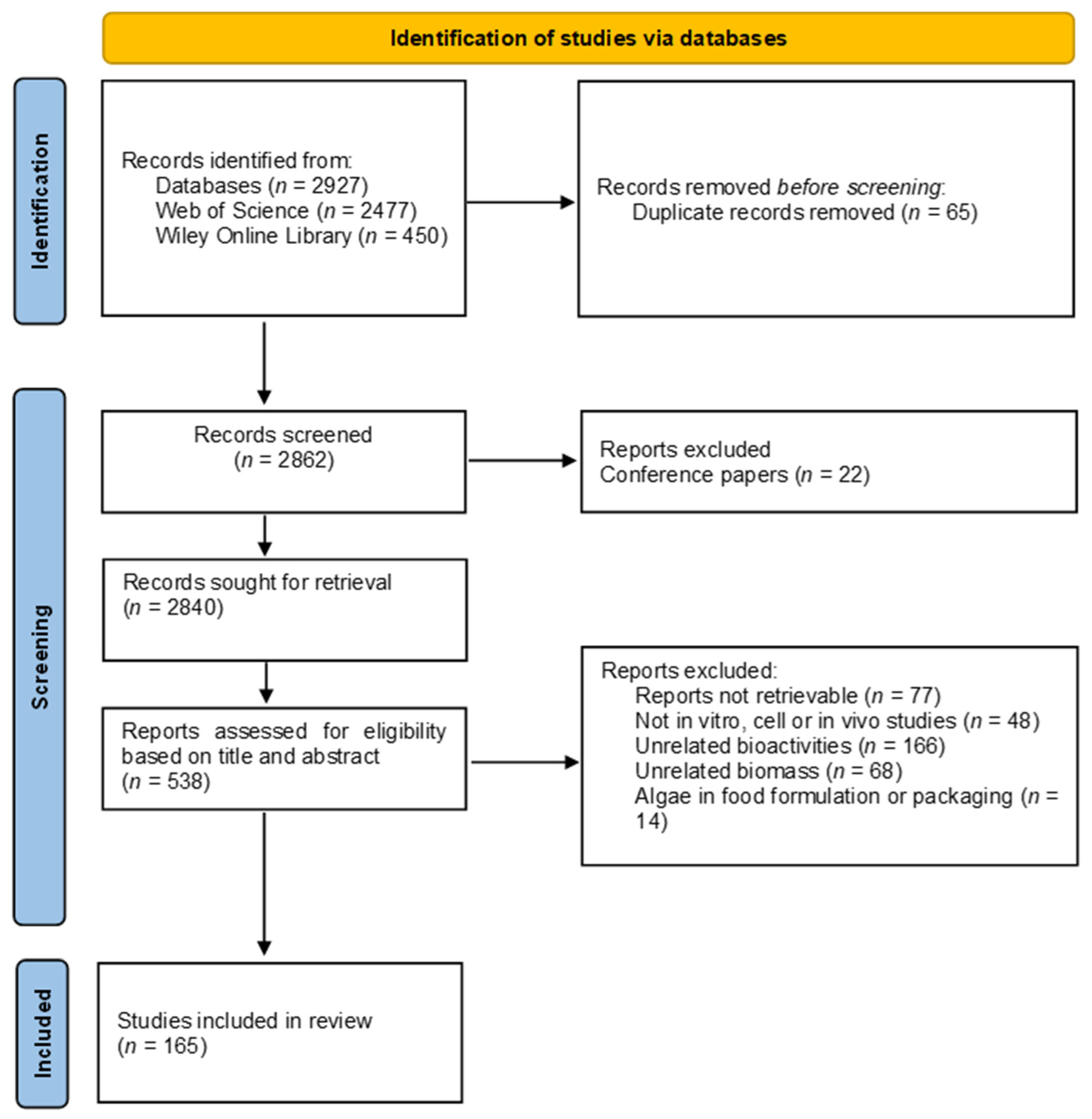
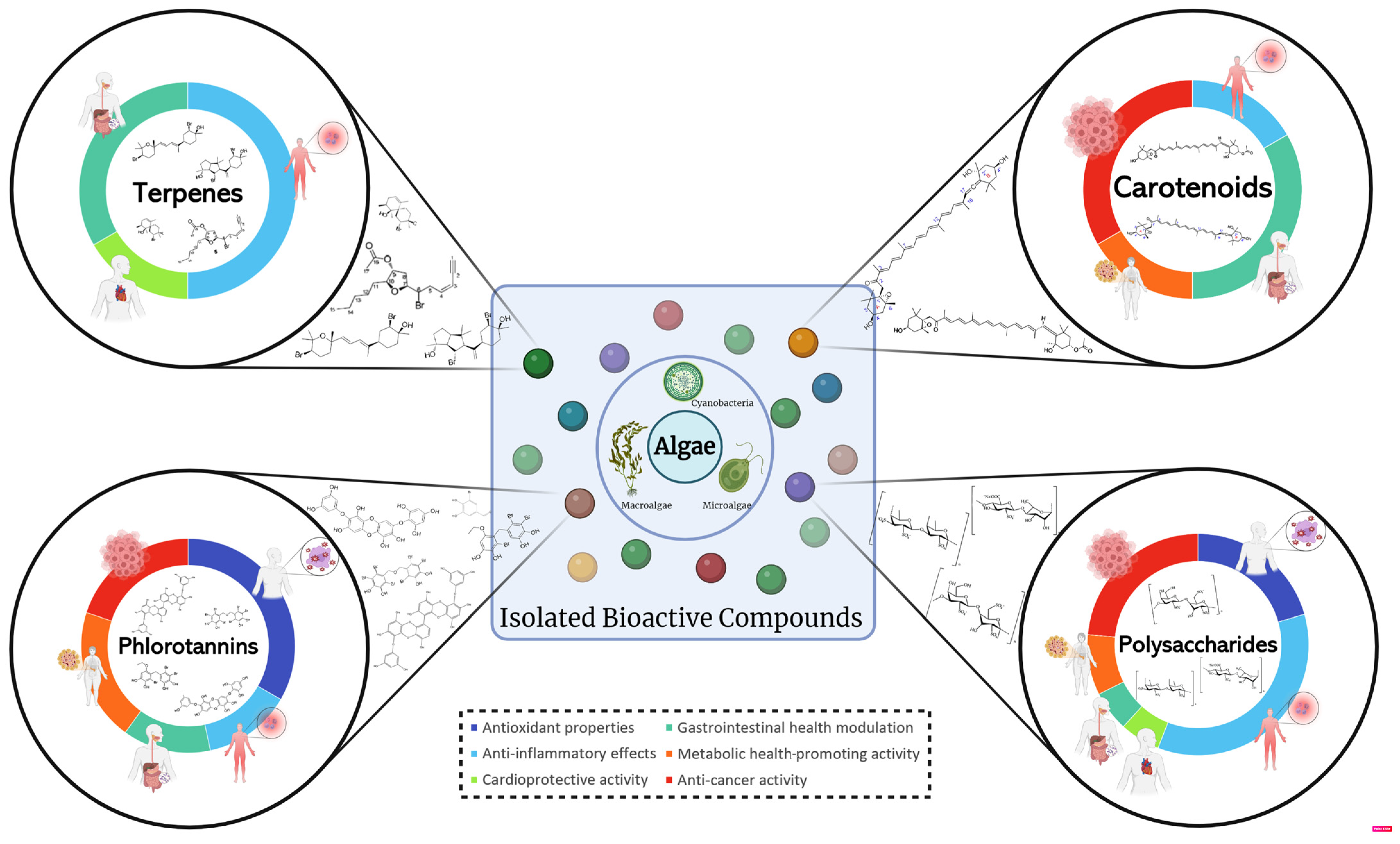
Disclaimer/Publisher’s Note: The statements, opinions and data contained in all publications are solely those of the individual author(s) and contributor(s) and not of MDPI and/or the editor(s). MDPI and/or the editor(s) disclaim responsibility for any injury to people or property resulting from any ideas, methods, instructions or products referred to in the content. |
© 2024 by the authors. Licensee MDPI, Basel, Switzerland. This article is an open access article distributed under the terms and conditions of the Creative Commons Attribution (CC BY) license (https://creativecommons.org/licenses/by/4.0/).