Abstract
The increasing prevalence of antimicrobial resistance and the persistent challenge of infectious diseases highlight the critical necessity for novel approaches that integrate pathogen management with swift detection methods. Carbon dots (CDs) are a versatile class of fluorescent nanomaterials that have garnered increasing attention owing to their tunable surface chemistry, strong photoluminescence, high stability, and biocompatibility. Recent studies demonstrate that CDs possess broad-spectrum antibacterial and antifungal activities via multiple mechanisms, including the generation of reactive oxygen species, disruption of membranes, inhibition of biofilms, and synergistic interactions with conventional antimicrobials. The performance is significantly affected by precursor selection, heteroatom doping, and surface functionalization, with minimum inhibitory concentrations reported to range from highly potent at the microgram level to moderate at elevated concentrations. The intrinsic fluorescence of CDs, in addition to their antimicrobial activity, facilitates their use as sensitive and selective probes for microbial detection, allowing for rapid and real-time monitoring in biomedical, food safety, and environmental settings. This review summarizes recent advancements in the antimicrobial properties of carbon dots (CDs) and their fluorescence-based applications in microbial detection. It emphasizes their theranostic potential and future prospects as multifunctional nanomaterials for combating infectious diseases and ensuring microbial safety.
1. Introduction
Pathogenic microorganisms play a critical role in the global spread of infectious diseases, contribute substantially to morbidity and mortality rates and place a heavy burden on healthcare systems worldwide, especially in low- and middle-income countries, where access to timely and effective treatment remains limited [1]. Moreover, the global health landscape is further complicated by the emergence of new, previously unrecognized, or even re-emerging infectious agents that have the potential to cause widespread epidemics. Emerging infectious diseases include new illnesses like COVID-19 and familiar ones like influenza and tuberculosis that are spreading more quickly or appearing again in certain regions [2,3]. What is particularly concerning is bacterial co-infections, which were identified in up to 16% of critically ill patients, further complicating treatment and increasing the risk of severe outcomes [4]. Antibiotics are indeed the most prescribed medications for the treatment of bacterial infections. However, the growing resistance to existing antibiotics, coupled with the slow pace of new antibiotic development, underscores the urgent need to explore and develop alternative antimicrobial agents for effective treatment [5,6].
The integration of nanotechnology and microbiology has created new opportunities for addressing bacterial infections and shows significant promise [7]. Recent advancements in nanotechnology have facilitated significant progress in the synthesis and application of nanomaterials, leading to the creation of highly engineered structures with distinct physicochemical properties. Nanomaterials such as metal nanoparticles, metal oxides, and carbon-based nanostructures have been thoroughly investigated for their antimicrobial properties [8]. Among these materials, carbon dots (CDs) have garnered significant research interest for their antimicrobial properties, attributed to their advantageous physicochemical characteristics and functional versatility.
CDs are a new and versatile category of zero-dimensional carbon-based nanomaterials, generally measuring less than 10 nm in diameter, characterized by their inherent photoluminescence and diverse surface chemistry [9]. Following their unexpected discovery in 2004 during the purification of single-walled carbon nanotubes and subsequent formal characterization in 2006 [10], they have attracted significant attention across multiple scientific fields, including optoelectronics, bioimaging, drug delivery, and antimicrobial therapy, owing to their remarkable physicochemical and biological properties [11]. CDs function as antimicrobials mainly via photodynamic and, in certain instances, photothermal mechanisms, which are facilitated by their photosensitizing characteristics [12]. Their ability to generate reactive oxygen species (ROS) under light irradiation leads to oxidative damage in microbial cells [13]. This antimicrobial activity is strongly influenced by the surface functional groups of CDs, which affect solubility, surface charge, membrane interactions, and ROS generation [14]. Additionally, these same surface properties allow CDs to function as probes for microbial detection. Functionalized carbon dots can selectively adhere to bacterial cells and demonstrate fluorescence alterations upon engagement, facilitating real-time pathogen identification. In some instances, CDs provide dual functionality, initially identifying bacteria through fluorescence modulation, subsequently eradicating them through light-activated antimicrobial mechanisms. This theranostic potential establishes CDs as valuable instruments for microbial detection and regulation, with extensive applications in biomedical diagnostics, food safety, and environmental surveillance.
The intrinsic fluorescence of carbon dots, alongside their antimicrobial functions, presents considerable potential for microbial detection. Carbon dots exhibit tunable emission, surface functionalization, and biocompatibility, allowing them to function as sensitive probes for rapid pathogen identification while concurrently delivering therapeutic effects via light-activated mechanisms. The dual functionality of CDs establishes them as promising theranostic agents, integrating antimicrobial activity with real-time diagnostic capabilities. This review highlights the antimicrobial efficacy of CDs and emphasizes their emerging role in fluorescence-based microbial detection.
2. Antimicrobial Mechanism Expressed by Carbon Dots (CDs)
The disruption of microbial cell structural integrity is one of the most widely recognized and effective antibacterial approaches [15]. In order to ensure their continued existence, both bacteria and fungi possess structural components that are vital to their life. Bacteria, which are unicellular prokaryotes, have essential components such as the cell wall, plasma membrane, cytoplasm, nucleoid region, and a variety of structural proteins [16]. On the other hand, fungi, which are eukaryotic microorganisms, have more complex structures, such as a cell wall that is rich in chitin, a cell membrane that contains ergosterol, organelles, and a nucleus that is clearly defined [17]. CDs, which are characterized by their extremely small size, high surface-area-to-volume ratio, and varied surface functions, have demonstrated the capability to interact with and disturb the critical structures of microorganisms.
The antibacterial efficiency of carbon dots (CDs) is closely associated with their structural attributes, such as particle size, surface charge, functional groups, passivation layers, and elemental doping [18]. Particle size is a crucial determinant among these factors. CDs smaller than 5 nm demonstrate enhanced antibacterial efficacy owing to their capacity to intimately engage with and infiltrate microbial cell membranes. Previous research showed that smaller carbon dots (CDs) around 2 nm were more effective antibacterial agents than larger ones (~5 nm), particularly against E. coli and S. aureus [19]. Beyond size, the shape and curvature of CDs are critical for targeting. CDs engineered to mimic the Gaussian membrane curvature of specific bacteria, like spherical S. aureus, can physically bind and disrupt their membranes via curvature-induced stress [20]. A smaller particle possesses a higher surface area-to-volume ratio, so probably augmenting interactions with microbial surfaces and promoting intracellular penetration [21]. The surfaces of most bacterial cells possess a negative charge attributed to teichoic acids and lipopolysaccharides [20]. Consequently, positively charged CDs exhibit enhanced efficacy in adhering to bacterial membranes through electrostatic interactions. This results in heightened membrane permeability, efflux of intracellular substances, and eventually, cellular lysis. Research has shown that increasing the zeta potential of CDs enhances their antibacterial efficacy against both Gram-positive and Gram-negative bacteria [22,23]. Not all effective antimicrobial CDs possess a positive charge. Under specific conditions, negatively charged CDs may demonstrate antibacterial properties. Highly anionic CDs have demonstrated improved antibacterial efficacy under laser irradiation [24]. In this instance, van der Waals forces surpassed electrostatic repulsion, facilitating the adherence of the CDs to the bacterial surface, resulting in ROS generation and subsequent damage to proteins and the cell wall. However, in a separate study, negatively charged CDs showed antimicrobial activity mechanism through electrostatic repulsion, obstructing nutrient absorption and resulting in microbial isolation and mortality [25].
The antibacterial efficacy of CDs is also significantly affected by their surface functional groups, which govern their interaction with bacterial cells and establish the total surface charge of the particles. Amino (–NH2) groups and quaternary ammonium moieties, which possess a positive charge, facilitate robust electrostatic attraction to negatively charged bacterial membranes, resulting in membrane rupture and subsequent bacterial death [26]. Structural changes in CDs by doping heteroatoms into the carbon core or surface group also have been reported to generate antimicrobial effect. Nitrogen-doped carbon dots and polyethyleneimine-coated carbon dots have demonstrated improved antibacterial efficacy while minimizing harm to mammalian cells [27]. Through the doping process, carbon dots exhibit a synergistic antimicrobial mechanism that integrates multiple modes of bacterial inhibition such as surface charges modification and photodynamic activation.
Another antimicrobial mechanism of CDs was explained via a light-activated photodynamic (photosensitizer), which entails the production of reactive oxygen species (ROS) [25,28]. When exposed to visible or natural light, CDs become photoexcited and transfer energy to adjacent oxygen molecules, resulting in the generation of ROS such as singlet oxygen (1O2) and hydroxyl radicals (•OH) [29]. Reactive species are pivotal in the inactivation of microbes. Specifically, the initial antimicrobial action involves the adhesion of CDs to the surface of bacterial cells, driven by electrostatic interactions between the positively charged CDs and the negatively charged bacterial cell walls, particularly in Gram-negative bacteria like Escherichia coli [30]. The process initiates with the adhesion of CDs to bacterial cell surfaces, mediated by electrostatic interactions. Upon activation by light, the resultant ROS target the bacterial cell membrane, leading to increased permeability and structural disruption. This damage enables reactive oxygen species (ROS) to infiltrate cells, inducing oxidative stress that affects essential biomolecules, including DNA, RNA, and proteins. Oxidative damage may impede gene expression, denature enzymes, and disrupt cellular functions, ultimately resulting in bacterial cell death via necrosis or programmed cell death [31]. Experimental studies have demonstrated that CDs exposed to light significantly reduce bacterial viability compared to dark conditions, underscoring the critical role of light in augmenting their bactericidal effects [32].
CDs function as antifungal agents by employing various mechanisms that compromise fungal cells, prevent biofilm formation, and disturbing the production of hyphae or yeast-transition [17,33]. Similarly to other positively charged nanoparticles, CDs with a positive surface charge demonstrate significant electrostatic attraction to the negatively charged fungal membrane, thereby enhancing cellular entry. Upon internalization, CDs can disturb the intracellular environment by hindering enzymatic functions, modifying ion concentrations, or exhibit strong photo-induced oxidative activity producing ROS such as singlet oxygen, mechanism notably linked to CDs enriched in electron-donating groups, including nitrogen-containing functionalities [34]. Disruptions can impair proteins and DNA, compromise membrane integrity, and result in the leakage of cytoplasmic contents [34,35]. Another antifungal strategy entails CDs to selectively bind to ergosterol, a crucial element of fungal membranes, thereby facilitating their attachment and traversal across the membrane barrier, leading to membrane disintegration and fungal cell death [36].
3. Antimicrobial Activities of Carbon Dots (CDs) Against Pathogenic Microorganisms
The swift emergence of antimicrobial resistance presents a significant risk to global public health, necessitating the urgent discovery and development of new antimicrobial drugs. Traditional antibiotics are progressively losing efficacy, resulting in enduring illnesses and elevated mortality rates. In this context, nanomaterials have emerged as viable alternatives, with carbon dots (CDs) receiving notable attention for their distinctive physicochemical properties, including adjustable surface functional groups, robust photoluminescence, high stability, and biocompatibility.
Antimicrobial susceptibility testing is an essential instrument for pharmacological development, epidemiological studies, and forecasting therapeutic results [37]. Evaluating the antimicrobial properties of CDs is essential as it assesses their capacity to suppress or eradicate pathogenic microorganisms, such as bacteria, fungus, and viruses. These investigations not only demonstrate their efficacy against clinically significant strains but also clarify mechanisms of action, including reactive oxygen species (ROS) production, membrane disruption, and biofilm inhibition [38]. Moreover, antimicrobial assays yield critical quantitative metrics, including minimum inhibitory concentration (MIC) and minimum bactericidal concentration (MBC), which are vital for comparing CDs with traditional antimicrobials and for informing their utilization in biomedical, food safety, and environmental domains.
Table 1 summarizes the results of antimicrobial activity of CDs from recent studies. There are three most common methodologies for evaluating the antimicrobial activity of CDs against several pathogenic microorganisms, namely disk diffusion, agar dilution, and broth microdilution.

Table 1.
Antimicrobial activity of CDs from the literature.
The disk or agar diffusion method, often referred to as the Kirby–Bauer test, is a widely used qualitative or semi-quantitative technique for evaluating antimicrobial activity. In this method, a standardized microbial suspension is evenly spread over the surface of an agar plate to create a uniform lawn of growth. Filter paper disks impregnated with the test compound or alternatively wells cut into the agar and filled with the agent, are then placed on the surface. During incubation, the compound diffuses radially through the agar, interacting with the microorganisms. If the agent is effective, it inhibits microbial growth, producing a clear circular area around the disk or well known as the zone of inhibition [80].
Studies employing disk diffusion and agar-well diffusion methods demonstrated varying levels of antimicrobial activity among different carbon dots (CDs). The most pronounced inhibition zones were observed with Taxus baccata–derived CDs, which produced zones up to 33 mm [71], and castor seed N-CQD1, with a zone of 30 mm [73], indicating strong antimicrobial potency. Moderate inhibition was shown by spermidine-derived CDs (22 mm) [48] and Prosopis juliflora leaf CDs (16.7 mm) [76], while avocado peel CQDs exhibited smaller but consistent zones of 8–13 mm [54]. CDs synthesized from citric acid with β-alanine or ethylenediamine showed relatively weaker activity, with zones ranging between 10 and 12 mm [43,57]. L-arginine–based CDs, including their HPMC-modified form, displayed only minimal inhibition zones of 1–2 mm [78], suggesting limited effectiveness by comparison. Disk diffusion is the most widely used AST method in microbiology laboratories because of its low cost and ease of performance and applicability of numerous bacterial and fungal species [81]. Nevertheless, this method cannot be used to determine the minimum inhibitory concentration of CDs and its matrices due to unquantified amounts of CDs diffused to the agar medium, and should be treated only as the method to provide initial qualitative information on antimicrobial potency of CDs [80,81].
The dilution method is one of the most reliable and standardized techniques for evaluating antimicrobial activity, as it allows determination of the minimum inhibitory concentration (MIC) and, when necessary, the minimum bactericidal concentration (MBC) of a compound [82]. Agar dilution and broth dilution methods are closely associated with methods of determining the minimum inhibitory concentration (MIC), the minimal quantity of a chemical that inhibits observable bacterial or fungal proliferation. The differentiation between the two resides in the medium employed: agar dilution integrates the antimicrobial agent into solid agar plates, whereas broth dilution utilizes liquid broth tubes. In both methodologies, repeated dilutions of the antimicrobial agent are formulated, and a standardized inoculum of bacteria is introduced [80]. The MIC is determined by the presence or absence of microbial growth. These methodologies are crucial for identifying appropriate concentration of CDs that give potent antimicrobial activity and for monitoring changes in microbial susceptibility over time.
The antibacterial efficiency of carbon dots (CDs) is significantly affected by their production method, doping elements, and surface chemistry. A variety of CDs have been evaluated against different bacterial strains, demonstrating significant differences in the minimum inhibitory concentrations (MICs) and minimum bactericidal concentrations (MBCs). Glucose-derived carbon dots synthesized via a one-step hydrothermal technique (GCDs) and their boron-doped variants (BGCDs) exhibited minimum inhibitory concentrations (MIC) ranging from 312 to 625 µg/mL and minimum bactericidal concentrations (MBC) up to 1250 µg/mL against Listeria monocytogenes and Escherichia coli [40]. The elevated findings suggest that a substantial concentration is necessary for both inhibition and total bacterial elimination, potentially indicating restricted interaction with bacterial membranes or diminished generation of reactive oxygen species (ROS). In contrast, CDs modified or doped with heteroatoms exhibited improved antibacterial efficacy. Sulfur and nitrogen co-doped carbon dots (SN-CDs), synthesized from thiourea and o-phenylenediamine, demonstrated minimum inhibitory concentrations (MICs) as low as 16 µg/mL against both E. coli and Staphylococcus aureus, presumably attributable to enhanced surface activity and augmented electron transfer resulting in reactive oxygen species (ROS) formation. Phosphorus-doped carbon dots produced from glucose and phosphoric acid exhibited MIC values between 128 and 256 µg/mL against E. coli, S. aureus, and Salmonella typhimurium [60]. The findings indicate that heteroatom doping alters the surface charge and binding capability of carbon dots while also improving their capacity to infiltrate bacterial biofilms and membranes. Supplementary antibacterial studies from functionalized carbon dots, specifically those produced from L-histidine or guanidine [54,55,57], further corroborate the influence of precursor chemistry. These CDs demonstrated MIC values ranging from 64 to 256 µg/mL against both Gram-positive and Gram-negative bacteria, with MBCs exhibiting analogous patterns. Conversely, CDs derived from basic precursors as citric acid, urea, or acetic acid without further functionalization typically necessitated elevated MICs—reaching up to 1500 µg/mL [41,61,62,63,64,65,66].
Beyond MIC/MBC/MFC and inhibition zone measurements, several studies in the table evaluated antimicrobial efficacy of carbon dots (CDs) using viable cell counts and log reduction assays. For instance, arginine-based CDs and their composites (Arg-Ag, Arg-AgCl) not only produced inhibition zones but also demonstrated strong antimicrobial effects through CFU reduction, confirming their bactericidal activity beyond diffusion-based methods [44]. Similarly, polymer–CD composites such as PANI–CuO, PANI–TiO2, and PANI–SiO2 were tested using CFU counting, where PANI–CuO and PANI–TiO2 at 1 mg/mL significantly inhibited Pseudomonas aeruginosa, and PANI–TiO2 completely eradicated Klebsiella pneumoniae ATCC 700603, highlighting direct bactericidal effects verified by culture viability rather than zone inhibition [45]. In another case, N@CDs derived from bovine serum albumin reduced MRSA (ATCC 43300) by approximately 3.5 log CFU at 0.5 mg/mL, with partial inhibition at 0.125 mg/mL, indicating a dose-dependent bactericidal effect based on viable cell count [46].
The antifungal efficacy of carbon dots (CDs) exhibits significant diversity based on the precursor and synthesis method employed. SP-CDs derived from L-serine and phytic acid exhibited the highest potency, attaining minimal MIC values of 2.5 µg/mL against Fusarium solani and 5 µg/mL against Candida albicans and Aspergillus fumigatus, but SN-CDs demonstrated reduced efficacy at 10–20 µg/mL [39]. In contrast, glucose-derived carbon dots (CDs) demonstrated only modest inhibition, with minimum inhibitory concentration (MIC) and minimum bactericidal concentration (MBC) values of 312/625 µg/mL [40]. Amine-rich carbon dots (CDs–NH2) exhibited a MIC of 397 µg/mL against Candida albicans, while also considerably inhibiting fungal adherence (79% at 125 µg/mL), suggesting anti-biofilm properties [41]. Correspondingly, elevated MIC/MFC values for A. fumigatus (156/625 µg/mL) and F. solani (312/655 µg/mL) indicate moderate-to-weak efficacy.
Plant-derived and doped carbon dots exhibited enhanced potential. N-doped carbon dots obtained from Trillium govanianum successfully suppressed multidrug-resistant Candida auris, exhibiting MIC/MFC values of 0.625/1.25 mg/mL, whereas ginger peel-derived carbon dots had MIC/MBC values of 0.29/0.58 mg/mL. Nitrogen-doped carbon quantum dots derived from citric acid and urea exhibited minimum inhibitory concentration (MIC) values of 52 µg/mL and minimum fungicidal concentration (MFC) of 104 µg/mL against Mucor indicus, Candida albicans, and Aspergillus flavus. Conversely, CQDs@AgNPs derived from watermelon peel had significantly lower efficacy, necessitating a concentration of 15 mg/mL to inhibit C. albicans [62]. While the findings for [72,73] are less comprehensive, the overarching trend indicates that SP-CDs [39] exhibit remarkable potency, whereas other CDs demonstrate varying degrees of efficacy, ranging from moderate to weak, contingent upon the carbon source, surface functionalization, and doping approach [39,40,41,59,60,62,72,73,78].
4. Fluorescence-Based Applications of CDs in Microbial Detection
Microbial contamination remains a critical global challenge in public health and food safety, necessitating rapid and reliable diagnostic platforms. Conventional methods, such as culture plating and polymerase chain reaction (PCR), are accurate but time consuming and labor intensive. Fluorescence-based nanomaterials have become an indispensable component of modern microbial diagnostics due to their sensitivity, rapid response, and ability to operate in complex biological environments [83]. CDs have emerged as a versatile class of fluorescent nanomaterials, offering remarkable photostability, water solubility, and ease of functionalization. In microbial detection, CDs have been exploited as sensing probes due to their tunable photoluminescence, low cytotoxicity, and compatibility with biological matrices. Rapid progress has been made in the development of fluorescence-based CD biosensors for bacteria and fungi, incorporating diverse mechanisms such as Förster resonance energy transfer (FRET), inner filter effect (IFE), ratiometric (Figure 1), and photoinduced electron transfer (PET) fluorescence. These systems have been integrated with recognition elements such as antibiotics, aptamers, and boronic acids, enabling species-specific or broad-spectrum microbial detection. Applications span food safety, environmental monitoring, and clinical diagnostics.
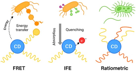
Figure 1.
Fluorescence based applications of CDs in microbial detection. CDs (blue nanodots) can detect microbes through diverse mechanisms.
In FRET-based systems, CDs act as fluorescent donors, transferring energy non-radiatively to an acceptor molecule or nanomaterial if the donor–acceptor distance is within 1–10 nm and there is sufficient spectral overlap. Microbial recognition events alter the spatial relationship between CDs and quenchers, leading to changes in fluorescence intensity [84]. The IFE occurs when the excitation or emission light of CDs is absorbed by nearby chromophores or nanoparticles, leading to apparent fluorescence quenching. In microbial detection, this effect has been harnessed in competitive assays, where microbial targets disrupt the absorber fluorophore interaction [85]. The advantage of IFE-based detection lies in its simplicity; no direct electron or energy transfer is required yet. It is highly sensitive to overlapping absorption spectra and solution turbidity, which must be controlled for reliable operation in food and clinical matrices [86]. Ratiometric fluorescence is an advanced detection strategy where CDs emit at two wavelengths, one responsive to microbial interactions and another serving as an internal reference. The ratiometric approach is especially valuable in real-world samples, where environmental interference often compromises single-emission assays [87]. PET mechanisms involve electron transfer between CDs and electron donors/acceptors in their environment, altering the emissive state of the CDs. Microbial metabolites, such as redox-active species, can modulate this electron transfer at the CD surface, leading to fluorescence quenching or enhancement. Additionally, functionalization of CDs with electron-rich ligands (e.g., phenolic or amine groups) has allowed PET interactions with microbial metabolites, offering selective fluorescence switching [88]. PET-based sensing is particularly useful for detecting enzymatic or metabolic activity, providing functional readouts that go beyond structural recognition of microbial surfaces. Reported CDs based fluorescent microbial assays could be seen in Table 2.
Table 2.
Comparison of reported CDs based fluorescent microbial assays.
5. Conclusions
Carbon dots (CDs) are recognized as multifunctional nanomaterials with significant potential in addressing microbial threats. The antimicrobial activity is derived from various mechanisms, such as the generation of reactive oxygen species, disruption of membranes, and inhibition of biofilms. Effectiveness is affected by the type of precursor, heteroatom doping, and surface chemistry. Simultaneously, the intrinsic fluorescence and surface functionalization of carbon dots (CDs) render them effective tools for microbial detection, facilitating rapid, sensitive, and real-time monitoring that addresses the limitations of traditional culture-based methods. This dual role underscores the theranostic potential of CDs, integrating diagnosis and treatment within a unified framework. Challenges persist in the standardization of antimicrobial assays, enhancement of detection selectivity, and the assurance of consistent large-scale synthesis with minimal toxicity. The future development of CDs should focus on the integration of their antimicrobial and fluorescence properties into practical applications, including biosensors, point-of-care devices, and smart antimicrobial coatings. Combining pathogen detection with targeted inactivation, CDs offer an advanced strategy for managing infectious diseases and maintaining microbial safety in biomedical, food, and environmental domains.
Author Contributions
Conceptualization, A.R. and S.S.; literature search, A.R., S.S. and R.H.B.S.; data curation, A.R. and S.S.; writing—original draft preparation, A.R., S.S. and R.H.B.S.; writing—review and editing, A.R., S.S. and Y.G.; supervision—Y.G. All authors have read and agreed to the published version of the manuscript.
Funding
This research received no external funding.
Institutional Review Board Statement
Not applicable.
Informed Consent Statement
Not applicable.
Data Availability Statement
All data presented in this review are from previously published studies, which have been appropriately cited. No new data collection involving humans or animals was conducted.
Conflicts of Interest
The authors declare no conflicts of interest.
References
- Lee, R.A.; Stripling, J.T.; Spellberg, B.; Centor, R.M. Short-Course Antibiotics for Common Infections: What Do We Know and Where Do We Go from Here? Clin. Microbiol. Infect. 2023, 29, 150–159. [Google Scholar] [CrossRef] [PubMed]
- Bagre, A.; Patel, P.R.; Naqvi, S.; Jain, K. Emerging Concerns of Infectious Diseases and Drug Delivery Challenges. In Nanotheranostics for Treatment and Diagnosis of Infectious Diseases; Academic Press: Cambridge, MA, USA, 2022; pp. 1–23. [Google Scholar] [CrossRef]
- Gulumbe, B.H.; Abdulrahim, A.; Ahmad, S.K.; Lawan, K.A.; Danlami, M.B. WHO Report Signals Tuberculosis Resurgence: Addressing Systemic Failures and Revamping Control Strategies. Decod. Infect. Transm. 2025, 3, 100044. [Google Scholar] [CrossRef]
- Chong, W.H.; Saha, B.K.; Ramani, A.; Chopra, A. State-of-the-Art Review of Secondary Pulmonary Infections in Patients with COVID-19 Pneumonia. Infection 2021, 49, 591–605. [Google Scholar] [CrossRef] [PubMed]
- Salam, M.A.; Al-Amin, M.Y.; Salam, M.T.; Pawar, J.S.; Akhter, N.; Rabaan, A.A.; Alqumber, M.A.A. Antimicrobial Resistance: A Growing Serious Threat for Global Public Health. Healthcare 2023, 11, 1946. [Google Scholar] [CrossRef]
- Oluwole, A.O.; Hernández-Rocamora, V.M.; Cao, Y.; Li, X.; Vollmer, W.; Robinson, C.V.; Bolla, J.R. Real-Time Biosynthetic Reaction Monitoring Informs the Mechanism of Action of Antibiotics. J. Am. Chem. Soc. 2024, 146, 7007–7017. [Google Scholar] [CrossRef]
- Liu, J.; Yuan, S.; Bremmer, A.; Hu, Q. Convergence of Nanotechnology and Bacteriotherapy for Biomedical Applications. Adv. Sci. 2024, 11, 2309295. [Google Scholar] [CrossRef]
- Parvin, N.; Joo, S.W.; Mandal, T.K. Nanomaterial-Based Strategies to Combat Antibiotic Resistance: Mechanisms and Applications. Antibiotics 2025, 14, 207. [Google Scholar] [CrossRef]
- Wang, S.; Wang, D.; Wang, G.; Zhang, M.; Sun, Y.; Ding, J. Antibacterial Carbon Dots. Mater. Today Bio 2025, 30, 101383. [Google Scholar] [CrossRef]
- Hussen, N.H.; Hasan, A.H.; FaqiKhedr, Y.M.; Bogoyavlenskiy, A.; Bhat, A.R.; Jamalis, J. Carbon Dot Based Carbon Nanoparticles as Potent Antimicrobial, Antiviral, and Anticancer Agents. ACS Omega 2024, 9, 9849–9864. [Google Scholar] [CrossRef]
- Wang, B.; Cai, H.; Waterhouse, G.I.N.; Qu, X.; Yang, B.; Lu, S. Carbon Dots in Bioimaging, Biosensing and Therapeutics: A Comprehensive Review. Small Sci. 2022, 2, 2200012. [Google Scholar] [CrossRef]
- Sun, L.; Zhao, Y.; Peng, H.; Zhou, J.; Zhang, Q.; Yan, J.; Liu, Y.; Guo, S.; Wu, X.; Li, B. Carbon Dots as a Novel Photosensitizer for Photodynamic Therapy of Cancer and Bacterial Infectious Diseases: Recent Advances. J. Nanobiotechnol. 2024, 22, 210. [Google Scholar] [CrossRef] [PubMed]
- Dong, X.; Liang, W.; Meziani, M.J.; Sun, Y.-P.; Yang, L. Carbon Dots as Potent Antimicrobial Agents. Theranostics 2020, 10, 671–686. [Google Scholar] [CrossRef]
- Mansuriya, B.D.; Altintas, Z. Carbon Dots: Classification, Properties, Synthesis, Characterization, and Applications in Health Care—An Updated Review (2018–2021). Nanomaterials 2021, 11, 2525. [Google Scholar] [CrossRef]
- AlQurashi, D.M.; AlQurashi, T.F.; Alam, R.I.; Shaikh, S.; Tarkistani, M.A.M. Advanced Nanoparticles in Combating Antibiotic Resistance: Current Innovations and Future Directions. J. Nanotheranostics 2025, 6, 9. [Google Scholar] [CrossRef]
- Galinier, A.; Delan-Forino, C.; Foulquier, E.; Lakhal, H.; Pompeo, F. Recent Advances in Peptidoglycan Synthesis and Regulation in Bacteria. Biomolecules 2023, 13, 720. [Google Scholar] [CrossRef]
- Sturabotti, E.; Camilli, A.; Leonelli, F.; Vetica, F. Carbon Dots as Bioactive Antifungal Nanomaterials. ChemMedChem 2024, 19, e202400463. [Google Scholar] [CrossRef]
- Guo, B.; Liu, G.; Hu, C.; Lei, B.; Liu, Y. The Structural Characteristics and Mechanisms of Antimicrobial Carbon Dots: A Mini Review. Mater. Adv. 2022, 3, 7726–7741. [Google Scholar] [CrossRef]
- Sun, B.; Wu, F.; Zhang, Q.; Chu, X.; Wang, Z.; Huang, X.; Li, J.; Yao, C.; Zhou, N.; Shen, J. Insight into the Effect of Particle Size Distribution Differences on the Antibacterial Activity of Carbon Dots. J. Colloid Interface Sci. 2021, 584, 505–519. [Google Scholar] [CrossRef]
- Hui, L.; Huang, J.; Chen, G.; Zhu, Y.; Yang, L. Antibacterial Property of Graphene Quantum Dots (Both Source Material and Bacterial Shape Matter). ACS Appl. Mater. Interfaces 2016, 8, 20–25. [Google Scholar] [CrossRef] [PubMed]
- Ullah, M.; Awan, U.A.; Ali, H.; Wahab, A.; Khan, S.U.; Naeem, M.; Ruslin, M.; Mustopa, A.Z.; Hasan, N. Carbon Dots: New Rising Stars in the Carbon Family for Diagnosis and Biomedical Applications. J. Nanotheranostics 2025, 6, 1. [Google Scholar] [CrossRef]
- Li, Y.-J.; Harroun, S.G.; Su, Y.-C.; Huang, C.-F.; Unnikrishnan, B.; Lin, H.-J.; Lin, C.-H.; Huang, C.-C. Synthesis of Self-Assembled Spermidine-Carbon Quantum Dots Effective against Multidrug-Resistant Bacteria. Adv. Healthc. Mater. 2016, 5, 2545–2554. [Google Scholar] [CrossRef]
- Bing, W.; Sun, H.; Yan, Z.; Ren, J.; Qu, X. Programmed Bacteria Death Induced by Carbon Dots with Different Surface Charge. Small 2016, 12, 4713–4718. [Google Scholar] [CrossRef] [PubMed]
- Sattarahmady, N.; Rezaie-Yazdi, M.; Tondro, G.H.; Akbari, N. Bactericidal Laser Ablation of Carbon Dots: An in Vitro Study on Wild-Type and Antibiotic-Resistant Staphylococcus aureus. J. Photochem. Photobiol. B Biol. 2017, 166, 323–332. [Google Scholar] [CrossRef] [PubMed]
- Zhu, C.; Li, H.; Wang, H.; Yao, B.; Huang, H.; Liu, Y.; Kang, Z. Negatively Charged Carbon Nanodots with Bacteria Resistance Ability for High-Performance Antibiofilm Formation and Anticorrosion Coating Design. Small 2019, 15, 1900007. [Google Scholar] [CrossRef]
- Xia, W.; Wu, Z.; Hou, B.; Cheng, Z.; Bi, D.; Chen, L.; Chen, W.; Yuan, H.; Koole, L.H.; Qi, L. Inactivation of Antibiotic Resistant Bacteria by Nitrogen-Doped Carbon Quantum Dots through Spontaneous Generation of Intracellular and Extracellular Reactive Oxygen Species. Mater. Today Bio 2025, 30, 101428. [Google Scholar] [CrossRef]
- Demirci, S.; McNally, A.B.; Ayyala, R.S.; Lawson, L.B.; Sahiner, N. Synthesis and Characterization of Nitrogen-Doped Carbon Dots as Fluorescent Nanoprobes with Antimicrobial Properties and Skin Permeability. J. Drug Deliv. Sci. Technol. 2020, 59, 101889. [Google Scholar] [CrossRef]
- Divsalar, E.; Tajik, H.; Moradi, M.; Molaei, R. Carbon Dot-Based Antimicrobial Photosensitizer: Synthesis, Characterization and Antimicrobial Performance against Food Borne Pathogens. Food Biosci. 2023, 56, 103220. [Google Scholar] [CrossRef]
- Deng, X.; Zhang, M.; Wang, Y.; Li, C.; Zhang, X.; Weng, S.; Li, Y. Carbon Dots with Selective Fluorescence Response to Hydroxyl Radical for Sensitive Detection of Bleomycin. Spectrochim. Acta Part A Mol. Biomol. Spectrosc. 2024, 306, 123582. [Google Scholar] [CrossRef]
- Slavin, Y.N.; Asnis, J.; Häfeli, U.O.; Bach, H. Metal Nanoparticles: Understanding the Mechanisms behind Antibacterial Activity. J. Nanobiotechnol. 2017, 15, 65. [Google Scholar] [CrossRef]
- Checa, J.; Aran, J.M. Reactive Oxygen Species: Drivers of Physiological and Pathological Processes. J. Inflamm. Res. 2020, 13, 1057–1073. [Google Scholar] [CrossRef]
- Meziani, M.J.; Dong, X.; Zhu, L.; Jones, L.P.; LeCroy, G.E.; Yang, F.; Wang, S.; Wang, P.; Zhao, Y.; Yang, L.; et al. Visible-Light-Activated Bactericidal Functions of Carbon “Quantum” Dots. ACS Appl. Mater. Interfaces 2016, 8, 10761–10766. [Google Scholar] [CrossRef] [PubMed]
- Ghirardello, M.; Ramos-Soriano, J.; Galan, M.C. Carbon Dots as an Emergent Class of Sustainable Antifungal Agents. ACS Nano 2025, 19, 24377–24403. [Google Scholar] [CrossRef]
- Zhao, X.; Zhang, S.; Zhang, M.; Zhang, Z.; Zhou, M.; Cao, J. Antifungal Performance and Mechanisms of Carbon Quantum Dots in Cellulosic Materials. ACS Nano 2025, 19, 14121–14136. [Google Scholar] [CrossRef]
- Nguyen, D.H.H.; El-Ramady, H.; Törős, G.; Muthu, A.; Elsakhawy, T.; Abdalla, N.; Alibrahem, W.; Kharrat Helu, N.; Prokisch, J. Food-Derived Carbon Dots: Formation, Detection, and Impact on Gut Microbiota. Foods 2025, 14, 2980. [Google Scholar] [CrossRef]
- de Oliveira, B.P.; de Castro Bessa, N.U.; do Nascimento, J.F.; de Paula Cavalcante, C.S.; dos Santos Fontenelle, R.O.; da Silva Abreu, F.O.M. Synthesis of Luminescent Chitosan-Based Carbon Dots for Candida albicans Bioimaging. Int. J. Biol. Macromol. 2023, 227, 805–814. [Google Scholar] [CrossRef]
- Wenzler, E.; Maximos, M.; Asempa, T.E.; Biehle, L.; Schuetz, A.N.; Hirsch, E.B. Antimicrobial Susceptibility Testing: An Updated Primer for Clinicians in the Era of Antimicrobial Resistance: Insights from the Society of Infectious Diseases Pharmacists. Pharmacother. J. Hum. Pharmacol. Drug Ther. 2023, 43, 264–278. [Google Scholar] [CrossRef] [PubMed]
- Zhang, X.; Bai, X.; Deng, X.; Peng, K.; Zheng, Z.; Xiao, J.; Zhang, R.; Huang, Z.; Huang, J.; Chen, M.; et al. Long-Term Antibacterial Activity of Guanidinium Carbon Dots without Detectable Resistance for the Effective Treatment of Pneumonia Caused by Gram-Negative Bacteria. Carbon 2023, 213, 118229. [Google Scholar] [CrossRef]
- Chen, H.; Geng, X.; Ning, Q.; Shi, L.; Zhang, N.; He, S.; Zhao, M.; Zhang, J.; Li, Z.; Shi, J.; et al. Biophilic Positive Carbon Dot Exerts Antifungal Activity and Augments Corneal Permeation for Fungal Keratitis. Nano Lett. 2024, 24, 4044–4053. [Google Scholar] [CrossRef]
- Ezati, P.; Rhim, J.-W.; Molaei, R.; Priyadarshi, R.; Roy, S.; Min, S.; Kim, Y.H.; Lee, S.-G.; Han, S. Preparation and Characterization of B, S, and N-Doped Glucose Carbon Dots: Antibacterial, Antifungal, and Antioxidant Activity. Sustain. Mater. Technol. 2022, 32, e00397. [Google Scholar] [CrossRef]
- Sturabotti, E.; Camilli, A.; Georgian Moldoveanu, V.; Bonincontro, G.; Simonetti, G.; Valletta, A.; Serangeli, I.; Miranda, E.; Amato, F.; Giacomo Marrani, A.; et al. Targeting the Antifungal Activity of Carbon Dots against Candida albicans Biofilm Formation by Tailoring Their Surface Functional Groups. Chem.—A Eur. J. 2024, 30, e202303631. [Google Scholar] [CrossRef]
- Wang, L.; Wang, T.; Hao, R.; Wang, Y. Construction Strategy and Mechanism of a Novel Wood Preservative with Excellent Antifungal Effects. Molecules 2024, 29, 1013. [Google Scholar] [CrossRef]
- Pandey, A.; Devkota, A.; Yadegari, Z.; Dumenyo, K.; Taheri, A. Antibacterial Properties of Citric Acid/β-Alanine Carbon Dots against Gram-Negative Bacteria. Nanomaterials 2021, 11, 2012. [Google Scholar] [CrossRef] [PubMed]
- Suner, S.S.; Sahiner, M.; Ayyala, R.S.; Bhethanabotla, V.R.; Sahiner, N. Nitrogen-Doped Arginine Carbon Dots and Its Metal Nanoparticle Composites as Antibacterial Agent. C 2020, 6, 58. [Google Scholar] [CrossRef]
- Maruthapandi, M.; Saravanan, A.; Luong, J.H.T.; Gedanken, A. Antimicrobial Properties of the Polyaniline Composites against Pseudomonas Aeruginosa and Klebsiella pneumoniae. J. Funct. Biomater. 2020, 11, 59. [Google Scholar] [CrossRef]
- Maruthapandi, M.; Natan, M.; Jacobi, G.; Banin, E.; Luong, J.H.T.; Gedanken, A. Antibacterial Activity against Methicillin-Resistant Staphylococcus aureus of Colloidal Polydopamine Prepared by Carbon Dot Stimulated Polymerization of Dopamine. Nanomaterials 2019, 9, 1731. [Google Scholar] [CrossRef]
- Marković, Z.M.; Budimir Filimonović, M.D.; Milivojević, D.D.; Kovač, J.; Todorović Marković, B.M. Antibacterial and Antibiofouling Activities of Carbon Polymerized Dots/Polyurethane and C60/Polyurethane Composite Films. J. Funct. Biomater. 2024, 15, 73. [Google Scholar] [CrossRef] [PubMed]
- Cui, T.; Fan, Y.; Liu, Y.; Fan, X.; Sun, Y.; Cheng, G.; Cheng, J. Antibacterial Activity and Mechanism of Self-Assembly Spermidine-Capped Carbon Dots against Staphylococcus aureus. Foods 2024, 13, 67. [Google Scholar] [CrossRef]
- Marković, Z.M.; Milivojević, D.D.; Kovač, J.; Todorović Marković, B.M. Phloroglucinol-Based Carbon Quantum Dots/Polyurethane Composite Films: How Structure of Carbon Quantum Dots Affects Antibacterial and Antibiofouling Efficiency of Composite Films. Polymers 2024, 16, 1646. [Google Scholar] [CrossRef]
- Marković, Z.; Dorontić, S.; Jovanović, S.; Kovač, J.; Milivojević, D.; Marinković, D.; Mojsin, M.; Todorović Marković, B. Biocompatible Carbon Dots/Polyurethane Composites as Potential Agents for Combating Bacterial Biofilms: N-Doped Carbon Quantum Dots/Polyurethane and Gamma Ray-Modified Graphene Quantum Dots/Polyurethane Composites. Pharmaceutics 2024, 16, 1565. [Google Scholar] [CrossRef]
- Shi, W.; Li, J.; Pu, J.; Cheng, G.; Liu, Y.; Xiao, S.; Cao, J. Epigynum Auritum-Derived Near-Infrared Carbon Dots for Bioimaging and Antimicrobial Applications. Molecules 2025, 30, 422. [Google Scholar] [CrossRef]
- Lee, W.; Ko, S. Synthesis and Characterization of Lignocellulose-Based Carbon Quantum Dots (CQDs) and Their Antimicrobial and Antioxidant Functionalities. Molecules 2025, 30, 667. [Google Scholar] [CrossRef]
- Singh, B.; Adcock, A.F.; Dumra, S.; Collins, J.; Yang, L.; Bunker, C.E.; Qian, H.; Meziani, M.J.; Sun, Y.-P. Microwave-Assisted Carbonization Processing for Carbon Dot-like Nanomaterials with Antimicrobial Properties. Micro 2025, 5, 14. [Google Scholar] [CrossRef]
- Fiallos, N.; Acuña, S.; Correa-Otero, D.; Venegas-Toloza, M.; Beldarrain, T.; Burgos, J.; Fuentes, F.; Bustamante, F.; Christiansen, G.; Roa, V.; et al. Centrifugal Partition Chromatography Is a Powerful Tool for the Isolation of Antibiofilm Quantum Carbon Dots Synthesized by Hydrothermal Treatment of Avocado Peels. Molecules 2025, 30, 1525. [Google Scholar] [CrossRef]
- Shamkhali, L.; Mobarez, A.M.; Siadat, S.D.; Pajavand, H. Synergistic Antibacterial Effects of Carbon Dots Derived from Lactobacillus acidophilus Alone and in Combination against Carbapenem-Resistant Klebsiella pneumoniae. J. Infect. Public Health 2025, 18, 102724. [Google Scholar] [CrossRef] [PubMed]
- Azizi, J.; Hamedi, H.; Javanbakht, S.; Mohammadi, R. Green Synthesis of Zeolitic Imidazolate Frameworks/Marshmallow Extract-Derived Carbon Quantum Dots for Improved Antibacterial Properties. Surf. Interfaces 2025, 72, 107215. [Google Scholar] [CrossRef]
- Ansari, P.; Ghobadifard, M.; Mohebbi, S.; Ashengroph, M. The Performance of Metal Carbon-Based Quantum Dots as an Anti-Bacterial Factor in Green Conditions. J. Organomet. Chem. 2025, 1026, 123495. [Google Scholar] [CrossRef]
- Peng, J.; Wang, X.; Rehman, M.U.; Ikram, M.; Liu, G.; Wu, F. Garlic-Derived Carbon Dots Hydrogel as Potent and Controllable Antibacterial Agents. Inorg. Chem. Commun. 2025, 181, 115162. [Google Scholar] [CrossRef]
- Vijeata, A.; Ghawri, H.; Chaudhary, G.R.; Raj, K.; Chaudhary, S. Engineering Fungicidal Nitrogen-Doped Carbon Dots from Trillium govanianum Rhizomes: A Theoretical and Experimental Approach against Candida auris. Appl. Surf. Sci. 2025, 709, 163789. [Google Scholar] [CrossRef]
- Ponnusamy, A.; Buatong, J.; Prodpran, T.; Kim, J.T.; Rhim, J.-W.; Benjakul, S. Natural Polysaccharide-Derived Carbon Dots from Ginger, Galangal, and Turmeric Rhizome Peels: Spectral and Functional Properties, and Cytotoxicity Assessment. Int. J. Biol. Macromol. 2025, 321, 146132. [Google Scholar] [CrossRef]
- Li, P.; Zhao, G.; Meng, L.; Wei, J.; Chen, J.; Xu, Y.; Zhang, D.; Ke, X.; Li, Z. Ginkgo Leaf Derived Amphiphilic and Biocompatible Carbon Dots for Sterilization and MRSA Biofilm Destruction. Process Biochem. 2025, 154, 172–183. [Google Scholar] [CrossRef]
- El Ghacham, S.; Hejji, L.; Aoulad El Hadj Ali, Y.; Azzouz, A.; Pérez-Villarejo, L.; Tamegart, L.; Souhail, B.; Castro, E. Green-Synthesized Carbon Quantum Dots–Silver Nanocomposites for Broad-Spectrum Antimicrobial and Wound Healing Applications. J. Drug Deliv. Sci. Technol. 2025, 108, 106964. [Google Scholar] [CrossRef]
- Taheri, Z.; Mirjalili, M.H.; Shahsavarani, H.; Ghassempour, A.; Rahmandoust, M. Single-Step Synthesized Carbon Quantum Dots from Centella asiatica Hairy Roots: Photoluminescent, Biocompatibility, Antibacterial and Anticancer Activity. Ind. Crops Prod. 2025, 229, 120999. [Google Scholar] [CrossRef]
- Du, J.; Liu, S.; Hou, J.; Wu, X.; Si, H.; Guo, X.; Zhuo, S. Curcumin-Derived Water-Soluble Carbon Dots without Detectable Resistance: Dual Potentials for Antimicrobial Activity and Infected Wound Healing. Diam. Relat. Mater. 2025, 153, 112075. [Google Scholar] [CrossRef]
- Kim, Y.H.; Khan, A.; Ahn, J.M.; Lee, J.H.; Yoon, K.S.; Rhim, J.-W. Effect of Carbon Quantum Dots Derived from Tangerine Peel on Emetic and Diarrheal Type of Bacillus cereus of Packaged Tofu. Food Control 2025, 175, 111303. [Google Scholar] [CrossRef]
- Amirsoleimani, A.; Bahrami, Z.; Abdoos, H.; Kafshdouzan, K. Synthesis and Characterization of Zn-Doped Carbon Dots Derived from Calendula Officinalis and Glucose: Antibacterial and Photoluminescence Properties. Carbon Trends 2025, 20, 100537. [Google Scholar] [CrossRef]
- Pei, S.; Lu, Z.; Sun, W.; Yan, K.; Zhou, J.; Sun, C.; Huang, J.; Luo, K.; Yang, X. Preparation, Characterization, and Antibacterial Activity of Rhodiola Carbon Dots. Russ. J. Gen. Chem. 2024, 94, 1991–1996. [Google Scholar] [CrossRef]
- Somasundaram, M.; Bepari, A.; Hussain, S.A.; Alneghery, L.M.; Al-Zharani, M.; Nasr, F.A.; Qurtam, A.A.; Manickam, P.; Niazi, S.K. Synthesis, Characterization of Polyphenolic Flavonoid Silymarin Encapsulated Carbon Quantum Dots (SL-CQDs) and Its Anticancer, Antibacterial Potential in In Vitro. J. Inorg. Organomet. Polym. 2025, 35, 1627–1639. [Google Scholar] [CrossRef]
- Wen, M.; Fu, X.; Li, T.; Ouyang, F.; Zha, G.; Zhu, L. Synthesis of P-Doped Carbon Quantum Dots from Keratin and Their Antibacterial Activity. Russ. J. Gen. Chem. 2023, 93, 2371–2377. [Google Scholar] [CrossRef]
- Dutta, A.; Begum, W.; Sarkar, S.; Dam, S.; Mandal, U. Highly Luminescent Nitrogen Doped Carbon Quantum Dots for Mercury Ion Sensing with Antibacterial Activity. J. Fluoresc. 2025, 1–14. [Google Scholar] [CrossRef] [PubMed]
- Kajani, A.A.; Bordbar, A.-K.; Pouresmaeili, A.; Mehrgardi, M.A. Green and One-Step Synthesis of Fluorescent Carbon Dots with the Significant Antibacterial Effects. Iran. J. Sci. 2025, 49, 577–584. [Google Scholar] [CrossRef]
- Gomaa, I.; Aleid, G.; EL-Moslamy, S.H.; AlShammari, A.; Al-Marshedy, S.; Alshammary, F.; Gharkan, J.; Abdel-Hameed, R.; Kamoun, E.A. Synergistic Efficacy of ZnO Quantum Dots, Ag NPs, and Nitazoxanide Composite against Multidrug-Resistant Human Pathogens as New Trend of Revolutionizing Antimicrobial Treatment. Discov. Nano 2024, 19, 164. [Google Scholar] [CrossRef]
- Elkun, S.; Ghali, M.; Sharshar, T.; Mosaad, M.M. Green Synthesis of Fluorescent N-Doped Carbon Quantum Dots from Castor Seeds and Their Applications in Cell Imaging and pH Sensing. Sci. Rep. 2024, 14, 27927. [Google Scholar] [CrossRef]
- Navaneethan, R.D.; Ravitchandiran, A.; Subramania, A.K.; Elayaperumal, M.; Rajaram, R.; Angaiah, S. Green Synthesis of Luminescent Carbon Dots from Ficus Benghalensis Aerial Roots for Bioimaging. J. Fluoresc. 2025, 35, 5979–5989. [Google Scholar] [CrossRef]
- Zhang, H.; He, J.; Xiong, Y.; Mu, H.; Deng, Y.; Zhao, Q. Antibacterial Mechanism Analysis and Structural Design of Amino Acid-Based Carbon Dots. J. Mater. Sci. 2023, 58, 4954–4969. [Google Scholar] [CrossRef]
- Prathap, N.; Balla, P.; Shivakumar, M.S.; Periyasami, G.; Karuppiah, P.; Ramasamy, K.; Venkatesan, S. Prosopis juliflora Hydrothermal Synthesis of High Fluorescent Carbon Dots and Its Antibacterial and Bioimaging Applications. Sci. Rep. 2023, 13, 9676. [Google Scholar] [CrossRef]
- Arif, M.; Sharaf, M.; Samreen; Dong, Q.; Wang, L.; Chi, Z.; Liu, C.-G. Bacteria-Targeting Chitosan/Carbon Dots Nanocomposite with Membrane Disruptive Properties Improve Eradication Rate of Helicobacter Pylori. J. Biomater. Sci. Polym. Ed. 2021, 32, 2423–2447. [Google Scholar] [CrossRef] [PubMed]
- Belal, A.; Almalki, A.H.; Farghali, A.A.; Mahmoud, R.; Atta, R.R.; Allah, A.E.; Hassan, W.H.; Lee, S.; Kotp, A.A.; Essam, D.; et al. Nitrogen-Doped Carbon Quantum Dots as a Novel Treatment for Black Fungal Bone Infections (Mucormycosis): In Vitro and in Vivo Study. Artif. Cells Nanomed. Biotechnol. 2024, 52, 131–144. [Google Scholar] [CrossRef] [PubMed]
- Xu, G.; Pan, P.; Hu, T.; Chen, Z.; Ying, H.; Cheng, Y. Antibacterial and Enhanced Stability of Carbon Dot Based on L-Arginine Modified by HPMC. Fuller. Nanotub. Carbon Nanostruct. 2024, 32, 78–86. [Google Scholar] [CrossRef]
- Hossain, T.J. Methods for Screening and Evaluation of Antimicrobial Activity: A Review of Protocols, Advantages, and Limitations. Eur. J. Microbiol. Immunol. 2024, 14, 97–115. [Google Scholar] [CrossRef]
- Balouiri, M.; Sadiki, M.; Ibnsouda, S.K. Methods for in Vitro Evaluating Antimicrobial Activity: A Review. J. Pharm. Anal. 2016, 6, 71–79. [Google Scholar] [CrossRef]
- Wiegand, I.; Hilpert, K.; Hancock, R.E.W. Agar and Broth Dilution Methods to Determine the Minimal Inhibitory Concentration (MIC) of Antimicrobial Substances. Nat. Protoc. 2008, 3, 163–175. [Google Scholar] [CrossRef]
- Salvi, A.; Kharbanda, S.; Thakur, P.; Shandilya, M.; Thakur, A. Biomedical application of carbon quantum dots: A review. Carbon Trends 2024, 17, 100407. [Google Scholar] [CrossRef]
- Jaiswal, K.S.; Kadamannil, N.N.; Jelinek, R. Carbon nanomaterials in microbial sensing and bactericidal applications. Curr. Opin. Colloid Interface Sci. 2023, 66, 101719. [Google Scholar] [CrossRef]
- Qiu, H.; Yang, H.; Gao, X.; Nie, C.; Gu, Y.; Shen, Y. Inner filter effect-based fluorescence assays toward environmental pesticides and antibiotics. Coord. Chem. Rev. 2023, 493, 215305. [Google Scholar] [CrossRef]
- Hou, R.; He, L.; Ji, X.; Rong, X.; Yin, Y.; Li, X.; Weng, Y.; Zhao, X. Recent advances in fluorescent aptamer-based sensors for food safety analysis. Trends Food Sci. Technol. 2025, 160, 105023. [Google Scholar] [CrossRef]
- Yin, Q.; Wang, Y.; Li, X.; Yang, D.; Yang, Y.; Yang, C.; Zhu, Y. Dual-Emission Carbon-Dot Ratiometric Fluorescence Sensor for Morphine Recognition in Biological Samples. Biosensors 2023, 13, 143. [Google Scholar] [CrossRef] [PubMed]
- Wang, Y.; Hu, R.; Lin, G.; Roy, I.; Yong, K.-T. Functionalized Quantum Dots for Biosensing and Bioimaging and Concerns on Toxicity. ACS Appl. Mater. Interfaces 2013, 5, 2786–2799. [Google Scholar] [CrossRef] [PubMed]
- Zhong, D.; Zhuo, Y.; Feng, Y.; Yang, X. Employing carbon dots modified with vancomycin for assaying Gram-positive bacteria like Staphylococcus aureus. Biosens. Bioelectron. 2015, 74, 546–553. [Google Scholar] [CrossRef] [PubMed]
- Li, K.; Fang, S.; Wu, T.; Zheng, C.; Zeng, Y.; He, J.; Zhang, Y.; Lu, Z. Aptamer-functionalized graphene quantum dots combined with artificial intelligence detect bacteria for urinary tract infections. Front. Cell. Infect. Microbiol. 2025, 15, 1555617. [Google Scholar] [CrossRef]
- Wang, S.; Liang, N.; Hu, X.; Li, W.; Guo, Z.; Zhang, X.; Huang, X.; Li, Z.; Zou, X.; Shi, J. Carbon dots and covalent organic frameworks based FRET immunosensor for sensitive detection of Escherichia coli O157:H7. Food Chem. 2024, 447, 138663. [Google Scholar] [CrossRef]
- Wang, F.; Xiao, M.; Qi, J.; Zhu, L. Paper-based fluorescence sensor array with functionalized carbon quantum dots for bacterial discrimination using a machine learning algorithm. Anal. Bioanal. Chem. 2024, 416, 3139–3148. [Google Scholar] [CrossRef] [PubMed]
- Bai, X.; Hou, X.; Ga, L.; Ai, J. Aptamer-carbon quantum dots and silver nanoparticles construct a FRET sensor for sensitive detection of E. coli. Biomed. Anal. 2024, 1, 162–173. [Google Scholar] [CrossRef]
- Yu, D.; Wang, L.; Zhou, H.; Zhang, X.; Wang, L.; Qiao, N. Fluorimetric Detection of Candida albicans Using Cornstalk N-Carbon Quantum Dots Modified with Amphotericin B. Bioconj. Chem. 2019, 30, 966–973. [Google Scholar] [CrossRef] [PubMed]
- Feng, X.; Yu, S.; Cai, J.; Wang, X. A ratiometric fluorescent platform based on xylan-derived carbon dots for detecting Vibrio parahaemolyticus. Ind. Crops Prod. 2025, 228, 120871. [Google Scholar] [CrossRef]

Disclaimer/Publisher’s Note: The statements, opinions and data contained in all publications are solely those of the individual author(s) and contributor(s) and not of MDPI and/or the editor(s). MDPI and/or the editor(s) disclaim responsibility for any injury to people or property resulting from any ideas, methods, instructions or products referred to in the content. |
© 2025 by the authors. Licensee MDPI, Basel, Switzerland. This article is an open access article distributed under the terms and conditions of the Creative Commons Attribution (CC BY) license (https://creativecommons.org/licenses/by/4.0/).