Article

Comprehensive Analysis of Stability and Variability of DNA Minimal I-Motif Structures

by
Koudai Ashida
,
Ayumi Kitabayashi
,
Kazuki Nishiyama
and
Shu-ichi Nakano
*
Department of Nanobiochemistry, Faculty of Frontiers of Innovative Research in Science and Technology (FIRST), Konan University, 7-1-20, Minatojima-minamimachi, Chuo-ku, Kobe 650-0047, Japan
*
Author to whom correspondence should be addressed.
Molecules 2025, 30(8), 1831; https://doi.org/10.3390/molecules30081831
Submission received: 13 March 2025 / Revised: 14 April 2025 / Accepted: 17 April 2025 / Published: 18 April 2025
(This article belongs to the Section Bioorganic Chemistry)

Abstract

:
Cytosine-rich DNA sequences form i-motif structures associated with various cellular functions including gene regulation. DNA sequences containing consecutive C residues are widely deemed essential for i-motif formation; however, some sequences lacking C-tracts have been reported to form minimal i-motif structures. We systematically investigated the variability in the minimal i-motif-forming DNA sequence comprising two TCGTTCCGT sequence units, which forms two C:C+ pairs and two G:C:G:T base tetrads. A comprehensive analysis of structural stability by DNA thermal melting temperature measurements revealed that oligonucleotides disrupting the formation of the base tetrad or its stacking interactions with a C:C+ pair prevent stable i-motif formation, and modifications to the sequence context and length of the lateral loops are difficult. This study further demonstrated that spermine effectively restores the stability reduction caused by creating a bulge, long loop, or dangling end within the minimal i-motif structure, which is less pronounced in the C-rich i-motif. The results suggest that the formation of minimal i-motifs with various sequences is facilitated in polyamine-rich environments, such as the nucleus of mammalian cells. These findings are valuable for identifying potential i-motif-forming sites lacking C-tracts in genomes and provide insights into the electrostatic interactions between i-motif structures and biological polyamines.

1. Introduction

Consecutive guanine-rich (G-rich) and cytosine-rich (C-rich) DNA sequences form noncanonical structures associated with various cellular functions, including gene regulation, replication, protein interactions, and mutagenesis [1,2]. Among these, G-rich sequences tend to adopt a four-stranded structure known as a G-quadruplex, stabilized by Hoogsteen-type hydrogen bonding within G-tetrads and stacking interactions among four tracts of consecutive G residues. The human genome contains multiple G-rich regions capable of forming stable G-quadruplexes, typically represented as GnL1GnL2GnL3Gn, where n denotes the number of G residues involved in G-tetrad formation and L1, L2, and L3 represent loop nucleotides of varying sequences and lengths [3,4]. Additionally, DNA sequences containing nucleotide insertions between G residues can form stable G-quadruplexes with bulges [5,6,7], and the ability to accommodate bulges increases the variability in G-quadruplex-forming sequences. On the other hand, C-rich sequences adopt an alternative four-stranded structure known as an i-motif, stabilized by the formation of hemiprotonated C–C pairs (C:C+ pairs). This structure is typically formed by sequences consisting of four consecutive C-tracts, represented as CnL1CnL2CnL3Cn, where n denotes the number of C residues involved in C:C+ pairing and L1, L2, and L3 represent loop nucleotides of varying sequences and lengths [8]. C-rich sequences are widely distributed throughout the human genome, particularly in telomeric and gene promoter regions [9,10,11], and meanwhile, i-motif structures are likely to be formed within the nuclei of human cells [12,13,14,15]. Because of the stabilizing effect of forming C:C+ pairs, i-motifs are stabilized under mildly acidic pH conditions. Their structural stability improves with the number of C:C+ pairs, with a minimum of three pairs required for i-motif formation at acidic pH and five pairs at neutral pH [16,17]. Notably, the overlapping area between intercalated C:C+ pairs is relatively small, suggesting weaker stabilizing stacking interactions, unlike that between the G-tetrads in G-quadruplexes. It has been proposed that i-motif stabilization is partly mediated by hydrogen bonding between the sugar moieties of the closely spaced DNA backbone and dipole–dipole interactions between C:C+ pairs [18]. Additionally, the sequence context and length of interior loops influence the stability of i-motifs, resulting from interactions with the i-motif core and steric and entropic effects [19,20].
I-motif structures are also formed by DNA sequences lacking C-tracts when stabilized by capping interactions at the ends of C:C+ pairs [21]. Short oligonucleotides such as TCGTTCCGT, CCGTTCCGT, and TCGTTTCGT adopt a dimeric i-motif structure consisting of two C:C+ pairs at both acidic and neutral pH. The structure is stabilized by capping interactions of hydrogen-bonded G:C:G:T, G:C:G:C, or G:T:G:T base tetrads and is referred to as a minimal i-motif [22,23]. These tetrad interactions are geometrically isomorphic, leading to a consensus sequence YCGXXYCGXnYCGXXYCG for a monomeric minimal i-motif containing three loops, where X represents any nucleotide forming each loop and Y denotes a pyrimidine nucleotide; and this sequence is prevalent in gene-regulating regions of development-related genes in the human genome [24]. Because of the unique structural features (Scheme 1), the stabilization mechanisms and molecular interactions of the minimal i-motif differ from those of the i-motif formed by C-rich sequences. However, much less work has been reported on minimal i-motif structures. For a deeper understanding and more accurate prediction of i-motif-forming sites in genomes, identifying i-motif-forming sequences lacking C-tracts is important. In this study, we explored the structural diversity of DNA minimal i-motifs by assessing whether the structure can accommodate bulges and by investigating i-motifs with different base tetrads. We systematically investigated the effects of nucleotide substitutions and insertions in the minimal i-motif-forming sequence comprising two TCGTTCCGT sequence units. Comparing the thermal stability of different DNA sequences highlighted the critical role of base stacking interactions in stabilizing the minimal i-motif. Our results further indicated that structural diversity, such as the creation of a bulge and alteration in the type of base tetrad, increases the variability in minimal i-motif-forming sequences. Notably, the minimal i-motif exhibits more tolerance to structural variations in the presence of spermine, which cannot be simply explained by electrostatic binding of spermine. The findings present notable features in minimal i-motifs, not observed for i-motifs formed by C-rich sequences.

2. Results and Discussion

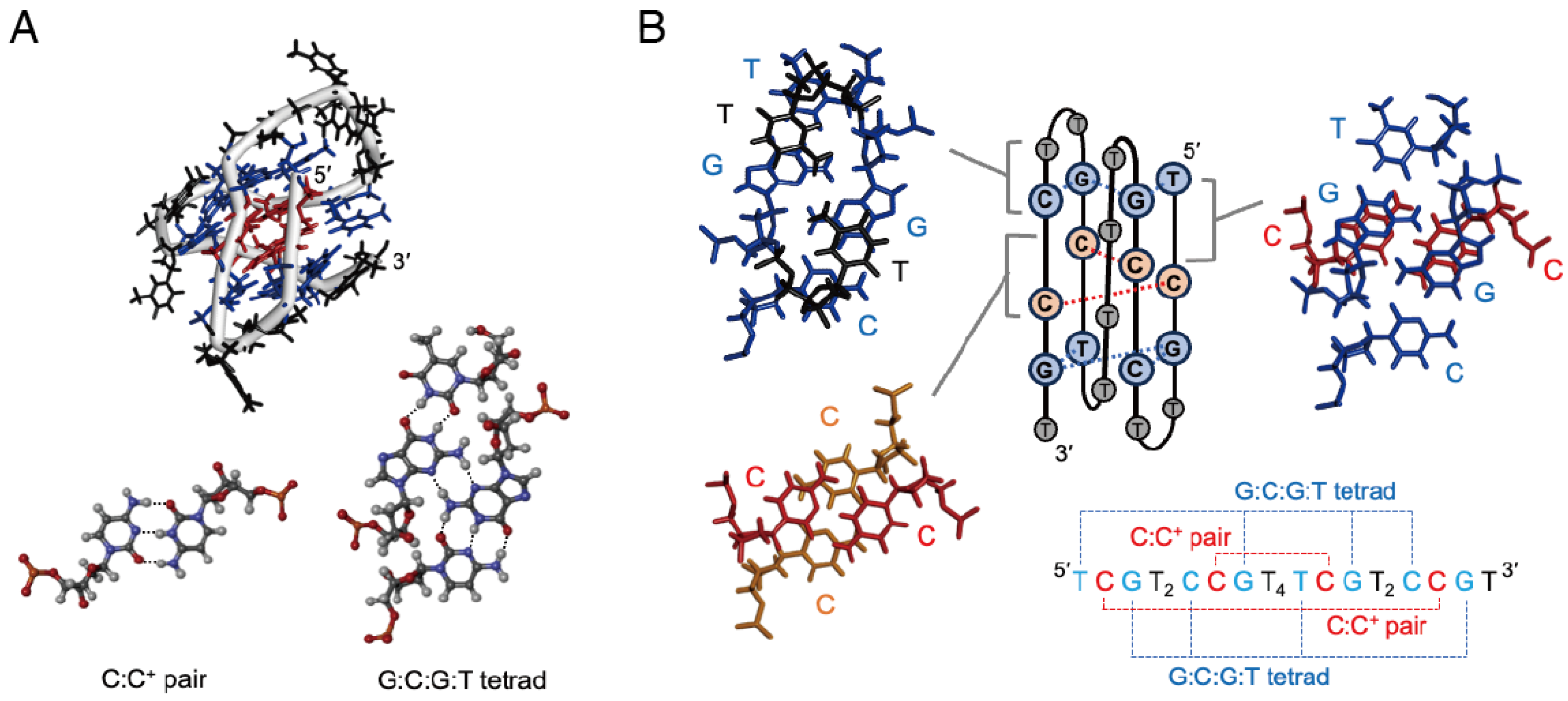
2.1. Formation of I-Motifs with Base Tetrads

Here, we study the variability in the i-motif-forming DNA sequence comprising two TCGTTCCGT sequence units, which forms two C:C+ pairs and two G:C:G:T tetrads. The base tetrad is formed through hydrogen-bonded G:G pairing at the minor groove of G residues, with each G also engaging in base pairing with either C (G:C pairing) or T (G:T pairing). The reference oligonucleotide TCGTTCCGTAAATCGTTCCGT (mini-iM) adopts a monomeric i-motif structure, where the C residues at positions 2, 7, 14, and 19 (numbered from the 5′ end) form a C:C+ pair and the nucleotides at positions 1, 3, 6, 8, 13, 15, 18, and 20 participate in a G:C:G:T tetrad (Scheme 2A). According to the reported nuclear magnetic resonance (NMR) structure in solution [24], these nucleotides engage in base stacking interactions (Scheme 1B), where the overlapping area between the G:C:G:T tetrad and the adjacent C:C+ pair is relatively large while the overlap between C:C+ pairs is relatively small. The base overlap also occurs between the G:C:G:T tetrad and the first T of each loop at positions 4, 9, and 16 or the 3′-terminal T at position 21.
The circular dichroism (CD) spectra of mini-iM in acidic pH solutions were consistent with those previously reported for a DNA minimal i-motif structure [24]. The spectra varied with the pH of the multi-component Britton–Robinson buffer (Figure 1A), indicating pH-dependent structural formation. Structural stability was assessed by monitoring the decrease in absorbance at 295 nm upon heating, a characteristic feature of i-motif melting [16,17,25,26]. The structure of mini-iM exhibited a two-state melting transition, with a melting temperature (Tm) of 38.1 °C at pH 5.9. This value demonstrated strong pH dependence (Figure 1B), consistent with the stabilization effect due to the formation of C:C+ pairs and the cation–π interactions between the protonated base pair and the base tetrad [27]. The Tm values of mini-iM in a simple phosphate buffer at a concentration of 10 mM or 50 mM adjusted to pH 6.0 were 36.2 °C or 37.3 °C, respectively, and these values are similar to those in the multi-component buffer adjusted to pH 5.9. Further investigations demonstrated that the composition of buffer solutions adjusted to pH 6.0 did not significantly influence the Tm of mini-iM (Table S1), in contrast to the observation for an i-motif formed by a C-rich sequence [28]. Accordingly, to compare the Tm values of different DNA sequences, we used the 10 mM phosphate buffer. A short oligonucleotide composed of a single TCGTTCCGT unit did not form an ordered structure, indicating that mini-iM adopts a monomeric i-motif structure under the condition.
The G residues at positions 3, 8, 15, and 20 in the sequence of mini-iM participate in base tetrad formation. To evaluate the importance of the G:C:G:T tetrad, oligonucleotides wherein these G residues were substituted with T were assessed. All substitutions resulted in a large decrease in Tm (Figure 1C), indicating the critical role of base tetrad capping interactions in the formation of the i-motif of mini-iM. Further investigations were conducted on oligonucleotides wherein T residues (at positions 1 or 13) paired with G were replaced with C or C residues (at positions 6 or 18) paired with G were replaced with T. These modifications altered the G:C:G:T tetrad to a G:T:G:T tetrad (T1C and T13C) or a G:C:G:C tetrad (C6T and C18T), respectively. The Tm of these oligonucleotides exceeded 30 °C (Figure 1C), confirming that i-motifs containing G:C:G:C or G:T:G:T tetrads are also stable; however, i-motifs containing a G:T:G:T tetrad are less stable than those containing a G:C:G:C tetrad. According to the thermodynamic parameters for i-motif formation (Table 1), the alterations of either G:C:G:T or G:C:G:C to G:T:G:T decreased the thermodynamic stability but did not generally reduce the enthalpic contribution. This suggests that the observed decrease in stability cannot be solely attributed to a reduction in the number of hydrogen bonds within the tetrad.

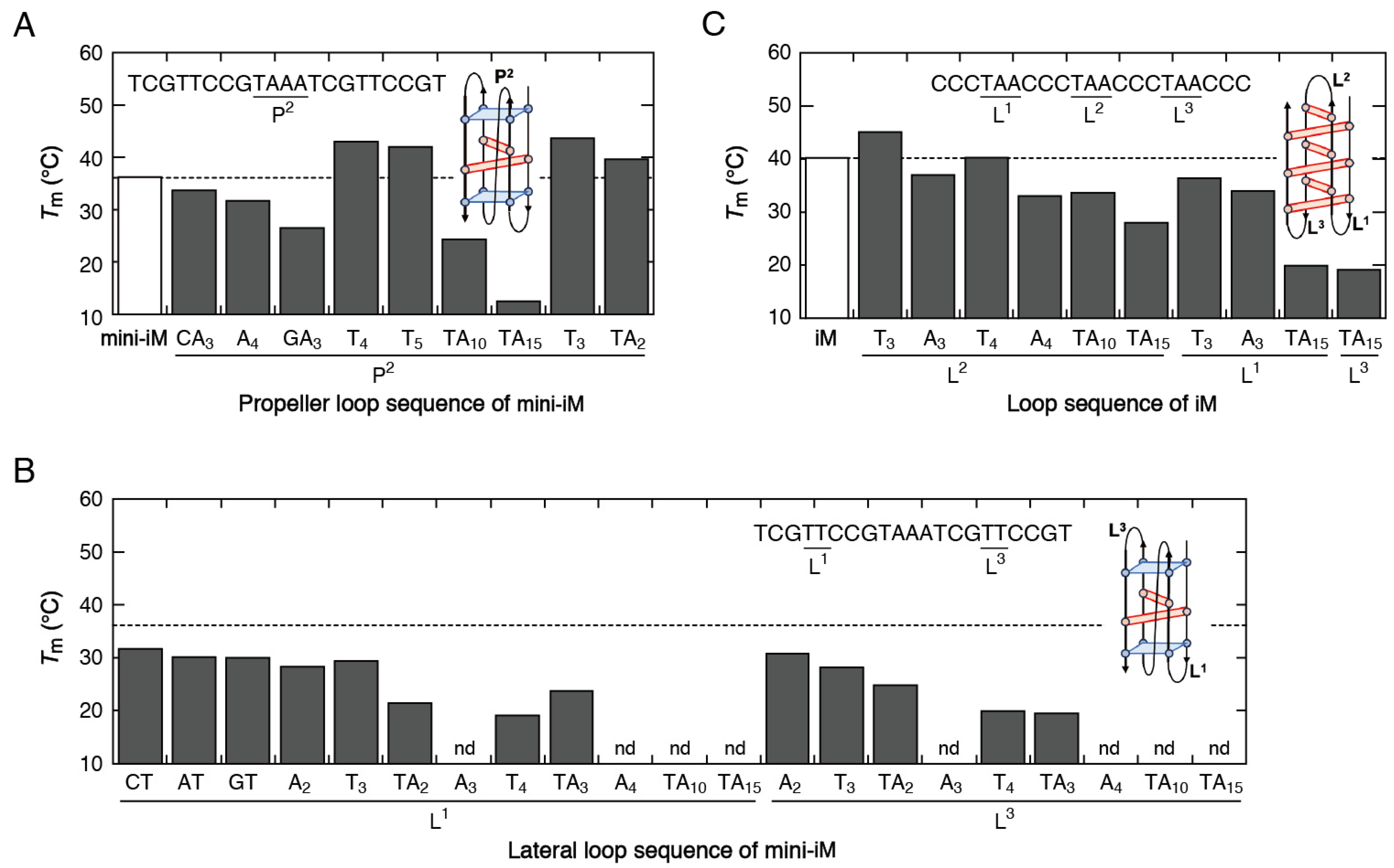
2.2. Variation in the Loop Sequences

The i-motif of mini-iM contains one propeller-type loop consisting of TAAA and two lateral loops consisting of TT. Substituting the first T of the propeller loop with C, A, or G lowered the Tm by 2.5 °C, 4.5 °C, or 9.7 °C, respectively (Figure 2A). Similarly, substituting the first T of the lateral loop with C, A, or G lowered the Tm by 4.5 °C, 6.1 °C, or 6.2 °C, respectively (Figure 2B). These results indicate that the first T of these loops has a stabilizing effect on the i-motif structure. Comparing loops with consecutive T and A residues further revealed a preference for T over A in both the propeller and lateral loops. Additionally, an oligonucleotide with a deletion of the 3′-terminal T of the sequence of mini-iM exhibited a Tm decrease of 6.4 °C, indicating the stabilizing effect of the 3′ dangling end. These results demonstrate the significant contribution of stacking interactions of the base tetrad with the T residues either as components of a loop or as a dangling end observed in the NMR structure [24], in stabilizing the minimal i-motif.
Loop length variations had different effects on the propeller and lateral loops. The propeller loop could be extended without inducing substantial destabilization, exhibiting a modest Tm decrease of 11.9 °C when the number of A residues in the loop was increased to 10 (Figure 2A). However, a substantial Tm decrease was observed when the number of A residues was increased to 15. The propeller loop could also be shortened to three nucleotides without disrupting minimal i-motif formation. In contrast, increasing the length of each lateral loop to more than three nucleotides effectively disrupted i-motif formation, except for the loops consisting of consecutive T residues (T3 and T4) and the loops containing a T residue at the first position (TA2 and TA3), which exhibited only moderate Tm decreases (Figure 2B). It is notable that the effects of loop sequence modifications were similar for both lateral loops, agreed with the structure formed by two repeating sequence units.
Next, we studied a typical i-motif structure formed by the C-rich sequence CCCTAACCCTAACCCTAACCC (iM), which has been reported to adopt a monomeric i-motif structure consisting of six C:C+ pairs and three TAA loops [29]. Although the sequence context and length of the loops influenced the stability of this C-rich i-motif (Figure 2C), the impacts were less pronounced compared to those on mini-iM. The data further indicated a preference for T over A. However, increasing the length of each loop of iM resulted in substantially smaller Tm decreases compared to those observed for mini-iM. These results suggest that the loop sequence exerts a greater influence on stabilizing the minimal i-motif than on the i-motif that lacks base tetrad capping interactions.

2.3. I-Motifs Containing a Bulge

Nucleotide insertions in the sequence of mini-iM can create a bulge within the minimal i-motif structure. Figure 3A presents the Tm values of oligonucleotides wherein either T or A was inserted between C and G (between positions 2 and 3, 7 and 8, 14 and 15, or 19 and 20). These insertions introduce a bulge that may disrupt interactions between the C:C+ pair and G:C:G:T tetrad. Regardless of the insertion position, T insertions led to substantial Tm decreases. For instance, inserting T between positions 2 and 3 lowered the Tm by 22.5 °C compared to the Tm of mini-iM. Similarly, A insertions induced substantial destabilization, with Tm decreases exceeding 19 °C. Overall, these insertions hindered stable i-motif formation. Furthermore, inserting an abasic nucleotide (a tetrahydrofuranyl residue) at these positions resulted in Tm decreases comparable to those caused by T or A insertions. It is likely that the primary factor disrupting i-motif formation is the extended distance between the C:C+ pair and G:C:G:T tetrad, weakening base stacking interactions and cation–π interactions, rather than steric hindrance from a specific nucleotide base. Additionally, the presence of an extra nucleotide within the i-motif core may further disrupt hydrogen bonding between the DNA backbones in the narrow grooves.
Inserting either A or G in a nucleotide upstream of a C residue involved in C:C+ pairing introduces a bulge that may disrupt base stacking interactions between intercalated C:C+ pairs. Figure 3B presents the Tm values of oligonucleotides with such insertions. Although the stacking interaction between C:C+ pairs is inherently weak owing to their small overlapping area, inserting A between two consecutive C residues (between positions 6 and 7, or 18 and 19) induced substantial Tm decreases to below 10 °C, effectively disrupting i-motif formation. In contrast, inserting A between T and C (between positions 1 and 2, or 13 and 14) resulted in more moderate Tm decreases. Similarly, inserting an abasic nucleotide between T and C resulted in smaller Tm decreases compared to the insertions between two consecutive C residues. On the other hand, G insertions led to relatively stable i-motif formation (with a Tm exceeding 25 °C), except for the insertion between positions 18 and 19. Moreover, increasing the number of G or A insertions from one to two in the positions exhibiting moderate Tm decreases did not cause a substantial decrease in Tm (Table S2). These results indicate that while many types of nucleotide insertions hinder stable i-motif formation, some insertions do not. For the i-motif of iM, inserting T or an abasic nucleotide between consecutive C residues resulted in only moderate Tm decreases across all insertion positions (Figure 3C). This comparison suggests that minimal i-motif structures have a relatively low capacity for accommodating bulges.

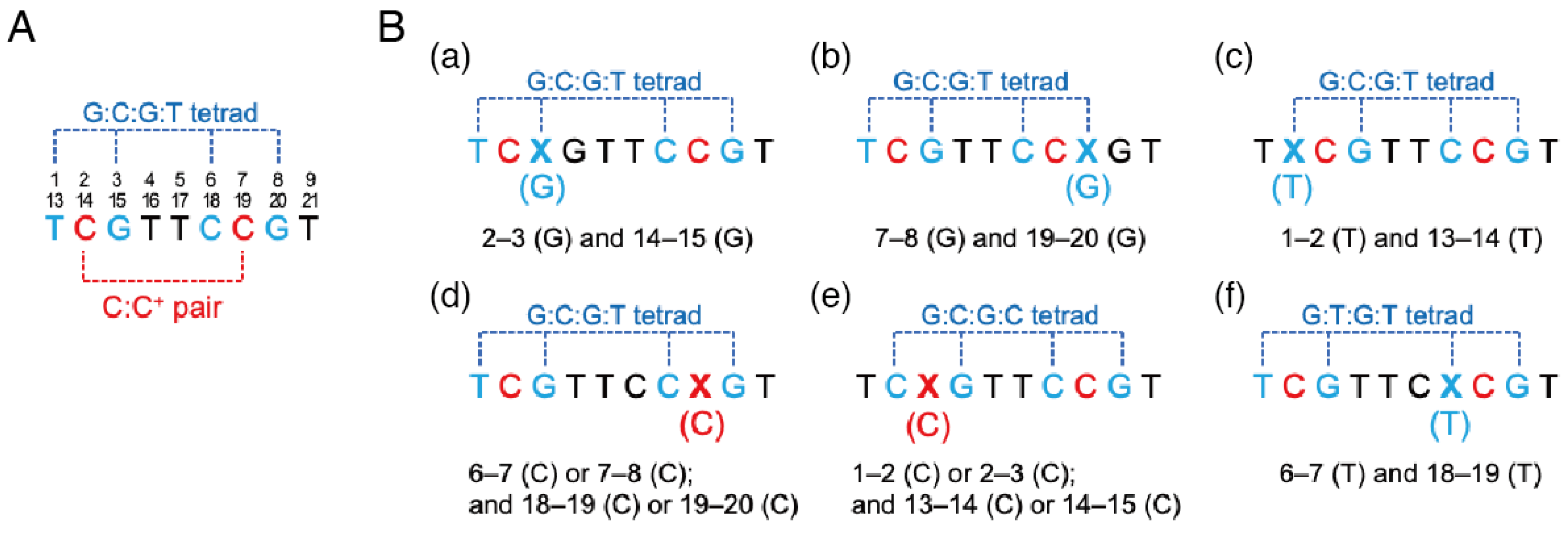
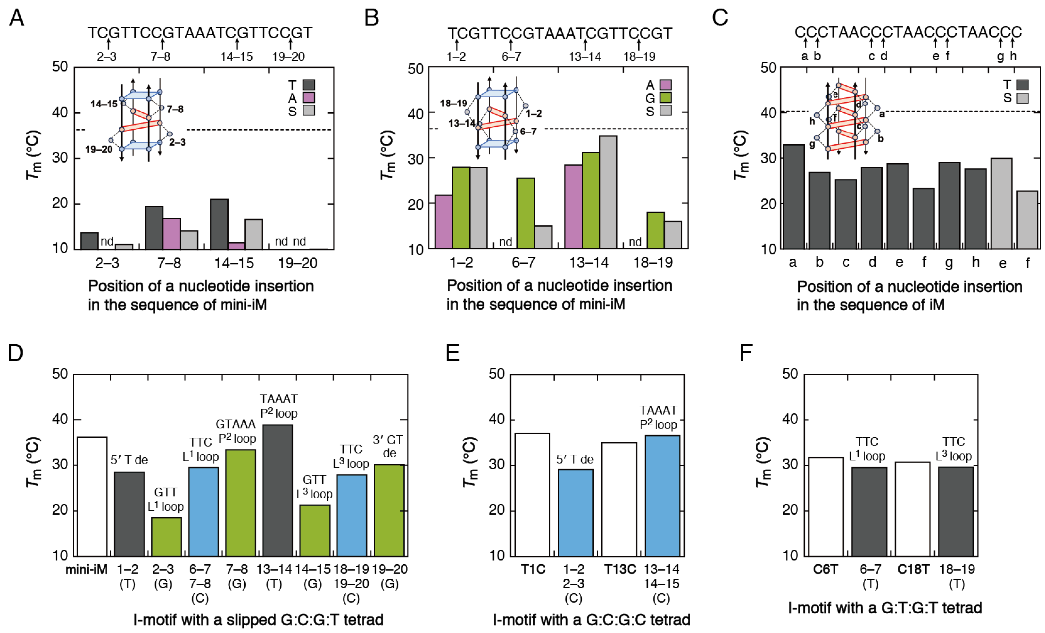
2.4. I-Motifs with a Slipped Base Tetrad

Some nucleotide insertions can induce slippage of base pairing between adjacent nucleotides. Figure 3D presents the Tm values of oligonucleotides forming a slipped G:C:G:T tetrad, resulting in an additional unpaired nucleotide as part of either an interior loop or a dangling end. Specifically, inserting G between C and G (between positions 2 and 3, or 14 and 15) led to the formation of a GTT lateral loop (Scheme 2(Ba)), and inserting G between C and G (between positions 7 and 8, or 19 and 20) led to the formation of either a GTAAA propeller loop or a 3′ GT dangling end (Scheme 2(Bb)). Alternatively, inserting T between T and C (between positions 1 and 2, or 13 and 14) led to the formation of either a 5′ T dangling end or a TAAAT propeller loop (Scheme 2(Bc)). Furthermore, inserting C in a nucleotide upstream or downstream of a C residue (between positions 6 and 7, 7 and 8, 18 and 19, or 19 and 20) induced slippage of both the C:C+ pair and the G:C:G:T tetrad, leading to the formation of a TTC lateral loop (Scheme 2(Bd)). The observed increases or decreases in Tm relative to the Tm of mini-iM aligned with expectations based on stabilizing stacking interactions between the tetrad and the first T of loops or the T at the 3′-end, as depicted in Figure 2A,B. The data further indicated that the formation of a 5′ T dangling end induced destabilization, likely owing to the structural proximity of the 5′-terminal nucleotide of the i-motif structure to propeller loop nucleotides.
Alternative base tetrads, G:C:G:C and G:T:G:T, could be formed through the slippage of base pairing within the G:C:G:T tetrad, because these tetrads exert similar stabilizing effects as indicated in Table 1. Figure 3E presents the Tm values of oligonucleotides with C insertion between C and G, leading to the formation of a G:C:G:C tetrad and either a 5′ T dangling end or a TAAAT propeller loop (Scheme 2(Be)). These structures exhibited only slight increases or decreases in Tm relative to the Tm of i-motifs containing a G:C:G:C tetrad (T1C and T13C), and these structural effects were similar to those observed for i-motifs consisting of two G:C:G:T tetrads. Figure 3F presents the Tm values of oligonucleotides with T insertions between two consecutive C residues, leading to the formation of a G:T:G:T tetrad and a TTC lateral loop (Scheme 2(Bf)). These structures did not exhibit substantial Tm changes relative to the Tm of i-motifs containing a G:T:G:T tetrad (C6T and C18T). Overall, this study demonstrates that nucleotide insertions, resulting in loop sequence modifications or bulge formation, have similar effects on minimal i-motif stability, regardless of the type of base tetrad formed.

2.5. Influence of the Molecular Environment in the Presence of Spermine

Polyamines are present at millimolar or higher concentrations in living cells and are important environmental factors regulating gene expression [30]. Spermine, having four protonation sites, is one of the most prevalent polyamines in mammalian cells. DNA interacts with spermine primarily through Coulombic interactions [31], and consequently, folded DNA structures are often stabilized in the presence of spermine. However, previous studies have reported that the stabilization effect of spermine on i-motif structures differs from the effect on duplex structures. For instance, 0.05 mM spermine increased the Tm of an i-motif formed by a C-rich sequence derived from the c-myc promoter by approximately 8 °C, whereas the same spermine concentration increased the Tm of a DNA duplex by more than 20 °C [32]. The observation cannot be simply explained by electrostatic binding of spermine to the i-motif. To understand the effect of spermine on i-motif stability, we measured the Tm of various minimal i-motif structures in its presence. Because DNA aggregation occurs at high spermine concentrations [33,34], many oligonucleotides aggregated in the presence of 1 mM spermine. However, no aggregation was observed with 0.1 mM spermine. Specifically, 0.1 mM spermine increased the Tm of mini-iM to 46.1 °C, representing a 9.9 °C increase over the Tm in the absence of spermine. The i-motifs containing a G:C:G:C or G:T:G:T tetrad (T1C, T13C, C6T, and C18T) exhibited Tm increases in the range of 10.2 °C to 12.1 °C, similar to the value observed for mini-iM. Other oligonucleotides designed based on the sequence of mini-iM were also stabilized in the presence of spermine; however, the extent of Tm changes showed considerable variations. Spermine increased the Tm of i-motifs with a long propeller loop by more than 20 °C, approaching the Tm of mini-iM (Figure 4A). It also notably stabilized i-motifs with any two-nucleotide lateral loop and a three- or four-nucleotide lateral loop containing a T residue at the first position, to a level comparable to the Tm of mini-iM (Figure 4B). This observation is striking, because the lateral loop sequence could not be readily altered in the absence of spermine. Spermine also increased the Tm of various minimal i-motif structures. In particular, i-motifs with a bulge between T and C and those with a dangling end exhibited stabilization to a level comparable to their reference i-motifs (Figure 4C–G). These results suggest that spermine can effectively restore the stability reduction caused by nucleotide substitutions or insertions in the sequence of mini-iM. Consequently, the molecular environment in the presence of spermine increases the likelihood of forming minimal i-motif structures containing a long loop, bulge, or dangling end.
In contrast to its effects on the i-motif of mini-iM, spermine did not increase the Tm of iM (39.7 °C) and only partially restored the stability reduction caused by either loop sequence modifications (Figure 4H) or insertions of T or an abasic nucleotide between consecutive C residues (Figure 4I). The observed difference in the effects of spermine between mini-iM and iM may be attributed to the different number of C:C+ pairs, as C protonation negatively impacts electrostatic binding with spermine. Previous studies have reported that polyamines interact weakly with i-motifs consisting of six C:C+ pairs and primarily bind to loop regions [32]. It is likely that spermine additionally binds to the core of minimal i-motif structures consisting of only two C:C+ pairs, forming a relatively stable complex.

2.6. Putative I-Motif-Forming Sequences Lacking C-Tracts

We identified nucleotide substitutions and insertions that did not disrupt minimal i-motif formation. Effects of the modifications were generally the same for each repeating sequence unit. This finding was used to establish a consensus sequence for an i-motif consisting of two C:C+ pairs and two base tetrads, based on a previously proposed consensus sequence, YCGXXYCG, where Y represents a pyrimidine nucleotide and X can be any nucleotide forming a lateral loop [24]. We found that the lateral loop length can be extended when the first nucleotide is T. Furthermore, the first nucleotide in this repeating sequence can be a purine nucleotide involved in bulge formation. Incorporating the additional information, broadened consensus sequences, YCGLYCG and YNCGLYCG, can be proposed, where Y represents a pyrimidine nucleotide, L denotes a loop consisting of either any two nucleotides or more than three nucleotides if the first nucleotide is T, and N represents any one or more bulge nucleotides. These sequences lead to an increase in the number of potential i-motif-forming sites in genomes. For example, considering the possibility of lengthening the lateral loop, a TCG triplet repeat sequence may serve as a potential minimal i-motif-forming sequence. Additionally, the insertion positions of stable bulge formation within the i-motif are speculated to accommodate a duplex bulge, as has been reported for the duplex bulge in a G-quadruplex, forming a G-quadruplex–duplex junction [35]. The potential for i-motif–duplex junction formation further expands the structural diversity of i-motifs. More importantly, the ability to form minimal i-motif structures containing a bulge, long loop, or dangling end is enhanced in the presence of spermine. This suggests that the polyamine-rich molecular environment of the nucleus of mammalian cells facilitates the formation of minimal i-motifs with various sequences. In particular, because both acidity levels and polyamine concentrations are higher in tumor cells than in normal cells [36,37], the formation of minimal i-motif structures is possibly more enhanced in tumor cells.

3. Materials and Methods

3.1. Preparation of Oligonucleotide and Buffer Solutions

All DNA oligonucleotides were purchased from Fasmac (Kanagawa, Atsugi, Japan). Oligonucleotide concentrations were determined by measuring absorbance at 260 nm, using molar extinction coefficients calculated based on those of mononucleotides and dinucleotides [38]. The buffer solutions used in this study were the Britton–Robinson buffer and a phosphate buffer. The Britton–Robinson buffer is a multi-component solution, consisting of 10 mM sodium dihydrogenphosphate (NaH2PO4), 10 mM boric acid (H3BO3), 10 mM acetic acid (CH3COOH), and 0.2 mM disodium ethylenediamine-N,N,N′,N′-tetraacetic acid (Na2EDTA), adjusted to a pH range of 5.0 to 9.0. The phosphate buffer consists of 10 mM disodium hydrogenphosphate (Na2HPO4) and 0.5 mM Na2EDTA, adjusted to pH 6.0, and its buffering capacity was verified by conducting experiments for directly measuring the pH of the solution. All reagents used for buffer preparation were purchased from Fujifilm Wako Pure Chemical Corp. (Osaka, Japan), except for Na2EDTA, which was obtained from Dojindo (Kumamoto, Japan). The pH of solutions was adjusted using a HORIBA F-52 pH meter (Kyoto, Japan).

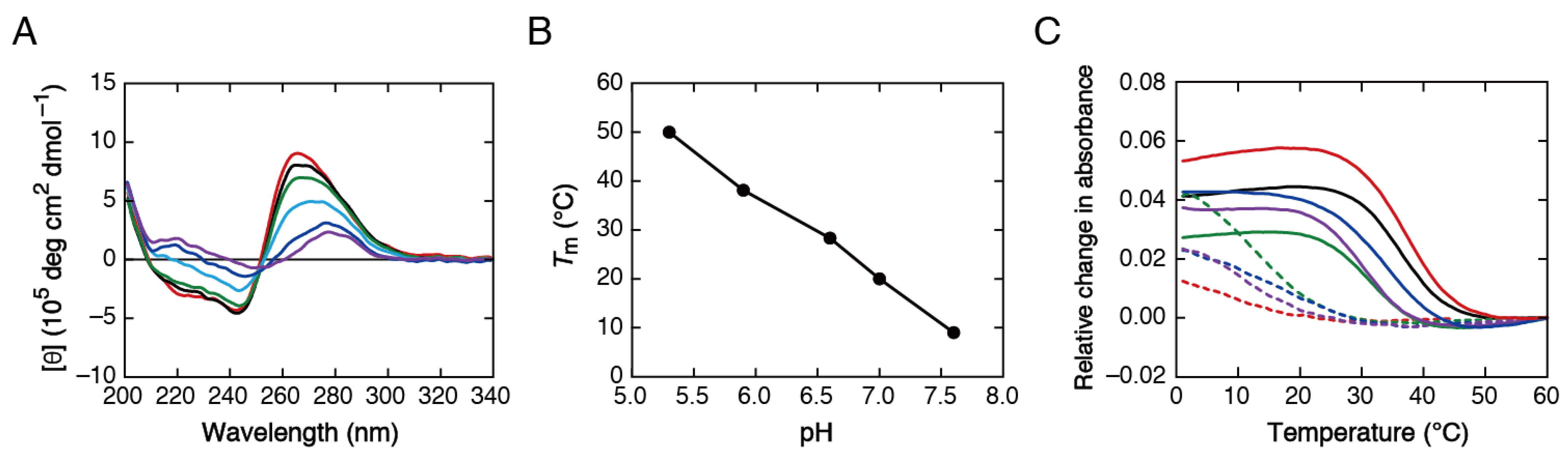
3.2. Measurement of CD Spectra

CD spectra were recorded using a JASCO J-820 CD spectropolarimeter (Tokyo, Japan) equipped with a temperature controller. Solutions containing oligonucleotides at a concentration of 5.0 µM in the Britton–Robinson buffer were prepared in a 1 mm cuvette sealed with an adhesive sheet. Before spectral measurements, oligonucleotides were annealed at 70 °C and then slowly cooled to 5.0 °C at a rate of −1.0 °C min−1. CD spectra were then recorded at 5.0 °C in triplicate, scanning from 340 to 200 nm at a rate of 50 nm min−1.

3.3. Measurement and Analysis of DNA Thermal Melting

The thermal melting of DNA structures was assessed by monitoring the hyperchromicity at 295 nm, reflecting cytosine protonation, at a heating rate of 0.5 °C min−1, using a Shimadzu UV-1800 spectrophotometer (Kyoto, Japan) equipped with a temperature controller. Solutions containing oligonucleotides at a concentration of 5.0 µM in a buffer solution were prepared in a 1 cm cuvette sealed with an adhesive sheet. Before melting curve measurements, oligonucleotides were annealed at a high temperature (typically above 60 °C) and then slowly cooled to 0 °C at a rate of −1.0 °C min−1.
Tm values for thermal melting upon heating were determined through the nonlinear fitting of the melting curve (absorbance vs. temperature) to a theoretical melting curve or through the first-derivative analysis for low-temperature melting [39]. Errors in Tm values were typically smaller than 0.5 °C, and thus, the theoretical statistical uncertainty in the difference between two Tm values is smaller than 0.7 °C. Thermodynamic parameters (∆G°, ∆H°, and ∆S°) for i-motif formation were determined through the nonlinear fitting of the melting curve to the van’t Hoff equation assuming a two-state melting transition [39,40], and values are reported as averages with standard deviations of four independent measurements.

4. Conclusions

This study presents the first comprehensive investigation into the stability and variability of DNA minimal i-motifs, possessing structural features distinct from those of i-motifs formed by C-rich sequences. Our findings demonstrate that base tetrad formation and its stacking interactions with C:C+ pairs are essential for stabilizing the minimal i-motif. Base stacking interactions between the tetrad and the first T of each loop further contribute to structural stability. A striking observation is that the sequence context and length of their lateral loops cannot be readily altered, unlike DNA loops in a duplex, G-quadruplex, and C-rich i-motif structures. Remarkably, spermine increases the variation in the lateral loop sequence ensuring the formation of a stable minimal i-motif. Furthermore, although the minimal i-motif is less likely to accommodate bulges than the C-rich i-motif, spermine increases the likelihood of forming minimal i-motif structures containing a bulge. These results suggest that the formation of minimal i-motifs with various sequences is facilitated in polyamine-rich cellular environments, such as the nucleus of mammalian cells. The findings are valuable for identifying potential i-motif-forming sites lacking C-tracts in genomes and provide insights into the electrostatic interactions between DNA structures involving protonated DNA bases and cationic cellular components.

Supplementary Materials

The following supporting information can be downloaded at: https://www.mdpi.com/article/10.3390/molecules30081831/s1, Table S1: Tm values of mini-iM and iM in different buffer solutions; Table S2: Tm values of oligonucleotides designed based on the sequence of mini-iM; Table S3: Tm values of oligonucleotides designed based on the sequence of iM.

Author Contributions

Data Curation and Analysis, K.A., A.K. and K.N.; Conceptualization, Methodology, Writing, and Funding Acquisition, S.-i.N. All authors have read and agreed to the published version of the manuscript.

Funding

This research was funded by Grants-in-Aid for Scientific Research, Japan Society for the Promotion of Science (JSPS KAKENHI grant number 23K04935).

Institutional Review Board Statement

Not applicable.

Informed Consent Statement

Not applicable.

Data Availability Statement

The data supporting the conclusions of this article will be made available by the authors on request.

Conflicts of Interest

The authors declare no conflicts of interest.

References

  1. Choi, J.; Majima, T. Conformational changes of non-B DNA. Chem. Soc. Rev. 2011, 40, 5893–5909. [Google Scholar] [CrossRef]
  2. Georgakopoulos-Soares, I.; Morganella, S.; Jain, N.; Hemberg, M.; Nik-Zainal, S. Noncanonical secondary structures arising from non-B DNA motifs are determinants of mutagenesis. Genome Res. 2018, 28, 1264–1271. [Google Scholar] [CrossRef] [PubMed]
  3. Chambers, V.S.; Marsico, G.; Boutell, J.M.; Di Antonio, M.; Smith, G.P.; Balasubramanian, S. High-throughput sequencing of DNA G-quadruplex structures in the human genome. Nat. Biotechnol. 2015, 33, 877–881. [Google Scholar] [CrossRef] [PubMed]
  4. Hänsel-Hertsch, R.; Spiegel, J.; Marsico, G.; Tannahill, D.; Balasubramanian, S. Genome-wide mapping of endogenous G-quadruplex DNA structures by chromatin immunoprecipitation and high-throughput sequencing. Nat. Protoc. 2018, 13, 551–564. [Google Scholar] [CrossRef]
  5. Mukundan, V.T.; Phan, A.T. Bulges in G-quadruplexes: Broadening the definition of G-quadruplex-forming sequences. J. Am. Chem. Soc. 2013, 135, 5017–5028. [Google Scholar] [CrossRef]
  6. Meier, M.; Moya-Torres, A.; Krahn, N.J.; McDougall, M.D.; Orriss, G.L.; McRae, E.K.S.; Booy, E.P.; McEleney, K.; Patel, T.R.; McKenna, S.A.; et al. Structure and hydrodynamics of a DNA G-quadruplex with a cytosine bulge. Nucleic Acids Res. 2018, 46, 5319–5331. [Google Scholar] [CrossRef] [PubMed]
  7. Papp, C.; Mukundan, V.T.; Jenjaroenpun, P.; Winnerdy, F.R.; Ow, G.S.; Phan, A.T.; Kuznetsov, V.A. Stable bulged G-quadruplexes in the human genome: Identification, experimental validation and functionalization. Nucleic Acids Res. 2023, 51, 4148–4177. [Google Scholar] [CrossRef]
  8. Skolakova, P.; Renciuk, D.; Palacky, J.; Krafcik, D.; Dvorakova, Z.; Kejnovska, I.; Bednarova, K.; Vorlickova, M. Systematic investigation of sequence requirements for DNA i-motif formation. Nucleic Acids Res. 2019, 47, 2177–2189. [Google Scholar] [CrossRef]
  9. Zanin, I.; Ruggiero, E.; Nicoletto, G.; Lago, S.; Maurizio, I.; Gallina, I.; Richter, S.N. Genome-wide mapping of i-motifs reveals their association with transcription regulation in live human cells. Nucleic Acids Res. 2023, 51, 8309–8321. [Google Scholar] [CrossRef]
  10. Roxo, C.; Pasternak, A. Switching off cancer—An overview of G-quadruplex and i-motif functional role in oncogene expression. Bioorg. Med. Chem. Lett. 2024, 116, 130038. [Google Scholar] [CrossRef]
  11. Pena Martinez, C.D.; Zeraati, M.; Rouet, R.; Mazigi, O.; Henry, J.Y.; Gloss, B.; Kretzmann, J.A.; Evans, C.W.; Ruggiero, E.; Zanin, I.; et al. Human genomic DNA is widely interspersed with i-motif structures. EMBO J. 2024, 43, 4786–4804. [Google Scholar] [CrossRef]
  12. Dzatko, S.; Krafcikova, M.; Hansel-Hertsch, R.; Fessl, T.; Fiala, R.; Loja, T.; Krafcik, D.; Mergny, J.L.; Foldynova-Trantirkova, S.; Trantirek, L. Evaluation of the stability of DNA i-motifs in the nuclei of living mammalian cells. Angew. Chem. Int. Ed. Engl. 2018, 57, 2165–2169. [Google Scholar] [CrossRef] [PubMed]
  13. Zeraati, M.; Langley, D.B.; Schofield, P.; Moye, A.L.; Rouet, R.; Hughes, W.E.; Bryan, T.M.; Dinger, M.E.; Christ, D. I-motif DNA structures are formed in the nuclei of human cells. Nat. Chem. 2018, 10, 631–637. [Google Scholar] [CrossRef] [PubMed]
  14. Cheng, M.; Qiu, D.; Tamon, L.; Istvankova, E.; Viskova, P.; Amrane, S.; Guedin, A.; Chen, J.; Lacroix, L.; Ju, H.; et al. Thermal and pH stabilities of i-DNA: Confronting in vitro experiments with models and in-cell NMR data. Angew. Chem. Int. Ed. Engl. 2021, 60, 10286–10294. [Google Scholar] [CrossRef] [PubMed]
  15. Viskova, P.; Istvankova, E.; Rynes, J.; Dzatko, S.; Loja, T.; Zivkovic, M.L.; Rigo, R.; El-Khoury, R.; Serrano-Chacon, I.; Damha, M.J.; et al. In-cell NMR suggests that DNA i-motif levels are strongly depleted in living human cells. Nat. Commun. 2024, 15, 1992. [Google Scholar] [CrossRef]
  16. Wright, E.P.; Huppert, J.L.; Waller, Z.A.E. Identification of multiple genomic DNA sequences which form i-motif structures at neutral pH. Nucleic Acids Res. 2017, 45, 2951–2959. [Google Scholar] [CrossRef]
  17. Fleming, A.M.; Ding, Y.; Rogers, R.A.; Zhu, J.; Zhu, J.; Burton, A.D.; Carlisle, C.B.; Burrows, C.J. 4n-1 Is a “sweet spot” in DNA i-motif folding of 2′-deoxycytidine homopolymers. J. Am. Chem. Soc. 2017, 139, 4682–4689. [Google Scholar] [CrossRef]
  18. Berger, I.; Egli, M.; Rich, A. Inter-strand C-H···O hydrogen bonds stabilizing four-stranded intercalated molecules: Stereoelectronic effects of O4′ in cytosine-rich DNA. Proc. Natl. Acad. Sci. USA 1996, 93, 12116–12121. [Google Scholar] [CrossRef]
  19. Gurung, S.P.; Schwarz, C.; Hall, J.P.; Cardin, C.J.; Brazier, J.A. The importance of loop length on the stability of i-motif structures. Chem. Commun. 2015, 51, 5630–5632. [Google Scholar] [CrossRef]
  20. McKim, M.; Buxton, A.; Johnson, C.; Metz, A.; Sheardy, R.D. Loop sequence context influences the formation and stability of the i-motif for DNA oligomers of sequence (CCCXXX)4, where X = A and/or T, under slightly acidic conditions. J. Phys. Chem. B 2016, 120, 7652–7661. [Google Scholar] [CrossRef]
  21. Abou Assi, H.; Garavis, M.; Gonzalez, C.; Damha, M.J. I-motif DNA: Structural features and significance to cell biology. Nucleic Acids Res. 2018, 46, 8038–8056. [Google Scholar] [CrossRef] [PubMed]
  22. Escaja, N.; Viladoms, J.; Garavis, M.; Villasante, A.; Pedroso, E.; Gonzalez, C. A minimal i-motif stabilized by minor groove G:T:G:T tetrads. Nucleic Acids Res. 2012, 40, 11737–11747. [Google Scholar] [CrossRef]
  23. Escaja, N.; Mir, B.; Garavis, M.; Gonzalez, C. Non-G base tetrads. Molecules 2022, 27, 5287. [Google Scholar] [CrossRef]
  24. Mir, B.; Serrano, I.; Buitrago, D.; Orozco, M.; Escaja, N.; Gonzalez, C. Prevalent sequences in the human genome can form mini i-motif structures at physiological pH. J. Am. Chem. Soc. 2017, 139, 13985–13988. [Google Scholar] [CrossRef]
  25. Mergny, J.-L.; Lacroix, L.; Han, X.; Leroy, J.-L.; Hélène, C. Intramolecular folding of pyrimidine oligodeoxynucleotides into an i-DNA motif. J. Am. Chem. Soc. 1995, 117, 8887–8898. [Google Scholar] [CrossRef]
  26. Benabou, S.; Aviñó, A.; Eritja, R.; González, C.; Gargallo, R. Fundamental aspects of the nucleic acid i-motif structures. RSC Adv. 2014, 4, 26956–26980. [Google Scholar] [CrossRef]
  27. Mir, B.; Soles, X.; Gonzalez, C.; Escaja, N. The effect of the neutral cytidine protonated analogue pseudoisocytidine on the stability of i-motif structures. Sci. Rep. 2017, 7, 2772. [Google Scholar] [CrossRef]
  28. Gao, B.; Hou, X.M. Opposite effects of potassium ions on the thermal stability of i-motif DNA in different buffer systems. ACS Omega 2021, 6, 8976–8985. [Google Scholar] [CrossRef]
  29. Phan, A.T.; Gueron, M.; Leroy, J.L. The solution structure and internal motions of a fragment of the cytidine-rich strand of the human telomere. J. Mol. Biol. 2000, 299, 123–144. [Google Scholar] [CrossRef]
  30. Childs, A.C.; Mehta, D.J.; Gerner, E.W. Polyamine-dependent gene expression. Cell Mol. Life Sci. 2003, 60, 1394–1406. [Google Scholar] [CrossRef]
  31. Lightfoot, H.L.; Hall, J. Endogenous polyamine function − the RNA perspective. Nucleic Acids Res. 2014, 42, 11275–11290. [Google Scholar] [CrossRef] [PubMed]
  32. Molnar, M.M.; Liddell, S.C.; Wadkins, R.M. Effects of polyamine binding on the stability of DNA i-motif structures. ACS Omega 2019, 4, 8967–8973. [Google Scholar] [CrossRef] [PubMed]
  33. Raspaud, E.; Chaperon, I.; Leforestier, A.; Livolant, F. Spermine-induced aggregation of DNA, nucleosome, and chromatin. Biophys. J. 1999, 77, 1547–1555. [Google Scholar] [CrossRef] [PubMed]
  34. Williams, A.M.; Poudyal, R.R.; Bevilacqua, P.C. Long tracts of guanines drive aggregation of RNA G-quadruplexes in the presence of spermine. Biochemistry 2021, 60, 2715–2726. [Google Scholar] [CrossRef]
  35. Ngoc Nguyen, T.Q.; Lim, K.W.; Phan, A.T. Duplex formation in a G-quadruplex bulge. Nucleic Acids Res. 2020, 48, 10567–10575. [Google Scholar] [CrossRef]
  36. Murray-Stewart, T.R.; Woster, P.M.; Casero, R.A., Jr. Targeting polyamine metabolism for cancer therapy and prevention. Biochem. J. 2016, 473, 2937–2953. [Google Scholar] [CrossRef]
  37. Novita Sari, I.; Setiawan, T.; Seock Kim, K.; Toni Wijaya, Y.; Won Cho, K.; Young Kwon, H. Metabolism and function of polyamines in cancer progression. Cancer Lett. 2021, 519, 91–104. [Google Scholar] [CrossRef]
  38. Richard, R.G. Handbook of Biochemistry and Molecular Biology: Nucleic Acids, 3rd ed.; CRC Press: Cleveland, OH, USA, 1975; Volume 1, p. 597. [Google Scholar]
  39. Puglisi, J.D.; Tinoco, I., Jr. Absorbance melting curves of RNA. Methods Enzymol. 1989, 180, 304–325. [Google Scholar] [CrossRef]
  40. Mergny, J.L.; Lacroix, L. Analysis of thermal melting curves. Oligonucleotides 2003, 13, 515–537. [Google Scholar] [CrossRef]
Scheme 1. (A) Structure and base–base interactions of the minimal i-motif determined through NMR spectroscopy (PDB ID: 5OGA). (B) The base overlap between C:C+ pairs, between the C:C+ pair and G:C:G:T tetrad, and between the tetrad and the first T residues of loops.
Molecules 30 01831 sch001
Scheme 2. (A) Base–base interactions and nucleotide numbering from the 5′ end of the sequence of mini-iM. (B) Slipped base interactions resulting from an insertion of X in the sequence of mini-iM: (a,b) G insertions between C and G, (c) T insertion between T and C, (d,e) C insertions between C and G, and (f) T insertion between two consecutive C residues.
Molecules 30 01831 sch002
Figure 1. (A) CD spectra of mini-iM in solutions with varying pH: 5.0 (red), 5.9 (black), 7.0 (green), 7.6 (light blue), 8.0 (blue), and 9.0 (purple). (B) Dependence of the Tm of mini-iM on solution pH. (C) Melting curves of mini-iM (black) and oligonucleotides with different substitutions: C replaced with T at position 1 (red) or 13 (blue); T replaced with C at position 6 (green) or 18 (purple); and T replaced with G at position 3 (dashed red line), 8 (dashed green line), 15 (dashed blue line), or 20 (dashed purple line).
Molecules 30 01831 g001
Figure 2. Tm values of oligonucleotides forming the i-motifs with varying loop sequences, designed based on the sequence of mini-iM (A,B) or iM (C). Data for oligonucleotides with a Tm below 10 °C are labeled as “nd”, and dashed horizontal lines represent the level of the Tm of mini-iM (36.2 °C, in panels (A,B)) or iM (40.2 °C, in panel (C)).
Molecules 30 01831 g002
Figure 3. Tm values of oligonucleotides containing nucleotide insertions in the sequence of mini-iM or iM. (A) Insertions of T, A, and S (abasic nucleotide) between C and G. (B) Insertions of A, G, and S in a nucleotide upstream of a C residue involved in C:C+ pairing. (C) Insertions of T and S between consecutive C residues in the sequence of iM. (DF) Insertions of T (black), G (green), and C (blue) that induce the formation of a slipped G:C:G:T, G:C:G:C, or G:T:G:T tetrad, resulting in an additional unpaired nucleotide as part of either a dangling end (de) or a loop. Data for oligonucleotides without insertions are indicated as white bars. Data for oligonucleotides with a Tm below 10 °C are labeled as “nd”, and dashed horizontal lines represent the level of the Tm of oligonucleotides without insertions.
Molecules 30 01831 g003
Figure 4. Tm values of oligonucleotides in the presence of 0.1 mM spermine (red), overlaid with the data obtained in the absence of spermine from Figure 2 and Figure 3. (AG) Data for minimal i-motif-forming sequences from Figure 2A,B and Figure 3A,B,D–F, respectively. (H,I) Data for C-rich i-motif-forming sequences from Figure 2C and Figure 3C, respectively. Data for oligonucleotides with a Tm below 10 °C are labeled as “nd”, and dashed horizontal lines represent the level of the Tm of mini-iM (46.1 °C, in panels (AD)) or iM (39.7 °C, in panels (H,I)).
Molecules 30 01831 g004
Table 1. Thermodynamic parameters for the formation of minimal i-motif structures with different base tetrads a.
DNA Sequence (from 5′ to 3′)Tm (°C)G° (kcal mol−1)H° (kcal mol−1)S° (cal mol−1 K−1)
mini-iMTCGTTCCGTAAATCGTTCCGT36.2 ± 0.50.12 ± 0.08−47.7 ± 2.1−154 ± 7
(two G:C:G:T tetrads)
T1CCCGTTCCGTAAATCGTTCCGT37.0 ± 0.5−0.01 ± 0.07−43.4 ± 1.3−140 ± 4
(G:C:G:C and G:C:G:T tetrads)
T13CTCGTTCCGTAAACCGTTCCGT35.0 ± 0.30.24 ± 0.04−37.4 ± 0.8−121 ± 3
(G:C:G:C and G:C:G:T tetrads)
C6TTCGTTTCGTAAATCGTTCCGT31.7 ± 0.30.77 ± 0.07−44.5 ± 2.1−146 ± 7
(G:T:G:T and G:C:G:T tetrads)
C18TTCGTTCCGTAAATCGTTTCGT30.7 ± 0.50.92 ± 0.13−45.1 ± 3.8−148 ± 12
(G:T:G:T and G:C:G:T tetrads)
a The values are averages and standard deviations of four independent measurements. ∆G° values correspond to values at 37.0 °C.
Disclaimer/Publisher’s Note: The statements, opinions and data contained in all publications are solely those of the individual author(s) and contributor(s) and not of MDPI and/or the editor(s). MDPI and/or the editor(s) disclaim responsibility for any injury to people or property resulting from any ideas, methods, instructions or products referred to in the content.

