Article

In Vitro Evaluation of Novel Furo[3,2-c]coumarins as Cholinesterases and Monoamine Oxidases Inhibitors

by
Mariagrazia Rullo
1,
Alice Benzi
2,
Lara Bianchi
2,
Massimo Maccagno
2,*,
Guglielmo Marcantoni Taddei
2,
Daniela Valeria Miniero
3,4,
Giuseppe Felice Mangiatordi
5,
Giovanni Lentini
1,
Leonardo Pisani
1,*,
Giovanni Petrillo
2 and
Cinzia Tavani
2
1
Department of Pharmacy—Pharmaceutical Sciences, University of Bari “Aldo Moro”, Via E. Orabona, 4, 70125 Bari, Italy
2
Department of Chemistry and Industrial Chemistry (DCCI), University of Genova, Via Dodecaneso 31, 16146 Genova, Italy
3
Department of Medicine & Surgery, LUM University Giuseppe Degennaro Torre Rossi, 70010 Puglia, Italy
4
Department of Biosciences, Biotechnologies and Environment, University of Bari Aldo Moro, Via E. Orabona 4, 70125 Bari, Italy
5
CNR, Institute of Crystallography, 70126 Bari, Italy
*
Authors to whom correspondence should be addressed.
Molecules 2025, 30(8), 1830; https://doi.org/10.3390/molecules30081830
Submission received: 3 March 2025 / Revised: 10 April 2025 / Accepted: 13 April 2025 / Published: 18 April 2025

Abstract

:
Coumarin represents a privileged structural motif that is quite common in nature-derived and synthetic bioactive molecules. Some of us have recently described the straightforward preparation of complex furo[3,2-c]coumarins through a sequential double coupling protocol. Aiming at finding novel chemical probes for the modulation of key anti-Alzheimer’s targets, a small subset of furo[3,2-c]coumarin prototypes and their non-aromatic synthetic precursors were tested in vitro as inhibitors of ChEs (acetyl- and butyrylcholinesterase, AChE and BChE) and MAOs (monoamine oxidases A and B, MAO A and MAO B). All compounds were low-micromolar AChE inhibitors devoid of toxic effects against SH-SY5Y cells. Lineweaver-Burk plots and docking simulations suggested mixed-type kinetics for inhibitor 3d (IC50 = 4.1 μM toward AChE). Its promising inhibitory profile encompasses additional, highly selective, activity against monoamine oxidase B, with a submicromolar IC50 value (561 nM).

Graphical Abstract

1. Introduction

Coumarin (1,2-benzopyrone, or 2H-1-benzopyran-2-one according to IUPAC classification) and its derivatives are lactones of o-hydroxycinnamic acid widely distributed in nature as free forms or glycosides [1]. More than 1300 structurally diverse coumarins have been identified from different natural sources [2], in particular within the plant kingdom. These oxygen-containing heterocycles can be mainly found in higher plants, where they are produced as secondary metabolites, along the shikimate and the general phenylpropanoid pathways [3], and enable defensive mechanisms. To a lesser extent, coumarins can be isolated from fungi and bacteria. Natural coumarins can be classified into six different subsets according to the core structure: simple coumarins, linear and angular furanocoumarins, linear and angular pyranocoumarins, biscoumarins, benzocoumarins, and coumestans [4].
Coumarin takes its name from coumarou, a vernacular French term used to indicate the bean of the Tonka tree (Dipteryx odorata) that was formerly known with the botanical name Coumaruna odorata [5]. Isolated by Vogel more than two centuries ago [6], coumarin itself is largely used as fragrance in perfume industry and cosmetic products thanks to its flavoring vanilla-type aroma. The consumption of certain types of cinnamon represents the main source for human exposure to coumarin and its derivatives, that can be found in lower concentrations also in many other edible plants (vegetables, apricots, strawberries, cherries, etc.). Hepatotoxic side effects have been proven in rats and dogs, although coumarin has not been classified as carcinogen by the International Agency for Research on Cancer (IARC).
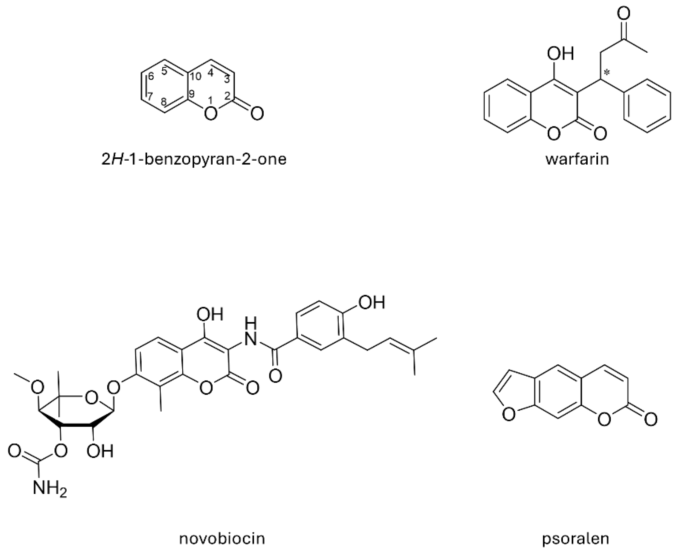
Although coumarin is not allowed as a food additive by the EU regulation [4], the nature-friendly heterocyclic core of coumarin is present in different classes of FDA approved drugs (Chart 1). Novobiocin and the more complex coumermycin are coumarin-containing topoisomerase inhibitors that act as antibiotics upon targeting bacterial DNA Gyrase B, thus interfering with ATPase activity and blocking ATP-dependent DNA negative supercoiling [7]. 4-Hydroxycoumarins as warfarin, acenocoumarol, and phenprocoumon are vitamin K antagonists used as oral anticoagulants under strict medical monitoring [8]. They prevent the γ-carboxylation reaction in the activation process of clotting factors [9].
From a synthetic viewpoint, the coumarin is an easy-to-handle and chemically tractable fragment, offering many anchoring points for exploring molecular diversity. Thus, over the years, huge efforts have been devoted to optimizing synthetic protocols targeted to suitably modified structures, e.g., by means of introducing different functional groups as substituents all around the heterocyclic core [10,11,12,13]. The presence of six branching positions (3-, 4-, 5-, 6-, 7-, 8-) makes coumarin a building block with outstanding scaffolding properties, devising molecular growing vectors toward all spatial directions [14,15]. These features increase the chances of finding suitable substitution patterns and appropriate geometries for binding interactions with potential drug targets [16]. Indeed, the chemical decoration of the coumarin core has long been exploited to developing bioactive molecules modulating the activity of different proteins (carriers, enzymes, and receptors) such as multidrug-resistance efflux pumps [17], acetylcholinesterase [18,19], STAT3 [20], monoamine oxidases A and B (MAO A and B) [21,22], carbonic anhydrases [23], aromatase [24], heat shock protein 90 [25], serine proteases [26], and many others. More recently, some of us have devoted great attention to the development of dual AChE-MAO B coumarin-based inhibitors as possible anti-Alzheimer’s agents [27,28,29,30].
Another possibility for coumarin core decoration/modification consists of the annulation approach, as also fused coumarins can be found in nature. Among these, linear furanocoumarins with the furane ring fused to the benzene ring of coumarin (psoralen, xanthotoxin, i.e., furo[3,2-g]chromenones) are well-studied compounds classified as primary photosensitizing agents that can be activated by UV light (320–380 nm wavelength) and produce phototoxicity through DNA cross-linking or fatty acids aberrations at the membrane phospholipid level. Other types of 3,4-heterocycle-fused coumarins, including angular furocoumarins [31], have been reported in the literature as bioactive compounds [32]. Recently, some of us became interested in novel fused-heteropolycycles characterized by the presence of furan or dihydrofuran rings as potential antioxidants mimiking phenolic and polyphenolic natural compounds [33]. Accordingly, a few classes of furo-fused heterocycles were synthesized by an annulation protocol exploiting, on one side, the bidentate O/C nucleophilic reactivity of phenolic moieties embedded in different heterocycles [34,35], and, on the other side, the bidentate C/C electrophilic character of nitrostilbenes. In particular, the application of the annulation protocol to 4-hydroxycoumarin [35] allowed the access to angular dihydrofuro[3,2-c]coumarins which were then subjected to oxidative aromatization to the corresponding furo[3,2-c]coumarins, characterized by previously unreported functionalization patterns.
Actually, both natural [36] and synthetic linear furo[3,2-g]coumarins [37] have been already described as cholinesterases (acetyl- and butyrylcholinesterase, AChE, and BChE) inhibitors. Anti-AChE activity still represents a key player in Alzheimer’s disease (AD) therapy [38], even if other mechanisms have been evoked as more valuable treatment strategies [39]. Along the so-called multitarget strategy to combat AD [40], the additional inhibition of MAOs has been envisaged as a powerful tool to counteract oxidative stress conditions [41], a common feature of neurodegenerative diseases.
With the aim of improving the portfolio of multitargeting blockers, in this manuscript, we report the in vitro biological evaluation of some angular furo[3,2-c]coumarins, made available by the annulation protocol cited above, as human AChE, BChE, MAO A, and MAO B inhibitors. In order to assess the significance (if any) of the spatial molecular arrangement on the inhibition activity, some precursor dihydrofuro[3,2-c]coumarins, characterized by a lower extent of coplanarity of the heterocyclic fused system, were also subjected to evaluation. Moreover, docking simulations were run in order to explain the plausible binding modes toward key target enzymes. The cytotoxicity of the whole series was assessed toward human neuroblastoma cells (SH-SY5Y) and is reported herein.

2. Results and Discussion

2.1. Synthesis of Dihydrofuro[3,2-c]coumarins 2 and Furo[3,2-c]coumarins 3

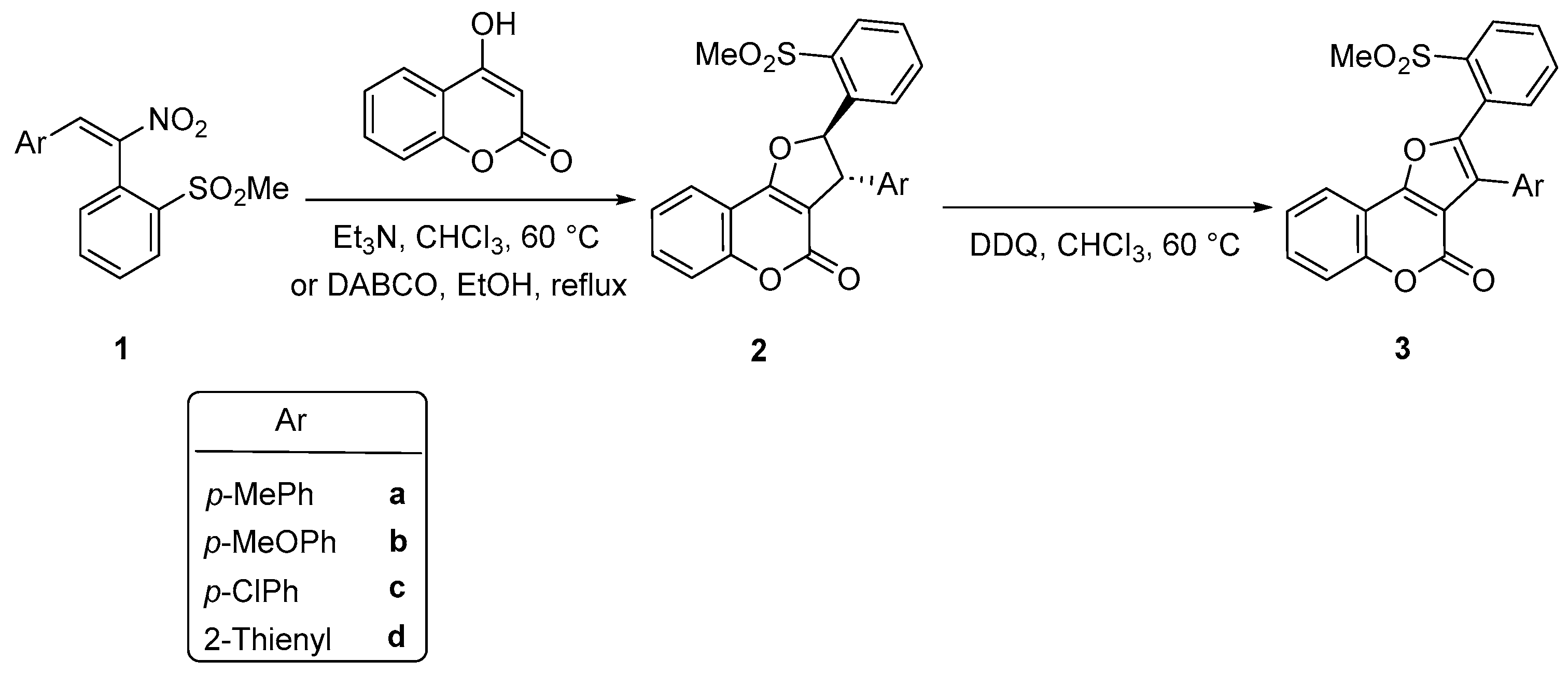
The dihydro[3,2-c]furocoumarins 2 and furo[3,2-c]coumarins 3 evaluated herein have been synthesized as recently reported by some of us via the sequence sketched in Scheme 1 [35] from 4-hydroxycoumarin and nitrostilbenes 1 [42], which generates the dihydro derivatives 2 as single (anti) diastereomers, then subjected to aromatization.
Experimental details for the synthesis of the model thiophene derivatives 2d and 3d are reported in the Experimental section. The syntheses of all the dihydrofuro- and furocoumarins tested herein have been already reported together with full physical and structural characterizations [35]. Compared to literature methods for the synthesis of angular furo[3,2-c]coumarins [31,32,43,44,45,46,47,48], the approach of Scheme 1 significantly enriches the available structural diversity as it allows to modulate the geometrical flexibility by means of the saturation degree of the furane ring (e.g., 2 vs. 3). In this regard, it should be remarked that, from a mechanistic viewpoint, the possibility to isolate the dihydrofurocoumarins 2 heavily rests on the presence of the electronwithdrawing o-methylsulfonyl-substituted benzene ring, which limits the negative charge delocalization onto the nitro group along with the initial Michael coupling, thus favoring its elimination upon ring closing to furane 2 [35].

2.2. In Vitro Biological Evaluation

All compounds have been tested as inhibitors of ChEs and MAOs from human sources, by applying the well-established spectrophotometric Ellman’s method [18,49]. Compounds 2ad were assayed as diastereomeric mixtures. Data are reported in Table 1 and indicated that no clear structure-activity relationships (SARs) can be derived. However, some in vitro results are worth noting. In fact, all compounds inhibited hAChE at the low micromolar level with IC50 values close to each other (4.0 μM < IC50 < 6.1 μM). Aromatized derivatives 3a, b, d were equipotent AChE inhibitors compared to the dihydro analogs 2a, b, d (2a vs. 3a, 2b vs. 3b, 2d vs. 3d). Both partially saturated and conjugated coumarins can be considered selective AChE inhibitors, since they exhibited very low percentage of inhibition (2a, 2b, 3b, 3d) or the absence of inhibitory activities toward hBChE (2c, 2d, 3a).
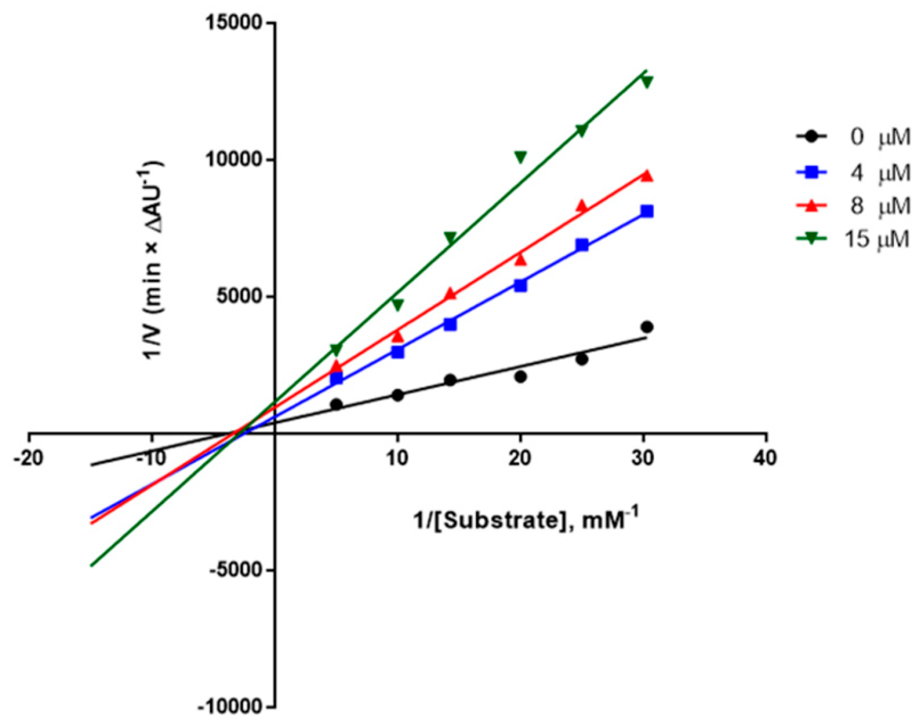
The most active achiral compound (3d, IC50 = 4.1 μM toward AChE) was enrolled in the investigation of inhibition kinetics. Lineweaver-Burk plots at different inhibitor concentrations (0–15 μM) in Figure 1 indicated a mixed-type or a non-competitive inhibition mechanism for AChE with a Ki = 9.7 ± 0.6 μM. This behavior can suggest the possibility for this compound to occupy the peripheral anionic subsite (PAS) of AChE, at least in part. From a three-dimensional viewpoint, the active site of AChE is characterized by the presence of a narrow gorge, about 20 Å long, spanning from the catalytic anionic subsite (CAS), close to the catalytic triad, to the peripheral anionic subsite [50,51]. PAS-binding property can have a beneficial effect against Alzheimer’s, since PAS has been postulated to exert a chaperone-like activity that accelerates amyloid deposition into fibrils [52].
Moreover, this series of compounds was also tested as potential inhibitors of both monoamine oxidase isoforms (MAO A and B). In fact, the blockade of MAO B can endorse the potential as neuroprotective or anti-Alzheimer’s tools [53,54]. As inferred by the inhibition data from Table 1, 2a, b showed comparable activity toward both isoforms, lacking the necessary selectivity toward MAO B that could reduce the risk of sympathomimetic side effects (e.g., cheese reaction [55]). Unfortunately, coumarin 2d was significantly more active toward MAO A than B.
Surprisingly, the aromatization of the furane ring provided selective, albeit moderate, MAO B inhibitors 3a and b, that were inactive toward the A isoform. It is worth noting that congener 3d inhibited hMAO B activity at the submicromolar level, returning an IC50 equal to 0.561 μM. Moreover, this compound was highly selective toward the B isoform, showing no inhibition of MAO A activity upon incubation at 10 μM concentration and deserving further attention as a potential multitarget tool.

2.3. Cytotoxicity

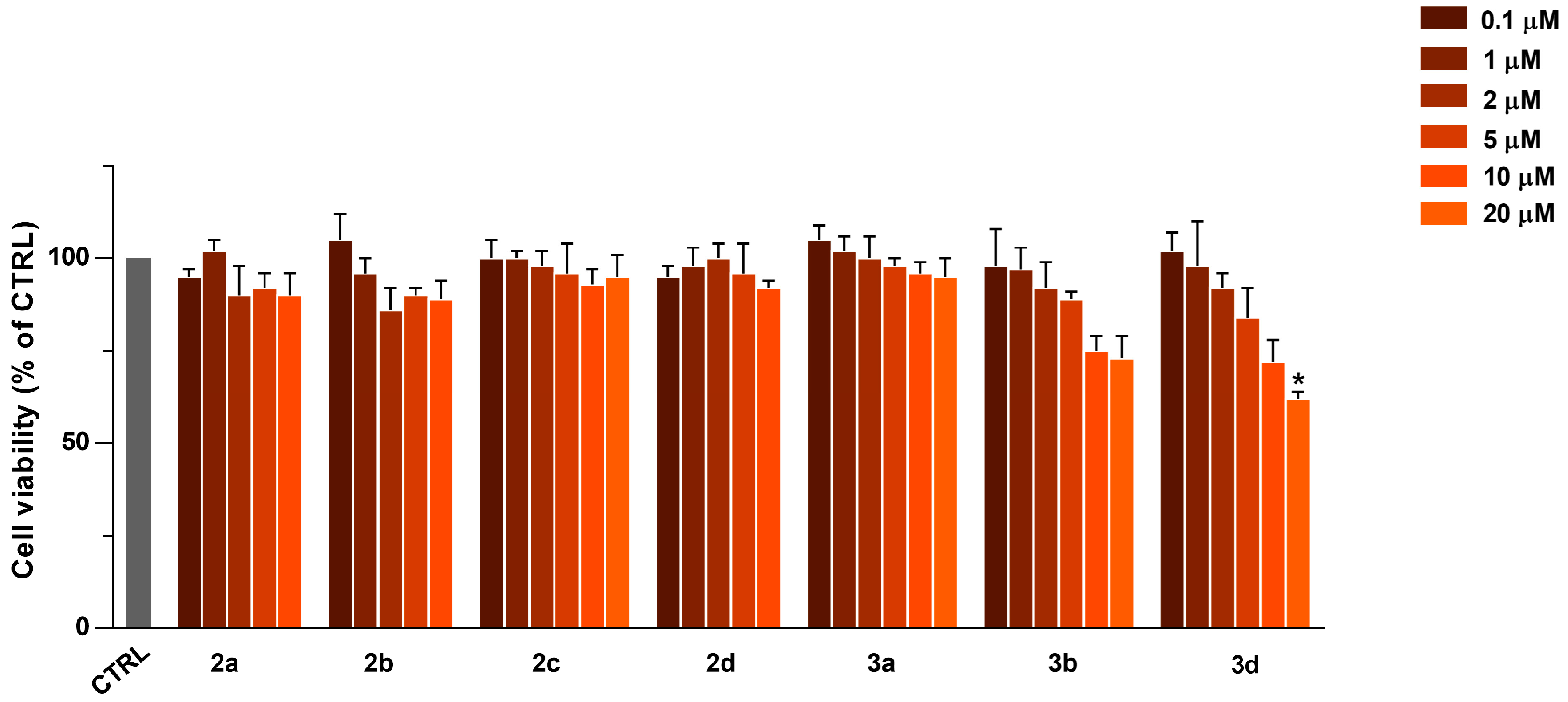
The cytotoxicity of compounds 2a–d and 3a, b, d was assessed upon co-incubation with human neuroblastoma cells and is reported as bar plots in Figure 2. For the diastereomer mixtures of compounds 2a, 2b, and 2d, the highest concentration tested was equal to 10 micromolars because of precipitation in the assay conditions at higher concentrations (20 μM). As can be observed in Figure 2, all compounds showed negligible cytotoxic effects at the studied concentrations, chosen within the same order of magnitude than IC50 values toward AChE.

2.4. Computational Studies

Molecular docking simulations were run for the most promising in vitro dual-targeting inhibitor (3d) within the binding site of AChE and MAO B to detect plausible binding poses and to better rationalize its biological activity. As for the protein template, high-resolution crystal structure of mus musculus AChE (mAChE) binary complex with a coumarin-based inhibitor (PDB code 7QAK [56]) was recruited, because of high sequence homology with the human enzyme and ligand similarity. Notably, the predicted binding mode for compound 3d appears to be primarily driven by π–π stacking interactions. In agreement with the kinetic analysis, inhibitor 3d settles in the PAS and in the mid-gorge of the target enzyme as illustrated in Figure 3A. The low inhibitory activity can be ascribed to several unfavorable interactions (i.e., steric clashes indicated by dotted orange and red lines in Figure 3) that were detected in the complex for all top-scored binding poses, being the computed docking score −6.573 kcal/mol for the best-ranked one. This value closely matches the in vitro experimental activity scale, hence supporting the reliability of the performed docking study. As a matter of fact, the free-energy calculation returned a much lower value for 3d than the score obtained upon redocking the coumarin-based native ligand of 7QAK crystallographic complex (docking score = −12.023 kcal/mol), possessing an IC50 at the nanomolar level (120 nM) toward hAChE [56]. In details, the sulphonyl-substituted phenyl ring of compound 3d is anchored to the PAS of the target enzyme by a sandwich-like arene-arene interaction directed to the indole side chain of Trp286 (distance between centroids: 3.5 Å) at the entrance of the enzymatic cavity and Tyr124 (distance between centroids: 3.4 Å). A similar interaction geometry was retrieved by donepezil, a standard AChE inhibitor marketed against AD, as shown in Figure 3B illustrating short-range favorable arene-arene interactions arranging the dimethoxyphenyl ring in front of both Trp286 (3.6 Å) and Tyr124 side chain (3.9 Å). However, donepezil behaves as a dual binding-site inhibitor able to fill the cavity from PAS to CAS, indeed it performs key additional interaction stacking parallel to Trp86 at the CAS level. The inhibitory potency of this drug, higher than both 3d and the native ligand, was further confirmed by the docking score calculated in our simulation (−12.549 kcal/mol). Docking protocols for 3d, including water molecules from the active site (e.g., 737 and 756), returned a lower docking score (−5.730 kcal/mol), likely due to steric clashes and water displacement by the bulky and hydrophobic ligand (data not shown).
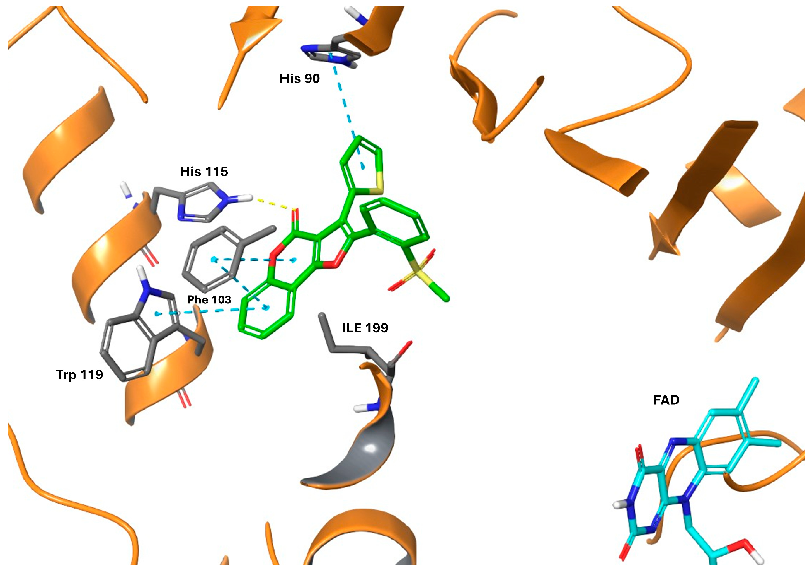
Thanks to ligand similarity, the X-ray binary complex coded as 7P4F in PDB [56] between human MAO B and a coumarin inhibitor was enrolled to dock 3d into this enzyme. Upon applying an induced fit protocol, to ensure target side chain flexibility, a good docking score was obtained (−12.602 kcal/mol) in agreement with the submicromolar potency of 3d as a MAO B inhibitor. As highlighted in Figure 4, the bulky ligand is accommodated within the entrance cavity and does not fill the substrate cavity gated by Ile199 and Tyr326, leaving the aromatic cage empty. The complex is stabilized by strong arene–arene interactions involving the coumarin motif. In detail, this heterocycle stacks parallel to Phe103 (3.7 Å distance), and the lactone carbonyl is hydrogen bonded with the His115 side chain (N···O distance of 3.2 Å). The thiophene ring of 3d interacts with His90.

3. Materials and Methods

3.1. Chemistry

Synthetic and analytic procedures are fully described in the cited ref. [35]. Hereinafter, for the sake of clarity, the experimental details for the synthesis of the model thiophene derivatives 2d and 3d are reported.

3.1.1. Reaction of Substrates 1d with 4-Hydroxycoumarin

In a round-bottom flask, the substrate 1d (0.16 mmol) was dissolved in ethanol (1.5 mL); then, 4-hydroxycoumarin (0.32 mmol) and 1,4 diazabicyclo[2.2.2]octane (DABCO, 0.24 mmol) were added. The reaction mixture was refluxed under magnetic stirring until the substrate disappeared (TLC). 2d precipitated as a white solid in EtOH (74%), so it could be separated by filtration, washed with ethanol, and subjected to aromatization with no need for further purification.

3.1.2. Aromatization of 2d with DDQ

The dihydrofurocoumarin 2d (0.12 mmol) was dissolved in chloroform (3.0 mL) and 2,3-dichloro-5,6-dicyano-p-benzoquinone (DDQ, 0.48 mmol) was added. The reaction was heated at 60 °C under magnetic stirring until completion (monitored by TLC). Then, an aqueous saturated NaHCO3 solution (8 mL) was added, and the resulting mixture was extracted with CHCl3 (3 × 15 mL). The combined organic layers were dried over anhydrous Na2SO4, filtered, and concentrated in vacuo. The reaction mixture was purified by column chromatography (petroleum ether/ethyl acetate 1:1) to afford the desired 3d (99%).

3.2. In Vitro Enzymatic Inhibition Assays

All human enzymes, substrates and reagents were from Sigma-Aldrich (Milan, Italy). Experiments were run in 96-well plates from Greiner Bio-One (Kremsmenster, Austria) with an Infinite M1000 Pro multiplate reader (Tecan, Cernusco sul Naviglio, Italy). The inhibition of ChEs was determined by following Ellman’s spectrophotometric assay in transparent, flat-bottom plates [18]. For studies addressing MAOs inhibition the spectrofluorimetric protocol was based on the oxidative deamination of kynuramine to 4-hydroxyquinoline measured in black, flat-bottom polystyrene plates [29]. Incubations were performed in triplicate and results were expressed as the mean ± SEM from 3 independent experiments. The values of IC50 were calculated by nonlinear regressions using GraphPad Prism 5.00 (GraphPad Software, San Diego, CA, USA). Kinetic studies were performed with the same test conditions, using six concentrations of substrate (from 0.033 to 0.2 mM) and four concentrations of inhibitor (0–15 μM). Apparent inhibition constants and kinetic parameters were calculated within the “Enzyme kinetics” module of Prism.

3.3. Molecular Docking Simulations

Docking simulations were performed using the crystal structure of compound 1 in ref [56] in complex with mAChE (PDB code 7QAK) and hMAO B (PDB code 7P4F) retrieved from the Protein Data Bank. The protein pretreatment to remove water molecules, assign bond orders, add hydrogen atoms, create disulfide bonds, fill in missing side chains and loops using Prime [59] and cap termini was carried out with the Protein Preparation Wizard module available from Schrödinger Release 2024-4. Protonation states at pH 7.0 ± 2.0 and tautomers for histidine residues were predicted with the Epik function and default parameters were used for the optimization of hydrogen-bond assignment. A restrained energy minimization step only on hydrogens was finally applied to the protein using the OPLS4 force field [60]. The ligand preparation was performed using the LigPrep [61] module in the Schrödinger Suite. Compound 3d (Table 1) was initially drafted in the Maestro [57] panel; then protonation states were calculated at pH 7.0 ± 2.0 retaining specific chiralities and applying the OPLS4 force field.
As for the docking calculations performed on AChE, the “Receptor Grid Generation” panel of Glide [58] was used to generate the grid files with grid points calculated within an enclosing box of 20 Å defined by the X-ray coordinates of 1 [56] from 7QAK. Docking calculations were performed for each ligand using the Glide module of Schrödinger, using standard (SP) mode and default settings (van der Waals radius scaling parameters: scaling factor of 0.80 and partial charge cut-off of 0.15; dock flexibly; add Epik state penalties to docking score; perform post-docking minimization).
For docking runs on hMAO B, the Induced Fit Docking protocol from Schrödinger Release 2024-4 was employed to ensure a reliable binding pose of compound 3d within the protein cavity. Specifically, the protocol was applied by centering the grid box on the co-crystallized ligand while maintaining all default parameters.
The scoring function of Glide known as “Docking score” was used for ranking the binding affinity. Pictures were elaborated using Maestro, available from the with Schrödinger Release 2024-4.

3.4. Cell-Based Experiments

The cell viability of human neuroblastoma SH-SY5Y cells was measured as previously reported in ref. [29]. To assess the potential cytotoxicity of 2ad, 3a,b, and 3d, compounds under study were co-incubated at 0.1–20 µM concentration range (0.1–10 µM for 2a, 2b, 2d) for 24 h. Then, viable cells were determined by MTT [3-(4,5-dimethylthiazol-2-yl)-2,5-diphenyl tetrazolium bromide] assay and results (as percentage) are referred to untreated cells (control, CTRL, cells without compounds, 100% viability). Briefly, cells were plated at a concentration of 4.5 × 10⁴ cells/well and cultured in Dulbecco’s modified Eagle’s medium (DMEM) high glucose supplemented with 10% (v/v) heat-inactivated fetal bovine serum (FBS), 2 mM L-glutamine, 100 U/mL penicillin, and 100 μg/mL streptomycin at 37 °C in an atmosphere of 5% CO2 in 96 wells/plate. At 80% confluence, the medium was removed, and cells in serum-free DMEM were treated with compounds 2ad, 3a,b, and 3d at concentrations ranging from 0.1 to 20 µM for 24 h at 37 °C in 5% CO2. Then, cells were incubated for 2 h at 37 °C and 5% CO2 with 0.5 mg/mL of MTT, and the formazan crystals in the cells were solubilized with absolute ethanol. The amount of the formazan product was determined by absorbance values measured at 545 nm using a multilabel plate counter Victor V3 (Perkin Elmer, Milan-Italy) with a reference wavelength of 690 nm. Cell viability was expressed as a percentage of the negative control (CTRL), represented by untreated cells, which was set at 100%. Data are the mean ± SD from three independent experiments, each performed in triplicate and analyzed using GraphPad Prism 5.0 (GraphPad Software Inc., San Diego, CA, USA). Statistical significance was calculated using a one-way analysis of variance (ANOVA) followed by Dunnett’s multiple comparison test. Levels of significance refer to untreated cells: * p < 0.05.

4. Conclusions

In this manuscript, we described the preparation and biological screening of novel oxygen-containing heterocycles, bearing a furo[3,2-c]coumarin motif. Studied angular coumarins were able to inhibit AChE at a low micromolar level and behaved as non-toxic compounds, showing negligible damage when co-incubated with human neuroblastoma cells. Compound 3d, carrying a 2-substitued thienyl ring and exhibiting IC50 = 4.1 μM toward hAChE, underwent further investigations. Its kinetic analysis showed a mixed-type or non-competitive binding mode that was further corroborated by docking simulations. This behavior outlined the possible occupancy of PAS, preventing pro-aggregating features in amyloid fibrilization. Additionally, 3d displayed a selective inhibition of hMAO B (IC50 = 0.561 μM). Taken together, these findings represent a piece of work highlighting the successful use of even more complex coumarins, such as angular-fused structures, in the discovery of bioactive molecules. Having in hand a straightforward synthetic protocol, hit optimization will be addressed to deepen structure–activity relationships.

Author Contributions

Conceptualization, C.T., G.P., G.L. and L.P.; methodology, C.T., G.P. and L.P.; software, G.F.M.; validation, L.P.; formal analysis, D.V.M. and L.P.; investigation, M.R., L.B., M.M., A.B., G.M.T. and D.V.M.; resources, C.T., L.B., G.F.M. and L.P.; data curation, L.P., L.B., M.M. and G.P.; writing—original draft preparation, L.B., D.V.M. and L.P.; writing—review and editing, L.P., G.L., L.B., M.M. and G.P.; visualization, L.P.; supervision, L.P., G.L., G.P. and M.M.; project administration, G.P.; funding acquisition, L.B. All authors have read and agreed to the published version of the manuscript.

Funding

Financial support was provided by grants from the Department of Chemistry and Industrial Chemistry (DCCI), University of Genova (‘Impact Boosting’ 2022).

Institutional Review Board Statement

Not applicable.

Informed Consent Statement

Not applicable.

Data Availability Statement

The data presented in this study are available on request from the corresponding authors.

Conflicts of Interest

The authors declare no conflicts of interest.

References

  1. Murray, R.D.H. Coumarins. Nat. Prod. Rep. 1995, 12, 477. [Google Scholar] [CrossRef]
  2. Hoult, J.R.S.; Payá, M. Pharmacological and Biochemical Actions of Simple Coumarins: Natural Products with Therapeutic Potential. Gen. Pharmacol. 1996, 27, 713–722. [Google Scholar] [CrossRef] [PubMed]
  3. Vogt, T. Phenylpropanoid Biosynthesis. Mol. Plant 2010, 3, 2–20. [Google Scholar] [CrossRef]
  4. Lončar, M.; Jakovljević, M.; Šubarić, D.; Pavlić, M.; Služek, V.B.; Cindrić, I.; Molnar, M. Coumarins in Food and Methods of Their Determination. Foods 2020, 9, 645. [Google Scholar] [CrossRef] [PubMed]
  5. Matos, M.J.; Santana, L.; Uriarte, E.; Abreu, O.A.; Molina, E.; Yordi, E.G. Coumarins—An Important Class of Phytochemicals. In Phytochemicals-Isolation, Characterisation and Role in Human Health; IntechOpen: London, UK, 2015. [Google Scholar]
  6. Vogel, A. Darstellung von Benzoesäure Aus Der Tonka-Bohne Und Aus Den Meliloten-Oder Steinklee-Blumen. Ann. Phys. 1820, 64, 161–166. [Google Scholar] [CrossRef]
  7. May, J.M.; Owens, T.W.; Mandler, M.D.; Simpson, B.W.; Lazarus, M.B.; Sherman, D.J.; Davis, R.M.; Okuda, S.; Massefski, W.; Ruiz, N.; et al. The Antibiotic Novobiocin Binds and Activates the ATPase That Powers Lipopolysaccharide Transport. J. Am. Chem. Soc. 2017, 139, 17221–17224. [Google Scholar] [CrossRef]
  8. Kasperkiewicz, K.; Ponczek, M.B.; Owczarek, J.; Guga, P.; Budzisz, E. Antagonists of Vitamin K-Popular Coumarin Drugs and New Synthetic and Natural Coumarin Derivatives. Molecules 2020, 25, 1465. [Google Scholar] [CrossRef]
  9. Shen, G.; Cui, W.; Zhang, H.; Zhou, F.; Huang, W.; Liu, Q.; Yang, Y.; Li, S.; Bowman, G.R.; Sadler, J.E.; et al. Warfarin Traps Human Vitamin K Epoxide Reductase in an Intermediate State during Electron Transfer. Nat. Struct. Mol. Biol. 2017, 24, 69–76. [Google Scholar] [CrossRef]
  10. Medina, F.G.; Marrero, J.G.; Macías-Alonso, M.; González, M.C.; Córdova-Guerrero, I.; Teissier García, A.G.; Osegueda-Robles, S. Coumarin Heterocyclic Derivatives: Chemical Synthesis and Biological Activity. Nat. Prod. Rep. 2015, 32, 1472–1507. [Google Scholar] [CrossRef]
  11. Stefanachi, A.; Leonetti, F.; Pisani, L.; Catto, M.; Carotti, A. Coumarin: A Natural, Privileged and Versatile Scaffold for Bioactive Compounds. Molecules 2018, 23, 250. [Google Scholar] [CrossRef]
  12. Ishii, H.; Kaneko, Y.; Miyazaki, H.; Harayama, T. A Convenient Synthesis of a Simple Coumarin. Chem. Pharm. Bull. 1991, 39, 3100–3102. [Google Scholar] [CrossRef]
  13. Fringuelli, F.; Piermatti, O.; Pizzo, F. One-Pot Synthesis of 3-Carboxycoumarins via Consecutive Knoevenagel and Pinner Reactions in Water. Synthesis 2003, 15, 2331–2334. [Google Scholar] [CrossRef]
  14. Setena, S.M.; Shah, N.M. The Chemistry of Coumarins. Chem. Rev. 1945, 36, 1–62. [Google Scholar] [CrossRef]
  15. Langer, P. Adventures in Coumarin Chemistry. Synlett 2023, 36, 29–43. [Google Scholar] [CrossRef]
  16. Annunziata, F.; Pinna, C.; Dallavalle, S.; Tamborini, L.; Pinto, A. An Overview of Coumarin as a Versatile and Readily Accessible Scaffold with Broad-Ranging Biological Activities. Int. J. Mol. Sci. 2020, 21, 4618. [Google Scholar] [CrossRef]
  17. Rullo, M.; Niso, M.; Pisani, L.; Carrieri, A.; Colabufo, N.A.; Cellamare, S.; Altomare, C.D. 1,2,3,4-Tetrahydroisoquinoline/2H-Chromen-2-One Conjugates as Nanomolar P-Glycoprotein Inhibitors: Molecular Determinants for Affinity and Selectivity over Multidrug Resistance Associated Protein 1. Eur. J. Med. Chem. 2019, 161, 433–444. [Google Scholar] [CrossRef]
  18. Pisani, L.; Catto, M.; De Palma, A.; Farina, R.; Cellamare, S.; Altomare, C.D. Discovery of Potent Dual Binding Site Acetylcholinesterase Inhibitors via Homo- and Heterodimerization of Coumarin-Based Moieties. ChemMedChem 2017, 12, 1349–1358. [Google Scholar] [CrossRef]
  19. Elsinghorst, P.W.; Wille, T.; Barić, D.; Mertens, M.D.; Baumann, M.; Küppers, J.; Gütschow, M. Aminoalkoxy-Substituted Coumarins: Synthesis and Evaluation for Reactivation of Inhibited Human Acetylcholinesterase. Arch. Pharm. 2022, 355, e2200208. [Google Scholar] [CrossRef]
  20. Cai, G.; Yu, W.; Song, D.; Zhang, W.; Guo, J.; Zhu, J.; Ren, Y.; Kong, L. Discovery of Fluorescent Coumarin-Benzo[b]Thiophene 1, 1-Dioxide Conjugates as Mitochondria-Targeting Antitumor STAT3 Inhibitors. Eur. J. Med. Chem. 2019, 174, 236–251. [Google Scholar] [CrossRef]
  21. Iacovino, L.G.; Manzella, N.; Resta, J.; Vanoni, M.A.; Rotilio, L.; Pisani, L.; Edmondson, D.E.; Parini, A.; Mattevi, A.; Mialet-Perez, J.; et al. Rational Redesign of Monoamine Oxidase A into a Dehydrogenase to Probe ROS in Cardiac Aging. ACS Chem. Biol. 2020, 15, 1795–1800. [Google Scholar] [CrossRef]
  22. Matos, M.J.; Novo, P.; Mayán, L.; Torres, I.; Uriarte, E.; Yáñez, M.; Fontenla, J.Á.; Ortuso, F.; Alcaro, S.; Procopio, F.; et al. 8-Amide and 8-Carbamate Substitution Patterns as Modulators of 7-Hydroxy-4-Methylcoumarin’s Antidepressant Profile: Synthesis, Biological Evaluation and Docking Studies. Eur. J. Med. Chem. 2023, 248, 115091. [Google Scholar] [CrossRef] [PubMed]
  23. Maresca, A.; Temperini, C.; Vu, H.; Pham, N.B.; Poulsen, S.A.; Scozzafava, A.; Quinn, R.J.; Supuran, C.T. Non-Zinc Mediated Inhibition of Carbonic Anhydrases: Coumarins Are a New Class of Suicide Inhibitors. J. Am. Chem. Soc. 2009, 131, 3057–3062. [Google Scholar] [CrossRef]
  24. Stefanachi, A.; Hanke, N.; Pisani, L.; Leonetti, F.; Nicolotti, O.; Catto, M.; Cellamare, S.; Hartmann, R.W.; Carotti, A. Discovery of New 7-Substituted-4-Imidazolylmethyl Coumarins and 4′-Substituted-2-Imidazolyl Acetophenones Open Analogues as Potent and Selective Inhibitors of Steroid-11β-Hydroxylase. Eur. J. Med. Chem. 2015, 89, 106–114. [Google Scholar] [CrossRef]
  25. Le Bras, G.; Radanyi, C.; Peyrat, J.F.; Brion, J.D.; Alami, M.; Marsaud, V.; Stella, B.; Renoir, J.M. New Novobiocin Analogues as Antiproliferative Agents in Breast Cancer Cells and Potential Inhibitors of Heat Shock Protein 90. J. Med. Chem. 2007, 50, 6189–6200. [Google Scholar] [CrossRef]
  26. Pochet, L.; Frederick, R.; Masereel, B. Coumarin and Isocoumarin as Serine Protease Inhibitors. Curr. Pharm. Des. 2005, 10, 3781–3796. [Google Scholar] [CrossRef] [PubMed]
  27. Rullo, M.; Cipolloni, M.; Catto, M.; Colliva, C.; Miniero, D.V.; Latronico, T.; de Candia, M.; Benicchi, T.; Linusson, A.; Giacchè, N.; et al. Probing Fluorinated Motifs onto Dual AChE-MAO B Inhibitors: Rational Design, Synthesis, Biological Evaluation, and Early-ADME Studies. J. Med. Chem. 2022, 65, 3962–3977. [Google Scholar] [CrossRef] [PubMed]
  28. Pisani, L.; Rullo, M.; Catto, M.; de Candia, M.; Carrieri, A.; Cellamare, S.; Altomare, C.D. Structure-Property Relationship Study of the HPLC Enantioselective Retention of Neuroprotective 7-[(1-Alkylpiperidin-3-Yl)Methoxy]Coumarin Derivatives on an Amylose-Based Chiral Stationary Phase. J. Sep. Sci. 2018, 41, 1376–1384. [Google Scholar] [CrossRef]
  29. La Spada, G.; Miniero, D.V.; Rullo, M.; Cipolloni, M.; Delre, P.; Colliva, C.; Colella, M.; Leonetti, F.; Liuzzi, G.M.; Mangiatordi, G.F.; et al. Structure-Based Design of Multitargeting ChEs-MAO B Inhibitors Based on Phenyl Ring Bioisosteres: AChE/BChE Selectivity Switch and Drug-like Characterization. Eur. J. Med. Chem. 2024, 274, 116511. [Google Scholar] [CrossRef]
  30. Rullo, M.; Catto, M.; Carrieri, A.; de Candia, M.; Altomare, C.D.; Pisani, L. Chasing ChEs-MAO B Multi-Targeting 4-Aminomethyl-7-Benzyloxy-2H-Chromen-2-Ones. Molecules 2019, 24, 4507. [Google Scholar] [CrossRef]
  31. Bruni, R.; Barreca, D.; Protti, M.; Brighenti, V.; Righetti, L.; Anceschi, L.; Mercolini, L.; Benvenuti, S.; Gattuso, G.; Pellati, F. Botanical Sources, Chemistry, Analysis, and Biological Activity of Furanocoumarins of Pharmaceutical Interest. Molecules 2019, 24, 2163. [Google Scholar] [CrossRef]
  32. Salehian, F.; Nadri, H.; Jalili-Baleh, L.; Youseftabar-Miri, L.; Abbas Bukhari, S.N.; Foroumadi, A.; Tüylü Küçükkilinç, T.; Sharifzadeh, M.; Khoobi, M. A Review: Biologically Active 3,4-Heterocycle-Fused Coumarins. Eur. J. Med. Chem. 2021, 212, 113034. [Google Scholar] [CrossRef]
  33. Fylaktakidou, K.C.; Hadjipavlou-Litina, D.J.; Litinas, K.E.; Nicolaides, D.N. Natural and Synthetic Coumarin Derivatives with Anti-Inflammatory/Antioxidant Activities. Curr. Pharm. Des. 2004, 10, 3813–3833. [Google Scholar] [CrossRef] [PubMed]
  34. Benzi, A.; Bianchi, L.; Giorgi, G.; Maccagno, M.; Petrillo, G.; Spinelli, D.; Tavani, C. An Easy Access to Furan-Fused Polyheterocyclic Systems. Molecules 2022, 27, 3147. [Google Scholar] [CrossRef]
  35. Benzi, A.; Bianchi, L.; Giorgi, G.; Lentini, G.; Maccagno, M.; Marcantoni Taddei, G.; Petrillo, G.; Tavani, C. An Appealing, Robust Access to Furo-Fused Heteropolycycles. Molecules 2025, 30, 948. [Google Scholar] [CrossRef] [PubMed]
  36. Youkwan, J.; Sutthivaiyakit, S.; Sutthivaiyakit, P. Citrusosides A-D and Furanocoumarins with Cholinesterase Inhibitory Activity from the Fruit Peels of Citrus Hystrix. J. Nat. Prod. 2010, 73, 1879–1883. [Google Scholar] [CrossRef]
  37. Shen, Q.; Peng, Q.; Shao, J.; Liu, X.; Huang, Z.; Pu, X.; Ma, L.; Li, Y.M.; Chan, A.S.C.; Gu, L. Synthesis and Biological Evaluation of Functionalized Coumarins as Acetylcholinesterase Inhibitors. Eur. J. Med. Chem. 2005, 40, 1307–1315. [Google Scholar] [CrossRef] [PubMed]
  38. Wang, H.; Zhang, H. Reconsideration of Anticholinesterase Therapeutic Strategies against Alzheimer’s Disease. ACS Chem. Neurosci. 2019, 10, 852–862. [Google Scholar] [CrossRef]
  39. Long, J.M.; Holtzman, D.M. Alzheimer Disease: An Update on Pathobiology and Treatment Strategies. Cell 2019, 179, 312–339. [Google Scholar] [CrossRef]
  40. Albertini, C.; Salerno, A.; de Sena Murteira Pinheiro, P.; Bolognesi, M.L. From Combinations to Multitarget-Directed Ligands: A Continuum in Alzheimer’s Disease Polypharmacology. Med. Res. Rev. 2021, 41, 2606–2633. [Google Scholar] [CrossRef]
  41. Sánchez-Rodríguez, R.; Munari, F.; Angioni, R.; Venegas, F.; Agnellini, A.; Castro-Gil, M.P.; Castegna, A.; Luisetto, R.; Viola, A.; Canton, M. Targeting Monoamine Oxidase to Dampen NLRP3 Inflammasome Activation in Inflammation. Cell. Mol. Immunol. 2021, 18, 1311–1313. [Google Scholar] [CrossRef]
  42. Petrillo, G.; Benzi, A.; Bianchi, L.; Maccagno, M.; Pagano, A.; Tavani, C.; Spinelli, D. Recent Advances in the Use of Conjugated Nitro or Dinitro-1,3-Butadienes as Building-Blocks for the Synthesis of Heterocycles. Tetrahedron Lett. 2020, 61, 152297. [Google Scholar] [CrossRef]
  43. Feng, X.; Qin, Z.; Cheng, X.; Liu, D.; Peng, Y.; Huang, H.; Song, B.; Bian, J.; Li, Z. Copper(II)-Catalyzed Tandem Reaction: Synthesis of Furo[3,2-c]Coumarin Derivatives and Evaluation for Photophysical Properties. J. Org. Chem. 2021, 86, 12537–12548. [Google Scholar] [CrossRef] [PubMed]
  44. Kolita, S.; Borah, P.; Naidu, P.S.; Bhuyan, P.J. DMSO and Iodine Mediated Reaction of 4-Hydroxycoumarins and Aldehydes/Aryl Methyl Ketones: Synthesis of Furo[3,2-c]Coumarins. Tetrahedron 2016, 72, 532–538. [Google Scholar] [CrossRef]
  45. Chen, Z.; Zeng, P.; Zhang, S.; Huang, X. Lewis-Acid-Mediated One-Pot Tandem Reactions for Synthesis of Structurally Diverse Furo[3,2-c]Coumarins. ChemistrySelect 2021, 6, 4539–4543. [Google Scholar] [CrossRef]
  46. Kerru, N.; Lalitha Gummidi, M.; Kumar Gangu, K.; Maddila, S.; Jonnalagadda, S.B. Synthesis of Novel Furo[3,2-c]Coumarin Derivatives through Multicomponent [4+1] Cycloaddition Reaction Using ZnO/FAp as a Sustainable Catalyst. ChemistrySelect 2020, 5, 4104–4110. [Google Scholar] [CrossRef]
  47. Rullo, M.; Pisani, L. 4-Hydroxycoumarins as Michael Donors in Asymmetric Routes to Polycyclic Coumarins (Microreview). Chem. Heterocycl. Compd. 2018, 54, 394–396. [Google Scholar] [CrossRef]
  48. Ghosh, M.; Hajra, A. DABCO-Promoted One-Pot Facile Synthesis of Angularly Fused Furoquinolinones and Furocoumarins. Eur. J. Org. Chem. 2015, 2015, 7836–7841. [Google Scholar] [CrossRef]
  49. Ellman, G.L.; Courtney, K.D.; Andres, V.; Feather-Stone, R.M. A New and Rapid Colorimetric Determination of Acetylcholinesterase Activity. Biochem. Pharmacol. 1961, 7, 88–95. [Google Scholar] [CrossRef]
  50. Colletier, J.P.; Fournier, D.; Greenblatt, H.M.; Stojan, J.; Sussman, J.L.; Zaccai, G.; Silman, I.; Weik, M. Structural Insights into Substrate Traffic and Inhibition in Acetylcholinesterase. EMBO J. 2006, 25, 2746–2756. [Google Scholar] [CrossRef]
  51. Ordentlich, A.; Barak, D.; Kronman, C.; Flashner, Y.; Leitner, M.; Segall, Y.; Ariel, N.; Cohen, S.; Velan, B.; Shafferman, A. Dissection of the Human Acetylcholinesterase Active Center Determinants of Substrate Specificity. Identification of Residues Constituting the Anionic Site, the Hydrophobic Site, and the Acyl Pocket. J. Biol. Chem. 1993, 268, 17083–17095. [Google Scholar] [CrossRef]
  52. Inestrosa, N.C.; Dinamarca, M.C.; Alvarez, A. Amyloid-Cholinesterase Interactions. Implications for Alzheimer’s Disease. FEBS J. 2008, 275, 625–632. [Google Scholar] [CrossRef] [PubMed]
  53. Rullo, M.; La Spada, G.; Miniero, D.V.; Gottinger, A.; Catto, M.; Delre, P.; Mastromarino, M.; Latronico, T.; Marchese, S.; Mangiatordi, G.F.; et al. Bioisosteric Replacement Based on 1,2,4-Oxadiazoles in the Discovery of 1H-Indazole-Bearing Neuroprotective MAO B Inhibitors. Eur. J. Med. Chem. 2023, 255, 115352. [Google Scholar] [CrossRef] [PubMed]
  54. Pisani, L.; Rullo, M.; Catto, M.; de Candia, M.; Carrieri, A.; Cellamare, S.; Altomare, C.D. Targeting Monoamine Oxidases with Multipotent Ligands: An Emerging Strategy in the Search of New Drugs against Neurodegenerative Diseases. Curr. Med. Chem. 2018, 41, 4568–4587. [Google Scholar] [CrossRef] [PubMed]
  55. Finberg, J.P.M.; Gillman, K. Selective Inhibitors of Monoamine Oxidase Type B and the “Cheese Effect”. In International Review of Neurobiology; Academic Press: Cambridge, MA, USA, 2011; Volume 100, pp. 169–190. [Google Scholar]
  56. Ekström, F.; Gottinger, A.; Forsgren, N.; Catto, M.; Iacovino, L.G.; Pisani, L.; Binda, C. Dual Reversible Coumarin Inhibitors Mutually Bound to Monoamine Oxidase B and Acetylcholinesterase Crystal Structures. ACS Med. Chem. Lett. 2022, 13, 499–506. [Google Scholar] [CrossRef]
  57. Schrödinger Release 2024-4: Maestro; Schrödinger, LLC.: New York, NY, USA, 2024.
  58. Schrödinger Release 2024-4: Glide; Schrödinger, LLC.: New York, NY, USA, 2024.
  59. Schrödinger Release 2024-4: Prime; Schrödinger, LLC.: New York, NY, USA, 2024.
  60. Schrödinger Release 2024-4: Force Fields; Schrödinger, LLC.: New York, NY, USA, 2024.
  61. Schrödinger Release 2024-4: LigPrep; Schrödinger, LLC.: New York, NY, USA, 2024.
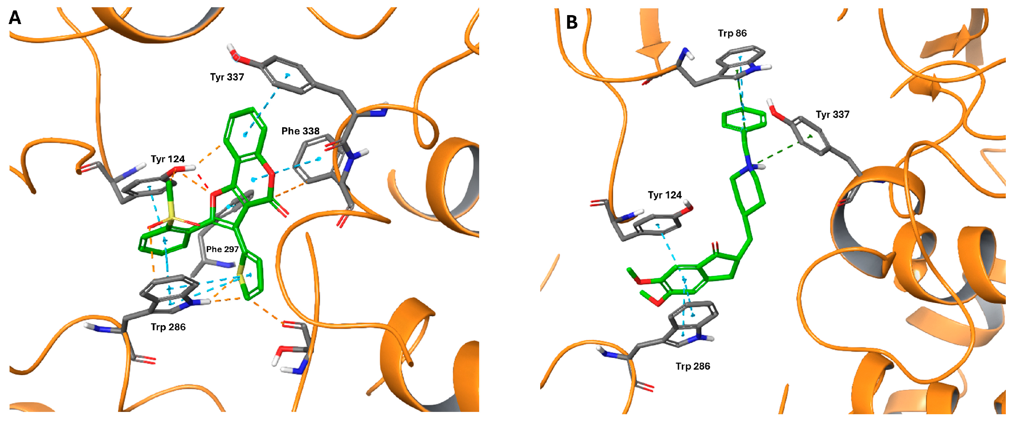
Chart 1. The numbering of 2H-1-benzopyran-2-one and some examples of approved drugs containing the heterocyclic core of coumarin.
Molecules 30 01830 ch001
Scheme 1. Synthesis of dihydrofuro[3,2-c]coumarins 2 and of relevant aromatized furo[3,2-c]coumarins 3.
Molecules 30 01830 sch001
Figure 1. Lineweaver-Burk plots of inhibition kinetics of compound 3d toward hAChE. Reciprocals of enzyme activity (hAChE) vs. reciprocals of substrate (S-acetylthiocholine) concentration in the presence of different concentrations (0–15 μM, reported with a different color code in insets) of inhibitor.
Molecules 30 01830 g001
Figure 2. Cell viability of neuroblastoma SH-SY5Y cells incubated with compounds 2a–d and 3a, b, d (0.1–10 µM or 0.1–20 µM concentration range, 24 h incubation), as determined by MTT assay. Bars represent the mean ± SD of three independent experiments, each performed in triplicates and referred to untreated control cells (CTRL, 100% value, in the absence of compounds). Statistical significance was calculated using one-way analysis of variance (ANOVA) followed by Dunnett’s multiple comparison test. Levels of significance are referred to untreated cells: * p < 0.05.
Molecules 30 01830 g002
Figure 3. Top-scored binding pose of compound 3d (A) and donepezil (B) docked within mAChE (PDB 7QAK, docking score = −6.573 kcal/mol and −12.549 kcal/mol, respectively). Ligands and relevant amino acid residues are rendered as sticks, while protein is represented as a cartoon. Colors are in accordance with the atom code, C atoms in green and gray for ligand and amino acid, respectively. Dotted blue lines represent π–π stacking; dotted orange and red lines represent bad and ugly contacts/clashes, respectively [57,58].
Molecules 30 01830 g003
Figure 4. Top-scored binding pose of compound 3d docked within hMAO B (PDB 7P4F, docking score = −12.602 kcal/mol). Ligands and relevant amino acid residues are rendered as sticks, while protein is represented as a cartoon. Colors are in accordance with the atom code, with C atoms in green and gray for ligands and amino acids, respectively. Dotted blue lines represent π−π stacking; dotted yellow lines represent hydrogen bonds [57,58].
Molecules 30 01830 g004
Table 1. In vitro inhibition data toward hChEs and hMAOs for compounds 2ad and 3a, b, d.
CompoundArIC50 (μM) or % Inhibition at 10 μM a
hAChE bhBChE chMAO A dhMAO B d
2a4-MePh5.5 ± 0.727 ± 12%47 ± 3%40 ± 8%
2b4-MeOPh6.1 ± 0.329 ± 11%45 ± 1%39 ± 6%
2c4-ClPh4.0 ± 0.7n.i. e16 ± 14%11 ± 6%
2d2-Thienyl4.0 ± 0.5n.i. e42 ± 4%17 ± 10%
3a4-MePh4.4 ± 0.5n.i. en.i. e32 ± 0%
3b4-MeOPh5.7 ± 1.019 ± 9%n.i. e43 ± 1%
3d2-Thienyl4.1 ± 0.721 ± 3%n.i. e0.561 ± 0.085
donepezil0.017 ± 0.004
tacrine 0.032 ± 0.011
clorgyline 0.020 ± 0.003
safinamide 0.037 ± 0.001
a Values are the mean of three independent experiments ± SEM. b Human AChE. c Human BChE. d Human recombinant MAO A and B on Supersomes. e n.i. = no inhibition or inhibition lower than 5% at 10 μM concentration.
Disclaimer/Publisher’s Note: The statements, opinions and data contained in all publications are solely those of the individual author(s) and contributor(s) and not of MDPI and/or the editor(s). MDPI and/or the editor(s) disclaim responsibility for any injury to people or property resulting from any ideas, methods, instructions or products referred to in the content.