Next Issue
Volume 21, January-1
Previous Issue
Volume 20, December-1
 
 
ijms-logo

Journal Browser

Journal Browser

Int. J. Mol. Sci., Volume 20, Issue 24 (December-2 2019) – 260 articles

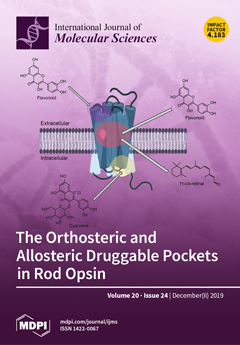
Cover Story (view full-size image): The G protein-coupled receptors (GPCRs) are at the forefront of the drug discovery initiative. Both the orthosteric and allosteric binding sites in GPCRs can be targeted by pharmacological treatments. Ocular degenerative disorders are often associated with impaired binding of the native chromophore 11-cis-retinal to rod opsin, resulting in aberrative protein folding and trafficking. These defects can be mitigated by pharmacological modulation with retinoid and nonretinoid molecules. View this paper.
  • Issues are regarded as officially published after their release is announced to the table of contents alert mailing list.
  • You may sign up for e-mail alerts to receive table of contents of newly released issues.
  • PDF is the official format for papers published in both, html and pdf forms. To view the papers in pdf format, click on the "PDF Full-text" link, and use the free Adobe Reader to open them.
Order results
Result details
Section
Select all
Export citation of selected articles as:
15 pages, 2691 KiB  
Article
“Salicylic Acid Mutant Collection” as a Tool to Explore the Role of Salicylic Acid in Regulation of Plant Growth under a Changing Environment
by Kamila Pluhařová, Hana Leontovyčová, Věra Stoudková, Romana Pospíchalová, Petr Maršík, Pavel Klouček, Anastasiia Starodubtseva, Oksana Iakovenko, Zuzana Krčková, Olga Valentová, Lenka Burketová, Martin Janda and Tetiana Kalachova
Int. J. Mol. Sci. 2019, 20(24), 6365; https://doi.org/10.3390/ijms20246365 - 17 Dec 2019
Cited by 18 | Viewed by 6247
Abstract
The phytohormone salicylic acid (SA) has a crucial role in plant physiology. Its role is best described in the context of plant response to pathogen attack. During infection, SA is rapidly accumulated throughout the green tissues and is important for both local and [...] Read more.
The phytohormone salicylic acid (SA) has a crucial role in plant physiology. Its role is best described in the context of plant response to pathogen attack. During infection, SA is rapidly accumulated throughout the green tissues and is important for both local and systemic defences. However, some genetic/metabolic variations can also result in SA overaccumulation in plants, even in basal conditions. To date, more than forty Arabidopsis thaliana mutants have been described as having enhanced endogenous SA levels or constitutively activated SA signalling pathways. In this study, we established a collection of mutants containing different SA levels due to diverse genetic modifications and distinct gene functions. We chose prototypic SA-overaccumulators (SA-OAs), such as bon1-1, but also “non-typical” ones such as exo70b1-1; the selection of OA is accompanied by their crosses with SA-deficient lines. Here, we extensively studied the plant development and SA level/signalling under various growth conditions in soil and in vitro, and showed a strong negative correlation between rosette size, SA content and PR1/ICS1 transcript signature. SA-OAs (namely cpr5, acd6, bon1-1, fah1/fah2 and pi4kβ1β2) had bigger rosettes under high light conditions, whereas WT plants did not. Our data provide new insights clarifying a link between SA and plant behaviour under environmental stresses. The presented SA mutant collection is thus a suitable tool to shed light on the mechanisms underlying trade-offs between growth and defence in plants. Full article
(This article belongs to the Special Issue Salicylic Acid Signalling in Plants)
Show Figures

Figure 1

23 pages, 1449 KiB  
Review
S1P/S1P Receptor Signaling in Neuromuscolar Disorders
by Elisabetta Meacci and Mercedes Garcia-Gil
Int. J. Mol. Sci. 2019, 20(24), 6364; https://doi.org/10.3390/ijms20246364 - 17 Dec 2019
Cited by 18 | Viewed by 7311
Abstract
The bioactive sphingolipid metabolite, sphingosine 1-phosphate (S1P), and the signaling pathways triggered by its binding to specific G protein-coupled receptors play a critical regulatory role in many pathophysiological processes, including skeletal muscle and nervous system degeneration. The signaling transduced by S1P binding appears [...] Read more.
The bioactive sphingolipid metabolite, sphingosine 1-phosphate (S1P), and the signaling pathways triggered by its binding to specific G protein-coupled receptors play a critical regulatory role in many pathophysiological processes, including skeletal muscle and nervous system degeneration. The signaling transduced by S1P binding appears to be much more complex than previously thought, with important implications for clinical applications and for personalized medicine. In particular, the understanding of S1P/S1P receptor signaling functions in specific compartmentalized locations of the cell is worthy of being better investigated, because in various circumstances it might be crucial for the development or/and the progression of neuromuscular diseases, such as Charcot–Marie–Tooth disease, myasthenia gravis, and Duchenne muscular dystrophy. Full article
(This article belongs to the Special Issue Sphingolipids: Metabolic Functions and Disorders)
Show Figures

Graphical abstract

20 pages, 3240 KiB  
Article
Cardiac Extracellular Vesicles (EVs) Released in the Presence or Absence of Inflammatory Cues Support Angiogenesis in Different Manners
by Christien Madlen Beez, Maria Schneider, Marion Haag, Kathleen Pappritz, Sophie Van Linthout, Michael Sittinger and Martina Seifert
Int. J. Mol. Sci. 2019, 20(24), 6363; https://doi.org/10.3390/ijms20246363 - 17 Dec 2019
Cited by 4 | Viewed by 3956
Abstract
Cells release extracellular vesicles (EVs) to communicate in a paracrine manner with other cells, and thereby influence processes, such as angiogenesis. The conditioned medium of human cardiac-derived adherent proliferating (CardAP) cells was recently shown to enhance angiogenesis. To elucidate whether their released EVs [...] Read more.
Cells release extracellular vesicles (EVs) to communicate in a paracrine manner with other cells, and thereby influence processes, such as angiogenesis. The conditioned medium of human cardiac-derived adherent proliferating (CardAP) cells was recently shown to enhance angiogenesis. To elucidate whether their released EVs are involved, we isolated them by differential centrifugation from the conditioned medium derived either in the presence or absence of a pro-inflammatory cytokine cocktail. Murine recipient cells internalized CardAP-EVs as determined by an intracellular detection of human proteins, such as CD63, by a novel flow cytometry method for studying EV–cell interaction. Moreover, endothelial cells treated for 24 h with either unstimulated or cytokine stimulated CardAP-EVs exhibited a higher tube formation capability on Matrigel. Interestingly, unstimulated CardAP-EVs caused endothelial cells to release significantly more vascular endothelial growth factor and interleukin (IL)-6, while cytokine stimulated CardAP-EVs significantly enhanced the release of IL-6 and IL-8. By nCounter® miRNA expression assay (NanoString Technologies) we identified microRNA 302d-3p to be enhanced in unstimulated CardAP-EVs compared to their cytokine stimulated counterparts, which was verified by quantitative polymerase chain reaction. This study demonstrates that both CardAP-EVs are pro-angiogenic by inducing different factors from endothelial cells. This would allow to select potent targets for a safe and efficient therapeutic application. Full article
(This article belongs to the Special Issue Extracellular Vesicles and Cell–Cell Communication)
Show Figures

Figure 1

14 pages, 2261 KiB  
Review
Vascular Diseases and Gangliosides
by Norihiko Sasaki and Masashi Toyoda
Int. J. Mol. Sci. 2019, 20(24), 6362; https://doi.org/10.3390/ijms20246362 - 17 Dec 2019
Cited by 14 | Viewed by 4351
Abstract
Vascular diseases, such as myocardial infarction and cerebral infarction, are most commonly caused by atherosclerosis, one of the leading causes of death worldwide. Risk factors for atherosclerosis include lifestyle and aging. It has been reported that lifespan could be extended in mice by [...] Read more.
Vascular diseases, such as myocardial infarction and cerebral infarction, are most commonly caused by atherosclerosis, one of the leading causes of death worldwide. Risk factors for atherosclerosis include lifestyle and aging. It has been reported that lifespan could be extended in mice by targeting senescent cells, which led to the suppression of aging-related diseases, such as vascular diseases. However, the molecular mechanisms underlying the contribution of aging to vascular diseases are still not well understood. Several types of cells, such as vascular (endothelial cell), vascular-associated (smooth muscle cell and fibroblast) and inflammatory cells, are involved in plaque formation, plaque rupture and thrombus formation, which result in atherosclerosis. Gangliosides, a group of glycosphingolipids, are expressed on the surface of vascular, vascular-associated and inflammatory cells, where they play functional roles. Clarifying the role of gangliosides in atherosclerosis and their relationship with aging is fundamental to develop novel prevention and treatment methods for vascular diseases based on targeting gangliosides. In this review, we highlight the involvement and possible contribution of gangliosides to vascular diseases and further discuss their relationship with aging. Full article
(This article belongs to the Special Issue Gangliosides: Modes of Action and Cell Fates)
Show Figures

Figure 1

15 pages, 5046 KiB  
Article
Sphingosine-1-Phosphate Enhances α1-Adrenergic Vasoconstriction via S1P2–G12/13–ROCK Mediated Signaling
by Cecília R. Panta, Éva Ruisanchez, Dorottya Móré, Péter T. Dancs, Andrea Balogh, Ágnes Fülöp, Margit Kerék, Richard L. Proia, Stefan Offermanns, Gábor J. Tigyi and Zoltán Benyó
Int. J. Mol. Sci. 2019, 20(24), 6361; https://doi.org/10.3390/ijms20246361 - 17 Dec 2019
Cited by 8 | Viewed by 3852
Abstract
Sphingosine-1-phosphate (S1P) has been implicated recently in the physiology and pathology of the cardiovascular system including regulation of vascular tone. Pilot experiments showed that the vasoconstrictor effect of S1P was enhanced markedly in the presence of phenylephrine (PE). Based on this observation, we [...] Read more.
Sphingosine-1-phosphate (S1P) has been implicated recently in the physiology and pathology of the cardiovascular system including regulation of vascular tone. Pilot experiments showed that the vasoconstrictor effect of S1P was enhanced markedly in the presence of phenylephrine (PE). Based on this observation, we hypothesized that S1P might modulate α1-adrenergic vasoactivity. In murine aortas, a 20-minute exposure to S1P but not to its vehicle increased the Emax and decreased the EC50 of PE-induced contractions indicating a hyperreactivity to α1-adrenergic stimulation. The potentiating effect of S1P disappeared in S1P2 but not in S1P3 receptor-deficient vessels. In addition, smooth muscle specific conditional deletion of G12/13 proteins or pharmacological inhibition of the Rho-associated protein kinase (ROCK) by Y-27632 or fasudil abolished the effect of S1P on α1-adrenergic vasoconstriction. Unexpectedly, PE-induced contractions remained enhanced markedly as late as three hours after S1P-exposure in wild-type (WT) and S1P3 KO but not in S1P2 KO vessels. In conclusion, the S1P–S1P2–G12/13–ROCK signaling pathway appears to have a major influence on α1-adrenergic vasoactivity. This cooperativity might lead to sustained vasoconstriction when increased sympathetic tone is accompanied by increased S1P production as it occurs during acute coronary syndrome and stroke. Full article
(This article belongs to the Special Issue Sphingolipids: Metabolic Functions and Disorders)
Show Figures

Figure 1

16 pages, 4337 KiB  
Article
Increase in IGF-1 Expression in the Injured Infraorbital Nerve and Possible Implications for Orofacial Neuropathic Pain
by Shiori Sugawara, Masamichi Shinoda, Yoshinori Hayashi, Hiroto Saito, Sayaka Asano, Asako Kubo, Ikuko Shibuta, Akihiko Furukawa, Akira Toyofuku and Koichi Iwata
Int. J. Mol. Sci. 2019, 20(24), 6360; https://doi.org/10.3390/ijms20246360 - 17 Dec 2019
Cited by 22 | Viewed by 4484
Abstract
Insulin-like growth factor-1 (IGF-1) is upregulated in the injured peripheral nerve bundle and controls nociceptive neuronal excitability associated with peripheral nerve injury. Here, we examined the involvement of IGF-1 signaling in orofacial neuropathic pain following infraorbital nerve injury (IONI) in rats. IONI promoted [...] Read more.
Insulin-like growth factor-1 (IGF-1) is upregulated in the injured peripheral nerve bundle and controls nociceptive neuronal excitability associated with peripheral nerve injury. Here, we examined the involvement of IGF-1 signaling in orofacial neuropathic pain following infraorbital nerve injury (IONI) in rats. IONI promoted macrophage accumulation in the injured ION, as well as in the ipsilateral trigeminal ganglion (TG), and induced mechanical allodynia of the whisker pad skin together with the enhancement of neuronal activities in the subnucleus caudalis of the spinal trigeminal nucleus and in the upper cervical spinal cord. The levels of IGF-1 released by infiltrating macrophages into the injured ION and the TG were significantly increased. The IONI-induced the number of transient receptor potential vanilloid (TRPV) subfamily type 4 (TRPV4) upregulation in TRPV subfamily type 2 (TRPV2)-positive small-sized, and medium-sized TG neurons were inhibited by peripheral TRPV2 antagonism. Furthermore, the IONI-induced mechanical allodynia was suppressed by TRPV4 antagonism in the whisker pad skin. These results suggest that IGF-1 released by macrophages accumulating in the injured ION binds to TRPV2, which increases TRPV4 expression in TG neurons innervating the whisker pad skin, ultimately resulting in mechanical allodynia of the whisker pad skin. Full article
(This article belongs to the Special Issue Orofacial Pain: Molecular Mechanisms, Diagnosis and Treatment)
Show Figures

Figure 1

14 pages, 2609 KiB  
Article
Proteomics Analysis of Early Developmental Stages of Zebrafish Embryos
by Kathiresan Purushothaman, Prem Prakash Das, Christopher Presslauer, Teck Kwang Lim, Steinar D. Johansen, Qingsong Lin and Igor Babiak
Int. J. Mol. Sci. 2019, 20(24), 6359; https://doi.org/10.3390/ijms20246359 - 17 Dec 2019
Cited by 26 | Viewed by 7311
Abstract
Zebrafish is a well-recognized organism for investigating vertebrate development and human diseases. However, the data on zebrafish proteome are scarce, particularly during embryogenesis. This is mostly due to the overwhelming abundance of egg yolk proteins, which tend to mask the detectable presence of [...] Read more.
Zebrafish is a well-recognized organism for investigating vertebrate development and human diseases. However, the data on zebrafish proteome are scarce, particularly during embryogenesis. This is mostly due to the overwhelming abundance of egg yolk proteins, which tend to mask the detectable presence of less abundant proteins. We developed an efficient procedure to reduce the amount of yolk in zebrafish early embryos to improve the Liquid chromatography–tandem mass spectrometry (LC–MS)-based shotgun proteomics analysis. We demonstrated that the deyolking procedure resulted in a greater number of proteins being identified. This protocol resulted in approximately 2-fold increase in the number of proteins identified in deyolked samples at cleavage stages, and the number of identified proteins increased greatly by 3–4 times compared to non-deyolked samples in both oblong and bud stages. Gene Ontology and Kyoto Encyclopedia of Genes and Genomes (KEGG) analysis revealed a high number of functional proteins differentially accumulated in the deyolked versus non-deyolked samples. The most prominent enrichments after the deyolking procedure included processes, functions, and components related to cellular organization, cell cycle, control of replication and translation, and mitochondrial functions. This deyolking procedure improves both qualitative and quantitative proteome analyses and provides an innovative tool in molecular embryogenesis of polylecithal animals, such as fish, amphibians, reptiles, or birds. Full article
(This article belongs to the Special Issue Regulation of Gene Expression During Embryonic Development)
Show Figures

Figure 1

23 pages, 2033 KiB  
Review
Targeting microRNAs as a Therapeutic Strategy to Reduce Oxidative Stress in Diabetes
by Giuseppina Emanuela Grieco, Noemi Brusco, Giada Licata, Laura Nigi, Caterina Formichi, Francesco Dotta and Guido Sebastiani
Int. J. Mol. Sci. 2019, 20(24), 6358; https://doi.org/10.3390/ijms20246358 - 17 Dec 2019
Cited by 32 | Viewed by 6081
Abstract
Diabetes mellitus is a group of heterogeneous metabolic disorders characterized by chronic hyperglycaemia as a consequence of pancreatic β cell loss and/or dysfunction, also caused by oxidative stress. The molecular mechanisms involved inβ cell dysfunction and in response to oxidative stress are also [...] Read more.
Diabetes mellitus is a group of heterogeneous metabolic disorders characterized by chronic hyperglycaemia as a consequence of pancreatic β cell loss and/or dysfunction, also caused by oxidative stress. The molecular mechanisms involved inβ cell dysfunction and in response to oxidative stress are also regulated by microRNAs (miRNAs). miRNAs are a class of negative gene regulators, which modulate pathologic mechanisms occurring in diabetes and its complications. Although several pharmacological therapies specifically targeting miRNAs have already been developed and brought to the clinic, most previous miRNA-based drug delivery methods were unable to target a specific miRNA in a single cell type or tissue, leading to important off-target effects. In order to overcome these issues, aptamers and nanoparticles have been described as non-cytotoxic vehicles for miRNA-based drug delivery. These approaches could represent an innovative way to specifically target and modulate miRNAs involved in oxidative stress in diabetes and its complications. Therefore, the aims of this review are: (i) to report the role of miRNAs involved in oxidative stress in diabetes as promising therapeutic targets; (ii) to shed light onto the new delivery strategies developed to modulate the expression of miRNAs in diseases. Full article
Show Figures

Figure 1

18 pages, 1589 KiB  
Article
Bioactive Polyphenols Modulate Enzymes Involved in Grapevine Pathogenesis and Chitinase Activity at Increasing Complexity Levels
by Antonio Filippi, Elisa Petrussa, Francesco Boscutti, Marco Vuerich, Urska Vrhovsek, Zohreh Rabiei and Enrico Braidot
Int. J. Mol. Sci. 2019, 20(24), 6357; https://doi.org/10.3390/ijms20246357 - 17 Dec 2019
Cited by 8 | Viewed by 3413
Abstract
The reduction of synthetic chemistry use in modern viticulture relies on either the biological control of microorganisms or the induction of pathogenesis-related proteins. In the present study, the effects of hydro-alcoholic plant extracts (PEs) (i.e., by-products of Vitis vinifera L., leaves of Olea [...] Read more.
The reduction of synthetic chemistry use in modern viticulture relies on either the biological control of microorganisms or the induction of pathogenesis-related proteins. In the present study, the effects of hydro-alcoholic plant extracts (PEs) (i.e., by-products of Vitis vinifera L., leaves of Olea europaea L. and Ailanthus altissima (Mill.) Swingle) were tested on purified enzymes activity involved in plant-pathogen interactions. The polyphenolic composition was assayed and analyzed to characterize the extract profiles. In addition, suspension cell cultures of grapevine were treated with PEs to study their modulation of chitinase activity. Application of grape marc’s PE enhanced chitinase activity at 4 g L−1. Additionally, foliar treatment of grape marc’s PE at two doses (4 g L−1 and 800 g L−1) on grapevine cuttings induced a concentration-dependent stimulation of chitinase activity. The obtained results showed that the application of bioactive compounds based on PEs, rich in phenolic compounds, was effective both at in vitro and ex/in vivo level. The overall effects of PEs on plant-pathogen interaction were further discussed by applying a multi-criteria decision analysis, showing that grape marc was the most effective extract. Full article
(This article belongs to the Special Issue Plant Elicitors of Resistance and the Future of Plant Protection)
Show Figures

Figure 1

18 pages, 1254 KiB  
Article
The Monetite Structure Probed by Advanced Solid-State NMR Experimentation at Fast Magic-Angle Spinning
by Yang Yu, Baltzar Stevensson, Michael Pujari-Palmer, Hua Guo, Håkan Engqvist and Mattias Edén
Int. J. Mol. Sci. 2019, 20(24), 6356; https://doi.org/10.3390/ijms20246356 - 17 Dec 2019
Cited by 19 | Viewed by 4230
Abstract
We present a solid-state nuclear magnetic resonance (NMR) spectroscopy study of the local 31 P and 1 H environments in monetite [CaHPO 4 ; dicalcium phosphate anhydrous (DCPA)], as well as their relative spatial proximities. Each of the three 1 H NMR peaks [...] Read more.
We present a solid-state nuclear magnetic resonance (NMR) spectroscopy study of the local 31 P and 1 H environments in monetite [CaHPO 4 ; dicalcium phosphate anhydrous (DCPA)], as well as their relative spatial proximities. Each of the three 1 H NMR peaks was unambiguously assigned to its respective crystallographically unique H site of monetite, while their pairwise spatial proximities were probed by homonuclear 1 H– 1 H double quantum–single quantum NMR experimentation under fast magic-angle spinning (MAS) of 66 kHz. We also examined the relative 1 H– 31 P proximities among the inequivalent {P1, P2} and {H1, H2, H3} sites in monetite; the corresponding shortest internuclear 1 H– 31 P distances accorded well with those of a previous neutron diffraction study. The NMR results from the monetite phase were also contrasted with those observed from the monetite component present in a pyrophosphate-bearing calcium phosphate cement, demonstrating that while the latter represents a disordered form of monetite, it shares all essential local features of the monetite structure. Full article
(This article belongs to the Special Issue NMR Characterization of Amorphous and Disordered Materials)
Show Figures

Figure 1

16 pages, 4651 KiB  
Article
Formin-like 1 (FMNL1) Is Associated with Glioblastoma Multiforme Mesenchymal Subtype and Independently Predicts Poor Prognosis
by Nayuta Higa, Yoshinari Shinsato, Muhammad Kamil, Takuro Hirano, Tomoko Takajo, Michiko Shimokawa, Kentaro Minami, Masatatsu Yamamoto, Kohichi Kawahara, Hajime Yonezawa, Hirofumi Hirano, Tatsuhiko Furukawa, Koji Yoshimoto and Kazunori Arita
Int. J. Mol. Sci. 2019, 20(24), 6355; https://doi.org/10.3390/ijms20246355 - 17 Dec 2019
Cited by 13 | Viewed by 3642
Abstract
Glioblastoma multiforme (GBM), the most common primary malignant brain tumor in adults, is characterized by rapid proliferation, aggressive migration, and invasion into normal brain tissue. Formin proteins have been implicated in these processes. However, the role of formin-like 1 (FMNL1) in cancer remains [...] Read more.
Glioblastoma multiforme (GBM), the most common primary malignant brain tumor in adults, is characterized by rapid proliferation, aggressive migration, and invasion into normal brain tissue. Formin proteins have been implicated in these processes. However, the role of formin-like 1 (FMNL1) in cancer remains unclear. We studied FMNL1 expression in glioblastoma samples using immunohistochemistry. We sought to analyze the correlation between FMNL1 expression, clinicopathologic variables, and patient survival. Migration and invasion assays were used to verify the effect of FMNL1 on glioblastoma cell lines. Microarray data were downloaded from The Cancer Genome Atlas and analyzed using gene set enrichment analysis (GSEA). FMNL1 was an independent predictor of poor prognosis in a cohort of 217 glioblastoma multiforme cases (p < 0.001). FMNL1 expression was significantly higher in the mesenchymal subtype. FMNL1 upregulation and downregulation were associated with mesenchymal and proneural markers in the GSEA, respectively. These data highlight the important role of FMNL1 in the neural-to-mesenchymal transition. Conversely, FMNL1 downregulation suppressed glioblastoma multiforme cell migration and invasion via DIAPH1 and GOLGA2, respectively. FMNL1 downregulation also suppressed actin fiber assembly, induced morphological changes, and diminished filamentous actin. FMNL1 is a promising therapeutic target and a useful biomarker for GBM progression. Full article
(This article belongs to the Section Molecular Biology)
Show Figures

Graphical abstract

11 pages, 1837 KiB  
Article
The Ubiquitin Proteasome System in Ischemic and Dilated Cardiomyopathy
by Sabine Spänig, Kristina Kellermann, Maja-Theresa Dieterlen, Thilo Noack, Sven Lehmann, Michael A. Borger, Jens Garbade, Yaron D. Barac and Fabian Emrich
Int. J. Mol. Sci. 2019, 20(24), 6354; https://doi.org/10.3390/ijms20246354 - 17 Dec 2019
Cited by 14 | Viewed by 3831
Abstract
Dilated (DCM) and ischemic cardiomyopathies (ICM) are associated with cardiac remodeling, where the ubiquitin–proteasome system (UPS) holds a central role. Little is known about the UPS and its alterations in patients suffering from DCM or ICM. The aim of this study is to [...] Read more.
Dilated (DCM) and ischemic cardiomyopathies (ICM) are associated with cardiac remodeling, where the ubiquitin–proteasome system (UPS) holds a central role. Little is known about the UPS and its alterations in patients suffering from DCM or ICM. The aim of this study is to characterize the UPS activity in human heart tissue from cardiomyopathy patients. Myocardial tissue from ICM (n = 23), DCM (n = 28), and control (n = 14) patients were used to quantify ubiquitinylated proteins, E3-ubiquitin-ligases muscle-atrophy-F-box (MAFbx)/atrogin-1, muscle-RING-finger-1 (MuRF1), and eukaryotic-translation-initiation-factor-4E (eIF4E), by Western blot. Furthermore, the proteasomal chymotrypsin-like and trypsin-like peptidase activities were determined fluorometrically. Enzyme activity of NAD(P)H oxidase was assessed as an index of reactive oxygen species production. The chymotrypsin- (p = 0.71) and caspase-like proteasomal activity (p = 0.93) was similar between the groups. Trypsin-like proteasomal activity was lower in ICM (0.78 ± 0.11 µU/mg) compared to DCM (1.06 ± 0.08 µU/mg) and control (1.00 ± 0.06 µU/mg; p = 0.06) samples. Decreased ubiquitin expression in both cardiomyopathy groups (ICM vs. control: p < 0.001; DCM vs. control: p < 0.001), as well as less ubiquitin-positive deposits in ICM-damaged tissue (ICM: 4.19% ± 0.60%, control: 6.28% ± 0.40%, p = 0.022), were detected. E3-ligase MuRF1 protein expression (p = 0.62), NADPH-oxidase activity (p = 0.63), and AIF-positive cells (p = 0.50). Statistical trends were detected for reduced MAFbx protein expression in the DCM-group (p = 0.07). Different levels of UPS components, E3 ligases, and UPS activation markers were observed in myocardial tissue from patients affected by DCM and ICM, suggesting differential involvement of the UPS in the underlying pathologies. Full article
(This article belongs to the Special Issue Heart Failure: From Molecular Basis to Therapy)
Show Figures

Figure 1

12 pages, 670 KiB  
Review
Pseudoxanthoma Elasticum, Kidney Stones and Pyrophosphate: From a Rare Disease to Urolithiasis and Vascular Calcifications
by Emmanuel Letavernier, Elise Bouderlique, Jeremy Zaworski, Ludovic Martin and Michel Daudon
Int. J. Mol. Sci. 2019, 20(24), 6353; https://doi.org/10.3390/ijms20246353 - 17 Dec 2019
Cited by 25 | Viewed by 4915
Abstract
Pseudoxanthoma elasticum is a rare disease mainly due to ABCC6 gene mutations and characterized by ectopic biomineralization and fragmentation of elastic fibers resulting in skin, cardiovascular and retinal calcifications. It has been recently described that pyrophosphate (a calcification inhibitor) deficiency could be the [...] Read more.
Pseudoxanthoma elasticum is a rare disease mainly due to ABCC6 gene mutations and characterized by ectopic biomineralization and fragmentation of elastic fibers resulting in skin, cardiovascular and retinal calcifications. It has been recently described that pyrophosphate (a calcification inhibitor) deficiency could be the main cause of ectopic calcifications in this disease and in other genetic disorders associated to mutations of ENPP1 or CD73. Patients affected by Pseudoxanthoma Elasticum seem also prone to develop kidney stones originating from papillary calcifications named Randall’s plaque, and to a lesser extent may be affected by nephrocalcinosis. In this narrative review, we summarize some recent discoveries relative to the pathophysiology of this mendelian disease responsible for both cardiovascular and renal papillary calcifications, and we discuss the potential implications of pyrophosphate deficiency as a promoter of vascular calcifications in kidney stone formers and in patients affected by chronic kidney disease. Full article
Show Figures

Figure 1

5 pages, 194 KiB  
Editorial
Editorial of Special Issue “Surface-Functionalized Nanoparticles as Drug Carriers”
by Buddhadev Layek and Jagdish Singh
Int. J. Mol. Sci. 2019, 20(24), 6352; https://doi.org/10.3390/ijms20246352 - 17 Dec 2019
Cited by 4 | Viewed by 2478
Abstract
Safe and effective delivery of therapeutics at the target site is the key to successful therapy. Nanocarriers can offer significant advantages over conventional dosage forms. Over the decades, nanoparticles have been extensively used to increase bioavailability, improve solubility and stability, reduce toxicities, and [...] Read more.
Safe and effective delivery of therapeutics at the target site is the key to successful therapy. Nanocarriers can offer significant advantages over conventional dosage forms. Over the decades, nanoparticles have been extensively used to increase bioavailability, improve solubility and stability, reduce toxicities, and facilitate the controlled release of therapeutics. Further, nanoparticles have often been surface-functionalized with a variety of ligands to enhance circulation half-life and increase target-specificity. Although nanotechnology has shown significant therapeutic benefits for multiple biomedical applications, limited nanoparticle-based formulations have progressed to clinical trials, and only a few have reached the pharmaceutical market. This editorial is an introduction to the special issue entitled Surface-Functionalized Nanoparticles as Drug Carriers. We outline the scope of the special issue, summarize the results and conclusions of the nine articles published in this issue, and provide perspective on the application of surface-functionalized nanoparticles in the drug delivery field. Full article
(This article belongs to the Special Issue Surface-Functionalized Nanoparticles as Drug Carriers)
20 pages, 1439 KiB  
Review
Pericytes in Microvessels: From “Mural” Function to Brain and Retina Regeneration
by Nunzia Caporarello, Floriana D’Angeli, Maria Teresa Cambria, Saverio Candido, Cesarina Giallongo, Mario Salmeri, Cinzia Lombardo, Anna Longo, Giovanni Giurdanella, Carmelina Daniela Anfuso and Gabriella Lupo
Int. J. Mol. Sci. 2019, 20(24), 6351; https://doi.org/10.3390/ijms20246351 - 17 Dec 2019
Cited by 96 | Viewed by 8895
Abstract
Pericytes are branched cells located in the wall of capillary blood vessels that are found throughout the body, embedded within the microvascular basement membrane and wrapping endothelial cells, with which they establish a strong physical contact. Pericytes regulate angiogenesis, vessel stabilization, and contribute [...] Read more.
Pericytes are branched cells located in the wall of capillary blood vessels that are found throughout the body, embedded within the microvascular basement membrane and wrapping endothelial cells, with which they establish a strong physical contact. Pericytes regulate angiogenesis, vessel stabilization, and contribute to the formation of both the blood-brain and blood-retina barriers by Angiopoietin-1/Tie-2, platelet derived growth factor (PDGF) and transforming growth factor (TGF) signaling pathways, regulating pericyte-endothelial cell communication. Human pericytes that have been cultured for a long period give rise to multilineage progenitor cells and exhibit mesenchymal stem cell (MSC) features. We focused our attention on the roles of pericytes in brain and ocular diseases. In particular, pericyte involvement in brain ischemia, brain tumors, diabetic retinopathy, and uveal melanoma is described. Several molecules, such as adenosine and nitric oxide, are responsible for pericyte shrinkage during ischemia-reperfusion. Anti-inflammatory molecules, such as IL-10, TGFβ, and MHC-II, which are increased in glioblastoma-activated pericytes, are responsible for tumor growth. As regards the eye, pericytes play a role not only in ocular vessel stabilization, but also as a stem cell niche that contributes to regenerative processes in diabetic retinopathy. Moreover, pericytes participate in melanoma cell extravasation and the genetic ablation of the PDGF receptor reduces the number of pericytes and aberrant tumor microvessel formation with important implications for therapy efficacy. Thanks to their MSC features, pericytes could be considered excellent candidates to promote nervous tissue repair and for regenerative medicine. Full article
(This article belongs to the Special Issue Small Vessel Disease: A Whole Brain Disease)
Show Figures

Figure 1

16 pages, 3334 KiB  
Article
Single-Molecule Long-Read Sequencing Reveals the Diversity of Full-Length Transcripts in Leaves of Gnetum (Gnetales)
by Nan Deng, Chen Hou, Fengfeng Ma, Caixia Liu and Yuxin Tian
Int. J. Mol. Sci. 2019, 20(24), 6350; https://doi.org/10.3390/ijms20246350 - 17 Dec 2019
Cited by 8 | Viewed by 3336
Abstract
The limitations of RNA sequencing make it difficult to accurately predict alternative splicing (AS) and alternative polyadenylation (APA) events and long non-coding RNAs (lncRNAs), all of which reveal transcriptomic diversity and the complexity of gene regulation. Gnetum, a genus with ambiguous phylogenetic [...] Read more.
The limitations of RNA sequencing make it difficult to accurately predict alternative splicing (AS) and alternative polyadenylation (APA) events and long non-coding RNAs (lncRNAs), all of which reveal transcriptomic diversity and the complexity of gene regulation. Gnetum, a genus with ambiguous phylogenetic placement in seed plants, has a distinct stomatal structure and photosynthetic characteristics. In this study, a full-length transcriptome of Gnetum luofuense leaves at different developmental stages was sequenced with the latest PacBio Sequel platform. After correction by short reads generated by Illumina RNA-Seq, 80,496 full-length transcripts were obtained, of which 5269 reads were identified as isoforms of novel genes. Additionally, 1660 lncRNAs and 12,998 AS events were detected. In total, 5647 genes in the G. luofuense leaves had APA featured by at least one poly(A) site. Moreover, 67 and 30 genes from the bHLH gene family, which play an important role in stomatal development and photosynthesis, were identified from the G. luofuense genome and leaf transcripts, respectively. This leaf transcriptome supplements the reference genome of G. luofuense, and the AS events and lncRNAs detected provide valuable resources for future studies of investigating low photosynthetic capacity of Gnetum. Full article
(This article belongs to the Section Molecular Biology)
Show Figures

Figure 1

18 pages, 3692 KiB  
Article
Sequential Transcriptome Changes in the Penumbra after Ischemic Stroke
by In-Ae Choi, Ji Hee Yun, Ji-Hye Kim, Hahn Young Kim, Dong-Hee Choi and Jongmin Lee
Int. J. Mol. Sci. 2019, 20(24), 6349; https://doi.org/10.3390/ijms20246349 - 16 Dec 2019
Cited by 16 | Viewed by 4413
Abstract
To investigate the changes in the expression of specific genes that occur during the acute-to-chronic post-stroke phase, we identified differentially expressed genes (DEGs) between naive cortical tissues and peri-infarct tissues at 1, 4, and 8 weeks after photothrombotic stroke. The profiles of DEGs [...] Read more.
To investigate the changes in the expression of specific genes that occur during the acute-to-chronic post-stroke phase, we identified differentially expressed genes (DEGs) between naive cortical tissues and peri-infarct tissues at 1, 4, and 8 weeks after photothrombotic stroke. The profiles of DEGs were subjected to the Kyoto Encyclopedia of Genes and Genomes (KEGG) pathway and gene ontology analyses, followed by string analysis of the protein–protein interactions (PPI) of the products of these genes. We found 3771, 536, and 533 DEGs at 1, 4, and 8 weeks after stroke, respectively. A marked decrease in biological–process categories, such as brain development and memory, and a decrease in neurotransmitter synaptic and signaling pathways were observed 1 week after stroke. The PPI analysis showed the downregulation of Dlg4, Bdnf, Gria1, Rhoa, Mapk8, and glutamatergic receptors. An increase in biological–process categories, including cell population proliferation, cell adhesion, and inflammatory responses, was detected at 4 and 8 weeks post-stroke. The KEGG pathways of complement and coagulation cascades, phagosomes, antigen processing, and antigen presentation were also altered. CD44, C1, Fcgr2b, Spp1, and Cd74 occupied a prominent position in network analyses. These time-dependent changes in gene profiles reveal the unique pathophysiological characteristics of stroke and suggest new therapeutic targets for this disease. Full article
(This article belongs to the Special Issue Molecular Research on Neurodegenerative Diseases 2.0)
Show Figures

Figure 1

32 pages, 3762 KiB  
Article
Integrated Transcriptomics, Metabolomics, and Lipidomics Profiling in Rat Lung, Blood, and Serum for Assessment of Laser Printer-Emitted Nanoparticle Inhalation Exposure-Induced Disease Risks
by Nancy Lan Guo, Tuang Yeow Poh, Sandra Pirela, Mariana T. Farcas, Sanjay H. Chotirmall, Wai Kin Tham, Sunil S. Adav, Qing Ye, Yongyue Wei, Sipeng Shen, David C. Christiani, Kee Woei Ng, Treye Thomas, Yong Qian and Philip Demokritou
Int. J. Mol. Sci. 2019, 20(24), 6348; https://doi.org/10.3390/ijms20246348 - 16 Dec 2019
Cited by 25 | Viewed by 12595
Abstract
Laser printer-emitted nanoparticles (PEPs) generated from toners during printing represent one of the most common types of life cycle released particulate matter from nano-enabled products. Toxicological assessment of PEPs is therefore important for occupational and consumer health protection. Our group recently reported exposure [...] Read more.
Laser printer-emitted nanoparticles (PEPs) generated from toners during printing represent one of the most common types of life cycle released particulate matter from nano-enabled products. Toxicological assessment of PEPs is therefore important for occupational and consumer health protection. Our group recently reported exposure to PEPs induces adverse cardiovascular responses including hypertension and arrythmia via monitoring left ventricular pressure and electrocardiogram in rats. This study employed genome-wide mRNA and miRNA profiling in rat lung and blood integrated with metabolomics and lipidomics profiling in rat serum to identify biomarkers for assessing PEPs-induced disease risks. Whole-body inhalation of PEPs perturbed transcriptional activities associated with cardiovascular dysfunction, metabolic syndrome, and neural disorders at every observed time point in both rat lung and blood during the 21 days of exposure. Furthermore, the systematic analysis revealed PEPs-induced transcriptomic changes linking to other disease risks in rats, including diabetes, congenital defects, auto-recessive disorders, physical deformation, and carcinogenesis. The results were also confirmed with global metabolomics profiling in rat serum. Among the validated metabolites and lipids, linoleic acid, arachidonic acid, docosahexanoic acid, and histidine showed significant variation in PEPs-exposed rat serum. Overall, the identified PEPs-induced dysregulated genes, molecular pathways and functions, and miRNA-mediated transcriptional activities provide important insights into the disease mechanisms. The discovered important mRNAs, miRNAs, lipids and metabolites may serve as candidate biomarkers for future occupational and medical surveillance studies. To the best of our knowledge, this is the first study systematically integrating in vivo, transcriptomics, metabolomics, and lipidomics to assess PEPs inhalation exposure-induced disease risks using a rat model. Full article
Show Figures

Figure 1

16 pages, 4488 KiB  
Article
Knockout of the S-acyltransferase Gene, PbPAT14, Confers the Dwarf Yellowing Phenotype in First Generation Pear by ABA Accumulation
by Hongguang Pang, Qi Yan, Shuliang Zhao, Fang He, Jianfeng Xu, Baoxiu Qi and Yuxing Zhang
Int. J. Mol. Sci. 2019, 20(24), 6347; https://doi.org/10.3390/ijms20246347 - 16 Dec 2019
Cited by 21 | Viewed by 4789
Abstract
The development of dwarf fruit trees with smaller and compact characteristics leads to significantly increased fruit production, which is a major objective of pear (Pyrus bretschneideri) breeding. We identified the S-acylation activity of PbPAT14, an S-acyltransferase gene related to plant [...] Read more.
The development of dwarf fruit trees with smaller and compact characteristics leads to significantly increased fruit production, which is a major objective of pear (Pyrus bretschneideri) breeding. We identified the S-acylation activity of PbPAT14, an S-acyltransferase gene related to plant development, using a yeast (Saccharomyces cerevisiae) complementation assay, and also PbPAT14 could rescue the growth defect of the Arabidopsis mutant atpat14. We further studied the function of PbPAT14 by designing three guide RNAs for PbPAT14 to use in the CRISPR/Cas9 system. We obtained 22 positive transgenic pear lines via Agrobacterium-mediated transformation using cotyledons from seeds of Pyrus betulifolia (‘Duli’). Six of these lines exhibited the dwarf yellowing phenotype and were homozygous mutations according to sequencing analysis. Ultrastructure analysis suggested that this dwarfism was manifested by shorter, thinner stems due to a reduction in cell number. A higher level of endogenous abscisic acid (ABA) and a higher transcript level of the ABA pathway genes in the mutant lines revealed that the PbPAT14 function was related to the ABA pathway. Overall, our experimental results increase the understanding of how PATs function in plants and help elucidate the mechanism of plant dwarfism. Full article
(This article belongs to the Section Molecular Genetics and Genomics)
Show Figures

Figure 1

20 pages, 3243 KiB  
Article
Preventive Effects of Pyungwi-san against Dextran Sulfate Sodium- and Clostridium difficile-Induced Inflammatory Bowel Disease in Mice
by Meng Yang, Shambhunath Bose, Soo-Kyoung Lim and Hojun Kim
Int. J. Mol. Sci. 2019, 20(24), 6346; https://doi.org/10.3390/ijms20246346 - 16 Dec 2019
Cited by 14 | Viewed by 4744
Abstract
Several lines of evidence indicate that inflammatory bowel disease (IBD) is associated with Clostridium difficile (CD) infection as a consequence of gut dysbiosis. Currently available treatments of IBD are either not very effective or have adverse effects. Pyungwi-san (PWS), a traditional Chinese herbal [...] Read more.
Several lines of evidence indicate that inflammatory bowel disease (IBD) is associated with Clostridium difficile (CD) infection as a consequence of gut dysbiosis. Currently available treatments of IBD are either not very effective or have adverse effects. Pyungwi-san (PWS), a traditional Chinese herbal formulation, has long been used to treat gastrointestinal disorders. The present study was conducted to investigate the efficacy of PWS against dextran sulfate sodium (DSS) + CD-induced IBD in mice. The animals received DSS in drinking water for seven days to produce DSS-induced acute colitis. In the DSS + CD group, the DSS-fed animals were orally administered with CD spores twice during the DSS treatment period. We observed that exposure of DSS + CD-treated animals to PWS significantly decreased the disease activity index; prevented the shortening of colonic length and increases in spleen size and weight; restored colonic histological parameters by significantly increasing mucus thickness, crypt depth, and goblet cell numbers; protected the tight junction proteins; improved the profiles of pro-inflammatory and anti-inflammatory cytokines; and normalized the abundance ratio of the Firmicutes/Bacteroidetes in the gut. Thus, PWS exerted a number of protective effects on DSS + CD-induced colitis, which might be mediated via restoration of a balance in gut microbial communities. Full article
(This article belongs to the Section Molecular Pathology, Diagnostics, and Therapeutics)
Show Figures

Figure 1

27 pages, 2935 KiB  
Review
Coagulatory Defects in Type-1 and Type-2 Diabetes
by Amélie I. S. Sobczak and Alan J. Stewart
Int. J. Mol. Sci. 2019, 20(24), 6345; https://doi.org/10.3390/ijms20246345 - 16 Dec 2019
Cited by 56 | Viewed by 9976
Abstract
Diabetes (both type-1 and type-2) affects millions of individuals worldwide. A major cause of death for individuals with diabetes is cardiovascular diseases, in part since both types of diabetes lead to physiological changes that affect haemostasis. Those changes include altered concentrations of coagulatory [...] Read more.
Diabetes (both type-1 and type-2) affects millions of individuals worldwide. A major cause of death for individuals with diabetes is cardiovascular diseases, in part since both types of diabetes lead to physiological changes that affect haemostasis. Those changes include altered concentrations of coagulatory proteins, hyper-activation of platelets, changes in metal ion homeostasis, alterations in lipid metabolism (leading to lipotoxicity in the heart and atherosclerosis), the presence of pro-coagulatory microparticles and endothelial dysfunction. In this review, we explore the different mechanisms by which diabetes leads to an increased risk of developing coagulatory disorders and how this differs between type-1 and type-2 diabetes. Full article
(This article belongs to the Special Issue Molecular and Cellular Basis of Thrombotic Diseases)
Show Figures

Graphical abstract

20 pages, 1056 KiB  
Review
Partners in Crime: Towards New Ways of Targeting Calcium Channels
by Lucile Noyer, Loic Lemonnier, Pascal Mariot and Dimitra Gkika
Int. J. Mol. Sci. 2019, 20(24), 6344; https://doi.org/10.3390/ijms20246344 - 16 Dec 2019
Cited by 6 | Viewed by 4813
Abstract
The characterization of calcium channel interactome in the last decades opened a new way of perceiving ion channel function and regulation. Partner proteins of ion channels can now be considered as major components of the calcium homeostatic mechanisms, while the reinforcement or disruption [...] Read more.
The characterization of calcium channel interactome in the last decades opened a new way of perceiving ion channel function and regulation. Partner proteins of ion channels can now be considered as major components of the calcium homeostatic mechanisms, while the reinforcement or disruption of their interaction with the channel units now represents an attractive target in research and therapeutics. In this review we will focus on the targeting of calcium channel partner proteins in order to act on the channel activity, and on its consequences for cell and organism physiology. Given the recent advances in the partner proteins’ identification, characterization, as well as in the resolution of their interaction domain structures, we will develop the latest findings on the interacting proteins of the following channels: voltage-dependent calcium channels, transient receptor potential and ORAI channels, and inositol 1,4,5-trisphosphate receptor. Full article
(This article belongs to the Special Issue Calcium Signaling in Human Health and Diseases 2.0)
Show Figures

Figure 1

19 pages, 1695 KiB  
Review
The Roles of Auxin Biosynthesis YUCCA Gene Family in Plants
by Xu Cao, Honglei Yang, Chunqiong Shang, Sang Ma, Li Liu and Jialing Cheng
Int. J. Mol. Sci. 2019, 20(24), 6343; https://doi.org/10.3390/ijms20246343 - 16 Dec 2019
Cited by 132 | Viewed by 16444
Abstract
Auxin plays essential roles in plant normal growth and development. The auxin signaling pathway relies on the auxin gradient within tissues and cells, which is facilitated by both local auxin biosynthesis and polar auxin transport (PAT). The TRYPTOPHAN AMINOTRANSFERASE OF ARABIDOPSIS (TAA)/YUCCA (YUC) [...] Read more.
Auxin plays essential roles in plant normal growth and development. The auxin signaling pathway relies on the auxin gradient within tissues and cells, which is facilitated by both local auxin biosynthesis and polar auxin transport (PAT). The TRYPTOPHAN AMINOTRANSFERASE OF ARABIDOPSIS (TAA)/YUCCA (YUC) pathway is the most important and well-characterized pathway that plants deploy to produce auxin. YUCs function as flavin-containing monooxygenases (FMO) catalyzing the rate-limiting irreversible oxidative decarboxylation of indole-3-pyruvate acid (IPyA) to form indole-3-acetic acid (IAA). The spatiotemporal dynamic expression of different YUC gene members finely tunes the local auxin biosynthesis in plants, which contributes to plant development as well as environmental responses. In this review, the recent advances in the identification, evolution, molecular structures, and functions in plant development and stress response regarding the YUC gene family are addressed. Full article
(This article belongs to the Section Molecular Plant Sciences)
Show Figures

Figure 1

13 pages, 5925 KiB  
Article
Cancer Cell-Derived Granulocyte-Macrophage Colony-Stimulating Factor Is Dispensable for the Progression of 4T1 Murine Breast Cancer
by Teizo Yoshimura, Kaoru Nakamura, Chunning Li, Masayoshi Fujisawa, Tsuyoshi Shiina, Mayu Imamura, Tiantian Li, Naofumi Mukaida and Akihiro Matsukawa
Int. J. Mol. Sci. 2019, 20(24), 6342; https://doi.org/10.3390/ijms20246342 - 16 Dec 2019
Cited by 9 | Viewed by 4940
Abstract
We previously reported that 4T1 murine breast cancer cells produce GM-CSF that up-regulates macrophage expression of several cancer promoting genes, including Mcp-1/Ccl2, Ccl17 and Rankl, suggesting a critical role of cancer cell-derived GM-CSF in cancer progression. Here, we attempted to define [...] Read more.
We previously reported that 4T1 murine breast cancer cells produce GM-CSF that up-regulates macrophage expression of several cancer promoting genes, including Mcp-1/Ccl2, Ccl17 and Rankl, suggesting a critical role of cancer cell-derived GM-CSF in cancer progression. Here, we attempted to define whether 4T1 cell-derived GM-CSF contributes to the expression of these genes by 4T1tumors, and their subsequent progression. Intraperitoneal injection of anti-GM-CSF neutralizing antibody did not decrease the expression of Mcp-1, Ccl17 or Rankl mRNA by 4T1 tumors. To further examine the role of cancer cell-derived GM-CSF, we generated GM-CSF-deficient 4T1 cells by using the Crisper-Cas9 system. As previously demonstrated, 4T1 cells are a mixture of cells and cloning of cells by itself significantly reduced tumor growth and lung metastasis. By contrast, GM-CSF-deficiency did not affect tumor growth, lung metastasis or the expression of these chemokine and cytokine genes in tumor tissues. By in-situ hybridization, the expression of Mcp-1 mRNA was detected in both F4/80-expressing and non-expressing cells in tumors of GM-CSF-deficient cells. These results indicate that cancer cell-derived GM-CSF is dispensable for the tuning of the 4T1 tumor microenvironment and the production of MCP-1, CCL17 or RANKL in the 4T1 tumor microenvironment is likely regulated by redundant mechanisms. Full article
(This article belongs to the Special Issue Tumor Microenvironment 3.0)
Show Figures

Figure 1

27 pages, 3730 KiB  
Article
Genetic and Physiological Dissection of Photosynthesis in Barley Exposed to Drought Stress
by Agata Daszkowska-Golec, Anna Collin, Krzysztof Sitko, Agnieszka Janiak, Hazem M. Kalaji and Iwona Szarejko
Int. J. Mol. Sci. 2019, 20(24), 6341; https://doi.org/10.3390/ijms20246341 - 16 Dec 2019
Cited by 35 | Viewed by 6370
Abstract
Balanced photosynthesis under drought is essential for better survival and for agricultural benefits in terms of biomass and yield. Given the current attempts to improve the photosynthetic efficiency for greater crop yield, the explanation of the genetic basis of that process, together with [...] Read more.
Balanced photosynthesis under drought is essential for better survival and for agricultural benefits in terms of biomass and yield. Given the current attempts to improve the photosynthetic efficiency for greater crop yield, the explanation of the genetic basis of that process, together with the phenotypic analysis, is significant in terms of both basic studies and potential agricultural application. Therefore, the main objective of this study was to uncover the molecular basis of the photosynthesis process under drought stress in barley. To address that goal, we conducted transcriptomic examination together with detailed photosynthesis analysis using the JIP-test. Using this approach, we indicated that photosynthesis is a process that is very early affected in barley seedlings treated with severe drought stress. Rather than focusing on individual genes, our strategy was pointed to the identification of groups of genes with similar expression patterns. As such, we identified and annotated almost 150 barley genes as crucial core-components of photosystems, electron transport components, and Calvin cycle enzymes. Moreover, we designated 17 possible regulatory interactions between photosynthesis-related genes and transcription factors in barley. Summarizing, our results provide a list of candidate genes for future genetic research and improvement of barley drought tolerance by targeting photosynthesis. Full article
(This article belongs to the Collection Feature Papers in Molecular Genetics and Genomics)
Show Figures

Figure 1

17 pages, 6121 KiB  
Article
Extended Cleavage Specificities of Rabbit and Guinea Pig Mast Cell Chymases: Two Highly Specific Leu-Ases
by Yuan Zhongwei, Srinivas Akula, Zhirong Fu, Lawrence de Garavilla, Jukka Kervinen, Michael Thorpe and Lars Hellman
Int. J. Mol. Sci. 2019, 20(24), 6340; https://doi.org/10.3390/ijms20246340 - 16 Dec 2019
Cited by 8 | Viewed by 3896
Abstract
Serine proteases constitute the major protein content of mast cell (MC) secretory granules. These proteases can generally be subdivided into chymases and tryptases based on their primary cleavage specificity. Here, we presented the extended cleavage specificities of a rabbit β-chymase and a guinea [...] Read more.
Serine proteases constitute the major protein content of mast cell (MC) secretory granules. These proteases can generally be subdivided into chymases and tryptases based on their primary cleavage specificity. Here, we presented the extended cleavage specificities of a rabbit β-chymase and a guinea pig α-chymase. Analyses by phage display screening and a panel of recombinant substrates showed a marked similarity in catalytic activity between the enzymes, both being strict Leu-ases (cleaving on the carboxyl side of Leu). Amino acid sequence alignment of a panel of mammalian chymotryptic MC proteases and 3D structural modeling identified an unusual residue in the rabbit enzyme at position 216 (Thr instead of more common Gly), which is most likely critical for the Leu-ase specificity. Almost all mammals studied, except rabbit and guinea pig, express classical chymotryptic enzymes with similarly extended specificities, indicating an important role of chymase in MC biology. The rabbit and guinea pig are the only two mammalian species currently known to lack a classical MC chymase. Key questions are now how this major difference affects their MC function, and if genes of other loci can rescue the loss of a chymotryptic activity in MCs of these two species. Full article
Show Figures

Figure 1

17 pages, 2325 KiB  
Article
Metabolomic Profile of Oviductal Extracellular Vesicles across the Estrous Cycle in Cattle
by Julie Gatien, Pascal Mermillod, Guillaume Tsikis, Ophélie Bernardi, Sarah Janati Idrissi, Rustem Uzbekov, Daniel Le Bourhis, Pascal Salvetti, Carmen Almiñana and Marie Saint-Dizier
Int. J. Mol. Sci. 2019, 20(24), 6339; https://doi.org/10.3390/ijms20246339 - 16 Dec 2019
Cited by 32 | Viewed by 4476
Abstract
Oviductal extracellular vesicles (oEVs) have been proposed as key modulators of gamete/embryo maternal interactions. The aim of this study was to examine the metabolite content of oEVs and its regulation across the estrous cycle in cattle. Oviductal EVs were isolated from bovine oviducts [...] Read more.
Oviductal extracellular vesicles (oEVs) have been proposed as key modulators of gamete/embryo maternal interactions. The aim of this study was to examine the metabolite content of oEVs and its regulation across the estrous cycle in cattle. Oviductal EVs were isolated from bovine oviducts ipsilateral and contralateral to ovulation at four stages of the estrous cycle (post-ovulatory stage, early and late luteal phases, and pre-ovulatory stage). The metabolomic profiling of EVs was performed by proton nuclear magnetic resonance spectroscopy (NMR). NMR identified 22 metabolites in oEVs, among which 15 were quantified. Lactate, myoinositol, and glycine were the most abundant metabolites throughout the estrous cycle. The side relative to ovulation had no effect on the oEVs’ metabolite concentrations. However, levels of glucose-1-phosphate and maltose were greatly affected by the cycle stage, showing up to 100-fold higher levels at the luteal phase than at the peri-ovulatory phases. In contrast, levels of methionine were significantly higher at peri-ovulatory phases than at the late-luteal phase. Quantitative enrichment analyses of oEV-metabolites across the cycle evidenced several significantly regulated metabolic pathways related to sucrose, glucose, and lactose metabolism. This study provides the first metabolomic characterization of oEVs, increasing our understanding of the potential role of oEVs in promoting fertilization and early embryo development. Full article
(This article belongs to the Special Issue Embryo-Maternal Interactions Underlying Reproduction in Mammals)
Show Figures

Figure 1

28 pages, 3377 KiB  
Review
The Puzzling Role of Neuron-Specific PMCA Isoforms in the Aging Process
by Tomasz Boczek, Tomasz Radzik, Bozena Ferenc and Ludmila Zylinska
Int. J. Mol. Sci. 2019, 20(24), 6338; https://doi.org/10.3390/ijms20246338 - 16 Dec 2019
Cited by 14 | Viewed by 5396
Abstract
The aging process is a physiological phenomenon associated with progressive changes in metabolism, genes expression, and cellular resistance to stress. In neurons, one of the hallmarks of senescence is a disturbance of calcium homeostasis that may have far-reaching detrimental consequences on neuronal physiology [...] Read more.
The aging process is a physiological phenomenon associated with progressive changes in metabolism, genes expression, and cellular resistance to stress. In neurons, one of the hallmarks of senescence is a disturbance of calcium homeostasis that may have far-reaching detrimental consequences on neuronal physiology and function. Among several proteins involved in calcium handling, plasma membrane Ca2+-ATPase (PMCA) is the most sensitive calcium detector controlling calcium homeostasis. PMCA exists in four main isoforms and PMCA2 and PMCA3 are highly expressed in the brain. The overall effects of impaired calcium extrusion due to age-dependent decline of PMCA function seem to accumulate with age, increasing the susceptibility to neurotoxic insults. To analyze the PMCA role in neuronal cells, we have developed stable transfected differentiated PC12 lines with down-regulated PMCA2 or PMCA3 isoforms to mimic age-related changes. The resting Ca2+ increased in both PMCA-deficient lines affecting the expression of several Ca2+-associated proteins, i.e., sarco/endoplasmic Ca2+-ATPase (SERCA), calmodulin, calcineurin, GAP43, CCR5, IP3Rs, and certain types of voltage-gated Ca2+ channels (VGCCs). Functional studies also demonstrated profound changes in intracellular pH regulation and mitochondrial metabolism. Moreover, modification of PMCAs membrane composition triggered some adaptive processes to counterbalance calcium overload, but the reduction of PMCA2 appeared to be more detrimental to the cells than PMCA3. Full article
Show Figures

Figure 1

18 pages, 1166 KiB  
Review
Humanized Mice as an Effective Evaluation System for Peptide Vaccines and Immune Checkpoint Inhibitors
by Yoshie Kametani, Yusuke Ohno, Shino Ohshima, Banri Tsuda, Atsushi Yasuda, Toshiro Seki, Ryoji Ito and Yutaka Tokuda
Int. J. Mol. Sci. 2019, 20(24), 6337; https://doi.org/10.3390/ijms20246337 - 16 Dec 2019
Cited by 20 | Viewed by 5851
Abstract
Peptide vaccination was developed for the prevention and therapy of acute and chronic infectious diseases and cancer. However, vaccine development is challenging, because the patient immune system requires the appropriate human leukocyte antigen (HLA) recognition with the peptide. Moreover, antigens sometimes induce a [...] Read more.
Peptide vaccination was developed for the prevention and therapy of acute and chronic infectious diseases and cancer. However, vaccine development is challenging, because the patient immune system requires the appropriate human leukocyte antigen (HLA) recognition with the peptide. Moreover, antigens sometimes induce a low response, even if the peptide is presented by antigen-presenting cells and T cells recognize it. This is because the patient immunity is dampened or restricted by environmental factors. Even if the immune system responds appropriately, newly-developed immune checkpoint inhibitors (ICIs), which are used to increase the immune response against cancer, make the immune environment more complex. The ICIs may activate T cells, although the ratio of responsive patients is not high. However, the vaccine may induce some immune adverse effects in the presence of ICIs. Therefore, a system is needed to predict such risks. Humanized mouse systems possessing human immune cells have been developed to examine human immunity in vivo. One of the systems which uses transplanted human peripheral blood mononuclear cells (PBMCs) may become a new diagnosis strategy. Various humanized mouse systems are being developed and will become good tools for the prediction of antibody response and immune adverse effects. Full article
(This article belongs to the Special Issue Peptides for Health Benefits 2019)
Show Figures

Figure 1

15 pages, 1244 KiB  
Review
Complement and Complement Targeting Therapies in Glomerular Diseases
by Sofia Andrighetto, Jeremy Leventhal, Gianluigi Zaza and Paolo Cravedi
Int. J. Mol. Sci. 2019, 20(24), 6336; https://doi.org/10.3390/ijms20246336 - 16 Dec 2019
Cited by 21 | Viewed by 6745
Abstract
The complement cascade is part of the innate immune system whose actions protect hosts from pathogens. Recent research shows complement involvement in a wide spectrum of renal disease pathogenesis including antibody-related glomerulopathies and non-antibody-mediated kidney diseases, such as C3 glomerular disease, atypical hemolytic [...] Read more.
The complement cascade is part of the innate immune system whose actions protect hosts from pathogens. Recent research shows complement involvement in a wide spectrum of renal disease pathogenesis including antibody-related glomerulopathies and non-antibody-mediated kidney diseases, such as C3 glomerular disease, atypical hemolytic uremic syndrome, and focal segmental glomerulosclerosis. A pivotal role in renal pathogenesis makes targeting complement activation an attractive therapeutic strategy. Over the last decade, a growing number of anti-complement agents have been developed; some are approved for clinical use and many others are in the pipeline. Herein, we review the pathways of complement activation and regulation, illustrate its role instigating or amplifying glomerular injury, and discuss the most promising novel complement-targeting therapies. Full article
Show Figures

Figure 1

Previous Issue
Back to TopTop