
X Chromosome Inactivation in Carriers of Fabry Disease: Review and Meta-Analysis


Abstract
:1. Anderson-Fabry Disease
1.1. Inheritance
1.2. Pathophysiology
1.3. Clinical Presentation
Cardiac Manifestations
1.4. Genotype-Phenotype Correlation
1.5. Treatment
2. Female Carriers of Fabry Disease
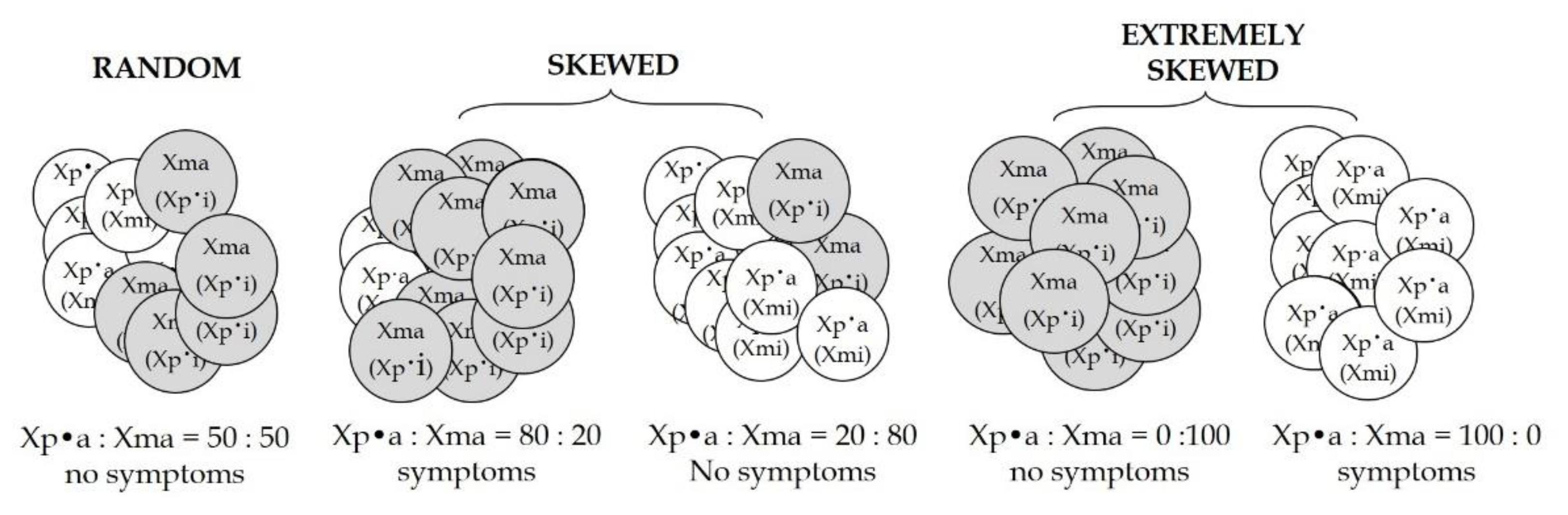
3. Skewed X-Chromosome Inactivation
3.1. Skewed XCI in the Normal (Healthy) Asymptomatic Females
3.2. Skewed XCI in Carriers of Inherited X-Linked Disorders
4. Meta-Analysis
5. Discussion
Author Contributions
Funding
Institutional Review Board Statement
Informed Consent Statement
Data Availability Statement
Conflicts of Interest
References
- Desnick, R.J.; Brady, R.; Barranger, J.; Collins, A.J.; Germain, D.P.; Goldman, M.; Grabowski, G.; Packman, S.; Wilcox, W.R. Fabry disease, an under-recognized multisystemic disorder: Expert recommendations for diagnosis, management, and enzyme replacement therapy. Ann. Intern. Med. 2003, 138, 338–346. [Google Scholar] [CrossRef] [Green Version]
- Gibas, A.L.; Klatt, R.; Johnson, J.; Clarke, J.T.R.; Katz, J. Disease rarity, carrier status, and gender: A triple disadvantage for women with fabry disease. J. Genet. Couns. 2008, 17, 528–537. [Google Scholar] [CrossRef] [Green Version]
- Spada, M.; Pagliardini, S.; Yasuda, M.; Tukel, T.; Thiagarajan, G.; Sakuraba, H.; Ponzone, A.; Desnick, R.J. High Incidence of Later-Onset Fabry Disease Revealed by Newborn Screening. Am. J. Hum. Genet. 2006, 79, 31–40. [Google Scholar] [CrossRef] [Green Version]
- Garman, S.C.; Garboczi, D.N. The molecular defect leading to fabry disease: Structure of human α-galactosidase. J. Mol. Biol. 2004, 337, 319–335. [Google Scholar] [CrossRef]
- Scriver, C.R.; Beaudet, A.L.; Sly, W.S.; Valle, D.; Childs, B.; Kinzler, K.W.; Vogelstein, B. The Metabolic and Molecular Bases of Inherited Disease, 8th ed.; McGraw-Hill: New York, NY, USA, 2001. [Google Scholar]
- Schiffmann, R.; Kopp, J.B.; Iii, H.A.A.; Sabnis, S.; Moore, D.F.; Weibel, T.; Balow, J.E.; Brady, R.O. Enzyme replacement therapy in fabry disease. JAMA 2001, 285, 2743–2749. [Google Scholar] [CrossRef] [PubMed]
- Hsu, M.-J.; Chang, F.-P.; Lu, Y.-H.; Hung, S.-C.; Wang, Y.-C.; Yang, A.-H.; Lee, H.-J.; Sung, S.-H.; Wang, Y.-F.; Yu, W.-C.; et al. Identification of lysosomal and extralysosomal globotriaosylceramide (Gb3) accumulations before the occurrence of typical pathological changes in the endomyocardial biopsies of Fabry disease patients. Genet. Med. 2018, 21, 224–232. [Google Scholar] [CrossRef] [PubMed]
- Von Scheidt, W.; Eng, C.M.; Fitzmaurice, T.F.; Erdmann, E.; Hübner, G.; Olsen, E.G.; Christomanou, H.; Kandolf, R.; Bishop, D.F.; Desnick, R.J. An Atypical Variant of Fabry’s Disease with Manifestations Confined to the Myocardium. N. Engl. J. Med. 1991, 324, 395–399. [Google Scholar] [CrossRef] [PubMed]
- Nakao, S.; Kodama, C.; Takenaka, T.; Tanaka, A.; Yasumoto, Y.; Yoshida, A.; Kanzaki, T.; Enriquez, A.L.; Eng, C.M.; Tanaka, H.; et al. Fabry disease: Detection of undiagnosed hemodialysis patients and identification of a “renal variant” phenotype. Kidney Int. 2003, 64, 801–807. [Google Scholar] [CrossRef] [PubMed] [Green Version]
- Yoshitama, T.; Nakao, S.; Takenaka, T.; Teraguchi, H.; Sasaki, T.; Kodama, C.; Tanaka, A.; Kisanuki, A.; Tei, C. Molecular genetic, biochemical, and clinical studies in three families with cardiac Fabry’s disease. Am. J. Cardiol. 2001, 87, 71–75. [Google Scholar] [CrossRef]
- Nakao, S.; Takenaka, T.; Maeda, M.; Kodama, C.; Tanaka, A.; Tahara, M.; Yoshida, A.; Kuriyama, M.; Hayashibe, H.; Sakuraba, H.; et al. An Atypical Variant of Fabry’s Disease in Men with Left Ventricular Hypertrophy. N. Engl. J. Med. 1995, 333, 288–293. [Google Scholar] [CrossRef]
- Wilcox, W.R.; Oliveira, J.P.; Hopkin, R.; Ortiz, A.; Banikazemi, M.; Feldt-Rasmussen, U.; Sims, K.; Waldek, S.; Pastores, G.M.; Lee, P.; et al. Females with Fabry disease frequently have major organ involvement: Lessons from the Fabry Registry. Mol. Genet. Metab. 2008, 93, 112–128. [Google Scholar] [CrossRef]
- Hůlková, H.; Ledvinová, J.; Poupĕtová, H.; Bultas, J.; Zeman, J.; Elleder, M. Postmortem diagnosis of Fabry disease in a female heterozygote leading to the detection of undiagnosed manifest disease in the family. Cas. Lek. Ceskych 1999, 138, 660–664. [Google Scholar]
- Laney, D.A.; Peck, D.S.; Atherton, A.M.; Manwaring, L.; Christensen, K.M.; Shankar, S.P.; Grange, D.K.; Wilcox, W.R.; Hopkin, R.J. Fabry disease in infancy and early childhood: A systematic literature review. Genet. Med. 2015, 17, 323–330. [Google Scholar] [CrossRef] [Green Version]
- Moura, A.P.; Hammerschmidt, T.G.; Deon, M.; Giugliani, R.; Vargas, C.R. Investigation of correlation of urinary globotriaosylceramide (Gb3) levels with markers of renal function in patients with Fabry disease. Clin. Chim. Acta 2018, 478, 62–67. [Google Scholar] [CrossRef]
- Vedder, A.C.; Linthorst, G.E.; Van Breemen, M.J.; Groener, J.E.M.; Bemelman, F.J.; Strijland, A.; Mannens, M.M.A.M.; Aerts, J.; Hollak, C.E.M. The Dutch Fabry cohort: Diversity of clinical manifestations and Gb3 levels. J. Inherit. Metab. Dis. 2007, 30, 68–78. [Google Scholar] [CrossRef] [PubMed]
- Laney, D.A. Interfamily variability in patients with classical Fabry disease. Mol. Genet. Metab. 2019, 126, S90–S91. [Google Scholar] [CrossRef]
- Tuttolomondo, A.; Simonetta, I.; Duro, G.; Pecoraro, R.; Miceli, S.; Colomba, P.; Zizzo, C.; Nucera, A.; Daidone, M.; Di Chiara, T.; et al. Inter-familial and intra-familial phenotypic variability in three Sicilian families with Anderson-Fabry disease. Oncotarget 2017, 8, 61415–61424. [Google Scholar] [CrossRef] [Green Version]
- Effraimidis, G.; Feldt-Rasmussen, U.; Rasmussen, Å.K.; Lavoie, P.; Abaoui, M.; Boutin, M.; Auray-Blais, C. Globotriaosylsphingosine (lyso-Gb3) and analogues in plasma and urine of patients with Fabry disease and correlations with long-term treatment and genotypes in a nationwide female Danish cohort. J. Med. Genet. 2020. [Google Scholar] [CrossRef] [PubMed]
- Aerts, J.M.; Groener, J.E.; Kuiper, S.; Donker-Koopman, W.E.; Strijland, A.; Ottenhoff, R.; van Roomen, C.; Mirzaian, M.; Wijburg, F.A.; Linthorst, G.E.; et al. Elevated globotriaosylsphingosine is a hallmark of Fabry disease. Proc. Natl. Acad. Sci. USA 2008, 105, 2812–2817. [Google Scholar] [CrossRef] [PubMed] [Green Version]
- Rombach, S.; Dekker, N.; Bouwman, M.; Linthorst, G.; Zwinderman, A.; Wijburg, F.; Kuiper, S.; Weerman, M.V.B.; Groener, J.; Poorthuis, B.; et al. Plasma globotriaosylsphingosine: Diagnostic value and relation to clinical manifestations of Fabry disease. Biochim. Biophys. Acta Mol. Basis Dis. 2010, 1802, 741–748. [Google Scholar] [CrossRef] [PubMed] [Green Version]
- Togawa, T.; Kodama, T.; Suzuki, T.; Sugawara, K.; Tsukimura, T.; Ohashi, T.; Ishige, N.; Suzuki, K.; Kitagawa, T.; Sakuraba, H. Plasma globotriaosylsphingosine as a biomarker of Fabry disease. Mol. Genet. Metab. 2010, 100, 257–261. [Google Scholar] [CrossRef]
- Seydelmann, N.; Wanner, C.; Störk, S.; Ertl, G.; Weidemann, F. Fabry disease and the heart. Best Pract. Res. Clin. Endocrinol. Metab. 2015, 29, 195–204. [Google Scholar] [CrossRef] [Green Version]
- Shu, L.; Vivekanandan-Giri, A.; Pennathur, S.; Smid, B.E.; Aerts, J.; Hollak, C.E.; Shayman, J.A. Establishing 3-nitrotyrosine as a biomarker for the vasculopathy of Fabry disease. Kidney Int. 2014, 86, 58–66. [Google Scholar] [CrossRef] [Green Version]
- Biancini, G.B.; Vanzin, C.S.; Rodrigues, D.B.; Deon, M.; Ribas, G.S.; Barschak, A.; Manfredini, V.; Netto, C.B.; Jardim, L.B.; Giugliani, R.; et al. Globotriaosylceramide is correlated with oxidative stress and inflammation in Fabry patients treated with enzyme replacement therapy. Biochim. Biophys. Acta Mol. Basis Dis. 2012, 1822, 226–232. [Google Scholar] [CrossRef] [Green Version]
- Shen, J.-S.; Meng, X.-L.; Moore, D.F.; Quirk, J.M.; Shayman, J.A.; Schiffmann, R.; Kaneski, C.R. Globotriaosylceramide induces oxidative stress and up-regulates cell adhesion molecule expression in Fabry disease endothelial cells. Mol. Genet. Metab. 2008, 95, 163–168. [Google Scholar] [CrossRef] [Green Version]
- De Francesco, P.N.; Mucci, J.M.; Ceci, R.; Fossati, C.A.; Rozenfeld, P.A. Fabry disease peripheral blood immune cells release inflammatory cytokines: Role of globotriaosylceramide. Mol. Genet. Metab. 2013, 109, 93–99. [Google Scholar] [CrossRef]
- Mauhin, W.; Lidove, O.; Masat, E.; Mingozzi, F.; Mariampillai, K.; Ziza, J.-M.; Benveniste, O. Innate and Adaptive Immune Response in Fabry Disease. JIMD Rep. 2015, 22, 1–10. [Google Scholar] [CrossRef] [PubMed] [Green Version]
- Beer, G.; Reinecke, P.; Gabbert, H.E.; Hort, W.; Kuhn, H. Fabry disease in patients with hypertrophic cardiomyopathy (HCM). Z. Kardiol. 2002, 91, 992–1002. [Google Scholar] [CrossRef] [PubMed]
- Hayashi, Y.; Hanawa, H.; Jiao, S.; Hasegawa, G.; Ohno, Y.; Yoshida, K.; Suzuki, T.; Kashimura, T.; Obata, H.; Tanaka, K.; et al. Elevated endomyocardial biopsy macrophage-related markers in intractable myocardial diseases. Inflammation 2015, 38, 2288–2299. [Google Scholar] [CrossRef] [PubMed]
- Rombach, S.; Twickler, T.; Aerts, J.; Linthorst, G.; Wijburg, F.; Hollak, C. Vasculopathy in patients with Fabry disease: Current controversies and research directions. Mol. Genet. Metab. 2010, 99, 99–108. [Google Scholar] [CrossRef]
- Del Pinto, R.; Ferri, C. The role of immunity in fabry disease and hypertension: A Review of a novel common pathway. High Blood Press. Cardiovasc. Prev. 2020, 27, 539–546. [Google Scholar] [CrossRef] [PubMed]
- Rombach, S.M.; van den Bogaard, B.; De Groot, E.; Groener, J.E.; Poorthuis, B.J.; Linthorst, G.E.; van den Born, B.-J.H.; Hollak, C.E.; Aerts, J.M. Vascular aspects of fabry disease in relation to clinical manifestations and elevations in plasma globotriaosylsphingosine. Hypertension 2012, 60, 998–1005. [Google Scholar] [CrossRef] [Green Version]
- Weidemann, F.; Sanchez-Niño, M.D.; Politei, J.; Oliveira, J.-P.; Wanner, C.; Warnock, D.G.; Ortiz, A. Fibrosis: A key feature of Fabry disease with potential therapeutic implications. Orphanet J. Rare Dis. 2013, 8, 116. [Google Scholar] [CrossRef] [PubMed] [Green Version]
- Brady, R.O. Clinical Features of and Recent Advances in Therapy for Fabry Disease. JAMA 2000, 284, 2771–2775. [Google Scholar] [CrossRef] [PubMed]
- Zar-Kessler, C.; Karaa, A.; Sims, K.B.; Clarke, V.; Kuo, B. Understanding the gastrointestinal manifestations of Fabry disease: Promoting prompt diagnosis. Ther. Adv. Gastroenterol. 2016, 9, 626–634. [Google Scholar] [CrossRef] [Green Version]
- Grünfeld, J.-P.; Lidove, O.; Joly, D.; Barbey, F. Renal disease in Fabry patients. J. Inherit. Metab. Dis. 2001, 24, 71–74. [Google Scholar] [CrossRef]
- Ries, M.; Moore, D.F.; Robinson, C.J.; Tifft, C.J.; Rosenbaum, K.N.; Brady, R.O.; Schiffmann, R.; Krasnewich, D. Quantitative dysmorphology assessment in Fabry disease. Genet. Med. 2006, 8, 96–101. [Google Scholar] [CrossRef] [Green Version]
- Cox-Brinkman, J.; Vedder, A.C.; Hollak, C.E.M.; Richfield, L.; Mehta, A.; Orteu, K.; Wijburg, F.A.; Hammond, P. Three-dimensional face shape in Fabry disease. Eur. J. Hum. Genet. 2007, 15, 535–542. [Google Scholar] [CrossRef]
- Papaxanthos-Roche, A.; Maillard, A.; Chansel-Debordeaux, L.; Albert, M.; Patrat, C.; Lidove, O.; Germain, D.P.; Perez, P.; Lacombe, D. Semen and male genital tract characteristics of patients with Fabry disease: The Fertifabry multicentre observational study. Basic Clin. Androl. 2019, 29, 7. [Google Scholar] [CrossRef]
- Laney, D.A.; Clarke, V.; Foley, A.; Hall, E.W.; Gillespie, S.E.; Holida, M.; Simmons, M.; Wadley, A.; Baumgartner, M.; Patterson, M.; et al. The impact of Fabry disease on reproductive fitness. JIMD Rep. 2017, 37, 85–97. [Google Scholar] [CrossRef] [Green Version]
- Papaxanthos-Roche, A.; Deminiere, C.; Bauduer, F.; Hocké, C.; Mayer, G.; Lacombe, D. Azoospermia as a new feature of Fabry disease. Fertil. Steril. 2007, 88, 212.e15–212.e18. [Google Scholar] [CrossRef] [PubMed]
- Weidemann, F.; Strotmann, J.M.; Niemann, M.; Herrmann, S.; Wilke, M.; Beer, M.; Voelker, W.; Ertl, G.; Emmert, A.; Wanner, C.; et al. Heart valve involvement in fabry cardiomyopathy. Ultrasound Med. Biol. 2009, 35, 730–735. [Google Scholar] [CrossRef] [PubMed]
- Weidemann, F.; Wanner, C.; Breunig, F. Nomen est omen. Fabry disease. Eur. Heart J. Cardiovasc. Imaging 2008, 9, 831–832. [Google Scholar] [CrossRef] [PubMed] [Green Version]
- Teekakirikul, P.; Kelly, M.A.; Rehm, H.L.; Lakdawala, N.K.; Funke, B.H. Inherited Cardiomyopathies. J. Mol. Diagn. 2013, 15, 158–170. [Google Scholar] [CrossRef] [Green Version]
- Wolf, C.M. Hypertrophic cardiomyopathy: Genetics and clinical perspectives. Cardiovasc. Diagn. Ther. 2019, 9, S388–S415. [Google Scholar] [CrossRef]
- Ho, C.Y.; Charron, P.; Richard, P.; Girolami, F.; Van Spaendonck-Zwarts, K.Y.; Pinto, Y. Genetic advances in sarcomeric cardiomyopathies: State of the art. Cardiovasc. Res. 2015, 105, 397–408. [Google Scholar] [CrossRef] [Green Version]
- Walsh, R.; Exome Aggregation Consortium; Thomson, K.L.; Ware, J.S.; Funke, B.H.; Woodley, J.; McGuire, K.J.; Mazzarotto, F.; Blair, E.; Seller, A.; et al. Reassessment of Mendelian gene pathogenicity using 7855 cardiomyopathy cases and 60,706 reference samples. Genet. Med. 2017, 19, 192–203. [Google Scholar] [CrossRef] [Green Version]
- Alfares, A.A.; Kelly, M.A.; McDermott, G.; Funke, B.H.; Lebo, M.S.; Baxter, S.B.; Shen, J.; McLaughlin, H.M.; Clark, E.H.; Babb, L.J.; et al. Results of clinical genetic testing of 2912 probands with hypertrophic cardiomyopathy: Expanded panels offer limited additional sensitivity. Genet. Med. 2015, 17, 880–888. [Google Scholar] [CrossRef] [Green Version]
- Gersh, B.J.; Maron, B.J.; Bonow, R.O.; Dearani, J.A.; Fifer, M.A.; Link, M.S.; Naidu, S.S.; Nishimura, R.A.; Ommen, S.R.; Rakowski, H.; et al. 2011 ACCF/AHA guideline for the diagnosis and treatment of hypertrophic cardiomyopathy. J. Thorac. Cardiovasc. Surg. 2011, 142, e153–e203. [Google Scholar] [CrossRef] [Green Version]
- Linhart, A.; Lubanda, J.-C.; Palecek, T.; Bultas, J.; Karetová, D.; Ledvinová, J.; Elleder, M.; Aschermann, M. Cardiac manifestations in Fabry disease. J. Inherit. Metab. Dis. 2001, 24, 75–83. [Google Scholar] [CrossRef]
- Pieroni, M.; Chimenti, C.; Ricci, R.; Sale, P.; Russo, M.A.; Frustaci, A. Early detection of Fabry cardiomyopathy by tissue doppler imaging. Circulation 2003, 107, 1978–1984. [Google Scholar] [CrossRef] [PubMed] [Green Version]
- Shah, J.S.; Hughes, D.; Sachdev, B.; Tome, M.; Ward, D.; Lee, P.; Mehta, A.B.; Elliott, P.M. Prevalence and clinical significance of cardiac arrhythmia in anderson-fabry disease. Am. J. Cardiol. 2005, 96, 842–846. [Google Scholar] [CrossRef] [PubMed]
- Krämer, J.; Niemann, M.; Liu, D.; Hu, K.; Machann, W.; Beer, M.; Wanner, C.; Ertl, G.; Weidemann, F. Two-dimensional speckle tracking as a non-invasive tool for identification of myocardial fibrosis in Fabry disease. Eur. Heart J. 2013, 34, 1587–1596. [Google Scholar] [CrossRef] [Green Version]
- Moon, J.; Sachdev, B.; Elkington, A.G.; McKenna, W.J.; Mehta, A.; Pennell, D.; Leed, P.J.; Elliott, P. Gadolinium enhanced cardiovascular magnetic resonance in Anderson-Fabry disease Evidence for a disease specific abnormality of the myocardial interstitium. Eur. Heart J. 2003, 24, 2151–2155. [Google Scholar] [CrossRef] [PubMed] [Green Version]
- Viggiano, E.; Marabotti, A.; Politano, L.; Burlina, A. Galactose-1-phosphate uridyltransferase deficiency: A literature review of the putative mechanisms of short and long-term complications and allelic variants. Clin. Genet. 2017, 93, 206–215. [Google Scholar] [CrossRef] [PubMed]
- Schaefer, E.; Mehta, A.; Gal, A. Genotype and phenotype in Fabry disease: Analysis of the Fabry Outcome Survey. Acta Paediatr. 2007, 94, 87–92. [Google Scholar] [CrossRef] [PubMed]
- Ries, M.; Gal, A. Genotype–phenotype correlation in Fabry disease. In Fabry Disease: Perspectives from 5 Years of FOS; Mehta, A., Beck, M., Sunder-Plassmann, G., Eds.; Oxford PharmaGenesis: Oxford, UK, 2006; ISBN 978-1-903539-03-3. [Google Scholar]
- Pan, X.; Ouyang, Y.; Wang, Z.; Ren, H.; Shen, P.; Wang, W.; Xu, Y.; Ni, L.; Yu, X.; Chen, X.; et al. Genotype: A Crucial but Not Unique Factor Affecting the Clinical Phenotypes in Fabry Disease. PLoS ONE 2016, 11, e0161330. [Google Scholar] [CrossRef] [Green Version]
- Koca, S.; Tümer, L.; Okur, I.; Erten, Y.; Bakkaloğlu, S.; Biberoğlu, G.; Kasapkara, Ç.; Küçükçongar, A.; Dalgıç, B.; Oktar, S.Ö.; et al. High incidence of co-existing factors significantly modifying the phenotype in patients with Fabry disease. Gene 2019, 687, 280–288. [Google Scholar] [CrossRef]
- Mignani, R.; Moschella, M.; Cenacchi, G.; Donati, I.; Flachi, M.; Grimaldi, D.; Cerretani, D.; De Giovanni, P.; Montevecchi, M.; Rigotti, A.; et al. Different renal phenotypes in related adult males with Fabry disease with the same classic genotype. Mol. Genet. Genom. Med. 2017, 5, 438–442. [Google Scholar] [CrossRef]
- Lukas, J.; Giese, A.; Markoff, A.; Grittner, U.; Kolodny, E.; Mascher, H.; Lackner, K.J.; Meyer, W.; Wree, P.; Saviouk, V.; et al. Functional characterisation of alpha-galactosidase a mutations as a basis for a new classification system in Fabry disease. PLoS Genet. 2013, 9, e1003632. [Google Scholar] [CrossRef] [Green Version]
- Smid, B.E.; Van Der Tol, L.; Biegstraaten, M.; Linthorst, G.E.; Hollak, C.E.M.; Poorthuis, B.J.H.M. Plasma globotriaosylsphingosine in relation to phenotypes of Fabry disease. J. Med. Genet. 2015, 52, 262–268. [Google Scholar] [CrossRef]
- El Dib, R.; Gomaa, H.; Carvalho, R.P.; Camargo, S.E.A.; Bazan, R.; Barretti, P.; Barreto, F.C. Enzyme replacement therapy for Anderson-Fabry disease. Cochrane Database Syst. Rev. 2016, 7, CD006663. [Google Scholar] [CrossRef] [PubMed]
- Arends, M.; Biegstraaten, M.; Wanner, C.; Sirrs, S.; Mehta, A.; Elliott, P.; Oder, D.; Watkinson, O.T.; Bichet, D.G.; Khan, A.; et al. Agalsidase alfa versus agalsidase beta for the treatment of Fabry disease: An international cohort study. J. Med. Genet. 2018, 55, 351–358. [Google Scholar] [CrossRef] [Green Version]
- Arends, M.; Biegstraaten, M.; Hughes, D.; Mehta, A.; Elliott, P.; Oder, D.; Watkinson, O.T.; Vaz, F.; Van Kuilenburg, A.B.P.; Wanner, C.; et al. Retrospective study of long-term outcomes of enzyme replacement therapy in Fabry disease: Analysis of prognostic factors. PLoS ONE 2017, 12, e0182379. [Google Scholar] [CrossRef]
- Weidemann, F.; Niemann, M.; Breunig, F.; Herrmann, S.; Beer, M.; Störk, S.; Voelker, W.; Ertl, G.; Wanner, C.; Strotmann, J. Long-Term Effects of Enzyme Replacement Therapy on Fabry Cardiomyopathy. Circulation 2009, 119, 524–529. [Google Scholar] [CrossRef] [Green Version]
- Arends, M.; Wijburg, F.A.; Wanner, C.; Vaz, F.M.; van Kuilenburg, A.B.; Hughes, D.A.; Biegstraaten, M.; Mehta, A.; Hollak, C.E.; Langeveld, M. Favourable effect of early versus late start of enzyme replacement therapy on plasma globotriaosylsphingosine levels in men with classical Fabry disease. Mol. Genet. Metab. 2017, 121, 157–161. [Google Scholar] [CrossRef] [PubMed]
- Citro, V.; Cammisa, M.; Liguori, L.; Cimmaruta, C.; Lukas, J.; Cubellis, M.V.; Andreotti, G. The large phenotypic spectrum of fabry disease requires graduated diagnosis and personalized therapy: A Meta-analysis can help to differentiate missense mutations. Int. J. Mol. Sci. 2016, 17, 2010. [Google Scholar] [CrossRef] [PubMed] [Green Version]
- Germain, D.; Hughes, D.; Nicholls, K.; Bichet, D.-G.; Giugliani, R.; Wilcox, W.R.; Feliciani, C.; Shankar, S.P.; Ezgu, F.; Amartino, H.; et al. Treatment of Fabry’s Disease with the Pharmacologic Chaperone Migalastat. N. Engl. J. Med. 2016, 375, 545–555. [Google Scholar] [CrossRef] [PubMed]
- Liguori, L.; Monticelli, M.; Allocca, M.; Mele, B.H.; Lukas, J.; Cubellis, M.V.; Andreotti, G. Pharmacological chaperones: A Therapeutic approach for diseases caused by destabilizing missense mutations. Int. J. Mol. Sci. 2020, 21, 489. [Google Scholar] [CrossRef] [PubMed] [Green Version]
- Simonetta, I.; Tuttolomondo, A.; Daidone, M.; Miceli, S.; Pinto, A. Treatment of Anderson-FabryDisease. Curr. Pharm. Des. 2020, 26, 5089–5099. [Google Scholar] [CrossRef] [PubMed]
- Felis, A.; Whitlow, M.; Kraus, A.; Warnock, D.G.; Wallace, E. Current and investigational therapeutics for fabry disease. Kidney Int. Rep. 2020, 5, 407–413. [Google Scholar] [CrossRef] [PubMed]
- Cammisa, M.; Correra, A.; Andreotti, G.; Cubellis, M.V. Fabry_CEP: A tool to identify Fabry mutations responsive to pharmacological chaperones. Orphanet J. Rare Dis. 2013, 8, 111. [Google Scholar] [CrossRef] [Green Version]
- Porto, C.; Pisani, A.; Rosa, M.; Acampora, E.; Avolio, V.; Tuzzi, M.R.; Visciano, B.; Gagliardo, C.; Materazzi, S.; la Marca, G.; et al. Synergy between the pharmacological chaperone 1-deoxygalactonojirimycin and the human recombinant alpha-galactosidase A in cultured fibroblasts from patients with Fabry disease. J. Inherit. Metab. Dis. 2011, 35, 513–520. [Google Scholar] [CrossRef] [PubMed] [Green Version]
- Hughes, D.A.; Nicholls, K.; Shankar, S.P.; Sunder-Plassmann, G.; Koeller, D.; Nedd, K.; Vockley, G.; Hamazaki, T.; Lachmann, R.; Ohashi, T.; et al. Oral pharmacological chaperone migalastat compared with enzyme replacement therapy in Fabry disease: 18-month results from the randomised phase III ATTRACT study. J. Med. Genet. 2017, 54, 288–296. [Google Scholar] [CrossRef] [PubMed]
- Van Der Veen, S.J.; Hollak, C.E.M.; Van Kuilenburg, A.B.P.; Langeveld, M. Developments in the treatment of Fabry disease. J. Inherit. Metab. Dis. 2020, 43, 908–921. [Google Scholar] [CrossRef] [Green Version]
- Ortiz, A.; Germain, D.P.; Desnick, R.J.; Politei, J.; Mauer, M.; Burlina, A.; Eng, C.; Hopkin, R.J.; Laney, D.; Linhart, A.; et al. Fabry disease revisited: Management and treatment recommendations for adult patients. Mol. Genet. Metab. 2018, 123, 416–427. [Google Scholar] [CrossRef]
- Pinto, L.L.C.; Vieira, T.A.; Giugliani, R.; Schwartz, I.V. Expression of the disease on female carriers of X-linked lysosomal disorders: A brief review. Orphanet J. Rare Dis. 2010, 5, 14. [Google Scholar] [CrossRef] [Green Version]
- Deegan, P.B.; Bähner, F.; Barba, M.; Hughes, D.A.; Beck, M. Fabry disease in females: Clinical characteristics and effects of enzyme replacement therapy. In Fabry Disease: Perspectives from 5 Years of FOS; Mehta, A., Beck, M., Sunder-Plassmann, G., Eds.; Oxford PharmaGenesis: Oxford, UK, 2006; ISBN 978-1-903539-03-3. [Google Scholar]
- Doi, Y.; Toda, G.; Yano, K. Sisters with atypical Fabry’s disease with complete atrioventricular block. Heart 2003, 89, e2. [Google Scholar] [CrossRef] [PubMed]
- Baehner, F.; Kampmann, C.; Whybra, C.; Miebach, E.; Wiethoff, C.M.; Beck, M. Enzyme replacement therapy in heterozygous females with Fabry disease: Results of a phase IIIB study. J. Inherit. Metab. Dis. 2003, 26, 617–627. [Google Scholar] [CrossRef]
- Ro, L.S.; Chen, S.T.; Tang, L.M.; Hsu, W.C.; Chang, H.S.; Huang, C.C. Current Perception Threshold Testing in Fabry’s Disease. Muscle Nerve 1999, 22, 1531–1537. [Google Scholar] [CrossRef]
- Whybra, C.; Kampmann, C.; Willers, I.; Davies, J.; Winchester, B.; Kriegsmann, J.; Brühl, K.; Gal, A.; Bunge, S.; Beck, M. Anderson-Fabry disease: Clinical manifestations of disease in female heterozygotes. J. Inherit. Metab. Dis. 2001, 24, 715–724. [Google Scholar] [CrossRef]
- Germain, D.; Benistan, K.; Angelova, L. X-linked inheritance and its implication in the diagnosis and management of female patients in Fabry disease. Revue Méd. Interne 2010, 31, S209–S213. [Google Scholar] [CrossRef]
- MacDermot, K.D.; Holmes, A.; Miners, A.H. Anderson-Fabry disease: Clinical manifestations and impact of disease in a cohort of 60 obligate carrier females. J. Med. Genet. 2001, 38, 769–775. [Google Scholar] [CrossRef] [PubMed] [Green Version]
- Chowdhury, M.M.; Holt, P.J. Pain in Anderson-Fabry’s disease. Lancet 2001, 357, 887. [Google Scholar] [CrossRef]
- Ramaswami, U.; Parini, R.; Pintos-Morell, G. Natural history and effects of enzyme replacement therapy in children and ad-olescents with Fabry disease. In Fabry Disease: Perspectives from 5 Years of FOS; Mehta, A., Beck, M., Sunder-Plassmann, G., Eds.; Oxford PharmaGenesis: Oxford, UK, 2006; ISBN 978-1-903539-03-3. [Google Scholar]
- Pintos-Morell, G.; Beck, M. Fabry disease in children and the effects of enzyme replacement treatment. Eur. J. Nucl. Med. Mol. Imaging 2009, 168, 1355–1363. [Google Scholar] [CrossRef] [PubMed] [Green Version]
- Fukushima, M.; Tsuchiyama, Y.; Nakato, T.; Yokoi, T.; Ikeda, H.; Yoshida, S.; Kusumoto, T.; Itoh, K.; Sakuraba, H. A female heterozygous patient with fabry’s disease with renal accumulation of trihexosylceramide detected with a monoclonal antibody. Am. J. Kidney Dis. 1995, 26, 952–955. [Google Scholar] [CrossRef]
- Yuen, N.W.-F.; Lam, C.-W.; Chow, T.-C.; Chiu, M.-C. A Characteristic dissection microscopy appearance of a renal biopsy of a fabry heterozygote. Nephron 1997, 77, 354–356. [Google Scholar] [CrossRef]
- Migeon, B.R. X Inactivation, Female Mosaicism, and Sex Differences in Renal Diseases. J. Am. Soc. Nephrol. 2008, 19, 2052–2059. [Google Scholar] [CrossRef] [PubMed] [Green Version]
- Mehta, A.; Beck, M.; Sunder-Plassmann, G. Fabry Disease: Perspectives from 5 Years of FOS; Oxford PharmaGenesis: Oxford, UK, 2006; ISBN 978-1-903539-03-3. [Google Scholar]
- Murata, R.; Takatsu, H.; Noda, T.; Nishigaki, K.; Tsuchiya, K.; Takemura, G.; Kanoh, M.; Kunishima, A.; Sano, K.; Minatoguchi, S.; et al. Fifteen-year follow-up of a heterozygous fabry’s disease patient associated with pre-excitation syndrome. Intern. Med. 1999, 38, 476–481. [Google Scholar] [CrossRef] [Green Version]
- Koitabashi, N.; Utsugi, T.; Seki, R.; Okamoto, E.; Sando, Y.; Kaneko, Y.; Nagai, R. Biopsy-proven cardiomyopathy in heterozygous fabry’s disease. Jpn. Circ. J. 1999, 63, 572–575. [Google Scholar] [CrossRef] [PubMed] [Green Version]
- Wang, R.; Lelis, A.; Mirocha, J.; Wilcox, W.R. Heterozygous Fabry women are not just carriers, but have a significant burden of disease and impaired quality of life. Genet. Med. 2007, 9, 34–45. [Google Scholar] [CrossRef] [PubMed] [Green Version]
- Kampmann, C.; Baehner, F.; Whybra, C.; Martin, C.; Wiethoff, C.M.; Ries, M.; Gal, A.; Beck, M. Cardiac manifestations of Anderson–Fabry disease in heterozygous females. J. Am. Coll. Cardiol. 2002, 40, 1668–1674. [Google Scholar] [CrossRef] [Green Version]
- Baig, S.; Edward, N.C.; Kotecha, D.; Liu, B.; Nordin, S.; Kozor, R.; Moon, J.C.; Geberhiwot, T.; Steeds, R.P. Ventricular arrhythmia and sudden cardiac death in Fabry disease: A systematic review of risk factors in clinical practice. Europace 2017, 20, f153–f161. [Google Scholar] [CrossRef]
- Niemann, M.; Herrmann, S.; Hu, K.; Breunig, F.; Strotmann, J.; Beer, M.; Machann, W.; Voelker, W.; Ertl, G.; Wanner, C.; et al. Differences in fabry cardiomyopathy between female and male patients: Consequences for diagnostic assessment. JACC Cardiovasc. Imaging 2011, 4, 592–601. [Google Scholar] [CrossRef] [PubMed] [Green Version]
- Fujii, K.; Minami, N.; Hayashi, Y.; Nishino, I.; Nonaka, I.; Tanabe, Y.; Takanashi, J.-I.; Kohno, Y. Homozygous female Becker muscular dystrophy. Am. J. Med. Genet. Part A 2009, 149A, 1052–1055. [Google Scholar] [CrossRef]
- Baiget, M.; Tizzano, E.; Volpini, V.; Del Rio, E.; Perez-Vidal, T.; Gallano, P. DMD carrier detection in a female with mosaic Turner’s syndrome. J. Med. Genet. 1991, 28, 209–210. [Google Scholar] [CrossRef] [Green Version]
- Satre, V.; Monnier, N.; Devillard, F.; Amblard, F.; Lunardi, P.J. Prenatal diagnosis of DMD in a female foetus affected by Turner syndrome. Prenat. Diagn. 2004, 24, 913–917. [Google Scholar] [CrossRef]
- Quan, F.; Janas, J.; Toth-Fejel, S.; Johnson, D.B.; Wolford, J.K.; Popovich, B.W. Uniparental disomy of the entire X chromosome in a female with Duchenne muscular dystrophy. Am. J. Hum. Genet. 1997, 60, 160–165. [Google Scholar] [PubMed]
- Soltanzadeh, P.; Friez, M.J.; Dunn, D.; von Niederhausern, A.; Gurvich, O.L.; Swoboda, K.; Sampson, J.B.; Pestronk, A.; Connolly, A.; Florence, J.M.; et al. Clinical and genetic characterization of manifesting carriers of DMD mutations. Neuromuscul. Disord. 2010, 20, 499–504. [Google Scholar] [CrossRef] [Green Version]
- Azofeifa, J.; Voit, T.; Cremer, M. X-chromosome methylation in manifesting and healthy carriers of dystrophinopathies: Concordance of activation ratios among first degree female relatives and skewed inactivation as cause of the affected phenotypes. Hum. Gen. 1995, 96, 167–176. [Google Scholar] [CrossRef]
- Viggiano, E.; Picillo, E.; Ergoli, M.; Cirillo, A.; Del Gaudio, S.; Politano, L. Skewed X-chromosome inactivation plays a crucial role in the onset of symptoms in carriers of Becker muscular dystrophy. J. Gene Med. 2017, 19, e2952. [Google Scholar] [CrossRef]
- Viggiano, E.; Ergoli, M.; Picillo, E.; Politano, L. Determining the role of skewed X-chromosome inactivation in developing muscle symptoms in carriers of Duchenne muscular dystrophy. Hum. Gen. 2016, 135, 685–698. [Google Scholar] [CrossRef]
- Viggiano, E.; Picillo, E.; Cirillo, A.; Politano, L. Comparison of X-chromosome inactivation in Duchenne muscle/myocardium-manifesting carriers, non-manifesting carriers and related daughters. Clin. Genet. 2012, 84, 265–270. [Google Scholar] [CrossRef]
- Cho, S.Y.; Lam, C.-W.; Tong, S.-F.; Siu, W.-K. X-linked glycogen storage disease IXa manifested in a female carrier due to skewed X chromosome inactivation. Clin. Chim. Acta 2013, 426, 75–78. [Google Scholar] [CrossRef] [PubMed]
- Garagiola, I.; Mortarino, M.; Siboni, S.M.; Boscarino, M.; Mancuso, M.E.; Biganzoli, M.; Santagostino, E.; Peyvandi, F. X Chromosome inactivation: A modifier of factor VIII and IX plasma levels and bleeding phenotype in Haemophilia carriers. Eur. J. Hum. Genet. 2021, 29, 241–249. [Google Scholar] [CrossRef] [PubMed]
- Ørstavik, K.H. X chromosome inactivation in clinical practice. Hum. Gen. 2009, 126, 363–373. [Google Scholar] [CrossRef] [PubMed]
- Pugacheva, E.M.; Tiwari, V.K.; Abdullaev, Z.; Vostrov, A.A.; Flanagan, P.T.; Quitschke, W.W.; Loukinov, D.I.; Ohlsson, R.; Lobanenkov, V.V. Familial cases of point mutations in the XIST promoter reveal a correlation between CTCF binding and pre-emptive choices of X chromosome inactivation. Hum. Mol. Genet. 2005, 14, 953–965. [Google Scholar] [CrossRef] [Green Version]
- Plenge, R.M.; Hendrich, B.D.; Schwartz, C.; Arena, J.F.; Naumova, A.; Sapienza, C.; Winter, R.M.; Willard, H.F. A promoter mutation in the XIST gene in two unrelated families with skewed X-chromosome inactivation. Nat. Genet. 1997, 17, 353–356. [Google Scholar] [CrossRef] [PubMed]
- Knudsen, G.; Pedersen, J.; Klingenberg, O.; Lygren, I.; Ørstavik, K. Increased skewing of X chromosome inactivation with age in both blood and buccal cells. Cytogenet. Genome Res. 2007, 116, 24–28. [Google Scholar] [CrossRef] [PubMed]
- Sharp, A.; Robinson, D.; Jacobs, P. Age- and tissue-specific variation of X chromosome inactivation ratios in normal women. Qual. Life Res. 2000, 107, 343–349. [Google Scholar] [CrossRef]
- Migeon, B.R. The Role of X inactivation and cellular mosaicism in women’s health and sex-specific diseases. JAMA 2006, 295, 1428–1433. [Google Scholar] [CrossRef] [PubMed]
- Amos-Landgraf, J.; Cottle, A.; Plenge, R.M.; Friez, M.; Schwartz, C.E.; Longshore, J.; Willard, H.F. X Chromosome–Inactivation Patterns of 1005 Phenotypically Unaffected Females. Am. J. Hum. Genet. 2006, 79, 493–499. [Google Scholar] [CrossRef] [Green Version]
- Bolduc, V.; Chagnon, P.; Provost, S.; Dube, M.-P.; Belisle, C.; Gingras, M.; Mollica, L.; Busque, L. No evidence that skewing of X chromosome inactivation patterns is transmitted to offspring in humans. J. Clin. Investig. 2008, 118, 333–341. [Google Scholar] [CrossRef] [Green Version]
- Gale, R.E.; Fielding, A.K.; Harrison, C.N.; Linch, D.C. Acquired skewing of X-chromosome inactivation patterns in myeloid cells of the elderly suggests stochastic clonal loss with age. Br. J. Haematol. 1997, 98, 512–519. [Google Scholar] [CrossRef] [Green Version]
- Fey, M.F.; Liechti-Gallati, S.; von Rohr, A.; Borisch, B.; Theilkäs, L.; Schneider, V.; Oestreicher, M.; Nagel, S.; Ziemiecki, A.; Tobler, A. Clonality and x-inactivation patterns in hematopoietic cell populations detected by the highly informative m27 beta dna probe. Blood 1994, 83, 931–938. [Google Scholar] [CrossRef] [PubMed] [Green Version]
- Christensen, K.; Kristiansen, M.; Hagen-Larsen, H.; Skytthe, A.; Bathum, L.; Jeune, B.; Andersen-Ranberg, K.; Vaupel, J.W.; Orstavik, K.H. X-Linked genetic factors regulate hematopoietic stem-cell kinetics in females. Blood 2000, 95, 2449–2451. [Google Scholar] [CrossRef] [PubMed]
- Busque, L.; Mio, R.; Mattioli, J.; Brais, E.; Blais, N.; Lalonde, Y.; Maragh, M.; Gilliland, D.G. Nonrandom X-inactivation patterns in normal females: Lyonization ratios vary with age. Blood 1996, 88, 59–65. [Google Scholar] [CrossRef] [PubMed] [Green Version]
- Lanasa, M.C.; Hogge, W.; Kubik, C.J.; Ness, R.B.; Harger, J.; Nagel, T.; Prosen, T.; Markovic, N.; Hoffman, E. A novel X chromosome–linked genetic cause of recurrent spontaneous abortion. Am. J. Obstet. Gynecol. 2001, 185, 563–568. [Google Scholar] [CrossRef]
- Kristiansen, M.; Knudsen, G.; Tanner, S.; McEntagart, M.; Jungbluth, H.; Muntoni, F.; Sewry, C.; Gallati, S.; Ørstavik, K.; Wallgren-Pettersson, C. X-inactivation patterns in carriers of X-linked myotubular myopathy. Neuromuscul. Disord. 2003, 13, 468–471. [Google Scholar] [CrossRef]
- Espinós, C.; Lorenzo, J.I.; Casaña, P.; Martínez, F.; Aznar, J.A. Haemophilia B in a female caused by skewed inactivation of the normal X-chromosome. Haematologica 2000, 85, 1092–1095. [Google Scholar]
- Okumura, K.; Fujimori, Y.; Takagi, A.; Murate, T.; Ozeki, M.; Yamamoto, K.; Katsumi, A.; Matsushita, T.; Naoe, T.; Kojima, T. Skewed X chromosome inactivation in fraternal female twins results in moderately severe and mild haemophilia B. Haemophilia 2008, 14, 1088–1093. [Google Scholar] [CrossRef] [PubMed]
- Devriendt, K.; Matthijs, G.; Legius, E.; Schollen, E.; Blockmans, D.; Van Geet, C.; Degreef, H.; Cassiman, J.J.; Fryns, J.P. Skewed X-chromosome inactivation in female carriers of dyskeratosis congenita. Am. J. Hum. Genet. 1997, 60, 581–587. [Google Scholar] [PubMed]
- Fahim, A.T.; Sullivan, L.S.; Bowne, S.J.; Jones, K.D.; Wheaton, D.K.; Khan, N.W.; Heckenlively, J.R.; Jayasundera, K.; Branham, K.H.; Andrews, C.; et al. X-Chromosome inactivation is a biomarker of clinical severity in female carriers of rpgr-associated x-linked retinitis pigmentosa. Ophthalmol. Retin. 2020, 4, 510–520. [Google Scholar] [CrossRef] [PubMed]
- Bushby, K.; Goodship, J.; Nicholson, L.; Johnson, M.; Haggerty, I.; Gardner-Medwin, D. Variability in clinical, genetic and protein abnormalities in manifesting carriers of Duchenne and Becker muscular dystrophy. Neuromuscul. Disord. 1993, 3, 57–64. [Google Scholar] [CrossRef]
- Sumita, D.R.; Vainzof, M.; Campiotto, S.; Cerqueira, A.M.; Cánovas, M.; Otto, P.A.; Passos-Bueno, M.R.; Zatz, M. Absence of Correlation between Skewed X Inactivation in Blood and Serum Creatine-Kinase Levels in Duchenne/Becker Female Carriers. Am. J. Med. Genet. 1998, 80, 356–361. [Google Scholar] [CrossRef]
- Seemann, N.; Selby, K.; McAdam, L.; Biggar, D.; Kolski, H.; Goobie, S.; Yoon, G.; Campbell, C. Symptomatic dystrophinopathies in female children. Neuromuscul. Disord. 2011, 21, 172–177. [Google Scholar] [CrossRef]
- Matthews, P.M.; Benjamin, D.; Van Bakel, I.; Squier, M.; Nicholson, L.; Sewry, C.; Barnes, P.; Hopkin, J.; Brown, R.; Hilton-Jones, D.; et al. Muscle X-inactivation patterns and dystrophin expression in Duchenne muscular dystrophy carriers. Neuromuscul. Disord. 1995, 5, 209–220. [Google Scholar] [CrossRef]
- Pegoraro, E.; Schimke, R.N.; Arahata, K.; Hayashi, Y.; Stern, H.; Marks, H.; Glasberg, M.R.; Carroll, J.E.; Taber, J.W.; Wessel, H.B.; et al. Detection of new paternal dystrophin gene mutations in isolated cases of dystrophinopathy in females. Am. J. Hum. Genet. 1994, 54, 989–1003. [Google Scholar]
- Pegoraro, E.; Schimke, R.N.; Garcia, C.; Stern, H.; Cadaldini, M.; Angelini, C.; Barbosa, E.; Carroll, J.; Marks, W.A.; Neville, H.E.; et al. Genetic and biochemical normalization in female carriers of Duchenne muscular dystrophy: Evidence for failure of dystrophin production in dystrophin-competent myonuclei. Neurology 1995, 45, 677–690. [Google Scholar] [CrossRef] [PubMed]
- Azofeifa, J.; Cremer, M.; Waldherr, R. X-chromosome methylation ratios as indicators of chromosomal activity: Evidence of intraindividual divergencies among tissues of different embryonal origin. Hum. Gen. 1996, 97, 330–333. [Google Scholar] [CrossRef] [PubMed]
- Fialkow, P.J. Primordial cell pool size and lineage relationships of five human cell types. Ann. Hum. Genet. 1973, 37, 39–48. [Google Scholar] [CrossRef]
- Lupski, J.R.; Garcia, C.A.; Zoghbi, H.Y.; Hoffman, E.; Fenwick, R.G. Discordance of muscular dystrophy in monozygotic female twins: Evidence supporting asymmetric splitting of the inner cell mass in a manifesting carrier of Duchenne dystrophy. Am. J. Med. Genet. 1991, 40, 354–364. [Google Scholar] [CrossRef]
- Dobrovolny, R.; Dvořáková, L.; Ledvinová, J.; Magage, S.; Bultas, J.; Lubanda, J.-C.; Elleder, M.; Karetová, D.; Pavlikova, M.; Hřebíček, M. Relationship between X-inactivation and clinical involvement in Fabry heterozygotes. Eleven novel mutations in the α-galactosidase A gene in the Czech and Slovak population. J. Mol. Med. 2005, 83, 647–654. [Google Scholar] [CrossRef]
- Maier, E.; Osterrieder, S.; Whybra, C.; Ries, M.; Gal, A.; Beck, M.; Roscher, A.; Muntau, A. Disease manifestations and X inactivation in heterozygous females with Fabry disease. Acta Paediatr. 2006, 95, 30–38. [Google Scholar] [CrossRef]
- Echevarria, L.; Benistan, K.; Toussaint, A.; Dubourg, O.; Hagege, A.; Eladari, D.; Jabbour, F.; Beldjord, C.; De Mazancourt, P.; Germain, D. X-chromosome inactivation in female patients with Fabry disease. Clin. Genet. 2016, 89, 44–54. [Google Scholar] [CrossRef]
- Morrone, A.; Cavicchi, C.; Bardelli, T.; Antuzzi, D.; Parini, R.; Di Rocco, M.; Feriozzi, S.; Gabrielli, O.; Barone, R.; Pistone, G.; et al. Fabry disease: Molecular studies in Italian patients and X inactivation analysis in manifesting carriers. J. Med. Genet. 2003, 40, 103e. [Google Scholar] [CrossRef] [Green Version]
- Rossanti, R.; Nozu, K.; Fukunaga, A.; Nagano, C.; Horinouchi, T.; Yamamura, T.; Sakakibara, N.; Minamikawa, S.; Ishiko, S.; Aoto, Y.; et al. X-chromosome inactivation patterns in females with Fabry disease examined by both ultra-deep RNA sequencing and methylation-dependent assay. Clin. Exp. Nephrol. 2021, 1–7. [Google Scholar] [CrossRef]
- Cox, R.P.; Krauss, M.R.; Balis, M.E.; Dancis, J. Evidence for transfer of enzyme product as the basis of metabolic cooperation between tissue culture fibroblasts of lesch-nyhan disease and normal cells. Proc. Natl. Acad. Sci. USA 1970, 67, 1573–1579. [Google Scholar] [CrossRef] [Green Version]
- Fratantoni, J.C.; Hall, C.W.; Neufeld, E.F. Hurler and Hunter Syndromes: Mutual correction of the defect in cultured fibroblasts. Science 1968, 162, 570–572. [Google Scholar] [CrossRef]
- Viggiano, E.; Madej-Pilarczyk, A.; Carboni, N.; Picillo, E.; Ergoli, M.; Del Gaudio, S.; Marchel, M.; Nigro, G.; Palladino, A.; Politano, L.; et al. X-Linked emery–dreifuss muscular dystrophy: Study of x-chromosome inactivation and its relation with clinical phenotypes in female carriers. Genes 2019, 10, 919. [Google Scholar] [CrossRef] [PubMed] [Green Version]
- Frustaci, A. Pathology and Function of Conduction Tissue in Fabry Disease Cardiomyopathy. Circ Arrhythm Electrophysiol. 2015, 8, 799–805. [Google Scholar] [CrossRef] [PubMed] [Green Version]


| Articles | Age | Skewed XCI | ||||
|---|---|---|---|---|---|---|
| Tissue Analyzed | Mild MSSI Score (Total Subjects) | Moderate-Severe MSSI Score (Total Subjects) | Cardiac Involvement (Total Subjects) | No Cardiac Involvement (Total Subjects) | ||
| Dobrovolny et al., 2005 | Young/Adult | L, U, SE | 7 (24) | 4 (14) | n.d. | n.d. |
| Maier et al., 2006 | Young/Adult | L | 5 (10) | 5 (18) | 6 (16) | 4 (12) |
| Echeivarra et al., 2015 | Young/Adult | L, U, SE, skin | 3 (35) | 7 (21) | 7 (41) | 3 (8) |
| Morrone et. al., 2003 | Young/Adult | L | n.d. | n.d. | 2 (0) | 2 (4) |
| Rossanti et al., 2021 | Adult | L, | n.d. | n.d. | 0 (5) | 1 (2) |
Publisher’s Note: MDPI stays neutral with regard to jurisdictional claims in published maps and institutional affiliations. |
© 2021 by the authors. Licensee MDPI, Basel, Switzerland. This article is an open access article distributed under the terms and conditions of the Creative Commons Attribution (CC BY) license (https://creativecommons.org/licenses/by/4.0/).
Share and Cite
Viggiano, E.; Politano, L. X Chromosome Inactivation in Carriers of Fabry Disease: Review and Meta-Analysis. Int. J. Mol. Sci. 2021, 22, 7663. https://doi.org/10.3390/ijms22147663
Viggiano E, Politano L. X Chromosome Inactivation in Carriers of Fabry Disease: Review and Meta-Analysis. International Journal of Molecular Sciences. 2021; 22(14):7663. https://doi.org/10.3390/ijms22147663
Chicago/Turabian StyleViggiano, Emanuela, and Luisa Politano. 2021. "X Chromosome Inactivation in Carriers of Fabry Disease: Review and Meta-Analysis" International Journal of Molecular Sciences 22, no. 14: 7663. https://doi.org/10.3390/ijms22147663