Article

Decrease in ITGA7 Levels Is Associated with an Increase in α-Synuclein Levels in an MPTP-Induced Parkinson’s Disease Mouse Model and SH-SY5Y Cells

1
Department of Meridian and Acupoint, College of Korean Medicine, Kyung Hee University, Seoul 02453, Korea
2
Department of Meridian and Acupoint, College of Korean Medicine, Sang Ji University, Wonju 26339, Korea
3
Research Institute of Korean Medicine, Sang Ji University, Wonju 26339, Korea
*
Authors to whom correspondence should be addressed.
Int. J. Mol. Sci. 2021, 22(23), 12616; https://doi.org/10.3390/ijms222312616
Submission received: 29 October 2021 / Revised: 19 November 2021 / Accepted: 20 November 2021 / Published: 23 November 2021

Abstract

:
We investigated the potential association between integrin α7 (ITGA7) and alpha-synuclein (α-syn) in a methyl-4-phenyl-1,2,3,6-tetrahydropyridine (MPTP)-induced Parkinson’s disease (PD) mouse model. Tyrosine hydroxylase (TH), ITGA7, and α-syn expression in the substantia nigra (SN) of the brain were observed to examine the pathological characteristics of PD. To determine the relationship between ITGA7 and PD, the expression of TH and α-syn was investigated after ITGA7 siRNA knockdown in SH-SY5Y cells. The ITGA7 microarray signal was decreased in the SN of the MPTP group, indicating reduced ITGA7 expression compared to that in the control. The expression patterns of ITGA7 in the control group and those of α-syn in the MPTP group were similar on immunohistochemical staining. Reduction in ITGA7 expression by ITGA7 siRNA administration induced a decrease in TH expression and an increase in α-syn expression in SH-SY5Y cells. The decreased expression of ITGA7 significantly decreased the expression of bcl2 and increased the bax/bcl2 ratio in SH-SY5Y cells. These results suggest that reduced ITGA7 expression may be related to increased α-syn expression and apoptosis of dopaminergic cells in an MPTP-induced PD mouse model. To the best of our knowledge, this is the first study to show an association between ITGA7 and PD.

Graphical Abstract

1. Introduction

As life expectancy and the prevalence of degenerative brain diseases increase, the number of patients with Parkinson’s disease (PD), the second most common degenerative brain disease after Alzheimer’s disease, is increasing rapidly [1]. The main symptoms of PD are motor function-related symptoms, including slow movement, tremor, rigidity, and postural instability [2]. PD is characterized by the loss of dopamine-secreting neurons in the substantia nigra (SN) located in the midbrain, but the cause of this decrease is still unknown [3]. The major histopathological feature of PD is the deposition of Lewy bodies, and the major protein component of these intracellular deposits is a fibrillar aggregate of α-synuclein (α-syn) [4]. Increased expression of α-syn was observed in both patients with PD and PD animal models and is reported to be closely related to PD pathology [5]. Self-aggregation of these proteins leads to the formation of an amphipathic helical structure, which may result in membrane association [6].
Integrin α7 (ITGA7), also known as the integrin subunit alpha 7 and integrin alpha 7 chain 3, encodes an extracellular matrix (ECM)-binding protein [7]. Integrins are a major family of cell surface receptors that mediate ECM adhesion and are closely implicated in the regulation of various cellular functions, including embryonic development, tumor cell growth and metastasis, and programmed cell death [8]. This protein functions as a receptor for laminin-1, a basement membrane protein on the surface of skeletal myoblasts and muscle fibers [9].
Abnormal integrin expression is associated with several human diseases [10]. Defects in ITGA7 are associated with congenital myopathy [11]. ITGA7-deficient mice display significant hyperplasia, hypertrophy of arteries and arterioles, and malformation of skeletal muscles [12,13]. In addition, ITGA7 deficiency is common in muscular dystrophy and myopathy [14]. α-syn and integrin were first reported in 2005, in a case of MSA, another synucleinopathy [15], and there are also recent studies reporting integrins and α-syn [16,17].
Based on these previous studies, we hypothesized that ITGA7 expression is involved in the pathological changes in PD. The main symptoms of PD are motor function-related symptoms; therefore, we speculated that the motor function-related symptoms of PD may be related to myopathy due to a decrease in ITGA7 expression. Herein, we report that ITGA7 expression is reduced in the SN in a 1-methyl-4-phenyl-1,2,3,6-tetrahydropyridine (MPTP)-induced Parkinsonism mouse model. We further showed that the reduction in ITGA7 expression induces an increase in α-syn expression in SH-SY5Y cells treated with MPP+. To the best of our knowledge, this is the first study to show an association between ITGA7 and PD.

2. Results

2.1. Reduction in Tyrosine Hydroxylase Expression in a Mouse Model of Chronic MPTP-Induced PD

MPTP and saline were injected intraperitoneally once daily in the control (CTL) and MPTP groups, respectively. After four weeks, to confirm that a chronic MPTP-induced PD mouse model was established, we analyzed the changes in tyrosine hydroxylase (TH) expression in the SN and striatum (ST). The expression of TH was significantly decreased in MPTP mice (Figure 1c,d) compared to that in the CTL mice in both the SN and ST. Similar to the results of the Western blot analysis, it was confirmed that the expression of TH was significantly reduced in the SN and ST (p < 0.005) treated with MPTP (Figure 1).

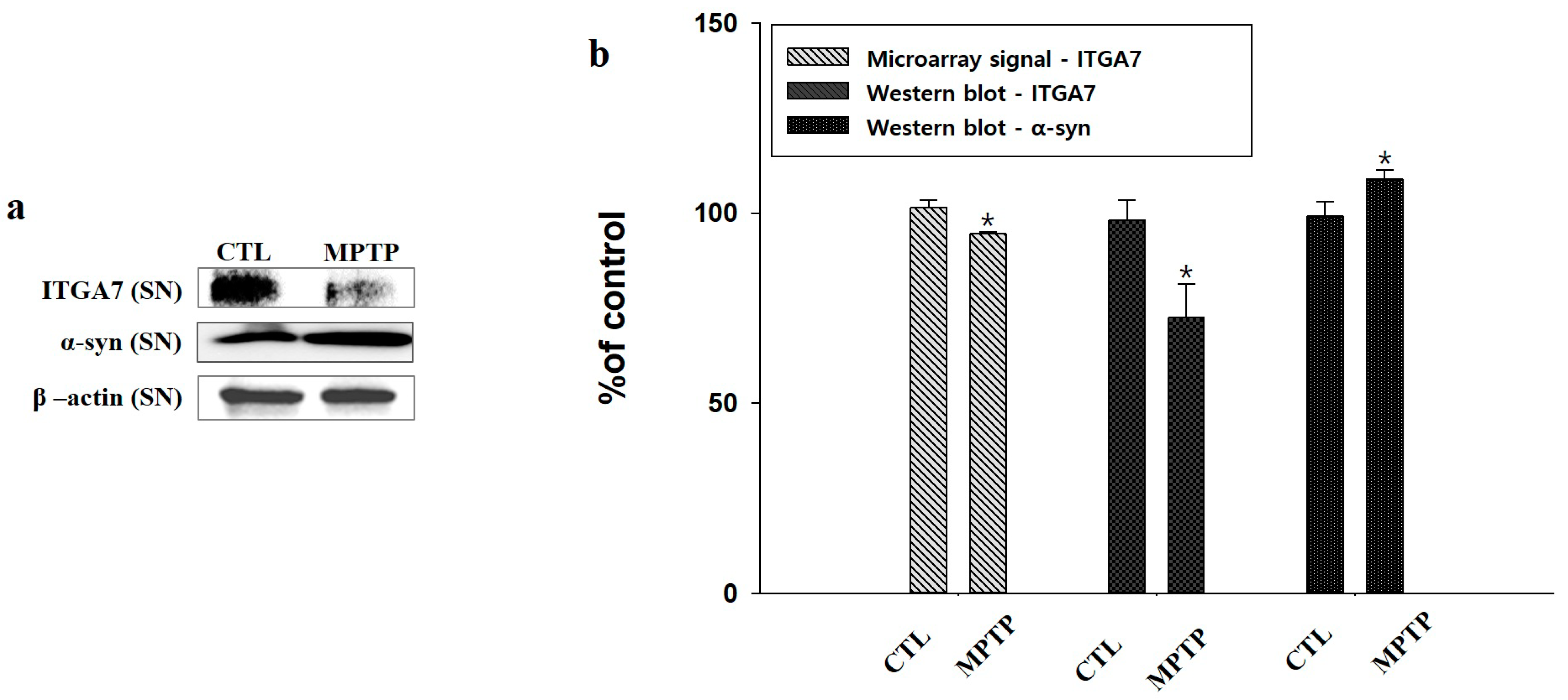
2.2. Microarray Analysis and Western Blot Analysis of ITGA7 in the Substantia Nigra

Microarray signal analysis of the SN confirmed that the ITGA7 signal in MPTP-treated mice was decreased compared to that in CTL (p < 0.05, n = 2, Figure 2b). Therefore, when ITGA7 expression was evaluated by Western blot analysis, it was confirmed that the expression of ITGA7 was significantly decreased in the MPTP group compared to that in the CTL group (p < 0.05, n = 3, Figure 2a,b). α-syn expression was evaluated using Western blot analysis, and it was confirmed that the expression of α-syn was significantly increased in the MPTP group compared to that in the CTL group (p < 0.05, n = 3, Figure 2a,b).

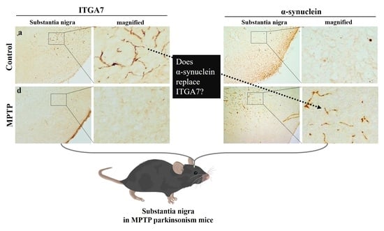
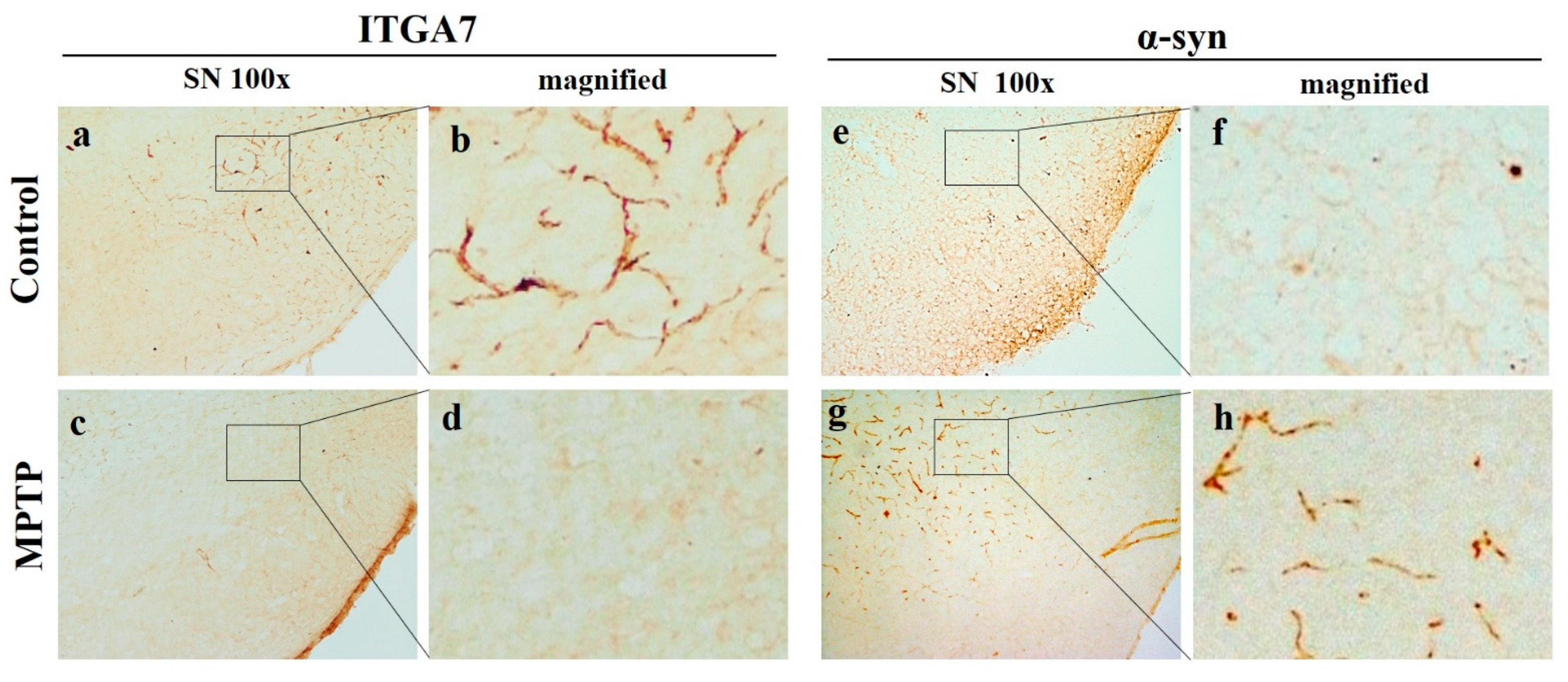
2.3. Reduction in ITGA7 Expression and Increase in α-Syn Expression in the Substantia Nigra in a Mouse Model of Chronic MPTP-Induced PD

Immunohistochemical analysis (IHC) showed that the expression of ITGA7 in the SN was significantly decreased in the MPTP group (Figure 3c,d) when compared to that in the CTL group (Figure 3a,b). Conversely, the expression of α-syn was remarkably increased in the MPTP group (Figure 3e–h). Although the expression of ITGA7 and α-syn was inversely proportional, the patterns were similar.

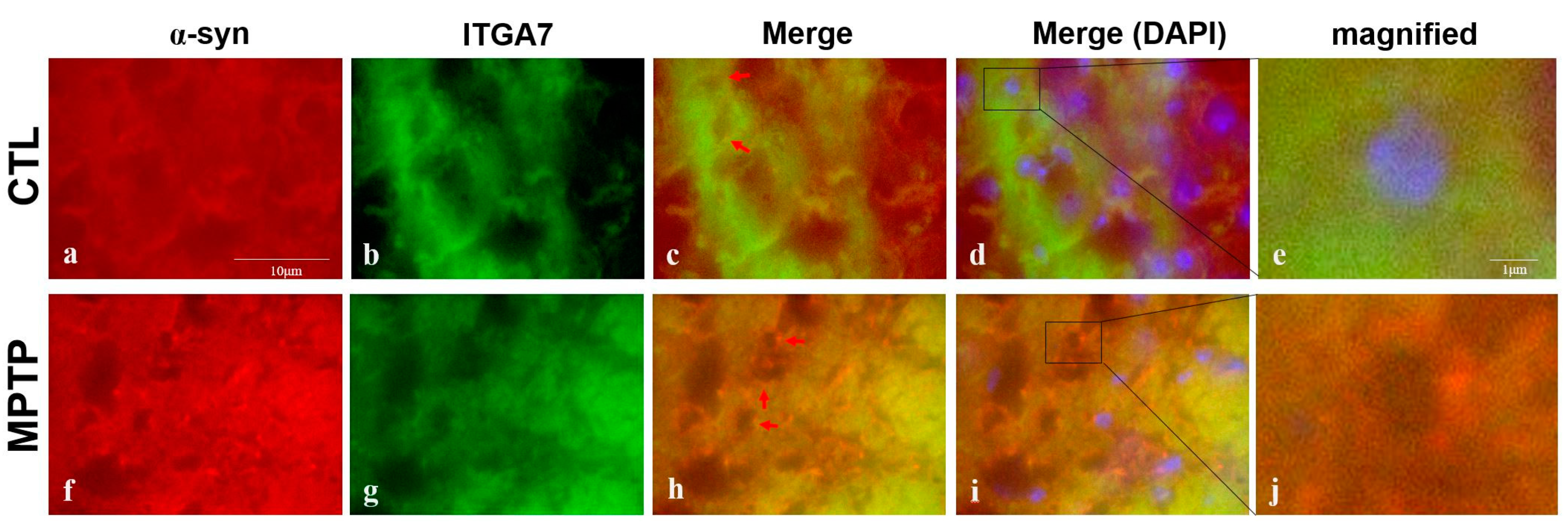
2.4. Immunofluorescence Analysis of ITGA7 Co-Localized with a α-Syn in the Substantia Nigra

Immunofluorescence analysis of ITGA7 and α-syn revealed co-localization and showed that ITGA7 expression was stronger in the CTL group (Figure 4a) than in the MPTP group (Figure 4f). Conversely, α-syn was more strongly expressed in the MPTP group than in the CTL group (Figure 4b,g). When ITGA7 and α-syn were merged (Figure 4c,d,h,i), the expression of ITGA7 was stronger in the CTL group (Figure 4d), whereas the expression of α-syn was stronger in the MPTP group (Figure 4i).

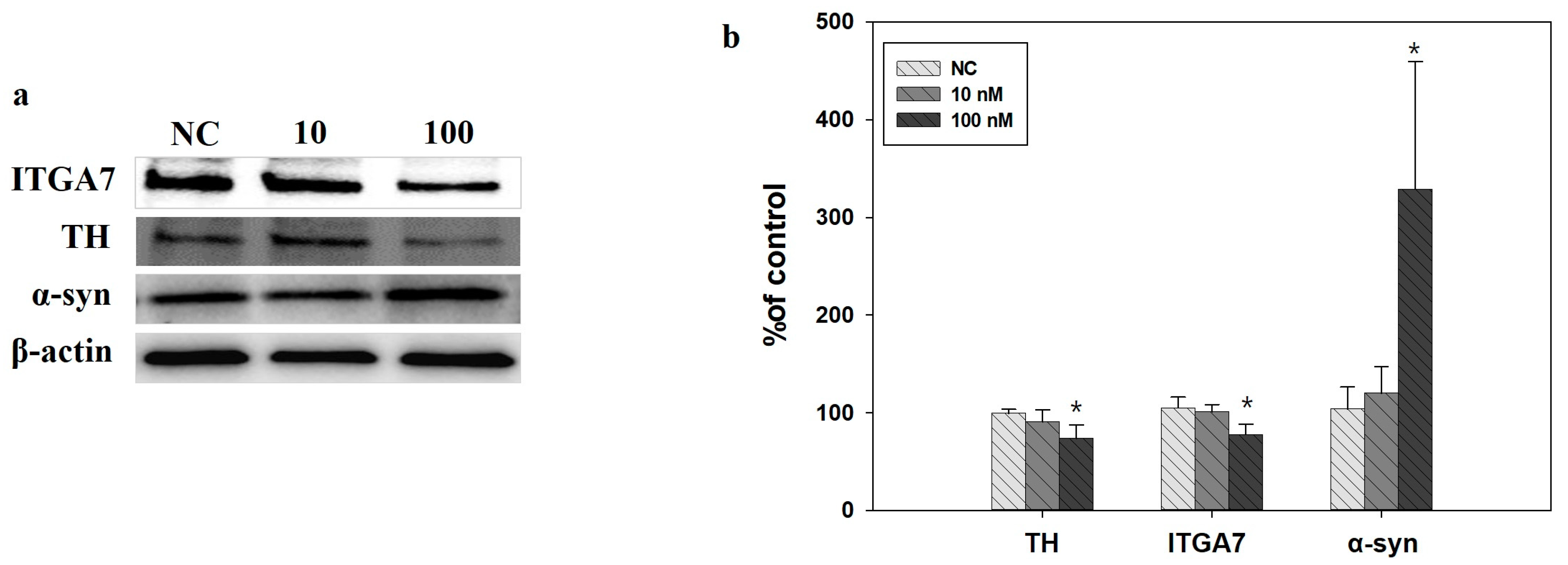
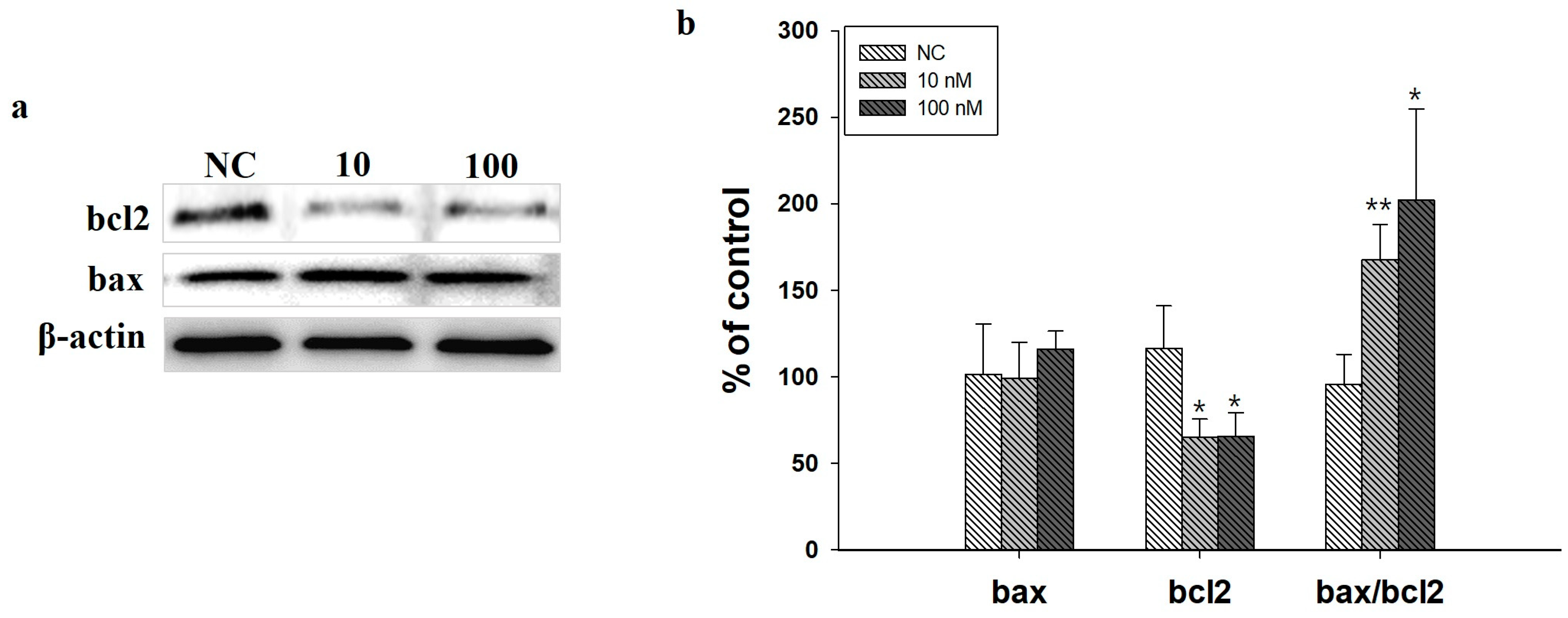
2.5. Western Blot Analysis in SH-SY5Y Cells Induced by ITGA7 siRNA

ITGA7 siRNA administration decreased ITGA7 and TH expression in SH-SY5Y cells (p < 0.05, n = 3) but increased α-syn expression (p < 0.05, n = 3) (Figure 5a,b). In addition, ITGA7 siRNA administration reduced the expression of bcl2 (p < 0.05), while bax expression did not increase significantly, resulting in a significantly increased bax/bcl2 ratio (Figure 6).

2.6. Immunofluorescence Analysis of ITGA7 Revealed Co-Localization with α-Syn in SH-SY5Y Cells

Immunofluorescence analysis of ITGA7 and α-syn revealed their co-localization, and the expression of ITGA7 was stronger in the CTL than in MPP+-treated SH-SY5Y cells (Figure 7b,f). Conversely, the expression of α-syn was stronger in the MPP+-treated group than in the CTL group (Figure 7a,e). When α-syn and ITGA7 were combined, ITGA7 expression was strong in the CTL group and α-syn was strongly expressed in the MPP+-treated group (Figure 7c,g).

3. Discussion

It has previously been reported that an increase in α-syn expression is an important step in the pathology of PD [18,19], and our results suggest that one of the causes of this increase may be a decrease in ITGA7 expression. The mechanism by which dopaminergic cell destruction occurs following increased α-syn expression has been reported to be related to its effect on membrane fusion machinery, which regulates neurotransmitter release [20,21]. α-syn is highly concentrated at the presynaptic nerve terminals in both soluble and membrane-bound states [22,23]. Membrane binding by α-syn is likely physiologically important because α-syn remodels membranes [24]. Moreover, membranes were found to be important for the neuropathological effects of α-syn. Importantly, several studies have shown the involvement of aberrant α-syn-membrane interactions in cytotoxicity [19]. The self-aggregation of α-syn promotes membrane disruption, which can result from the formation of an amphipathic helical structure of α-syn [6]. Modern views of PD encompass the cell-to-cell trans-synaptic spread of α-syn [25,26] and the existence of many α-syn polymorphs that might be responsible for different synucleinopathies [27,28]. The cell membrane containing integrins is thought to play an important role in the intercellular synaptic diffusion of α-syn. Although studies have shown the link between PD and integrins [15,29], studies in Chinese [30], Swedes [31], and Taiwanese [32] reported that ITGA8 was not associated with PD.
ITGA7 encodes the ECM-binding protein. Integrins are a major family of cell surface receptors that mediate ECM adhesion and are strongly implicated in the regulation of various cellular functions, including programmed cell death [33]. Consistent with these previous studies, our results demonstrated that decreased ITGA7 expression induced by ITGA7 siRNA administration decreased TH expression in SH-SY5Y cells but increased α-syn expression. In addition, the decreased expression of ITGA7 reduced the expression of bcl2 and significantly increased the bax/bcl2 ratio. Increased bax induces apoptosis by stimulating the release of pro-apoptotic proteins such as cytochrome C from the mitochondrial intermembrane space to the cytoplasm [34]. As bcl2, an anti-apoptotic protein, can inhibit Ca2+ or bax-mediated cytochrome C release, a decrease in bcl2 plays an important role in inducing apoptosis [35]. These results suggest that the decreased expression of ITGA7 could be associated with the increased expression of α-syn and apoptosis of dopaminergic cells. In addition, since the protein encoded by ITGA7 is involved in the membrane and the aggregated form of α-syn promotes membrane destruction, an increase in α-syn expression due to a decrease in ITGA7 expression may contribute to transmembrane pathogenesis. In particular, ITGA7 may be involved in the mechanism by which α-syn acts on the membrane. Immunofluorescent analysis of the SN regions demonstrated that ITGA7 and α-syn were localized near the membrane, and the MPTP group showed stronger α-syn expression and weaker ITGA7 expression than the CTL group.
Considering that the increase in cell membrane α-syn expression leads to the destruction of dopaminergic cells [36], the increase in α-syn expression may be caused by a decrease in the expression of ITGA7, which is present in the cell membrane.
In the IHC results of this experiment, the expression pattern of ITGA7 in the CTL group was quite similar to that of α-syn expressed in the MPTP group. The decrease in ITGA7 expression induced by ITGA7 siRNA in SH-SY5Y cells confirmed that the expression of α-syn was inversely increased. Based on these results, we believe that additional research on whether α-syn expression increases to replace the decrease in ITGA7 expression is needed.

4. Materials and Methods

4.1. MPTP-Induced Parkinson’s Disease Mouse Model

The study protocol was approved by the Institutional Animal Care and Use Committee (IACUC) of Sangji University (IACUC 2018-6). Four-week-old male C57BL/6 mice (20–22 g; DBL, Daejeon, Korea) were used. The mice were divided into two groups: control (CTL) and MPTP groups. The mice in the CTL group were administered 0.9% (100 μL) saline intraperitoneally once every 24 h for 4 weeks. In the MPTP group, MPTP-HCl (20 mg/kg of free base) in 0.9% (100 μL) saline was intraperitoneally injected once a day for 4 weeks at 24-h intervals.

4.2. Cell lines and Cultures

Human-derived SH-SY5Y cells were grown at 37 °C in a humidified CO2 environment in 5% minimum essential medium (MEM; Welgen, Namcheon-myeon, Korea) supplemented with 10% fetal bovine serum (FBS; Lonza, Walkersville, MD, USA), 100 U/mL penicillin, and 100 mg/mL streptomycin.

4.3. ITGA7 Small Interfering RNA

Stealth siRNA against ITGA7 (5′-GAC AUG CAC UAC CUC GUC U-3′) and negative control duplexes (i.e., scrambled siRNA against ITGA7, 5-UUC UCC GAA CGU GUC ACG UTT-3′) were purchased from Bioneer Inc. (Daejeon, Korea). SH-SY5Y cells were treated with ITGA7 siRNA for 24 h. Before siRNA transfection, SH-SY5Y cells were incubated in Opti-MEM medium (Gibco, Amarillo, TX, USA). Transfection reagent was used in a 3.5:1 transfection reagent-to-duplex RNA ratio (Promega, Madison, WI, USA) in Opti-MEM medium.

4.4. MPP+ Treatment

SH-SY5Y cells were treated for 18 h with 500 μM MPP + iodide (Sigma, St. Louis, MO, USA).

4.5. Western Blot

The SN and ST regions and SH-SY5Y cells were homogenized in radioimmunoprecipitation assay buffer (RIPA) on ice for 30 min for Western blot analysis. The lysate was centrifuged at 12,000 rpm at 4 °C for 20 min, and the protein concentration of the supernatant was measured using the bicinchoninic acid (BCA) method. The protein was separated by sodium dodecyl sulfate–polyacrylamide gel electrophoresis (SDS-PAGE) (4–15% Tris-Bis mini gel) and transferred to a polyvinylidene fluoride membrane. The membranes were blocked with 3% BSA at 37 °C for 1 h and 30 min and then incubated with anti-ITGA7 (1:1000), anti-TH (1:2000), anti-bcl2 (1000), anti-bax (1: 1000), β-actin antibody (1:2000), or anti-α-syn (1:500) antibodies overnight at 4 °C. Subsequently, after washing three times for 15 min with TBST, the membrane was treated using horseradish peroxidase (HRP)-conjugated anti-mouse or anti-rabbit IgG antibody (1:5000) for 1 h. After washing with TBST, the membrane was visualized using a chemiluminescent substrate. Membrane band density was determined using ImageJ (https://rsbweb.nih.gov/ij/, accessed on 11 July 2021).

4.6. Immunohistochemistry

Mouse brains were fixed in 4% paraformaldehyde postfixed in 0.05 M sodium phosphate buffer containing 4% paraformaldehyde for 24 h at 4 °C, dehydrated with sucrose overnight at 4 °C, and then cryosectioned. Coronal sections of the brains (40 μm thickness) were cut using a cryomicrotome. Subsequently, each slice was treated with anti-integrin alpha 7 (1:1000, Santa Cruz Biotechnology, Santa Cruz, CA, USA), anti-TH antibody (1:2000, Santa Cruz Biotechnology), and anti-α-syn antibody (1:500, Santa Cruz Biotechnology), and then incubated overnight at 4 °C. The sections were then treated with biotinylated anti-mouse IgG, avidin-biotin-peroxidase complex, and diaminobenzidine hydrogen peroxide solution.

4.7. Immunofluorescence

After incubation with the primary antibodies and biotinylated anti-mouse IgG, each section was treated with fluorescein avidin DCS (Vector Laboratories, Burlington, ON, Canada). The sections were then treated with an avidin/biotin blocking kit and M.O.M mouse Ig blocking reagent (Vector Laboratories), after which they were stained with anti-α-syn or anti-ITGA7 IgG at 4 °C overnight. Each section was treated with biotinylated anti-mouse IgG, followed by incubation with rhodamine D (Vector Laboratories, Burlington, ON, Canada). Photographic documentation was performed using a Nikon X-cite series 120Q microscope (Nikon, Tokyo, Japan), and the exposure parameters were the same for each group of samples.

4.8. Statistical Analysis

Statistical analysis was performed using Student’s t-test or analysis of variance (ANOVA) with SPSS 25 (SPSS Inc., Chicago, IL, USA) software. All values are shown as the mean ± standard error.

5. Conclusions

In this study, ITGA7 expression was found to be reduced in the SN of an MPTP-induced Parkinsonism mouse model. In addition, the reduced expression of ITGA7 may be related to the increased expression of α-syn and apoptosis of dopaminergic cells. Our results suggest that a decrease in ITGA7 expression may be one of the causes of this increase in α-syn expression, which induces PD pathology.

Author Contributions

Conceptualization, S.Y.; methodology, S.H. and M.H.S.; software, S.H. and M.H.S.; validation, S.Y.; formal analysis, S.H. and M.H.S.; investigation, S.Y.; resources, S.Y. and M.H.S.; data curation, S.Y. and S.H.; writing—original draft preparation, S.Y. and S.H.; writing—review and editing, S.Y. and S.L.; visualization, S.Y., S.L. and M.H.S.; supervision, S.Y.; project administration, S.Y.; funding acquisition, S.Y. All authors have read and agreed to the published version of the manuscript.

Funding

This study was supported by a National Research Foundation Q17 of Korea (NRF) grant funded by the Korean Government’s Ministry of Science, ICT and Future Planning (MSIP; No. 2020R1I1A3A04036500).

Institutional Review Board Statement

This study was approved by the Institutional Animal Care and Use Committee of Sang Ji University (approval code: 2016-07; date of approval 29 March 2016).

Data Availability Statement

All data analyzed in this study are included in this published article.

Conflicts of Interest

The authors declare no conflict of interest.

References

  1. Hattori, N. Etiology and pathogenesis of Parkinson’s disease: From mitochondrial dysfunctions to familial Parkinson’s disease. Rinsho Shinkeigaku 2004, 44, 241–262. [Google Scholar] [PubMed]
  2. Jankovic, J. Parkinson’s disease: Clinical features and diagnosis. J. Neurol. Neurosurg. Psychiatry 2008, 79, 368–376. [Google Scholar] [CrossRef] [PubMed] [Green Version]
  3. Dauer, W.; Przedborski, S. Parkinson’s disease: Mechanisms and models. Neuron 2003, 39, 889–909. [Google Scholar] [CrossRef] [Green Version]
  4. Baba, M.; Nakajo, S.; Tu, P.H.; Tomita, T.; Nakaya, K.; Lee, V.M.; Trojanowski, J.Q.; Iwatsubo, T. Aggregation of alpha-synuclein in Lewy bodies of sporadic Parkinson’s disease and dementia with Lewy bodies. Am. J. Pathol. 1998, 152, 879–884. [Google Scholar]
  5. Spillantini, M.G.; Schmidt, M.L.; Lee, V.M.Y.; Trojanowski, J.Q.; Jakes, R.; Goedert, M. α-Synuclein in Lewy bodies. Nature 1997, 388, 839–840. [Google Scholar] [CrossRef] [PubMed]
  6. Beyer, K. Mechanistic aspects of Parkinson’s disease: Alpha-synuclein and the biomembrane. Cell Biochem. Biophys. 2007, 47, 285–299. [Google Scholar] [CrossRef] [PubMed]
  7. Carrasco-Garcia, E.; Auzmendi-Iriarte, J.; Matheu, A. Integrin α7: A novel promising target in glioblastoma stem cells. Stem Cell Investig. 2018, 5, 2. [Google Scholar] [CrossRef]
  8. Wu, Z.; Kong, X. Integrin α7 knockdown suppresses cell proliferation, migration, invasion and EMT in hepatocellular carcinoma. Exp. Ther. Med. 2021, 21, 1. [Google Scholar] [CrossRef] [PubMed]
  9. Klaffky, E.; Williams, R.; Yao, C.C.; Ziober, B.; Kramer, R.; Sutherland, A. Trophoblast-specific expression and function of the integrin alpha 7 subunit in the peri-implantation mouse embryo. Dev. Biol. 2001, 239, 161–175. [Google Scholar] [CrossRef] [Green Version]
  10. Tan, L.-Z.; Song, Y.; Nelson, J.; Yu, Y.P.; Luo, J.-H. Integrin α7 Binds Tissue Inhibitor of Metalloproteinase 3 to Suppress Growth of Prostate Cancer Cells. Am. J. Pathol. 2013, 183, 831–840. [Google Scholar] [CrossRef] [Green Version]
  11. Cohn, R.D.; Mayer, U.; Saher, G.; Herrmann, R.; van der Flier, A.; Sonnenberg, A.; Sorokin, L.; Voit, T. Secondary reduction of alpha7B integrin in laminin alpha2 deficient congenital muscular dystrophy supports an additional transmembrane link in skeletal muscle. J. Neurol. Sci. 1999, 163, 140–152. [Google Scholar] [CrossRef]
  12. Mayer, U.; Saher, G.; Fassler, R.; Bornemann, A.; Echtermeyer, F.; von der Mark, H.; Miosge, N.; Poschl, E.; von der Mark, K. Absence of integrin alpha 7 causes a novel form of muscular dystrophy. Nat. Genet. 1997, 17, 318–323. [Google Scholar] [CrossRef] [PubMed]
  13. Flintoff-Dye, N.L.; Welser, J.; Rooney, J.; Scowen, P.; Tamowski, S.; Hatton, W.; Burkin, D.J. Role for the alpha7beta1 integrin in vascular development and integrity. Dev. Dyn. 2005, 234, 11–21. [Google Scholar] [CrossRef] [PubMed]
  14. Pegoraro, E.; Cepollaro, F.; Prandini, P.; Marin, A.; Fanin, M.; Trevisan, C.P.; El-Messlemani, A.H.; Tarone, G.; Engvall, E.; Hoffman, E.P.; et al. Integrin alpha 7 beta 1 in muscular dystrophy/myopathy of unknown etiology. Am. J. Pathol. 2002, 160, 2135–2143. [Google Scholar] [CrossRef]
  15. Tsuboi, K.; Grzesiak, J.J.; Bouvet, M.; Hashimoto, M.; Masliah, E.; Shults, C.W. Alpha-synuclein overexpression in oligodendrocytic cells results in impaired adhesion to fibronectin and cell death. Mol. Cell. Neurosci. 2005, 29, 259–268. [Google Scholar] [CrossRef]
  16. Surguchev, A.A.; Surguchov, A. Integrins-A missing link in synuclein’s pathogenic mechanism. J. Neurosci. Res. 2019, 97, 539–542. [Google Scholar] [CrossRef]
  17. Surguchev, A.A.; Emamzadeh, F.N.; Surguchov, A. Cell Responses to Extracellular alpha-Synuclein. Molecules 2019, 24, 305. [Google Scholar] [CrossRef] [Green Version]
  18. Campos-Sousa, R.N.; Quagliato, E.; da Silva, B.B.; de Carvalho, R.M., Jr.; Ribeiro, S.C.; de Carvalho, D.F. Urinary symptoms in Parkinson’s disease: Prevalence and associated factors. Arq. Neuropsiquiatr. 2003, 61, 359–363. [Google Scholar] [CrossRef] [Green Version]
  19. Nakamura, K.; Nemani, V.M.; Azarbal, F.; Skibinski, G.; Levy, J.M.; Egami, K.; Munishkina, L.; Zhang, J.; Gardner, B.; Wakabayashi, J.; et al. Direct membrane association drives mitochondrial fission by the Parkinson disease-associated protein alpha-synuclein. J. Biol. Chem. 2011, 286, 20710–20726. [Google Scholar] [CrossRef] [Green Version]
  20. Stefanis, L. α-Synuclein in Parkinson’s disease. Cold Spring Harb. Perspect. Med. 2012, 2, a009399. [Google Scholar] [CrossRef] [Green Version]
  21. Burre, J.; Sharma, M.; Tsetsenis, T.; Buchman, V.; Etherton, M.R.; Sudhof, T.C. Alpha-synuclein promotes SNARE-complex assembly in vivo and in vitro. Science 2010, 329, 1663–1667. [Google Scholar] [CrossRef] [Green Version]
  22. Davidson, W.S.; Jonas, A.; Clayton, D.F.; George, J.M. Stabilization of alpha-synuclein secondary structure upon binding to synthetic membranes. J. Biol. Chem. 1998, 273, 9443–9449. [Google Scholar] [CrossRef] [PubMed] [Green Version]
  23. Nuscher, B.; Kamp, F.; Mehnert, T.; Odoy, S.; Haass, C.; Kahle, P.J.; Beyer, K. Alpha-synuclein has a high affinity for packing defects in a bilayer membrane: A thermodynamics study. J. Biol. Chem. 2004, 279, 21966–21975. [Google Scholar] [CrossRef] [PubMed] [Green Version]
  24. West, A.; Brummel, B.E.; Braun, A.R.; Rhoades, E.; Sachs, J.N. Membrane remodeling and mechanics: Experiments and simulations of alpha-Synuclein. Biochim. Biophys. Acta 2016, 1858 Pt B, 1594–1609. [Google Scholar] [CrossRef] [PubMed]
  25. Ferreira, N.; Goncalves, N.P.; Jan, A.; Jensen, N.M.; van der Laan, A.; Mohseni, S.; Vaegter, C.B.; Jensen, P.H. Trans-synaptic spreading of alpha-synuclein pathology through sensory afferents leads to sensory nerve degeneration and neuropathic pain. Acta Neuropathol. Commun. 2021, 9, 31. [Google Scholar] [CrossRef]
  26. Jan, A.; Goncalves, N.P.; Vaegter, C.B.; Jensen, P.H.; Ferreira, N. The Prion-Like Spreading of Alpha-Synuclein in Parkinson’s Disease: Update on Models and Hypotheses. Int. J. Mol. Sci. 2021, 22, 8338. [Google Scholar] [CrossRef]
  27. Ferreira, N.; Gram, H.; Sorrentino, Z.A.; Gregersen, E.; Schmidt, S.I.; Reimer, L.; Betzer, C.; Perez-Gozalbo, C.; Beltoja, M.; Nagaraj, M.; et al. Multiple system atrophy-associated oligodendroglial protein p25alpha stimulates formation of novel alpha-synuclein strain with enhanced neurodegenerative potential. Acta Neuropathol. 2021, 142, 87–115. [Google Scholar] [CrossRef]
  28. Peng, C.; Gathagan, R.J.; Covell, D.J.; Medellin, C.; Stieber, A.; Robinson, J.L.; Zhang, B.; Pitkin, R.M.; Olufemi, M.F.; Luk, K.C.; et al. Cellular milieu imparts distinct pathological alpha-synuclein strains in alpha-synucleinopathies. Nature 2018, 557, 558–563. [Google Scholar] [CrossRef] [PubMed]
  29. Lill, C.M.; Roehr, J.T.; McQueen, M.B.; Kavvoura, F.K.; Bagade, S.; Schjeide, B.M.; Schjeide, L.M.; Meissner, E.; Zauft, U.; Allen, N.C.; et al. Comprehensive research synopsis and systematic meta-analyses in Parkinson’s disease genetics: The PDGene database. PLoS Genet. 2012, 8, e1002548. [Google Scholar] [CrossRef] [Green Version]
  30. Fang, J.; Yi, K.; Guo, M.; An, X.; Qu, H.; Lin, Q.; Bi, M.; Ma, Q. Analysis of LRRK2, SNCA, and ITGA8 Gene Variants with Sporadic Parkinson’s Disease Susceptibility in Chinese Han Population. Parkinsons Dis. 2016, 2016, 3474751. [Google Scholar]
  31. Ran, C.; Mehdi, R.N.; Fardell, C.; Xiang, F.; Nissbrandt, H.; Sydow, O.; Wirdefeldt, K.; Belin, A.C. No Association Between rs7077361 in ITGA8 and Parkinson’s Disease in Sweden. Open Neurol. J. 2016, 10, 25–29. [Google Scholar] [CrossRef] [Green Version]
  32. Wu, H.C.; Chen, C.M.; Chen, Y.C.; Fung, H.C.; Chang, K.H.; Wu, Y.R. DLG2, but not TMEM229B, GPNMB, and ITGA8 polymorphism, is associated with Parkinson’s disease in a Taiwanese population. Neurobiol. Aging 2018, 64, 158.e1–158.e6. [Google Scholar] [CrossRef] [PubMed]
  33. Desgrosellier, J.S.; Cheresh, D.A. Integrins in cancer: Biological implications and therapeutic opportunities. Nat. Rev. Cancer 2010, 10, 9–22. [Google Scholar] [CrossRef] [PubMed] [Green Version]
  34. Hsu, Y.T.; Wolter, K.G.; Youle, R.J. Cytosol-to-membrane redistribution of Bax and Bcl-X(L) during apoptosis. Proc. Natl. Acad. Sci. USA 1997, 94, 3668–3672. [Google Scholar] [CrossRef] [Green Version]
  35. Oltvai, Z.N.; Milliman, C.L.; Korsmeyer, S.J. Bcl-2 heterodimerizes in vivo with a conserved homolog, Bax, that accelerates programmed cell death. Cell 1993, 74, 609–619. [Google Scholar] [CrossRef]
  36. Burre, J.; Sharma, M.; Sudhof, T.C. alpha-Synuclein assembles into higher-order multimers upon membrane binding to promote SNARE complex formation. Proc. Natl. Acad. Sci. USA 2014, 111, E4274–E4283. [Google Scholar] [CrossRef] [PubMed] [Green Version]
Figure 1. Immunohistochemistry analysis of tyrosine hydroxylase (TH) expression in the substantia nigra (SN; a,c) and the striatum (ST; b,d) in the control (CTL) and 1-methyl-4-phenyl-1,2,3,6-tetrahydropyridine-treated (MPTP) groups. TH expression decreased in the ST and SN regions of the brains of MPTP-treated mice. Western blot analyses (e) showed that TH expression was significantly decreased in MPTP-treated mice (f). ** denotes p < 0.005 compared to CTL. All values are expressed as mean ± standard error, and statistical analysis was performed using Student’s t-test.
Ijms 22 12616 g001
Figure 2. Integrin alpha 7 (ITGA7) expression was decreased and α-synuclein (α-syn) expression was increased in the MPTP group (MPTP) compared to that in the control group (CTL) in the SN. (a) Western blot analysis verified the decreased expression of ITGA7 and the increased expression of α-syn in MPTP-treated mice. (b) Histogram shows a significant decrease in ITGA7 expression, detected by Western blot analysis, and the decreased signal of microarray in the MPTP group compared to that in the CTL group. Histogram shows a significant increase in α-syn in the MPTP group compared to that in the CTL group. * denotes p < 0.05 compared to CTL. All values are expressed as mean ± standard error, and statistical analysis was performed using Student’s t-test.
Ijms 22 12616 g002
Figure 3. Representative images of (ITGA7) and α-synuclein (α-syn) expression in the SN in a chronic 1-methyl-4-phenyl-1,2,3,6-tetrahydropyridine (MPTP)-induced Parkinson’s disease mouse model. The expression of ITGA7 (ad) was decreased (c,d) and the expression of α-syn (eh) was increased (g,h) in in the MPTP group. Images of (b,d,f,h) are magnified images of squares in (a,c,e,g).
Ijms 22 12616 g003
Figure 4. Immunofluorescence images of ITGA7 co-localized with α-syn in the SN in an MPTP-induced Parkinson’s disease mouse model. SN regions were immunofluorescently labeled with anti-ITGA7 (b,g) and anti-α-syn (a,f) antibodies by using Rhodamine Avidin (a,f, red) and were then double immunolabeled with ITGA7 antibodies by using Fluorescein Avidin (b,g, green). The middle panels (c,h) show merged images of the individual α-syn (a,f) and ITGA7 (b,g) panels. In the control group (c), ITGA7 was more strongly expressed (green) and in the MPTP group (h), intense red fluorescence was observed. (d,i) show merged images with DAPI. Images of (e) and (j) show enlarged images of the square in (d) and (i). (scale bar, 10 µm).
Ijms 22 12616 g004
Figure 5. (a) Western immunoblot analysis showed that the administration of ITGA7 siRNA decreased ITGA7 and TH expression but increased α-synuclein (α-syn) expression in the SH-SY5Y cells. (b) NC, negative control siRNA treatment (100 nM for 24 h); 10, ITGA7 siRNA treatment (10 nM for 24 h); 100, ITGA7 siRNA treatment (100 nM for 24 h). * denotes p < 0.05 compared to NC. All values are expressed as mean ± standard error, and statistical analysis was performed using ANOVA.
Ijms 22 12616 g005
Figure 6. (a) Western immunoblot analysis revealed that the administration of ITGA7 siRNA significantly decreased bcl2 expression and increased the bax/bcl2 ratio in SH-SY5Y cells. (b) NC, negative control siRNA treatment (100 nM for 24 h); 10, ITGA7 siRNA treatment (10 nM for 24 h); 100, ITGA7 siRNA treatment (100 nM for 24 h). * denotes p < 0.05 and ** p < 0.001 compared to NC. All values are expressed as mean ± standard error, and statistical analysis was performed using ANOVA.
Ijms 22 12616 g006
Figure 7. Immunofluorescence analysis of ITGA7 co-localized with α-syn in SH-SY5Y cell. The cells were immunofluorescently labeled with anti-ITGA7 (b,f) and anti-α-synuclein (a,e) antibodies using Rhodamine Avidin (a,e, red) and were then double immunolabeled with ITGA7 antibodies using Fluorescein Avidin (b,f, green). The panels on the right (c,g) show merged images of the individual left (a,e) and middle (b,f) panels. (d,h) show merged images with DAPI (scale bar, 10 µm). White arrows indicate regions of interest.
Ijms 22 12616 g007
Publisher’s Note: MDPI stays neutral with regard to jurisdictional claims in published maps and institutional affiliations.

