Article

New Insights of OLFM2 and OLFM4 in Gut-Liver Axis and Their Potential Involvement in Nonalcoholic Fatty Liver Disease

by
Laia Bertran
1,†,
Rosa Jorba-Martin
2,†,
Andrea Barrientos-Riosalido
1,
Marta Portillo-Carrasquer
1,
Carmen Aguilar
1,
David Riesco
3,
Salomé Martínez
4,
Margarita Vives
5,
Fàtima Sabench
5,
Daniel Del Castillo
5,
Cristóbal Richart
1 and
Teresa Auguet
1,3,*
1
Grup de Recerca GEMMAIR (AGAUR)-Medicina Aplicada, Departament de Medicina i Cirurgia, Universitat Rovira i Virgili (URV), Institut d’Investigació Sanitària Pere Virgili (IISPV), 43007 Tarragona, Spain
2
Servei de Cirurgia General i de l’Aparell Digestiu, Hospital Universitari de Tarragona Joan XXIII, Universitat Rovira i Virgili (URV), Institut d’Investigació Sanitària Pere Virgili (IISPV), Mallafré Guasch, 4, 43007 Tarragona, Spain
3
Servei Medicina Interna, Hospital Universitari de Tarragona Joan XXIII, Mallafré Guasch, 4, 43007 Tarragona, Spain
4
Servei Anatomia Patològica, Hospital Universitari de Tarragona Joan XXIII, Mallafré Guasch, 4, 43007 Tarragona, Spain
5
Servei de Cirurgia, Hospital Sant Joan de Reus, Departament de Medicina i Cirurgia, Universitat Rovira i Virgili (URV), Institut d’Investigació Sanitària Pere Virgili (IISPV), Avinguda Doctor Josep Laporte, 2, 43204 Reus, Spain
*
Author to whom correspondence should be addressed.
The authors contributed equally to this work.
Int. J. Mol. Sci. 2022, 23(13), 7442; https://doi.org/10.3390/ijms23137442
Submission received: 9 June 2022 / Revised: 29 June 2022 / Accepted: 3 July 2022 / Published: 4 July 2022
(This article belongs to the Special Issue The Molecular Aspects of Gut-Brain Communications)

Abstract

:
Olfactomedins (OLFMs) are a family of glycoproteins that play a relevant role in embryonic development and in some pathological processes. Although OLFM2 is involved in the regulation of the energy metabolism and OLFM4 is an important player in inflammation, innate immunity and cancer, the role of OLFMs in NAFLD-related intestinal dysbiosis remains unknown. In this study, we analysed the hepatic mRNA expression of OLFM2 and the jejunal expression of OLFM4 in a well-established cohort of women with morbid obesity (MO), classified according to their hepatic histology into normal liver (n = 27), simple steatosis (n = 26) and nonalcoholic steatohepatitis (NASH, n = 16). Our results showed that OLFM2 hepatic mRNA was higher in NASH, in advanced degrees of steatosis and in the presence of lobular inflammation. Additionally, we obtained positive correlations between hepatic OLFM2 and glucose, cholesterol, trimethylamine N-oxide and deoxycholic acid levels and hepatic fatty acid synthase, and negative associations with weight and jejunal Toll-like receptors (TLR4) and TLR5 expression. Regarding jejunal OLFM4, we observed positive correlations with circulating interleukin (IL)-8, IL-10, IL-17 and jejunal TLR9. In conclusion, OLFM2 in the liver seems to play a relevant role in NAFLD progression, while OLFM4 in the jejunum could be involved in gut dysbiosis-related inflammatory events.

1. Introduction

Nonalcoholic fatty liver disease (NAFLD) is characterized by ectopic fat accumulation in hepatic tissue in the absence of secondary causes of steatosis, such as alcohol intake and/or viral infection [1,2]. NAFLD is a general term used for a variety of liver conditions that comprises many stages from simple steatosis (SS) to nonalcoholic steatohepatitis (NASH), this last one defined by the presence of hepatic inflammation, ballooning and/or fibrosis [3,4], which can trigger cirrhosis or hepatocellular carcinoma if left untreated [5]. Despite NAFLD being the most extended chronic hepatic disorder and the existence of many studies characterizing it, its underlying molecular mechanisms remain incompletely understood, and a specific treatment is not currently approved. Thus, understanding the pathogenic processes behind NAFLD provides the basis for identifying the onset and progression of the disease [6].
The severity of NAFLD is related to different comorbidities, such as obesity and type 2 diabetes mellitus (T2DM), and recently it was also related to intestinal dysbiosis [7]. The intestinal microbiota seem to play an important role in human health, since it is known to be altered in pathological conditions, leading to intestinal dysbiosis [8,9]. The altered microbiota trigger an increase in the permeability of the intestinal barrier, which induces the release of microbiota-derived mediators such as lipopolysaccharides (LPS), short-chain fatty acids (SCFAs), choline metabolites, bile acids (BAs) and endogenous ethanol [10]. Recently, the relevance of the gut-liver axis and its communication through the portal vein has been described. When the permeability of the intestinal barrier is corrupted, the liver is exposed to components of the microbiota contributing to inflammation and liver injury and thus to the pathogenesis of NAFLD [11].
In this regard, the gut-liver axis [12] and other crosstalk systems, such as the trade-off of adipokines in the adipose tissue-liver axis [13], have reinforced the fact that NAFLD is a multisystemic condition influenced by metabolic and inflammatory processes occurring in related tissues [14]. Thus, evaluating NAFLD as a disorder with hepatic and extrahepatic manifestations [15] would help us to understand the physiological processes involved in this disease.
The seven groups (I–VII) of the olfactomedin (OLFM) family of glycoproteins are located in C-terminal regions, except those from groups II and VI. OLFMs play a key role in cell–cell signalling and cell adhesion and differentiation, which suggests involvement in many molecular mechanisms of cancer [16]. Although the biological functions of OLFM domain-containing proteins remain mostly elusive, continuous evidence indicates that these proteins may play relevant roles in normal development and pathological processes [17]. Additionally, the participation of OLFMs in haematopoiesis and the early development of the nervous system has been reported. Additionally, OLFMs have been associated with diseases, such as gastrointestinal cancer and glaucoma, since the interruption of their expression leads to the development of disturbances and lethality [16].
On the one hand, olfactomedin 2 (OLFM2; also known as noelin-2) is a secretory glycoprotein that is a member of OLFM group I together with OLFM1 and 3. This protein is mainly expressed in neurons, but also in the liver [18] and adipose tissue [18,19]. Moreover, González-García et al. demonstrated that OLFM2 is involved in the regulation of energy metabolism [20].
On the other hand, olfactomedin 4 (OLFM4) belongs to OLFM group V. This glycoprotein was found to be endogenously expressed in bone marrow and the small intestine, stomach, colon, prostate [21] and pancreas [22]. In addition, this protein has been related to multiple signalling pathways and factors, including the NF-κB [23] WNT/β-catenin pathway [24,25] and Notch proteins [26,27], which are strongly related to pathological processes in hepatic diseases, such as NAFLD [28,29,30]. Furthermore, OLFM4 is an important player in the cellular process of inflammation, and it has important roles in innate immunity against bacterial infection, gastrointestinal inflammation, and cancer [17,31].
OLFM2 seems to have a role in the regulation of the lipid metabolism, and OLFM4 may participate in gastrointestinal inflammation in relation to dysbiosis. Furthermore, the Human Protein Atlas has reported OLMF2 expression in the liver and OLFM4 expression in the small intestine [18,19]. Thus, the current study aimed to analyse OLFM2 mRNA expression in liver biopsies and OLFM4 mRNA expression in jejunum samples in a well-established cohort of women with morbid obesity (MO) with different NAFLD grades to explore the role of OLFMs in the pathogenesis of NAFLD.

2. Results

2.1. Baseline Characteristics of Subjects

The clinical and biochemical measurements of the cohort formed by women with MO (BMI > 40 kg/m2) classified according to the hepatic histology as normal liver (NL, n = 27), SS (n = 26) and NASH (n = 16), are shown in Table 1. The participants did not present significant differences between groups in terms of weight, body mass index (BMI), systolic blood pressure (SBP), diastolic blood pressure (DBP), homeostatic model assessment method-insulin resistance (HOMA1-IR), insulin, glycosylated haemoglobin (HbA1c), cholesterol, high density lipoprotein cholesterol (HDL-C), low density lipoprotein cholesterol (LDL-C), aspartate-aminotransferase (AST), alanine-aminotransferase (ALT) and gamma-glutamyltransferase (GGT). However, in this analysis, we found higher levels of glucose and alkaline phosphatase (ALP) in the SS group than in the NL group, increased levels of triglycerides in the NASH cohort compared to NL women and increased levels of ALP in SS subjects in comparison to NASH patients.

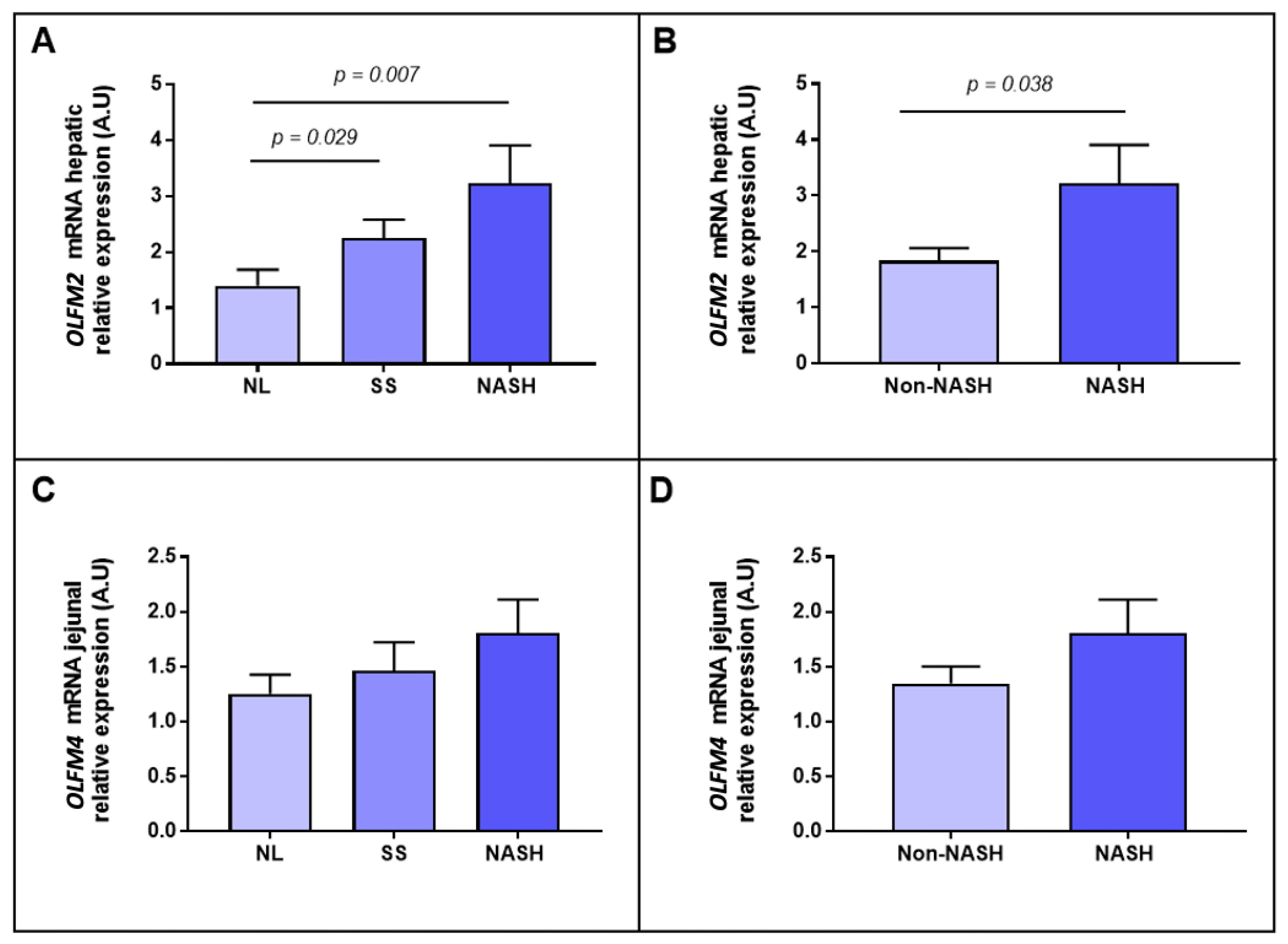
2.2. Evaluation of the Relative mRNA Abundance of OLFM2 and OLFM4 According to Hepatic Histology

To achieve the aim of this study, that was to explore the role of OLFMs in NAFLD progression, we evaluated the OLFM2 mRNA expression in liver samples in a cohort of women with MO. Moreover, given that the importance of the gut-liver axis in NAFLD progression has been reported, we also analysed the jejunal OLFM4 mRNA expression.
First, when we analysed OLFM2 and OLFM4 relative mRNA expressions between NL and NAFLD in hepatic and jejunal samples, respectively, we only reported significant differences between groups in the case of OLFM2 (p = 0.005). Then, we subclassified the patients according to the hepatic histopathological grades into NL, SS and NASH. In this sense we found a higher expression of hepatic OLFM2 in SS and NASH women compared to the NL cohort (Figure 1A). Later, when the subjects were divided according to the presence of NASH, an increase in OLFM2 relative expression in the NASH group was observed in comparison with the non-NASH group, as shown in Figure 1B. However, regarding OLFM4, we did not report significant differences between groups (Figure 1C,D).

2.3. Evaluation of the Relative mRNA Abundance of OLFM2 and OLFM4 According to the Severity of Steatosis

To deepen the knowledge of the link between OLFMs and NAFLD, first we wanted to focus on the hepatic lipid content degree, so we classified the cohort into different grades of steatosis. In this sense, we reported an enhanced expression of hepatic OLFM2 mRNA in moderate and severe stages of steatosis in comparison to those subjects without steatosis (Figure 2A); while we did not find significant differences when OLFM4 was analysed, as graphically represented in Figure 2B.

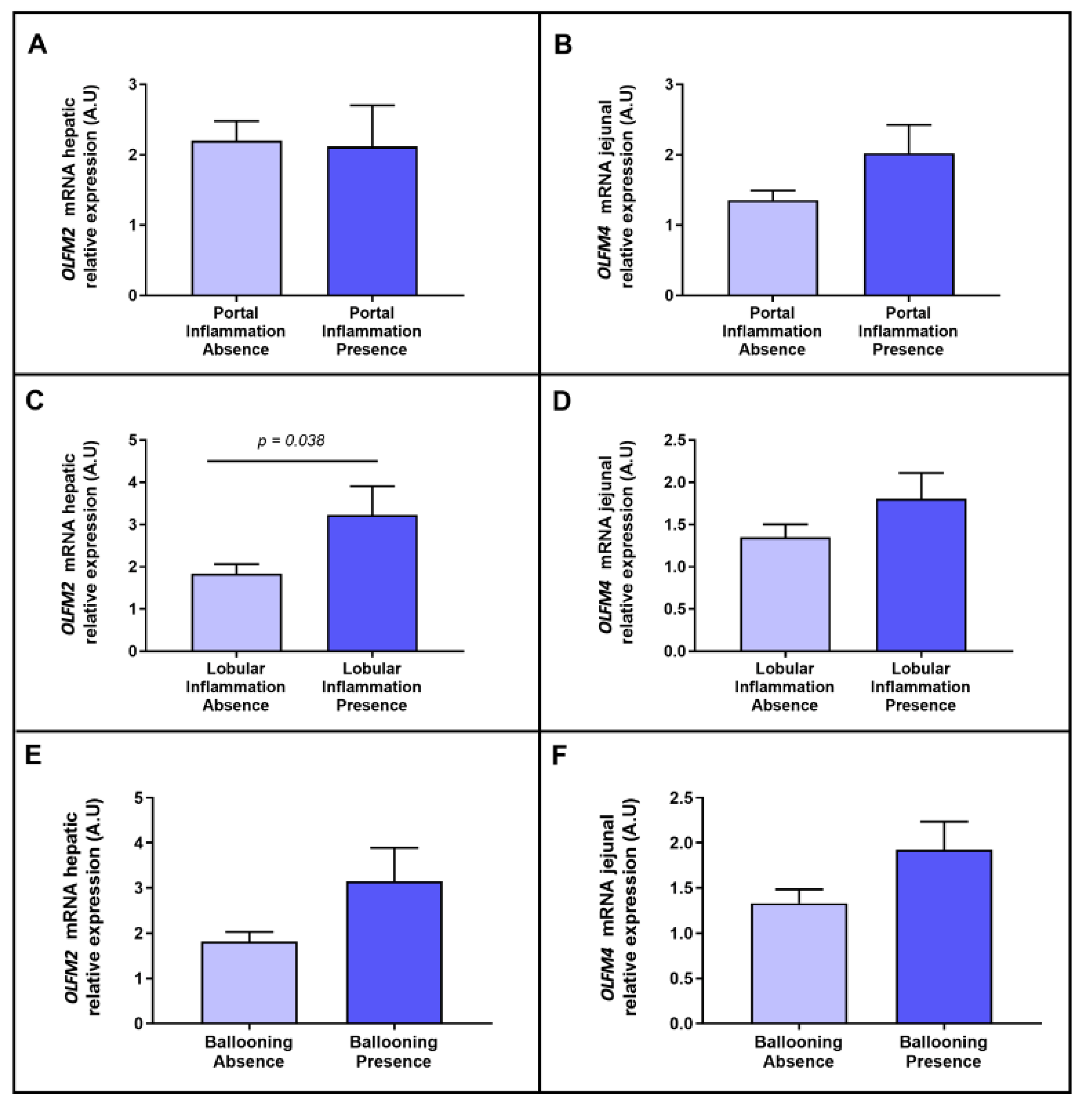
2.4. Evaluation of the Relative mRNA Abundance of OLFM2 and OLFM4 According to NASH-Related Parameters

Later, we wanted to focus the analysis on the main parameters of the advanced stage of NAFLD, such as inflammation and hepatocellular ballooning; we therefore evaluated their link with OLFM2 and OLFM4. Concerning portal inflammation, we did not observe differential expressions of OLFM2 in the liver and OLFM4 in the jejunum between groups (Figure 3A,B). Regarding lobular inflammation, we found an increase in OLFM2 hepatic expression in the cohort presenting this type of inflammation (Figure 3C), but we did not find differences with regard to OLFM4 jejunal expression (Figure 3D). Moreover, non-significant differences in OLFM2 and OLFM4 were reported when the cohort was classified based on the presence of hepatocyte ballooning (Figure 3E,F).

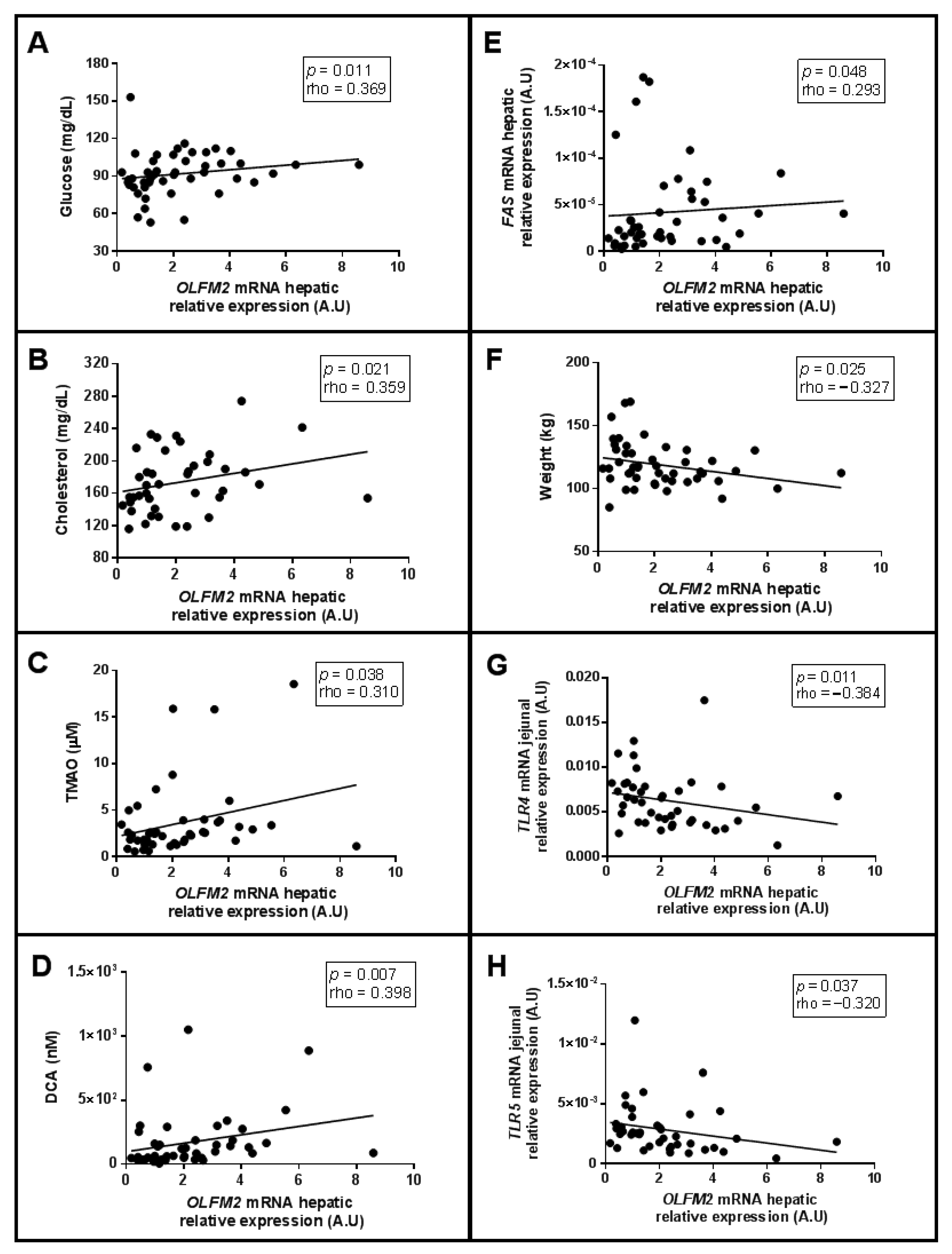
2.5. Correlations of Relative mRNA Abundance of Hepatic OLFM2 and Jejunal OLFM4, with Clinical and Biochemical-Related Parameters

To broaden the study of OLFM2, we analysed their correlations with different parameters related to glucose and lipid metabolism, gut microbiota and inflammation in a cohort of women with MO. First, we observed positive correlations between hepatic OLFM2 mRNA expression and glucose, cholesterol, trimethylamine N-oxide (TMAO), deoxycholic acid (DCA) levels and fatty acid synthase (FAS) mRNA expression in the liver, as shown in Figure 4A–E, respectively. We also found a negative association between hepatic OLFM2 mRNA expression and weight, jejunal TLR4 and jejunal TLR5 expression, as was graphically represented in Figure 4F–H.
On the other hand, when we focused on the OLFM4 in the jejunum, we observed positive correlations between OLFM4 and circulating interleukin (IL)-8, IL-10, IL-17 levels and jejunal TLR9 expression, as shown in Figure 5A–D, respectively.

3. Discussion

This study is important as it is the first to report an interesting association between OLFMs and NAFLD in hepatic and jejunal samples of women with MO. It is already known that more in-depth studies of the molecular mechanisms involved in NAFLD are necessary to find new therapeutic targets. In this sense, given that OLFMs are strongly linked to cellular processes [16,17] and have been related to some disorders, such as obesity and hepatocarcinoma (HCC), among others [20,31,32,33,34], we wanted to evaluate the possible roles of OLFM2 and OLFM4 in NAFLD pathogenesis.
Regarding our first finding, when patients were classified according to their hepatic histopathology, OLFM2 mRNA expression in the liver increased when hepatic health worsened. Moreover, a significant increase in OLFM2 expression in the presence of NASH was observed when the cohort was classified into non-NASH and NASH groups. These facts seem to suggest that OLFM2 is involved in NAFLD progression, especially in NASH. Although OLFM2 mRNA abundance has been found in liver tissues [18], its expression has not been previously evaluated, and this protein has not been related to NAFLD. Therefore, OLFM2 in liver tissues seems to play a role in NAFLD progression in subjects with obesity, but further studies are needed in this field.
Then, when we evaluated hepatic OLFM2 and jejunal OLFM4 based on the steatosis stage of the disease, we found a significant increase in hepatic OLFM2 in patients with moderate and severe steatosis grades. Later, when we analysed the role of these OLFMs in accordance with steatohepatitis parameters, such as lobular and portal inflammation and hepatocyte ballooning, we found an increase in hepatic OLFM2 expression in patients with lobular inflammation. These findings indicate that OLFM2 increases as the disease progresses, since it is greatly increased in NASH, which is usually characterized by hepatic inflammation and elevated lipid content [3,35]. As we have previously mentioned, this is a novel finding; thus, no previous reports exist in this regard, not even reports on the expression of this protein in the liver. More studies are necessary to corroborate this evidence.
Unfortunately, although OLFM4 seems to play relevant roles as an anti-inflammatory molecule in Helicobacter pylori infection [31] and gastric and intestinal disorders [33,34], which could suggest that OLFM4 is involved in NAFLD pathogenesis through microbiota changes and the gut-liver axis, in our study, the mRNA expression of this protein in jejunal samples was not significantly different between the hepatic histopathological groups. In the literature there are no other studies in this area, but probably in those patients with MO who present an intestinal dysbiosis associated with obesity, there is an activation of inflammasomes and also of the immune system [36,37] that gives rise to an inflammatory microenvironment capable of masking the possible relationship that could have expression in the jejunum of OLFM4 with liver histology. Therefore, to further investigate this relationship, additional studies should be performed in subjects of normal weight.
To deepen the knowledge of OLFMs in regard to NAFLD, we analysed several correlations. Positive associations between hepatic OLFM2 mRNA and hepatic FAS mRNA expression and cholesterol and glucose levels, which are parameters closely related to NAFLD, were found [38,39]. When there is an excess of carbohydrates in the diet, glucose can be stored as lipids in the liver, and FAS, a lipogenic enzyme, synthesizes triglycerides from free fatty acids; therefore, its action enhances hepatic steatosis, the principal characteristic of NAFLD [39,40]. In addition, another study reported increased FAS mRNA expression and high glucose levels in people with NAFLD [41], so our current results are consistent with previously mentioned publications showing dysregulation of hepatic lipogenesis in NAFLD [42,43]. All this information, as well as our findings of positive correlations of OLFM2 in the liver with glucose and cholesterol levels and hepatic FAS, corroborates our previous hypothesis that hepatic expression of OLFM2 increases as NAFLD progresses, when at the same time, the metabolic imbalance also worsens [44].
Additionally, we found a negative significant correlation between hepatic OLFM2 and weight. This association is difficult to explain, given that González-García et al. observed that a global lack of OLFM2 induces weight loss [20]. This contradiction could be explained by the fact that our cohort presented a degree of severe obesity (BMI between 40 and 46 Kg/m2), and perhaps some correlations may have been skewed due to such excessive BMI.
On the other hand, we also observed a positive correlation of hepatic OLFM2 with DCA and TMAO levels. TMAO is a gut microbiota-dependent metabolite, and some publications [42,45,46] reported increased TMAO levels in patients with NAFLD. The same authors suggested that TMAO could contribute to the development and severity of this disease due to its role in glucose regulation, cholesterol homeostasis and lipid absorption [45,47]. An increase in DCA levels, which are involved in the metabolism of ingested lipids, was also seen in NAFLD patients [48]. Additionally, Grzych et al. showed high levels of DCA in the presence of NASH [49]. Therefore, these positive associations of hepatic OLFM2 with DCA and TMAO levels remain consistent with previous results, reinforcing the hypothesis that OLFM2 may play an important role in the progression of NAFLD.
Later, we wanted to analyse whether OLFM2 in the liver is associated with jejunal TLRs, since these are involved in gut-liver axis crosstalk that can influence NAFLD appearance and progression [50]. In this regard, although we previously observed that hepatic OLFM2 mRNA expression is enhanced when the disease becomes severe, we observed a negative correlation between jejunal TLR4 and 5 with hepatic OLFM2 mRNA expression. This is a contradictory result given that TLRs are known to be involved in NAFLD progression [50], but this result may be because women in our cohort followed a very low-calorie diet for 3 weeks before bariatric surgery [51]. In this sense, Macedo Rogero et al. demonstrated that omega-3 polyunsaturated fatty acids, usually included in the very low-calorie diet [52], attenuate the activation of the TLR4 signalling pathway, exerting an anti-inflammatory effect [53] and inhibiting lipid accumulation [54]. Concerning TLR5, this receptor has been shown to not necessarily be altered by diet [55]. Otherwise, TLR5 seems to play a key role in liver protection against intestinal dysbiosis-induced NAFLD in mice [56], preventing gut inflammation-related disorders [57]. Moreover, an increase in TLR5 seems to contribute to liver regeneration events [58]. Hence, in this case, the negative correlation of TLR5 with OLFM2 mRNA expression in the liver makes sense given that OLFM2 increases as NAFLD worsens, while TLR5 in the gut could be decreased due to the high inflammation pattern induced by intestinal dysbiosis and liver damage. Similarly, mouse models without TLR5 expression were used to present obesity, hepatic steatosis and insulin resistance, the main characteristics of our study cohort [59].
Focusing on OLFM4, as stated above, its relative mRNA expression in the jejunum did not present significant differences between NAFLD groups, but we did observe positive associations with IL-8, IL-10 and IL-17 levels and jejunal TLR9 expression. In this sense, circulating IL-8 and IL-17 are defined as proinflammatory cytokines that present enhanced circulating levels in NAFLD [60,61,62]. Therefore, in this work, OLFM4 seems to be related to a proinflammatory state, despite previously being linked to an anti-inflammatory role in other tissues [31,63]. Hence, we cannot definitively conclude that OLFM4 in the jejunum is involved in gut dysbiosis-related inflammation because our patients presented low-grade chronic inflammation due to obesity [64], but it would be interesting to evaluate OLFM4 jejunal abundance in lean subjects.
IL-10 is a well-defined anti-inflammatory cytokine [65] with protective effects in liver injury [66], but high levels of circulating IL-10 have sometimes been reported in subjects with obesity [67]. Given that IL-10 attenuates the secretion of proinflammatory cytokines, a continuous increase in IL-10 levels could be explained by the competition of IL-10 with proinflammatory cytokines in an attempt to balance the inflammatory state of these patients [67]. Moreover, IL-10 exhibits feedback regulation to inhibit proinflammatory cytokine production [68]. These findings could explain the positive correlation between OLFM4 mRNA in the jejunum, which was previously correlated with proinflammatory cytokines, and IL-10 circulating levels, since this anti-inflammatory cytokine seems to counteract the low-grade chronic inflammatory pattern of our MO patients.
TLR9 is a receptor that is closely linked to metabolism and inflammatory events [69], and it has been suggested it plays a relevant role in NAFLD pathogenesis [50]. Additionally, it was clinically demonstrated that TLR9 is a key driver of NASH [69,70]. Hence, the positive association of TLR9 with OLFM4 jejunal expression also makes sense, as it is consistent with its correlation with proinflammatory cytokines. This finding reinforces the possible inflammatory role of OLFM4 in the jejunum, but more studies are needed.
In summary, we have carried out a novel study showing that the hepatic expression of OLFM2 seems to increase as NAFLD progresses, and this protein seems to be associated with NASH and with a severe steatosis pattern in NAFLD patients. Additionally, OLFM4 was suggested to be more related to inflammation that occurs due to intestinal dysbiosis, usually linked to NAFLD. In this sense, we are pioneers in demonstrating a relationship between OLFM2 and NAFLD and between OLFM4 and inflammation related to gut dysbiosis. However, there are some limitations to this study; for example, in the literature, there is little or no experience in this field. In addition, this is a cross-sectional study, so a causal relationship cannot be confirmed. Lastly, a cohort of patients made up of only MO women was used, so these results cannot be extrapolated to other sexes or other groups of people with obesity, overweight or normal weight. Hence, future studies with a larger cohort and assessing the role of these proteins in lean subjects with NAFLD, are needed to understand the specific role of these OLFMs in the pathogenesis of NAFLD and their relationship with the gut-liver axis. Moreover, to analyse OLFMs expression in in vivo and in vitro models of liver lipid overload would also be necessary. In addition, it could also be interesting to evaluate the OLFM4 expression in other sections of the gastrointestinal tract to elucidate its involvement in gut inflammation and its relationship to NAFLD.

4. Materials and Methods

4.1. Subjects

The institutional review board (Institut Investigació Sanitària Pere Virgili (IISPV) CEIm; 23c/2015; 11 May 2015) approved this research. Informed written consent was obtained from all participants. The cohort was formed of 69 Caucasian women with MO (BMI > 40 kg/m2). Liver and jejunum biopsies were collected during a planned laparoscopic bariatric surgery and liver samples were indicated for clinical diagnosis. The exclusion criteria were as follows: (1) an intake of ethanol higher than 10 g/day or other toxins; (2) patients who had infectious disease or neoplastic disease or an acute or chronic hepatic disease different from NAFLD; (3) menopausal women or women using contraceptives in order to avoid the interference of hormones which can cause biases in glucose and lipid metabolism as well as in cytokine determinations; (4) patients treated with fibrates because this treatment can interfere with the metabolism of some microbiota-derived metabolites studied in this work.

4.2. Sample Size

The work was mainly focused on defining the specific role of OLFM2 in hepatic tissue and OLFM4 in jejunum samples in MO patients with or without NAFLD. To achieve our objective, sample sizes were calculated using a GRANMO calculator accepting an alpha risk of 0.05 and a beta risk of less than 0.2 in a bilateral contrast; 25 subjects in the first group (NL: control group) and 50 in the second (NAFLD: SS and NASH) are needed to detect as statistically significant the difference between two proportions, which for group 1 is expected to be of 0.33 and group 2 of 0.67. The ARCSINUS approach was used.

4.3. Liver Pathology

Liver samples were classified by the method described elsewhere [71,72], using haematoxylin and eosin and Masson’s trichrome stains. They were then scored by an experienced hepatopathologist. According to their hepatic histopathology, women with MO were classified into NL (n = 27) and NAFLD (n = 42). Patients with NAFLD were subclassified SS (micro/macrovesicular steatosis without inflammation or fibrosis, n = 26), and NASH (Brunt grades 1–2, n = 16). None of the patients with NASH in our cohort presented fibrosis.

4.4. Biochemical Analyses

Physical, anthropometric, and biochemical evaluation were performed on all the studied cohort. Blood samples were extracted through a BD Vacutainer® system by specialized nurses, after overnight fasting and before bariatric surgery. Venous blood samples were obtained in tubes with or without ethylenediaminetetraacetic acid, which were separated in plasma and serum aliquots by centrifugation (3500 rpm, 4 °C, 15 min). Conventional automated analyser was used to analyse biochemical parameters. IR was estimated using homeostatic model assessment for IR (HOMA1-IR). Cytokines, such as interleukin (IL)-1β, IL-6, IL-8, IL-10, IL-17, TNF-α and adiponectin, were determined using multiplex sandwich immunoassays and the MILLIPLEX MAP Human Adipokine Magnetic Bead Panel 1 (HADK1MAG-61K, Millipore, Billerica, MA, USA), the MILLIPLEX MAP Human High-Sensitivity T Cell Panel (HSTCMAG28SK, Millipore, Billerica, MA, USA), and the Bio-Plex 200 instrument, according to the manufacturer’s instructions. Absolute quantification of circulating bile acids, choline, trimethylamine (TMA), trimethylamine N-oxide (TMAO), betaine and short-chain fatty acids were analysed by liquid chromatography coupled to triple-quadrupole-mass spectrometry (LC-QqQ). All these analyses were assessed at the Center for Omic Sciences (Rovira i Virgili University-Eurecat).

4.5. mRNA Expression in Liver and Jejunum

Hepatic and jejunal samples were collected during bariatric surgery and conserved in tubes with RNAlater (Qiagen, Hilden, Germany) at 4 °C. Samples were then processed and stored at −80 °C. RNeasy mini kit (Qiagen, Barcelona, Spain) was used to extract total RNA from the liver and the jejunum. Reverse transcription to cDNA was performed with the High-Capacity RNA-to-cDNA Kit (Applied Biosystems, Madrid, Spain). Real-time quantitative PCR was carried out with the TaqMan Assay predesigned by Applied Biosystems for the detection of OLMF2 (Hs01017934_m1) mRNA in the liver and OLFM4 (Hs00197437_m1) mRNA in the jejunum; we also evaluated the mRNA of some hepatic lipid metabolism-related genes such as sterol-regulatory-element-binding protein 1c (SREBP1c) (Hs01088691_m1), liver X receptor alpha (LXRα) (Hs00173195_m1), fatty acid synthase (FAS) (Hs00188012_m1); and TLRs in jejunum (TLR2 (Hs02621280_s1), TLR4 (Hs00152939_m1), TLR5 (Hs05021301_s1), TLR9 (Hs00370913_s1)). The expression of each gene was calculated standardized to the mRNA expression of 18S RNA (Fn04646250_s1) for hepatic genes, and glyceraldehyde-3-phosphate dehydrogenase (GAPDH) (Hs02786624_g1) for jejunal genes, after they were normalized using the control group (NL) as a reference. All reactions were duplicated in 96-well plates using the QuantStudio™ 7 Pro Real-Time PCR System (Applied Biosystem, Foster City, CA, USA).

4.6. Statistical Analysis

The data were analysed using the SPSS/PC+ for Windows statistical package (version 27.0; SPSS, Chicago, IL, USA). The distribution of variables was obtained using the Kolmogorov–Smirnov test. All results were expressed as the median and the interquartile range (25th–75th). The different comparative analyses were assessed using Mann–Whitney U test to compare groups. The coefficient of correlation (rho) between variables was calculated using Spearman’s method. p-values < 0.05 were statistically significant. Graphics were elaborated using GraphPad Prism software (version 7.0; GraphPad, San Diego, CA, USA).

5. Conclusions

In conclusion, the first novel finding of this study is that OLFM2 expression in the liver seems to play a relevant role in NAFLD progression in women presenting MO. Secondly, our results suggested that jejunal OLFM4 expression could be involved in gut dysbiosis-related inflammatory events in women with MO. In any case, more research in this field is necessary to confirm these hypotheses.

Author Contributions

Conceptualization, T.A., L.B. and R.J.-M.; methodology, L.B., M.P.-C., A.B.-R. and C.A.; software, T.A., L.B., M.P.-C. and A.B.-R.; validation, L.B., M.P.-C., A.B.-R., C.A., R.J.-M., S.M. and D.R.; formal analysis, L.B., M.P.-C., A.B.-R. and T.A.; investigation, T.A., L.B., M.P.-C., A.B.-R., C.A., R.J.-M., S.M., M.V., F.S., D.R., D.D.C. and C.R.; resources, S.M., M.V., F.S. and D.D.C.; data curation, T.A. and L.B. writing—original draft preparation, L.B., M.P.-C. and A.B.-R.; writing—review and editing, T.A., L.B. and R.J.-M.; visualization, T.A. and L.B.; supervision, T.A. and C.R.; project administration, T.A.; funding acquisition, T.A. and C.R. All authors have read and agreed to the published version of the manuscript.

Funding

This study was funded by funds from Agència de Gestió d’Ajuts Universitaris de Recerca (AGAUR 2017 SGR 357 to Cristóbal Richart), the Grup de Recerca en Medicina Aplicada URV (2016 PFR-URV-B2-72 to Cristóbal Richart), the Investigador actiu Program from the URV (2021-10-837-AUGUET to Teresa Auguet) and own financing.

Institutional Review Board Statement

The study was conducted in accordance with the Declaration of Helsinki, and approved by the Institutional Review Board (or Ethics Committee) of Institut Investigació Sanitària Pere Virgili (IISPV) (CEIm; 23c/2015; 11 May 2015).

Informed Consent Statement

Informed consent was obtained from all subjects involved in the study.

Conflicts of Interest

The authors declare no conflict of interest.

References

  1. Cobbina, E.; Akhlaghi, F. Non-Alcoholic Fatty Liver Disease (NAFLD)—Pathogenesis, Classification, and Effect on Drug Metabolizing Enzymes and Transporters. Drug Metab. Rev. 2017, 49, 197–211. [Google Scholar] [CrossRef] [PubMed]
  2. Pappachan, J.M.; Babu, S.; Krishnan, B.; Ravindran, N.C. Non-Alcoholic Fatty Liver Disease: A Clinical Update. J. Clin. Transl. Hepatol. 2017, 5, 384–393. [Google Scholar] [CrossRef] [PubMed] [Green Version]
  3. Cataldo, I.; Sarcognato, S.; Sacchi, D.; Cacciatore, M.; Baciorri, F.; Mangia, A.; Cazzagon, N.; Guido, M. Pathology of Non-Alcoholic Fatty Liver Disease. Pathologica 2021, 113, 194–202. [Google Scholar] [CrossRef] [PubMed]
  4. Farrell, G.C.; Teoh, N.C.; Mccuskey, R.S. Hepatic Microcirculation in Fatty Liver Disease. Anat. Rec. Adv. Integr. Anat. Evol. Biol. 2008, 291, 684–692. [Google Scholar] [CrossRef]
  5. Manne, V.; Handa, P.; Kowdley, K.V. Pathophysiology of Nonalcoholic Fatty Liver Disease/Nonalcoholic Steatohepatitis. Clin. Liver Dis. 2018, 22, 23–37. [Google Scholar] [CrossRef]
  6. Kaufmann, B.; Reca, A.; Wang, B.; Friess, H.; Feldstein, A.E.; Hartmann, D. Mechanisms of Nonalcoholic Fatty Liver Disease and Implications for Surgery. Langenbecks Arch. Surg. 2021, 406, 1–17. [Google Scholar] [CrossRef]
  7. Chalasani, N.; Younossi, Z.; Lavine, J.E.; Charlton, M.; Cusi, K.; Rinella, M.; Harrison, S.A.; Brunt, E.M.; Sanyal, A.J. The Diagnosis and Management of Nonalcoholic Fatty Liver Disease. Hepatology 2018, 67, 328–357. [Google Scholar] [CrossRef]
  8. Kataoka, K. The Intestinal Microbiota and Its Role in Human Health and Disease. J. Med. Investig. 2016, 63, 27–37. [Google Scholar] [CrossRef] [Green Version]
  9. Bäckhed, F.; Ding, H.; Wang, T.; Hooper, L.V.; Koh, G.Y.; Nagy, A.; Semenkovich, C.F.; Gordon, J.I. The Gut Microbiota as an Environmental Factor That Regulates Fat Storage. Proc. Natl. Acad. Sci. USA 2004, 101, 15718–15723. [Google Scholar] [CrossRef] [Green Version]
  10. Imajo, K.; Yoneda, M.; Ogawa, Y.; Wada, K.; Nakajima, A. Microbiota and Nonalcoholic Steatohepatitis. Semin. Immunopathol. 2014, 36, 115–132. [Google Scholar] [CrossRef]
  11. Milosevic, I.; Vujovic, A.; Barac, A.; Djelic, M.; Korac, M.; Radovanovic Spurnic, A.; Gmizic, I.; Stevanovic, O.; Djordjevic, V.; Lekic, N.; et al. Gut-Liver Axis, Gut Microbiota, and Its Modulation in the Management of Liver Diseases: A Review of the Literature. Int. J. Mol. Sci. 2019, 20, 395. [Google Scholar] [CrossRef] [Green Version]
  12. Albillos, A.; de Gottardi, A.; Rescigno, M. The Gut-Liver Axis in Liver Disease: Pathophysiological Basis for Therapy. J. Hepatol. 2020, 72, 558–577. [Google Scholar] [CrossRef] [Green Version]
  13. Azzu, V.; Vacca, M.; Virtue, S.; Allison, M.; Vidal-Puig, A. Adipose Tissue-Liver Cross Talk in the Control of Whole-Body Metabolism: Implications in Nonalcoholic Fatty Liver Disease. Gastroenterology 2020, 158, 1899–1912. [Google Scholar] [CrossRef]
  14. Konrad, D.; Wueest, S. The Gut-Adipose-Liver Axis in the Metabolic Syndrome. Physiology 2014, 29, 304–313. [Google Scholar] [CrossRef]
  15. Kaya, E.; Yilmaz, Y. Metabolic-Associated Fatty Liver Disease (MAFLD): A Multi-Systemic Disease Beyond the Liver. J. Clin. Transl. Hepatol. 2022, 10, 329–338. [Google Scholar] [CrossRef]
  16. Anholt, R.R.H. Olfactomedin Proteins: Central Players in Development and Disease. Front. Cell Dev. Biol. 2014, 2, 6. [Google Scholar] [CrossRef] [Green Version]
  17. Tomarev, S.I.; Nakaya, N. Olfactomedin Domain-Containing Proteins: Possible Mechanisms of Action and Functions in Normal Development and Pathology. Mol. Neurobiol. 2009, 40, 122–138. [Google Scholar] [CrossRef] [Green Version]
  18. Fagerberg, L.; Hallström, B.M.; Oksvold, P.; Kampf, C.; Djureinovic, D.; Odeberg, J.; Habuka, M.; Tahmasebpoor, S.; Danielsson, A.; Edlund, K.; et al. Analysis of the Human Tissue-Specific Expression by Genome-Wide Integration of Transcriptomics and Antibody-Based Proteomics. Mol. Cell. Proteomics 2014, 13, 397–406. [Google Scholar] [CrossRef] [Green Version]
  19. Kutyavin, V.I.; Chawla, A. BCL6 Regulates Brown Adipocyte Dormancy to Maintain Thermogenic Reserve and Fitness. Proc. Natl. Acad. Sci. USA 2019, 116, 17071–17080. [Google Scholar] [CrossRef] [Green Version]
  20. González-García, I.; Freire-Agulleiro, Ó.; Nakaya, N.; Ortega, F.J.; Garrido-Gil, P.; Liñares-Pose, L.; Fernø, J.; Labandeira-Garcia, J.L.; Diéguez, C.; Sultana, A.; et al. Olfactomedin 2 Deficiency Protects against Diet-Induced Obesity. Metabolism 2022, 129, 155122. [Google Scholar] [CrossRef]
  21. Zhang, J.; Liu, W.-L.; Tang, D.C.; Chen, L.; Wang, M.; Pack, S.D.; Zhuang, Z.; Rodgers, G.P. Identification and Characterization of a Novel Member of Olfactomedin-Related Protein Family, HGC-1, Expressed during Myeloid Lineage Development. Gene 2002, 283, 83–93. [Google Scholar] [CrossRef]
  22. Liu, W.; Chen, L.; Zhu, J.; Rodgers, G.P. The Glycoprotein HGC-1 Binds to Cadherin and Lectins. Exp. Cell Res. 2006, 312, 1785–1797. [Google Scholar] [CrossRef]
  23. Liu, W.; Rodgers, G.P. Olfactomedin 4 Expression and Functions in Innate Immunity, Inflammation, and Cancer. Cancer Metastasis Rev. 2016, 35, 201–212. [Google Scholar] [CrossRef]
  24. Liu, W.; Li, H.; Hong, S.-H.; Piszczek, G.P.; Chen, W.; Rodgers, G.P. Olfactomedin 4 Deletion Induces Colon Adenocarcinoma in ApcMin/+ Mice. Oncogene 2016, 35, 5237–5247. [Google Scholar] [CrossRef] [Green Version]
  25. Bertran, L.; Portillo-Carrasquer, M.; Aguilar, C.; Porras, J.A.; Riesco, D.; Martínez, S.; Vives, M.; Sabench, F.; Gonzalez, E.; Del Castillo, D.; et al. Deregulation of Secreted Frizzled-Related Protein 5 in Nonalcoholic Fatty Liver Disease Associated with Obesity. Int. J. Mol. Sci. 2021, 22, 6895. [Google Scholar] [CrossRef]
  26. Auguet, T.; Bertran, L.; Binetti, J.; Aguilar, C.; Martínez, S.; Guiu-Jurado, E.; Sabench, F.; Adalid, L.; Porras, J.A.; Riesco, D.; et al. Hepatocyte Notch Signaling Deregulation Related to Lipid Metabolism in Women with Obesity and Nonalcoholic Fatty Liver. Obesity 2020, 28, 1487–1493. [Google Scholar] [CrossRef]
  27. VanDussen, K.L.; Carulli, A.J.; Keeley, T.M.; Patel, S.R.; Puthoff, B.J.; Magness, S.T.; Tran, I.T.; Maillard, I.; Siebel, C.; Kolterud, Å.; et al. Notch Signaling Modulates Proliferation and Differentiation of Intestinal Crypt Base Columnar Stem Cells. Development 2012, 139, 488–497. [Google Scholar] [CrossRef] [Green Version]
  28. Tian, Y.; Mok, M.; Yang, P.; Cheng, A. Epigenetic Activation of Wnt/β-Catenin Signaling in NAFLD-Associated Hepatocarcinogenesis. Cancers 2016, 8, 76. [Google Scholar] [CrossRef]
  29. Xu, H.; Wang, L. The Role of Notch Signaling Pathway in Non-Alcoholic Fatty Liver Disease. Front. Mol. Biosci. 2021, 8, 792667. [Google Scholar] [CrossRef]
  30. Daniel, P.V.; Dogra, S.; Rawat, P.; Choubey, A.; Khan, A.S.; Rajak, S.; Kamthan, M.; Mondal, P. NF-ΚB P65 Regulates Hepatic Lipogenesis by Promoting Nuclear Entry of ChREBP in Response to a High Carbohydrate Diet. J. Biol. Chem. 2021, 296, 100714. [Google Scholar] [CrossRef]
  31. Liu, W.; Yan, M.; Liu, Y.; Wang, R.; Li, C.; Deng, C.; Singh, A.; Coleman, W.G.; Rodgers, G.P. Olfactomedin 4 Down-Regulates Innate Immunity against Helicobacter Pylori Infection. Proc. Natl. Acad. Sci. USA 2010, 107, 11056–11061. [Google Scholar] [CrossRef] [PubMed] [Green Version]
  32. Wang, X.-Y.; Chen, S.-H.; Zhang, Y.-N.; Xu, C.-F. Olfactomedin-4 in Digestive Diseases: A Mini-Review. World J. Gastroenterol. 2018, 24, 1881–1887. [Google Scholar] [CrossRef] [PubMed]
  33. Liu, W.; Zhu, J.; Cao, L.; Rodgers, G.P. Expression of HGC-1 Is Correlated with Differentiation of Gastric Carcinoma. Histopathology 2007, 51, 157–165. [Google Scholar] [CrossRef] [PubMed]
  34. Grover, P.K.; Hardingham, J.E.; Cummins, A.G. Stem Cell Marker Olfactomedin 4: Critical Appraisal of Its Characteristics and Role in Tumorigenesis. Cancer Metastasis Rev. 2010, 29, 761–775. [Google Scholar] [CrossRef]
  35. Schuster, S.; Cabrera, D.; Arrese, M.; Feldstein, A.E. Triggering and Resolution of Inflammation in NASH. Nat. Rev. Gastroenterol. Hepatol. 2018, 15, 349–364. [Google Scholar] [CrossRef]
  36. Inczefi, O.; Bacsur, P.; Resál, T.; Keresztes, C.; Molnár, T. The Influence of Nutrition on Intestinal Permeability and the Microbiome in Health and Disease. Front. Nutr. 2022, 9, 718710. [Google Scholar] [CrossRef]
  37. Sala, P.; Torrinhas, R.S.M.d.M.; Fonseca, D.C.; Machado, N.M.; Singer, J.; Singer, P.; Ravacci, G.R.; Belarmino, G.; Ferreira, B.A.M.; Marques, M.; et al. Intestinal Expression of Toll-like Receptor Gene Changes Early after Gastric Bypass Surgery and Association with Type 2 Diabetes Remission. Nutrition 2020, 79–80, 110885. [Google Scholar] [CrossRef]
  38. Enjoji Impact of Cholesterol Metabolism and the LXRα-SREBP-1c Pathway on Nonalcoholic Fatty Liver Disease. Int. J. Mol. Med. 2009, 23, 603–608. [CrossRef] [Green Version]
  39. Donnelly, K.L.; Smith, C.I.; Schwarzenberg, S.J.; Jessurun, J.; Boldt, M.D.; Parks, E.J. Sources of Fatty Acids Stored in Liver and Secreted via Lipoproteins in Patients with Nonalcoholic Fatty Liver Disease. J. Clin. Investig. 2005, 115, 1343–1351. [Google Scholar] [CrossRef] [Green Version]
  40. Parlati, L.; Régnier, M.; Guillou, H.; Postic, C. New Targets for NAFLD. JHEP Rep. 2021, 3, 100346. [Google Scholar] [CrossRef]
  41. Auguet, T.; Berlanga, A.; Guiu-Jurado, E.; Martinez, S.; Porras, J.; Aragonès, G.; Sabench, F.; Hernandez, M.; Aguilar, C.; Sirvent, J.; et al. Altered Fatty Acid Metabolism-Related Gene Expression in Liver from Morbidly Obese Women with Non-Alcoholic Fatty Liver Disease. Int. J. Mol. Sci. 2014, 15, 22173–22187. [Google Scholar] [CrossRef] [Green Version]
  42. Aragonès, G.; Colom-Pellicer, M.; Aguilar, C.; Guiu-Jurado, E.; Martínez, S.; Sabench, F.; Antonio Porras, J.; Riesco, D.; Del Castillo, D.; Richart, C.; et al. Circulating Microbiota-Derived Metabolites: A “liquid Biopsy? Int. J. Obes. 2020, 44, 875–885. [Google Scholar] [CrossRef] [Green Version]
  43. Paul, B.; Lewinska, M.; Andersen, J.B. Lipid Alterations in Chronic Liver Disease and Liver Cancer. JHEP Rep. 2022, 4, 100479. [Google Scholar] [CrossRef]
  44. Friedman, S.L.; Neuschwander-Tetri, B.A.; Rinella, M.; Sanyal, A.J. Mechanisms of NAFLD Development and Therapeutic Strategies. Nat. Med. 2018, 24, 908–922. [Google Scholar] [CrossRef]
  45. Chen, Y.; Liu, Y.; Zhou, R.; Chen, X.; Wang, C.; Tan, X.; Wang, L.; Zheng, R.; Zhang, H.; Ling, W.; et al. Associations of Gut-Flora-Dependent Metabolite Trimethylamine-N-Oxide, Betaine and Choline with Non-Alcoholic Fatty Liver Disease in Adults. Sci. Rep. 2016, 6, 19076. [Google Scholar] [CrossRef]
  46. Gao, X.; Liu, X.; Xu, J.; Xue, C.; Xue, Y.; Wang, Y. Dietary Trimethylamine N-Oxide Exacerbates Impaired Glucose Tolerance in Mice Fed a High Fat Diet. J. Biosci. Bioeng. 2014, 118, 476–481. [Google Scholar] [CrossRef]
  47. Tan, X.; Liu, Y.; Long, J.; Chen, S.; Liao, G.; Wu, S.; Li, C.; Wang, L.; Ling, W.; Zhu, H. Trimethylamine N-Oxide Aggravates Liver Steatosis through Modulation of Bile Acid Metabolism and Inhibition of Farnesoid X Receptor Signaling in Nonalcoholic Fatty Liver Disease. Mol. Nutr. Food Res. 2019, 63, 1900257. [Google Scholar] [CrossRef]
  48. Jaillier-Ramírez, A.M.; Waitzberg, D.; Becerra Romero, A. Relación Entre Los Ácidos Biliares y La Microbiota Intestinal ¿es Posible Considerarlo Como Un Factor Etiológico En Diversas Colangiopatías? Una Revisión Narrativa. Rev. Nutr. Clínica Metab. 2021, 4, 40–55. [Google Scholar] [CrossRef]
  49. Grzych, G.; Chávez-Talavera, O.; Descat, A.; Thuillier, D.; Verrijken, A.; Kouach, M.; Legry, V.; Verkindt, H.; Raverdy, V.; Legendre, B.; et al. NASH-Related Increases in Plasma Bile Acid Levels Depend on Insulin Resistance. JHEP Rep. 2021, 3, 100222. [Google Scholar] [CrossRef]
  50. Miura, K. Role of Gut Microbiota and Toll-like Receptors in Nonalcoholic Fatty Liver Disease. World J. Gastroenterol. 2014, 20, 7381. [Google Scholar] [CrossRef]
  51. Bettini, S.; Belligoli, A.; Fabris, R.; Busetto, L. Diet Approach before and after Bariatric Surgery. Rev. Endocr. Metab. Disord. 2020, 21, 297–306. [Google Scholar] [CrossRef] [PubMed]
  52. Muscogiuri, G.; Barrea, L.; Laudisio, D.; Pugliese, G.; Salzano, C.; Savastano, S.; Colao, A. The Management of Very Low-Calorie Ketogenic Diet in Obesity Outpatient Clinic: A Practical Guide. J. Transl. Med. 2019, 17, 356. [Google Scholar] [CrossRef] [PubMed]
  53. Rogero, M.; Calder, P. Obesity, Inflammation, Toll-Like Receptor 4 and Fatty Acids. Nutrients 2018, 10, 432. [Google Scholar] [CrossRef] [PubMed] [Green Version]
  54. Ferreira, D.F.; Fiamoncini, J.; Prist, I.H.; Ariga, S.K.; de Souza, H.P.; de Lima, T.M. Novel Role of TLR4 in NAFLD Development: Modulation of Metabolic Enzymes Expression. Biochim. Biophys. Acta BBA Mol. Cell Biol. Lipids 2015, 1851, 1353–1359. [Google Scholar] [CrossRef]
  55. Zhang, W.; Hartmann, R.; Tun, H.M.; Elson, C.O.; Khafipour, E.; Garvey, W.T. Deletion of the Toll-Like Receptor 5 Gene Per Se Does Not Determine the Gut Microbiome Profile That Induces Metabolic Syndrome: Environment Trumps Genotype. PLoS ONE 2016, 11, e0150943. [Google Scholar] [CrossRef]
  56. Etienne-Mesmin, L.; Vijay-Kumar, M.; Gewirtz, A.T.; Chassaing, B. Hepatocyte Toll-Like Receptor 5 Promotes Bacterial Clearance and Protects Mice Against High-Fat Diet–Induced Liver Disease. Cell. Mol. Gastroenterol. Hepatol. 2016, 2, 584–604. [Google Scholar] [CrossRef] [Green Version]
  57. Chassaing, B.; Ley, R.E.; Gewirtz, A.T. Intestinal Epithelial Cell Toll-like Receptor 5 Regulates the Intestinal Microbiota to Prevent Low-Grade Inflammation and Metabolic Syndrome in Mice. Gastroenterology 2014, 147, 1363–1377.e17. [Google Scholar] [CrossRef] [Green Version]
  58. Zhang, W.; Wang, L.; Sun, X.-H.; Liu, X.; Xiao, Y.; Zhang, J.; Wang, T.; Chen, H.; Zhan, Y.-Q.; Yu, M.; et al. Toll-like Receptor 5-Mediated Signaling Enhances Liver Regeneration in Mice. Mil. Med. Res. 2021, 8, 16. [Google Scholar] [CrossRef]
  59. Vijay-Kumar, M.; Aitken, J.D.; Carvalho, F.A.; Cullender, T.C.; Mwangi, S.; Srinivasan, S.; Sitaraman, S.V.; Knight, R.; Ley, R.E.; Gewirtz, A.T. Metabolic Syndrome and Altered Gut Microbiota in Mice Lacking Toll-Like Receptor 5. Science 2010, 328, 228–231. [Google Scholar] [CrossRef] [Green Version]
  60. Glass, O.; Henao, R.; Patel, K.; Guy, C.D.; Gruss, H.J.; Syn, W.; Moylan, C.A.; Streilein, R.; Hall, R.; Mae Diehl, A.; et al. Serum Interleukin-8, Osteopontin, and Monocyte Chemoattractant Protein 1 Are Associated With Hepatic Fibrosis in Patients With Nonalcoholic Fatty Liver Disease. Hepatol. Commun. 2018, 2, 1344–1355. [Google Scholar] [CrossRef]
  61. Ruiz de Morales, J.M.G.; Puig, L.; Daudén, E.; Cañete, J.D.; Pablos, J.L.; Martín, A.O.; Juanatey, C.G.; Adán, A.; Montalbán, X.; Borruel, N.; et al. Critical Role of Interleukin (IL)-17 in Inflammatory and Immune Disorders: An Updated Review of the Evidence Focusing in Controversies. Autoimmun. Rev. 2020, 19, 102429. [Google Scholar] [CrossRef]
  62. Gomes, A.L.; Teijeiro, A.; Burén, S.; Tummala, K.S.; Yilmaz, M.; Waisman, A.; Theurillat, J.-P.; Perna, C.; Djouder, N. Metabolic Inflammation-Associated IL-17A Causes Non-Alcoholic Steatohepatitis and Hepatocellular Carcinoma. Cancer Cell 2016, 30, 161–175. [Google Scholar] [CrossRef]
  63. Chin, K.L.; Aerbajinai, W.; Zhu, J.; Drew, L.; Chen, L.; Liu, W.; Rodgers, G.P. The Regulation of OLFM4 Expression in Myeloid Precursor Cells Relies on NF-ΚB Transcription Factor. Br. J. Haematol. 2008, 143, 421–432. [Google Scholar] [CrossRef]
  64. Engin, A. The Pathogenesis of Obesity-Associated Adipose Tissue Inflammation. In Obesity and Lipotoxicity; Engin, A.B., Engin, A., Eds.; Advances in Experimental Medicine and Biology; Springer International Publishing: Cham, Switzerland, 2017; Volume 960, pp. 221–245. ISBN 978-3-319-48380-1. [Google Scholar]
  65. Couper, K.N.; Blount, D.G.; Riley, E.M. IL-10: The Master Regulator of Immunity to Infection. J. Immunol. 2008, 180, 5771–5777. [Google Scholar] [CrossRef]
  66. Cintra, D.E.; Pauli, J.R.; Araújo, E.P.; Moraes, J.C.; de Souza, C.T.; Milanski, M.; Morari, J.; Gambero, A.; Saad, M.J.; Velloso, L.A. Interleukin-10 Is a Protective Factor against Diet-Induced Insulin Resistance in Liver. J. Hepatol. 2008, 48, 628–637. [Google Scholar] [CrossRef]
  67. Esposito, K.; Pontillo, A.; Giugliano, F.; Giugliano, G.; Marfella, R.; Nicoletti, G.; Giugliano, D. Association of Low Interleukin-10 Levels with the Metabolic Syndrome in Obese Women. J. Clin. Endocrinol. Metab. 2003, 88, 1055–1058. [Google Scholar] [CrossRef]
  68. Tedgui, A.; Mallat, Z. Anti-Inflammatory Mechanisms in the Vascular Wall. Circ. Res. 2001, 88, 877–887. [Google Scholar] [CrossRef] [Green Version]
  69. Shepard, C.R. TLR9 in MAFLD and NASH: At the Intersection of Inflammation and Metabolism. Front. Endocrinol. 2021, 11, 613639. [Google Scholar] [CrossRef]
  70. He, Y.; Hwang, S.; Cai, Y.; Kim, S.; Xu, M.; Yang, D.; Guillot, A.; Feng, D.; Seo, W.; Hou, X.; et al. MicroRNA-223 Ameliorates Nonalcoholic Steatohepatitis and Cancer by Targeting Multiple Inflammatory and Oncogenic Genes in Hepatocytes. Hepatology 2019, 70, 1150–1167. [Google Scholar] [CrossRef]
  71. Kleiner, D.E.; Brunt, E.M.; Van Natta, M.; Behling, C.; Contos, M.J.; Cummings, O.W.; Ferrell, L.D.; Liu, Y.-C.; Torbenson, M.S.; Unalp-Arida, A.; et al. Design and Validation of a Histological Scoring System for Nonalcoholic Fatty Liver Disease. Hepatology 2005, 41, 1313–1321. [Google Scholar] [CrossRef]
  72. Brunt, E.M.; Janney, C.G.; Di Bisceglie, A.M.; Neuschwander-Tetri, B.A.; Bacon, B.R. Nonalcoholic Steatohepatitis: A Proposal for Grading and Staging The Histological Lesions. Am. J. Gastroenterol. 1999, 94, 2467–2474. [Google Scholar] [CrossRef]
Figure 1. Differential relative mRNA abundance of OLFM2 in hepatic tissue between women with MO (A) classified as NL, SS and NASH and (B) classified according to the presence or absence of NASH. Differential relative mRNA abundance of OLFM4 in jejunal tissue between women with MO (C) classified as NL, SS and NASH and (D) classified according to the presence or absence of NASH. OLFM, olfactomedin; NL, normal liver; SS, simple steatosis; NASH, nonalcoholic steatohepatitis; A.U arbitrary units. Differences between groups were calculated using Mann–Whitney test and p < 0.05 was considered statistically significant.
Ijms 23 07442 g001
Figure 2. Differential relative mRNA abundance of (A) OLFM2 in hepatic tissue and (B) OLFM4 in jejunal samples between women with MO classified according to differences grades of steatosis into absence, mild, moderate and severe. OLFM, olfactomedin. A.U arbitrary units. Differences between groups were calculated using Mann–Whitney test and p < 0.05 was considered statistically significant.
Ijms 23 07442 g002
Figure 3. Differential relative mRNA abundance of hepatic OLFM2 between, (A) absence or presence of portal inflammation, (C) lobular inflammation absence or presence and (E) ballooning absence or presence. Differential relative mRNA abundance of jejunal OLFM4 between, (B) absence or presence of portal inflammation, (D) lobular inflammation absence or presence and (F) ballooning absence or presence. OLFM, olfactomedin. A.U arbitrary units. Differences between groups were calculated using Mann–Whitney test. p < 0.05 was considered statistically significant.
Ijms 23 07442 g003
Figure 4. Significant correlations between OLFM2 hepatic mRNA expression and (A) glucose, (B) cholesterol (C) TMAO, (D) DCA levels and (E) FAS in liver, (F) weight, (G) jejunal TLR4 and (H) TLR5 using Spearman’s method. OLFM, olfactomedin; TMAO, Trimethylamine N-oxide dihydrate; DCA, deoxycholic acid; TLR, Toll-like receptor FAS, fatty acid synthase; RE, relative expression; A.U, arbitrary units. Correlation coefficient (rho) was calculated using Spearman test. p < 0.05 was considered statistically significant.
Ijms 23 07442 g004
Figure 5. Significantly positive correlations between OLFM4 jejunal mRNA expression (A) IL-8, (B) IL-10, (C) IL-17 levels and (D) TLR9 in jejunum using Spearman’s method. OLFM, olfactomedin; TLR, Toll-like receptor; IL, interleukin; RE, relative expression; A.U, arbitrary units. Correlation coefficient (rho) was calculated using Spearman test. p < 0.05 was considered statistically significant.
Ijms 23 07442 g005
Table 1. Anthropometric and biochemical variables of women in the studied cohort.
VariablesNL (n = 27)SS (n = 26)NASH (n = 16)
Weight (kg)117.00 (107.00–131.00)114.00 (108.98–128.60)110.50 (104.33–120.75)
BMI (kg/m2)43.50 (40.89–46.88)44.35 (40.87–46.80)44.19 (40.69–45.80)
SBP (mmHg)120.00 (100.00–132.50)117.50 (108.50–127.00)115.00 (102.00–127.00)
DBP (mmHg)63.00 (57.50–73.00)62.00 (59.50–73.75)64.00 (55.00–70.00)
HOMA1-IR2.05 (1.03–3.45)2.52 (1.38–3.68)1.63 (1.26–4.23)
Glucose (mg/dL)85.00 (76.00–93.00)93.00 (87.25–107.00) *91.50 (82.25–101.75)
Insulin (mUI/L)9.57 (5.55–16.82)10.17 (7.23–13.93)7.19 (5.14–26.02)
HbA1c (%)5.50 (5.30–5.70)5.55 (5.30–5.95)5.55 (5.15–6.13)
TG (mg/dL)106.50 (94.00–136.00)117.50 (82.25–172.50)153.00 (116.50–256.50) *
Cholesterol (mg/dL)170.00 (148.25–209.50)171.15 (136.25–194.25)183.90 (152.75–229.50)
HDL-C (mg/dL)40.60 (32.05–48.50)43.50 (33.75–47.00)37.80 (33.50–48.50)
LDL-C (mg/dL)107.90 (86.00–134.20)104.10 (77.20–126.25)94.00 (79.30–128.03)
AST (UI/L)20.00 (15.50–36.50)23.00 (17.00–35.00)27.00 (17.25–43.50)
ALT (UI/L)22.50 (16.00–37.50)31.00 (22.00–32.25)32.00 (16.25–41.00)
GGT (UI/L)18.00 (15.25–26.25)21.00 (16.00–32.25)25.50 (18.00–28.75)
ALP (Ul/L)58.50 (49.25–71.25)74.00 (64.00–86.25) *63.00 (55.00–74.50) $
NL, normal liver; SS, simple steatosis; NASH, nonalcoholic steatohepatitis; BMI, body mass index; SBP, systolic blood pressure; DBP, diastolic blood pressure; HOMA1-IR, homeostatic model assessment method-insulin resistance; HbA1c, glycosylated haemoglobin; TG, triglycerides; HDL-C, high density lipoprotein cholesterol; LDL-C, low density lipoprotein cholesterol; AST, aspartate aminotransferase; ALT, alanine aminotransferase; GGT, gamma-glutamyltransferase; ALP, alkaline phosphatase. Data are expressed as the median (interquartile range). * Significant differences vs. NL group (p < 0.05). $ Significant differences vs. SS group (p < 0.05).
Publisher’s Note: MDPI stays neutral with regard to jurisdictional claims in published maps and institutional affiliations.

