
Serum Amyloid A3 Promoter-Driven Luciferase Activity Enables Visualization of Diabetic Kidney Disease

 , and
, and
Abstract
:1. Introduction
2. Results
2.1. Gene Expression Patterns of Saa3 and Other Fibro-Inflammatory Markers Are Altered with DN Development
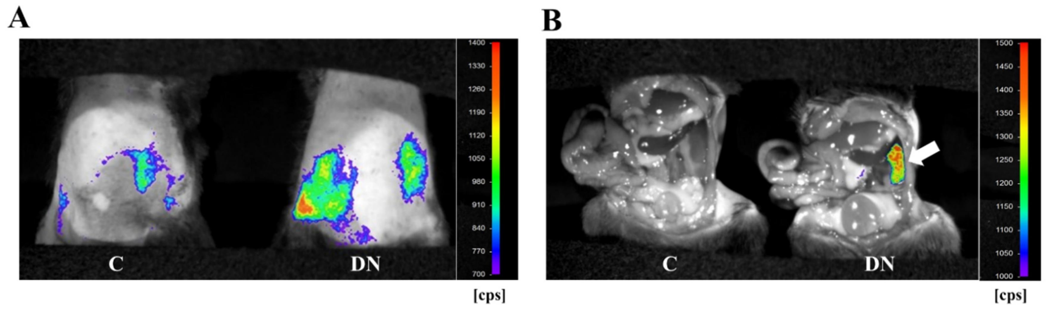
2.2. Non-Invasive High-Resolution Bioluminescence Imaging Detected Diabetes Kidney Disease in the HFD/Multiple Low-Dose STZ-Induced DN Model
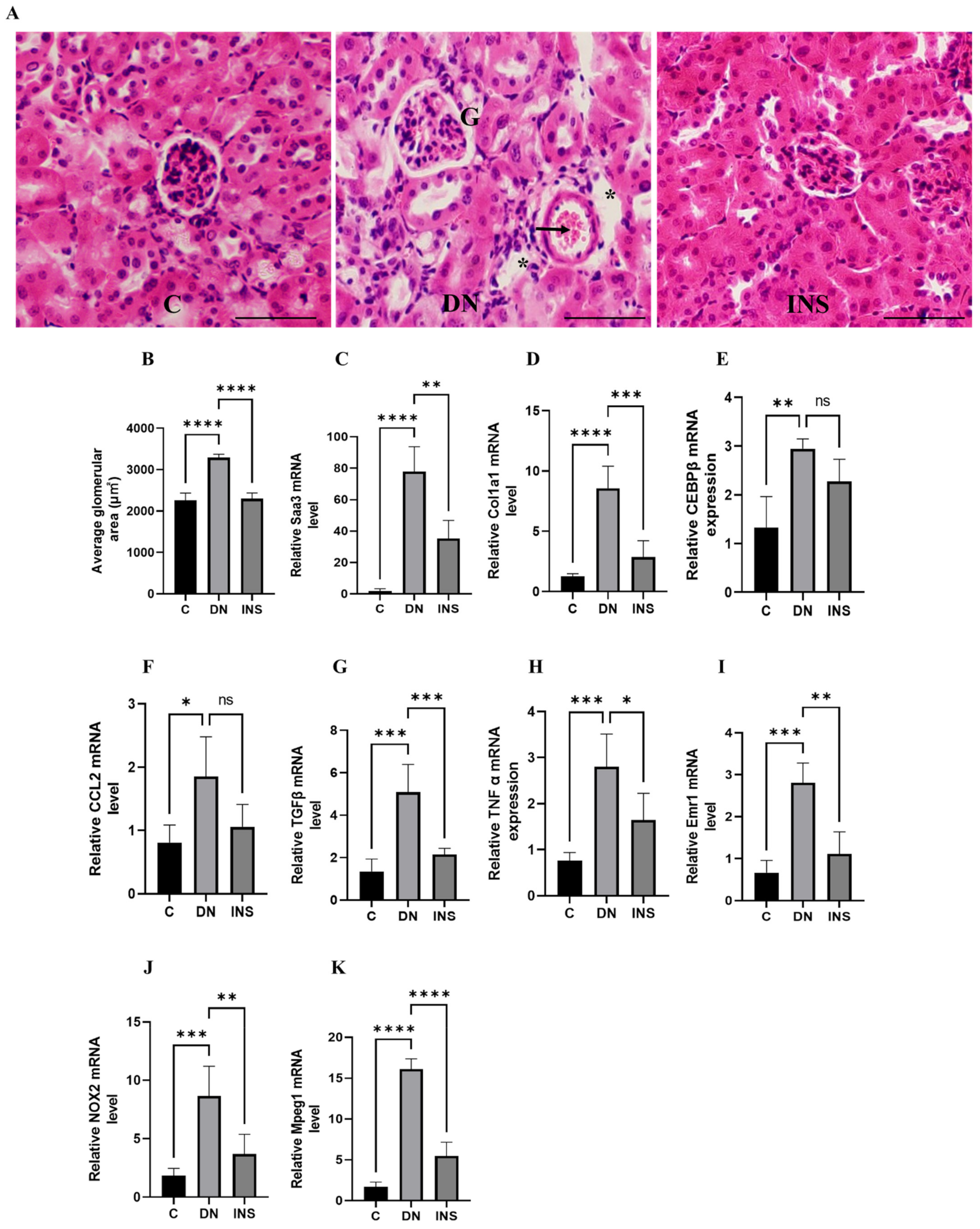
2.3. Histological, Biochemical, and Molecular Validation of In Vivo Bioluminescence Signals from the Renal Tissues of Two-Moderate-Dose STZ-Induced DN Model
3. Discussion
4. Materials and Methods
4.1. Experimental Animals
4.2. Diabetic Nephropathy Animal Models
4.3. DNA Microarray
4.4. RT-PCR
4.5. In Vivo Bioluminescent Imaging
4.6. Histological Analysis
4.7. Measurement of Plasma BUN Level
4.8. Statistical Analysis
Supplementary Materials
Author Contributions
Funding
Institutional Review Board Statement
Informed Consent Statement
Data Availability Statement
Conflicts of Interest
References
- Thomas, M.C.; Brownlee, M.; Susztak, K.; Sharma, K.; Jandeleit-Dahm, K.A.; Zoungas, S.; Rossing, P.; Groop, P.H.; Cooper, M.E. Diabetic kidney disease. Nat. Rev. Dis. Primers 2015, 1, 15018. [Google Scholar] [CrossRef]
- Hussain, S.; Jamali, M.C.; Habib, A.; Hussain, M.S.; Akhtar, M.; Najmi, A.K. Diabetic kidney disease: An overview of prevalence, risk factors, and biomarkers. Clin. Epidemiol. Glob. Health 2021, 9, 2–6. [Google Scholar] [CrossRef]
- Su, C.H.; Hsu, Y.C.; Thangudu, S.; Chen, W.Y.; Huang, Y.T.; Yu, C.C.; Shih, Y.H.; Wang, C.J.; Lin, C.L. Application of multiparametric MR imaging to predict the diversification of renal function in miR29a-mediated diabetic nephropathy. Sci. Rep. 2021, 11, 1909. [Google Scholar] [CrossRef] [PubMed]
- Wasung, M.E.; Chawla, L.S.; Madero, M. Biomarkers of renal function, which and when? Clin. Chim. Acta 2015, 438, 350–357. [Google Scholar] [CrossRef] [PubMed]
- Griffin, B.R.; Faubel, S.; Edelstein, C.L. Biomarkers of drug-induced kidney toxicity. Ther. Drug Monit. 2019, 41, 213. [Google Scholar] [CrossRef] [PubMed]
- Navarro-González, J.F.; Mora-Fernández, C.; De Fuentes, M.M.; García-Pérez, J. Inflammatory molecules and pathways in the pathogenesis of diabetic nephropathy. Nat. Rev. Nephrol. 2011, 7, 327–340. [Google Scholar] [CrossRef]
- Matoba, K.; Takeda, Y.; Nagai, Y.; Kawanami, D.; Utsunomiya, K.; Nishimura, R. Unraveling the role of inflammation in the pathogenesis of diabetic kidney disease. Int. J. Mol. Sci. 2019, 20, 3393. [Google Scholar] [CrossRef] [Green Version]
- Moreno, J.A.; Gomez-Guerrero, C.; Mas, S.; Sanz, A.B.; Lorenzo, O.; Ruiz-Ortega, M.; Opazo, L.; Mezzano, S.; Egido, J. Targeting inflammation in diabetic nephropathy: A tale of hope. Expert Opin. Investig. Drugs. 2018, 27, 917–930. [Google Scholar] [CrossRef]
- Qian, Y.; Feldman, E.; Pennathur, S.; Kretzler, M.; Brosius, F.C. From fibrosis to sclerosis: Mechanisms of glomerulosclerosis in diabetic nephropathy. Diabetes 2008, 57, 1439–1445. [Google Scholar] [CrossRef] [Green Version]
- Gross, S.; Gammon, S.T.; Moss, B.L.; Rauch, D.; Harding, J.; Heinecke, J.W.; Ratner, L.; Piwnica-Worms, D. Bioluminescence imaging of myeloperoxidase activity in vivo. Nat. Med. 2009, 15, 455–461. [Google Scholar] [CrossRef] [Green Version]
- Wessels, J.; Busse, A.C.; Mahrt, J.; Dullin, C.; Grabbe, E.; Mueller, G.A. In vivo imaging in experimental preclinical tumor research—A review. Cytom. A 2007, 71, 542–549. [Google Scholar] [CrossRef] [PubMed]
- Sanada, Y.; Yamamoto, T.; Satake, R.; Yamashita, A.; Kanai, S.; Kato, N.; Van De Loo, F.A.; Nishimura, F.; Scherer, P.E.; Yanaka, N. Serum amyloid A3 gene expression in adipocytes is an indicator of the interaction with macrophages. Sci. Rep. 2016, 6, 38697. [Google Scholar] [CrossRef]
- Kumrungsee, T.; Kariya, T.; Hashimoto, K.; Koyano, T.; Yazawa, N.; Hashimoto, T.; Sanada, Y.; Matsuyama, M.; Sotomaru, Y.; Sakurai, H.; et al. The serum amyloid A3 promoter-driven luciferase reporter mice is a valuable tool to image early renal fibrosis development and shows the therapeutic effect of glucosyl-hesperidin treatment. Sci. Rep. 2019, 9, 14101. [Google Scholar] [CrossRef]
- Mohammed-Ali, Z.; Carlisle, R.E.; Nademi, S.; Dickhout, J.G. Animal models of kidney disease. In Animal Models for the Study of Human Disease, 2nd ed.; Michael, C.P., Ed.; Academic Press: New York, NY, USA, 2017; pp. 379–417. [Google Scholar]
- Breyer, M.D.; Böttinger, E.; Brosius, F.C.; Coffman, T.M.; Harris, R.C.; Heilig, C.W.; Sharma, K. Mouse models of diabetic nephropathy. J. Am. Soc. Nephrol. 2005, 16, 27–45. [Google Scholar] [CrossRef]
- Chow, F.Y.; Nikolic-Paterson, D.J.; Ozols, E.; Atkins, R.C.; Rollins, B.J.; Tesch, G.H. Monocyte chemoattractant protein-1 promotes diabetic renal injury in streptozotocin-treated mice. Kidney Int. 2006, 69, 73–80. [Google Scholar] [CrossRef] [PubMed] [Green Version]
- Ye, R.D.; Sun, L. Emerging functions of serum amyloid A in inflammation. J. Leukoc. Biol. 2015, 98, 923–929. [Google Scholar] [CrossRef] [PubMed]
- Anderberg, R.J.; Meek, R.L.; Hudkins, K.L.; Cooney, S.K.; Alpers, C.E.; Leboeuf, R.C.; Tuttle, K.R. Serum amyloid A and inflammation in diabetic kidney disease and podocytes. Lab. Investig. 2015, 95, 250–262. [Google Scholar] [CrossRef] [Green Version]
- Dieter, B.P.; McPherson, S.M.; Afkarian, M.; de Boer, I.H.; Mehrotra, R.; Short, R.; Barbosa-Leiker, C.; Alicic, R.Z.; Meek, R.L.; Tuttle, K.R. Serum amyloid A and risk of death and end-stage renal disease in diabetic kidney disease. J. Diabetes Complicat. 2016, 30, 1467–1472. [Google Scholar] [CrossRef] [PubMed] [Green Version]
- Chawla, A.; Chawla, R.; Jaggi, S. Microvasular and macrovascular complications in diabetes mellitus: Distinct or continuum? Indian J. Endocrinol. Metab. 2016, 20, 546. [Google Scholar] [CrossRef]
- Tesch, G.H.; Allen, T.J. Rodent models of streptozotocin-induced diabetic nephropathy (Methods in Renal Research). Nephrology 2007, 12, 261–266. [Google Scholar] [CrossRef]
- Brosius, F.C.; Alpers, C.E.; Bottinger, E.P.; Breyer, M.D.; Coffman, T.M.; Gurley, S.B.; Harris, R.C.; Kakoki, M.; Kretzler, M.; Leiter, E.H.; et al. Mouse models of diabetic nephropathy. J. Am. Soc. Nephrol. 2009, 20, 2503–2512. [Google Scholar] [CrossRef] [PubMed] [Green Version]
- Brouwers, B.; Pruniau, V.P.; Cauwelier, E.J.; Schuit, F.; Lerut, E.; Ectors, N.; Declercq, J.; Creemers, J.W. Phlorizin pretreatment reduces acute renal toxicity in a mouse model for diabetic nephropathy. J. Biol. Chem. 2013, 288, 27200–27207. [Google Scholar] [CrossRef] [PubMed] [Green Version]
- Niewczas, M.A.; Pavkov, M.E.; Skupien, J.; Smiles, A.; Dom, Z.I.M.; Wilson, J.M.; Park, J.; Nair, V.; Schlafly, A.; Saulnier, P.J.; et al. A signature of circulating inflammatory proteins and development of end-stage renal disease in diabetes. Nat. Med. 2019, 25, 805–813. [Google Scholar] [CrossRef]
- Usui, H.K.; Shikata, K.; Sasaki, M.; Okada, S.; Matsuda, M.; Shikata, Y.; Ogawa, D.; Kido, Y.; Nagase, R.; Yozai, K.; et al. Macrophage scavenger receptor-a–deficient mice are resistant against diabetic nephropathy through amelioration of microinflammation. Diabetes 2007, 56, 363–372. [Google Scholar] [CrossRef] [Green Version]
- Guha, M.; Xu, Z.G.; Tung, D.; Lanting, L.; Natarajan, R. Specific down-regulation of connective tissue growth factor attenuates progression of nephropathy in mouse models of type 1 and type 2 diabetes. FASEB J. 2007, 21, 3355–3368. [Google Scholar] [CrossRef]
- Okuma, H.; Mori, K.; Nakamura, S.; Sekine, T.; Ogawa, Y.; Tsuchiya, K. Ipragliflozin Ameliorates Diabetic Nephropathy Associated with Perirenal Adipose Expansion in Mice. Int. J. Mol. Sci. 2021, 22, 7329. [Google Scholar] [CrossRef] [PubMed]
- Xu, Q.; Li, B.; Wang, Y.; Wang, C.; Feng, S.; Xue, L.; Chen, J.; Jiang, H. Identification of VCAN as hub gene for diabetic kidney disease immune injury using integrated bioinformatics analysis. Front. Physiol. 2021, 12, 547. [Google Scholar] [CrossRef]
- Zheng, W.; Guo, J.; Liu, Z.S. Effects of metabolic memory on inflammation and fibrosis associated with diabetic kidney disease: An epigenetic perspective. Clin. Epigenetics 2021, 13, 87. [Google Scholar] [CrossRef]
- Meek, R.L.; LeBoeuf, R.C.; Saha, S.A.; Alpers, C.E.; Hudkins, K.L.; Cooney, S.K.; Anderberg, R.J.; Tuttle, K.R. Glomerular cell death and inflammation with high-protein diet and diabetes. Nephrol. Dial. Transpl. 2013, 28, 1711–1720. [Google Scholar] [CrossRef] [Green Version]
- Eklund, K.K.; Niemi, K.; Kovanen, P.T. Immune functions of serum amyloid A. Crit. Rev. Immunol. 2012, 32, 335–348. [Google Scholar] [CrossRef]
- Sorić Hosman, I.; Kos, I.; Lamot, L. Serum amyloid A in inflammatory rheumatic diseases: A compendious review of a renowned biomarker. Front. Immunol. 2021, 11, 631299. [Google Scholar] [CrossRef]
- Chami, B.; Hossain, F.; Hambly, T.W.; Cai, X.; Aran, R.; Fong, G.; Vellajo, A.; Martin, N.J.; Wang, X.; Dennis, J.M.; et al. Serum amyloid A stimulates vascular and renal dysfunction in apolipoprotein E-deficient mice fed a normal chow diet. Front. Immunol. 2019, 10, 380. [Google Scholar] [CrossRef] [PubMed]
- Den Hartigh, L.J.; Wang, S.; Goodspeed, L.; Ding, Y.; Averill, M.; Subramanian, S.; Wietecha, T.; O’Brien, K.D.; Chait, A. Deletion of serum amyloid A3 improves high fat high sucrose diet-induced adipose tissue inflammation and hyperlipidemia in female mice. PLoS ONE 2014, 9, e108564. [Google Scholar] [CrossRef] [PubMed] [Green Version]
- Thaler, R.; Sturmlechner, I.; Spitzer, S.; Riester, S.M.; Rumpler, M.; Zwerina, J.; Klaushofer, K.; Van Wijnen, A.J.; Varga, F. Acute-phase protein serum amyloid A3 is a novel paracrine coupling factor that controls bone homeostasis. FASEB J. 2015, 29, 1344–1359. [Google Scholar] [CrossRef] [PubMed] [Green Version]
- Dieter, B.P.; Meek, R.L.; Anderberg, R.J.; Cooney, S.K.; Bergin, J.L.; Zhang, H.; Nair, V.; Kretzler, M.; Brosius, F.C.; Tuttle, K.R. Serum amyloid A and Janus kinase 2 in a mouse model of diabetic kidney disease. PLoS ONE 2019, 14, e0211555. [Google Scholar] [CrossRef]
- Goldin, A.; Beckman, J.A.; Schmidt, A.M.; Creager, M.A. Advanced glycation end products: Sparking the development of diabetic vascular injury. Circulation 2006, 114, 597–605. [Google Scholar] [CrossRef] [Green Version]
- Chen, E.S.; Song, Z.; Willett, M.H.; Heine, S.; Yung, R.C.; Liu, M.C.; Groshong, S.D.; Zhang, Y.; Tuder, R.M.; Moller, D.R. Serum Amyloid A Regulates Granulomatous Inflammation in Sarcoidosis through Toll-like Receptor-2. Am. J. Respir. Crit. Care Med. 2010, 181, 360–373. [Google Scholar] [CrossRef] [PubMed]
- Fischer, A.H.; Jacobson, K.A.; Rose, J.; Zeller, R. Hematoxylin and eosin staining of tissue and cell sections. Cold Spring Harb. Protoc. 2008, 5, pdb.prot4986. [Google Scholar] [CrossRef]


| Gene Symbol | Gene Description | Fold in HFD/Multiple Low-Dose STZ-Induced DN | Fold in Two Moderate-Dose STZ-Induced DN |
|---|---|---|---|
| Inflammation and immune response | |||
| Ccl7 | chemokine (C-C motif) ligand 7 | 9.57 | 9.21 |
| C3 | complement component 3 | 8.32 | 4.03 |
| Cxcl1 | chemokine (C-X-C motif) ligand 1 | 6.02 | 2.96 |
| Cxcl13 | chemokine (C-X-C motif) ligand 13 | 5.07 | 3.72 |
| Ccl2 | chemokine (C-C motif) ligand 2 | 4.68 | 3.69 |
| Saa3 | serum amyloid A 3 | 4.67 | 6.25 |
| Cxcl10 | chemokine (C-X-C motif) ligand 10 | 4.57 | 4.24 |
| Ifi27l2a | interferon, alpha-inducible protein 27 like 2A | 4.44 | 1.73 |
| Ccl8 | chemokine (C-C motif) ligand 8 | 4.20 | 5.87 |
| Irf7 | interferon regulatory factor 7 | 4.16 | 1.80 |
| Sftpd | surfactant associated protein D | 3.84 | 13.99 |
| Saa2 | serum amyloid A 2 | 3.94 | 3.02 |
| Saa1 | serum amyloid A 1 | 3.79 | 5.72 |
| Oasl1 | 2′-5′ oligoadenylate synthetase-like 1 | 3.69 | 2.96 |
| Il1f6 | interleukin 1 family, member 6 | 3.66 | 8.42 |
| Oas1a | 2′-5′ oligoadenylate synthetase 1A | 3.60 | 1.86 |
| Ifi27 | interferon, alpha-inducible protein 27 | 3.53 | 1.93 |
| Ccl12 | chemokine (C-C motif) ligand 12 | 3.50 | 1.50 |
| Oas1f | 2′-5′ oligoadenylate synthetase 1F | 3.34 | 2.05 |
| Ccl3 | chemokine (C-C motif) ligand 3 | 3.17 | 1.94 |
| Ifit2 | interferon-induced protein with tetratricopeptide repeats 2 | 3.05 | 2.52 |
| Tlr2 | Toll-like receptor 2 | 2.90 | 2.27 |
| Saa4 | serum amyloid A 4 | 2.89 | 3.12 |
| Tnfrsf1b | tumor necrosis factor receptor superfamily, member 1b | 2.86 | 1.88 |
| Il1rn | interleukin 1 receptor antagonist | 2.84 | 11.50 |
| B2m | beta-2 microglobulin | 2.76 | 2.04 |
| Ccl9 | chemokine (C-C motif) ligand 9 | 2.56 | 2.08 |
| Ifit1 | interferon-induced protein with tetratricopeptide repeats 1 | 2.54 | 2.33 |
| Gbp6 | guanylate binding protein 6 | 2.50 | 2.24 |
| Oas1d | 2′-5′ oligoadenylate synthetase 1D | 2.45 | 4.78 |
| C4b | complement component 4B | 2.34 | 1.86 |
| Ltc4s | leukotriene C4 synthase | 2.31 | 2.76 |
| Ccl5 | chemokine (C-C motif) ligand 5 | 2.18 | 2.61 |
| Mpeg1 | macrophage expressed gene 1 | 1.57 | 1.57 |
| Cebpb | CCAAT/enhancer binding protein (C/EBP), beta | 1.35 | 1.44 |
| Adgre1 | adhesion G protein-coupled receptor E1 | 2.15 | 1.87 |
| Fibrosis marker | |||
| Col1a1 | collagen, type I, alpha 1 | 2.31 | 1.69 |
| Col3a1 | collagen, type III, alpha 1 | 2.50 | 1.75 |
| Col17a1 | collagen, type XVII, alpha 1 | 2.51 | 3.55 |
| Col2a1 | collagen, type II, alpha 1 | 2.17 | 1.59 |
| Col12a1 | collagen, type XII, alpha 1 | 2.26 | 2.13 |
| Tnc | tenascin C | 2.08 | 2.06 |
| Areg | Amphiregulin | 2.01 | 8.97 |
| Itgav | integrin alpha V | 5.06 | 1.69 |
| Fn1 | fibronectin 1 | 3.31 | 2.89 |
| Timp1 | tissue inhibitor of metalloproteinase 1 | 4.66 | 3.01 |
| Mmp2 | matrix metallopeptidase 2 | 2.30 | 2.72 |
| Mmp3 | matrix metallopeptidase 3 | 5.07 | 3.46 |
| Fbn1 | fibrillin 1 | 2.41 | 2.53 |
| Atf3 | activating transcription factor 3 | 2.70 | 2.45 |
| Lox | lysyl oxidase | 2.33 | 2.05 |
| Cellular senescence and apoptosis | |||
| Rprm | reprimo, TP53 dependent G2 arrest mediator candidate | 1.24 | 1.46 |
| Trp53inp1 | transformation related protein 53 inducible nuclear protein 1 | 2.16 | 3.87 |
| Tnfrsf10b | tumor necrosis factor receptor superfamily, member 10b | 1.61 | 4.64 |
| Cdkn1a | cyclin-dependent kinase inhibitor 1A (P21) | 12.22 | 15.15 |
| Ddias | DNA damage-induced apoptosis suppressor | 3.19 | 3.71 |
| Bcl2a1b | B cell leukemia/lymphoma 2 related protein A1b | 1.82 | 1.76 |
| Casp12 | caspase 12 | 1.43 | 1.75 |
| Aen | apoptosis enhancing nuclease | 1.43 | 1.66 |
| Casp4 | caspase 4 | 1.56 | 1.57 |
| Naip1 | NLR family, apoptosis inhibitory protein 1 | 4.49 | 2.60 |
| Bcl2a1c | B cell leukemia/lymphoma 2 related protein A1c | 2.05 | 1.65 |
| Bak1 | BCL2-antagonist/killer 1 | 1.49 | 1.50 |
| Bbc3 | BCL2 binding component 3 | 2.23 | 2.75 |
| Top2a | topoisomerase (DNA) II alpha | 11.37 | 3.92 |
| Bub1 | BUB1, mitotic checkpoint serine/threonine kinase | 8.76 | 4.72 |
| Bub1b | BUB1B, mitotic checkpoint serine/threonine kinase | 4.76 | 1.86 |
| Chek1 | checkpoint kinase 1 | 1.98 | 3.36 |
| Mad2l1 | mitotic checkpoint component Mad2 | 1.95 | 1.97 |
| Target Gene | Sequence (5′–3′) | |
|---|---|---|
| L19 | Forward Reverse | GGCATAGGGAAGAGGAAGG GGATGTGCTCCATGAGGATGC |
| Saa3 | Forward Reverse | AAGGGTCTAGAGACATGTGG ACTTCTGAACAGCCTCTCTG |
| EMR1 (Adgre1) | Forward Reverse | ATTGTGGAAGCATCCGAGAC GTAGGAATCCCGCAATGATG |
| TNFα | Forward Reverse | CGTCGTAGCAAACCACCAAG TTGAAGAGAACCTGGGAGTAGACA |
| Mpeg1 | Forward Reverse | GCTTGCCTCTGCATTTCTTC TCTTCTGCTCCAGGTTTTGG |
| CEBPβ | Forward Reverse | GAAGACGGTGGACAAGCTGA TGCTCCACCTTCTTCTGCAG |
| Ccl2 (MCP-1) | Forward Reverse | GGTCCCTGTCATGCTTCTGG CCTTCTTGGGGTCAGCACAG |
| Col1a1 | Forward Reverse | CCCAAGGAAAAGAAGCACGTC ACATTAGGCGCAGGAAGGTCA |
| NOX2 | Forward Reverse | AGCTATGAGGTGGTGATGTTAGTGG TGCACAGCAAAGTGATTGGC |
| TGFβ | Forward Reverse | GGCACCATCCATGACATGAA TTCTCTGTGGAGCTGAAGCAAT |
Publisher’s Note: MDPI stays neutral with regard to jurisdictional claims in published maps and institutional affiliations. |
© 2022 by the authors. Licensee MDPI, Basel, Switzerland. This article is an open access article distributed under the terms and conditions of the Creative Commons Attribution (CC BY) license (https://creativecommons.org/licenses/by/4.0/).
Share and Cite
Saliu, T.P.; Yazawa, N.; Hashimoto, K.; Miyata, K.; Kudo, A.; Horii, M.; Kamesawa, M.; Kumrungsee, T.; Yanaka, N. Serum Amyloid A3 Promoter-Driven Luciferase Activity Enables Visualization of Diabetic Kidney Disease. Int. J. Mol. Sci. 2022, 23, 899. https://doi.org/10.3390/ijms23020899
Saliu TP, Yazawa N, Hashimoto K, Miyata K, Kudo A, Horii M, Kamesawa M, Kumrungsee T, Yanaka N. Serum Amyloid A3 Promoter-Driven Luciferase Activity Enables Visualization of Diabetic Kidney Disease. International Journal of Molecular Sciences. 2022; 23(2):899. https://doi.org/10.3390/ijms23020899
Chicago/Turabian StyleSaliu, Tolulope Peter, Nao Yazawa, Kotaro Hashimoto, Kenshu Miyata, Ayane Kudo, Mayu Horii, Mion Kamesawa, Thanutchaporn Kumrungsee, and Noriyuki Yanaka. 2022. "Serum Amyloid A3 Promoter-Driven Luciferase Activity Enables Visualization of Diabetic Kidney Disease" International Journal of Molecular Sciences 23, no. 2: 899. https://doi.org/10.3390/ijms23020899