Article

Engineering of Bacillus Promoters Based on Interacting Motifs between UP Elements and RNA Polymerase (RNAP) α-Subunit

1
Key Laboratory of Industrial Biotechnology, Ministry of Education, School of Biotechnology, Jiangnan University, Wuxi 214122, China
2
National Engineering Research Center for Cereal Fermentation and Food Biomanufacturing, Jiangnan University, 1800 Lihu Avenue, Wuxi 214122, China
3
Jiangsu Provincial Engineering Research Center for Bioactive Product Processing, Jiangnan University, 1800 Lihu Avenue, Wuxi 214122, China
*
Author to whom correspondence should be addressed.
Int. J. Mol. Sci. 2022, 23(21), 13480; https://doi.org/10.3390/ijms232113480
Submission received: 6 October 2022 / Revised: 1 November 2022 / Accepted: 1 November 2022 / Published: 3 November 2022
(This article belongs to the Section Molecular Microbiology)

Abstract

:
Bacillus genetics need more versatile promoters for gene circuit engineering. UP elements are widely distributed in noncoding regions and interact with the α-subunit of RNA polymerase (RNAP). They can be applied as a standard element for synthetic biology. Characterization of the binding motif between UP elements and RNAP may assist with rational and effective engineering. In this study, 11 Bacillus constitutive promoters were screened for strength in Bacillus licheniformis. The motif in UP elements from a strong native promoter, PLan, was characterized. The influence of specific sequences on RNAP binding and expression strength was investigated both in vitro and in vivo. It was found that sequences up to 50 base pairs upstream of the consensus motif significantly contributed to α-CTD (the alpha subunit carboxy-terminal domain) association. Meanwhile, two repeats of a proximal subsite were able to more strongly activate the expression (by 8.2-fold) through strengthening interactions between UP elements and RNAP. Based the above molecular basis, a synthetic UP element, UP5-2P, was constructed and applied to nine wild-type promoters. Fluorescence polarization results demonstrated that it had an apparent effect on promoter–α-CTD interactions, and elevated expression strength was observed for all the engineered promoters. The highest improved core promoter, Pacpp, was more strongly activated by 7.4-fold. This work thus develops a novel strategy for Bacillus promoter engineering.

1. Introduction

Well-evaluated promoters of diversified strength are crucial elements for complex in vivo expression in synthetic biology. To this end, they are believed not only to be standardized, but also to exhibit predictable and reliable activity under a variety of different conditions [1,2]. Strong heterologous overexpression has been achieved in Escherichia coli based on the specific interaction between the bacteriophage-derived, T7-specific RNA polymerase (RNAP) and the strong T7 promoter. Recent research concerning this Gram-negative bacterium has focused more on the engineering of native promoters to create highly regulated gene expression circuits [3,4,5]. Although multiple native promoters have been identified in Bacillus, fewer methods for their rational engineering have been shown to be highly efficient, compared with that of E. coli ones.
Structurally, bacterial promoters contain two recognition elements for RNAP and the assembled sigma factor: the classical −35/−10 hexamer and the UP element. The −35 and −10 elements, or the promoter core sequences, recognized by the sigma subunit of RNAP, determine to a great extent the strength of a promoter [6]. A vast majority of promoter engineering approaches are based on evolutionary or semi-rational strategies within these regions [7,8,9]. Robust libraries can be obtained through saturation mutagenesis or error-prone PCR. Then, a series of engineered promoters with broad expression profiles can be screened with the assistance of fluorescent proteins. The most frequently encountered issue here concerns the inability to produce a synthetic promoter with higher expression levels than a natural alternative. Accumulating experimental evidence has offered a detailed picture of the molecular mechanism underlying UP element-dependent promoter stimulation in E. coli [10]. Recently, Alper et al. provided an example of successfully engineered E. coli promoters in a study that employed designing and optimizing UP elements of the promoter, with strengths up to 9.4-fold higher than their wild-type sequences [6]. This UP element was reported to contact the alpha subunit carboxy-terminal domain (α-CTD) of the RNAP, thus exerting significant control on promoter activity in a manner that is independent of the promoter core sequences.
Bacillus RNAP also contains α2 (dimer), β, β′, and σ subunits [11]. The holoenzyme assembled with a major sigma factor σA (analogous to σ70 in E. coli) binds to promoters to initiate transcription. Kang et al. designed synthetic Bacillus promoters by shuffling the promoter core sequences recognized by different sigma factors [12]. A set of strong promoters were identified, with a strength up to 18.6-fold higher with respect to that of the commonly used promoter P43. Although current genetic and biochemical information has proved that the UP elements were involved in α-CTD binding, which is required for high expression of multiple Bacillus promoters, modifying promoter activity through the engineering of UP elements has received limited attention in the field. Here, we seek to engineer a previously verified α-CTD-binding Bacillus redox-sensitive promoter TrxA [13], based on interacting motifs between UP elements and α-CTD of RNAP. To do so, we first prepared purified protein of α-CTD and located the UP sequence of TrxA via DNase I footprinting and electrophoretic mobility shift assay (EMSA). Then, the native one was replaced by predicted UP elements from other promoters. The synthetic promoters were characterized both in vitro for DNA–protein interaction and in vivo for the strength of expression of Enhanced Green Fluorescent Protein (EGFP). The results were analyzed for correlation, and finally, four UP elements were proved to more strongly activate expression of the TrxA core promoter by up to 7.3-fold. This work thus develops a novel strategy for Bacillus promoter engineering through simply altering the UP elements alone in a manner that is independent of the core promoter itself.

2. Results

2.1. The Selected Promoters Vary in Strength and Stage-Specific Expression

Eleven promoters, both wild-type and artificial, were selected to be incorporated into a library for structural and functional comparison, listed in Table 1. Then, a fluorescence method was adopted to evaluate the strength and the trends over cultivation time. As the wild-type cells also exhibit false-positive signals, and the fluorescence may decay in cell cultures, the two issues that influence the results the most are the cell loading volume and sampling time for fluorescence measurements. To this end, a method for EGFP-assisted screening of the constitutive promoters in B. licheniformis was developed by using BlR2G as an example. Fluorescent signals from cells loaded with different concentrations were investigated. Results showed that an OD600 range of 0.8–2.5 in a single well of the 96-well microtiter plate guaranteed a linear correlation between cell counts and the fluorescent signals (Figure S1).
Then, the cell cultures corresponding to different promoters were sampled at 12, 24, 36, and 48 h, respectively, and relative fluorescence intensity (fluorescence intensity (Au)/OD600) was used to demonstrate the promoter activities (Figure 1). The selected 11 constitutive promoters showed a wide range of estimated activities, with relative fluorescence intensity being the highest (64896) in PLan at 48 h and the lowest (860) in P43 at 24 h. More importantly, comparison between different sampling times revealed huge variations in transcription initiation. For example, Pshuttle09 was the most active promoter at both 12 and 24 h, with a relative fluorescence intensity of more than 2.3-fold higher than that of the other promoters. From 24 h to 36 h, however, PLan experienced a great boost in relative fluorescence intensity (by more than 13-fold). Its activity continued to increase by 2-fold from 36 h to 48 h. At the same time, 9 of 11 promoters showed a significantly decreasing trend in activity. When relating the above results to cell growth of the strains, we found that 24 h was near the division point between exponential and stationary, so that these promoters exhibited transcription initiation patterns corresponding to different cell growth stages. Finally, the estimated promoter activity of PLan could be as high as 75-fold higher when the most frequently used promoter P43 was used as a reference. This promoter was then selected for further analysis and engineering.

2.2. PLan Was Predicted to Be Involved in Quorum Sensing (QS)

In B. licheniformis, PLan exhibited much higher activity than the other ones tested. This promoter was mined and predicted based on our previous transcriptome data. We wondered why it mediated hyper-expression and if it could be a source of UP elements for promoter engineering. Therefore, its genome loci were retrieved to find the possible answers. The highly expressed transcript RS00925 mediated by PLan is located within a lantibiotic biosynthesis gene cluster and codes for the putative propeptide. The amino acid sequence of this peptide is close to the one reported in B. subtilis and other bacteria [22,23]. However, one additional gene cluster with an unknown phenotype was observed just upstream of the PLan promoter. Inside this cluster, four genes have been annotated as being signal transduction histidine kinase (LanC), transcriptional regulatory protein (LanB), and two hypothetical proteins (LanD and LanA). Subsequent sequence alignment indicated that those sequences were consistent with the agr QS system containing AgrBDCA, which was well characterized in Gram-positive Staphylococcus aureus. This is related to cell density-dependent control of behaviors such as virulence and biofilm formation [24,25,26]. Specifically, LanC shares 23% identity with AgrC, LanB shares 21% identity with AgrB, LanD shares 30% identity with AgrD, and LanA shares 24% identity with AgrA. Notably, highly conserved or similar amino residues could be observed in specific domains between these four pairs of proteins. As a result, we can conclude that PLan is involved in an agr-like QS system. Most QS systems possess the characteristic of autoinducer-dependent transcriptional activation to ensure hyper expression of QS operons. It has also been reported that C-terminal domain of the alpha subunit of RNA Polymerase (α-CTD) plays an important role in this activation by making contact with the promoter regions [27,28]. We therefore examined whether this is also true for the QS system predicted in B. licneniformis.

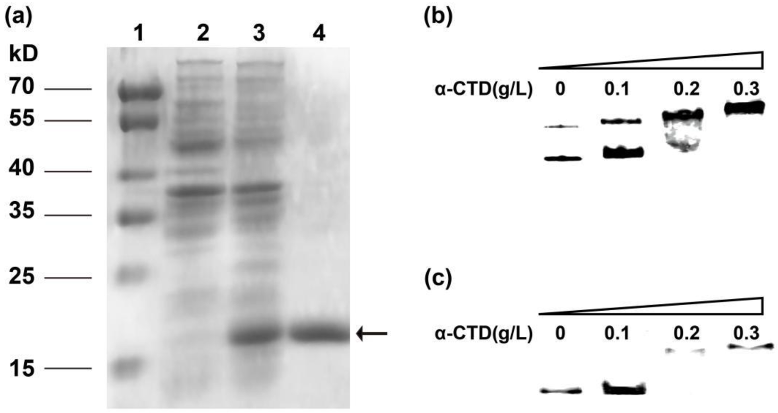
2.3. Heterologously Expressed α-CTD Directly Interacts with Both PtrxA and PLan In Vitro

α-CTD from B. licheniformis was expressed by using pET-28(a)/DE3 system in E. coli. After the construct EcCTD was cultivated and induced, cell lysate was subjected to SDS-PAGE (sodium dodecyl sulfate-polyacrylamide gel electrophoresis). Compared with the control one, the sample corresponding to EcCTD apparently showed a band of 15 kD, which is consistent with the predicted molecular weight of α-CTD (Figure 2). As a His-tag was fused into the 3′end of the recombinant protein, purified α-CTD could be easily obtained by using a Mag-Beads His-Tag Kit. Subsequent SDS-PAGE confirmed that the purified sample exhibited a single band of ~15 kD, and this sample can be used for in vitro experiments on interaction between DNA and protein. PtrxA has been reported to specifically contact with α-CTD through UP elements in B. subtilis. Therefore, this sequence was chosen as a reference DNA sample to examine whether the recombinant α-CTD was structurally positive. Then, EMSA was used to characterize the DNA–protein interaction. As can be seen from the chromatography, the migration of the 343-bp DNA fragment was retarded when it was incubated with α-CTD. In addition, a clear protein concentration response relationship was observed in the EMSA gel, within a range of 0.1–0.3 g/L. This indicated a correct fold recognition of recombinant α-CTD which could bind to the reference probe.
We therefore performed additional EMSA to investigate whether PLan can interact with α-CTD. The probe corresponding to PLan was first amplified by PCR with biotin labeled at the 5′-end. When we incubated the probe with increasing amounts of α-CTD (from 0.1 to 0.3 g/L), different shifted bands were observed. It could be inferred that the complex α-CTD-PLan was formed in a protein concentration-dependent manner, confirming that α-CTD specifically bound to the promoter PLan.

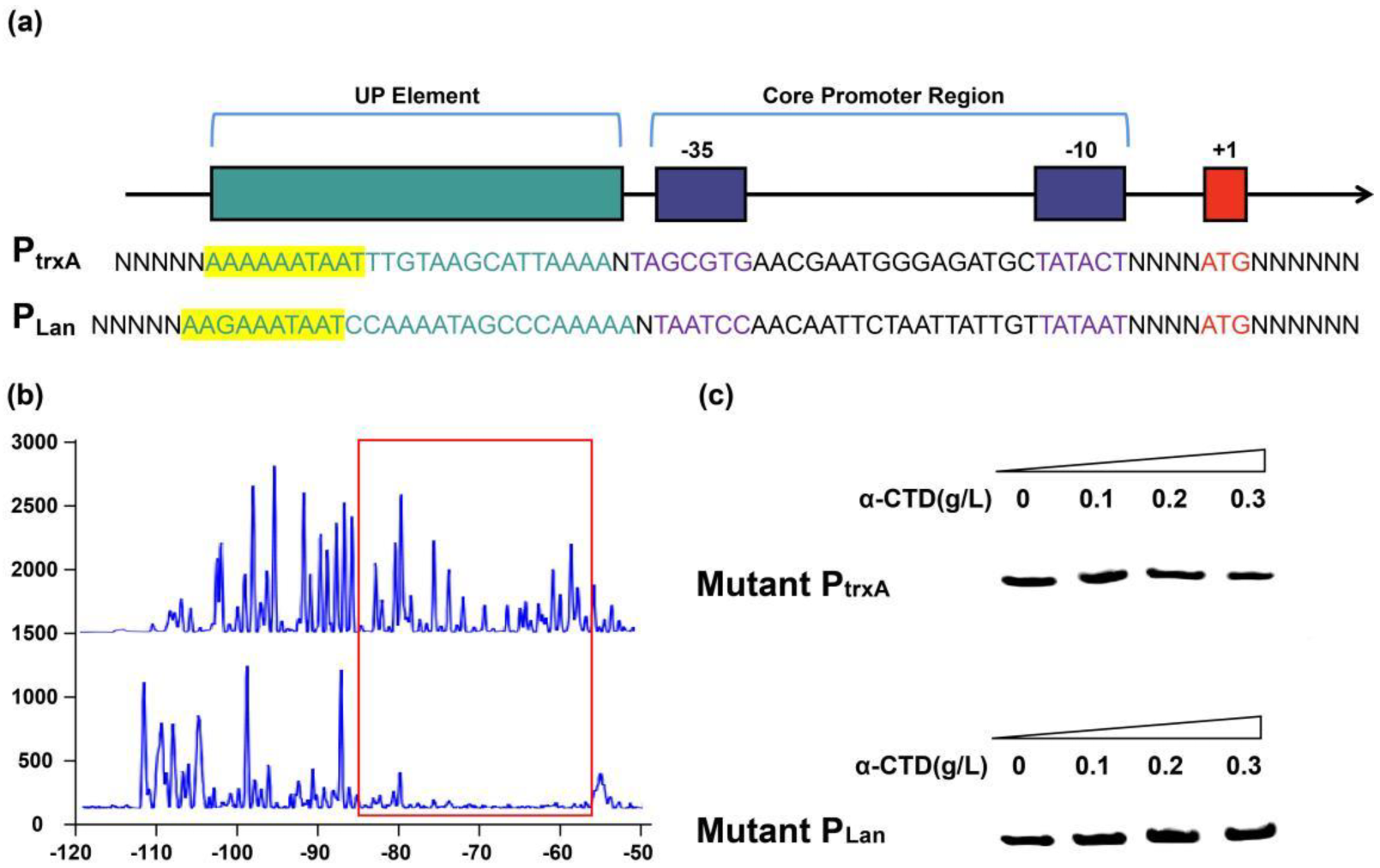
2.4. α-CTD Binds to an UP Element 50 bp Upstream of TSS

It is known that in B. subtilis, α-CTD can interact with specific UP elements located in a region upstream of the PtrxA core promoter. This UP element is believed to act as an activating sequence for certain promoters and has been consistently shown to increase promoter strength by facilitating RNA polymerase association with DNA. To determine the precise α-CTD binding site or the UP element, DNase I footprinting assays were performed with 5′-6-carboxyfluorescein (6-FAM)-labeled probes containing a sequence upstream of PLan core promoter region. As can be seen in Figure 3, the DNA region from −57 to −85 was protected from DNase I digestion. It was predicted to be an UP element (named UPL) able to interact with α-CTD. This predicted UP element was further verified by EMSA performed with α-CTD and truncated PLan, in which UPL was removed through overlapping PCR. As expected from DNase I footprinting data, no shifted band was obtained with the truncated PLan, from which a positive UP element could be inferred. Compared with previously reported UP elements in PtrxA (terms as UPA here), UPL contained a high-consensus sequence of “AAGAAATAAT” and sheared adenine (A) and thymine (T) dinucleotide repeat tracts. This has been proven to be a crucial element for RNAP recognition and binding in B. subtilis [29]. However, there were significant differences between the two UP elements in terms of genomic location. For example, UPA is located in the −40 to −31 region. UPL, in comparison, is located at a position further upstream (−60) of the TSS. Their flanking sequences also share little similarity. The influence of this newly found UP element on promoter activity remains elusive and needs further investigation.

2.5. UP Elements with More Flanking Base Pairs Ensured Higher Promoter Activity

UP elements as expression activators have been tested in different bacteria [30]. It was suggested that sequences flanking the UP element could influence promoter activity through certain DNA secondary structures. We have proven that the newly found UP element shares a consensus motif of 10 bp with that in PtrxA. On this basis, we then selected the truncated UP element fragments with different lengths in flanking base pairs as DNA probes, and evaluated their interactions with α-CTD in vitro and the performance on expression activity. In this effort, we sought to identify a stronger UP element based on the ability to amplify expression of the PtrxA core promoter. For in vitro investigation, Fluorescence polarization (FP) provides a nonradioactive approach for measuring protein–DNA interactions. An increase in molecular volume due to binding of a small fluorescent molecule to a protein impedes the fluorescent molecule’s rotational motion in solution and results in an increase in polarization. According to this principle, the interaction between α-CTD and the engineered promoters with different flanking base pairs in UP elements was indicated by the degree of polarization change (DPC). It showed a clear correlation between the lengths of motif-flanking sequences and the polarization values (Figure 4). As shifted polarization values suggested stronger interactions, conformational changes of the DNA probes potentially induced by α-CTD interaction were revealed. It was found that sequences up to 50 base pairs upstream of the consensus motif significantly contribute to α-CTD association (p < 0.0001).
To demonstrate the PtrxA promoter activities mediated by the above different UP elements, cells harboring recombinant vectors were cultivated and collected for promoter strength detection, by using the same fluorescence method described in promoter selection. Wild-type PtrxA promoter was used as the control. As shown in Figure 5, all the synthetic promoters with UP elements embedded upstream of −35 sites displayed stronger activities than the wild-type one. The strongest promoter exhibited significantly higher strength (up to 5.6-fold) than the control. Consistent with the results in vitro, strong positive correlations were seen between the lengths in flanking base pairs of UP elements and the promoter activities, indicating that UP elements with more flanking base pairs ensured higher promoter activity. UP5 exhibited a potential stem-loop structure consisting of palindromic and inverted repeat sequences between the proximal subsite and the distal subsite. This is consistent with the best-characterized bacterial UP element, the one in the rrnB P1 promoter, in which the subsites are located between positions −40 and −60 with respect to the transcription start site [31]. Both subsites have been reported to stimulate transcription with varying degrees. We also found that there is no base preference in UP5. This is unexpected as previously characterized UP elements were reported to be rich in AT or CG tracts to facilitate interaction with α-CTD [32]. These findings prompted us to evaluate synthetic UP elements to improve promoter activities based on UP5.

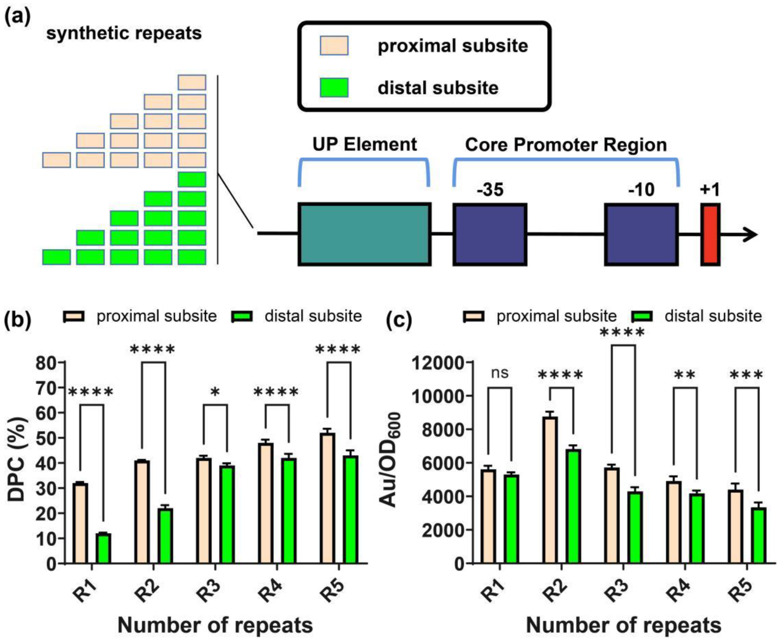
2.6. Repeated Specific Subsite in UP Elements Improved Promoter Activity

According to the existing literature, it is possible that placing UP element sequences in tandem could further activate gene expression. However, the specific role of proximal subsites and distal subsites in activating expression remains unclear. Thus, we first attempted to improve promoter activity by inserting repeated UP5 sequences in the wild-type UP element (the semi-rational strategy). Several synthetic promoters harboring 1 to 5 tandem repeats of intact UP5 upstream of PtrxA were constructed (Figure 6). In preliminary screening, the promoter–α-CTD interactions were tested in vitro by FP. The results showed that although the polarization values seemed positively correlated with the number of repeats, the DPC caused by α-CTD interaction did not show significant distinctions. An EGFP-assisted secondary screening also showed that no activated expression was obtained compared with that of the initial synthetic promoter PtrxA-UP5. These results indicate that simple repeats of UP5 could not improve promoter activity by providing an extended interaction region for α-CTD to bind.
As α-CTD binds to UP elements at both the proximal subsite and the distal subsite, we hypothesized that each subsite could influence the initiation of transcription individually. It is therefore worth trying to engineer promoters using the respective subsite as an activator. To do so, we designed two sets of synthetic promoters with tandem repeats of “CAAAATAGC” (proximal) and “GGCATTTTG” (distal), inserted upstream of UP5 sequences in PtrxA-UP5, respectively. After those synthetic promoters were synthesized, they were labeled and incubated with α-CTD. FP results exhibited a wide distribution for DPC. The most repeats (five) of the proximal subsite had the highest DPC of 52%, suggesting a significantly changed interaction. The same repeat number of the distal subsite also had a DPC of 35%. Seven in ten synthetic promoters exhibited polarization values corresponding to stronger binding. In terms of fluorescence intensity, only four in ten synthetic promoters were higher than that of the parent promoter with only a single copy of UP5. Remarkably, the strongest performing sequence was not the most repeats of R5 but R2 (UP5-2P) for the proximal subsite, which more strongly activated PtrxA expression by 8.2-fold. Likewise, R2 for the distal subsite also exhibited the highest expression intensity. These results indicate that repeats of the specific subsite within an UP element could improve the downstream promoter performance by facilitating promoter–α-CTD interactions. Excessively strong interactions between UP elements and RNAP could decrease gene expression.

2.7. Synthetic UP Elements Improved Activity of Varied Core Promoters

The above results motivated us to investigate whether the synthetic UP element, UP5-2P, could function independently to improve the performance of promoters other than PtrxA, especially those without a known α-CTD binding site. For this purpose, UP5-2P was inserted upstream of the other nine constitutive promoters screened previously. Just as predicted, as shown in Figure 7, FP results demonstrate that UP5-2P had an apparent effect on promoter–α-CTD interactions, with DPC values ranging from 33 to 85%. Fluorescence measurements of the EGFP reporting gene corroborated the above results in that elevated intensities were observed for all the engineered constructs. The most improved core promoter, Pacpp, was able to be more strongly activated by 7.4-fold. Interestingly, all these promoters have not been reported to have an α-CTD binding site other than PtrxA and PspovG. These results suggest that the function of UP5-2P is transferable when it is embedded upstream of multiple core promoters.

3. Discussion

The genus Bacillus respresents a group of important bacterial hosts for synthetic biology, especially for its excellent performance at an industrial scale [33]. However, the development of the Bacillus expression system lags far behind E. coli or yeast. This can be largely attributed to the lack of strong promoters. The most currently used promoters are wild-type ones and originated in B. subtilis, such as P43 and PHpaII. The strongest constitutive promoter reported in B. subtilis is a hybrid one, PShuttle-09, whose activity was 8 times that of P43. Interestingly, this hybrid promoter was assembled artificially from two B. licheniformis genes: ylyB and luxS [34]. In our current study, PShuttle-09 was first evaluated in B. licheniformis, and the results also suggested that it was stronger than most reported Bacillus promoters within the first 24 h of cultivation. However, the activities of PspovG and PLan were highly significant after 24 h, and PLan exceeded PShuttle-09 by more than 28-fold in activity after 48 h. These results indicated that in addition to activities, theses promoters exhibited various patterns of transcription initiation corresponding to specific stages of cell growth. Based on the time point with the highest fluorescent intensity, PtrxA and PShuttle-09 can be classified as “exponential active promoters”and PspovG and PLan as “stationary active promoters” in B. licheniformis. It has also been reported that the expression of a series of genes was significantly upregulated during the transition from exponential growth to the stationary stage [35]. This type of cellular response may be controlled by two-component systems (quorum-sensing systems or other transcriptional regulators) to cope with the environmental changes. The promoter PLan in this study was supposed to be involved in an uncharacterized quorum-sensing system. In addition to the obvious time-course in the changes of fluorescent signals, sequence alignment also suggested that the gene cluster transcribed from PLan may serve the functions of recognition and secretion of cell–cell signaling molecules. This is the first time a B. licheniformis quorum-sensing-based promoter has been described; it may be used as a strong expression element for synthetic biology. Unlike the exponential active pattern of most currently used promoters [36], the cell concentration-dependent promoter may contribute to the rational design of a synthetic circuit which better balances cell growth and target compound biosynthesis.
RNAP has two points of physical contact with a promoter: the core promoter sequence (-10 and -35 element) and the UP element. This study confirms that the interaction between the specific DNA sequences in the UP element and α-CTD domain of RNAP can be utilized for rational engineering of promoters in Bacillus. As emphasized by Phillips et al., RNAP binding to the UP element occurs independently of the other promoter element [37]. We showed that manipulation of a well-characterized α-CTD binding motif could enhance the binding of RNAP to the promoter, which contributed to an improved expression intensity. This is similar to the case of the tandem promoter strategy to increase the strength. Researchers anticipated that tandem repetitive promoters might trap plentiful RNAPs, which in turn enhances gene expression [38]. Perhaps the most important finding in this study is that two repeats of the proximal/distal subsites strengthened the expression of the promoter the most, although five repeats of them were characterized as binding RNAP more tightly. Urtecho measured the expression strength of promoters in a library containing over 10,000 constructs and postulated that addition of an UP element would serve a compensatory role for promoters with weak −10 and −35 elements [39]. Based on their data, Phillips et al. further modeled the relationship between expression strength and RNAP–promoter free energy ΔERNAP [37]. They suggested that sufficiently strong RNAP–promoter binding energy could decrease gene expression because RNAP was glued to the promoter and unable to initiate transcription. Although these results were obtained in E. coli, the conclusion is consistent with our findings in B. licheniformis. A moderately repeated element, UP5-2P, more strongly activated PtrxA expression by 8.2-fold. Moreover, the wide range of activation or inhibition seen in the tandem proximal/distal subsite constructs suggests that the UP element has a significant effect on promoter function. A standardized UP element can be developed as a plug-and-play regulatory element for genetic circuit designing, in which the expression levels of the target gene could be tuned to an appropriate input level for a certain pathway. A clear advantage of this type of synthetic element is its portable and modular characteristics. We showed that when UP5-2P was placed upstream of nine constitutive promoters, all of them showed increased expression strength. This may be because these promoters were weaker compared with PLan, and they received the greatest benefit upon addition of the UP element. We propose that the inhibition effect of a strong UP element, such as UP5-2P, would also be general and efficient for multiple wild-type promoters.
Finally, this study shows that UP element manipulation alone is competitive against other methods of promoter engineering and can be applied to modulate the activity of promoters with different core regions. This is especially beneficial for Bacillus hosts, as it is now inconvenient to construct complex genetic circuits due to a limited range of promoter activity. As UP elements are also likely to be important components of promoters recognized by secondary sigma factors, it is hypothesized that these UP elements could activate core promoters corresponding to various other sigma factors to a predictable extent.

4. Materials and Methods

4.1. Media and Strain Cultivation

The bacterial strains and plasmids used in this study are listed in Table 2. The reagents and medium for Bacillus transformation were prepared according to Li [33]. E. coli and Bacillus were grown in terrific broth (TB) based on Xiao’s methods [40]. Ampicillin (100 μg/mL) was added when necessary to maintain the plasmids in E. coli. Bacillus transformants were grown with 10 μg/mL erythromycin or 20 μg/mL tetracycline. Cultivation was performed at 37 °C in Luria–Bertani (LB) broth unless otherwise stated.

4.2. Plasmid Construction

For the purpose of strength evaluation, the selected promoters reported from other Bacilli strains and the predicted promoters in this study were cloned into pHY300-PLK, together with the egfp gene. All pHY plasmid constructs are listed in Table 1, and the primers used for amplification are listed in Supplementary Table S1. First, an egfp fragment was cloned and inserted between the HindIII and SalI sites of pHY300-PLK. Then, promoters from different origins were assembled upstream of egfp at the HindIII site by using primer pairs of HpaII-F/R ~ R2-F/R. Specifically, PHpaII was cloned using the plasmid pMA5 as the template. PspovG, PydzA, P43, and PTrxA were amplified from genomic DNA of Bacillus subtilis 168. PglvA, PLan, PDS, and Pshuttle-09 were amplified from genomic DNA of B. licheniformis B1391. Finally, Pacpp and Pr2 were synthesized according to the genomic sequence of B. megaterium DSM319 and B. amyloliquefaciens XH7, respectively.
For construction of synthetic promoters with different flanking sequence lengths, UP0~UP5 were introduced by primer pairs UP0-F~UP5-F and GFP-R. These PCR reactions were performed by using the plasmid pTrxAGFP as the template. The amplified fragments were also assembled in plasmid pHY300-PLK and then transformed into E. coli JM109.
For the construction of synthetic promoters with a repeated specific subsite in the UP element, UP5-1P~UP5-5P and UP5-1D~UP5-5D were introduced by primer pairs UP5-1P-F~UP5-5P-F, UP5-1D-F~UP5-5D-F, and GFP-R. These PCR reactions were performed by using the plasmid pTrxAGFPUP5 as the template. The amplified fragments were also assembled in plasmid pHY300-PLK and then transformed into E. coli JM109.
For the construction of synthetic promoters with the engineered UP element, UP5-2P, upstream of different parent promoters, primer pairs UPHpaII-F ~ UPR2-F and GFP-R were used to incorporate the engineered UP element, by using plasmids pHpaIIGFP~pR2GFP as the templates, respectively. The amplified fragments were also assembled in plasmid pHY300-PLK and then transformed into E. coli JM109.
The gene encoding α-CTD in the core enzyme of RNA polymerase was amplified using primer pairs CTD-F/R and gnomic DNA of B1391. This was then cloned between BamHI and SalI sites of pET-28(a) and transformed into competent E. coli DE3. Standard cloning and E. coli transformations were performed according to Sambrook and Russell [41]. To transform plasmids into B. licheniformis, a method of electrotransformation was used [42]. PCR reactions used PhantaTM Super-Fidelity DNA Polymerase from Vazyme biotech (Nanjing, China) and followed supplier instructions; primers were purchased from Sangon Biotech (Shanghai, China). All restriction enzymes were purchased from New England Biolabs (Ipswich, MA, USA). Fermentas T4 DNA ligase samples were purchased from ThermoFisher Scientific (Waltham, MA, USA). Vectors were isolated using an AxyPrep Plasmid Miniprep Kit from Axygen biosciences (Corning, CA, USA), and DNA purification was performed with an Axygen Magnetic Beads DNA purification Kit (Corning, CA, USA).

4.3. Fluorescence Measurements

Overnight culture of the recombinant B. licheniformis involved inoculation in 30 mL of TB medium supplemented with 20 g/L glucose at an inoculation volume of 3%. This was then kept at 37 °C with orbital shaking at 250 rpm. The culture (1 mL) was sampled at different time points. Cells were collected by centrifugation at 9000 g, washed with 0.9% saline, and diluted to OD600 = 0.5–1.0. GFP fluorescence was measured (SparK plate reader, Tecan, Männedorf, Switzerland) using a 96-well microtiter plate at an excitation wavelength of 485 nm, emission wavelength of 535 ± 15 nm, and a gain value of 100. Average fluorescence and standard deviation were calculated from the geometric mean fluorescence values of technical triplicates.

4.4. Purification of Recombinant α-CTD

The strain EcCTD harboring pCTD was inoculated in 30 mL TB media. When the OD600 of the cell culture reached 0.8, IPTG was added at a concentration 0.5 mM. After 12 h of incubation, cells were harvested by centrifugation and washed twice with a phosphate buffer (pH 7.4). They were lysed using a Sonics VCX 750 (Sonics and Materials, Inc., USA) ultrasonic processor and the crude enzyme was subjected to affinity purification using Mag-Beads His-Tag (Sangon biotech, Shanghai, China), following the manufacture’s instruction.

4.5. Electrophoretic Mobility Shift Assays (EMSA)

EMSA was carried out following Zhang’s report [43], with some modifications. Primers labeled with biotin were synthesized by Sangon biotech (Shanghai, China) and are listed in Supplementary Table S1. They were used for amplification of DNA probes. Different concentrations of purified α-CTD proteins were added to the reaction system containing 10 nM of the biotin-labeled fragment. After incubated at 25 °C for 20 min, the reaction samples were subjected to EMSA gel and transferred to the film, with an ice bath maintained throughout the process. The transferred nylon film was then irradiated with ultraviolet light to crosslink the sample. Finally, the image was obtained by ECL luminescence at different time intervals using a ChemiDoc XRS+ gel imager (Bio-Rad, Hercules, CA, USA).

4.6. Fluorescence Polarization (FP)

DNA probes were amplified by PCR using FAM labeled primers (synthesized by Sangon biotech) and the different promoters as templates. All the primers are shown in Supplementary Table S1. Labeled probe (100 nM) was incubated with 30 μg purified α-CTD protein in a binding buffer (25 mM Tris-HCl, 3 mM NaCl, 3 mM MgCl2, and 0.1 mM DTT) at room temperature for 20 min according to the method of Zhang [43]. Then, the total volume was added to 100 μL using the same binding buffer, and the sample was subjected to a multifunctional enzyme marker (BioTek Instruments, Winooski, VT, USA) for polarization value measurement. In this experiment, an excitation wavelength of 485 nm and an emission wavelength of 528 nm were used. The degree of polarization change (DPC) was defined as follows:
D e g r e e   o f   p o l a r i z a t i o n   c h a n g e % = p r o b e   p o l a r i z a t i o n   v a l u e p r o t e i n   i n c u b a t e d   p r o b e   p o l a r i z a t i o n   v a l u e   p r o b e   p o l a r i z a t i o n   v a l u e × 100 %

4.7. DNaseI Footprinting

DNase I footprinting experiments were performed as described in a previous study [44]. The DNA probes were prepared by PCR using the different promoters as templates with the FAM-labeled primers (synthesized by Sangon biotech) (Supplementary Table S1). The purified PCR product was incubated with the indicated amounts of purified α-CTD proteins at 37 °C for 20 min prior to digestion with 0.02 U DNase I (New England Biolabs, Beverley, MA, USA) for 30 s. The cleavage reaction was stopped by adding the same volume of stop solution (200 mM NaCl, 30 mM EDTA, 1% SDS) followed by phenol extraction and EtOH precipitation. DNase I digestion reactions were analyzed by capillary electrophoresis in an ABI 3730xl DNA Analyzer (Applied Biosystems, Foster City, CA, USA) with PeakScanner software v1.0 (Applied Biosystems, Foster City, CA, USA).

4.8. Statistical Analysis

All experiments were performed at least three times, and the results were expressed as the means ± standard deviations (SDs). Statistical analyses were performed using Student’s t test and ANOVA followed by a Tukey’s post hoc test. p values of less than 0.05 indicated significant results.

Supplementary Materials

The following supporting information can be downloaded at https://www.mdpi.com/article/10.3390/ijms232113480/s1.

Author Contributions

Conceptualization, G.S. and Y.L.; methodology, X.M.; software, Z.D.; validation, Y.L., S.X., and Z.G.; formal analysis, Y.L.; writing—original draft preparation, G.S.; writing—review and editing, G.S.; supervision, L.Z.; funding acquisition. All authors have read and agreed to the published version of the manuscript.

Funding

This research was funded by the National Key Research and Development Program of China (2018YFA0900504, 2020YFA0907700 and 2018YFA0900300) from the Ministry of Science and Technology, the National Natural Foundation of China (32172174, 31401674), the National First-Class Discipline Program of Light Industry Technology and Engineering (LITE2018-22) from the Ministry of Education, and the Top-notch Academic Programs Project of Jiangsu Higher Education Institutions from Jiangsu Province.

Conflicts of Interest

The authors declare no conflict of interest.

References

  1. Segall-Shapiro, T.H.; Sontag, E.D.; Voigt, C.A. Engineered promoters enable constant gene expression at any copy number in bacteria. Nat. Biotechnol. 2018, 36, 352–358. [Google Scholar] [CrossRef] [PubMed]
  2. Guiziou, S.; Sauveplane, V.; Chang, H.J.; Clerte, C.; Declerck, N.; Jules, M.; Bonnet, J. A part toolbox to tune genetic expression in Bacillus subtilis. Nucleic Acids Res. 2016, 44, 7495–7508. [Google Scholar] [PubMed] [Green Version]
  3. Kedzierska-Mieszkowska, S.; Potrykus, K.; Arent, Z.; Krajewska, J. Identification of sigma(E)-dependent promoter upstream of clpb from the pathogenic Spirochaete Leptospira interrogans by applying an E. coli two-plasmid system. Int. J. Mol. Sci. 2019, 20, 6325. [Google Scholar]
  4. Hauk, P.; Stephens, K.; McKay, R.; Virgile, C.R.; Ueda, H.; Ostermeier, M.; Ryu, K.S.; Sintim, H.O.; Bentley, W.E. Insightful directed evolution of Escherichia coli quorum sensing promoter region of the lsrACDBFG operon: A tool for synthetic biology systems and protein expression. Nucleic Acids Res. 2016, 44, 10515–10525. [Google Scholar]
  5. Liu, X.Y.; Gupta, S.T.P.; Bhimsaria, D.; Reed, J.L.; Rodriguez-Martinez, J.A.; Ansari, A.Z.; Raman, S. De novo design of programmable inducible promoters. Nucleic Acids Res. 2019, 47, 10452–10463. [Google Scholar] [PubMed]
  6. Presnell, K.V.; Flexer-Harrison, M.; Alper, H.S. Design and synthesis of synthetic UP elements for modulation of gene expression in Escherichia coli. Synth. Syst. Biotechnol. 2019, 4, 99–106. [Google Scholar] [CrossRef]
  7. Cazier, A.P.; Blazeck, J. Advances in promoter engineering: Novel applications and predefined transcriptional control. Biotechnol. J. 2021, 16, 2100239. [Google Scholar] [CrossRef]
  8. Wang, Y.; Wang, H.C.; Wei, L.; Li, S.L.; Liu, L.Y.; Wang, X.W. Synthetic promoter design in Escherichia coli based on a deep generative network. Nucleic Acids Res. 2020, 48, 6403–6412. [Google Scholar] [CrossRef]
  9. Feng, X.F.; Marchisio, M.A.; Novel, S. cerevisiae Hybrid synthetic promoters based on foreign core promoter sequences. Int. J. Mol. Sci. 2021, 22, 5704. [Google Scholar] [CrossRef]
  10. Rangel-Chavez, C.P.; Galan-Vasquez, E.; Pescador-Tapia, A.; Delaye, L.; Martinez-Antonio, A. RNA polymerases in strict endosymbiont bacteria with extreme genome reduction show distinct erosions that might result in limited and differential promoter recognition. PLoS ONE 2021, 16, e0239350. [Google Scholar] [CrossRef]
  11. Shin, Y.; Hedglin, M.; Murakami, K.S. Structural basis of reiterative transcription from the pyrG and pyrBI promoters by bacterial RNA polymerase. Nucleic Acids Res. 2020, 48, 2144–2155. [Google Scholar] [PubMed] [Green Version]
  12. Wang, Y.; Shi, Y.; Hu, L.; Du, G.; Chen, J.; Kang, Z. Engineering strong and stress-responsive promoters in Bacillus subtilis by interlocking sigma factor binding motifs. Synth. Syst. Biotechnol. 2019, 4, 197–203. [Google Scholar] [CrossRef] [PubMed]
  13. Nakano, M.M.; Lin, A.; Zuber, C.S.; Newberry, K.J.; Brennan, R.G.; Zuber, P. Promoter recognition by a complex of spx and the c-terminal domain of the rna polymerase alpha subunit. PLoS ONE 2010, 5, e8664. [Google Scholar] [CrossRef] [PubMed] [Green Version]
  14. Yang, H.; Ma, Y.; Zhao, Y.; Shen, W.; Chen, X. Systematic engineering of transport and transcription to boost alkaline alpha-amylase production in Bacillus subtilis. Appl. Microbiol. Biot. 2020, 104, 2973–2985. [Google Scholar] [CrossRef] [PubMed]
  15. Liu, X.; Yang, H.; Zheng, J.; Ye, Y.; Pan, L. Identification of strong promoters based on the transcriptome of Bacillus licheniformis. Biotechnol. Lett. 2017, 39, 873–881. [Google Scholar] [PubMed]
  16. Carter, H.L., 3rd; Moran, C.P., Jr. New RNA polymerase sigma factor under spo0 control in Bacillus subtilis. Proc. Natl. Acad. Sci. USA 1986, 83, 9438–9442. [Google Scholar] [CrossRef] [Green Version]
  17. Meinhardt, F.; Stahl, U.; Ebeling, W. Highly efficient expression of homologous and heterologous genes in Bacillus megaterium. Appl. Microbiol. Biot. 1989, 30, 343–350. [Google Scholar] [CrossRef]
  18. Yang, H.; Qu, J.; Zou, W.; Shen, W.; Chen, X. An overview and future prospects of recombinant protein production in Bacillus subtilis. Appl. Microbiol. Biot. 2021, 105, 6607–6626. [Google Scholar] [CrossRef]
  19. Yu, X.; Xu, J.; Liu, X.; Chu, X.; Wang, P.; Tian, J.; Wu, N.; Fan, Y. Identification of a highly efficient stationary phase promoter in Bacillus subtilis. Sci. Rep. 2015, 5, 1–9. [Google Scholar] [CrossRef] [Green Version]
  20. Liu, X.; Wang, H.; Wang, B.; Pan, L. Efficient production of extracellular pullulanase in Bacillus subtilis ATCC6051 using the host strain construction and promoter optimization expression system. Microb. Cell Factories 2018, 17, 1–12. [Google Scholar] [CrossRef] [Green Version]
  21. Liu, Y.; Shi, C.; Li, D.; Chen, X.; Li, J.; Zhang, Y.; Yuan, H.; Li, Y.; Lu, F. Engineering a highly efficient expression system to produce BcaPRO protease in Bacillus subtilis by an optimized promoter and signal peptide. Int. J. Biol. Macromol. 2019, 138, 903–911. [Google Scholar] [PubMed]
  22. Knerr, P.J.; van der Donk, W.A. Discovery, biosynthesis, and engineering of lantipeptides. Annu. Rev. Biochem. 2012, 81, 479–505. [Google Scholar] [CrossRef] [PubMed]
  23. Dischinger, J.; Josten, M.; Szekat, C.; Sahl, H.-G.; Bierbaum, G. Production of the novel two-peptide lantibiotic lichenicidin by Bacillus licheniformis DSM 13. PLoS ONE 2009, 4, e6788. [Google Scholar] [CrossRef] [PubMed]
  24. George, E.A.; Muir, T.W. Molecular mechanisms of agr quorum sensing in virulent staphylococci. Chembiochem 2007, 8, 847–855. [Google Scholar] [CrossRef]
  25. Ji, G.Y.; Beavis, R.C.; Novick, R.P. Cell density control of staphylococcal virulence mediated by an octapeptide pheromone. Proc. Natl. Acad. Sci. USA 1995, 92, 12055–12059. [Google Scholar] [CrossRef] [Green Version]
  26. Geisinger, E.; George, E.A.; Muir, T.W.; Novick, R.P. Identification of ligand specificity determinants in AgrC, the Staphylococcus aureus quorum-sensing receptor. J. Biol. Chem. 2008, 283, 8930–8938. [Google Scholar] [CrossRef] [Green Version]
  27. Finney, A.H.; Blick, R.J.; Murakami, K.; Ishihama, A.; Stevens, A.M. Role of the C-terminal domain of the alpha subunit of RNA polymerase in LuxR-dependent transcriptional activation of the lux operon during quorum sensing. J. Bacteriol. 2002, 184, 4520–4528. [Google Scholar] [CrossRef] [Green Version]
  28. Ball, A.S.; van Kessel, J.C. The master quorum-sensing regulators LuxR/HapR directly interact with the alpha subunit of RNA polymerase to drive transcription activation in Vibrio harveyi and Vibrio cholerae. Mol. Microbiol. 2019, 111, 1317–1334. [Google Scholar]
  29. Birch, C.A.; Davis, M.J.; Mbengi, L.; Zuber, P. Exploring the amino acid residue requirements of the rna polymerase (RNAP) alpha subunit c-terminal domain for productive interaction between spx and RNAP of Bacillus subtilis. J. Bacteriol. 2017, 199, e00124–17. [Google Scholar]
  30. Chen, H.; Tang, H.; Ebright, R.H. Functional interaction between RNA polymerase alpha subunit C-terminal domain and sigma(70) in UP-element- and activator-dependent transcription. Mol. Cell 2003, 11, 1621–1633. [Google Scholar] [CrossRef]
  31. Rao, L.; Ross, W.; Appleman, J.A.; Gaal, T.; Leirmo, S.; Schlax, P.J.; Record, M.T.; Gourse, R.L. Factor-independent activation of rrnb p1–an extended promoter with an upstream element that dramatically increases promoter strength. J. Mol. Biol. 1994, 235, 1421–1435. [Google Scholar] [CrossRef] [PubMed]
  32. Estrem, S.T.; Gaal, T.; Ross, W.; Gourse, R.L. Identification of an UP element consensus sequence for bacterial promoters. Proc. Natl. Acad. Sci. USA 1998, 95, 9761–9766. [Google Scholar] [CrossRef] [PubMed] [Green Version]
  33. Li, Y.R.; Jin, K.; Zhang, L.; Ding, Z.Y.; Gu, Z.H.; Shi, G.Y. Development of an inducible secretory expression system in Bacillus licheniformis based on an engineered xylose operon. J. Agric. Food Chem. 2018, 66, 9456–9464. [Google Scholar] [PubMed]
  34. Yang, M.M.; Zhang, W.W.; Ji, S.Y.; Cao, P.H.; Chen, Y.L.; Zhao, X. Generation of an artificial double promoter for protein expression in Bacillus subtilis through a promoter trap system. PLoS ONE 2013, 8, e56321. [Google Scholar]
  35. Li, Y.R.; Liu, X.; Zhang, L.; Ding, Z.Y.; Xu, S.; Gu, Z.H.; Shi, G.Y. Transcriptional changes in the xylose operon in Bacillus licheniformis and their use in fermentation optimization. Int. J. Mol. Sci. 2019, 20, 4615. [Google Scholar] [CrossRef] [PubMed]
  36. Kang, X.M.; Cai, X.; Huang, Z.H.; Liu, Z.Q.; Zheng, Y.G. Construction of a highly active secretory expression system in Bacillus subtilis of a recombinant amidase by promoter and signal peptide engineering. Int. J. Biol. Macromol. 2020, 143, 833–841. [Google Scholar] [CrossRef]
  37. Einav, T.; Phillips, R. How the avidity of polymerase binding to the-35/-10 promoter sites affects gene expression. Proc. Natl. Acad. Sci. USA 2019, 116, 13340–13345. [Google Scholar] [CrossRef] [PubMed] [Green Version]
  38. Zhao, P.; Ma, C.L.; Xu, L.D.; Tian, P.F. Exploiting tandem repetitive promoters for high-level production of 3-hydroxypropionic acid. Appl. Microbiol. Biotechnol. 2019, 103, 4017–4031. [Google Scholar] [CrossRef]
  39. Urtecho, G.; Tripp, A.D.; Insigne, K.D.; Kim, H.; Kosuri, S. Systematic Dissection of Sequence Elements controlling sigma 70 promoters using a genomically encoded multiplexed reporter assay in Escherichia coli. Biochemistry 2019, 58, 1539–1551. [Google Scholar] [CrossRef]
  40. Xiao, F.X.; Li, Y.R.; Zhang, Y.P.; Wang, H.R.; Zhang, L.; Ding, Z.Y.; Gu, Z.H.; Xu, S.; Shi, G.Y. A new CcpA binding site plays a bidirectional role in carbon catabolism in Bacillus licheniformis. Iscience 2021, 24, 102400. [Google Scholar] [CrossRef]
  41. Sambrook, J.; Russell, D.W.; Sambrook, J. The Condensed Protocols: From Molecular Cloning: A Laboratory Manual; Cold Spring Harbor Laboratory Press: Cold Spring Harbor, NY, USA, 2006. [Google Scholar]
  42. Li, Y.R.; Wang, H.R.; Zhang, L.; Ding, Z.Y.; Xu, S.; Gu, Z.H.; Shi, G.Y. Efficient genome editing in Bacillus licheniformis mediated by a conditional CRISPR/Cas9 system. Microorganisms 2020, 8, 754. [Google Scholar] [CrossRef] [PubMed]
  43. Zhang, Y.P.; Li, Y.R.; Xiao, F.X.; Wang, H.R.; Zhang, L.; Ding, Z.Y.; Xu, S.; Gu, Z.H.; Shi, G.Y. Engineering of a biosensor in response to malate in Bacillus licheniformis. Acs Synth. Biol. 2021, 10, 1775–1784. [Google Scholar] [CrossRef] [PubMed]
  44. Zannoni, A.; Pelliciari, S.; Musiani, F.; Chiappori, F.; Roncarati, D.; Scarlato, V. Definition of the binding architecture to a target promoter of hp1043, the essential master regulator of Helicobacter pylori. Int. J. Mol. Sci. 2021, 22, 7848. [Google Scholar] [CrossRef] [PubMed]
Figure 1. The strength and the trends over cultivation time of the selected promoters, characterized by the fluorescent method. (a) Relative fluorescence intensity at 12 h, (b) relative fluorescence intensity at 24 h, (c) relative fluorescence intensity at 36 h, (d) relative fluorescence intensity at 48 h. Au—Arbitrary unit. Results are expressed as mean of replicates. Columns with **** or * represent statistically significant differences between groups at p < 0.0001 or p < 0.05, respectively.
Ijms 23 13480 g001
Figure 2. Purification of recombinant α-CTD and in vitro characterization of the interaction between α-CTD and PtrxA/PLan by EMSA. (a) Recombinant α-CTD was purified by using affinity chromatography. Lane 1—standard molecular marker; lane 2—cell lysate of the control DE3 strain; lane 3—cell lysate of EcCTD; lane 4—purified α-CTD. The molecular weight of recombinant α-CTD is indicated by an arrow. (b) EMSA gels of α-CTD protein with increased loading of oligonucleotide probes corresponding to PtrxA. (c) EMSA gels of α-CTD protein with increased loading of oligonucleotide probes corresponding to PLan.
Ijms 23 13480 g002
Figure 3. Characterization of α-CTD binding site of the promoters PtrxA and PLan. (a) Illustration of the UP element and the core promoter region in the promoters PtrxA and PLan. (b) α-CTD binding site was predicted by DNase footprinting in PLan. (c) EMSA gels of α-CTD protein with oligonucleotide probes corresponding to mutant PtrxA and PLan with UPA or UPL truncated. The predicted binding sites of both promoters were thereby confirmed.
Ijms 23 13480 g003
Figure 4. The influence of different flanking sequence lengths on the interaction between the promoters and α-CTD. (a) Illustration of the synthetic promoters with different flanking sequence lengths. (b) Interaction between the synthetic promoters and α-CTD were investigated by FP, and the degree of polarization was calculated. Results are expressed as mean of replicates; columns with **** represent statistically significant differences between groups at p < 0.0001.
Ijms 23 13480 g004
Figure 5. Expression strength of the synthetic promoters with different lengths in flanking base pairs. (a) Expression strength was compared by the florescent method, using wild-type PtrxA as the control. (b) The predicted structure of UPL. Au—Arbitrary unit. Results are expressed as mean of replicates; columns with ****, ***, ** or * represent statistically significant differences between groups at p < 0.0001, p < 0.001, p < 0.01 or p < 0.05, respectively.
Ijms 23 13480 g005
Figure 6. Illustration of the synthetic promoters and the characterization of their DPC and expression strength. (a) Different repeats of proximal/distal subsites were inserted upstream of the core promoter region of PtrxA-UP5. (b) The DPC was investigated between synthetic promoters harboring different repeats of proximal/distal subsites. (c) The expression strength of the synthetic promoters was detected by the fluorescent method. Au—Arbitrary unit. Results are expressed as mean of replicates; columns with ****, ***, ** or * represent statistically significant differences between groups at p < 0.0001, p < 0.001, p < 0.01 or p < 0.05, respectively.
Ijms 23 13480 g006
Figure 7. Characterization of DPC and expression strength of different promoters inserted with UP5-2P. Au—Arbitrary unit. Au is Arbitrary unit. Results are expressed as mean of replicates; columns with **** represent statistically significant differences between groups at p < 0.0001.
Ijms 23 13480 g007
Table 1. The selected promoters of Bacillus origin.
PromotersDescriptionReference
PHpaIIStrong constitutive promoter in B. subtilis[14]
PglvAUpstream of ynfA in B. licheniformis[15]
PspovGUpstream of spovG in B. subtilis[16]
PacppUpstream of acpP in B. megaterium[17]
PydzAUpstream of ydzA in B. subtilis[18]
P43σ55 and σ37 promoter in B. subtilis[19]
PLanUpstream of lanC in B. licheniformisThis study
PTrxAUpstream of trxA in B. subtilis[13]
PDSUpstream of QGI43659.1 in B. licheniformisThis study
Pr2σw promoter in B. amyloliquefaciens[20]
Pshuttle-09Artificial promoter in B. subtilis[21]
Table 2. Bacterial strains and plasmids used in this study.
Strain or PlasmidDescriptionSource
Strains
Escherichia coli JM109F′, traD36, proAB +. lacIq,(lacZ), M15/(lac-proAB), glnV44, e14−, gyrA96, recA1, relA1, endA1, thi, hsdR17Our lab
E. coli BL21(DE3)F-ompT gal dcm lon hsdSB (rB- mB) λ(DE3)Our lab
Bacillus licheniformis CICIM B1391Wild-typeOur lab
EcCTDBL21, harboring pCTDThis work
BlHpaIIGB. licheniformis CICIM B1391 harboring pHpaIIGFPThis work
BlSpovGGB. licheniformis CICIM B1391 harboring pSpovGGFPThis work
BlYdzAGB. licheniformis CICIM B1391 harboring pYdzAGFPThis work
Bl43GB. licheniformis CICIM B1391 harboring p43GFPThis work
BlTrxAGB. licheniformis CICIM B1391 harboring pTrxAGFPThis work
BlGlvAGB. licheniformis CICIM B1391 harboring pGlvAGFPThis work
BlLanGB. licheniformis CICIM B1391 harboring pLanGFPThis work
BlDSGB. licheniformis CICIM B1391 harboring pDSGFPThis work
Bl09GB. licheniformis CICIM B1391 harboring p09GFPThis work
BlAcppGB. licheniformis CICIM B1391 harboring pAcppGFPThis work
BlR2GB. licheniformis CICIM B1391 harboring pR2GFPThis work
BlTrxAGUP0B. licheniformis CICIM B1391 harboring pTrxAGFPUP0This work
BlTrxAGUP1B. licheniformis CICIM B1391 harboring pTrxAGFPUP1This work
BlTrxAGUP2B. licheniformis CICIM B1391 harboring pTrxAGFPUP2This work
BlTrxAGUP3B. licheniformis CICIM B1391 harboring pTrxAGFPUP3This work
BlTrxAGUP4B. licheniformis CICIM B1391 harboring pTrxAGFPUP4This work
BlTrxAGUP5B. licheniformis CICIM B1391 harboring pTrxAGFPUP5This work
BlTrxAGUP5P1B. licheniformis CICIM B1391 harboring pTrxAGFPUP5P1This work
BlTrxAGUP5P2B. licheniformis CICIM B1391 harboring pTrxAGFPUP5P2This work
BlTrxAGUP5P3B. licheniformis CICIM B1391 harboring pTrxAGFPUP5P3This work
BlTrxAGUP5P4B. licheniformis CICIM B1391 harboring pTrxAGFPUP5P4This work
BlTrxAGUP5P5B. licheniformis CICIM B1391 harboring pTrxAGFPUP5P5This work
BlTrxAGUP5D1B. licheniformis CICIM B1391 harboring pTrxAGFPUP5D1This work
BlTrxAGUP5D2B. licheniformis CICIM B1391 harboring pTrxAGFPUP5D2This work
BlTrxAGUP5D3B. licheniformis CICIM B1391 harboring pTrxAGFPUP5D3This work
BlTrxAGUP5D4B. licheniformis CICIM B1391 harboring pTrxAGFPUP5D4This work
BlTrxAGUP5D5B. licheniformis CICIM B1391 harboring pTrxAGFPUP5D5This work
BlHpaIIGUP5P2B. licheniformis CICIM B1391 harboring pHpaIIUP5P2GFPThis work
BlSpovGGUP5P2B. licheniformis CICIM B1391 harboring pSpovGUP5P2GFPThis work
BlYdzAGUP5P2B. licheniformis CICIM B1391 harboring pYdzAUP5P2GFPThis work
Bl43GUP5P2B. licheniformis CICIM B1391 harboring p43UP5P2GFPThis work
BlGlvAGUP5P2B. licheniformis CICIM B1391 harboring pGlvAUP5P2GFPThis work
BlDSGUP5P2B. licheniformis CICIM B1391 harboring pDSUP5P2GFPThis work
Bl09GUP5P2B. licheniformis CICIM B1391 harboring p09UP5P2GFPThis work
BlAcppGUP5P2B. licheniformis CICIM B1391 harboring pAcppUP5P2GFPThis work
BlR2GUP5P2B. licheniformis CICIM B1391 harboring pR2UP5P2GFPThis work
Plasmids
pMD19-TE. coli cloning vector, ApRTaKaRa
pMA5E. coli/Bacillus shuttle vector, NeoR/ApR, PHpaIIOur lab
pHY300-PLKE. coli/Bacillus shuttle vector, ApR/TetROur lab
pET-28(a)E. coli expression vector, KanRTaKaRa
pCTDpET-28a derivative with α-CTD encoding gene in B. licheniformisThis work
pHpaIIGFPpGFP derivative with promoter PHpaIIThis work
pSpovGGFPpGFP derivative with promoter PSpovGThis work
pYdzAGFPpGFP derivative with promoter PYdzAThis work
p43GFPpGFP derivative with promoter P43This work
pTrxAGFPpGFP derivative with promoter PTrxAThis work
pGlvAGFPpGFP derivative with promoter PGlvAThis work
pLanGFPpGFP derivative with promoter PLanThis work
pDSGFPpGFP derivative with promoter PDSThis work
p09GFPpGFP derivative with promoter Pshuttle-09This work
pAcppGFPpGFP derivative with promoter PacppThis work
pR2GFPpGFP derivative with promoter Pr2This work
pTrxAGFPUP0pTrxAGFP derivative with UP0 insertedThis work
pTrxAGFPUP1pTrxAGFP derivative with UP1 insertedThis work
pTrxAGFPUP2pTrxAGFP derivative with UP2 insertedThis work
pTrxAGFPUP3pTrxAGFP derivative with UP3 insertedThis work
pTrxAGFPUP4pTrxAGFP derivative with UP4 insertedThis work
pTrxAGFPUP5pTrxAGFP derivative with UP5 insertedThis work
pTrxAGFPUP5P1pTrxAGFPUP5 derivative with 1 proximal subsite insertedThis work
pTrxAGFPUP5P2pTrxAGFPUP5 derivative with 2 proximal subsites insertedThis work
pTrxAGFPUP5P3pTrxAGFPUP5 derivative with 3 proximal subsites insertedThis work
pTrxAGFPUP5P4pTrxAGFPUP5 derivative with 4 proximal subsites insertedThis work
pTrxAGFPUP5P5pTrxAGFPUP5 derivative with 5 proximal subsites insertedThis work
pTrxAGFPUP5D1pTrxAGFPUP5 derivative with 1 distal subsite insertedThis work
pTrxAGFPUP5D2pTrxAGFPUP5 derivative with 2 distal subsites insertedThis work
pTrxAGFPUP5D3pTrxAGFPUP5 derivative with 3 distal subsites insertedThis work
pTrxAGFPUP5D4pTrxAGFPUP5 derivative with 4 distal subsites insertedThis work
pTrxAGFPUP5D5pTrxAGFPUP5 derivative with 5 distal subsites insertedThis work
pHpaIIUP5P2GFPpGFP derivative with promoter PHpaII-UP5-2PThis work
pSpovGUP5P2GFPpGFP derivative with promoter PSpovG-UP5-2PThis work
pYdzAUP5P2GFPpGFP derivative with promoter PYdzA-UP5-2PThis work
p43UP5P2GFPpGFP derivative with promoter P43-UP5-2PThis work
pGlvAUP5P2GFPpGFP derivative with promoter PGlvA-UP5-2PThis work
pDSUP5P2GFPpGFP derivative with promoter PDS-UP5-2PThis work
p09UP5P2GFPpGFP derivative with promoter Pshuttle-09-UP5-2PThis work
pAcppUP5P2GFPpGFP derivative with promoter Pacpp-UP5-2PThis work
pR2UP5P2GFPpGFP derivative with promoter Pr2-UP5-2PThis work
Publisher’s Note: MDPI stays neutral with regard to jurisdictional claims in published maps and institutional affiliations.

