Article

Granulosa Cell Specific Loss of Adar in Mice Delays Ovulation, Oocyte Maturation and Leads to Infertility

1
Department of Cell Biology and Physiology, University of Kansas Medical Center, Kansas City, KS 66160, USA
2
Osteo-Pharma BV, 5349 AB Oss, North Brabant, The Netherlands
*
Author to whom correspondence should be addressed.
These authors contributed equally to the work.
Int. J. Mol. Sci. 2022, 23(22), 14001; https://doi.org/10.3390/ijms232214001
Submission received: 25 August 2022 / Revised: 8 November 2022 / Accepted: 10 November 2022 / Published: 13 November 2022
(This article belongs to the Special Issue Physiology of Gametes and Fertilization)

Abstract

:
Adenosine deaminases acting on RNA-(ADAR) comprise one family of RNA editing enzymes that specifically catalyze adenosine to inosine (A-to-I) editing. A granulosa cell (GC) specific Adar depleted mouse model [Adar flox/flox:Cyp19a1-Cre/+ (gcAdarKO)] was used to evaluate the role of ADAR1 during the periovulatory period. Loss of Adar in GCs led to failure to ovulate at 16 h post-hCG, delayed oocyte germinal vesicle breakdown and severe infertility. RNAseq analysis of GC collected from gcAdarKO and littermate control mice at 0 and 4 h post-hCG following a super-ovulatory dose of eCG (48 h), revealed minimal differences after eCG treatment alone (0 h), consistent with normal folliculogenesis observed histologically and uterine estrogenic responses. In contrast, 300 differential expressed genes (DEGs; >1.5-fold change and FDRP < 0.1) were altered at 4 h post-hCG. Ingenuity pathway analysis identified many downstream targets of estrogen and progesterone pathways, while multiple genes involved in inflammatory responses were upregulated in the gcAdarKO GCs. Temporal expression analysis of GCs at 0, 4, 8, and 12 h post-hCG of Ifi44, Ifit1, Ifit3b, and Oas1g and Ovgp1 confirmed upregulation of these inflammatory and interferon genes and downregulation of Ovgp1 a glycoprotein involved in oocyte zona pellucida stability. Thus, loss of ADAR1 in GCs leads to increased expression of inflammatory and interferon response genes which are temporally linked to ovulation failure, alterations in oocyte developmental progression and infertility.

Graphical Abstract

1. Introduction

Ovulation or release of a fertilizable egg from the large preovulatory follicle represents the culmination of follicular growth and onset of corpus luteum formation. Disruption of ovulation is a major cause of infertility as well as a prime target for many contraceptive mechanisms, as such the transcriptional, post-transcriptional and cell-signaling events that occur within the ovary following the ovulatory surge of LH has garnered much research [1,2]. Uniquely, post-transcriptional gene regulatory events have primarily focused on microRNA regulation within the ovarian somatic cells, while in the oocyte all aspects from splicing to translation (i.e., the boundaries of post-transcriptional gene regulation) have been extensively studied in the transcriptionally quiescent germinal vesicle oocyte [3].
Post-transcriptional gene regulatory studies have revealed a ubiquity of mRNA modifications from deposition of chemical moieties as seen in N6-methyladenosine to complete catalysis of transition as seen in adenosine to inosine (A-to-I) editing [4]. In the case of A-to-I editing, three members of the adenosine deaminases acting on dsRNA (ADARs) enzyme family exist, with Adar (ADAR1) and Adarb1 (ADAR2) being catalytically active, while Adarb2 (ADAR3) does not catalyze the A-to-I transition [5]. All ADARs maintain the dsRNA binding domain, thus preserving the potential of ADAR3, in addition to ADAR1 and ADAR2, to elicit effects through non-canonical mechanisms [6]. Additionally, Adar generates two isoforms; a small constitutively expressed p110 isoform generally found in the nucleus, and a larger interferon-induced cytoplasmic p150 isoform [7]. The functional implications of ADARs at the molecular level, includes alterations of coding sequences, modification of alternative splicing acceptor sites, changes of 5’ and 3’ untranslated regions, including modification of miRNA biogenesis to target recognition sites [8,9], as well as in the ability to sequester transcripts from degradation [10]. Effects of ADARs are ubiquitous, however ADAR2 is commonly considered important primarily in the brain, while ADAR1 (Adar) is more strongly expressed in most other tissues [11]. To identify A-to-I edits, RNA seq results are compared to DNA seq results and guanines in locations where adenosine should be present represent putative deamination events as inosines are recognized by reverse transcriptase as guanine residues [12,13].
The status of A-to-I editing as the most prevalent mammalian RNA editing event [11,14,15], begs the question of its physiological role. However, organism-wide Adar deletion in mice leads to embryonic lethality due to liver disintegration and hematopoietic insufficiency, preventing investigation in adult tissues [16]. Implementing models for spatially and temporally controlled Adar deletion revealed ADAR1’s critical role in inducing organ fibrosis because of decreased cell proliferation and survival in the murine liver and the adult heart [17,18]. ADAR1 is proposed to protect against stress-induced apoptosis in developing vertebrae, heart, liver, and mouse embryonic fibroblasts [19]. Exploration of ADAR1’s role in the female reproductive system as another highly proliferative tissue, where inflammation is closely associated with the ovulatory process [20,21] has not been undertaken. A-to-I editing occurs in mammalian (human, porcine, and murine) ovarian tissues [22,23,24,25,26]. In fact, ovarian and testicular tissue exhibited the greatest levels of A-to-I edits of all tissues in zebrafish [24] and in the pig, the ovary exhibited the second highest level of editing for a tissue after the brain [22,24]. Whole organ studies of a dynamic organ, such as the ovary, however, leaves many unresolved questions, including the identities of the cell types within the ovary where A-to-I editing occurs, whether Adar expression is hormonally regulated and lastly its functional role. Due to the unique presence of germ cells, in the ovary and testis, it is easy to speculate that oocytes may account for the elevated A-to-I editing seen in ovarian tissue. Indeed, our laboratory and others demonstrated that mouse oocytes collected at germinal vesicle (GV), and metaphase II (MII) stages displayed abundant A-to-I RNA editing [13,25,26,27] and ADAR1 detection by Western blot in as few as 10-20 oocytes [13] supports this premise. The role of ADAR1 in the ovarian somatic cells particularly the highly proliferative follicular granulosa cell which are critical for all aspects of ovarian function including folliculogenesis, ovulation, and formation of the corpus luteum need to be studied to know whether ADAR1 has any role in these tissue events.
ADAR1’s role in mural granulosa cells (mGCs) function of the periovulatory follicle will be examined by targeted deletion of the gene in mGC at the transition from pre-antral to an antral follicle (i.e., onset of Cyp19A1 expression). Detailed molecular, tissue and whole animal functional assessments of the conditional KO and littermate control, will address ADAR1’s role in the female reproductive system and fertility.

2. Results

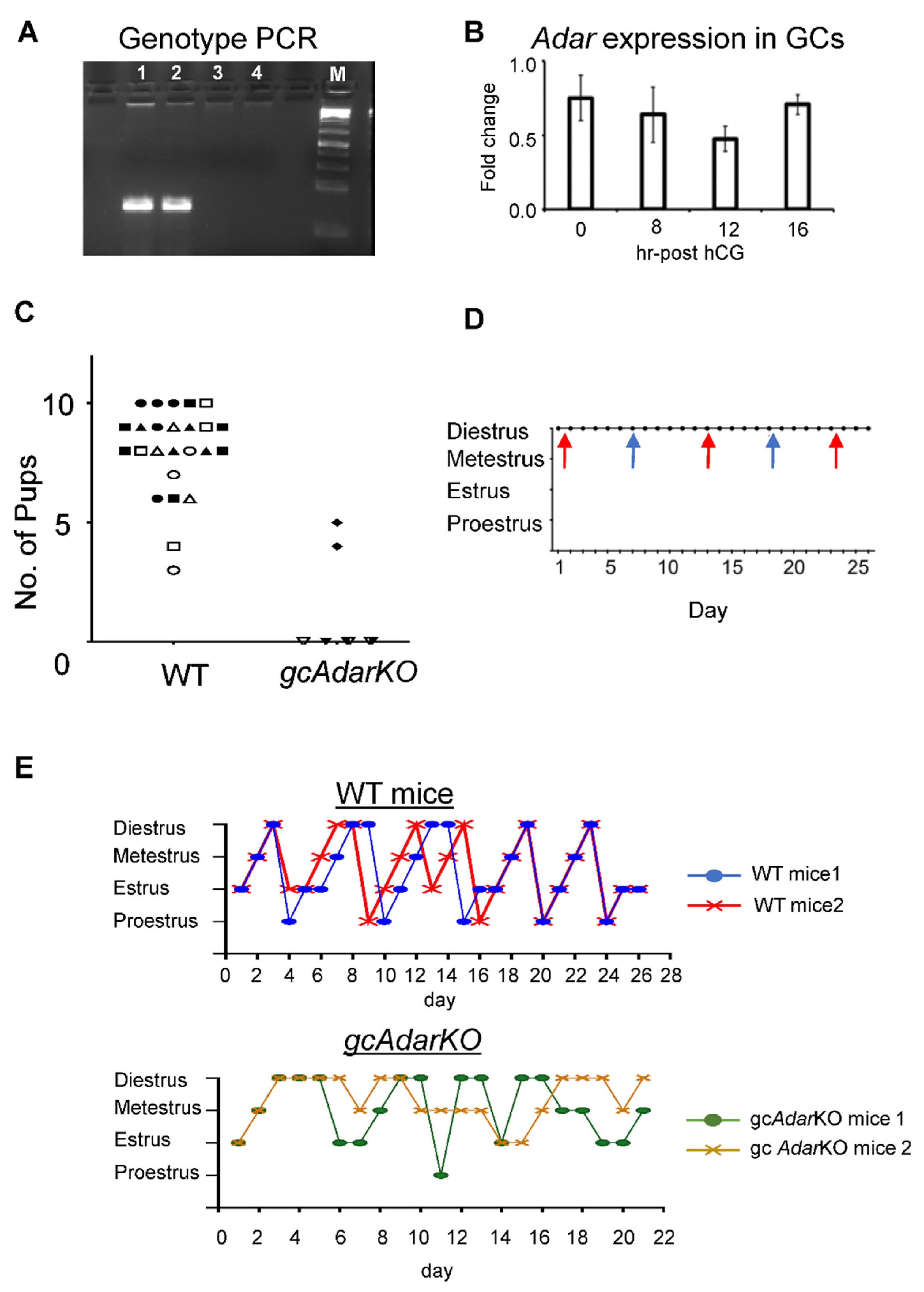
2.1. Conditional Deletion of Adar in mGC Renders Female Mice Infertile

Granulosa specific Adar KO was confirmed by genotyping using deletion specific primers in gcAdarKO and WT (littermate control) mice mGC (Figure 1A). Adar expression did not vary in mGC following hCG (Figure 1B). Control female littermates (Adar flox/flox or flox/+, lacking the Cyp19a1-Cre) all produced at least three litters each with an average of 8.0 ± 0.4 pups per litter, while 4 of 5 gcAdarKO female mice failed to produce any offspring over a 7-month breeding trial, while the single gcAdarKO female that did deliver pups, had two litters of four and five pups and no further litters (Figure 1C). At 5 months of the breeding trial, vaginal cytology and breeding was confirmed by visualization of a seminal plug or presence of sperm in vaginal smears over a 26-d period in gcAdarKO females. Plugs or sperm were found every 10–12 days (Figure 1D). Cytology indicative of pseudopregnancy was observed in each smear between breeding events in the breeding trial animals. At the end of breeding trial, female mice were euthanized, and gross reproductive tract morphology appeared normal with uterine horns appearing normal in width and length as were ovaries.

2.2. Estrous Cyclicity Disturbances in gcAdarKO Mice

Estrous cycle staging of WT (control littermates) mice progressed chronologically as expected, from proestrus, to estrus, metestrus and diestrus before repeating, while gcAdarKO exhibited erratic estrous cycle patterns, with prolonged periods of metestrus and diestrus observed. Proestrus was almost never identified (Figure 1E).

2.3. Histological Analysis of Ovaries

Ovaries collected from gcAdarKO and WT (littermate control) mice at the completion of 7 months breeding trial exhibited follicles of various sizes in central sections of ovarian tissue in both genotypes (Figure 2A). A preponderance of multinucleated yellow to brown cells were visualized in the stroma of the ovarian tissue of the gcAdarKO females, and picrosirius red staining (PSR) revealed an increase in extracellular matrix deposition, presumed to be collagen, in the gcAdarKO ovaries when compared to the breeding trial WT ovarian tissue (Figure 2B,C). In order to ensure this difference was not due to the intervening pregnancies that occurred only in the WT females, ovarian PSR staining was compared in another experimental trial using virgin aged WT and gcAdarKO females. No significant differences in extracellular matrix deposition were found across a 16-month analysis of aged WT animals or in the gcAdarKO (2–16 m) females (Supplementary Figure S1).

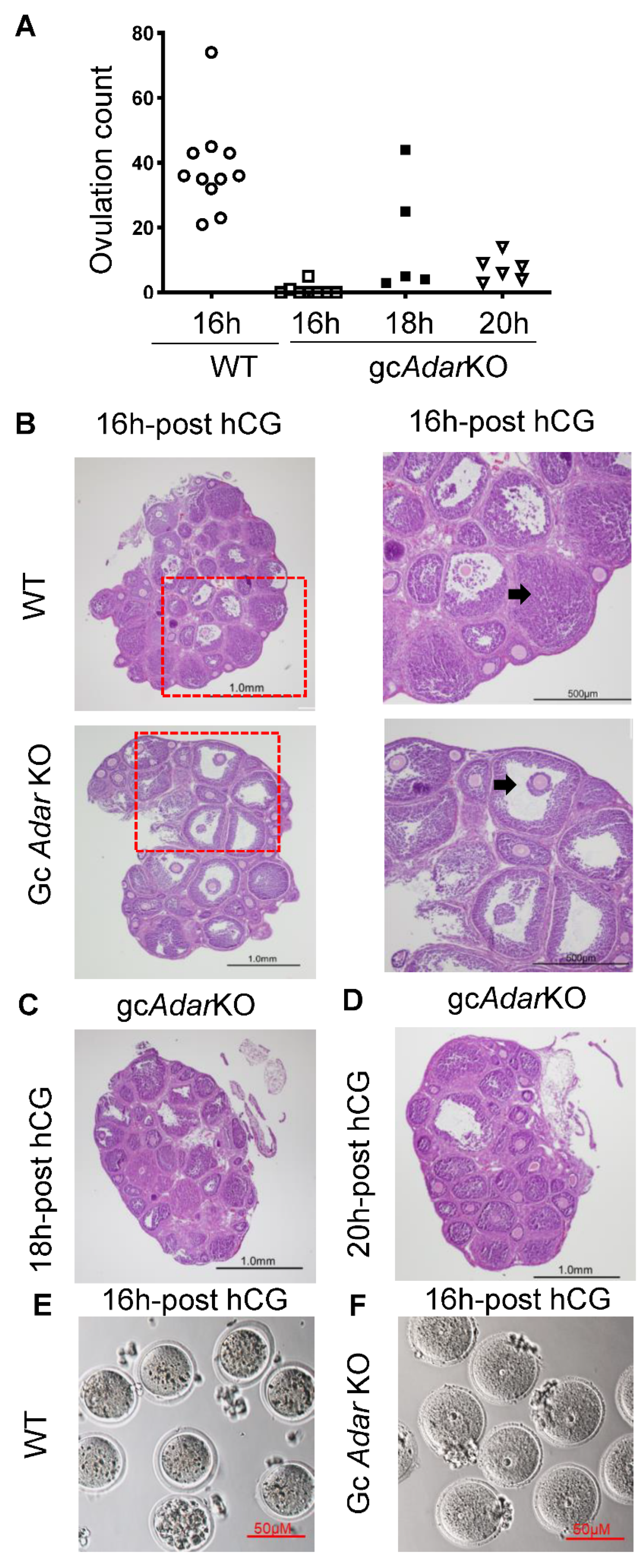
2.4. Delayed Ovulation in gcAdarKO Mice

Superovulation of immature (21-day-old) WT females ovulated more (p < 0.05; 44.0 ± 8.0 mean ± SEM; n = 11) than gcAdarKO mice (0.75 ± 0.62; n = 8) at 16 h post-hCG. However, when collection was completed at 18 h or 20 h following hCG administration, eggs were recovered (16.02 ± 8.06; n = 5 and 7.3 ± 1.6; n = 6, respectively) in gcAdarKO females (Figure 3A). Eggs from WT mice were released as one large, cloud-like mass of multiple COCs from the oviduct, while eggs from gcAdarKO mice were released as individual COCs. As expected at 16 h post-hCG, control ovaries displayed eosinophilic corpora lutea and clear evidence of follicle rupture (Figure 3B), while gcAdarKO ovaries follicles did not display the normal evidence of luteinization, and numerous trapped oocytes with densely compacted cumulus cells were observed (Figure 3B). At 16 h post-hCG, oocytes isolated from aspirated follicles of gcAdarKO mice showed intact germinal vesicle, while the ovulated oocytes from WT mice showed germinal vesicle breakdown (Figure 3E,F). At 18 and 20 h post-hCG, trapped oocytes were observed in adjacent luteinized follicles and non-luteinized follicles in gcAdarKO ovaries (Figure 3C,D).

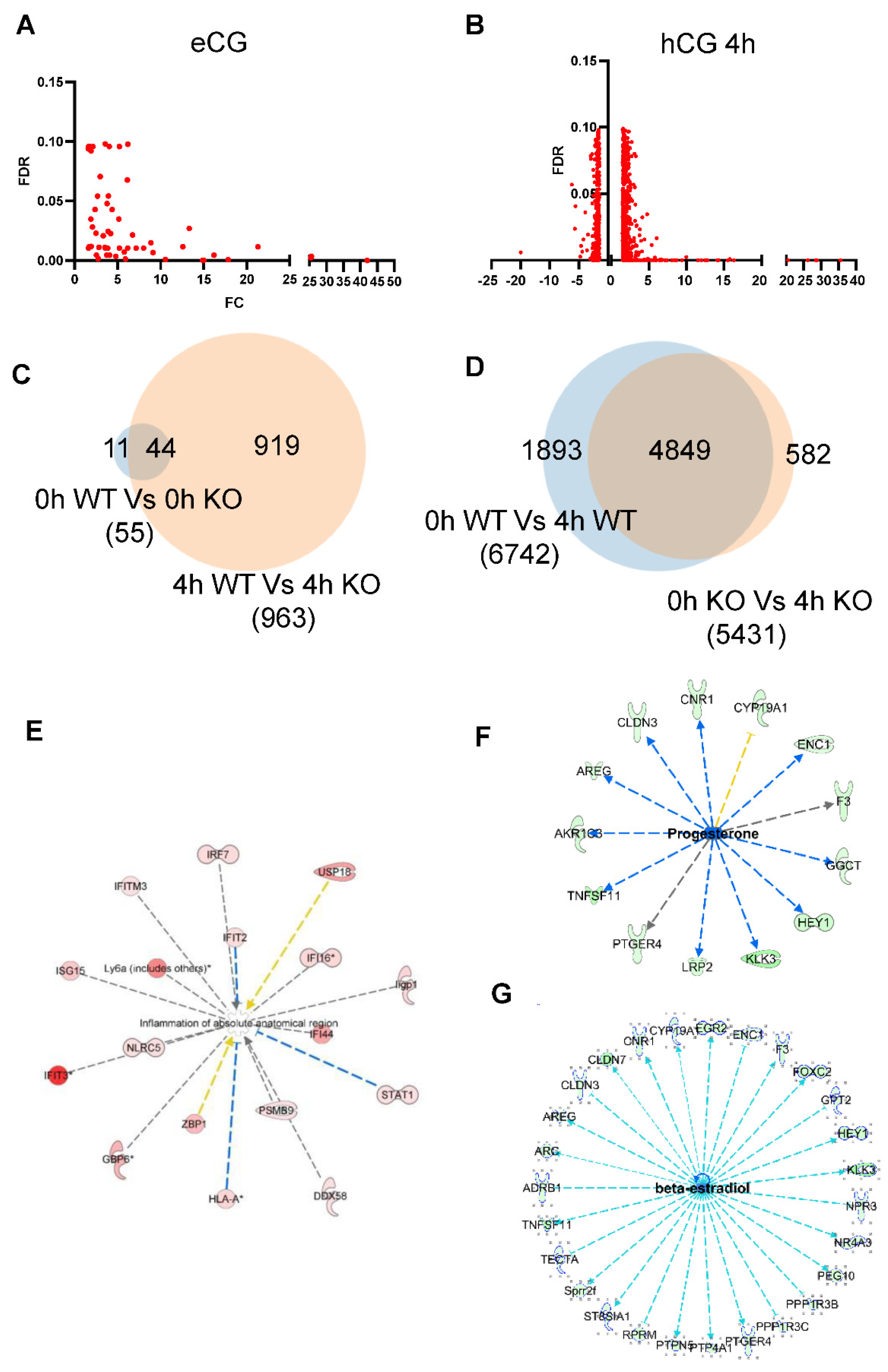
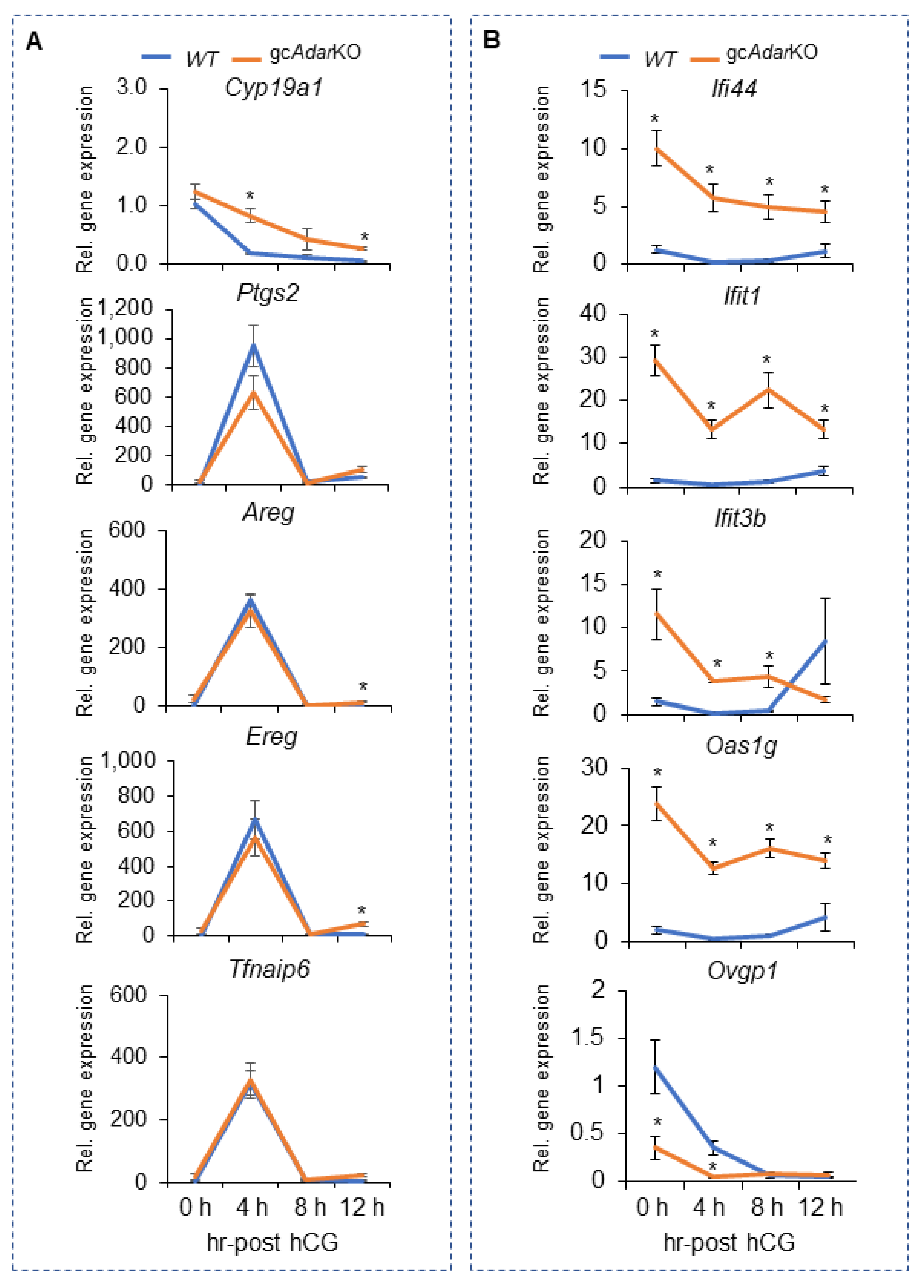
2.5. Transcriptome Analysis of mGCs from gcAdarKO Mice

Comparison of mGC gene expression at 0 h (i.e., eCG treatment only) for WT and gcAdarKO indicated a total of 55 differentially expressed genes (DEGs) with a fold change ≥ 1.5 and FDRP value < 0.1 (Figure 4A,C), all of which were upregulated. Comparison at 4 h post-hCG showed a total of 300 DEGs with fold change ≥1.5 and FDRP value < 0.1 between the two genotypes, of which 213 were upregulated and 87 downregulated (Figure 4B,C) of these only 31 DEG exhibited a fold change ≥5.
Comparison of DEGs in 0 h (WT versus gcAdarKO) and 4 h hCG (WT versus gcAdarKO) groups showed 40 commonly upregulated genes (Figure 4C). Comparison of DEGs from 0 h to 4 h hCG across the WT and gcAdarKO genotypes showed 4849 common DEGs with fold change ≥1.5 and FDRP value < 0.1 (Figure 4D) and 582 DEGs are specific for gcAdarKO. Complete lists of DEG in mGCs from WT and gcAdarKO at 0 and 4 h post-hCG treatment are presented in the Supplementary Tables S1 and S2.

2.6. Adar Regulated Pathways in mGCs

IPA analysis of the RNA seq data revealed upregulation of several genes related to the inflammatory response pathway (Figure 4E). At the same time genes downstream of progesterone and estradiol signaling were slightly downregulated (Figure 4F,G). The LH induced genes Ptgs2, Areg, Ereg, and Tfnaip6 all exhibited an almost identical pattern of expression across time for the WT and gcAdarKO, while that of Cyp19a1, which is down regulated by the LH surge showed a delay in the gcAdarKO (Figure 5A). Expression analysis of interferon induced genes Ifi44, Ifit1, Ifit3b and Oas1g were confirmed at different time periods of gonadotropin stimulation by qRT-PCR. Expression of Ifi44, Ifit1, and Oas1g genes were significantly greater in gcAdarKO compared to WT mGCs (Figure 5B) at all time points. Expression of Ifit3b was significantly greater at 0, 4 and 8 h post-hCG but decreased significantly at 12 h in gcAdarKO compared to WT mice (Figure 5B). Expression of Ovgp1 a glycoprotein involved in zona pellucida stability was also decreased significantly at 0 and 4 h post-hCG (Figure 5B).

3. Discussion

Granulosa cell specific knockdown of Adar guided by Cyp19a1-Cre recombinase activity produced female mice with severely diminished fertility. Despite observing positive breeding events, most gcAdarKO mice of the breeding trial failed to produce pups. Rhythmic estrous cycling was not observed in gcAdarKO mice, indicating a failure in ovarian estrous patterning. While ovulation could be induced in gcAdarKO mice, ovulation was significantly delayed as indicated by the failure to detect oocytes in the oviducts of superovulated mice 16 h post-hCG followed by modest recovery rates of oviductal eggs at 18 and 20 h post-hCG. There was a defect in the germinal vesicle breakdown in gcAdarKO mice ovaries as observed from the oocytes isolated by puncturing the antral follicles at 16 h post-hCG. Since there was no ovulation 16 h post-hCG in gcAdarKO mice, we by necessity collected oocytes from the gcAdarKO mice, by aspiration from the large periovulatory follicles. It is important to note that germinal vesicle breakdown occurs at 2-4 h after hCG administration in wildtype mice [28], and we did not observe this in the gcAdarKO mice even at 16 h after hCG. Luteinization of the mGC was also delayed as was cumulus expansion. Thus, it appears that gcAdarKO females are unable to initiate proper ovulation and differentiation into luteal cells, likely resulting in a dyssynchronous oviductal and uterine environment such that the oocytes that do reach the oviduct are not able to properly implant and maintain a pregnancy.
Foundational studies of established critical ovarian / ovulatory factors have observed a similar trapped oocyte phenotype, sometimes with “normal” luteinization and sometimes with disrupted differentiation of the mGC layer [21]. Indeed, loss of CEBP/β, expression reduced ovulation in response to gonadotropins [29]. The present study similarly describes a delay that does not result in absolute infertility, illustrated by the gcAdarKO dam that produced two smaller litters of pups and the recovery of a small number of eggs from the oviducts of immature superovulated gcAdarKO mice, akin to what is seen in CEBP/β knockout mice. COX-2−/− females are also infertile due to defective ovulation and fertilization [30]. The importance of timely expression of COX-2 is highlighted in mice lacking the transcription factor CEBP/β.
Ovulation itself is often explained as an inflammatory process [31]. The ovulatory surge of LH induces many inflammation mediators within the ovary including: leukocyte infiltration, edema swelling, tissue digestion and repair [21]. The ovary is a unique tissue that requires a delicate balance in inflammation to allow for ovulation and yet not so much that it impacts fertility. In contrast to hormonal control of mGC function, the extent of inflammatory cues and curtails have not been as well defined. A recent study displayed long term effects of manipulating the inflammasome within the ovary by suppression of NLRP3 resulting in an improved reproductive rate at 12 months of age [32]. Interestingly, lack of Adar induces inflammatory pathways in many tissues [7,16,33,34]. Thus, it is possible that an increase in inflammatory responses in the mural granulosa Adar knockouts could be altering the delicate balance needed, ultimately preventing normal temporal sequences essential for proper ovulation.
RNA seq analysis of GCs from gcAdarKO mice revealed upregulation of several inflammatory pathway genes including interferon induced protein with tetratricopeptide repeats 1 (IFIT1) which is reported to be a negative regulator of inflammatory genes [35]. Likewise, IFIT3b was upregulated in gcAdarKO after hCG in mGCs and this protein acts by binding to IFIT1 to extend its half-life, thereby regulating its specificity and activity [36]. It is possible that the delay in ovulation and presence of trapped oocytes at 16 h after hCG as observed in gcAdarKO could be due to blockade of the inflammatory response caused by IFIT1 and IFIT3B. It is well established that LH induces the expression of interferon-alpha (IFN-α), which is involved in the differentiation of preovulatory follicles [37]. Moreover, IFNα or IFNβ alone can induce the cumulus cell expansion and thereby ovulation [38]. IFI44 similar to IFIT1 and IFIT3B is another negative regulator of IFNα and β response and it was also upregulated [39]. Thus, upregulation of three negative regulators of the interferon pathway, prior to the LH surge, are thus likely blocking the positive LH induced IFNα and β effects on cumulus expansion, differentiation and ultimately ovulation.
Loss of Adar in other tissues has been shown to induce fibrosis in addition to inflammation [17]. Our initial analyses of the mating trial animals suggested that loss of ADAR1 might also be increasing ovarian fibrosis as collagen content was increased in the gcAdarKO mice. However, in a subsequent matched study of virgin WT and gcAdarKO mice from 2 to 16 months of age indicated that this change was likely due to a reduction in ovarian collagen content in the pregnant WT mice that experienced consecutive and almost continuous pregnancy compared to the infertile gcAdarKO mice. Similarly, we likewise did not observe a marked change in collagen deposition (i.e., PSR staining) between 2-16 months of age in the WT mice similar to prior studies [40], where the first evidence was observed at 22 months of age. It remains an interesting phenomena that pregnancy impacts ovarian collagen deposition, as well as the fact that Adar loss did not impact ovarian stromal fibrosis and for this reason we thought it was worth presenting the results of these two experiments. It is also worth mentioning that one aspect that differs between other tissues where ADAR1 is depleted, and our gcAdarKO is the ephemeral nature of the follicle and the subsequent corpus luteum. At the conclusion of the described breeding trial, presence of eosinophil structures, increased macrophage presence was also observed in the gcAdarKO, interestingly increased eosinophil abundance was reported to play a regulatory role in corpus luteum formation [41], and it is noted that luteal structures were not widely observed in the gcAdarKO mice.
In addition to the inflammatory genes altered in the gcAdarKO mice, several other gene families showed marked changes. The oligoadenylate synthetase like protein family of genes consists of three classes in humans. They are OAS1, OAS2, OAS3 [42]. In mice eight subtypes of genes, i.e., Oas1a, Oas1b, Oas1c, Oas1d, Oas1e, Oas1f, Oas1g, and Oas1h are found to be homologues to OAS1 in humans [43]. Among these eight subtypes OAS1A and OAS1G alone have the ability to bind dsRNA and convert ATP into 2’, 5’ linked oligomers of adenosine (2-5A) [44,45]. OAS1D was reported to interact with OAS1A and inhibit OAS1A activity and block IFN/OAS/RNAsL-mediated RNA degradation in oocytes. Oas1d null mice show reduced fertility due to the lack of this protective effect [46]. We observed Adar plays a similar role and regulates the expression of Oas1g. Thus, lack of Adar caused higher expression of Oas1g, which in turn could mediate RNA degradation in oocytes and subsequently reduce fertility. A significantly down regulated gene following loss of Adar in granulosa cells, oviduct specific glycoprotein 1 (OVGP1) binds to the zona pellucida and helps the zona pellucida in acquiring resistance to proteolytic digestion during oviductal transit [47,48,49]. Its reduced expression by granulosa cells prior to ovulation could compromise the oocyte and oocyte-cumulus interaction and ultimately impact events such as cumulus expansion and oocyte maturation, which are vital for ovulation.

4. Materials and Methods

4.1. Animals and Generation of Conditional Knockout

All procedures involving animals were reviewed and approved by the Institutional Animal Care and Use Committee at the University of Kansas Medical Center and were performed in accordance with the Guiding Principles for the Care and Use of Laboratory Animals. Experiments were performed on conditional knockout female mice, Adar flox/flox Cyp19a1-Cre/+ (gcAdarKO) and littermate controls (i.e., Adarflox/flox or flox/+ lacking the Cyp19a1-Cre transgene hereafter collectively referred to as wild-type (WT)) generated by breeding homozygous Adar flox/flox female mice with heterozygous Adar flox/+ Cyp19a1-Cre/+ males. Adar flox/flox mice were obtained from Dr. Stuart H Orkin’s lab [50] and the Cyp19a1-Cre mice from Dr. Jan A. Gossen [51]. Mice were maintained in a humidity and temperature-controlled environment with a 14 h light, 10 h dark cycle (7 am to 9 pm) with ad libitum access to food and water.

4.2. Assessment of Fertility

Fertility was assessed by pairing female gcAdarKO mice (42 days of age, n = 5) as well as control littermates (WT) lacking Cyp19a1-Cre (n=6) with adult (7–8-week-old) wild-type C57BL/6J males of known fertility. All females were continually exposed to males for 7 months. Female mice were euthanized at the conclusion of the breeding trial and ovaries were dissected away from the uterus proximal to the uterotubal junction, rinsed in sterile PBS and placed in cold 4% paraformaldehyde (PFA).
Estrous cyclicity was examined by vaginal cytology in a group of sexually mature gcAdarKO and WT 42-day old female mice in the absence of males for at least 21 days. Additionally, at ~5 months of the breeding trial, the vaginal cytology of the gcAdarKO females in the presence of fertile males was examined for 26 days. Vaginal cytology for control females in the breeding trial was not conducted due to continuous pregnancy. All cycle staging was performed at 8:00-10:00 a.m. and approximately 200 µl of sterile saline was gently flushed into and aspirated from vaginal opening using a glass dropper. The aspirated lavage was then placed on glass slides and allowed to dry at room temperature. Smears were stained with 1% crystal violet in water (w/v) for 2 minutes, followed by gentle rinsing with water. Samples were mounted with 15 µl of glycerol and coverslip for microscopic visualization on a Nikon YS2-T microscope at 10× magnification. Presence and relative abundance of leukocytes, nucleated epithelial cells, and cornified squamous epithelial cells were used to categorize mice into proestrus, estrus, metestrus, and diestrus [52]. Presence of sperm or visualization of a seminal plug preventing lavage in breeding trial females was recorded as a positive mating.

4.3. Induction of Superovulation by Gonadotropins

Superovulation was induced in 21-day old mice via an intraperitoneal (i.p.) injection of 5 IU of equine chorionic gonadotropin (eCG; Lee Biosolutions, Maryland Heights, MO, USA) followed by 5 IU of human chorionic gonadotropin (hCG) 46–48 h later with relevant lengths of hCG (Millipore Sigma, St. Louis, MO, USA) exposure described for each experiment. To determine ovulation rates, mice were euthanized at 16, 18, and 20 h post-hCG administration and ovaries and oviducts were collected by dissection distal to the uterotubal junction to ensure complete removal of the oviduct without disruption. Typically, ovulation in mice occurs between 12–15 h post-hCG, thus the standard is to collect at 16 h post-hCG for assessment of ovulation rate [53]. In this study an additional 18 and 20 h post-hCG collection were included to determine if ADAR1 was temporally affecting the ovulatory process. The oviduct and bursal encapsulated ovary were washed in dPBS before separation in warmed FHM media (Millipore Sigma) supplemented with bovine serum albumin (BSA; 4 mg/ml, Millipore Sigma) in 35 mm dishes. Cumulus oocyte complexes (COCs) were expressed from oviducts and transferred to 25 µl FHM media droplets containing 5 µL of hyaluronidase (Millipore Sigma) at 10 mg/mL for ten minutes to remove cumulus cells. Freed oocytes were then counted, morphologically evaluated and then discarded.

4.4. Histology

Ovaries were prepared for histological analysis by immediately placing them into cold 4% paraformaldehyde fixative. Fixation occurred overnight (~16 h) at 4 °C before ovaries were transferred to 70% ethanol prior to dehydration and paraffin embedding. Tissues were sectioned (Leica RM2035) at 7 µm thickness, placed on ProbeOn Plus slides in preparation for hematoxylin and eosin or picrosirius red (PSR) staining for visualization of collagen deposition [40]. PSR intensity was quantified using FIJI/ImageJ (NIH).
PSR staining of aged (2, 6, 9, 12 and 16 m old) virgin gcAdarKO (n=5, 6, 5, 5, 3) and WT (n=3, 4, 3, 3, 3) ovary sections were imaged at 20x using brightfield microscopy (Nikon 80i). A sampling of three random regions of the ovaries were imaged and uploaded to the ImageJ software. Non-tissue space was cut out using the selection tool. Scale calibration, image gray scaling, and measurement of positive staining were done following NIH ImageJ PSR staining analysis protocol [54,55]. Threshold for staining intensity was maintained across all samples.

4.5. Isolation of Mural Granulosa Cell RNA

Immature (21 d old) and mature (6–8-week-old) WT and gcAdarKO mice were superovulated as described above, at specific time points post-hCG administration mice were euthanized, and the ovaries were dissected free of the ovarian bursa and then rinsed in sterile dPBS to remove blood and tissue debris. Ovaries were then placed in dishes of FHM with 4 mg/ml BSA. Large antral follicles visualized under a Nikon SMZ1000 with a Nikon NI-150 high intensity illuminator were individually punctured with insulin syringes (28 gauge) to allow expulsion of mGCs and cumulus oocyte complexes. Smaller follicles were left with the remaining ovarian tissue which was removed from the dish before naked oocytes and cumulus oocyte complexes were removed and discarded. Mural GCs and the surrounding media was then collected, placed into a 1.5 ml Eppendorf tube and gently pelletized at 800× g, excess media was aspirated using a pipette, and 500 µL of TRI Reagent (Invitrogen, Waltham, MA) was applied to the cell pellet. Solubilized cell pellets were frozen at −80 °C overnight followed by completion of the manufacturer’s RNA extraction protocol (Invitrogen).

4.6. Quantitative RT-PCR Analysis and RNA Sequencing

Extracted total RNA was subjected to RNA quality analysis using an Agilent 2100 Bioanalyzer (Santa Clara, CA, USA) and samples with RIN values over 9 were selected for RNA sequencing, samples with RIN values above 8 were used for qRT-PCR analyses. RNA seq was performed on WT and gcAdarKO mGCs collected after eCG only injection (0 h) and 4 h post-hCG (n = 3 for each sample), while qRT-PCR was evaluated over a broader range of time points (0, 4, 8, and 12 h post-hCG; n = 6 for each sample). cDNA library preparation and sequencing were performed by KUMC Genomic Core. RNA sequencing was performed on Illumina NovaSeq 6000 sequencing machine (Illumina, San Diego, CA, USA). Sequencing generated between 28 to 32 million reads per samples. The read quality was assessed using the fastQC software. Sequenced reads were mapped to mouse genome (GRCm38 re197) using STAT software. Expression normalization and differential gene expression (DEG) were calculated using the DESeq2 (version 1.26.0). All RNA seq files are available at Sequence Read Archive portal (Bio Project: PRJNA794072 and SRR17423715-SRR17423728). Superscript II reverse transcription was used for the cDNA preparation, followed by qRT-PCR on a Applied Biosystems-Quant Studio 7 Flex using Power SYBR green master mix. Primers used in the present study are listed in Table 1. To normalize gene expression Rn18S was used in the comparative 2-∆∆CT method.

4.7. Pathway Analyses

Using the Meta-chart software, two Venn diagrams were generated, (1) by overlapping DEGs of 0 h WT and gcAdarKO and 4 h WT and gcAdarKO granulosa cell RNA seq results, (2) by overlapping DEGs of 0 and 4 h WT with 0 and 4 h gcAdarKO mGCs results. The resultant DEG from these groups were subjected to Ingenuity Pathway Analyses to determine the functional pathways.

4.8. Statistics

Statistical analysis was performed using GraphPad Prism (Version 9 GraphPad Software Inc. San Diego, CA, USA). T-tests were performed to determine significance of PSR staining intensity (breeding trial animals) and litter size between WT and gcAdarKO groups. ANOVA was used to determine the significance of the number of eggs retrieved following superovulation and for the PSR staining time course from aged females. Quantitative RT-PCR results were analyzed by ANOVA followed by Duncan’s post-hoc mean separation tests. p value < 0.05 was considered significant.

5. Conclusions

In conclusion granulosa specific Adar knockout mice have relatively normal folliculogenesis but failed to respond appropriately to the ovulatory surge of LH/hCG resulting abnormal estrous cyclicity, failure to ovulate at the normal time (i.e., prior to 16 h post-hCG) and infertility. RNA seq analysis revealed upregulation of a subset of genes which includes Ifit1, Ifit3b, Ifi44, Oas1g, which are involved in the interferon and inflammatory pathways, consistent with ADAR1′s role in preventing dsRNA mediated interferon activation. The lack of Adar, thus lead to an altered inflammatory and interferon response within the granulosa cells, delaying ovulation and oocyte maturation.

Supplementary Materials

The following supporting information can be downloaded at: https://www.mdpi.com/article/10.3390/ijms232214001/s1.

Author Contributions

R.N.N. and L.K.C. designed the research; R.N.N., V.P.C., A.R., X.H. and J.A.G. performed the research; R.N.N., V.P.C. and L.K.C. analyzed the data; R.N.N., V.P.C. and L.K.C. wrote the manuscript. All authors have read and agreed to the published version of the manuscript.

Funding

This work was supported by the National Institutes of Health (HD094545 to L.K.C). R.N.N was supported by Kathleen M. Osborn fellowship in Reproductive Physiology.

Institutional Review Board Statement

The animal study protocol was approved by the University of Kansas Medical Center Animal Care and Use committee (IACUC 2020-2550).

Informed Consent Statement

Not applicable.

Data Availability Statement

All RNA seq files are available at Sequence Read Archive portal https://www.ncbi.nlm.nih.gov/sra (Bio Project: PRJNA794072 and SRR17423715-SRR17423728).

Acknowledgments

The authors acknowledge the University of Kansas Medical Center Internal Support and Genomics Core and their funding: Kansas Intellectual and Developmental Disabilities Research Center (NIH U54 HD090216).

Conflicts of Interest

The authors declare no conflict of interest.

References

  1. Russell, D.L.; Robker, R.L. Molecular mechanisms of ovulation: Co-ordination through the cumulus complex. Hum. Reprod. Update 2007, 13, 289–312. [Google Scholar] [CrossRef] [PubMed]
  2. Liu, D.T.; Brewer, M.S.; Chen, S.; Hong, W.; Zhu, Y. Transcriptomic signatures for ovulation in vertebrates. Gen. Comp. Endocrinol. 2017, 247, 74–86. [Google Scholar] [CrossRef] [PubMed]
  3. Wu, Y.; Li, M.; Yang, M. Post-Translational Modifications in Oocyte Maturation and Embryo Development. Front. Cell Dev. Biol. 2021, 9, 645318. [Google Scholar] [CrossRef] [PubMed]
  4. Harcourt, E.M.; Kietrys, A.M.; Kool, E.T. Chemical and structural effects of base modifications in messenger RNA. Nature 2017, 541, 339–346. [Google Scholar] [CrossRef] [PubMed] [Green Version]
  5. Bass, B.L. RNA editing by adenosine deaminases that act on RNA. Annu. Rev. Biochem. 2002, 71, 817–846. [Google Scholar] [CrossRef] [PubMed] [Green Version]
  6. Chen, C.X.; Cho, D.S.; Wang, Q.; Lai, F.; Carter, K.C.; Nishikura, K. A third member of the RNA-specific adenosine deaminase gene family, ADAR3, contains both single- and double-stranded RNA binding domains. RNA 2000, 6, 755–767. [Google Scholar] [CrossRef]
  7. Pestal, K.; Funk, C.C.; Snyder, J.M.; Price, N.D.; Treuting, P.M.; Stetson, D.B. Isoforms of RNA-Editing Enzyme ADAR1 Independently Control Nucleic Acid Sensor MDA5-Driven Autoimmunity and Multi-organ Development. Immunity 2015, 43, 933–944. [Google Scholar] [CrossRef] [Green Version]
  8. Tomaselli, S.; Bonamassa, B.; Alisi, A.; Nobili, V.; Locatelli, F.; Gallo, A. ADAR enzyme and miRNA story: A nucleotide that can make the difference. Int. J. Mol. Sci. 2013, 14, 22796–22816. [Google Scholar] [CrossRef] [Green Version]
  9. Nishikura, K. A-to-I editing of coding and non-coding RNAs by ADARs. Nat. Rev. Mol. Cell Biol. 2016, 17, 83–96. [Google Scholar] [CrossRef] [Green Version]
  10. Sakurai, M.; Shiromoto, Y.; Ota, H.; Song, C.; Kossenkov, A.V.; Wickramasinghe, J.; Showe, L.C.; Skordalakes, E.; Tang, H.-Y.; Speicher, D.W. ADAR1 controls apoptosis of stressed cells by inhibiting Staufen1-mediated mRNA decay. Nat. Struct. Mol. Biol. 2017, 24, 534–543. [Google Scholar] [CrossRef]
  11. Eisenberg, E.; Levanon, E.Y. A-to-I RNA editing—Immune protector and transcriptome diversifier. Nat. Rev. Genet. 2018, 19, 473–490. [Google Scholar] [CrossRef] [PubMed]
  12. Ramaswami, G.; Zhang, R.; Piskol, R.; Keegan, L.P.; Deng, P.; O’connell, M.A.; Li, J.B. Identifying RNA editing sites using RNA sequencing data alone. Nat. Methods 2013, 10, 128–132. [Google Scholar] [CrossRef] [PubMed] [Green Version]
  13. Brachova, P.; Alvarez, N.S.; Hong, X.; Gunewardena, S.; Vincent, K.A.; Latham, K.E.; Christenson, L.K. Inosine RNA modifications are enriched at the codon wobble position in mouse oocytes and eggs. Biol. Reprod. 2019, 101, 938–949. [Google Scholar] [CrossRef] [PubMed]
  14. Picardi, E.; Manzari, C.; Mastropasqua, F.; Aiello, I.; D’Erchia, A.M.; Pesole, G. Profiling RNA editing in human tissues: Towards the inosinome Atlas. Sci. Rep. 2015, 5, 14941. [Google Scholar] [CrossRef] [Green Version]
  15. O’Connell, M. RNA modification and the epitranscriptome; the next frontier. RNA 2015, 21, 703–704. [Google Scholar] [CrossRef] [Green Version]
  16. Hartner, J.C.; Schmittwolf, C.; Kispert, A.; Müller, A.M.; Higuchi, M.; Seeburg, P.H. Liver disintegration in the mouse embryo caused by deficiency in the RNA-editing enzyme ADAR1. J. Biol. Chem. 2004, 279, 4894–4902. [Google Scholar] [CrossRef] [Green Version]
  17. Ben-Shoshan, S.O.; Kagan, P.; Sultan, M.; Barabash, Z.; Dor, C.; Jacob-Hirsch, J.; Harmelin, A.; Pappo, O.; Marcu-Malina, V.; Ben-Ari, Z.; et al. ADAR1 deletion induces NFκB and interferon signaling dependent liver inflammation and fibrosis. RNA Biol. 2017, 14, 587–602. [Google Scholar] [CrossRef] [Green Version]
  18. El Azzouzi, H.; Vilaça, A.P.; Feyen, D.A.M.; Gommans, W.M.; de Weger, R.A.; Doevendans, P.A.F.; Sluijter, J.P.G. Cardiomyocyte Specific Deletion of ADAR1 Causes Severe Cardiac Dysfunction and Increased Lethality. Front. Cardiovasc. Med. 2020, 7, 30. [Google Scholar] [CrossRef] [Green Version]
  19. Wang, Q.; Miyakoda, M.; Yang, W.; Khillan, J.; Stachura, D.L.; Weiss, M.J.; Nishikura, K. Stress-induced apoptosis associated with null mutation of ADAR1 RNA editing deaminase gene. J. Biol. Chem. 2004, 279, 4952–4961. [Google Scholar] [CrossRef] [Green Version]
  20. Richards, J.S.; Russell, D.L.; Ochsner, S.; Espey, L.L. Ovulation: New dimensions and new regulators of the inflammatory-like response. Annu. Rev. Physiol. 2002, 64, 69–92. [Google Scholar] [CrossRef]
  21. Duffy, D.M.; Ko, C.; Jo, M.; Brannstrom, M.; Curry, T.E. Ovulation: Parallels With Inflammatory Processes. Endocr. Rev. 2019, 40, 369–416. [Google Scholar] [CrossRef] [PubMed] [Green Version]
  22. Zhang, Y.; Zhang, L.; Yue, J.; Wei, X.; Wang, L.; Liu, X.; Gao, H.; Hou, X.; Zhao, F.; Yan, H.; et al. Genome-wide identification of RNA editing in seven porcine tissues by matched DNA and RNA high-throughput sequencing. J. Anim. Sci. Biotechnol. 2019, 10, 24. [Google Scholar] [CrossRef] [PubMed] [Green Version]
  23. Picardi, E.; D’Erchia, A.M.; Lo Giudice, C.; Pesole, G. REDIportal: A comprehensive database of A-to-I RNA editing events in humans. Nucleic Acids Res. 2017, 45, D750–D757. [Google Scholar] [CrossRef] [PubMed] [Green Version]
  24. Buchumenski, I.; Holler, K.; Appelbaum, L.; Eisenberg, E.; Junker, J.P.; Levanon, E.Y. Systematic identification of A-to-I RNA editing in zebrafish development and adult organs. Nucleic Acids Res. 2021, 49, 4325–4337. [Google Scholar] [CrossRef] [PubMed]
  25. Wang, L.Y.; Guo, J.; Cao, W.; Zhang, M.; He, J.; Li, Z. Integrated sequencing of exome and mRNA of large-sized single cells. Sci. Rep. 2018, 8, 1–11. [Google Scholar] [CrossRef] [PubMed] [Green Version]
  26. Qiu, S.; Li, W.; Xiong, H.; Liu, D.; Bai, Y.; Wu, K.; Zhang, X.; Yang, H.; Ma, K.; Hou, Y. Single-cell RNA sequencing reveals dynamic changes in A-to-I RNA editome during early human embryogenesis. BMC Genom. 2016, 17, 1–11. [Google Scholar] [CrossRef] [PubMed] [Green Version]
  27. Brachova, P.; Alvarez, N.S.; Christenson, L.K. Loss of Cnot6l Impairs Inosine RNA Modifications in Mouse Oocytes. Int. J. Mol. Sci. 2021, 22, 1191. [Google Scholar] [CrossRef]
  28. Edwards, R.G.; Gates, A.H. Timing of the stages of the maturation divisions, ovulation, fertilization and the first cleavage of eggs of adult mice treated with gonadotrophins. J. Endocrinol. 1959, 18, 292–304. [Google Scholar] [CrossRef]
  29. Sterneck, E.; Tessarollo, L.; Johnson, P.F. An essential role for C/EBPbeta in female reproduction. Genes Dev. 1997, 11, 2153–2162. [Google Scholar] [CrossRef]
  30. Lim, H.; Paria, B.C.; Das, S.K.; Dinchuk, J.E.; Langenbach, R.; Trzaskos, J.M.; Dey, S.K. Multiple female reproductive failures in cyclooxygenase 2-deficient mice. Cell 1997, 91, 197–208. [Google Scholar] [CrossRef]
  31. Espey, L.L. Ovulation as an inflammatory reaction—A hypothesis. Biol. Reprod. 1980, 22, 73–106. [Google Scholar] [CrossRef] [PubMed] [Green Version]
  32. Navarro-Pando, J.M.; Alcocer-Gómez, E.; Castejón-Vega, B.; Navarro-Villarán, E.; Condés-Hervás, M.; Mundi-Roldan, M.; Muntané, J.; Pérez-Pulido, A.J.; Bullon, P.; Wang, C.; et al. Inhibition of the NLRP3 inflammasome prevents ovarian aging. Sci. Adv. 2021, 7, eabc7409. [Google Scholar] [CrossRef] [PubMed]
  33. Liddicoat, B.J.; Chalk, A.M.; Walkley, C.R. ADAR1, inosine and the immune sensing system: Distinguishing self from non-self. Wiley Interdiscip. Rev. RNA 2016, 7, 157–172. [Google Scholar] [CrossRef] [PubMed]
  34. Bajad, P.; Ebner, F.; Amman, F.; Szabó, B.; Kapoor, U.; Manjali, G.; Hildebrandt, A.; Janisiw, M.P.; Jantsch, M.F. An internal deletion of ADAR rescued by MAVS deficiency leads to a minute phenotype. Nucleic Acids Res. 2020, 48, 3286–3303. [Google Scholar] [CrossRef]
  35. John, S.P.; Sun, J.; Carlson, R.J.; Cao, B.; Bradfield, C.J.; Song, J.; Smelkinson, M.; Fraser, I.D.C. IFIT1 Exerts Opposing Regulatory Effects on the Inflammatory and Interferon Gene Programs in LPS-Activated Human Macrophages. Cell Rep. 2018, 25, 95–106.e106. [Google Scholar] [CrossRef] [Green Version]
  36. Johnson, B.; VanBlargan, L.A.; Xu, W.; White, J.P.; Shan, C.; Shi, P.Y.; Zhang, R.; Adhikari, J.; Gross, M.L.; Leung, D.W.; et al. Human IFIT3 Modulates IFIT1 RNA Binding Specificity and Protein Stability. Immunity 2018, 48, 487–499.e485. [Google Scholar] [CrossRef] [Green Version]
  37. Lee, D.L.; Kim, S.H.; Kim, E.; Chun, S.Y.; Kim, T.S. Interferon-alpha is involved in the luteinizing hormone-induced differentiation of rat preovulatory granulosa cells. J. Interferon Cytokine Res. 2009, 29, 801–808. [Google Scholar] [CrossRef]
  38. Jang, Y.J.; Park, J.I.; Moon, W.J.; Dam, P.T.; Cho, M.K.; Chun, S.Y. Cumulus cell-expressed type I interferons induce cumulus expansion in mice. Biol. Reprod. 2015, 92, 20. [Google Scholar] [CrossRef] [Green Version]
  39. DeDiego, M.L.; Nogales, A.; Martinez-Sobrido, L.; Topham, D.J. Interferon-Induced Protein 44 Interacts with Cellular FK506-Binding Protein 5, Negatively Regulates Host Antiviral Responses, and Supports Virus Replication. mBio 2019, 10, e01839-19. [Google Scholar] [CrossRef] [Green Version]
  40. Briley, S.M.; Jasti, S.; McCracken, J.M.; Hornick, J.E.; Fegley, B.; Pritchard, M.T.; Duncan, F.E. Reproductive age-associated fibrosis in the stroma of the mammalian ovary. Reproduction 2016, 152, 245–260. [Google Scholar] [CrossRef]
  41. Aust, G.; Simchen, C.; Heider, U.; Hmeidan, F.A.; Blumenauer, V.; Spanel-Borowski, K. Eosinophils in the human corpus luteum: The role of RANTES and eotaxin in eosinophil attraction into periovulatory structures. Mol. Hum. Reprod. 2000, 6, 1085–1091. [Google Scholar] [CrossRef] [PubMed] [Green Version]
  42. Hovnanian, A.; Rebouillat, D.; Levy, E.R.; Mattei, M.G.; Hovanessian, A.G. The human 2’,5’-oligoadenylate synthetase-like gene (OASL) encoding the interferon-induced 56-kDa protein maps to chromosome 12q24.2 in the proximity of the 2’,5’-OAS locus. Genomics 1999, 56, 362–363. [Google Scholar] [CrossRef] [PubMed]
  43. Hovnanian, A.; Rebouillat, D.; Mattei, M.G.; Levy, E.R.; Marié, I.; Monaco, A.P.; Hovanessian, A.G. The human 2’,5’-oligoadenylate synthetase locus is composed of three distinct genes clustered on chromosome 12q24.2 encoding the 100-, 69-, and 40-kDa forms. Genomics 1998, 52, 267–277. [Google Scholar] [CrossRef] [PubMed]
  44. Kakuta, S.; Shibata, S.; Iwakura, Y. Genomic structure of the mouse 2’,5’-oligoadenylate synthetase gene family. J. Interferon Cytokine Res. 2002, 22, 981–993. [Google Scholar] [CrossRef]
  45. Mashimo, T.; Glaser, P.; Lucas, M.; Simon-Chazottes, D.; Ceccaldi, P.E.; Montagutelli, X.; Desprès, P.; Guénet, J.-L. Structural and functional genomics and evolutionary relationships in the cluster of genes encoding murine 2′,5′-oligoadenylate synthetases. Genomics 2003, 82, 537–552. [Google Scholar] [CrossRef]
  46. Yan, W.; Ma, L.; Stein, P.; Pangas, S.A.; Burns, K.H.; Bai, Y.; Schultz, R.M.; Matzuk, M.M. Mice deficient in oocyte-specific oligoadenylate synthetase-like protein OAS1D display reduced fertility. Mol. Cell Biol. 2005, 25, 4615–4624. [Google Scholar] [CrossRef] [Green Version]
  47. Hunter, R.H. Have the Fallopian tubes a vital rôle in promoting fertility? Acta Obstet. Gynecol. Scand. 1998, 77, 475–486. [Google Scholar]
  48. Algarra, B.; Han, L.; Soriano-Úbeda, C.; Avilés, M.; Coy, P.; Jovine, L.; Jiménez-Movilla, M. The C-terminal region of OVGP1 remodels the zona pellucida and modifies fertility parameters. Sci. Rep. 2016, 6, 32556. [Google Scholar] [CrossRef] [Green Version]
  49. Choudhary, S.; Kumaresan, A.; Kumar, M.; Chhillar, S.; Malik, H.; Kumar, S.; Kaushik, J.K.; Datta, T.K.; Mohanty, A.K. Effect of recombinant and native buffalo OVGP1 on sperm functions and in vitro embryo development: A comparative study. J. Anim. Sci. Biotechnol. 2017, 8, 69. [Google Scholar] [CrossRef]
  50. Hartner, J.C.; Walkley, C.R.; Lu, J.; Orkin, S.H. ADAR1 is essential for the maintenance of hematopoiesis and suppression of interferon signaling. Nat. Immunol. 2009, 10, 109–115. [Google Scholar] [CrossRef] [Green Version]
  51. Fan, H.-Y.; Shimada, M.; Liu, Z.; Cahill, N.; Noma, N.; Wu, Y.; Gossen, J.; Richards, J.S. Selective expression of KrasG12D in granulosa cells of the mouse ovary causes defects in follicle development and ovulation. Development 2008, 135, 2127–2137. [Google Scholar] [CrossRef] [PubMed]
  52. McLean, A.C.; Valenzuela, N.; Fai, S.; Bennett, S.A. Performing vaginal lavage, crystal violet staining, and vaginal cytological evaluation for mouse estrous cycle staging identification. J. Vis. Exp. 2012, 67, e4389. [Google Scholar] [CrossRef] [PubMed] [Green Version]
  53. Masters, W.G., 3rd; Wheeler, M.B. Timing of induced ovulation in C.B-17/Icr-scid/scid and B6SJLF1 mice. Lab. Anim. Sci. 1996, 46, 663–666. [Google Scholar] [PubMed]
  54. Puchtler, H.; Waldrop, F.S.; Valentine, L.S. Polarization microscopic studies of connective tissue stained with picro-sirius red FBA. Beitr. Pathol. 1973, 150, 174–187. [Google Scholar] [CrossRef]
  55. Junqueira, L.C.; Bignolas, G.; Brentani, R.R. Picrosirius staining plus polarization microscopy, a specific method for collagen detection in tissue sections. Histochem. J. 1979, 11, 447–455. [Google Scholar] [CrossRef] [PubMed]
Figure 1. Granulosa specific Adar knockout leads to infertility (A) Genotyping PCR was performed using Adar deletion specific primers in granulosa cells from control (lanes 1 and 2) and gcAdarKO mice (lanes 3 and 4). ‘M’ represents DNA ladder. (B) Adar expression in ovarian granulosa cells at different time points post-hCG was determined using RT-qPCR. The expression was normalized with 18S rRNA. (C) Pups per litter from wild-type littermate control mice (n = 6) and gcAdarKO (n = 5) were counted over a 7-month period. Individual mice are indicated by unique icons. Each litter is plotted by the number of pups counted or placed at zero to indicate no litters were ever observed. (D) Estrous stage for representative gcAdarKO breeding trial mice as determined by vaginal cytology, presence of seminal plugs is indicated by arrows, (different colors represent individual animals, n = 2 shown of the 5). (E) Estrous cycle stages for representative wild-type and gcAdarKO females analyzed by vaginal smears collected from 42-day old mice for at least 21 days. Smears were stained with crystal violet staining and staged based upon cytology. Representative graphs for 2 WT and 2 gcAdarKO are shown. Wild-type mice displayed cyclical estrous patterns, while gcAdarKO had disrupted cycling with prolonged periods of diestrus and metestrus.
Ijms 23 14001 g001
Figure 2. Histological analyses. (A) Histology analyses was performed in the ovaries from wild-type (WT) littermate control and gcAdarKO (n = 5) breeding trial mice (n = 6). Sections from the middle region of tissue were selected for H&E staining. Control ovaries display corpora lutea (CL) and follicles of varying size. Ovaries from gcAdarKO mice display fewer corpora lutea and similar follicle sizes. Yellow-multinucleated (arrows) cells can be seen in all gcAdarKO ovaries. (B) Picrosirius red staining was completed in the ovaries of WT and gcAdarKO mice. (C) Wild-type mice (n = 3) exhibited red staining intensity of 45,133 ± 14,019 pixels/µm2, while gcAdarKO mice (n = 4) had increased (p = 0.04) intensity at 166,005 ± 36,643 pixels/µm2. * Denotes significant (p<0.05) difference between means ± SEM.
Ijms 23 14001 g002
Figure 3. Granulosa specific Adar knockout leads to delayed and altered ovulation. (A) Ovulated eggs retrieved from oviducts 16, 18, and 20 h after ovulation induction. Eggs were collected from oviducts of immature, 24-day-old WT and gcAdarKO after 46 h of eCG stimulation followed by 16, 18 and 20 h of hCG treatment. A delay and reduction in the number of eggs ovulated was observed in gcAdarKO mice compared to WT controls. (B) Histological analyses of ovaries from immature superovulated WT and gcAdarKO mice. Ovaries from WT mice display corpora lutea (CL) and ovulation sites, while gcAdarKO ovaries display trapped oocytes with lack of luteinization of follicles at 16 h post-hCG administration. (C, D) Variation in luteinized follicles with trapped oocytes (arrowhead) are observed at 18 and 20 h post-hCG administration. (E) Ovulated oocytes from WT mice 16 h post-hCG isolated from ampulla of the oviduct. (F) Oocytes from gcAdarKO ovaries, isolated by puncturing antral follicles with 28-gauge syringe.
Ijms 23 14001 g003
Figure 4. RNA seq analyses of gcAdarKO granulosa cells. (A) Volcano plot showing DEGs with fold change ≥1.5, FDR ≤ 0.1, between WT versus gcAdarKO granulosa cells 46 h after eCG injection. Each dot represents a gene. Total there are only 55 upregulated genes. (B) Volcano plot showing DEGs with fold change ≥1.5, FDRP ≤ 0.1, between WT versus gcAdarKO granulosa cells 4h after hCG injection in eCG primed mice. Each dot represents a gene. (C) Venn diagram comparing differentially expressed genes (DEGs) between WT versus gcAdarKO GCs 46 h after eCG injection and 4 h after hCG injection in eCG primed mice. (D) Venn diagram comparing DEGs 46 h after eCG versus 4 h after hCG injection in eCG primed mice in WT and gcAdarKO. (EG) IPA analysis revealed three major pathways related to ovarian physiology. (E) Inflammation pathway, all genes were highly upregulated in gcAdarKO mice granulosa cells. * Denotes significant upregulated genes (p < 0.05). (F) progesterone and (G) estradiol downstream signaling pathways were identified and all of these genes were slightly downregulated.
Ijms 23 14001 g004
Figure 5. Temporal expression of known LH regulated genes and of selected genes that exhibited difference following granulosa cell deletion of Adar. Quantitative RT-PCR analysis of (A) well-known LH regulated genes (Cyp19a1, Ptgs2, Areg, Ereg, and Tfnaip6) and (B) select gcAdarKO differentially expressed genes (Ifi44, Ifit1, Ifit3b, Oas1g and Ovgp1) in mural GCs from mice (21 days old) collected 46 h after eCG (0 h) or after eCG followed by hCG for 4, 8, 12 h are depicted and shown. qRT-PCR data represent the mean ± SEM n ≥ 6. * Denotes a significant difference (p < 0.05) between genotypes.
Ijms 23 14001 g005
Table 1. List of primers used in the qPCR assays.
SymbolReference mRNAForward Primer and Reverse PrimersAmplicon
Ifi44NM_001370771.1F: CCAACTGACTGCTCGCAATA
R: AATAGGACCCAGCAGCAGAA
190 bp
Ifit1NM_008331.3F:ATGGGAGAGAATGCTGATGG
R: AGGAACTGGACCTGCTCTGA
137 bp
Ifit3bNM_001005858.3F: CCATGTTCCGCCTAGAAGAA
R: TCTCCCATCCTCAGCAGTTT
130 bp
Oas1gNM_011852.3F:GCTGGGAGACCCAGGAAG
R: GCACCTTGGAAGCATCTCTC
180 bp
Ovgp1NM_007696.2F: TTGTGGCCAAGAATCTGCAG
R: GCAAGTGTGGAGAGCATAGC
148 bp
Rn18sNR_046237.1F:GCAATTATTCCCCATGAACG
R: GGCCTCACTAAACCATCCAA
123 bp
Publisher’s Note: MDPI stays neutral with regard to jurisdictional claims in published maps and institutional affiliations.

