Article

Effect and Mechanism Analysis of Pig FUT8 Gene on Resistance to Escherichia coli F18 Infection

1
Key Laboratory for Animal Genetics, Breeding, Reproduction and Molecular Design of Jiangsu Province, College of Animal Science and Technology, Yangzhou University, Yangzhou 225009, China
2
Joint International Research Laboratory of Agriculture & Agri-Product Safety, Yangzhou University, Yangzhou 225009, China
*
Authors to whom correspondence should be addressed.
These authors contributed equally to this work.
Int. J. Mol. Sci. 2022, 23(23), 14713; https://doi.org/10.3390/ijms232314713
Submission received: 4 October 2022 / Revised: 16 November 2022 / Accepted: 22 November 2022 / Published: 25 November 2022

Abstract

:
Post-weaning diarrhea caused by enterotoxigenic Escherichia coli F18 (E. coli F18) causes significant economic losses for pig producers. Fucosyltransferase 8 (FUT8) is a glycosyltransferase that catalyzes core fucosylation; however, its role in mediating the resistance to E. coli F18 infection in pigs remains unknown. In this study, we systematically verified the relationship between FUT8 expression and E. coli resistance. The results showed that FUT8 was expressed in all detected tissues of Meishan piglets and that its expression was significantly increased in the duodenum and jejunum of E. coli F18-sensitive individuals when compared to E. coli F18-resistant individuals. FUT8 expression increased after exposure to E. coli F18 (p < 0.05) and decreased significantly after LPS induction for 6 h (p < 0.01). Then, the IPEC-J2 stable cell line with FUT8 interference was constructed, and FUT8 knockdown decreased the adhesion of E. coli F18ac to IPEC-J2 cells (p < 0.05). Moreover, we performed a comparative transcriptome study of IPEC-J2 cells after FUT8 knockdown via RNA-seq. In addition, further expression verification demonstrated the significant effect of FUT8 on the glycosphingolipid biosynthesis and Toll-like signaling pathways. Moreover, the core promoter of FUT8, which was located at −1213 bp to −673 bp, was identified via luciferase assay. Interestingly, we found a 1 bp C base insertion mutation at the −774 bp region, which could clearly inhibit the transcriptional binding activity of C/EBPα to an FUT8 promoter. Therefore, it is speculated that FUT8 acts in a critical role in the process of E. coli infection; furthermore, the low expression of FUT8 is conducive to the enhancement of E. coli resistance in piglets. Our findings revealed the mechanism of pig FUT8 in regulating E. coli resistance, which provided a theoretical basis for the screening of E. coli resistance in Chinese local pig breeds.

1. Introduction

Post-weaning diarrhea (PWD) is one of the most common major diseases for piglets, which, in turn, causes huge economic losses to the pig industry [1,2,3]. Escherichia coli is the most common pathogen of PWD in the swine industry. In order to prevent the disease, there has been an overuse of antibacterial drugs and antibiotics, which has led to some instances of antibacterial and antibiotic resistance in certain pathogens [4]. Escherichia coli is specifically attached to receptors on the surface of epithelial cells of the small intestine. It can cause immune reactions and pathological changes in intestinal cells, as well as cause diarrhea. It produces enterotoxins mainly through bacterial adhesins, resulting in immune responses and pathological changes in intestinal cells and, also, causing diarrhea. Therefore, the key to investigating the resistance of pigs to E. coli lies in the expression of the F18 receptor, the integrity of the intestinal barrier, and the differences in intestinal immunity. Studies have shown that glycosyltransferase (FUT8) changes core fucosylation and catalyzes the transfer of α-1,6-bound fucose to the innermost N-acetylglucosamine layer in order to complete core fucosylation [5]; as such, the biological function of core fucosylation in mammalian cells has been extensively studied. FUT8 is widely involved in a variety of life processes, including in the differential diagnosis of tumors [6], cancer [7], immune response [8,9], the regulation of gut microbiota [10], and cell adhesion [11]. In addition, it has been shown that down-regulation of the FUT8 expression inhibits the invasion and migration ability of breast cancer cells [12]. Some studies have shown that key proteins regulated by FUT8 play an important role in immune responses and cell adhesion [13]. Recently, it has been shown that by inducing trinitrophenol and lipopolysaccharide, the degree of inflammation in FUT8-deficient (FUT8 −/−) mice showed significantly lower inflammation in colitis than in wild-type (FUT8 +/+) mice [14,15]. Xiaoli et al. noted that insertion/deletion and duplication of SVS in genes such as FUT8 and MDM2 resulted in the loss of stop codon or frameshift mutation, as well as the aberrant alternative splicing of transcripts. These genes are involved in cell lamin filament, intermediate filament cytoskeletons, supramolecular complexes, cell differentiation, regulation of macromolecule metabolic processes, etc. [16]. However, the roles of FUT8 regulating E. coli F18 susceptibility in IPEC-J2 cells remains unclear.
The promoter region consists of transcription start sites (TSSs) that recognize and specifically bind cis-acting elements in the promoter region. Meanwhile, single-nucleotide polymorphisms (SNPs) and base mutations in the promoter region cause changes in genetic expression [17]. The concept of single-nucleotide polymorphisms, which has been seen as one of the hotspots in the field of life science, was put forward in the early 1990s. SNPs and single-nucleotide mutations are due to substitutions of only one base. SNPs or mutations can be associated with susceptibility to disease, pathogenesis, and the efficacy of specific drugs. Clinical detection of SNPs or mutations is important [18]. SNPs could be responsible for individual diversity, genome evolution, the most common familial traits, difference in drug responses between individuals, and complex and common diseases (such as diabetes, obesity, hypertension, and psychiatric disorders [19]). Nevertheless, the regulatory mechanism of the pig FUT8 promoter in E. coli F18 is not fully understood.
In this study, we systematically investigated the relationship between FUT8 expression and E. coli resistance at the cellular and tissue levels. Then, we explored the effect of the FUT8 gene on signaling pathways and tight junctions by using FUT8 knockdown and transcriptome sequencing. Moreover, we obtained the sequences of the FUT8 promoter region from the NCBI (http://www.NCBI.Nlm.nih.govn/, accessed on 7 August 2022) database and identified the core promoter of FUT8 via luciferase assay. Additionally, we screened for SNP mutations in the promoter region of the FUT8 gene in Meishan pigs and analyzed the effect of key transcription factors in promoter regions on SNP mutation sites. This study elucidated the mechanism of FUT8 regulation on E. coli F18 resistance and provides a theoretical basis of strategies for the bioengineering regulation in E. coli F18 resistance of pigs.

2. Results

2.1. Association Analysis of FUT8 Expression and E. coli F18 Infection

In order to preliminarily investigate the role of FUT8 in the resistance of piglets to E. coli F18 infection, we detected the expression level of FUT8 in different tissues of 35-day-old Meishan pigs and the duodenal tissues of E. coli F18-sensitive and -resistant piglets. RT-qPCR detection showed (Figure 1A) that FUT8 was expressed in the 12 tissues tested, with high expression in the spleen, lung, thymus, lymph node, jejunum, and ileum, but low expression in heart and muscle tissues (Figure 1A). In addition, the expression of FUT8 in the duodenum and jejunum of the resistant group was significantly down-regulated (p < 0.05), as shown in Figure 1B. Moreover, immunohistochemical analysis showed that FUT8 was mainly distributed in small intestinal epithelial mucosa cells of sensitive piglets (Figure S1). Then, we further analyzed the expression changes in FUT8 within IPEC-J2 cells upon E. coli F18 invasion. Through the processes of Western blotting (Figure 1C) and RT-qPCR (Figure 1E) testing, it was observed that the expression of FUT8 was significantly increased in F18ac-treated cells. To explore the role of FUT8 in IPEC-J2 cells, we detected the subcellular localization of FUT8 using nuclear-cytoplasmic fractionation. We observed that FUT8 was present primarily in the cytoplasm of normal IPEC-J2 cells or E. coli F18-infected IPEC-J2 cells (Figure S2). Within Escherichia coli, LPS was used to stabilize the outer membrane of this bacterium; further, LPS-induced IPEC-J2 cells were found to induce cellular immune responses. Our findings demonstrated that FUT8 was significantly up-regulated in IPEC-J2 cells after LPS induction for 6 h (Figure 1D,F), which suggested that FUT8 may play a critical role in regulating E. coli F18-induced immune responses.

2.2. Establishment of IPEC-J2 Cell Lines with FUT8 Interference

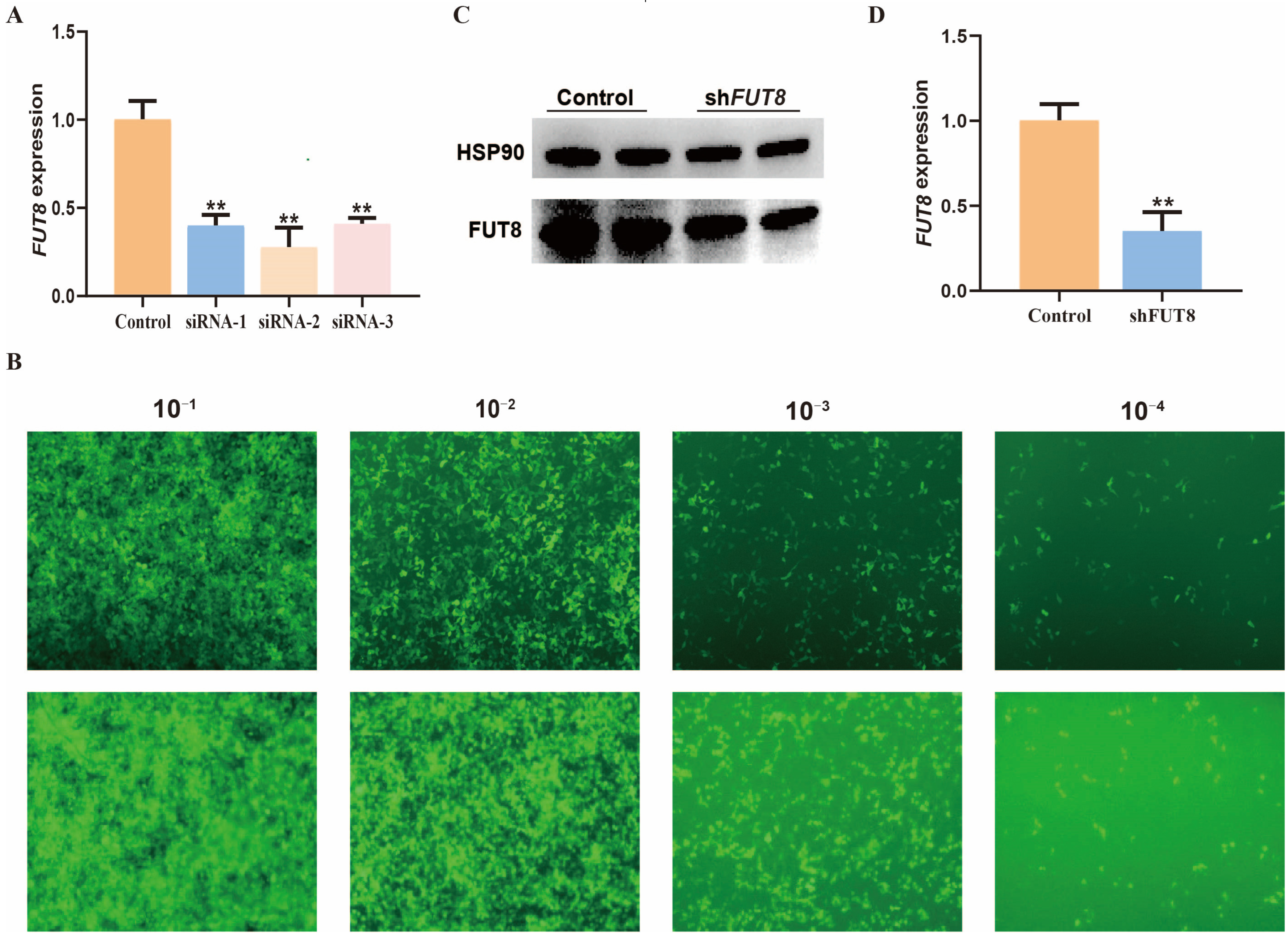
In order to understand the regulatory role of FUT8 in IPEC-J2 cells, we constructed three siRNA vectors for the FUT8 gene and determined that the optimal interference efficiency of FUT8 in IPEC-J2 cells was 62.5% via RT-qPCR testing, as shown in Figure 2A (p < 0.01); furthermore, this interference sequence siRNA was selected for lentiviral packaging. The recombinant interfering lentiviral vector and packaging plasmid were co-transfected with 293T cells and, after 24 h of incubation, 293T cells expressed green fluorescence that indicated that the lentiviral packaging was successful. The virus concentrates were collected for titer determination, which is shown in Figure 2B. Further, the expression of green fluorescent was observed in 10−1, 10−2, 10−3, and 10−4 virus concentrates. Moreover, the number of fluorescent cells gradually decreased with increasing dilution. According to the fluorescent cells observed in the 10−4 virus concentrate, the virus titer was calculated to be 4 × 10⁸ TU/mL, which met the concentration requirements for cell infection. We found that the transcription and protein levels of FUT8 were significantly downgraded in the shFUT8 group (p < 0.01), which indicated that the IPEC-J2 cell line with stable interference of FUT8 was successfully constructed by the Western blotting (Figure 2C) and RT-qPCR (Figure 2D) tests.

2.3. Effect of FUT8 Knockdown on the Adhesion Ability of E. coli F18

Regarding future insight into how FUT8 regulates susceptibility to E. coli F18 infection, we evaluated the effect of FUT8 expression on the level of adhesion of E. coli F18-expressing fimbriae to IPEC-J2 cells. Colony counting (Figure 3A), and expression detection of F18-fimbriae protein (PILIN) (Figure 3C), showed significantly lower numbers of E. coli F18ac adhering to IPEC-J2 cells in the shFUT8 group (p < 0.01). Further, there was no significance in the E. coli F18ab invasion (p > 0.05). In addition, the adhesion ability of E. coli F18ac adhesion to the IPEC-J2 cells was also assessed by Gram staining (Figure 3B), indirect immunofluorescence (Figure 3D), and scanning electron microscopy (Figure S3). Our results showed that FUT8 knockdown significantly decreased the level of E. coli F18ac adhesion at IPEC-J2 cells.

2.4. Transcriptome Sequencing Analysis of IPEC-J2 Cells after FUT8 Knockdown by RNA-seq

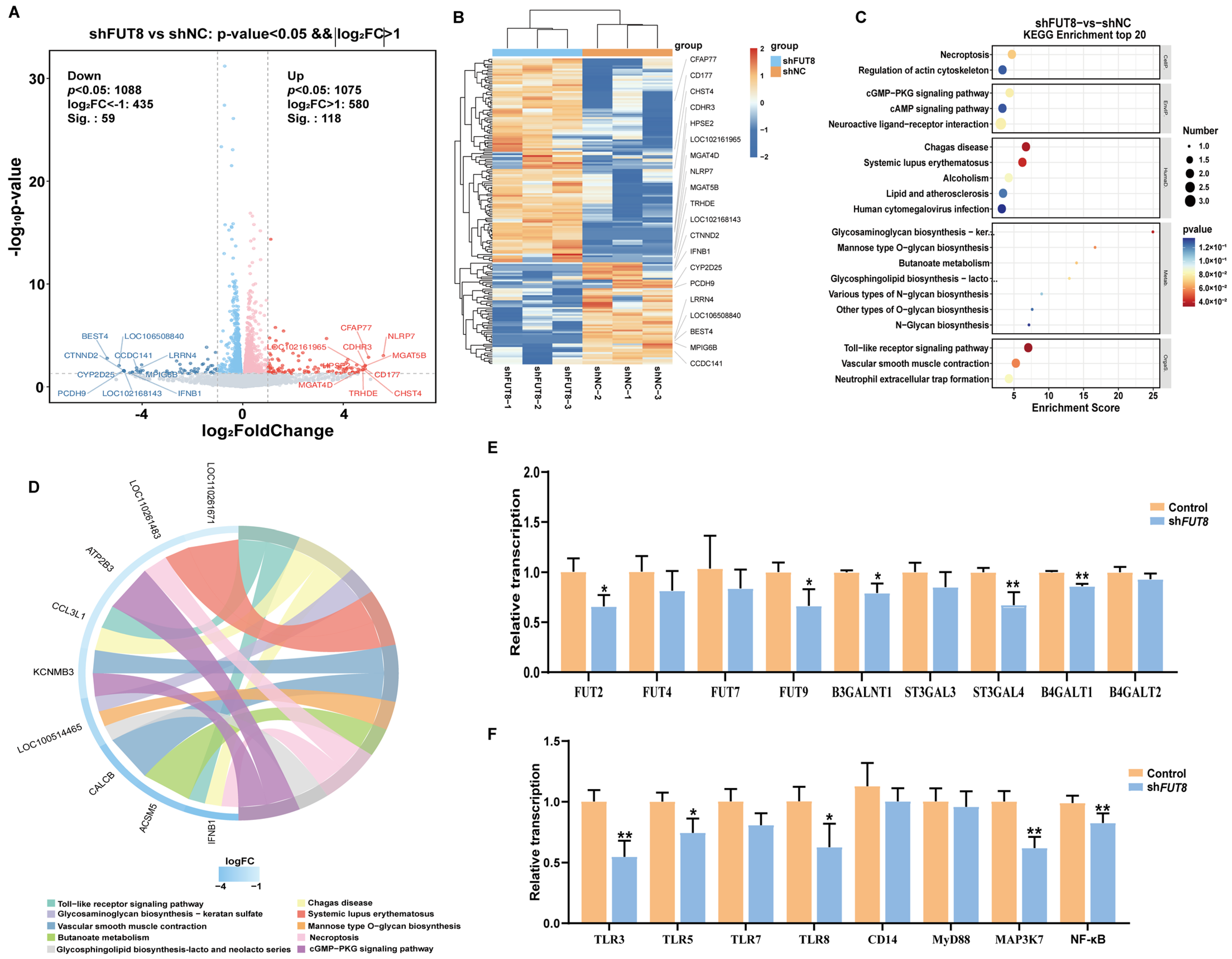
We next investigated the molecular mechanism of the FUT8 gene in the regulation of E. coli F18 infection. We performed a comparative transcriptome sequencing of six RNA libraries (shFUT8-1, shFUT8-2, shFUT8-3, shNC-1, shNC-2, and shNC-3) using the Illumina HiSeq 2500 platform. A total of 299.65 and 300.26 million clean reads after filtering were obtained from the shFUT8 and shNC libraries, respectively (see Supplementary Table S1). A |log2 (fold change)| >1 and p < 0.05 were used as the standard thresholds for screening the differentially expressed genes (DEGs). A total of 177 DEGs were obtained, of which 118 were up-regulated (66.67%) and 59 were down-regulated (33.33%) in the shFUT8 group (see Supplementary Table S2; Figure 4A). The expression profiles of the DEGs in the two groups were visualized using a heat map; the DEGs in the shFUT8 and shNC samples were clustered separately, whereas for each of them the three replicates were clustered together (Figure 4B). Further gene ontology (GO) function annotations of transcripts were shown in Figure S4. Kyoto Encyclopedia of Genes and Genome (KEGG) enrichment analyses were performed in order to explore the potential functions of the DEGs (Figure 4C). Furthermore, these DEGs were enriched in 20 pathways, including 41 significantly enriched Toll-like receptor signaling pathways (i.e., p < 0.05) (see Supplementary Table S3). Among them, the pathway of the “Glycosphingolipid biosynthesis-lacto and neolacto series” (ko00940) was enriched in the shFUT8 and shNC samples (Figure 4D), which probably played an important role in the regulation of E. coli F18 receptor formation [20,21].
To confirm the effect of FUT8 on the key pathways in the IPEC-J2 cells, the expression of genes in the cellular Toll-like receptor signaling and glycosphingolipid biosynthesis pathways in the IPEC-J2 cells were examined via RT-qPCR testing. Results showed that the expressions of FUT2, FUT9, B3GALNT1, STAGAL4, and B4GALT1 from the glycosphingolipid biosynthesis pathway were all significantly decreased (p < 0.05) in IPEC-J2 cells following FUT8 knockdown (shown in Figure 4E). Additionally, the expressions of TLR3, TLR5, TLR8, MAP3K7, and NF-κB from the Toll-like receptor signaling pathway were also significantly down-regulated in FUT8-knockdown IPEC-J2 cells (p < 0.05, Figure 4F). The above results indicated that FUT8-mediated Toll-like receptor signaling and glycosphingolipid biosynthesis pathways may act as critical roles in the process of IPEC-J2 cell responses to E. coli F18 infection.

2.5. Analysis of the Effect of FUT8 on Tight Junction Genes Expression

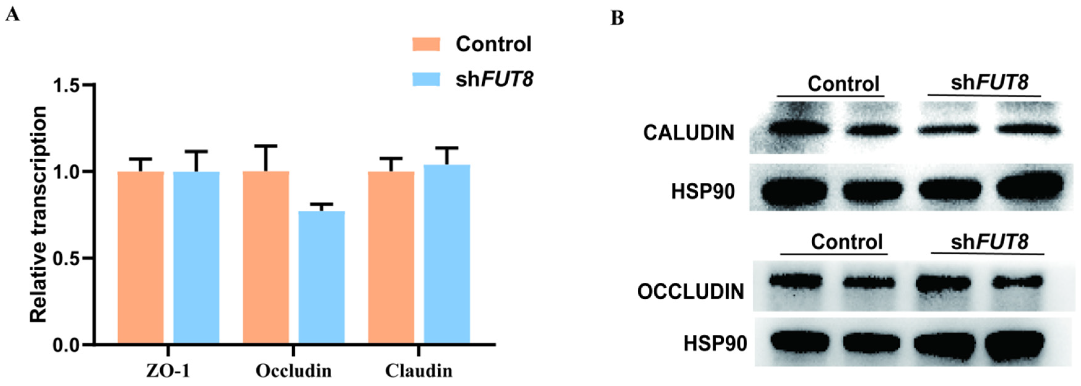
In addition, we analyzed the expression changes in tight junction protein genes in FUT8-knockdown IPEC-J2 cells and found that there were no significant changes in the expression of mRNA (Figure 5A) and protein (Figure 5B) levels after FUT8 knockdown (p > 0.05). These results indicated that FUT8 knockdown may have no influence on the tight junction of IPEC-J2 cells.

2.6. Identification Analysis of Pig FUT8 Core Promoter Region

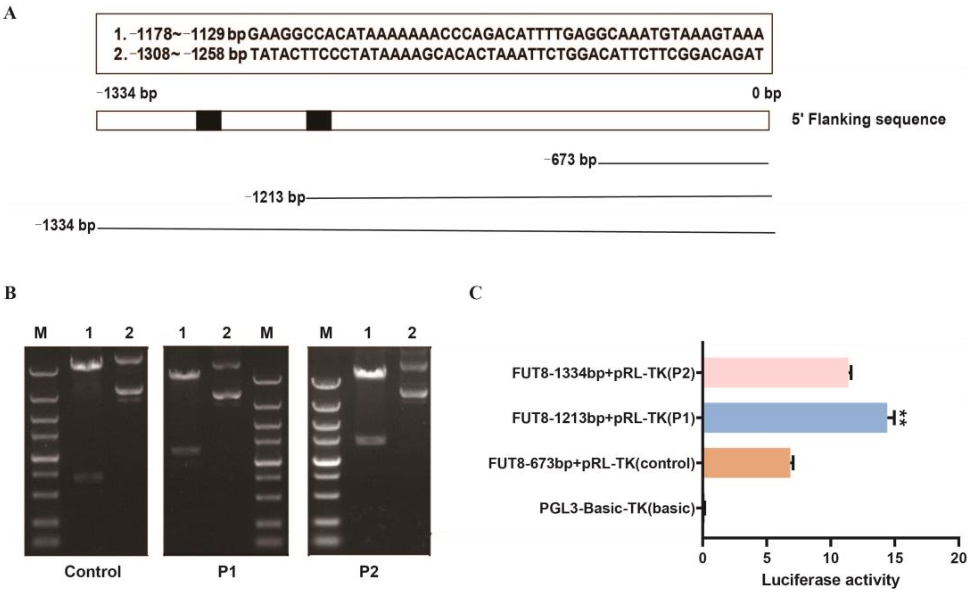
To further explore the regulatory mechanism of FUT8 expression, we focused on the promoter analysis of pig FUT8 (GenBank: XM_005666322.3). The approximately 1300 bp sequence upstream of the transcription start site was used as a template in order to predict the core promoter region using BDGP software (https://www.fruitfly.org/seq_tools/promoter.html). We found two possible promoter regions (−1178~−1129 and −1308~−1258). According to the prediction results, the 1300 bp upstream sequence was divided into three fragments, namely, −673–0 bp (control), −1213–0 bp (P1), and −1334–0 bp (P2) (Figure 6A). As shown in Figure 6B, PCR amplification products were assessed by agarose gel electrophoresis. The luciferase assay showed that the luciferase intensity of pRL-P1 was significantly higher than that of the other transfected groups (Figure 6C). Our results suggested that the core promoter region of the pig the FUT8 gene is located at −1213 bp to −673 bp.

2.7. Important SNP and Transcription Factor Identification Analysis of Pig the FUT8 Gene Promoters

In order to investigate the genetic variation in the promoter region of the pig FUT8 gene, we sequenced the PCR product of this core promoter region in the DNA bulk of 400 Meishan pigs and detected a 1 bp (C base) insertion mutation at the −774 bp upstream of the FUT8 gene, which was verified by sequencing; furthermore, the agarose indicated that the FUT8 wild-type and mutant vectors were constructed successfully (Figure 7A,B). Next, we constructed wild-type and mutant vectors of FUT8 and further transfected the recombinant plasmids of PGL3-FUT8-mut or PGL3-FUT8-wt sequences into 293T cells for luciferase activity analysis, as shown in Figure 7C. Results showed that the luciferase activity was significantly decreased in cells transfected with PGL3-FUT8-mut plasmids compared to the controls (p < 0.01). In order to further identify the important transcription factors in regulating FUT8 expression, we presented the potential transcription factor binding sites such as MY8, BCL6, STAT1, REST, C/EBPα, and CREBBP, as shown in Figure 7D, of which C/EBPα was found in the 1 bp (C base) insertion mutation region of the pig FUT8 gene. Herein, we performed dual-luciferase activity assays in order to investigate the effects of C/EBPα on the transcriptional activity within the FUT8 promoter. As shown in Figure 7E, C/EBPα led to the inhibition of transcriptional activity. Our results indicated that the 1 bp C base insertion mutation at the −774 bp region probably inhibited the transcriptional binding activity of C/EBPα to the FUT8 promoter, and then decreased the FUT8 expression.

3. Discussion

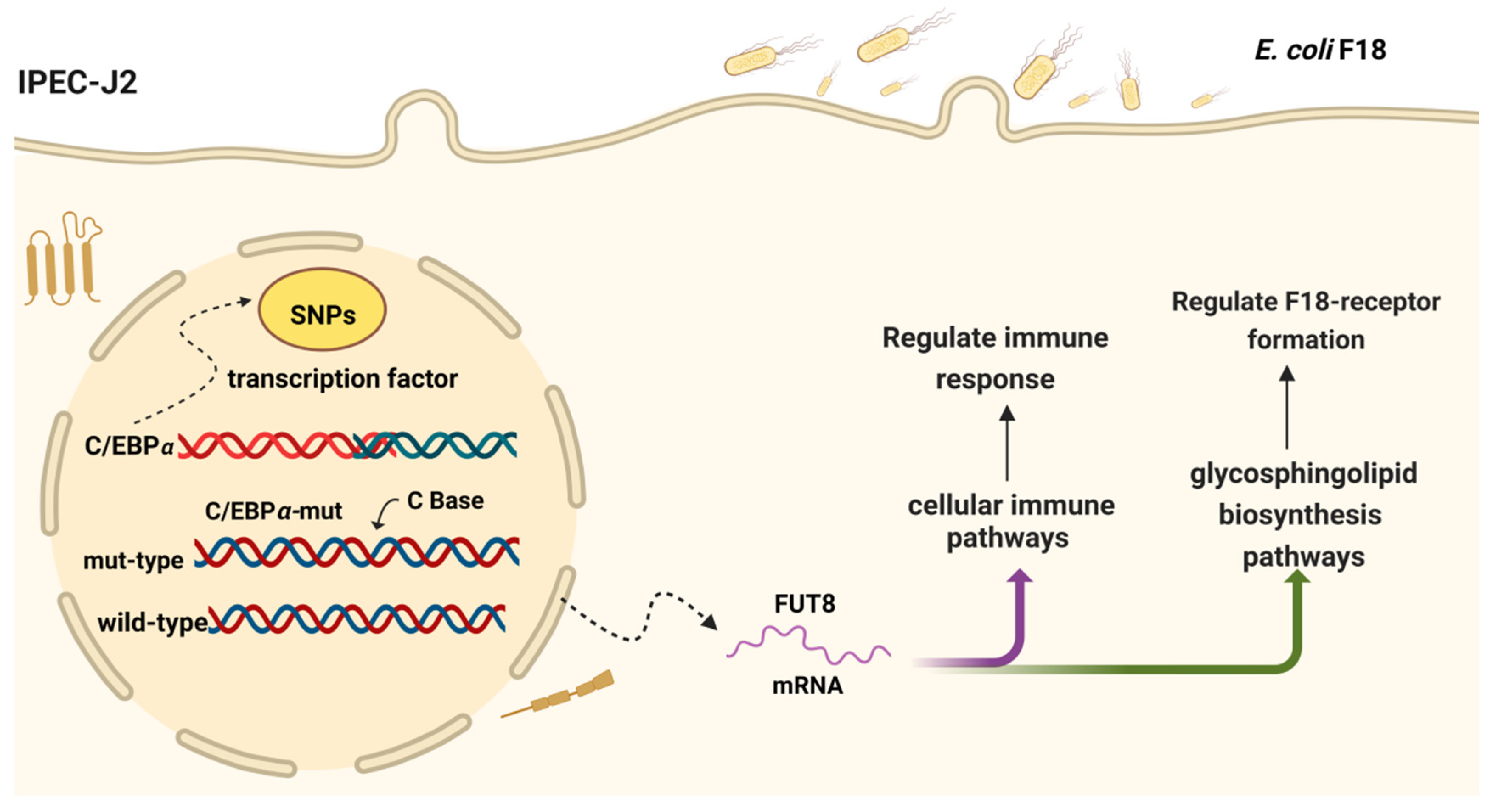
The major causative agent of post-weaning diarrhea (PWD) in piglets is enterotoxin-producing Escherichia coli, which is a Gram-negative bacterium. E. coli F18 binds to the cellular E. coli F18 receptor through bacterial fimbriae, which adheres to the IPEC-J2 and then produces LPS and enterotoxins, which damage the cells and impede the barrier function of the intestinal epithelial cells, leading to metabolic dysregulation and inflammatory reactions [22,23,24,25]. Therefore, the resistance of piglets to E. coli F18 exposure depends on the expression of the E. coli F18 receptor in the porcine intestinal epithelium and the ability of the body to regulate intestinal immunity. The potential regulatory role of FUT8 in IPEC-J2 cells after E. coli F18 exposure was better understood by using IPEC-J2 to mimic the adhesion phenomenon of E. coli F18 in vitro. In this study, we successfully utilized IPEC-J2 cells in order to simulate the phenomenon of E. coli F18 adhesion in vitro. Moreover, it is crucial to enhance our comprehension of the underlying mechanisms behind FUT8 regulating E. coli F18 susceptibility. Briefly, an SNP (C base insertion mutation) was detected in the −774 bp region of the FUT8 core promoter (−1213 bp to −673 bp), and most likely inhibited the transcriptional binding activity of C/EBPα to the FUT8 promoter, which then lead to the decrease in FUT8 expression and the enhancing of the resistance to E. coli F18 infection (Figure 8).
Here, we identify an important regulatory role for FUT8 in the regulating of susceptibility to E. coli F18, as well as glycosylation, which is an important mode of the post-translational modification of proteins. FUT8 is a very important glycosyltransferase that catalyzes the modification of core protein fucose and plays an important role in regulating the normal physiological function of glycoproteins [26]. FUT8-catalyzed core fucosylation plays a role in a variety of life processes in the body and is involved in the regulation of a variety of physiological and pathological processes. Studies have shown that the core fucose level is increased in tumor tissues compared with normal tissues [27] and that the aberrant expression of FUT8 promotes the proliferation and invasion of malignant tumors such as liver cancer, breast cancer, and non-small cell lung cancer [28,29,30]. It is also important in lectin-mediated cytokine production that is induced by immune cells, where the affinity of the antibody to the receptor is reduced by 98–99% after the Fc region of the antibody has been glycosylated by core rockweed [31]. Li et al. found that FUT8 affected the intestinal microbiota profile of mice, with FUT8 −/− mice having a significantly different intestinal microbiota when compared to FUT8 +/+ mice, and the different microbiota were mainly Gram-negative bacteria [10]. Shinzaki et al. found that defects in FUT8 resulted in a lack of core rockweed glycosylation of the TCR (T cell receptor) in the mouse intestinal mucosa, such that TCR-mediated inflammatory signals cannot be activated, thereby resulting in improved colonic inflammation in mice [32,33]. Currently, the research on FUT8 has focused on tumor and intestinal inflammation—mainly in humans and mice—and there are few studies on the relationship between the expression level of the pig FUT8 gene and the drug resistance of Escherichia coli.
Intestinal epithelial cells are the primary site of defense against invading heterologous pathogens that can directly react with Gram-negative bacteria and LPS; further, it is this process that results in the development of diarrhea in piglets [34,35]. Therefore, this study selected IPEC-J2 as the research object at the cellular level. First, we examined the expression of FUT8 at the mRNA and protein levels following E. coli F18 infection and LPS induction by 1 µg/mL. The results showed that compared with the control group, the expression level of FUT8 in IPEC-J2 cells infected with E. coli F18ac was significantly increased (p < 0.05), and the expression level of the FUT8 gene had no significant change after infection with E. coli F18ab (p > 0.05). The expression of the FUT8 gene increased in a stepwise manner after LPS induction by 1 µg/mL, and the expression of the FUT8 gene was significantly increased after 6 h of LPS induction by 1 µg/mL (p < 0.01). LPS is an endotoxin released by Escherichia coli adhesion to intestinal epithelial cells and plays an important role in the inflammatory response. This suggests that the expression of the FUT8 gene is closely related to the collective resistance of E. coli infection, and FUT8 may be involved in the life process of E. coli F18 resistance regulation. Studies have shown that changes in intestinal mucosal glycosylation in patients with IBD affect the integrity of the intestinal mucosa, which is inseparable from bacterial damage to mucosal epithelial cells causing intestinal inflammation. In IBD patients, FUT8 expression is higher in moderately to severely inflammatory mucosa than in mildly inflammatory mucosa [14,36]. These findings suggest that the high expression of FUT8 may exacerbate bacterial exposure of intestinal cells, leading to an inflammatory response in the body.
In this study, in order to investigate the relationship between the expression of the FUT8 gene and E. coli F18 resistance, the stable interference of the FUT8 gene in IPEC-J2 cells was constructed; further, the effect of down-regulation of the FUT8 gene expression on the ability of E. coli F18 to adhere to IPEC-J2 cells was comprehensively analyzed by the processes of bacterial counting, F18-fimbriae protein (PILIN), indirect immunofluorescence, Gram staining, and scanning electron microscopy. After FUT8 gene interference, IPEC-J2 cells were infected with E. coli F18ac and E. coli F18ab; colony counting results showed that there was no significant change in the number of E. coli F18ab adhesion after FUT8 gene interference; additionally, E. coli F18ac was significantly decreased (p < 0.01). F18-fimbriae protein (PILIN) showed a consistent trend with colony counts. Based on the above experimental results, we analyzed the adhesion ability of E. coli using indirect immunofluorescence techniques and Gram staining observation after the E. coli infection. The results showed that there was no significant difference in the E. coli F18ab group after E. coli infection; in addition, E. coli F18ac was significantly decreased. Fucosyltransferases in mammals can be divided into two categories according to the way they are linked to sugar chains, one is FUT1 to FUT11 transferases other than FUT8, and the other is FUT8 unique fucosyltransferases [37,38]. Kashiwazaki et al. showed that FUT9 knockout mice had very low virus titers after infection with the virus and did not change significantly after inoculation with endotoxin [39]. Cai et al. have experimentally demonstrated that low expression of the FUT8 gene reduces cellular activities, such as adhesion binding and migration infection, and improves resistance to disease [40,41]. FUT8 expression is increased in ovarian cancer and the down-regulation of FUT8 significantly inhibits the invasion and spread of tumor cells [42,43]. Munkley et al. showed that core fucosylation increased locally at sites of intestinal inflammation, and that expression of FUT8 was positively correlated with inflammation [44]. These studies have shown that fucosyltransferase is involved in a variety of life processes in the body. Moreover, FUT8—as the only core fucosyltransferase—plays an important role in various physiological activities such as cancer, cell proliferation, migration, and immune and inflammatory responses in the body. Therefore, we speculate that the down-regulation of FUT8 expression may help pigs to resist E. coli infection, pathogen adhesion, and pathogen colonization through a series of signaling and immune regulations.
In order to verify that the FUT8 signaling pathway plays a role in E. coli infection with IPEC-J2 cells, we used RT-qPCR to detect the expression levels of key genes in the signaling pathway. We found that the significantly down-regulated expressions of TLR3, TLR5, TLR8, MAP3K7, and NF-κB were all present in FUT8-knockdown IPEC-J2 cells. MAP3K7 knockout mice have reduced production of inflammatory cytokines in cells [45]. Moreover, TLR5 is a cell surface receptor for innate immunity after pathogens invade the body. It is a receptor for the recognition of Gram-negative bacterial flagellin by the organism and plays an important regulatory role in E. coli invasion into the organism [46]. Low expression of TLR5 helps to inhibit immune responses, reduce cell damage, and promote resistance to Escherichia coli infection in weaned piglets [47]. TLR8 recognizes the nucleic acid components of microorganisms, which in turn initiate antiviral immune responses and promote the expression of inflammatory cytokines [48]. Additionally, we found that FUT8 also significantly affected the glycosphingolipid biosynthesis pathways. Studies showed that glycosphingolipid biosynthesis correlated with the generation of the receptor for E. coli F18 [19,20]. Thus, we speculated that FUT8 most likely regulated E. coli F18 susceptibility via the activation or suppression of TLR signaling (which is related to immune response) and glycosphingolipid biosynthesis (which is related to the formation of the E. coli F18 receptor).
In this study, the core promoter region was predicted and the presence of the core promoter region was verified using a dual-luciferase assay. In order to investigate the genetic variation in the promoter sequence of the pig FUT8 gene, we sequenced the PCR product of the FUT8 gene promoter sequence and detected a 1-bp insertion mutation at the −774 bp upstream of the FUT8 gene in Meishan pigs. Then, we constructed wild-type and mutant vectors with this base insertion. Through conducting this and performing dual-luciferase assays, it was revealed that the 1-bp insertion inhibited transcriptional activity. In order to investigate whether the 1-bp insertion affects the binding of the FUT8 promoter DNA sequence to the transcription factor, we predicted the transcription factors that may bind to the insertion sequence and performed a dual-luciferase assay, which showed that co-transfection with C/EBPα resulted in significantly lower activity in the mutant than in the wild-type. The transcription factor CCAAT enhancer binding protein alpha (C/EBPa) is critical for cell proliferation; in addition, evidence suggests that C/EBPα may negatively control cell proliferation. C/EBPa is essential for granulocyte formation and is regulated by multiple mechanisms in acute myeloid leukemia, which is one of the major regulators of granulopoiesis [49]. During granulocyte formation, C/EBPα regulates differentiation at several steps, including the transition from common myeloid progenitor cells to granulocyte-macrophage progenitor cells [50]. Ayumi Hashimoto et al. demonstrated that therapeutic up-regulation of the transcription factor C/EBPα causes the inactivation of immune-suppressive myeloid cells inactivating and is an effective antitumor response across different tumor models and cancer patients [51]. This author’s finding, i.e., that the DNA-binding variant of the wild-type C/EBPα inhibited the formation of 3T3-L1 preadipocyte colonies, prompted further experiments with estradiol-regulated C/EBPα-ER, which was found to directly inhibit 3T3-L1 cell cycle progression [52]. More and more studies indicate that C/EBPα is down-regulated by mechanisms in acute myeloid leukemia, thereby highlighting that C/EBPα is a myeloid tumor suppressor [53]. Combined with the analysis of the above experimental results, we preliminarily proposed that the low expression of FUT8 would inhibit E. coli F18 adhesion cells and ultimately improve the resistance of piglets to E. coli. However, further electrophoretic mobility shift assays (EMSA) and co-immunoprecipitation (CoIP) assays should be performed to determine the binding of C/EBPα to the FUT8 promoter. Moreover, we will explore in depth the function of FUT8 by RNA overexpression and CRISPR/Cas9 knockout. Our study will help to solve the problem of breeding screening for E. coli resistance in local pig breeds in China and provides a theoretical basis for the genetic breeding of E. coli resistance in the future.

4. Materials and Methods

4.1. Ethics Statement

All the experiments were approved by the Institutional Animal Care and Use Committee (IACUC) of Yangzhou University (pig: SYXK (Su) 2012-0029) and were performed according to the Animal Ethics Procedures and Guidelines of the People’s Republic of China.

4.2. Reagents and Animal Material

The following antibodies were used for indirect immunofluorescence assay (IFA) and Western blot analysis: E. coli (ab137967, rabbit, 1:200) and HSP90 (ab59459, mouse, 1:500), which were both purchased from Abcam (Shanghai, China). FUT8 (NBP1-79869, rabbit, 1:1000) was purchased from NOVUS Ltd. Co. (Littleton, CO, USA). Additionally, IgG (Q6005, rabbit, 1:200) was from Dia-an biotech (Wuhan, China). Experimental pigs (Meishan) were acquired from Kunshan Conservation Ltd(Jiangsu, China). In the previous study, the experimental piglets were challenged with a daily dose of 4.6 × 10⁸ CFU of E. coli F18 strain, and the differences in susceptibility were assessed by assays, such as E. coli F18 bacteria counting and histopathological and in vitro adherence assays of intestinal porcine epithelial cells [54]. Ultimately, we selected three resistant piglets and three sensitive piglets for further analysis. All the experimental pigs were euthanized via the intravenous injection of pentobarbital sodium and duodenum, liver, heart, spleen, lung, stomach, kidney, muscle, lymph node, and thymus. Furthermore, the jejunum tissues of the 35-day-old Meishan pigs were collected, followed by storing them in liquid nitrogen in situ for further use.

4.3. E. coli F18 Exposure and LPS Induction

IPEC-J2 and E. coli F18ab and F18ac organisms were maintained in our laboratory and LPS was purchased from Sigma, USA. In this study, E. coli F18 and LPS-induced cells were used to detect the expression of FUT8 at the mRNA and protein levels, respectively. IPEC-J2 cells were seeded in 12-well plates at a density of 5 × 10⁴ cells per well and cell culture medium was used to dilute E. coli to 1.0 × 10⁹ CFU/mL and then stimulated for 4 h, which is when the cell density reached about 80%. LPS by 1 µg/mL was induced at four time points: 0 h, 2 h, 4 h, and 6 h, and the group was set with three replicates in each group.

4.4. Primer Design and Sequence Synthesis

Quantitative PCR primers were based on their coding sequences in the NCBI (http://www.NCBI.Nlm.nih.govn/ (accessed on 1 May 2022)) database. The GAPDH gene was used as an internal reference gene. All the primers are shown in Supplementary Table S4.

4.5. Cell Culture

IPEC-J2 was seeded into a 12-well plate at a density of 5 × 10⁴ per well; three designed pairs of siRNA sequences (see Supplementary Table S5) were transfected into cells when the density reached approximately 60%. Moreover, the expression of FUT8 was detected by RT-qPCR testing. Gateway recombination technology was used in order to screen the siRNA-2 and the interfering vector was cloned into the lentiviral vector. After 24 h, the expression of green fluorescent was observed and puromycin was added to screen the positive cells. When the positive cells were stably expressed, the cells were collected for mRNA and protein detection in order to analyze the expression of the FUT8 gene in IPEC-J2 cells.

4.6. RT-qPCR Analysis

Total RNA was extracted from IPEC-J2 cells and tissues using a Trizol reagent (Takara, Dalian, China). The purity, as well as the total RNA concentration, was evaluated via a NanoDrop 1000 (Thermo, Massachusetts, USA); then, the RNA was kept at −80 °C. The synthesis of cDNA was performed via 4 µL of 5 × HiScript III qPCR SuperMix II (Vazyme Biotech Co., Ltd., Nanjing, China) along with a total RNA of 1000 ng and RNase-free ddH2O. RT-qPCR was performed in a 20 µL reaction system consisting of 2 µL Cdna (100–500 ng), 0.4 µL upstream and downstream primers (10 µmol/L), 10 µL Hieff UNICON® Universal Blue qPCR SYBR Green Master Mix (Yeasen, Shanghai, China), and 7.2 µL double-distilled water. All experiments were performed in triplicates. The RT-qPCR instrument ABI7500 was employed for the evaluation of qPCR and the parameters were as follows: 95 °C for 5 min, 40 cycles of 95 °C for 10 s, and 60 °C for 30 s. Relative expression was calculated by the 2−∆∆Ct method and interference efficiency was calculated by 1–2−∆∆Ct.

4.7. Western Blot Analysis

Total proteins were extracted using RIPA lysate (Cwbio, Beijing, China) from Thermo Fisher Scientific. A bicinchoninic acid (BCA) kit from Nanjing Keygen Biotech (Nanjing, China) was used for normalizing the protein levels (Biosharp, Beijing, China). The SDS-PAGE of the protein samples was subjected to electrophoresis at 120 V for 90 min in 10% gel; then, the protein sample was transferred to a PVDF membrane. Subsequently, they were blocked with skimmed milk powder and incubated with the corresponding primary antibody (1:1000) overnight at 4 °C. This was then followed by incubation of the corresponding secondary antibody (goat anti-mouse IgG HRP, goat anti-rabbit IgG HRP; CWBIO, Beijing, China). HSP90 (1:5000, Proteintech Group, Inc, Rosemont, IL, USA) was used as an equivalent protein internal reference.

4.8. Subcellular Fraction Extraction

Nucleocytoplasmic RNA of IPEC-J2 was isolated and extracted using the Cytoplasmic and Nuclear RNA Purification Kit (Norgen Biotek, Thorold, ON, Canada).

4.9. Immunohistochemical Analysis

Jejunum tissues were collected from E. coli F18-resistant and -susceptible individuals. Serial sections of paraffin-embedded tissues were first treated with 3% peroxide for 15 min to quench endogenous peroxidase. The sections were rinsed for 15 min with 0.01M PBS (Jiangsu KeyGEN BioTECH Corp., Ltd.) and then incubated at room temperature for 2 h with the following primary antibodies: rabbit anti-FUT8. Whereafter, they were sequentially incubated with the HRP conjugated secondary antibody (Jackson ImmunoResearch Laboratories, West Grove, PA, USA., 1:50) for 30 min at room temperature with diaminobenzidine (DAB) as the substrate chromogen (Jiangsu KeyGEN BioTECH Corp, Jiangsu, China), the sections were counterstained with hematoxylin.

4.10. In vitro Adherence Assays with E. coli F18

IPEC-J2 cells were inoculated into 12-well plates at a density of 5 × 10⁴ per well and placed in a constant temperature incubator at 37 °C. E. coli F18ab and E. coli F18ac fimbriae standard strains were inoculated into the LB culture medium and then incubated for 12 h on a rotating shaker table (200 rpm). PBS was used for the wash and the bacteria collected was centrifuged at a rotating speed of 4000 rev/min for 10 min at three times; next, the cell culture medium was used to dilute E. coli to 1.0 × 10⁹ CFU/mL, and then added to 12-well culture plates. Cells were cultured for 4 h in an incubator under the conditions of 37 °C; further, F18-fimbriae protein, colony count, Gram staining, scanning electron microscopy (SEM), and indirect immunofluorescence assays were all used in order to detect E. coli adherence to cells [47,55,56].

4.11. Indirect Immunofluorescence (IFA)

IPEC-J2 cells (E. coli F18ab exposure, E. coli F18ac exposure, shFUT8, and control) were gently washed three times with PBS and were then fixed with 4% paraformaldehyde (Solarbio, Beijing, China) for 0.5 h. Next, they were washed with PBS three times again, then the cells were treated with 500 μL 0.5% Triton X-100 for 15 min. Then, washing with PBS and goat blocking serum was added, they were incubated for 2 h, and then the cells were incubated with an E. coli antibody (1:200) (Gene Tex, San Antonio, TX, USA) and secondary antibody IgG (1:200) (Affinity, Cincinnati, OH, USA), respectively. Subsequently, the cells were incubated with DAPI (1:1000) (Solarbio, Beijing, China), and then washed three times with 0.5% PBST. Finally, cells were mounted using a fade-resistant fluorescent medium and were analyzed immediately by confocal microscopy.

4.12. Single-Nucleotide Polymorphisms in the Porcine FUT8 Gene Promote

According to the promoter sequence of the porcine FUT8 gene (GenBank: XM005666322.3), the PCR primers were designed by Prime Premier 5.0 software. PCR assays were conducted in a 50 µL volume containing 200 ng of the DNA template, 10 µL of PCR Master Mix, and 25 pmol of each forward and reverse primer. The thermal conditions were as follows: 95 °C for 5 min, 36 cycles of 95 °C for 30 s, 55 °C for 30 s, 72 °C for 60 s, and a final extension at 72 °C for 10 min. The PCR products were checked by electrophoresis in a 2% agarose gel, and then sequenced by Sangon Biotech (Sangon, Shanghai, China).

4.13. Dual-luciferase Reporter Assays

The 2000-bp upstream sequence of the transcription start site of the pig FUT8 gene (GenBank: XM005666322.3) was obtained from the NCBI (http://www.NCBI.Nlm.nih.govn/, accessed on 7 August 2022) database and the BDGP (http://www.fruitfly.org/seq_tools/promoter.html, accessed on 7 August 2022) was used in order to predict the core promoter region. Different promoter fragments were ligated into the pGL3-Basic vector in order to construct recombinant plasmids. Positive clones of recombinant plasmids were examined by sequencing (Shanghai Bioengineering Co., Ltd., Shanghai, China). When the density of 293T cells reached a 70–80% confluence in the 12-well plates, the expression vector and pRL-TK vector were co-transfected into the cells. Dual-luciferase assay was performed in strict accordance with the kit instructions. Luciferase activity was analyzed on a luminometer equipped with a Dual-Luciferase Assay System (Promega Corporation, Madison, WI, USA). The relative fluorescence intensity was calculated as Firefly Luc (Ff)/Renilla-Luc (Rn). The transcription factors in the core promoter region of FUT8 were predicted using the Alibaba software.

4.14. Transcription Factor Prediction

Alibaba (http://www.gene-regulation.com/pub/programs/alibaba2/index.html?, accessed on 16 August 2022) and TFDB (http://bioinfo.life.hust.edu.cn/AnimalTFDB/, accessed on 16 August 2022) were used in order to screen out the important transcription factor binding sites (C/EBPα), which were based on the sequence of the FUT8 promoter region, as well as in the conducted validation experiments by dual-luciferase activity assay.

4.15. Statistical Analysis

In order to conduct statistical analyses, the SPSS v.20 software (IBM Corp, Armonk, NY, USA) and GraphPad Prism 6.0 software (GraphPad Inc., La Jolla, CA, USA) were used. The relative quantitative results were examined via the 2−∆∆Ct method. The analysis was performed using Student’s t-test (two-tailed). * p < 0.05 and ** p < 0.01 were considered statistically significant.

5. Conclusions

In conclusion, our study showed that the down-regulation of FUT8 correlates with E. coli F18 resistance. Moreover, we found that the insertion mutation (1 bp C base) located at −774 bp in the FUT8 promoter (−1213 bp to −673 bp) probably inhibited FUT8 expression by affecting the binding of C/EBPα to the FUT8 promoter, thereby improving the E. coli F18 resistance in piglets. Our results revealed the regulatory mechanism of the pig FUT8 gene affecting the resistance to E. coli F18 infection. This finding will provide a theoretical reference for the molecular breeding of combating bacterial diarrhea in Chinese local pig breeds.

Supplementary Materials

The following supporting information can be downloaded at: https://www.mdpi.com/article/10.3390/ijms232314713/s1.

Author Contributions

Conceived and designed the experiments: Z.W. and W.B. Performed the experiments: L.W. and Y.W. Analyzed the data: L.W. and Z.W. Contributed reagents/materials/analysis tools: S.W. Contributed to the writing of the manuscript: L.W. All authors have read and agreed to the published version of the manuscript.

Funding

This study was supported by the Open Competition Mechanism in order to select the best candidates’ foundation for the breeding industry prosperity of Jiangsu Province, China (JBGS [2021]098), as well as for the Key Research and Development Project (Modern Agriculture) of Jiangsu Province (BE2019341), China Postdoctoral Science Foundation (2021M702764), and the Priority Academic Program Development of Jiangsu Higher Education Institutions.

Institutional Review Board Statement

The study was conducted according to the guidelines of the Declaration of the State Council of the People’s Republic of China, and approved by the Ethics Committee of Yangzhou University (permit number: SYXK (Su) IACUC 2012-0029; Approval Date: 9 December 2012).

Informed Consent Statement

Not applicable.

Data Availability Statement

The data presented in this study are available on request from the corresponding author.

Conflicts of Interest

The authors declare that they have no competing interests.

Abbreviations

FUT8Fucosyltransferase 8
PWDPost-weaning diarrhea
E. coliEscherichia coli
IPEC-J2Intestinal porcine epithelial cell
IFAIndirect immunofluorescence assay
SEMScanning electron microscopy
LPSLipopolysaccharide
FBSFetal bovine serum
DMEMDulbecco’s modified Eagle’s medium
NF-κBNuclear factor κB
SNPsSingle-nucleotide polymorphisms
DEGsDifferentially expressed genes
TSSsTranscription start sites
KEGGKyoto Encyclopedia of Genes and Genome
EMSAElectrophoretic mobility shift assay
CoIPCo-immunoprecipitation
TUTransducing units

References

  1. Boldin, B. Persistence and spread of gastro-intestinal infections: The case of enterotoxigenic Escherichia coli in piglets. Bull. Math. Biol. 2008, 70, 2077–2101. [Google Scholar] [CrossRef] [PubMed]
  2. Imberechts, H.; Bertschinger, H.U.; Stamm, M.; Sydler, T.; Pohl, P.; De Greve, H.; Hernalsteens, J.P.; Van Montagu, M.; Lintermans, P. Prevalence of F107 fimbriae on Escherichia coli isolated from pigs with oedema disease or postweaning diarrhoea. Vet. Microbiol. 1994, 40, 219–230. [Google Scholar] [CrossRef] [PubMed]
  3. Fairbrother, J.M.; Nadeau, É.; Gyles, C.L. Escherichia coli in postweaning diarrhea in pigs: An update on bacterial types, pathogenesis, and prevention strategies. Anim. Health Res. Rev. 2005, 6, 17–39. [Google Scholar] [CrossRef] [PubMed] [Green Version]
  4. Sinha, R.; Sahoo, N.R.; Shrivastava, K.; Kumar, P.; Qureshi, S.; De Kumar, U.; Kumar, A.; Kumar, G.V.P.P.S.R.; Bhushan, B. Resistance to ETEC F4/F18-mediated piglet diarrhoea: Opening the gene black box. Trop. Anim. Health Prod. 2019, 51, 1307–1320. [Google Scholar] [CrossRef] [PubMed]
  5. Agrawal, P.; Fontanals-Cirera, B.; Sokolova, E.; Jacob, S.; Vaiana, C.A.; Argibay, D.; Davalos, V.; McDermott, M.; Nayak, S.; Darvishian, F.; et al. A Systems Biology Approach Identifies FUT8 as a Driver of Melanoma Metastasis. Cancer Cell. 2017, 31, 804–819. [Google Scholar] [CrossRef] [PubMed] [Green Version]
  6. Liang, C.; Fukuda, T.; Isaji, T.; Duan, C.; Song, W.; Wang, Y.; Gu, J. 1,6-Fucosyltransferase contributes to cell migration and proliferation as well as to cancer stemness features in pancreatic carcinoma. Biochim. Biophys. Acta Gen. Subj. 2021, 1865, 129870. [Google Scholar] [CrossRef]
  7. Bastian, K.; Scott, E.; Elliott, D.J.; Munkley, J. FUT8 Alpha-(1,6)-Fucosyltransferase in Cancer. Int. J. Mol. Sci. 2021, 22, 455. [Google Scholar] [CrossRef]
  8. Zahid, D.; Zhang, N.; Fang, H.; Gu, J.; Li, M.; Li, W. Loss of core fucosylation suppressed the humoral immune response in Salmonella typhimurium infected mice. J. Microbiol. Immunol. Infect. 2021, 54, 606–615. [Google Scholar] [CrossRef] [PubMed]
  9. Li, W.; Yu, R.; Ma, B.; Yang, Y.; Jiao, X.; Liu, Y.; Cao, H.; Dong, W.; Liu, L.; Ma, K.; et al. Core fucosylation of IgG B cell receptor is required for antigen recognition and antibody production. J. Immunol. 2015, 194, 2596–2606. [Google Scholar] [CrossRef] [PubMed] [Green Version]
  10. Li, M.; Bai, Y.Q.; He, C.Y.; Yan, Q.L.; Fang, H.; Li, Z. Intestinal microbial structure of core fucosyltransferase FUT8 gene knockout mice. Chin. J. Microecol. 2017, 29, 5. [Google Scholar]
  11. Osumi, D.; Takahashi, M.; Miyoshi, E.; Yokoe, S.; Taniguchi, N. Core fucosylation of E-cadherin enhances cell–cell adhesion in human colon carcinoma WiDr cells. Cancer Sci. 2009, 100, 888–895. [Google Scholar] [CrossRef] [PubMed]
  12. Ma, M.; Guo, D.; Tan, Z.; Du, J.; Guan, F.; Li, X. Fucosyltransferase 8 regulation and breast cancer suppression by transcription factor activator protein 2γ. Cancer Sci. 2021, 112, 3190–3204. [Google Scholar] [CrossRef] [PubMed]
  13. Ihara, H.; Okada, T.; Taniguchi, N.; Ikeda, Y. Involvement of the α-helical and Src homology 3 domains in the molecular assembly and enzymatic activity of human α1, 6-fucosyltransferase, FUT8. Biochim. Biophys. Acta. Gen. Subj. 2020, 1864, 129596. [Google Scholar] [CrossRef] [PubMed]
  14. Fujii, H.; Shinzaki, S.; Iijima, H.; Wakamatsu, K.; Iwamoto, C.; Sobajima, T.; Kuwahara, R.; Hiyama, S.; Hayashi, Y.; Takamatsu, S. Core Fucosylation on T Cells, Required for Activation of T-Cell Receptor Signaling and Induction of Colitis in Mice, Is Increased in Patients with Inflammatory Bowel Disease. Gastroenterology 2016, 150, 1620–1632. [Google Scholar] [CrossRef] [PubMed] [Green Version]
  15. Nakayama, K.; Wakamatsu, K.; Fujii, H.; Shinzaki, S.; Takamatsu, S.; Kitazume, S.; Kamada, Y.; Takehara, T.; Taniguchi, N.; Miyoshi, E. Core fucose is essential glycosylation for CD14-dependent Toll-like receptor 4 and Toll-like receptor 2 signalling in macrophages. J. Biochem. 2019, 165, 227–237. [Google Scholar] [CrossRef] [PubMed]
  16. Xiaoli, L.; Fengbin, H.; Shihui, H.; Xi, N.; Sheng, L.; Zhou, W.; Xueqin, R.; Jiafu, W. Detection of genomic structure variants associated with wrinkled skin in Xiang pig by next generation sequencing. Aging 2021, 13, 24710–24739. [Google Scholar] [CrossRef] [PubMed]
  17. Zi, C.; Wu, Z.; Wang, J.; Huo, Y.B.; Zhu, G.; Wu, S.; Bao, W. Transcriptional activity of the FUT1 gene promoter region in pigs. Int. J. Mol. Sci. 2013, 14, 24126–24134. [Google Scholar] [CrossRef] [PubMed] [Green Version]
  18. Matsuda, K. PCR-Based Detection Methods for Single-Nucleotide Polymorphism or Mutation: Real-Time PCR and Its Substantial Contribution Toward Technological Refinement. Adv. Clin. Chem. 2017, 80, 45–72. [Google Scholar]
  19. Shastry, B.S. SNPs: Impact on gene function and phenotype. Methods Mol. Biol. 2009, 578, 3–22. [Google Scholar] [PubMed]
  20. Dong, W.H.; Dai, C.H.; Sun, L.; Wang, J.; Sun, S.Y.; Zhu, G.Q.; Wu, S.L.; Bao, W.B. Expression of key glycosphingolipid biosynthesis-globo series pathway genes in Escherichia coli F18-resistant and Escherichia coli F18-sensitive piglets. Anim. Genet. 2016, 47, 428–435. [Google Scholar] [CrossRef] [PubMed]
  21. Wu, Z.; Fan, H.; Jin, J.; Gao, S.; Huang, R.; Wu, S.; Bao, W. Insight into mechanisms of pig lncRNA FUT3-AS1 regulating E. coli F18-bacterial diarrhea. PLoS Pathog. 2022, 18, e1010584. [Google Scholar] [CrossRef] [PubMed]
  22. Aarestrup, F.M.; Jorsal, S.E.; Ahrens, P.; Jensen, N.E.; Meyling, A. Molecular characterization of Escherichia coli strains isolated from pigs with edema disease. J. Clin. Microbiol. 1997, 35, 20–24. [Google Scholar] [CrossRef] [PubMed]
  23. Batisson, I.; Guimond, M.P.; Girard, F.; An, H.; Zhu, C.; Oswald, E.; Fairbrother, J.M.; Jacques, M.; Harel, J. Characteriza-tion of the novel factor paa involved in the early steps of the adhesion mechanism of attaching and effacing Escherichia coli. Infect. Immun. 2003, 71, 4516–4525. [Google Scholar] [CrossRef] [PubMed] [Green Version]
  24. Nagy, B.; Fekete, P.Z. Enterotoxigenic Escherichia coli (ETEC) in farm animals. Vet. Res. 1999, 30, 259–284. [Google Scholar] [PubMed]
  25. Sham, H.P.; Shames, S.R.; Croxen, M.A.; Ma, C.; Chan, J.M.; Khan, M.A.; Wickham, M.E.; Deng, W.; Finlay, B.B.; Vallance, B.A. Attaching and effacing bacterial effector NleC suppresses epithelial inflammatory responses by inhibiting NF-κB and p38 mitogen-activated protein kinase activation. Infect. Immun. 2011, 79, 3552–3562. [Google Scholar] [CrossRef] [PubMed] [Green Version]
  26. Fukuda, T.; Hashimoto, H.; Okayasu, N.; Kameyama, A.; Onogi, H.; Nakagawasai, O.; Nakazawa, T.; Kurosawa, T.; Hao, Y.; Isaji, T.; et al. a1,6-Fucosyltransferase-deficient mice exhibit multiple behavioral abnormalities associated with a schizophrenia-like phenotype: Importance of the balance between the dopamine and serotonin systems. J. Biol. Chem. 2011, 286, 18434–18443. [Google Scholar] [CrossRef] [Green Version]
  27. Keeley, T.S.; Yang, S.; Lau, E. The Diverse Contributions of Fucose Linkages in Cancer. Cancers 2019, 11, 1241. [Google Scholar] [CrossRef] [Green Version]
  28. Li, S.; Liu, X.Y.; Pan, Q.; Wu, J.; Liu, Z.H.; Wang, Y.; Liu, M.; Zhang, X.L. Hepatitis C Virus-Induced FUT8 Causes 5-FU Drug Resistance in Human Hepatoma Huh7.5.1 Cells. Viruses 2019, 11, 378. [Google Scholar] [CrossRef] [Green Version]
  29. Tu, C.F.; Wu, M.Y.; Lin, Y.C.; Kannagi, R.; Yang, R.B. FUT8 promotes breast cancer cell invasiveness by remodeling TGF-β receptor core fucosylation. Breast Cancer Rev. 2017, 19, 111. [Google Scholar] [CrossRef] [Green Version]
  30. Li, F.; Zhao, S.; Cui, Y.; Guo, T.; Qiang, J.; Xie, Q.; Yu, W.; Guo, W.; Deng, W.; Gu, C.; et al. a1,6-Fucosyltransferase (FUT8) regulates the cancer-promoting capacity of cancer-associated fibroblasts (CAFs) by modifying EGFR core fucosylation (CF) in non-small cell lung cancer (NSCLC). Am. J. Cancer Res. 2020, 10, 816–837. [Google Scholar] [PubMed]
  31. Sonneveld, M.E.; van der Schoot, C.E.; Vidarsson, G. The Elements Steering Pathogenesis in IgG-Mediated Alloimmune Diseases. J. Clin. Immunol. 2016, 36, 76–81. [Google Scholar] [CrossRef] [PubMed]
  32. Pham, D.; Vincentz, J.W.; Firulli, A.B.; Kaplan, M.H. Twist1 regulates Ifng expression in Th1 cells by interfering with Runx3 function. J. Immunol. 2012, 189, 832–840. [Google Scholar] [CrossRef] [PubMed]
  33. Shinzaki, S.; Iijima, H.; Fujii, H.; Kamada, Y.; Naka, T.; Takehara, T.; Miyoshi, E. A novel pathogenesis of inflammatory bowel disease from the perspective of glyco-immunology. J. Biochem. 2017, 161, 409–415. [Google Scholar] [CrossRef] [PubMed] [Green Version]
  34. Fermín, S.D.M.; Isabel, R.C.; Cristina, M.; Olga, M.A. Intestinal inflammation and mucosal barrier function. Inflamm. Bowel Dis. 2014, 20, 2394–2404. [Google Scholar]
  35. Moonens, K.; Bouckaert, J.; Coddens, A.; Tran, T.; Panjikar, S.; De Kerpel, M.; Cox, E.; Remaut, H.; De Greve, H. Structural insight in histo-blood group binding by the F18 fimbrial adhesin FedF. Mol. Microbiol. 2012, 86, 82–95. [Google Scholar] [CrossRef]
  36. Theodoratou, E.; Campbell, H.; Ventham, N.T.; Kolarich, D.; Pučić-Baković, M.; Zoldoš, V.; Fernandes, D.; Pemberton, I.K.; Rudan, I.; Kennedy, N.A. The role of glycosylation in IBD. Nat. Rev. Gastroenterol. Hepatol. 2014, 11, 588–600. [Google Scholar] [CrossRef]
  37. Schadendorf, D.; Hodi, F.S.; Robert, C.; Weber, J.S.; Margolin, K.; Hamid, O.; Patt, D.; Chen, T.T.; Berman, D.M.; Wolchok, J.D. Pooled Analysis of Long-Term Survival Data from Phase II and Phase III Trials of Ipilimumab in Unresectable or Metastatic Melanoma. J. Clin. Oncol. 2015, 33, 1889–1894. [Google Scholar] [CrossRef] [PubMed] [Green Version]
  38. Liang, S.; Latchman, Y.; Buhlmann, J.; Tomczak, M.; Horwitz, B.; Freeman, G.; Sharpe, A. Regulation of PD-1, PD-L1, and PD-L2 expression during normal and autoimmune responses. Eur. J. Immunol. 2010, 33, 2706–2716. [Google Scholar] [CrossRef] [PubMed]
  39. Kashiwazaki, H.; Kakizaki, M.; Ikehara, Y.; Togayachi, A.; Narimatsu, H.; Watanabe, R. Mice lacking α1,3-fucosyltransferase 9 exhibit modulation of in vivo immune responses against pathogens. Pathol. Int. 2014, 64, 199–208. [Google Scholar] [CrossRef]
  40. Cai, Y.J.; Zheng, X.F.; Lu, C.H.; Jiang, Q.; Liu, Q.; Xin, Y.H. Effect of FUT3 gene silencing with miRNA on proliferation, invasion and migration abilities of human KATO-III gastric cancer cell line. Cell Mol. Biol. 2016, 62, 15–20. [Google Scholar]
  41. Orntoft, T.F.; Vestergaard, E.M.; Holmes, E.; Jakobsen, J.S.; Grunnet, N.; Mortensen, M.; Johnson, P.; Bross, P.; Gregersen, N.; Skorstengaard, K. Influence of Lewis α1-3/4-L-Fucosyltransferase (FUT3) Gene Mutations on Enzyme Activity, Erythrocyte Phenotyping, and Circulating Tumor Marker Sialyl-Lewis a Levels. J. Biol. Chem. 1996, 271, 32260–32268. [Google Scholar] [CrossRef] [PubMed] [Green Version]
  42. Faghfuri, E.; Faramarzi, M.A.; Nikfar, S.; Abdollahi, M. Nivolumab and pembrolizumab as immune-modulating monoclonal antibodies targeting the PD-1 receptor to treat melanoma. Expert. Rev. Anticancer Ther. 2015, 15, 981–993. [Google Scholar] [CrossRef] [PubMed]
  43. Munkley, J.; Mills, I.G.; Elliott, D.J. The role of glycans in the development and progression of prostate cancer. Nat. Rev. Urol. 2016, 13, 324–333. [Google Scholar] [CrossRef]
  44. Chen, J.Q.; Szodoray, P.; Zeher, M. Toll-Like Receptor Pathways in Autoimmune Diseases. Clin. Rev. Allergy. Immunol. 2016, 50, 1–17. [Google Scholar] [CrossRef] [PubMed]
  45. Sato, S.; Sanjo, H.; Takeda, K.; Ninomiya-Tsuji, J.; Yamamoto, M.; Kawai, T.; Matsumoto, K.; Takeuchi, O.; Akira, S. Essential function for the kinase TAK1 in innate and adaptive immune responses. Nat. Immunol. 2005, 6, 1087–1095. [Google Scholar] [CrossRef] [PubMed]
  46. Kravchenko, V.V.; Kaufmann, G.F. Bacterial inhibition of inflammatory responses via TLR-independent mechanisms. Cell. Microbiol. 2013, 15, 527–536. [Google Scholar] [CrossRef] [PubMed]
  47. Dai, C.; Yang, L.; Jin, J.; Wang, H.; Wu, S.L.; Bao, W.B. Regulation and Molecular Mechanism of TLR5 on Resistance to Escherichia coli F18 in Weaned Piglets. Animals 2019, 9, 735. [Google Scholar] [CrossRef] [PubMed] [Green Version]
  48. Liu, Y.J. IPC: Professional type 1 interferon-producing cells and plasmacytoid dendritic cell precursors. Annu. Rev. Immunol. 2005, 23, 275–306. [Google Scholar] [CrossRef] [PubMed]
  49. Umek, R.M.; Friedman, A.D.; McKnight, S.L. CCAAT-enhancer binding protein: A component of a differentiation switch. Science 1991, 251, 288–292. [Google Scholar] [CrossRef]
  50. Rosenbauer, F.; Tenen, D.G. Transcription factors in myeloid development: Balancing differentiation with transformation. Nat. Rev. Immunol. 2007, 7, 105–117. [Google Scholar] [CrossRef]
  51. Zhang, P.; Iwasaki-Arai, J.; Iwasaki, H.; Fenyus, M.L.; Dayaram, T.; Owens, B.M.; Shigematsu, H.; Levantini, E.; Huettner, C.S.; Lekstrom-Himes, J.A.; et al. Enhancement of hematopoietic stem cell repopulating capacity and self-renewal in the absence of the transcription factor C/EBP alpha. Immunity 2004, 21, 853–863. [Google Scholar] [CrossRef] [PubMed] [Green Version]
  52. Hashimoto, A.; Sarker, D.; Reebye, V.; Jarvis, S.; Sodergren, M.H.; Kossenkov, A.; Sanseviero, E.; Raulf, N.; Vasara, J.; Andrikakou, P. Upregulation of C/EBPα Inhibits Suppressive Activity of Myeloid Cells and Potentiates Antitumor Response in Mice and Patients with Cancer. Clin. Cancer. Res. 2021, 27, 5961–5978. [Google Scholar] [CrossRef] [PubMed]
  53. Wang, Q.F.; Cleaves, R.; Kummalue, T.; Nerlov, C.; Friedman, A.D. Cell cycle inhibition mediated by the outer surface of the C/EBPalpha basic region is required but not sufficient for granulopoiesis. Oncogene 2003, 22, 2548–2557. [Google Scholar] [CrossRef] [PubMed] [Green Version]
  54. Wu, Z.C.; Dong, W.H.; Liu, Y.; Yang, J.S.; Zhu, G.Q.; Wu, S.L.; Bao, W.B. Attack Experiment and Phenotype Analysis of Meishan piglets by E. coli F18 Strain. Chin. J. Anim. Vet. Sci. 2014, 45, 1608–1615. (In Chinese) [Google Scholar]
  55. Liu, L.; Wang, J.; Zhao, Q.; Zi, C.; Wu, Z.; Su, X.; Huo, Y.; Zhu, G.Q.; Wu, S.L.; Bao, W.B. Genetic variation in exon 10 of the BPI gene is associated with Escherichia coli F18 susceptibility in Sutai piglets. Gene 2013, 523, 70–75. [Google Scholar] [CrossRef] [PubMed]
  56. Dai, C.; Gan, L.; Qin, W.; Zi, C.; Zhu, G.Q.; Wu, S.L.; Bao, W.B. Use of fluorescence quantitative polymerase chain reaction (PCR) for the detection of Escherichia coli adhesion to pig intestinal epithelial cells. Pol. J. Vet. Sci. 2016, 19, 619–625. [Google Scholar] [CrossRef] [PubMed]
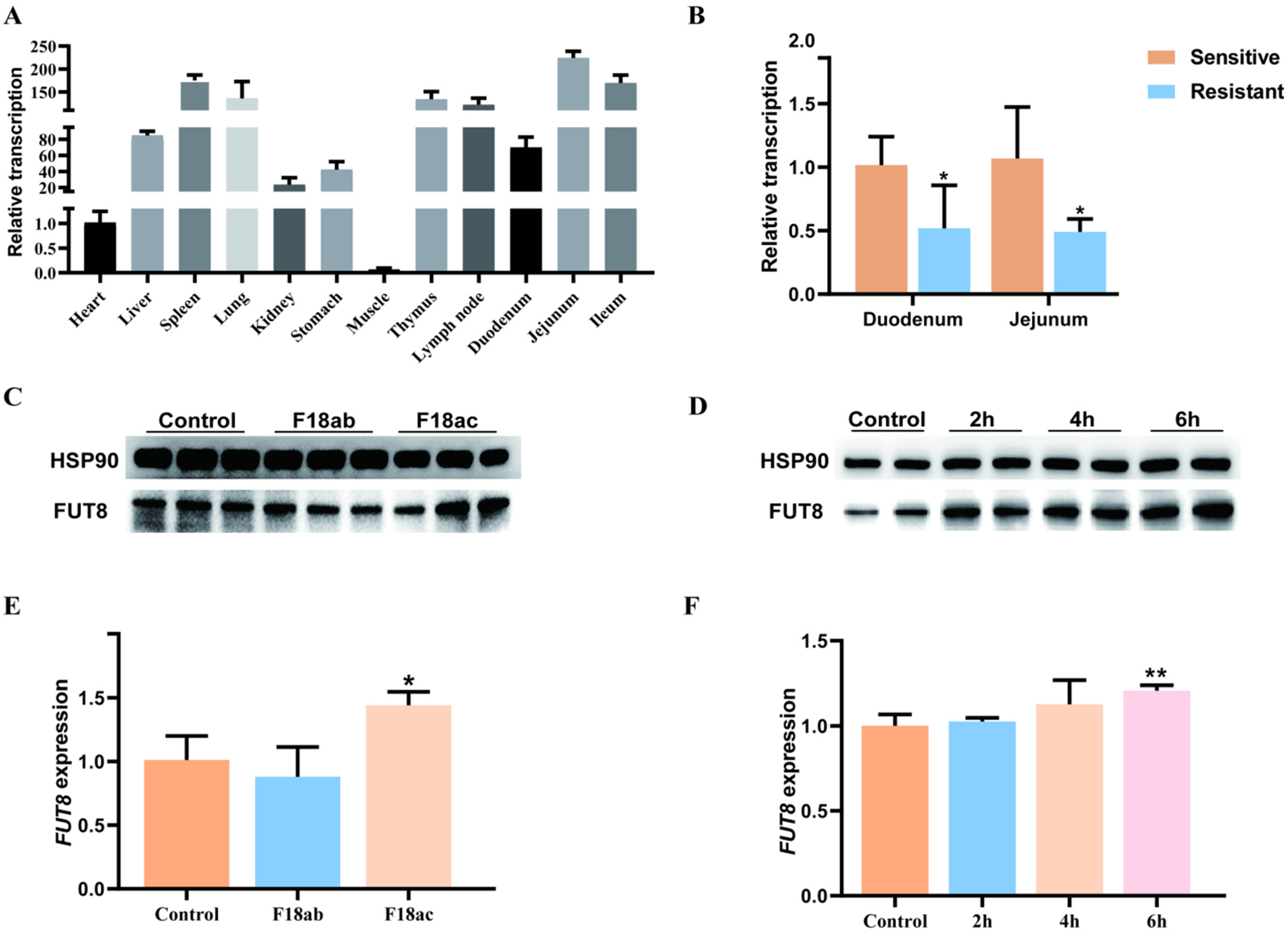
Figure 1. The relationship between the expression of FUT8 and E. coli infection was analyzed at the tissue and cell levels. (A) Expression of the FUT8 gene in 12 tissues of 35-day-old Meishan pigs. (B) Differential expression analysis of the FUT8 gene in intestinal tissues between E. coli F18-resistant and -sensitive Meishan piglets. (C) Expression levels of FUT8 in IPEC-J2 cells with E. coli infection were determined by Western blotting. (D) FUT8 expression analysis in IPEC-J2 cells was determined using Western blotting, which was induced by 1 µg/mL LPS at 0 h, 2 h, 4 h, and 6 h. (E) Expression of FUT8 in IPEC-J2 cells with E. coli infection was determined by RT-qPCR testing. (F) FUT8 expression in IPEC-J2 cells with 1 µg/mL LPS at 0 h, 2 h, 4 h, and 6 h induction was determined using RT-qPCR testing. * Represents a significant difference (p < 0.05) and ** represents an extremely significant difference (p < 0.01).
Ijms 23 14713 g001
Figure 2. Establishment of cell lines with FUT8 interference. (A) The interference efficiency of siRNA on the FUT8 gene within IPEC-J2 cells. (B) Detection of lentiviral titer (24 h, 40×). (C,D) Knockdown efficiency of the FUT8 gene in shFUT8 cells, which was determined using Western blotting (C) and RT-qPCR (D) tests. ** p < 0.01.
Ijms 23 14713 g002
Figure 3. Analysis of the effect of FUT8 expression on E. coli adhesion. (A) Expression detection of E. coli F18 fimbriae gene (PILIN) via relative quantification in FUT8-silenced IPEC-J2 cells. (B) Gram staining assay. An optical microscope (400×) was used to observe cells. (C) Colony count for F18ab and F18ac adhered shFUT8 cell. (D) Indirect immunofluorescence assay, blue fluorescence indicates nuclear staining via DAPI, and red fluorescence indicates staining with the anti-E. coli antibody. Cells were observed under a fluorescence microscope. * p < 0.05 and ** p < 0.01.
Ijms 23 14713 g003
Figure 4. Identification and analysis of differentially expressed genes (DEGs) in IPEC-J2 cells following FUT8 knockdown. (A) Statistical analysis of the number of up- and down-regulated DEGs identified between the shFUT8 and shNC samples. (B) Heat map analysis of DEGs. (C) KEGG pathway enrichment analysis for the DEGs. (D) Visualization analysis of key pathways and key DEGs. (E,F) Effect of FUT8 knockdown on the expression level of glycosphingolipid biosynthesis and Toll-like signaling pathway genes was analyzed using RT-qPCR testing. * p < 0.05 and ** p < 0.01.
Ijms 23 14713 g004
Figure 5. Effect of the FUT8 genes on the expression levels of tight junction genes. (A) qRT-PCR analysis of mRNA expression of tight junction protein in FUT8-knockdown IPEC-J2 cells. (B) Western blot analysis of protein expression of tight junction genes in FUT8-knockdown IPEC-J2 cells. HSP90 was used as an internal reference.
Ijms 23 14713 g005
Figure 6. Impact of Pig FUT8 on the core promoter region. (A) Prediction of FUT8 core promoter region and truncation of detection fragment. (B) Agarose gel electrophoresis of PCR products that were digested by restriction enzymes. Lane 1: plasmid digested by KpnI and XhoI; Lane 2: plasmid DNA; Lane M: DL5000 marker. (C) Luciferase assay of different vectors. Basic: negative control. ** p < 0.01.
Ijms 23 14713 g006
Figure 7. Promoter polymorphism and transcription factor analysis. (A) SNP mutation sites were identified via PCR sequencing, i.e., wild-type sequence and mutant sequences. Inserted sequences are marked with red boxes. (B) Mutant and wild-type sequences were verified by agarose gel. Lane 1: plasmid digested by KpnI and XhoI; Lane 2: plasmid DNA, Lane M: DL5000 marker. (C) Relative luciferase activity of wild-type (wt) or mutant-type (mut) FUT8 luciferase reporter. (D) Transcription factor binding sites were predicted from the insertional mutated sequences. (E) Effects of C/EBPα on FUT8 transcriptional activity by dual-luciferase activity assay. ns p > 0.05 and ** p < 0.01.
Ijms 23 14713 g007
Figure 8. The diagram of the regulation mechanism of E. coli F18 infection of the FUT8 gene in IPEC-J2 cells.
Ijms 23 14713 g008
Publisher’s Note: MDPI stays neutral with regard to jurisdictional claims in published maps and institutional affiliations.