Article

Construction of scFv Antibodies against the Outer Loops of the Microsporidium Nosema bombycis ATP/ADP-Transporters and Selection of the Fragment Efficiently Inhibiting Parasite Growth

by
Viacheslav V. Dolgikh
1,*,
Igor V. Senderskiy
1,
Sergej A. Timofeev
1,
Vladimir S. Zhuravlyov
1,
Alexandra V. Dolgikh
2,
Elena V. Seliverstova
3,
Diloram A. Ismatullaeva
4 and
Bakhtiyar A. Mirzakhodjaev
4
1
All-Russian Institute of Plant Protection, Podbelsky Chausse 3, 196608 Saint-Petersburg, Russia
2
All-Russia Research Institute for Agricultural Microbiology, Podbelsky Chausse 3, 196608 Saint-Petersburg, Russia
3
Sechenov Institute of Evolutionary Physiology and Biochemistry, Thorez 44, 194223 Saint-Petersburg, Russia
4
Scientific Research Institute of Sericulture, Ipakchi Str. 1, Tashkent 100069, Uzbekistan
*
Author to whom correspondence should be addressed.
Int. J. Mol. Sci. 2022, 23(23), 15307; https://doi.org/10.3390/ijms232315307
Submission received: 26 October 2022 / Revised: 23 November 2022 / Accepted: 1 December 2022 / Published: 4 December 2022
(This article belongs to the Section Molecular Microbiology)

Abstract

:
Traditional sanitation practices remain the main strategy for controlling Bombyx mori infections caused by microsporidia Nosema bombycis. This actualizes the development of new approaches to increase the silkworm resistance to this parasite. Here, we constructed a mouse scFv library against the outer loops of N. bombycis ATP/ADP carriers and selected nine scFv fragments to the transporter, highly expressed in the early stages of the parasite intracellular growth. Expression of selected scFv genes in Sf9 cells, their infection with different ratios of microsporidia spores per insect cell, qPCR analysis of N. bombycis PTP2 and Spodoptera frugiperda COXI transcripts in 100 infected cultures made it possible to select the scFv fragment most effectively inhibiting the parasite growth. Western blot analysis of 42 infected cultures with Abs against the parasite β-tubulin confirmed its inhibitory efficiency. Since the VL part of this scFv fragment was identified as a human IgG domain retained from the pSEX81 phagemid during library construction, its VH sequence should be a key antigen-recognizing determinant. Along with the further selection of new recombinant Abs, this suggests the searching for its natural mouse VL domain or “camelization” of the VH fragment by introducing cysteine and hydrophilic residues, as well as the randomization of its CDRs.

Graphical Abstract

1. Introduction

Microsporidia are a large group of fungi-related obligate intracellular parasites that infect almost all types of animals, including mammals and humans [1]. Being widespread entomopathogens [2,3], microsporidia infect many domestic and beneficial insects [4,5], causing significant economic damage.
Pébrine is a devastating microsporidiosis for the world’s sericulture. This disease of the silkworm Bombyx mori is caused by the microsporidium Nosema bombycis [6]. Since the first description of pébrine in France in 1845, traditional sanitation practices such as culling of infected insects and eggs, using healthy animals, remain the main strategy to control N. bombycis infection [7]. This actualizes the search and development of new approaches to increase the silkworm resistance to N. bombycis.
Most of these studies are focused on the search for parasite genes as effective targets for suppression the growth of N. bombycis using RNA interference (RNAi) methods [8,9,10,11]. Along with double-stranded RNA (dsRNA) and small RNA (siRNA) molecules used to specifically suppress the expression of microsporidia genes, metal (silver) nanoparticles may also be effective in the treatment of infections caused by N. bombycis [12].
As an alternative to the treatment of infected insects with various compounds, their resistance to intracellular parasites may be increased by the infection-triggered biosynthesis of certain molecules that inhibit the microsporidia growth in infected cells. Along with dsRNA molecules that specifically suppress pathogen gene transcription, recombinant single chain antibodies (Abs) (scFv fragments) against important parasite proteins, heterologously expressed in the infected host cell, should also suppress the development of microsporidia. The N. bombycis SWP12 spore wall protein was found to be the first target for scFv Abs that suppress pathogen growth in an infected Sf9 cell culture [13]. Recently, two scFv fragments against N. bombycis hexokinase were fused to the F-box domain of Drosophila melanogaster to ubiquitinylate the parasite’s secretory enzyme for its proteasome degradation and suppress parasite growth [14].
Since the molecular size of scFv fragments is about 30 kDa, it should be sufficient to block the transmembrane channel of parasite transporters without additional modifications of recombinant Abs. One of the most interesting and important microsporidia membrane transporters are plastid-bacterial (non-mitochondrial) ATP/ADP carriers (AACs) [15,16,17], first discovered in parasitic bacteria [18,19] and plant plastids [20]. In the case of microsporidia, these solute transporters with 12 transmembrane domains import host-derived ATP to provide energy for intracellular growth of the pathogen.
Previously, we fused 18 outer hydrophilic loops of three N. bombycis AACs (NbAACs) contacting with the cytoplasm of an infected host cell [21]. The chimeric gene and its fragments, encoding loops of individual transporters, were overexpressed in E. coli as insoluble inclusion bodies (IBs). In this study, we constructed an scFv immune library against this chimeric antigen NbAAC1-3, isolated the first nine recombinant Abs against the NbAAC1 transporter, and selected the most effective inhibitory fragment which was promising for further modifications.

2. Results

2.1. Constructing of scFv Immune Library, Its Panning and Selection of NbAAC1-Specific Abs

To obtain an antigen for mice immunization, we overexpressed the NbAAC1-3 chimeric protein carrying the outer loops of three N. bombycis ATP/ADP transporters [21] in E. coli and purified it by SDS-PAGE followed by gel electroelution (Figure 1A). Thoroughly dialyzed fractions containing pure protein were used to immunize mice, and sera were analyzed by immunoblotting with NbAAC1-3 fragments carrying loops of individual transporters (Figure 1B).
A mouse whose serum showed approximately equal levels of polyclonal Abs to the loops of three N. bombycis transporters was chosen to construct the scFv immune library.
The representativeness of the constructed library was about 10 million E. coli transformants, and we infected 4 × 10⁹ bacterial cells (200 mL of culture with initial OD600 0.025) with helper phage. Thus, the number of bacterial transformants used in the library panning exceeded its diversity by four hundred times. In order not to divide the pool of phages into three parts, we cut out NbAAC1, NbAAC2, and NbAAC3 immobilized on nitrocellulose as different figures for the first round of panning and incubated them with viruses in one tube. The second and third rounds of selection were performed separately for each chimeric protein.
Heterologous expression of 180 selected genes in E. coli (60 bacterial transformants for each chimeric protein) followed by dot blot analysis of their antigen-binding capacity allowed us to select 10 NbAAC1-specific scFv variants (Figure 2A). Dot (Figure 2B) and Western (Figure 2C) blotting confirmed the specificity of these recombinant Abs to NbAAC1. At the same time, we did not find any scFv Ab specific for NbAAC2 or NbAAC3 chimeric proteins.
An additional analysis of 96 colonies carrying scFv genes selected against NbAAC2 and the same number of colonies with genes panned against NbAAC3 also did not reveal any producer of recombinant Abs specific to these proteins. Some expressed scFv Abs non-specifically stained all four or three chimeric proteins placed in the corners of nitrocellulose squares (Figure 2A), but none of the examined fragments specifically recognized NbAAC2 or NbAAC3. This result suggests the lower immunogenicity of NbAAC2 and NbAAC3 compared to NbAAC1.

2.2. Originality of Selected scFv Sequences

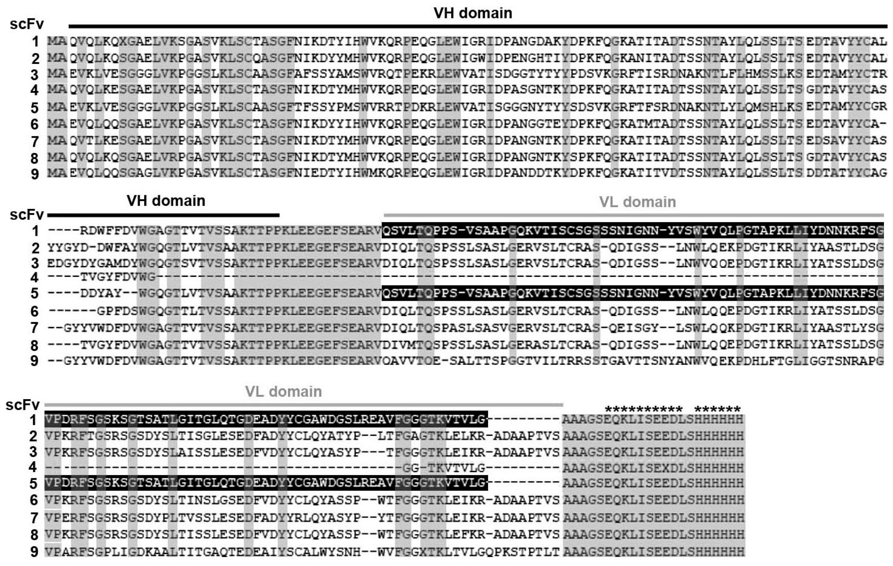
Analysis of the amino acid (a/a) sequences of 10 selected recombinant Abs (Table 1, Figure 3) demonstrated (1) 100% identity of independently selected scFv3 and 10, (2) the same VL part in scFv1 and 5, identified as a human IgG domain from the commercial pSEX81 phagemid that was retained during library construction, (3) no VL domain in scFv4, (4) no identical VL or VH fragments in other scFv Abs. VH domains of scFv4, 7 and 8 showed 91–93% identity, differing only in a few a/a residues. However, the identity of the VL parts of scFv7 and 8 was 81% and this domain was absent in scFv4. Similarly, scFv fragments 3, 6 and 8 showed the greatest similarity of VL domains (93–95% identity), but their VH parts differed more strongly (Table 1). Variable fragments of selected recombinant Abs have 87–93% (VHs) and 91–98% (VLs) identity with the most similar sequences deposited in GenBank. No fragment was completely identical to any of the deposited sequences.

2.3. Heterologous Expression of Selected scFv Fragments in Sf9 Cells

To express nine different NbAAC1-specific scFv fragments in insect cells, their genes were cloned into the pIB/V5-His plasmid. Lipofection of Lepidopteran Sf9 cultures with engineered plasmids followed by selection of transformants in the presence of the antibiotic blasticidin S and immunoblotting with anti-c-myc Abs demonstrated heterologous expression of cloned genes in insect cells (Figure 4). To correlate scFv accumulation in the transformants with the total protein concentrations in the samples, we repeated SDS-PAGE with the same aliquots and stained the gel with Coomassie. Since the lowest content of scFv 3, 6 and 8 in transformants correlated with lower protein concentrations in these samples, this generally indicates efficient expression of recombinant Abs in Sf9 cells. At the same time, the studied scFv fragments were differently expressed in the blasticidin-selected transformants, and the concentration of scFv1 significantly exceeded the content of other recombinant Abs. The single VH domain scFv4 showed the smallest molecular weight. The fragments scFv1 and 5 carrying VL part of a human IgG from the pSEX81 phagemid were also smaller than the other Abs.

2.4. Expression of NbAAC1-Specific scFv Abs Differently Affected the Parasite Intracellular Growth

To select a recombinant Ab with maximal N. bombycis inhibitory activity, nine blasticidin-resistant transformants were infected with 20 microsporidia spores per insect cell, and expression of the N. bombycis polar tube protein PTP2 gene was analyzed 7 days after inoculation, using real-time qPCR. To control the quality of RNA isolation and cDNA synthesis, the cutworm Spodoptera frugiperda cytochrome c oxidase subunit I (COXI) transcripts were analyzed in the same cDNA samples as a reference. A total of 64 independent infections repeated 4 to 9 times for each scFv variant were analyzed.
As shown in Table 2, the number of N. bombycis PTP2 transcripts relative to insect COXI ones varied in cell cultures expressing different scFv fragments. The most efficient development of the parasite was observed in cultures expressing scFv4, 6, and 8. The ΔCq (5.2–6) and NbPTP2 Cq (19.9–20.2) values in these variants were significantly different (p-value < 0.05) from the values detected in the cultures expressing six other Abs (6.9–8.5 and 20.9–22.7, respectively). Cloning of PCR products, specifically amplified with primers SfCOXI and NbPTP2, constructing of calibration curves and counting of transcripts in analyzed cDNA samples showed that cultures with the lowest (expression of scFv4) and highest (expression of scFv5) ΔCq demonstrated a tenfold difference in the number of NbPTP2 transcripts per thousand of SfCOXI ones (11 and 1.1 copies, respectively). The constructing of calibration curves also showed that PCR efficiencies was 92.4% and 122.4% for primers SfCOXI and NbPTP2, respectively.
Analyzing these results, we hypothesized that simultaneously effective (ΔCq ≤ 6) and inefficient (ΔCq ≥ 7) growth of microsporidia in infected cultures expressing the same scFv fragment may be due to its low suppressing activity in some experimental replicates, along with poor activation of spores or their aggregation and adhesion in others. Since scFv5, 7 and 9 showed the highest ΔCq values along with inefficient growth of N. bombycis (ΔCq ≥ 7) in all independently infected cell cultures (Table 2, right column), they were chosen for the next experiment. Retention of a human IgG VL domain from the commercial phagemid pSEX81 in scFv5 was an interesting result of the experiment, as this recombinant Ab showed the highest N. bombycis inhibitory activity. Since scFv fragments 5 and 1 carry the same “artificial” VL domain, the latter was also included in the next experiment.

2.5. The Effectiveness of scFv5 to Suppress Intracellular Growth of N. bombycis Was Confirmed by Additional Infections and qPCR Assays

Sf9 transformants expressing the four scFv Abs selected in the first experiment were infected with lower doses of N. bombycis spores. In this experiment, we reduced the multiplicity of infection to ten and three spores per insect cell (Table 3).
As expected, the SfCOXI Cq values in Table 2 and Table 3 were approximately the same (13.5–15.3 and 13.8–15.5, respectively), and the decrease in infection load was accompanied by an increase in NbPTP2 Cq (19.9–22.7 and 22.1–26.1, respectively). At the same time, we did not observe a clear relationship between a decrease in the multiplicity of infection from ten to three spores per cell and the number of parasite transcripts in cDNA samples. In any case, both infections at a ratio of ten and three spores per insect cell confirmed the highest suppressive activity of scFv5, showing a statistically significant difference in ΔCq values (Table 3). As in the first experiment, scFv5 again demonstrated the highest Cq, ΔCq values and the lowest number of NbPTP2 transcripts per million SfCOXI ones.
In the final qPCR experiment, we used another pair of NbPTP2 and SfCOXI-specific primers, and also added a control variant of cell cultures transformed with a plasmid carrying the gene encoding eGFP. Cell cultures expressing five scFv fragments and eGFP were infected with 50 microsporidia spores per insect cell, and after 7 days of cultivation, the content of parasite and insect transcripts was analyzed by qPCR with SfCOXI-2/NbPTP2-2 primers (Table 4). As in previous experiments, the results of qPCR with a new pair of primers showed that of all of the analyzed variants, scFv5 most effectively inhibits the intracellular growth of the pathogen, demonstrating the highest values of Cq NbPTP2-2 and ΔCq SfCOXI-2/NbPTP2-2 (Table 4). The lowest Cq and ΔCq showed scFv8 and these values were similar to those observed for the eGFP-expressing control cultures.

2.6. Western Blot Analysis of Infected Transformants with Abs against N. bombycis β-Tubulin Confirmed the Inhibitory Activity of scFv5

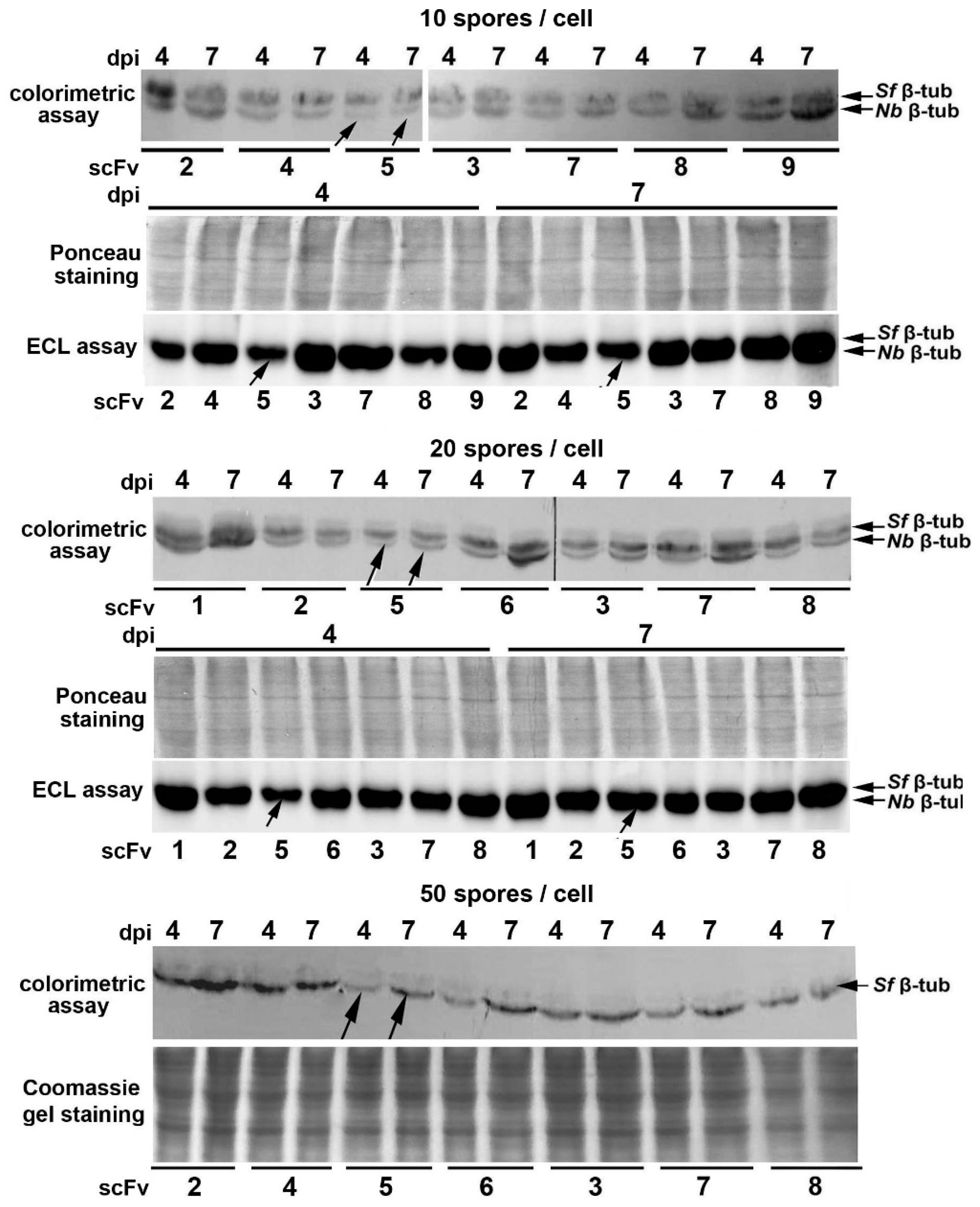
Previously, we produced polyclonal Abs against N. bombycis β-tubulin overexpressed in E. coli and demonstrated the promise of Western blotting for detecting intracellular growth of the parasite in infected Sf9 cell cultures [22]. In this study, we attempted to use immunoblotting with these Abs to further confirm the N. bombycis-inhibitory efficacy of scFv5 compared to other selected recombinant Abs. Blasticidin-selected transformants were infected with microsporidia at a ratio of 10, 20 or 50 spores per cell, and after cultivation for 1.5 h, 4 and 7 days, the samples for SDS-PAGE were prepared. According to the results of immunoblotting, parasite β-tubulin was not detected in freshly infected cultures (1.5 h after infection, not shown), but its detectable accumulation was found in 42 samples of 4- and 7-day cell cultures (Figure 5).
When cell cultures were infected with 10 or 20 spores per cell, Abs against N. bombycis β-tubulin stained two protein bands. The lower band corresponded to the parasite β-tubulin, and the upper one, to the homologous S. frugiperda protein [22]. When cell cultures were infected with 50 spores per cell, the parasite protein band was stained more brightly.
Although visual assessment of protein band staining after immunoblotting is not quantitative, this method provided additional evidence for the high N. bombycis-inhibitory activity of scFv5. A comparison of the staining intensity of the parasite β-tubulin in samples of cell cultures infected with three doses of spores on the 4th and 7th day after inoculation showed that in each of these six groups, the lowest growth of the pathogen was observed in the case of scFv5 expression (Figure 5, indicated by arrows).

2.7. Identification of Complementarity Determining Regions (CDRs) of the scFv5 Unique VH Domain

A blast analysis of the scFv5 VH domain a/a sequence showed that the most similar fragment deposited in GenBank (accession number 5DO2_C) has 86.9% identity.
The identification of the scFv5 VH CDR1 and CDR2 did not show similarity to corresponding sequences of other selected Abs, except for the VH part of scFv3 (Figure 6). In this case, the identity of CDR1 and CDR2 regions was 78% and 82%, respectively. As expected, the highly heterogeneous CDR3 region in the VH part of scFv5 was significantly different from the corresponding regions of all other selected VH fragments. In GenBank, we found only one VH domain with a CDR3 identical to the same scFv5 region (Figure 6, accession number BCB24250.1), but its CDR1 and CDR2 were significantly different from those of scFv5. The CDR1 and CDR2 of two other VH fragments (Figure 6, AAO19046.1 and CCQ13148.1) with most similar CDR3 were also not identical to scFv5 regions. The unique sequence of the VH domain of N. bombycis-inhibitory scFv5 was deposed in GenBank.

3. Discussion

To construct an immune library of scFv Abs specific to NbAAC1-3 fragments, we purified this chimeric protein of any bacterial impurities and immunized several mice to select a variant with high content of IgGs against the outer loops of each of the three N. bombycis ATP/ADP transporters. The representativeness of the constructed library (107 transformants) was also doubled compared to the previously constructed one against the microsporidium Vairimorpha ceranae hexokinase [23]. Despite this, we failed to isolate any recombinant Abs specific to loops of the second and third N. bombycis transporters. Since NbAAC2- and NbAAC3-specific Abs were detected in immune serum of the mouse selected for library construction (Figure 1B), the proportion of their VH and VL transcripts in the total pool of synthesized cDNA was probably insufficient to form specific scFv Abs.
Fortunately, transcriptome analysis of N. bombycis mature spores and sporoplasms (microsporidia embryos infecting new host cells) [24] demonstrated that the expression of NbAAC1 gene (accession number KB909159.1; 154353 FPKM (fragments per kilobase per million) for sporoplasms and 146706 ones for spores) was about 1500 and 15000 times higher than the transcription levels of NbAAC2 (KB908963.1; 113 and 128 FPKM) and NbAAC3 (KB909008.1; 9 and 4 FPKM, respectively) genes. This indicates that the NbAAC1 transporter plays the main role in the energy supply of the first stages of the parasite intracellular development and can be considered as the most promising target for microsporidia-suppressing recombinant Abs.
Since gene sequencing of the first 10 selected Abs showed that scFv3 and 10 were identical, nine different variants were expressed in Sf9 insect cells. The presence of the recombinant c-myc peptide at the C-terminus of Abs made it possible to confirm their expression in insect cell cultures by immunoblotting before searching for a variant that maximally suppresses parasite growth and is promising for further studies. Previously, it was shown that the analysis of microsporidia transcripts involved in sporogenesis but not expressed at the initial stages of infection may be successfully used to assay the parasite growth in cell cultures, regardless of a multiplicity of their infection [25]. In the case of infection of Sf9 cells with N. bombycis spores, the level of expression of the gene encoding the parasite PTP2 may serve as a such marker [22]. To match the number of parasite and insect host transcripts in the same cDNA samples, we used the expression level of the S. frugiperda COXI gene as a reference. Mitochondrial transcripts are polyadenylated in insect cells [26], and the gene encoding COXI is highly expressed [27].
Although the formation of N. bombycis spores in B. mori ovarian tissue cultures was observed as early as 2 days after infection [28], qPCR analysis of the parasite β-tubulin gene copies [13] and immunoblotting with Abs against the same protein [22] demonstrated the parasite growth in Sf9 cells between days 4 and 7 after inoculation. This is most likely due to the immediate germination of first-generation spores to infect new insect cells. Previously, the formation of such “early” spores was described for the Nosema species, including N. bombycis [29,30,31]. With these data in mind, we analyzed infected cultures at day 7 post-infection in the case of qPCR, and samples for immunoblotting were prepared using cell cultures 1.5 h, 4 and 7 days after inoculation.
The first infection of 64 cell cultures expressing nine recombinant Abs with 20 spores per insect cell showed that (1) the number of parasite transcripts per thousand of insect ones can vary in 10 times for transformants expressing different Abs, (2) scFv5 demonstrate the most effective N. bombycis-inhibiting activity. Subsequent experiments with different numbers of infecting spores per insect cell, other NbPTP2-2 and SfCOXI-2 primers, GFP-expressing control cell cultures, and immunoblotting confirmed the highest inhibitory activity of scFv5 compared to other selected variants. Interesting features of this recombinant Ab are (1) the retention of a human IgG VL fragment from the pSEX81 phagemid, (2) the unique sequence of its VH domain, including the CDRs.
Three years ago, we constructed an immune scFv library against fat body proteins of locust Locusta migratoria infected with the microsporidium Paranosema (Antonospora) locustae and selected scFv fragments specific to three secretory proteins of the parasite [32]. The following sequencing showed that two of them had the same VL domain of a human IgG (unpublished data). Recently, we also selected a recombinant Ab against the microsporidium V. ceranae hexokinase containing this VL domain from the commercial vector pSEX81 [23], and in this study we found it in two anti-VcAAC1 scFv fragments. Some characteristics of such scFv Abs with an “artificial” VL part should be similar to those of single domain Abs (sdAbs) carrying natural VHs of camelids [33,34] and cartilaginous fishes [35,36], or to “camelized” nanobodies [37,38]. Since a high diversity of VH CDR3 loops is sufficient to provide antigen specificity of any Ab, and this region is a key determinant of antigen recognition [39], the functional role of such “artificial” VL fragment in scFv molecules may be associated with an increase in their thermal stability and a decrease in aggregation tendencies. In the case of sdAbs it is ensured by a special repertoire of hydrophilic and cysteine residues in proteins [40,41,42]. Since we have found the “artificial” VL domain in several specific recombinant Abs, its presence does not appear to be critical for the antigen-binding activity of these scFv fragments.
Along with the search for new inhibitory Abs, further modification of scFv5 should be aimed at finding its native VL fragment, which is present in a natural mouse IgG not found in the constructed library, which appears to be insufficiently representative. Cloning a pool of mouse VL fragments into the pSEX81 phagemid with previously inserted scFv5 VH part followed by panning the new library can solve this problem. Another direction may be connected with the camelization of its VH fragment, which implies the engineering introduction of additional disulfide bonds to stabilize the tertiary structure [43], hydrophilic residues, as well as randomization of CDRs and especially CDR3.
The scFv3 fragment with the similar VH domain could also be the subject of such study. Although it did not show very effective N. bombycis inhibitory activity, (1) it is the only recombinant Ab selected twice; (2) it showed high specificity on dot blots (Figure 2A, scFv3, 10) and the lowest level of expression in Sf9 cells (Figure 4, scFv3); (3) its VH sequence similarity to that of scFv5 suggests recognition of the same epitope; (4) its relatively extended (12 a/a) CDR3 of the VH domain (Figure 6) is not identical to any sequence annotated in GenBank. It is possible that the replacement of its VL domain with another one or engineering modifications of its VH part will help to construct scFv or sdAb that effectively suppress the intracellular growth of N. bombycis.

4. Materials and Methods

4.1. Bacterial Overexpression of Chimeric Proteins and Development of Immune scFv Library

The constructing of plasmids and bacterial overexpression of the NbAACb chimeric protein carrying 18 outer hydrophilic domains of three N. bombycis ATP/ADP carriers, as well as its fragments NbAAC1, NbAAC2, NbAAC3, consisting of loops of individual transporters, were described previously [21]. To eliminate the C-terminal tag of NbAACb, the encoding sequence was amplified using its copy, previously inserted into the tagless variant of pRSETa [21] as a template, Phusion Flash High-Fidelity PCR Master Mix (Thermo Fisher Scientific, Waltham, MA, USA), forward T7 and reverse NbAAC3 (Table 5) primers.
The gel-purified PCR-product was re-cloned in the tagless pRSETa plasmid at the BamHI/HindIII sites, and the NbAAC1-3 devoid of any tags was overexpressed in E. coli, as it was previously described for other chimeric proteins [21].

4.2. Antigen Preparation and Immunization of Mice

To immunize mice, the NbAAC1-3, forming insoluble inclusion bodies (IBs) in E. coli, was solubilized in the presence of SDS-PAGE sample buffer (62.5 mM Tris-Cl (pH 6.8), 2% SDS, 5% 2-mercaptoethanol, 10% glycerol) at 95 °C for 15 min, and the chimeric protein was purified by SDS-PAGE in 12% gel using a PROTEAN® II xi cell (Bio-Rad, Hercules, CA, USA) followed by electroelution using the same manufacturer’s whole gel eluter. After electroelution, fractions containing pure chimeric protein were thoroughly dialyzed against TBS (50 mM Tris-Cl (pH 7.4), 150 mM NaCl) mixed with an equal volume of Freund’s adjuvant (complete for first injection, incomplete for subsequent ones) and four mice were immunized with four intraperitoneal injections (80 μg of protein per injection) at 10-day intervals. Ten days after the last immunization, sera were analyzed by immunoblotting with NbAAC1-3, NbAAC1, NbAAC2, NbAAC3 proteins transferred onto a nitrocellulose membrane. For staining of the chimeric proteins, we used immune sera, anti-mouse IgG polyclonal Abs conjugated with horseradish peroxidase (HRP) (Bio-Rad, Hercules, CA, USA) and 4-chloro-1-naphthol (Merck, Darmstadt, Germany) as a colorimetric substrate for HRP reaction [44].

4.3. Construction of the scFv Immune Library

For cDNA synthesis and amplification of IgG VH and VL variable domains, we used (1) RNA from mice whose serum contained Abs to the loops of all three N. bombycis transporters, (2) the Mouse IgG Library primer set (Progen Biotechnik, Heidelberg, Germany), (3) DreamTaq Green PCR Master mix (Thermo Fisher Scientific, Waltham, MA, USA). The gel-purified PCR products were cloned into the pSEX81 phagemid vector (Progen Biotechnik, Heidelberg, Germany) at the NcoI/HindIII (VH domains) and MluI/NotI (VL domains) restriction sites to construct an immune scFv library with a representativeness of about 10 million E. coli transformants (XL1-Blue MRF’ strain). All stages of library constructing, storage, infection with the M13 KO7ΔpIII hyperphage (Progen Biotechnik, Heidelberg, Germany), and phage production have been described previously [32].

4.4. Library Panning

Phages produced in 200 mL of bacterial culture were precipitated with PEG, resuspended in 3 mL of phage dilution buffer, and blocked with 4 volumes of TTBS (TBS plus 0.1% Tween-20) with 3% BSA for 30 min. NbAAC1, NbAAC2, and NbAAC3 transferred to a nitrocellulose membrane after SDS-PAGE were cut into triangles, squares, and stripes respectively, washed in TTBS, blocked in the presence of 3% BSA for 3 h at 25 °C, placed in a 15 mL tube with page suspension and incubated overnight at 4 °C. After incubation, the nitrocellulose figurines were thoroughly washed with TTBS, TBS, and phages bound to individual chimeric proteins were separately eluted in 0.5 mL of 0.1 M triethylamine for 5 min. Aspirated eluates were immediately neutralized with the same volume of 1 M Tris-Cl (pH 7.5) and stored on ice until bacteria infecting. E. coli XL1-Blue MRF’ cells were infected with eluted phages as described previously [23]. Their re-infection with helper phage, PEG precipitation, incubation with immobilized antigen, elution of bound phages, followed by re-infection of bacteria allowed for the second and third rounds of selection.

4.5. Bacterial Expression and Analysis of Selected scFv Fragments

The re-cloning of selected scFv genes into the pOPE101 plasmid (Progen Biotechnik, Heidelberg, Germany), their expression in E. coli, and the analysis of the ability of selected scFv antibodies to recognize NbAAC1-3, NbAAC1, NbAAC2, NbAAC3 by dot blotting and immunoblotting were performed as described previously [23,32]. The isolation of plasmid DNA from transformants producing NbAAC-specific scFv-fragments was followed by sequencing of their genes with pOPE101seq primers (Table 5) and alignment using Clustal W of MegAlign from DNASTAR’s Lasergene sequence analysis software [45]. Complementarity determining regions (CDRs) of the sequenced VH regions were predicted according to the instructions of Andrew C.R. Martin’s group at UCL (http://www.bioinf.org.uk/abs/info.html).
To express the scFv-encoding genes in insect cells, they were re-amplified with pOPEpIB primers (Table 5) and cloned into the pIB/V5-His vector at the SacI/XhoI restriction sites. The eGFP coding gene was PCR amplified using eGFP forward and reverse primers (Table 5), Phusion Flash High-Fidelity PCR Master Mix (Thermo Fisher Scientific, Waltham, MA, USA), pEGFP-N3 plasmid (Clontech Lab Inc., San Jose, CA, USA) as a template, and cloned into the same vector at the EcoRI/XhoI sites. Plasmids with inserted genes were purified, verified by sequencing with pIB seq forward and reverse primers (Table 5), and used to transfect Sf9 cells.

4.6. Heterologous Expression of scFv Abs in Sf9 Cells and Their Infection with N. bombycis Spores

S. frugiperda (Lepidoptera: Noctuidae) derived Sf9 cells from the ECACC General collection (ECACC 89070101) were cultured in Sf-900III SFM (Thermo Fisher Scientific, Waltham, MA, USA) and regularly maintained according to the manufacturer’s instructions. Transformation (lipofection) of adhesion cell cultures with 10 µg of pure plasmid DNA and selection of blasticidin-resistant transformants were performed as described previously [23].
To confirm the expression of the studied scFv fragments in Sf9 cells, 5 × 10⁵ blasticidin-selected non-infected transformants or control untransformed Sf9 cells were sonicated in 0.1 mL TBS and analyzed by immunoblotting with anti-c-Myc monoclonal Abs (Merck, Darmstadt, Germany) and HRP- conjugated anti-mouse IgG polyclonal Abs (Bio-Rad, Hercules, CA, USA). ChemiDoc MP Imaging System (Bio-Rad, Hercules, CA, USA) and Clarity™ Western ECL substrate (Bio-Rad, Hercules, CA, USA) were used to detect the HRP reaction.
N. bombycis spores were obtained from the Uzbek Research Institute of Sericulture in Tashkent, Uzbekistan. Their (1) isolation from fat bodies of experimentally infected Bombyx mori 5th instar caterpillars, (2) purification in the Percoll density gradient, (3) sterilization with the antiseptic Multicide (Sante Pharm, Moscow, Russia) [46], polar tube extrusion activation [47] have also been described previously [22]. To obtain infection rates of fifty, twenty, ten and three spores per insect cell, 2.5 × 10⁷, 107, 5 × 10⁶ or 1.5 × 10⁶ activated spores in 20 µL of 10 mM KOH were added to a culture plate well containing 5 × 10⁵ Sf9 cells in 500 µL of SF-900 III SFM medium (Thermo Fisher Scientific, Waltham, MA, USA). Infected cell cultures were maintained at 27 °C for 7 days without control of humidity and CO2 concentration. After culturing, pelleted at 600× g for 5 min cells were stored at −80 °C until RNA isolation, cDNA synthesis and RT-qPCR analysis.

4.7. qPCR Analysis of N. bombycis Growth in Infected Cultures

The isolation of total RNA using Trizol, DNAse I and RNA grade glycogen (Thermo Fisher Scientific, Waltham, MA, USA) was performed according to the manufacturer’s instructions. cDNA was synthesized in 20 μL reaction mixture containing 1 μg RNA, 1 μM oligo-dT primers, 0.6 mM dNTPs, 100 U RevertAid M-MuLV reverse transcriptase (RT), 20 U RNase inhibitor (all reagents were produced by Thermo Fisher Scientific, Waltham, MA, USA) as described previously [23].
To assay the content of N. bombycis polar tube protein PTP2 gene transcripts in infected transformants relative to the transcripts of the reference host gene of S. frugiperda COXI by real-time qPCR, cDNA, two pairs of primers SfCOXI/NbPTP2 or SfCOXI-2/NbPTP2-2 (Table 5) were mixed with 5× qPCRmix-HS SYBR (Evrogen, Moscow, Russia), following the manufacturer’s recommendations. For analysis, a CFX Opus real-time PCR detection system with a C1000 thermal cycler (Bio-Rad, Hercules, CA, USA) was used. Quantification cycle values (Cq, number of cycles required for fluorescence to reach a quantification threshold) were determined using the supplied software. To assess statistically significant differences between ΔCq values, one-way analysis of variance (one-way ANOVA) followed by Tukey’s test for post-hoc test (Table 2 and Table 3) or Kruskal-Wallis one-way analysis of variance followed by Dunn’s post-hoc test and relevant R functions (Table 4) were used.
To determine the number of transcripts in the analyzed samples, amplified with SfCOXI and NbPTP2 primers (Table 5) DNA fragments were cloned into the pAL-TA vector (Evrogen, Moscow, Russia), and qPCR analysis performed on 105–108 or 102–106 plasmid copies, respectively, was followed by construction of calibration curves and counting of transcripts in samples using NEBioCalculator v1.15.0 (https://nebiocalculator.neb.com/#!/qPCRGen).
The efficiency of the SfCOXI and NbPTP2 primers for qPCR was determined using the ThermoFisher Scientific QPCR Efficiency Calculator.

4.8. Analysis of N. bombycis β-Tubulin Accumulation in Infected Transformants

Sf9 cell cultures expressing selected scFv Abs were infected with 10, 20 and 50 spores per insect cell and cultured for 1.5 h, 4 and 7 days. After cultivation, they were analyzed by immunoblotting with polyclonal Abs against the parasite β-tubulin as described previously [22].

Author Contributions

V.V.D. and I.V.S. conceived and designed the experiments; S.A.T., V.S.Z. and V.V.D. carried out bacterial expression and purification of chimeric proteins; E.V.S. and I.V.S. performed all experiments with mice, immune sera analysis and immunoblotting; V.V.D., S.A.T. and V.S.Z. carried out construction the scFv library, its panning, expression of recombinant Abs in E. coli and analysis of their antigen-binding activity; V.S.Z. constructed plasmids for heterologous expression of scFv genes in insect cells; B.A.M. and D.A.I. performed cultivation of microsporidia N. bombycis in silkworms and isolation of spores; I.V.S. performed all experiments with transfection, cultivation and infection of Sf9 cell cultures; S.A.T. carried out RNA isolation from infected cell cultures, cloning of NbPTP2 and SfCOXI fragments, construction of calibration curves; V.V.D. performed synthesis of cDNA samples and their assay by real-time qPCR; A.V.D. carried out the statistical analysis; V.V.D. and I.V.S. supervised and edited the manuscript. All authors have read and agreed to the published version of the manuscript.

Funding

This work was supported by the Russian Science Foundation (RSF 18-16-00054).

Institutional Review Board Statement

The animal study protocol was approved by the Bioethics Committee of I.M. Sechenov Institute of Evolutionary Physiology and Biochemistry Russian Academy of Science (Protocol no. 3 of 19 March 2019).

Informed Consent Statement

Not applicable.

Data Availability Statement

Not applicable.

Acknowledgments

The sequencing was performed using equipment of the Core Centrum “Genomic Technologies, Proteomics and Cell Biology” in All-Russia Research Institute for Agricultural Microbiology.

Conflicts of Interest

The authors declare no conflict of interest.

References

  1. Didier, E.S.; Weiss, L.M. Overview of microsporidia and microsporidiosis. Protistology 2008, 5, 243–255. [Google Scholar]
  2. Solter, L.F.; Becnel, J.J.; Oi, D.H. Microsporidian entomopathogens. In Insect Pathology, 2nd ed.; Vega, F.E., Kaya, H.K., Eds.; Elsevier Inc.: Amsterdam, The Netherlands, 2012; pp. 221–263. [Google Scholar]
  3. Becnel, J.J.; Andreadis, T.G. Microsporidia in insects. In Microsporidia: Pathogens of Opportunity, 1st ed.; Weiss, L.M., Becnel, J.J., Eds.; John Wiley and Sons Inc.: Hoboken, NJ, USA, 2014; pp. 521–570. [Google Scholar]
  4. James, R.R.; Li, Z. From silkworms to bees: Diseases of beneficial Insects. In Insect Pathology, 2nd ed.; Vega, F.E., Kaya, H.K., Eds.; Elsevier Inc.: Amsterdam, The Netherlands, 2012; pp. 429–459. [Google Scholar]
  5. Bjørnson, S.; Oi, D. Microsporidia biological control agents and pathogens of beneficial insects. In Microsporidia: Pathogens of Opportunity, 1st ed.; Weiss, L.M., Becnel, J.J., Eds.; John Wiley and Sons Inc.: Hoboken, NJ, USA, 2014; pp. 635–670. [Google Scholar]
  6. Pan, G.; Xu, J.; Li, T.; Xia, Q.; Liu, S.L.; Zhang, G.; Li, S.; Li, C.; Liu, H.; Yang, L.; et al. Comparative genomics of parasitic silkworm microsporidia reveal an association between genome expansion and host adaptation. BMC Genom. 2013, 14, 186. [Google Scholar] [CrossRef] [PubMed] [Green Version]
  7. Hukuhara, T. The epizootiology of pebrine, one of the great scourges of sericulture. J. Biochem. Biotech. 2017, 1, 1–3. [Google Scholar] [CrossRef]
  8. Huang, Y.; Zheng, S.; Mei, X.; Yu, B.; Sun, B.; Li, B.; Wei, J.; Chen, J.; Li, T.; Pan, G.; et al. A secretory hexokinase plays an active role in the proliferation of Nosema bombycis. PeerJ 2018, 6, e5658. [Google Scholar] [CrossRef] [PubMed] [Green Version]
  9. Zheng, S.; Hang, Y.; Huang, H.; Yu, B.; Zhou, N.; Wei, J.; Pan, G.; Li, C.; Zhou, Z. The role of NbTMP1, a surface protein of sporoplasm, in Nosema bombycis infection. Parasit. Vectors 2021, 14, 81. [Google Scholar] [CrossRef]
  10. Chen, Y.; Wei, E.; Chen, Y.; He, P.; Wang, R.; Wang, Q.; Tang, X.; Zhang, Y.; Zhu, F.; Shen, Z. Identification and subcellular localization analysis of membrane protein Ycf 1 in the microsporidian Nosema bombycis. PeerJ 2022, 10, e13530. [Google Scholar] [CrossRef]
  11. Yao, M.; Wang, R.; Chen, Y.; He, P.; Wei, E.; Zhu, F.; Wang, Q.; Zhang, Y.; Tang, X.; Shen, Z. Identification and subcellular localization analysis of CCTα in microsporidian Nosema bombycis. Infect. Genet. Evol. 2022, 102, 105309. [Google Scholar] [CrossRef]
  12. Dong, Z.; Wu, Q.; Long, J.; Lu, B.; Zheng, N.; Hu, C.; Chen, P.; Hu, N.; Lu, C.; Pan, M. Silver nanoparticles are effective in controlling microsporidia. Mater. Sci. Eng. C 2021, 125, 112106. [Google Scholar] [CrossRef] [PubMed]
  13. Huang, Y.; Chen, J.; Sun, B.; Zheng, R.; Li, B.; Li, Z.; Tan, Y.; Wei, J.; Pan, G.; Li, C.; et al. Engineered resistance to Nosema bombycis by in vitro expression of a single-chain antibody in Sf9-III cells. PLoS ONE 2018, 13, e0193065. [Google Scholar] [CrossRef] [PubMed] [Green Version]
  14. Zhang, R.; Zheng, S.; Huang, H.; Sun, X.; Huang, Y.; Wei, J.; Pan, G.; Li, C.; Zhou, Z. Expression of anti-NbHK single-chain antibody in fusion with NSlmb enhances the resistance to Nosema bombycis in Sf9-III cells. Bull. Entomol. Res. 2022, 112, 502–508. [Google Scholar] [CrossRef]
  15. Tsaousis, A.D.; Kunji, E.R.; Goldberg, A.V.; Lucocq, J.M.; Hirt, R.P.; Embley, T.M. A novel route for ATP acquisition by the remnant mitochondria of Encephalitozoon cuniculi. Nature 2008, 453, 553–556. [Google Scholar] [CrossRef]
  16. Heinz, E.; Hacker, C.; Dean, P.; Mifsud, J.; Goldberg, A.V.; Williams, T.A.; Nakjang, S.; Gregory, A.; Hirt, R.P.; Lucocq, J.M. Plasma membrane-located purine nucleotide transport proteins are key components for host exploitation by microsporidian intracellular parasites. PLoS Pathog. 2014, 10, e1004547. [Google Scholar] [CrossRef] [Green Version]
  17. Dean, P.; Sendra, K.M.; Williams, T.A.; Watson, A.K.; Major, P.; Nakjang, S.; Kozhevnikova, E.; Goldberg, A.V.; Kunji, E.R.S.; Hirt, R.P.; et al. Transporter gene acquisition and innovation in the evolution of microsporidia intracellular parasites. Nat. Commun. 2018, 9, 1709. [Google Scholar] [CrossRef] [Green Version]
  18. Plano, G.V.; Winkler, H.H. Identification and initial topological analysis of the Rickettsia prowazekii ATP/ADP translocase. J. Bacteriol. 1991, 173, 3389–3396. [Google Scholar] [CrossRef] [Green Version]
  19. Tjaden, J.; Winkler, H.H.; Schwöppe, C.; Van Der Laan, M.; Möhlmann, T.; Neuhaus, H.E. Two nucleotide transport proteins in Chlamydia trachomatis, one for net nucleoside triphosphate uptake and the other for transport of energy. J. Bacteriol. 1999, 181, 1196–1202. [Google Scholar] [CrossRef] [Green Version]
  20. Kampfenkel, K.; Mohlmann, T.; Batz, O.; Van Montagu, M.; Inze, D.; Neuhaus, H.E. Molecular characterization of an Arabidopsis thaliana cDNA encoding a novel putative adenylate translocator of higher plants. FEBS Lett. 1995, 374, 351–355. [Google Scholar] [PubMed]
  21. Dolgikh, V.V.; Timofeev, S.A.; Zhuravlyov, V.S.; Senderskiy, I.V. Construction and heterologous overexpression of two chimeric proteins carrying outer hydrophilic loops of Vairimorpha ceranae and Nosema bombycis ATP/ADP carriers. J. Invertebr. Pathol. 2020, 171, 107337. [Google Scholar] [CrossRef] [PubMed]
  22. Dolgikh, V.V.; Senderskiy, I.V.; Zhuravlyov, V.S.; Ignatieva, A.N.; Timofeev, S.A.; Ismatullaeva, D.A.; Mirzakhodjaev, B.A. Molecular analysis of the microsporidia Vairimorpha ceranae and Nosema bombycis growth in the lepidoptera Sf9 cell culture. Protistology 2022, 16, 21–29. [Google Scholar] [CrossRef]
  23. Dolgikh, V.V.; Zhuravlyov, V.S.; Senderskiy, I.V.; Ignatieva, A.N.; Timofeev, S.A.; Seliverstova, E.V. Heterologous expression of scFv fragment against Vairimorpha (Nosema) ceranae hexokinase in Sf9 cell culture inhibits microsporidia intracellular growth. J. Invertebr. Pathol. 2022, 191, 107755. [Google Scholar] [CrossRef]
  24. He, Q.; Luo, J.; Xu, J.; Wang, C.; Meng, X.; Pan, G.; Li, T.; Zhou, Z. Morphology and transcriptome analysis of Nosema bombycis sporoplasm and insights into the initial infection of microsporidia. mSphere 2020, 5, e00958-19. [Google Scholar] [CrossRef] [Green Version]
  25. Gisder, S.; Genersch, E. Identification of candidate agents active against N. ceranae infection in honey bees: Establishment of a medium throughput screening assay based on N. ceranae infected cultured cells. PLoS ONE 2015, 10, e0117200. [Google Scholar] [CrossRef] [PubMed]
  26. Bratic, A.; Clemente, P.; Calvo-Garrido, J.; Maffezzini, C.; Felser, A.; Wibom, R.; Wedell, A.; Freyer, C.; Wredenberg, A. Mitochondrial Polyadenylation Is a One-Step Process Required for mRNA Integrity and tRNA Maturation. PLoS Genet. 2016, 12, e1006028. [Google Scholar] [CrossRef] [Green Version]
  27. Gao, S.; Ren, Y.; Sun, Y.; Wu, Z.; Ruan, J.; He, B.; Zhang, T.; Yu, X.; Tian, X.; Bu, W. PacBio full-length transcriptome profiling of insect mitochondrial gene expression. RNA Biol. 2016, 13, 820–825. [Google Scholar] [CrossRef] [Green Version]
  28. Ishihara, R. The life cycle of Nosema bombycis as revealed in tissue culture cells of Bombyx mori. J. Invertebr. Pathol. 1969, 14, 316–320. [Google Scholar] [CrossRef]
  29. Iwano, H.; Ishihara, R. Intracellular germination of spores of a Nosema sp. immediately after their formation in cultured cell. J. Invertebr. Pathol. 1989, 54, 125–127. [Google Scholar] [CrossRef]
  30. Iwano, H.; Ishihara, R. Dimorphism of spores of Nosema spp. in cultured cell. J. Invertebr. Pathol. 1991, 57, 211–219. [Google Scholar] [CrossRef]
  31. Iwano, H.; Kurtti, T.J. Identification and isolation of dimorphic spores from Nosema furnacalis (Microspora: Nosematidae). J. Invertebr. Pathol. 1995, 65, 230–236. [Google Scholar] [CrossRef]
  32. Tsarev, A.A.; Senderskiy, I.V.; Timofeev, S.A.; Zhuravlyov, V.S.; Dolgikh, V.V. Recombinant single chain antibodies as an instrument to search proteins involved in interaction of microsporidia and other intracellular parasites with infected host cell. Protistology 2019, 13, 5–13. [Google Scholar] [CrossRef]
  33. Hamers-Casterman, C.; Atarhouch, T.; Muyldermans, S.; Robinson, G.; Hamers, C.; Songa, E.B.; Bendahman, N.; Hamers, R. Naturally occurring antibodies devoid of light chains. Nature 1993, 363, 446–448. [Google Scholar] [CrossRef]
  34. Muyldermans, S.; Atarhouch, T.; Saldanha, J.; Barbosa, J.A.; Hamers, R. Sequence and structure of VH domain from naturally occurring camel heavy chain immunoglobulins lacking light chains. Protein Eng. 1994, 7, 1129–1135. [Google Scholar] [CrossRef]
  35. Greenberg, A.S.; Avila, D.; Hughes, M.; Hughes, A.; McKinney, E.C.; Flajnik, M.F. A new antigen receptor gene family that undergoes rearrangement and extensive somatic diversification in sharks. Nature 1995, 374, 168–173. [Google Scholar] [CrossRef]
  36. Zielonka, S.; Empting, M.; Grzeschik, J.; Könning, D.; Barelle, C.J.; Kolmar, H. Structural insights and biomedical potential of IgNAR scaffolds from sharks. MAbs 2015, 7, 15–25. [Google Scholar] [CrossRef] [Green Version]
  37. Riechmann, L.; Muyldermans, S. Single domain antibodies: Comparison of camel VH and camelised human VH domains. J. Immunol. Methods 1999, 231, 25–38. [Google Scholar] [CrossRef]
  38. Muyldermans, S.; Cambillau, C.; Wyns, L. Recognition of antigens by single-domain antibody fragments: The superfluous luxury of paired domains. Trends. Biochem. Sci. 2001, 26, 230–235. [Google Scholar] [CrossRef]
  39. Xu, J.L.; Davis, M.M. Diversity in the CDR3 region of VH is sufficient for most antibody specificities. Immunity 2000, 13, 37–45. [Google Scholar] [CrossRef] [Green Version]
  40. Rouet, R.; Dudgeon, K.; Christie, M.; Langley, D.; Christ, D. Fully human VH single domains that rival the stability and cleft recognition of camelid antibodies. J. Biol. Chem. 2015, 290, 11905–11917. [Google Scholar] [CrossRef] [Green Version]
  41. Shinozaki, N.; Hashimoto, R.; Fukui, K.; Uchiyama, S. Efficient generation of single domain antibodies with high affinities and enhanced thermal stabilities. Sci. Rep. 2017, 7, 5794. [Google Scholar] [CrossRef] [PubMed] [Green Version]
  42. Bekker, G.J.; Ma, B.; Kamiya, N. Thermal stability of single-domain antibodies estimated by molecular dynamics simulations. Protein Sci. 2019, 28, 429–438. [Google Scholar] [CrossRef] [Green Version]
  43. McAuley, A.; Jacob, J.; Kolvenbach, C.G.; Westland, K.; Lee, H.J.; Brych, S.R.; Rehder, D.; Kleemann, G.R.; Brems, D.N.; Matsumura, M. Contributions of a disulfide bond to the structure, stability, and dimerization of human IgG1 antibody CH3 domain. Protein Sci. 2008, 17, 95–106. [Google Scholar] [CrossRef] [PubMed] [Green Version]
  44. Dolgikh, V.V.; Seliverstova, E.V.; Naumov, A.M.; Senderskiy, I.V.; Pavlova, O.A.; Beznoussenko, G.V. Heterologous expression of pyruvate dehydrogenase E1 subunits of the microsporidium Paranosema (Antonospora) locustae and immunolocalization of the mitochondrial protein in amitochondrial cells. FEMS Microbiol. Lett. 2009, 293, 285–291. [Google Scholar] [CrossRef] [PubMed] [Green Version]
  45. Burland, T.G. DNASTAR’s Lasergene sequence analysis software. Methods Mol. Biol. 2000, 132, 71–91. [Google Scholar] [PubMed]
  46. Tetz, G.V.; Artemenko, N.K.; Yankovskii, G.M.; Kever, L.V.; Komissarchik, Y.Y.; Tetz, V.V. Effects of multicide, antibacterial drug, on Staphylococcus biomembranes. Bull. Exp. Biol. Med. 2017, 163, 780–784. [Google Scholar] [CrossRef] [PubMed]
  47. Ohshima, K. On the function of the polar filament of Nosema bombycis. Parasitology 1937, 29, 220–224. [Google Scholar] [CrossRef]
Figure 1. (A) NbAAC1-3 chimeric protein carrying the outer loops of three N. bombycis ATP/ADP transporters was overexpressed in E. coli, purified by SDS-PAGE and gel electroelution. The pure protein fractions, marked with asterisks, were dialyzed and used to immunize mice. (B) Western blot analysis of immune sera from individual mice allowed us to select a mouse whose serum showed similar levels of Abs to the loops of all three N. bombycis transporters (marked with an asterisk) and use it to construct the scFv library. As a negative control, we used the serum of a mouse immunized with the chimeric VcAAC1-4 protein carrying the outer loops of the microsporidium Vairimorpha ceranae four ATP/ADP transporters [21] also overexpressed in E. coli and purified.
Ijms 23 15307 g001
Figure 2. (A) Dot blots of 10 selected recombinant Abs showed their specific recognition of the chimeric proteins NbAAC1 and NbAAC1-3 but not NbAAC2 and NbAAC3. Dot blot analysis of some recombinant Abs selected against NbAAC2 and NbAAC3 demonstrated non-specific staining of three or four chimeric proteins placed at the corners of nitrocellulose squares, but none of the scFv variants specifically recognized these NbAAC1-3 fragments. (B) As a control, we used the VcAAC1-4 chimeric protein carrying the outer loops of four microsporidia Vairimorpha ceranae ATP/ADP transporters recognized by previously constructed VcAAC1- and VcAAC3-specific recombinant Abs. (C) Binding of selected scFv fragments to the NbAAC1 protein band, but not to NbAAC2 or NbAAC3, was shown by immunoblotting.
Ijms 23 15307 g002
Figure 3. Multiple a/a sequence alignment of nine selected scFv antibodies. VH and VL domains are indicated by black and gray horizontal lines respectively, gray rectangles indicate conserved residues present in all selected scFv fragments, the human VL domain from the pSEX81 phagemid retained in scFv1 and scFv5 is marked in inverted color, recombinant c-Myc and polyHis peptides are marked with asterisks.
Ijms 23 15307 g003
Figure 4. Western blotting with anti-c-myc monoclonal and HRP-conjugated anti-mouse IgG Abs confirmed expression of the nine scFv fragments in blasticidin-selected transformants. The concentration of scFv1 in the Sf9 cell culture significantly exceeded the content of other recombinant Abs. As expected, the scFv4 Ab with a single VH domain showed the lowest molecular weight, and the scFv1 and 5 fragments retaining the VL part from the pSEX81 phagemid were also smaller than the other Abs. To detect the HRP reaction, ChemiDoc MP Imaging System and Clarity™ Western ECL substrate were used. For the ECL assay of the nitrocellulose membrane, the optimal autoexposure settings of the Imaging System were used. To obtain a longer exposure, the very bright scFv1 lane was removed before the second run of analysis. To obtain the third small ECL image, we only analyzed the scFv3 and scFv6 lanes.
Ijms 23 15307 g004
Figure 5. Western blot analysis with Abs against N. bombycis β-tubulin showed that in 6 experimental groups representing equal number of Sf9 transformants infected with three doses of spores and cultivated for 4 and 7 days, the lowest N. bombycis growth was observed in the case of scFv5 expression (marked by arrows). The lower detected bands corresponded to the parasite β-tubulin, and the upper one, to the S. frugiperda homologous protein [22]. To detect the HRP reaction, 4-chloro-1-naphthol as a colorimetric substrate or ChemiDoc MP Imaging System and Clarity™ Western ECL substrate were used.
Ijms 23 15307 g005
Figure 6. Analysis of CDRs (complementarity determining regions) of VH domains of selected recombinant Abs and three annotated in GenBank fragments with CDR3 most similar to scFv5 one. CDRs of scFv5 are framed, and identical a/a residues are marked with gray rectangles.
Ijms 23 15307 g006
Table 1. Similarity (% identity) of the a/a sequences of the VH and VL fragments of the selected recombinant Abs.
VH9 *8765432
186.289.789.787.645.686.248.384.5
276.383.683.18554.482.850.4 36.12
35049.150.852.284.249.1 88.6373
481.993.190.58546.5 3636.91005
547.446.548.248.7 37.894.887.7376
685.886.786.7 82.836.983.583.3387
787.392.2 8194.836.99386.8388
885.3 35.334.536.242.335.73640.79
8765321VL
*—numbers of selected scFv Abs are in bold and underlined.
Table 2. Real-time qPCR analysis of N. bombycis PTP2 and S. frugiperda COXI transcripts in scFv-expressing Sf9 transformants infected with 20 microsporidia spores per insect cell and cultured for 7 days.
scFvCq Values * and Number of
Transcripts in 1 μL of cDNA
Samples (in Brackets)
ΔCq ** and Number of NbPTP2 Transcripts per 103 SfCOXI Ones
(in Brackets)
N Samples and Variants with
ΔCq ≥ 7
(in Brackets)
SfCOXINbPTP2
113.8 ± 0.2
(7.1 × 10⁶)
20.9 ± 0.6
(20.7 × 10³)
7.1 ± 0.6 a
(2.9)
9 (6)
214.2 ± 0.3
(5.4 × 10⁶)
21.6 ± 0.5
(12.3 × 10³)
7.4 ± 0.6 a
(2.3)
9 (4)
315.3 ± 0.5
(2.7 × 10⁶)
22.2 ± 0.4
(7.9 × 10³)
6.9 ± 0.2 a
(3)
6 (3)
414.7 ± 0.2
(3.9 × 10⁶)
19.9 ± 0.7
(43.3 × 10³)
5.2 ± 0.6 b
(11)
5 (1)
513.5 ± 0.3
(8.6 × 10⁶)
22 ± 0.3
(9.2 × 10³)
8.5 ± 0.5 a
(1.1)
8 (8)
6 14.3 ± 0.2
(5.1 × 10⁶)
20.1 ± 0.4
(37. 4 × 10³)
5.8 ± 0.4 b
(7.3)
9 (2)
714 ± 0.3
(6.2 × 10⁶)
21.8 ± 0.3
(10.6 × 10³)
7.8 ± 0.5 a
(1.7)
4 (4)
814.2 ± 0.5
(5.4 × 10⁶)
20.2 ± 0.4
(34.7 × 10³)
6 ± 0.6 b
(6.4)
8 (1)
914.7 ± 0.3
(3.9 × 10⁶)
22.7 ± 0.5
(5.4 × 10³)
8 ± 0.3 a
(1.4)
6 (6)
*—mean of values from 4–9 independent infections (n = 12–27) ± SE; **—different letters indicate statistically significant differences (p-value < 0.05) between scFv variants as analyzed by one-way analysis of variance (one-way ANOVA) followed by Tukey’s test for post-hoc analysis.
Table 3. Real-time qPCR analysis of N. bombycis PTP2 and S. frugiperda COXI transcripts in 7-day old Sf9 cultures expressing four recombinant Abs and infected with ten and three microsporidia spores per insect cell.
scFvSpores per CellCq Values * and Number of Transcripts
in 1 μL of cDNA Samples (in Brackets)
ΔCq ** and Number of NbPTP2 Transcripts per 106 SfCOXI Ones (in Brackets)
SfCOXINbPTP2
11015.5 ± 0.4
(2.3 × 10⁶)
23.2 ± 0.2
(3.8 × 10³)
7.7 ± 0.5 a
(1.7 × 10³)
51014.4 ± 0.2
(5.1 × 10⁶)
25.1 ± 0.3
(1.2 × 10³)
10.7 ± 0.4 b
(2.4 × 10²)
71014.8 ± 0.2
(3.7 × 10⁶)
23.7 ± 0.2
(2.6 × 10³)
8.9 ± 0.2 a
(7 × 10²)
91013.8 ± 0.3
(7.1 × 10⁶)
22.1 ± 0.3
(8.5 × 10³)
8.3 ± 0.9 a
(1.2 × 10³)
1314.7 ± 0.1
(3.9 × 10⁶)
22.4 ± 0.1
(6.8 × 10³)
7.7 ± 0.2 a
(1.7 × 10³)
5315.5 ± 0.3
(2.3 × 10⁶)
26.1 ± 0.2
(4.4 × 10²)
10.6 ± 0.2 b
(1.9 × 10²)
7315.3 ± 0.6
(2.7 × 10⁶)
24.1 ± 0.9
(1.9 × 10³)
8.8 ± 0.4 a
(7 × 10²)
9314.9 ± 0.2
(3.4 × 10⁶)
23.9 ± 0.2
(2.2 × 10³)
9 ± 0.3 a
(6.5 × 10²)
*—mean of values from three independent infections (n = 9) ± SE; **—different letters indicate statistically significant differences (p-value < 0.05) between ΔCq values in scFv5 and other samples as analyzed by one-way analysis of variance (one-way ANOVA) followed by Tukey’s test for post-hoc analysis.
Table 4. Real-time qPCR analysis of N. bombycis PTP2 and S. frugiperda COXI transcripts with primers SfCOXI-2 and NbPTP2-2 in Sf9 cultures expressing five NbAAC1-specific recombinant Abs and eGFP (control). Blasticidin-selected transformants were infected with 50 microsporidia spores per insect cell and incubated 7 days prior to assay.
scFvCq *ΔCq **
SfCOXI-2NbPTP2-2
219.4 ± 0.121.7 ± 0.12.3 ± 0.1 a
319.7 ± 0.822.1 ± 0.42.4 ± 0.5 a
520.6 ± 0.426.4 ± 15.8 ± 0.6 b
718.1 ± 0.320.4 ± 0.42.3 ± 0.4 a
819.2 ± 0.119.6 ± 0.10.4 ± 0.1 a
eGFP18.5 ± 0.219.1 ± 0.20.6 ± 0.2 a
*—mean of values from 2 independent infections (n = 6) ± SE; **—different letters indicate statistically significant differences (p-value < 0.05) between scFv variants as analyzed by Kruskal-Wallis test followed by Dunn’s post-hoc test.
Table 5. List of primers used for PCR amplification and sequencing of N. bombycis, S. frugiperda, scFv genes and their fragments.
PrimerSequence (5′-3′)
NbAAC3 rev aTGCAAGCTTACGGTGCAGCACGACGAATGTTGTC
pOPE101seq forAAGAGGAGAAATTAACCATGA
pOPE101seq revTCATTAGCACAGGCCTCTAGA
pOPEpIB for bACGGAGCTCGGTACCTGCTGCTGGCAGCTCAGCCGGCCATG
pOPEpIB rev cGCTGAATTCTCGAGTTAATGATGATGGTGATGATGGGATAG
GFP for dCATGAATTCATGGTGAGCAAGGGCGAGGAGCTG
GFP rev eTCACTCGAGTTACTTGTACAGCTCGTCCATGCCGA
pIB seq forCGCAACGATCTGGTAAACAC
pIB seq revGACAATACAAACTAAGATTTAGTCAG
SfCOXI forTTTGAGCAGGAATAGTAGGT
SfCOXI revTAAAGATGGGGGTAAAAGT
SfCOXI-2 forTACCGCATTTTTATTATTATTATC
SfCOXI-2 revGTTTCCTTTTTACCTCTTTCTTGA
NbPTP2 forTGGCATCAGTAGCTCCTCCTCAAG
NbPTP2 revACGGCCCTAGCTGCTGTTTCAA
NbPTP2-2 forCAATAATCCAGCCGAGTGTCAA
NbPTP2-2 revAGTGGGGTACCTTCAGCAGTTT
aHindIII site and stop codon are underlined; bSacI/KpnI sites are underlined; cEcoRI/XhoI and stop codon are underlined; dEcoRI site is underlined; eXhoI site and stop codon are underlined.
Publisher’s Note: MDPI stays neutral with regard to jurisdictional claims in published maps and institutional affiliations.