Article

The DmeRF System Is Involved in Maintaining Cobalt Homeostasis in Vibrio parahaemolyticus

1
Jiangsu Key Laboratory of Zoonosis, Yangzhou University, Yangzhou 225009, China
2
Joint International Research Laboratory of Agriculture and Agri-Product Safety of MOE, Yangzhou University, Yangzhou 225009, China
*
Author to whom correspondence should be addressed.
These authors contributed equally to this work.
Int. J. Mol. Sci. 2023, 24(1), 414; https://doi.org/10.3390/ijms24010414
Submission received: 23 October 2022 / Revised: 18 December 2022 / Accepted: 23 December 2022 / Published: 27 December 2022
(This article belongs to the Section Molecular Microbiology)

Abstract

:
Although cobalt (Co) is indispensable for life, it is toxic to cells when accumulated in excess. The DmeRF system is a well-characterized metal-response system that contributes to Co and nickel resistance in certain bacterial species. The Vibrio parahaemolyticus RIMD 2210633 genome also harbors a dmeRF operon that encodes a multiple antibiotic resistance regulator family transcriptional regulator and a cation diffusion facilitator family protein. Quantitative real-time PCR, growth curves analysis, inductively coupled plasma-mass spectrometry, β-galactosidase activity assays, electrophoretic mobility shift assays, and a mouse infection experiment were performed to characterize the function of the DmeRF system in V. parahaemolyticus. Zinc, copper, and Co significantly increase dmeF expression, with Co inducing the greatest increase. DmeF promotes V. parahaemolyticus growth under high-Co conditions. Additionally, increased accumulation of cellular Co in the ΔdmeF mutant indicates that DmeF is potentially involved in Co efflux. Moreover, DmeR represses the dmeRF operon by binding directly to its promoter in the absence of Co. Finally, the DmeRF system was not required for V. parahaemolyticus virulence in mice. Collectively, our data indicate that the DmeRF system is involved in maintaining Co homeostasis in V. parahaemolyticus and DmeR functioning as a repressor of the operon.

1. Introduction

Metals, such as cobalt (Co) and zinc (Zn), are indispensable for almost all organisms. Indeed, many proteins require metals as structural components or enzymatic cofactors [1]. Vertebrate hosts have evolved a strategy, termed nutritional immunity, to decrease the availability of metals to invading bacteria [2]. Consequently, bacteria have developed numerous and varied countermeasures such as the utilization of high-affinity metal acquisition systems [3]. Despite their essential roles, metals are harmful to bacteria when accumulated in excess [4]. Remarkably, increasing evidence shows that hosts exploit metal toxicity to defend against bacterial pathogens [5,6]. Metal homeostasis disturbance can lead to mismetalation (i.e., protein metalation with a non preferred metal) or the generation of reactive oxygen species (ROS) via the Fenton reaction; both are detrimental to cells [7]. Therefore, metal homeostasis should be tightly regulated. Since metals cannot be synthesized or degraded, bacteria maintain their homeostasis mainly through the modulation of metal import and export [4].
Co is a micronutrient that participates in various metabolic processes. Most notably, it is required by vitamin B12 and certain proteins [8,9]. Nonetheless, excessive amounts of Co lead to toxicity. It can cause cell damage by catalyzing the production of ROS [10]. Moreover, Co competes with iron (Fe) in a variety of metabolic processes [11,12,13]. To prevent Co toxicity, efflux systems, such as P1B-4-type ATPase and cation diffusion facilitator (CDF), are employed to eliminate excessive amounts of Co [14]. In Streptococcus suis, a P1B-4-type ATPase, PmtA, acts as a ferrous iron (Fe[II]) and Co efflux pump [15]. Other examples of Co-exporting P1B-4-type ATPases include CoaT from Synechocystis and CtpD from Mycobacterium smegmatis [16,17]. DmeF, a CDF family protein, was first identified as playing a crucial role in Co homeostasis in Cupriavidus metallidurans (formerly called Wautersia metallidurans) [18]. Subsequently, DmeF homologues have been shown to be essential for Co and nickel (Ni) resistance in certain Rhizobiaceae species, including Sinorhizobium meliloti, Agrobacterium fabrum (formerly called Agrobacterium tumefaciens), and Rhizobium leguminosarum [14,19,20,21]. The gene dmeR encodes a RcnR/CsoR family metal-responsive transcriptional regulator. Typically, dmeF and dmeR are organized in an operon (dmeRF) where DmeR modulates the transcription of the operon [14,19,20].
Vibrio parahaemolyticus (family Vibrionaceae and order Vibrionales) is a Gram-negative, halophilic, curved-rod-shaped bacterium that can be found in estuarine, coastal, and marine environments [22,23]. It is also a major food-borne pathogen that primarily causes acute gastroenteritis in humans. Generally, gastroenteritis caused by V. parahaemolyticus is associated with the ingestion of contaminated seafood [24]. Occasionally, V. parahaemolyticus causes wound infections (leading to cellulitis or necrotizing fasciitis) and septicemia [25]. In addition to human infections, V. parahaemolyticus is responsible for acute hepatopancreatic necrosis disease in shrimp, which results in considerable economic losses in shrimp aquaculture [26,27]. Whole genome sequencing of RIMD 2210633, a V. parahaemolyticus clinical isolate, has revealed that this bacterium uses a mechanism distinct from that of Vibrio cholerae to establish infection [28]. Furthermore, a locus encoding a DmeF homologue was identified in the genome.
This study aimed to characterize the function of the DmeRF system in V. parahaemolyticus. This system was found to play a central role in Co homeostasis. Moreover, the dmeRF operon is repressed by DmeR in the absence of Co.

2. Results

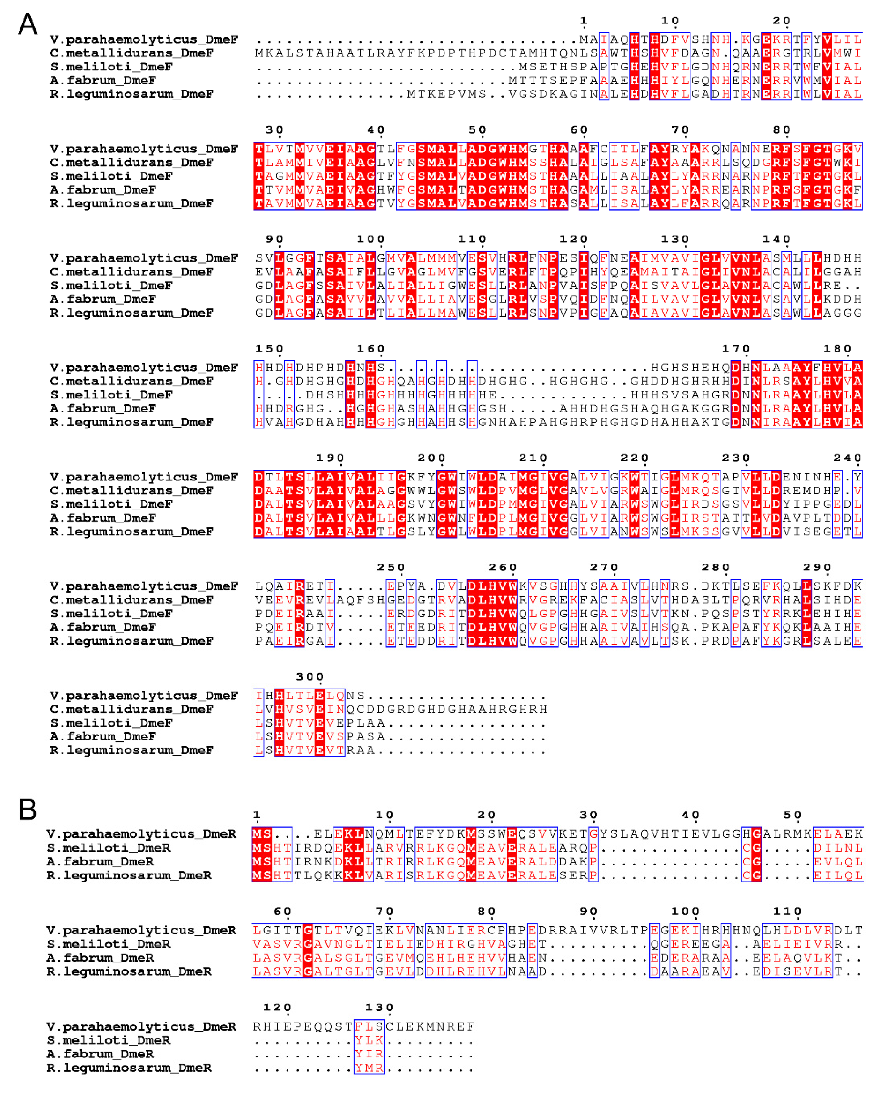
2.1. Identification of the DmeRF System in V. parahaemolyticus

In the genome of V. parahaemolyticus RIMD 2210633, the locus VP_RS21330 is annotated as dmeF, whose product is the CDF family Co/Ni efflux transporter DmeF. This protein exhibits 50.72%, 49.68%, 46.15%, and 44.68% amino acid sequence identity with DmeF from C. metallidurans, S. meliloti, A. fabrum, and R. leguminosarum, respectively. The locus VP_RS21325, immediately adjacent to and co-transcribed with dmeF (Figure S1), encodes a multiple antibiotic resistance regulator (MarR) family transcriptional regulator. No significant similarity was found between this protein and DmeR from the Rhizobiaceae species. Despite this, VP_RS21325 was designated as dmeR based on its regulatory role in dmeF expression (see Section 2.5). In the Rhizobiaceae species, dmeR is located upstream of dmeF [14,19,20,21], but in V. parahaemolyticus, the reverse applies (Figure 1). Multiple sequence alignment also showed that the DmeF proteins from V. parahaemolyticus, C. metallidurans, and the Rhizobiaceae species are highly conserved, while the DmeR protein from V. parahaemolyticus shows little homology with those of the Rhizobiaceae species (Figure 2). Together, these results revealed that the V. parahaemolyticus DmeF may have functions similar to its homologues from C. metallidurans and Rhizobiaceae.

2.2. The V. parahaemolyticus dmeF Gene Is Inducible by Zinc, Copper, and Cobalt

To evaluate the involvement of DmeF in the response of V. parahaemolyticus to metal toxicity, dmeF expression in RIMD 2,210,633 incubated with elevated levels of various metals was determined by quantitative real-time PCR (qRT-PCR) analysis. As seen in Figure 3, dmeF expression increased approximately 14-fold after the Co treatment compared to the H2O treatment. Furthermore, the Zn and Cu treatments increased dmeF expression by approximately 4-fold and 5-fold, respectively, while Fe(II), Mn, and Ni had no significant effect on dmeF expression (Figure 3).

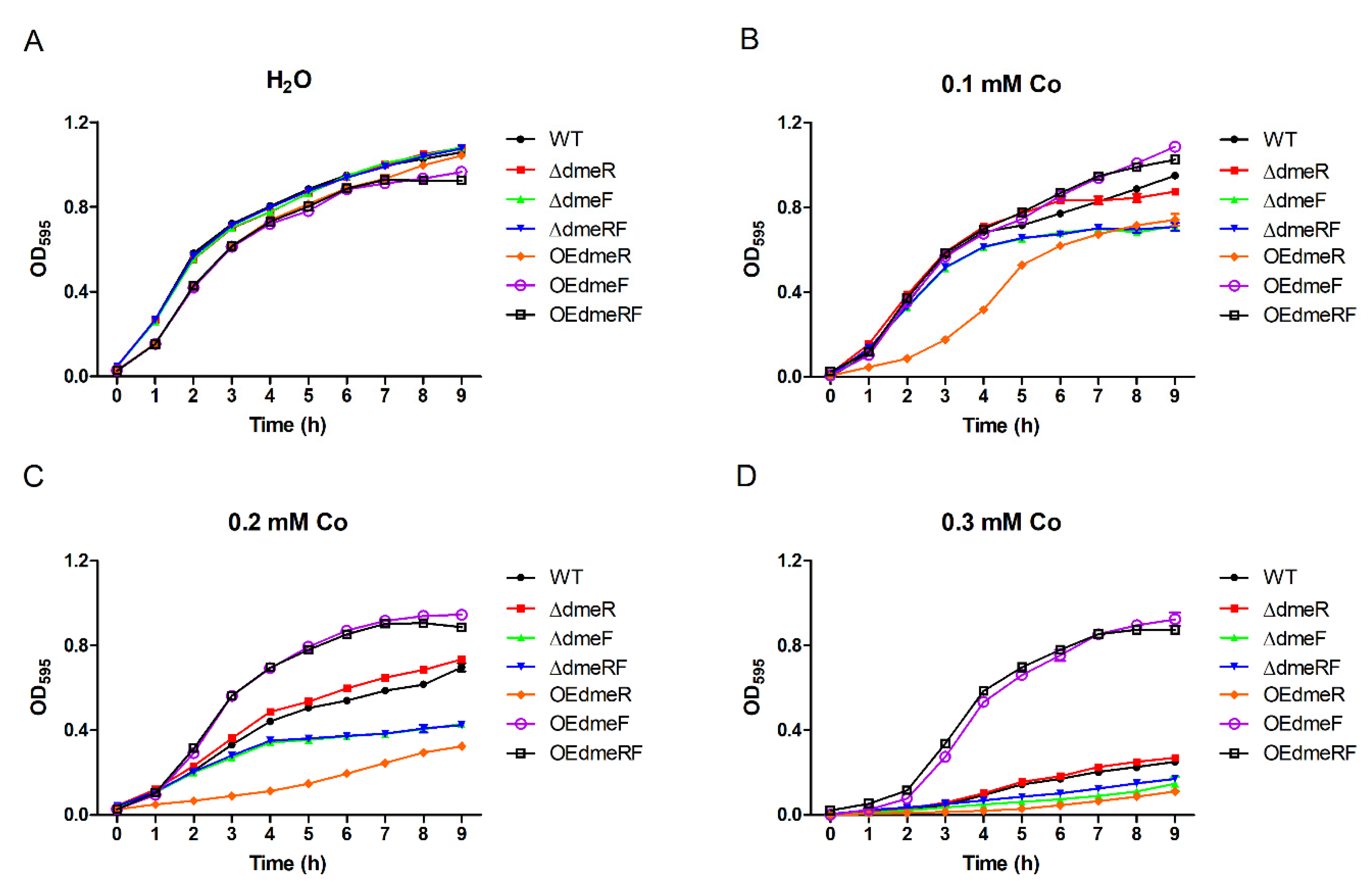
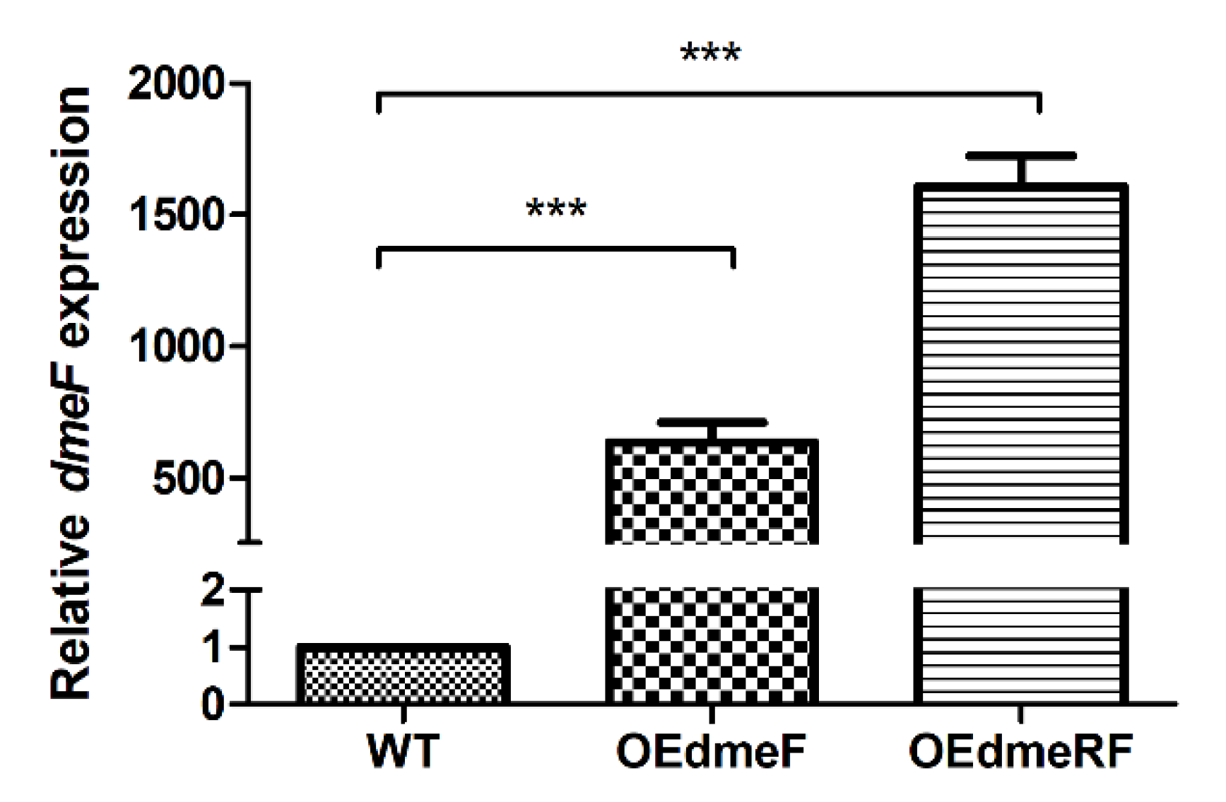
2.3. DmeF Contributes to V. parahaemolyticus Growth under High-Cobalt Conditions

To explore the role of DmeRF in V. parahaemolyticus physiology, we generated gene deletion mutants (including the single mutants ΔdmeR and ΔdmeF, and the double mutant ΔdmeRF) and overexpression strains in the corresponding mutant’s background (OEdmeR, OEdmeF, and OEdmeRF). These strains were verified by PCR analysis and DNA sequencing.
The sensitivities of the wild type (WT), mutants, and overexpression strains to various Co concentrations were determined by growth curves analysis. In the absence of Co, all the strains displayed similar growth (Figure 4A). However, in the presence of elevated concentrations of Co, the ΔdmeF and ΔdmeRF mutants exhibited severely decreased growth compared to the WT strain (Figure 4B−D). The ΔdmeR mutant exhibited similar growth compared to the WT strain, while the OEdmeF and OEdmeRF strains, in which dmeF expression is significantly upregulated (Figure 5), grew much better than the WT strain (Figure 4B−D). Surprisingly, decreased OEdmeR growth was also observed in the presence of elevated Co concentrations (Figure 4B−D).
The sensitivities of the WT, ΔdmeF, and OEdmeF strains to various other metals were also evaluated. Upon supplementation of Fe(II), Mn, Zn, Cu, and Ni, ΔdmeF displayed almost identical growth compared to the WT strain (Figure S2). OEdmeF exhibited either slight or moderate growth decreases in the presence of trisodium citrate dihydrate (TCD, as a control for the Fe[II] treatment), Fe(II), Mn, Zn, and Cu, whereas it showed increased growth compared to the WT strain in the presence of Ni (Figure S2A–F). Taken together, these results indicate that DmeF is involved in the resistance of V. parahaemolyticus to Co toxicity.

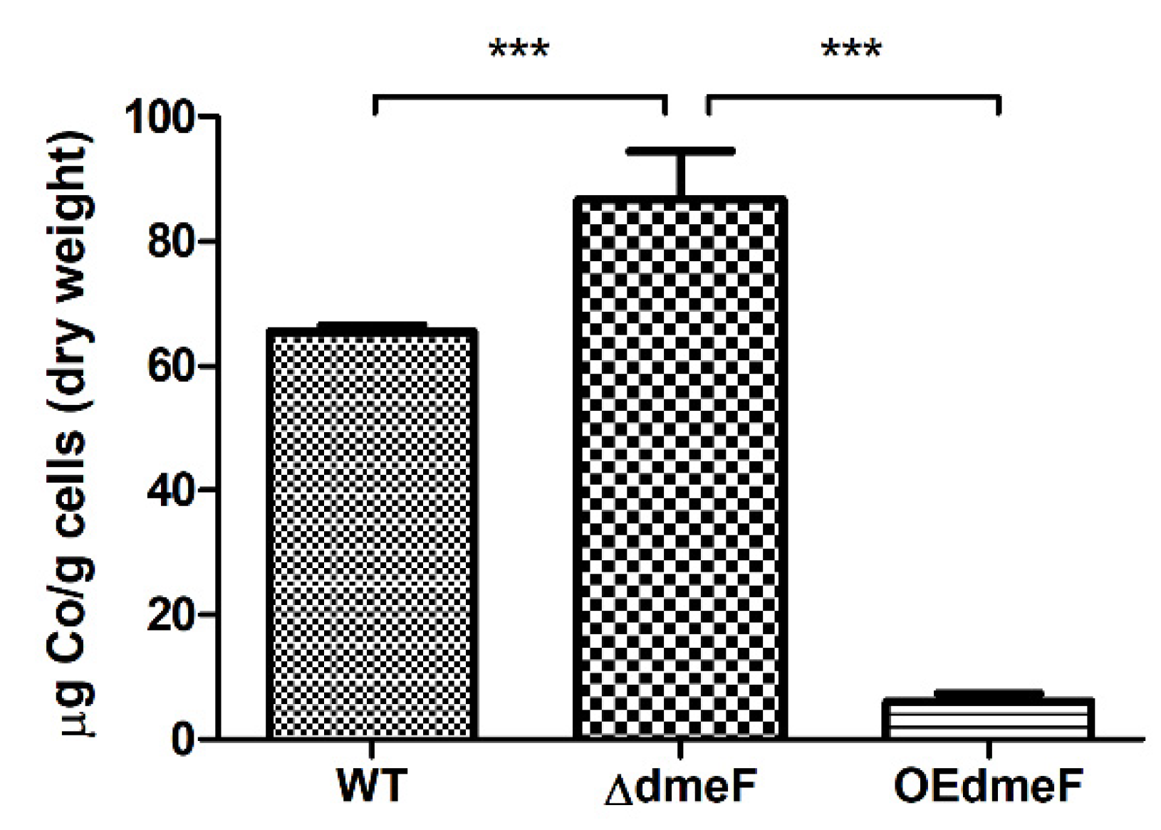
2.4. The ΔdmeF Mutant Accumulated Increased Levels of Cellular Cobalt Content

To better understand the mechanism underlying DmeF-mediated Co resistance, the WT, ΔdmeF, and OEdmeF strains grown in the presence of 0.1 mM Co were collected and analyzed for Co content using inductively coupled plasma-mass spectrometry (ICP-MS). As seen in Figure 6, the WT and ΔdmeF strains accumulated approximately 65 and 87 μg Co per g cells (dry weight), respectively. The OEdmeF strain, which had a higher expression of dmeF, accumulated only 6 μg Co per g cells (dry weight) (Figure 6). The cellular Co content in ΔdmeF was significantly higher than that in the WT and OEdmeF strains. Moreover, when growing in the presence of 1 mM Ni, ΔdmeF accumulated higher Ni content than the WT strain (although not significantly) and OEdemF (significantly) (Figure S3).

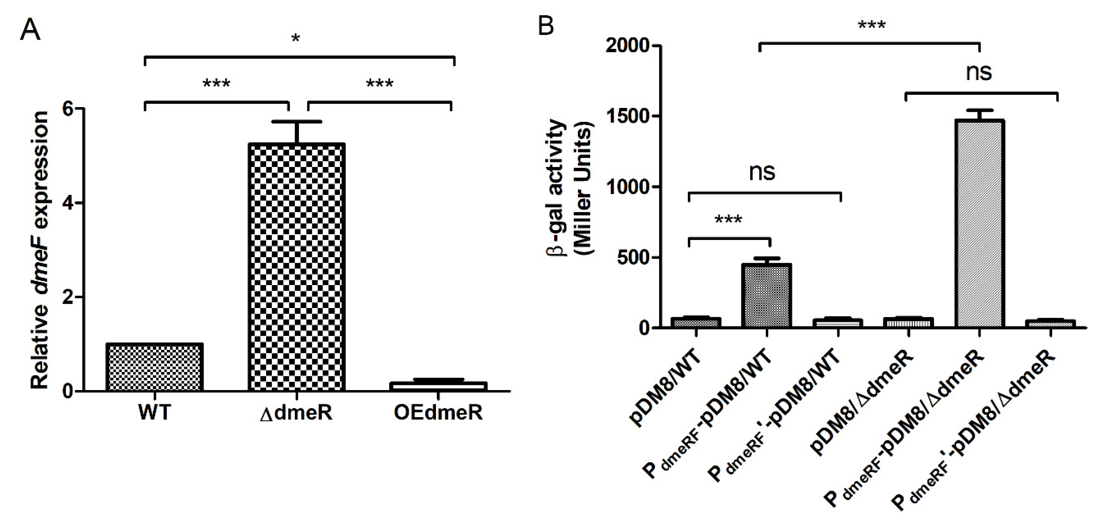
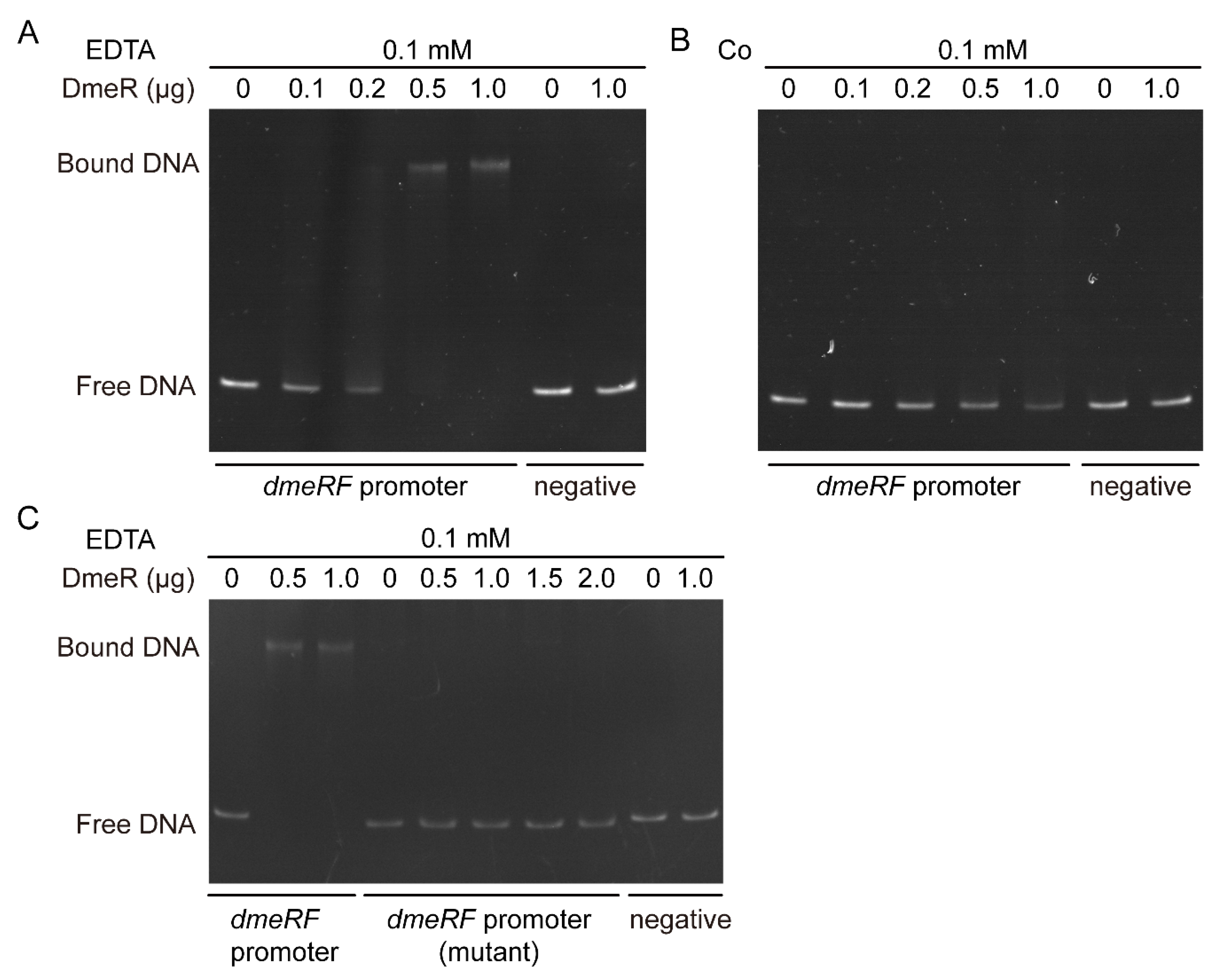
2.5. DmeR Negatively Regulates the dmeRF Operon by Binding Directly to the Promoter While Cobalt Inhibits the Interaction

To test whether DmeR regulates the dmeRF operon, dmeF expression in the WT, ΔdmeR, and OEdmeR strains was determined by qRT-PCR analysis. In ΔdmeR, dmeF expression was approximately 5-fold and 31-fold higher than that of the WT and OEdmeR strains, respectively (Figure 7A).
The promoter region of dmeRF contains a predicted binding site for DmeR that consists of a DNA sequence flanked by two inverted repeats (Figure S4A). The promotion of dmeRF was mutated by replacing the putative binding site with an unrelated DNA sequence, to generate PdmeRF’ (Figure S4B). The WT and ΔdmeR strains harboring either pDM8 (a plasmid carrying a promoterless lacZ gene), PdmeRF-pDM8 (pDM8 carrying the promoter of dmeRF), or PdmeRF’-pDM8 (pDM8 carrying the mutant promoter of dmeRF) were measured for β-galactosidase activity. As shown in Figure 7B, the β-galactosidase activity produced by ΔdmeR harboring PdmeRF-pDM8 (2039 Miller units) was significantly higher than that produced by the WT strain harboring the same plasmid (743 Miller units). In addition, the strain (either WT or ΔdmeR) harboring PdmeRF’-pDM8 produced β-galactosidase activity comparable to that harboring pDM8 (Figure 7B).
To further elucidate the regulatory mechanism of DmeR, electrophoretic mobility shift assays (EMSAs) were performed. Purified recombinant DmeR (rDmeR) was incubated with the promoter probes of either dmeRF (WT or mutant) or gyrB (as a negative control) in buffers containing EDTA or Co. As seen in Figure 8A, when 0.1 mM EDTA was present in the reactions, a single shifted band became visible as the concentration of rDmeR increased. In contrast, no shifted band was observed in reactions containing 0.5 mM Co (Figure 8B). Moreover, when incubated with the mutant promoter probe of dmeRF, no shifted band was observed in reactions containing EDTA (Figure 8C). Regardless of the presence of EDTA or Co, rDmeR was unable to shift the negative control probes (Figure 8A–C).
Together, these results indicate that DmeR represses the dmeRF operon by binding directly to its promoter in the absence of Co.

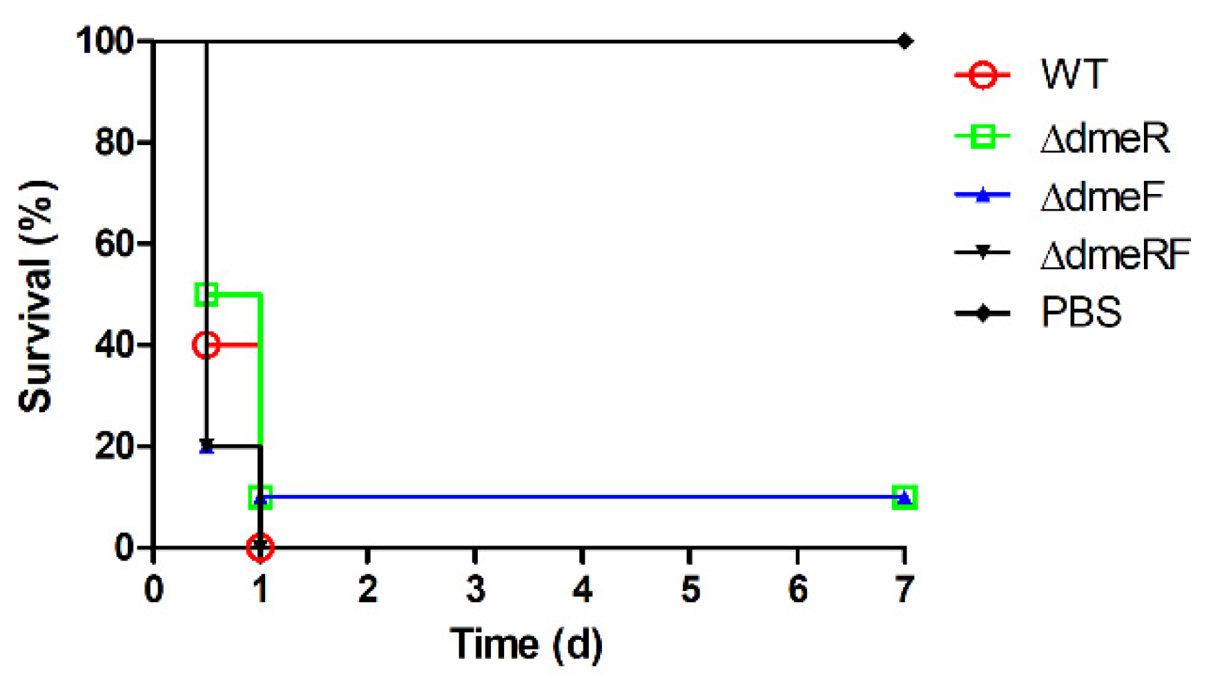
2.6. The DmeRF System Is Not Required for V. parahaemolyticus Virulence in Mice

To investigate the role of DmeRF in V. parahaemolyticus virulence, an experimental infection of C57BL/6 mice was conducted. Ten mice per treatment were intraperitoneally infected with either phosphate-buffered saline (PBS) or one of four V. parahaemolyticus strains: the WT, ΔdmeR, ΔdmeF, or ΔdmeRF strains. At 12 h post infection, the survival rates for mice in the WT, ΔdmeR, ΔdmeF, and ΔdmeRF groups were 40%, 50%, 20%, and 20%, respectively (Figure 9). The remaining mice in the WT and ΔdmeRF groups died during the following 12 h, and 10% of the mice infected with the ΔdmeR or ΔdmeF strains survived over the course of the experiment (Figure 9). In contrast, all mice injected with PBS survived. These results suggest that the DmeRF system has no significant role in V. parahaemolyticus virulence in mice.

3. Discussion

Even though the maintenance of metal homeostasis is essential for bacterial physiology and pathogenesis, not much is known about the mechanisms by which V. parahaemolyticus responds to metal overload. Metal efflux is one of the most important mechanisms employed by bacteria to reduce the damage caused by metal influxes [4]. The DmeRF system is a well-characterized metal-response system that is involved in Co resistance in C. metallidurans and Co/Ni resistance in certain Rhizobiaceae species [14,18,19,20,21]. The DmeRF system was demonstrated to contribute to the maintenance of Co homeostasis in V. parahaemolyticus by providing the following lines of evidence: (i) V. parahaemolyticus DmeF shares a high level of homology (approximately 44% to 51% amino acid sequence identity) with its homologues, all of which are involved in Co (and Ni) resistance; (ii) dmeF expression is significantly upregulated in response to Zn, Cu, and Co exposure; (iii) the dmeF deletion mutants exhibit increased sensitivity to Co stress, while the overexpression strains that have higher dmeF expression exhibit decreased sensitivity to Co stress; (iv) when cultured in a medium supplemented with Co, the cellular Co content in the ΔdmeF mutant is significantly higher than that in the WT and OEdmeF strains; and (v) in the presence of Co, DmeR dissociates from the dmeRF promoter, allowing the transcription of dmeF.
The role of the DmeRF system in the metal tolerances of C. metallidurans and certain Rhizobiaceae species is well studied [14,18,19,20,21]. Therefore, BlastP analyses and multiple sequence alignments were performed to examine the level of homology exhibited between the DmeRF systems of V. parahaemolyticus and their homologues in these other species. While the DmeF proteins share high levels of homology, no significant similarity was observed between the DmeR of V. parahaemolyticus and those of the Rhizobiaceae species. It is not surprising as V. parahaemolyticus DmeR is a MarR family regulator while the others belong to the RcnR/CsoR family. Moreover, the organization of the V. parahaemolyticus demRF operon is different from those of the Rhizobiaceae species. Consequently, it is worthwhile to explore the role of DmeRF in V. parahaemolyticus.
Typically, bacteria respond to metal excess by expressing specific genes. Therefore, dmeF expression in the presence of elevated levels of various metals was measured using qRT-PCR analysis. dmeF expression is induced by Zn, Cu, and Co, with Co serving as the most potent inducer. The expression of dmeF differs among V. parahaemolyticus, C. metallidurans, and the Rhizobiaceae species. In C. metallidurans, dmeF expression is constitutive and cannot be induced by metals [18]. By contrast, dmeF expression is strongly induced by Co, Ni, and Cu in S. meliloti [19], and specifically induced by Co and Ni in A. fabrum and R. leguminosarum, with Co being a more potent inducer [14,20]. Although Cu induces demF expression in S. meliloti, the ΔdmeF mutant exhibited no difference in growth compared with the WT strain under high-Cu conditions [19]. Similarly, in V. parahaemolyticus, there was no major difference in the growth of the ΔdmeF and WT strains under high-Zn or -Cu conditions. A similar observation has also been made for the Fe(II) and Co efflux pump PmtA in S. suis. While pmtA expression is induced by Fe(II), Co, and Ni, the ΔpmtA mutant displayed no growth decrease under Ni stress [15]. We speculate that excessive amounts of Zn or Cu may change DmeR conformation, resulting in partial derepression of the dmeRF operon.
In line with the induction of dmeF expression by Co, the dmeF deletion mutants exhibited obvious growth inhibition under high-Co conditions, whereas, the dmeF overexpression strains grew better than the WT strain in the presence of elevated Co levels. These results clearly suggest that DmeF is involved in Co resistance in V. parahaemolyticus, which is consistent with the observations in C. metallidurans and certain Rhizobiaceae species [14,18,19,20,21]. In A. fabrum, the ΔdmeF mutant accumulated significantly higher levels of cellular Co content than the WT strain when cultured in a Co-rich medium [14]. A similar observation was made in our study, indicating that DmeF potentially mediates Co resistance by Co efflux. Yet, growth of the ΔdmeF and ΔdmeRF strains was not completely inhibited by high concentrations of Co, suggesting that additional Co resistance systems likely exist in V. parahaemolyticus. Surprisingly, the OEdmeR strain, which showed very low dmeF expression, exhibited more severe growth inhibition than ΔdmeF and ΔdmeRF under Co stress. We speculate that DmeR may play a role in the regulation of other Co resistance systems. Interestingly, the OEdmeF strain exhibited increased growth compared to the WT strain in the presence of high concentrations of Ni. Consistent with their growth, OEdmeF accumulated significantly lower levels of cellular Ni content under Ni conditions. These results indicate that in V. parahaemolyticus, DmeF may be involved in Ni resistance, albeit in a less prominent role than in Co resistance.
qRT-PCR analysis, β-galactosidase activity assays, and EMSAs showed that DmeR, a MarR family regulator, represses the dmeRF operon by binding directly to the promoter in the absence of Co. In several studied Rhizobiaceae species, dmeF is co-transcribed with a gene encoding the RcnR/CsoR family regulator, whose repression of dmeRF transcription has been confirmed in A. fabrum [14,19,20]. Accordingly, we speculate that V. parahaemolyticus and Rhizobiaceae adopt regulators belonging to different families to modulate the conserved Co resistance system. Furthermore, the mechanisms of the DmeRF system in V. parahaemolyticus were proposed according to our results and the findings in A. fabrum [14]. In the presence of limited or normal concentrations of Co, DmeR binds to the promoter region of dmeRF, resulting in transcriptional repression, whereas under high-Co conditions, excessive amounts of Co result in DmeR dissociating from the promoter, probably by causing a conformational change, and consequently, the repression is relieved.
In A. fabrum, the inactivation of dmeF has no effect on bacterial virulence in Nicotiana benthamiana [14]. Likewise, the DmeRF system plays no significant role in V. parahaemolyticus virulence in mice. We speculate that the Co concentration is low in mouse tissues, hence the DmeRF system does not serve an important function during the infectious process.
In conclusion, a metal-response system, DmeRF, composed of the metal efflux pump DmeF and the regulator DmeR, has been identified and characterized in V. parahaemolyticus. The DmeRF system contributes to the maintenance of Co homeostasis in V. parahaemolyticus, and DmeR functions as a transcriptional repressor of the dmeRF operon in the absence of Co.

4. Materials and Methods

4.1. Bacterial Strains, Culture Conditions, Plasmids, and Primers

The bacterial strains and plasmids used in this study are listed in Table 1. All Escherichia coli strains, V. parahaemolyticus RIMD 2210633, and its derivatives were routinely grown at 37 °C in Luria-Bertani (LB) broth or on LB agar. When required, carbenicillin, chloramphenicol, and isopropyl β-D-1-thiogalactopyranoside (IPTG) were supplemented at 50 μg/mL, 25 μg/mL, and 1 mM, respectively. The primers used in this study are listed in Table 2.

4.2. RNA Extraction and qRT-PCR Analysis

An overnight culture of the RIMD 2,210,633 strain was diluted 1:100 in LB broth and grown to the early-exponential phase (OD600 of ~0.7). Seven 1 mL aliquots were removed, 2 μL of H2O was added to one and metal solutions were added to the rest, one to each, to create final concentrations of 1 mM FeSO4, 1 mM MnSO4, 0.5 mM ZnSO4, 1 mM CuSO4, 0.25 mM CoSO4, and 1 mM NiSO4. After further incubation at 37 °C for 15 min, bacterial cells were collected by centrifugation. Total RNA was isolated from the cell pellets using an Eastep Super Total RNA Isolation Kit (Promega, Shanghai, China). Three independent experiments were performed to obtain triplicate biological samples.
In another experiment, overnight cultures of the WT, ΔdmeR, and OEdmeR were separately diluted 1:100 in LB broth and grown to the early-exponential phase (OD600 of ~0.7). Then, bacterial cells were collected for RNA isolation as described above. Three independent experiments were performed to obtain triplicate biological samples.
After evaluations of RNA integrity and measurements of RNA concentrations, the qualified RNAs were subjected to qRT-PCR analysis. cDNA was generated from approximately 200 ng of RNA per sample using ToloScript RT EasyMix for qPCR (with 2-step gDNA Erase-Out) (TOLOBIO, Shanghai, China). Quantitative PCR was performed on the StepOnePlus Real-Time PCR System (Applied Biosystems, Waltham, MA, USA) using NovoStart SYBR qPCR SuperMix Plus (Novoprotein, Shanghai, China) and gene-specific primers (Table 2). Gene expression levels were analyzed using the 2− ΔΔCT method [32], with gyrB as the internal standard.

4.3. Construction of Gene Deletion and Overexpression Strains

The gene deletion mutants ΔdmeR, ΔdmeF, and ΔdmeRF were generated via allelic exchange using the pDM4 plasmid [29], as previously described [33]. The overexpression strains OEdmeR, OEdmeF, and OEdmeRF were constructed using the pMMB207 plasmid [30], as previously described [33].

4.4. Growth Evaluation

The WT, ΔdmeR, ΔdmeF, ΔdmeRF, OEdmeR, OEdmeF, and OEdmeRF strains were grown to the mid-exponential phase (OD600 of ~2) and diluted 1:100 in LB broth Supplemented with one of four concentrations of CoSO4 (0, 0.1, 0.2, or 0.3 mM) or specific high concentrations of another metal (2 mM FeSO4, 1 mM MnSO4, 1.25 mM ZnSO4, 2 mM CuSO4, or 1 mM NiSO4). To alleviate Fe(II) oxidation, 1 g/L of TCD was supplemented to the medium containing FeSO4 [34]. The cultures were transferred into 96-well plates with 200 μL per well and three wells per treatment. The plates were incubated in a shaker at 37 °C and 120 rpm, and the OD595 values were measured hourly using a CMax Plus plate reader (Molecular Devices, San Jose, CA, USA).

4.5. Intracellular Metal Content Analysis

The WT, ΔdmeF, and OEdmeF strains were grown to the mid-exponential phase (OD600 of ~2) and diluted 1:100 in LB broth supplemented with either 0.1 mM CoSO4 or 1 mM NiSO4. After incubation for another 6 h, bacterial cells were collected by centrifugation. Five independent experiments were performed to obtain five biological samples for each strain. Sample washing, drying, digestion, and dilution were performed as previously described [35]. Co/Ni content in these samples was analyzed by ICP-MS at Yangzhou University. The metal content was expressed as μg of Co/Ni per g of cells (dry weight).

4.6. Construction of LacZ Fusion Strains and β-galactosidase Activity Assays

The promoter of the dmeRF operon was mutated by replacing the putative binding site for DmeR with an unrelated DNA sequence. The DNA carrying the mutant promoter of dmeRF (PdmeRF’) was synthesized by Tsingke Biotechnology Co., Ltd. (Beijing, China).
The promoter of dmeRF (either WT or mutant) was cloned into pDM8, a plasmid carrying a promoterless lacZ gene [31]. The resulting plasmid PdmeRF-pDM8/PdmeRF’-pDM8 was transformed into E. coli S17-1 λpir and then conjugated into the WT strain and ΔdmeR. The WT strain and ΔdmeR harboring the empty pDM8 plasmid served as the control strains.
β-galactosidase activity assays were performed as previously described [36,37], with some modifications. Overnight cultures of the LacZ fusion strains were diluted 1:100 in LB broth and grown to the early-exponential phase (OD600 of ~0.7). Bacterial cells were collected from 1 mL of each culture by centrifugation. The cell pellets were resuspended in 1 mL PM buffer (60 mM Na2HPO4, 40 mM NaH2PO4, 10 mM KCl, 1 mM MgSO4, and 50 mM β-mercaptoethanol, pH 7.0). The A600 values of the bacterial suspensions were measured. For each suspension, 200 μL of suspension, 30 μL of chloroform, and 30 μL of 0.1% SDS were added to 500 μL of PM buffer, and then vortexed vigorously to lyse bacterial cells. The reaction was started by adding 200 μL of o-nitrophenyl-β-galactopyranoside (4 mg/mL in PM buffer). When the mixture turned yellowish, the reaction was stopped by adding 400 μL of 1 M Na2CO3. The mixture was centrifuged, and the A420 value of the supernatant was measured. β-galactosidase activity, in Miller units, was calculated as A420 × 1000 × min−1 × mL−1 × A600−1.

4.7. rDmeR Expression, Purification, and EMSAs

The dmeR gene was cloned into the pET-30a plasmid, and the resulting plasmid, pET30a-dmeR, was transformed into E. coli BL21(DE3). The strain was grown at 37 °C to the mid-exponential phase (OD600 of ~0.8). Then, 0.5 mM of isopropyl-β-D-thiogalactopyranoside (IPTG) was added to induce DmeR expression. After further growth at 28 °C for 4 h, bacterial cells were harvested by centrifugation. The cell pellets were resuspended in binding buffer (20 mM Tris-HCl, 500 mM NaCl, and 20 mM imidazole, pH 8.0) and lysed by sonication. rDmeR purification and assessment of its quality and concentration were performed as previously described [38].
EMSAs were performed as previously described [38]. DNA fragments containing the promoters of the dmeRF operon (WT or mutant) or gyrB (negative control) were PCR amplified and then purified. The 20 μL reaction mixtures contained 20 ng of the DNA fragments, varying amounts of rDmeR (0, 0.1, 0.2, 0.5, or 1.0 μg), and 200 ng of poly (dI:dC) in either EMSA buffer 1 (150 mM NaCl, 0.1 mM DTT, 0.1 mM EDTA, and 10 mM Tris, pH 7.4) or the Co containing EMSA buffer 2 (150 mM NaCl, 0.1 mM DTT, 0.5 mM CoSO4, and 10 mM Tris, pH 7.4). The mixtures were incubated at 25 °C for 30 min and then resolved by 6% native polyacrylamide gel electrophoresis in 0.5 × TBE buffer at 100 V for 2 h. After staining with SYBR Green I for 30 min, the gel was photographed.

4.8. Mouse Infection Experiment

A total of 50 female C57BL/6 mice (specific, pathogen-free, 8−12 weeks old) were randomly divided into five groups (10 mice per group). The WT, ΔdmeR, ΔdmeF, and ΔdmeRF strains were grown in LB broth at 30 °C for 12 h and adjusted to 1 × 10⁹ CFU/mL in PBS. For groups I to IV, the mice were intraperitoneally infected with 100 μL of the corresponding strain. Mice in group V were injected with 100 μL of PBS and served as the control. Mouse survival was recorded twice daily for seven days.

4.9. Bioinformatic and Statistical Analysis

BlastP was performed to analyze the amino acid sequence identities of homologous proteins. Clustal Omega (https://www.ebi.ac.uk/Tools/msa/clustalo/ (accessed on 12 October 2022)) was used for multiple sequence alignments; the results were visualized using ESPript 3.0 [39]. Promoters were predicted using BPROM (http://linux1.softberry.com/berry.phtml (accessed on 12 October 2022)).
GraphPad Prism 5 (San Diego, CA, USA) was used for statistical analysis. Gene expression, metal content, and β-galactosidase activity were analyzed by one-way analysis of variance with a Bonferroni’s post-test. The log-rank test was used for analyzing mouse survival curves.

Supplementary Materials

The following supporting information can be downloaded at: https://www.mdpi.com/article/10.3390/ijms24010414/s1, Figure S1: Co-transcription analysis of the dmeR and dmeF genes; Figure S2: Growth curves analysis of the V. parahaemolyticus strains in the presence of various metals. Figure S3: Cellular Ni content in the WT, ΔdmeF, and OEdmeF strains. Figure S4: Mutation of the promoter of the dmeRF operon.

Author Contributions

Conceptualization, C.Z.; methodology, C.Z., Y.Z. (Yuxuan Zhao), M.K., J.Y., X.Z., Y.S., Y.Z. (Yimeng Zhai) and J.Q.; validation, Y.Z. (Yuxuan Zhao), M.K. and J.Y.; formal analysis, C.Z.; investigation, Y.Z. (Yuxuan Zhao), M.K., J.Y., X.Z., Y.S., Y.Z. (Yimeng Zhai) and J.Q.; resources, C.Z.; data curation, Y.Z. (Yimeng Zhai) and J.Q.; writing—original draft preparation, C.Z. and J.Q.; writing—review and editing, C.Z.; visualization, C.Z.; supervision, C.Z.; project administration, C.Z.; funding acquisition, C.Z. All authors have read and agreed to the published version of the manuscript.

Funding

This research was funded by the National Natural Science Foundation of China, grant number 31802210 and the Interdisciplinary Project from Veterinary Science of Yangzhou University, grant number yzuxk202002.

Institutional Review Board Statement

The animal study protocol was approved by the Animal Welfare and Ethics Committees of Yangzhou University (No.: 202103099) and performed in strict accordance with the guidelines of the Institutional Administrative Committee and Ethics Committee of Laboratory Animals.

Informed Consent Statement

Not applicable.

Data Availability Statement

Not applicable.

Conflicts of Interest

The authors declare no conflict of interest.

References

  1. Foster, A.W.; Young, T.R.; Chivers, P.T.; Robinson, N.J. Protein metalation in biology. Curr. Opin. Chem. Biol. 2022, 66, 102095. [Google Scholar] [CrossRef] [PubMed]
  2. Skaar, E.P.; Raffatellu, M. Metals in infectious diseases and nutritional immunity. Metallomics 2015, 7, 926–928. [Google Scholar] [CrossRef] [PubMed]
  3. Hood, M.I.; Skaar, E.P. Nutritional immunity: Transition metals at the pathogen-host interface. Nat. Rev. Microbiol. 2012, 10, 525–537. [Google Scholar] [CrossRef] [PubMed] [Green Version]
  4. Chandrangsu, P.; Rensing, C.; Helmann, J.D. Metal homeostasis and resistance in bacteria. Nat. Rev. Microbiol. 2017, 15, 338–350. [Google Scholar] [CrossRef] [Green Version]
  5. Djoko, K.Y.; Ong, C.L.; Walker, M.J.; McEwan, A.G. The Role of Copper and Zinc Toxicity in Innate Immune Defense against Bacterial Pathogens. J. Biol. Chem. 2015, 290, 18954–18961. [Google Scholar] [CrossRef] [Green Version]
  6. Sheldon, J.R.; Skaar, E.P. Metals as phagocyte antimicrobial effectors. Curr. Opin. Immunol. 2019, 60, 1–9. [Google Scholar] [CrossRef]
  7. Akbari, M.S.; Doran, K.S.; Burcham, L.R. Metal Homeostasis in Pathogenic Streptococci. Microorganisms 2022, 10, 1501. [Google Scholar] [CrossRef]
  8. Osman, D.; Cooke, A.; Young, T.R.; Deery, E.; Robinson, N.J.; Warren, M.J. The requirement for cobalt in vitamin B12: A paradigm for protein metalation. Biochim. Biophys. Acta Mol. Cell Res. 2021, 1868, 118896. [Google Scholar] [CrossRef]
  9. Kobayashi, M.; Shimizu, S. Cobalt proteins. Eur. J. Biochem. 1999, 261, 1–9. [Google Scholar] [CrossRef]
  10. Leonard, S.; Gannett, P.M.; Rojanasakul, Y.; Schwegler-Berry, D.; Castranova, V.; Vallyathan, V.; Shi, X. Cobalt-mediated generation of reactive oxygen species and its possible mechanism. J. Inorg. Biochem. 1998, 70, 239–244. [Google Scholar] [CrossRef]
  11. Majtan, T.; Frerman, F.E.; Kraus, J.P. Effect of cobalt on Escherichia coli metabolism and metalloporphyrin formation. Biometals 2011, 24, 335–347. [Google Scholar] [CrossRef] [PubMed] [Green Version]
  12. Barras, F.; Fontecave, M. Cobalt stress in Escherichia coli and Salmonella enterica: Molecular bases for toxicity and resistance. Metallomics 2011, 3, 1130–1134. [Google Scholar] [CrossRef] [PubMed]
  13. Thorgersen, M.P.; Downs, D.M. Cobalt targets multiple metabolic processes in Salmonella enterica. J. Bacteriol. 2007, 189, 7774–7781. [Google Scholar] [CrossRef] [PubMed] [Green Version]
  14. Dokpikul, T.; Chaoprasid, P.; Saninjuk, K.; Sirirakphaisarn, S.; Johnrod, J.; Nookabkaew, S.; Sukchawalit, R.; Mongkolsuk, S. Regulation of the Cobalt/Nickel Efflux Operon dmeRF in Agrobacterium tumefaciens and a Link between the Iron-Sensing Regulator RirA and Cobalt/Nickel Resistance. Appl. Environ. Microbiol. 2016, 82, 4732–4742. [Google Scholar] [CrossRef] [Green Version]
  15. Zheng, C.; Jia, M.; Gao, M.; Lu, T.; Li, L.; Zhou, P. PmtA functions as a ferrous iron and cobalt efflux pump in Streptococcus suis. Emerg. Microbes. Infect. 2019, 8, 1254–1264. [Google Scholar] [CrossRef] [Green Version]
  16. Rutherford, J.C.; Cavet, J.S.; Robinson, N.J. Cobalt-dependent transcriptional switching by a dual-effector MerR-like protein regulates a cobalt-exporting variant CPx-type ATPase. J. Biol. Chem. 1999, 274, 25827–25832. [Google Scholar] [CrossRef] [Green Version]
  17. Raimunda, D.; Long, J.E.; Sassetti, C.M.; Argüello, J.M. Role in metal homeostasis of CtpD, a Co2+ transporting P(1B4)-ATPase of Mycobacterium smegmatis. Mol. Microbiol. 2012, 84, 1139–1149. [Google Scholar] [CrossRef] [Green Version]
  18. Munkelt, D.; Grass, G.; Nies, D.H. The chromosomally encoded cation diffusion facilitator proteins DmeF and FieF from Wautersia metallidurans CH34 are transporters of broad metal specificity. J. Bacteriol. 2004, 186, 8036–8043. [Google Scholar] [CrossRef] [Green Version]
  19. Li, Z.; Song, X.; Wang, J.; Bai, X.; Gao, E.; Wei, G. Nickel and cobalt resistance properties of Sinorhizobium meliloti isolated from Medicago lupulina growing in gold mine tailing. PeerJ 2018, 6, e5202. [Google Scholar] [CrossRef] [Green Version]
  20. Rubio-Sanz, L.; Prieto, R.I.; Imperial, J.; Palacios, J.M.; Brito, B. Functional and expression analysis of the metal-inducible dmeRF system from Rhizobium leguminosarum bv. viciae. Appl. Environ. Microbiol. 2013, 79, 6414–6422. [Google Scholar] [CrossRef]
  21. Rubio-Sanz, L.; Brito, B.; Palacios, J. Analysis of metal tolerance in Rhizobium leguminosarum strains isolated from an ultramafic soil. FEMS Microbiol. Lett. 2018, 365, fny010. [Google Scholar] [CrossRef] [PubMed] [Green Version]
  22. Letchumanan, V.; Chan, K.G.; Lee, L.H. Vibrio parahaemolyticus: A review on the pathogenesis, prevalence, and advance molecular identification techniques. Front. Microbiol. 2014, 5, 705. [Google Scholar] [CrossRef] [Green Version]
  23. Matsuda, S.; Hiyoshi, H.; Tandhavanant, S.; Kodama, T. Advances on Vibrio parahaemolyticus research in the postgenomic era. Microbiol. Immunol. 2020, 64, 167–181. [Google Scholar] [CrossRef]
  24. Zhang, L.; Orth, K. Virulence determinants for Vibrio parahaemolyticus infection. Curr. Opin. Microbiol. 2013, 16, 70–77. [Google Scholar] [CrossRef] [PubMed]
  25. Broberg, C.A.; Calder, T.J.; Orth, K. Vibrio parahaemolyticus cell biology and pathogenicity determinants. Microbes Infect. 2011, 13, 992–1001. [Google Scholar] [CrossRef] [PubMed] [Green Version]
  26. Lee, C.T.; Chen, I.T.; Yang, Y.T.; Ko, T.P.; Huang, Y.T.; Huang, J.Y.; Huang, M.F.; Lin, S.J.; Chen, C.Y.; Lin, S.S.; et al. The opportunistic marine pathogen Vibrio parahaemolyticus becomes virulent by acquiring a plasmid that expresses a deadly toxin. Proc. Natl. Acad. Sci. USA 2015, 112, 10798–10803. [Google Scholar] [CrossRef] [Green Version]
  27. Prachumwat, A.; Taengchaiyaphum, S.; Mungkongwongsiri, N.; Aldama-Cano, D.J.; Flegel, T.W.; Sritunyalucksana, K. Update on early mortality syndrome/acute hepatopancreatic necrosis disease by April 2018. J. World Aquacult. Soc. 2019, 50, 5–17. [Google Scholar] [CrossRef] [Green Version]
  28. Makino, K.; Oshima, K.; Kurokawa, K.; Yokoyama, K.; Uda, T.; Tagomori, K.; Iijima, Y.; Najima, M.; Nakano, M.; Yamashita, A.; et al. Genome sequence of Vibrio parahaemolyticus: A pathogenic mechanism distinct from that of V cholerae. Lancet 2003, 361, 743–749. [Google Scholar] [CrossRef]
  29. Milton, D.L.; O’Toole, R.; Horstedt, P.; Wolf-Watz, H. Flagellin A is essential for the virulence of Vibrio anguillarum. J. Bacteriol. 1996, 178, 1310–1319. [Google Scholar] [CrossRef] [Green Version]
  30. Morales, V.M.; Bäckman, A.; Bagdasarian, M. A series of wide-host-range low-copy-number vectors that allow direct screening for recombinants. Gene 1991, 97, 39–47. [Google Scholar] [CrossRef]
  31. Croxatto, A.; Chalker, V.J.; Lauritz, J.; Jass, J.; Hardman, A.; Williams, P.; Cámara, M.; Milton, D.L. VanT, a homologue of Vibrio harveyi LuxR, regulates serine, metalloprotease, pigment, and biofilm production in Vibrio anguillarum. J. Bacteriol. 2002, 184, 1617–1629. [Google Scholar] [CrossRef] [PubMed] [Green Version]
  32. Livak, K.J.; Schmittgen, T.D. Analysis of relative gene expression data using real-time quantitative PCR and the 2(T)(-Delta Delta C) method. Methods 2001, 25, 402–408. [Google Scholar] [CrossRef] [PubMed]
  33. Zhang, Y.; Liu, H.; Gu, D.; Lu, X.; Zhou, X.; Xia, X. Transcriptomic analysis of PhoR reveals its role in regulation of swarming motility and T3SS expression in Vibrio parahaemolyticus. Microbiol. Res. 2020, 235, 126448. [Google Scholar] [CrossRef] [PubMed]
  34. Guan, G.; Pinochet-Barros, A.; Gaballa, A.; Patel, S.J.; Arguello, J.M.; Helmann, J.D. PfeT, a P1B4 -type ATPase, effluxes ferrous iron and protects Bacillus subtilis against iron intoxication. Mol. Microbiol. 2015, 98, 787–803. [Google Scholar] [CrossRef] [PubMed] [Green Version]
  35. Zheng, C.K.; Wei, M.; Qiu, J.; Jia, M.D.; Zhou, X.H.; Jiao, X.A. TroR Negatively Regulates the TroABCD System and Is Required for Resistance to Metal Toxicity and Virulence in Streptococcus suis. Appl. Environ. Microbiol. 2021, 87, e0137521. [Google Scholar] [CrossRef]
  36. Gu, D.; Zhang, Y.; Wang, Q.; Zhou, X. S-nitrosylation-mediated activation of a histidine kinase represses the type 3 secretion system and promotes virulence of an enteric pathogen. Nat. Commun. 2020, 11, 5777. [Google Scholar] [CrossRef]
  37. Xiao, Y.; Chen, H.; Nie, L.; He, M.; Peng, Q.; Zhu, W.; Nie, H.; Chen, W.; Huang, Q. Identification of c-di-GMP/FleQ-Regulated New Target Genes, Including cyaA, Encoding Adenylate Cyclase, in Pseudomonas putida. mSystems 2021, 6, e00295-21. [Google Scholar] [CrossRef]
  38. Zheng, C.; Qiu, J.; Zhao, X.; Yu, S.; Wang, H.; Wan, M.; Wei, M.; Jiao, X. The AdcR-regulated AdcA and AdcAII contribute additively to zinc acquisition and virulence in Streptococcus suis. Vet. Microbiol. 2022, 269, 109418. [Google Scholar] [CrossRef]
  39. Robert, X.; Gouet, P. Deciphering key features in protein structures with the new ENDscript server. Nucleic. Acids Res. 2014, 42, W320–W324. [Google Scholar] [CrossRef]
Figure 1. Genetic structures of the dmeRF systems in V. parahaemolyticus and several Rhizobiaceae species. Arrows indicate the direction of transcription. The structure analyses were performed using the following genomes: V. parahaemolyticus RIMD 2210633, NC_004605.1; S. meliloti CCNWSX0020, AGVV01000035.1; A. fabrum C58, NC_003062.2; and R. leguminosarum UPM791, CP025509.1.
Ijms 24 00414 g001
Figure 2. Multiple sequence alignments of the DmeF (A) and DmeR (B) homologues. Identical residues are in white letters on a red background, and similar residues are in red letters on a white background. The GenBank accession numbers are as follows: V. parahaemolyticus DmeF, WP_005477256.1; C. metallidurans DmeF, ABF07084.1; S. meliloti DmeF, EHK76549.1; A. fabrum DmeF, AAK86697.2; R. leguminosarum DmeF, AVC48922.1; V. parahaemolyticus DmeR, WP_005463375.1; S. meliloti DmeR, EHK76548.1; A. fabrum DmeR, AAK86696.2; and R. leguminosarum DmeR, AVC48921.1. DmeR homologue has not been described in C. metallidurans; thus, the analysis does not include this protein.
Ijms 24 00414 g002
Figure 3. Induction of dmeF expression by various metals. V. parahaemolyticus RIMD 2,210,633 was incubated in H2O, 1 mM FeSO4, 1 mM MnSO4, 0.5 mM ZnSO4, 1 mM CuSO4, 0.25 mM CoSO4, or 1 mM NiSO4 for 15 min. Expression of dmeF is reported relative to the H2O treatment. Results represent the means and standard deviations (SD) from three biological replicates. The data were analyzed using one-way analysis of variance along with Bonferroni’s post-test. **, p < 0.01; ***, p < 0.001.
Ijms 24 00414 g003
Figure 4. Growth curves of the V. parahaemolyticus strains grown in the absence of Co (A) or in 0.1 mM (B), 0.2 mM (C), or 0.3 mM (D) solutions of Co. The experiments were performed at least three times; the results represent the means and SD from three wells in a representative experiment.
Ijms 24 00414 g004
Figure 5. qRT-PCR analysis of dmeF expression in the WT, OEdmeF, and OEdmeRF strains. Results represent the means and SD from three biological replicates. The data were analyzed using one-way analysis of variance along with Bonferroni’s post-test. ***, p < 0.001.
Ijms 24 00414 g005
Figure 6. Cellular Co content in the WT, ΔdmeF, and OEdmeF strains. These strains were grown in the presence of 0.1 mM CoSO4 for 6 h. Cellular Co content was analyzed by inductively coupled plasma-mass spectrometry. Results represent the means and SD from five biological replicates. The data were analyzed using one-way analysis of variance along with Bonferroni’s post-test. ***, p < 0.001.
Ijms 24 00414 g006
Figure 7. DmeR regulates the dmeRF operon. (A) qRT-PCR analysis of dmeF expression in the WT, ΔdmeR, and OEdmeR strains. Results represent the means and SD from three biological replicates. (B) Assays measuring the β-galactosidase activities of early-exponential phase cells of the WT and ΔdmeR strains harboring either pDM8 (a plasmid carrying a promoterless lacZ gene), PdmeRF-pDM8 (pDM8 carrying the promoter of dmeRF), or PdmeRF’-pDM8 (pDM8 carrying the mutant promoter of dmeRF). Results represent the means and SD from three independent experiments performed in duplicate. The data were analyzed using one-way analysis of variance along with Bonferroni’s post-test. ns, not significant; *, p < 0.05; ***, p < 0.001.
Ijms 24 00414 g007
Figure 8. Electrophoretic mobility shift assays. (A,B) Purified rDmeR was incubated with the promoter probes of either dmeRF or gyrB (negative control) in buffers containing EDTA (A) or Co (B). (C) Purified rDmeR was incubated with the promoter probes in buffers containing EDTA. The mutant demRF promoter probe was generated by replacing the putative binding site for DmeR with an unrelated DNA sequence. The rDmeR was added to each reaction mixture in the amounts indicated. The images are representative of at least three independent experiments.
Ijms 24 00414 g008
Figure 9. Survival curves of mice infected with one of four V. parahaemolyticus strains or PBS. Ten mice per treatment were intraperitoneally infected with 1 × 10⁸ CFU of the WT, ΔdmeR, ΔdmeF, or ΔdmeRF strains, or injected with 100 μL of PBS as the control. The data were analyzed using the log-rank test.
Ijms 24 00414 g009
Table 1. Bacterial strains and plasmids used in this study.
Strain or PlasmidRelevant Characteristics 1Source or Reference
Strains
E. coli
DH5α λpirCloning host for pDM4, pMMB207, and pDM8 Laboratory collection
S17-1 λpirConjugal donor for pDM4, pMMB207, and pDM8Laboratory collection
DH5αCloning host for pET-30aLaboratory collection
BL21(DE3)Expression host for pET-30aLaboratory collection
V. parahaemolyticus
RIMD 2,210,633 (WT)Clinical isolate, CarbR[28]
ΔdmeRdmeR deletion mutant of RIMD 2210633This study
ΔdmeFdmeF deletion mutant of RIMD 2210633This study
ΔdmeRFdmeR and dmeF double mutant of RIMD 2210633This study
OEdmeRdmeR overexpression strain in ΔdmeR backgroundThis study
OEdmeFdmeF overexpression strain in ΔdmeF backgroundThis study
OEdmeRFdmeRF overexpression strain in ΔdmeRF backgroundThis study
pDM8/WTRIMD 2,210,633 harboring pDM8This study
PdmeRF-pDM8/WTRIMD 2,210,633 harboring PdmeRF-pDM8This study
PdmeRF’-pDM8/WTRIMD 2,210,633 harboring PdmeRF’-pDM8This study
pDM8/ΔdmeRΔdmeR harboring pDM8This study
PdmeRF-pDM8/ΔdmeRΔdmeR harboring PdmeRF-pDM8This study
PdmeRF’-pDM8/ΔdmeRΔdmeR harboring PdmeRF’-pDM8This study
Plasmids
pDM4Suicide vector containing a sacB counterselectable marker; CmR[29]
pDM4-ΔdmeRKnockout vector for dmeR deletionThis study
pDM4-ΔdmeFKnockout vector for dmeF deletionThis study
pDM4-ΔdmeRFKnockout vector for dmeRF deletionThis study
pMMB207Wide-host-range low-copy-number vector; CmR[30]
pMMB207-dmeRpMMB207 containing dmeR and an additional ribosome-binding siteThis study
pMMB207-dmeFpMMB207 containing dmeF and an additional ribosome-binding siteThis study
pMMB207-dmeRFpMMB207 containing dmeRF and an additional ribosome-binding siteThis study
pDM8Plasmid containing the promoterless lacZ gene; CmR[31]
PdmeRF-pDM8pDM8 containing the promoter of dmeRFThis study
PdmeRF’-pDM8pDM8 containing the mutant promoter of dmeRFThis study
pET-30aExpression vector; KanRNovagen
pET30a-dmeRpET-30a containing dmeRThis study
1 CarbR, carbenicillin resistant; CmR, chloramphenicol resistant; KanR, kanamycin resistant.
Table 2. Primers used in this study.
PrimerSequence (5′-3′) 1Size (bp)Target Gene
QdmeF-FCACCAAAGCACCAACGATCC151An internal region of dmeF
QdmeF-RCGAGCACCAGGACCACAATC
QgyrB-FGGTGGTATTCAAGCGTTCGTTC116An internal region of gyrB
QgyrB-RTGCATTGCCACTTCTACCGAG
dmeR-LA-FTCCCCCGGGCCACCACAAACGCTCTCTG738The left arm of dmeR
dmeR-LA-RGATGAGCGAACCGGGAATTCTAGGTTTCA
dmeR-RA-FATTCCCGGTTCGCTCATCGTCAGCTATTT732The right arm of dmeR
dmeR-RA-RCCGCTCGAGTGTTCGCTTATCGCTATGCT
dmeR-in-FAAAGGTTGATTGCTGCTCTG325An internal region of dmeR
dmeR-in-RTTCTTGGGAACAGTCAGTCG
dmeR-out-FGCATTTTGTGTTGGTGTGACT338/733A fragment containing dmeR
dmeR-out-RCGATTGAGCCATACGCAG
OEdmeR-FTCCCCCGGGTAAGGAGGTAGGATAATAATGAGCGAACTAGAAAAGTTGAA417dmeR and an additional ribosome-binding site
OEdmeR-RAACTGCAGCTAGAATTCCCGGTTCATTTT
dmeF-LA-FTCCCCCGGGATTATGCTTGCCACCGCT714The left arm of dmeF
dmeF-LA-RCACGCACGATAAATAGCTGACGATGAGCGA
dmeF-RA-FTCAGCTATTTATCGTGCGTGTGTTGTGC723The right arm of dmeF
dmeF-RA-RCCGCTCGAGAGCACGCCATTACGATAGAG
OEdmeF-FTCCCCCGGGTAAGGAGGTAGGATAATAATGGCAATAGCACAACACAC929dmeF and an additional ribosome-binding site
OEdmeF-RAACTGCAGTAGTTCGCTCATCGTCAGC
dmeF-in-FCATCTAACCAAATCCATCCG291An internal region of dmeF
dmeF-in-RAATCCGTTCACCGTTTGTT
dmeF-out-FCAGCACTTCAATGGTATGGAC279/1157A fragment containing dmeF
dmeF-out-RAGTGATGAACGCCTTTCTTAGT
dmeR-LA-R2ACGCACGATACCGGGAATTCTAGGTTTCA
dmeF-RA-F2ATTCCCGGTATCGTGCGTGTGTTGTGC
OEdmeRF-FTCCCCCGGGTAAGGAGGTAGGATAATAATGGCAATAGCACAACACAC1344dmeRF and an additional ribosome-binding site
OEdmeRF-RAACTGCAGCGGATGAAACCTAGAATTCC
PdmeRF-FCGGATCCGGGGAATTCCCGGGTAAGCGGCTGATTCCCAAAC208The promoter of dmeRF, for β-galactosidase activity assays
PdmeRF-RAAGCTTATCGATTCGCCCGGGGCGTGTGTTGTGCTATTGCC
dmeR-FCGCGGATCCATGAGCGAACTAGAAAAGTTGAA417The dmeR gene
dmeR-RCCGCTCGAGCTAG
AATTCCCGGTTCATTTT
PdmeRF-F2TAAGCGGCTGATTCCCAAAC208The promoter of dmeRF, for EMSAs
PdmeRF-R2GCGTGTGTTGTGCTATTGCC
PgyrB-FCAAGGGCAACATCTTACAGC215The promoter of gyrB
PgyrB-RTCTATCCTGCCATGTTCCAC
1 The underlined sequences are restriction sites.
Disclaimer/Publisher’s Note: The statements, opinions and data contained in all publications are solely those of the individual author(s) and contributor(s) and not of MDPI and/or the editor(s). MDPI and/or the editor(s) disclaim responsibility for any injury to people or property resulting from any ideas, methods, instructions or products referred to in the content.

