Article

The Regulation of the Growth and Pathogenicity of Valsa mali by the Carbon Metabolism Repressor CreA

1
Mountain Tai Forest Ecosystem Research Station of State Forestry Administration, Key Laboratory of Ecological Protection and Safety Prevention of the Lower Yellow River, Forestry College, Shandong Agricultural University, Tai’an 271018, China
2
Shandong Research Center for Forestry Harmful Biological Control Engineering and Technology, College of Plant Protection, Shandong Agricultural University, Tai’an 271018, China
*
Authors to whom correspondence should be addressed.
Int. J. Mol. Sci. 2023, 24(11), 9252; https://doi.org/10.3390/ijms24119252
Submission received: 13 March 2023 / Revised: 18 May 2023 / Accepted: 18 May 2023 / Published: 25 May 2023
(This article belongs to the Special Issue Advances of Plants-Pathogen Interaction 2023)

Abstract

:
Carbon catabolite repression (CCR) is a very important mechanism for efficient use of carbon sources in the environment and is necessary for the regulation of fungal growth, development, and pathogenesis. Although there have been extensive studies conducted regarding this mechanism in fungi, little is yet known about the effects of CreA genes on Valsa mali. However, based on the results obtained in this study for the identification of the VmCreA gene in V. mali, it was determined that the gene was expressed at all stages of fungal growth, with self-repression observed at the transcriptional level. Furthermore, the functional analysis results of the gene deletion mutants (ΔVmCreA) and complements (CTΔVmCreA) showed that the VmCreA gene played an important role in the growth, development, pathogenicity, and carbon source utilization of V. mali.

1. Introduction

Valsa mali is a mycelial fungus. The destructive Valsa canker disease of apples is caused by the ascomycetes fungus V. mali [1]. The occurrence of apple tree rot disease is extremely serious worldwide. It has been found that almost all apple-growing areas are affected by apple Valsa canker, particularly those located in Eastern Asia [2,3].
During the colonization and infection process, the pathogenic fungi face various challenges from hostile environments, such as nutrient limitations and stress conditions. However, a carbon catabolite repression (CCR) mechanism could facilitate the fungi’s effective utilization of the surrounding physiological environment. CCR inhibits the expressions of the genes involved in the utilization of other carbon sources when fungi utilize preferred carbon sources, such as glucose [4]. In Saccaromyces cerevisiae, a key regulator of CCR is Mig1, which inhibits the transcription of the genes encoding GAL4 and SUC2 in the nucleus. The loss of glucose transfers Mig1 phosphorylation from the protein kinase Snf1 complex to the cytoplasm [5]. CreA is a homologue of yeast Mig1 in filamentous fungi. However, there are some glaring differences in the mechanism of the CCR. For example, CCR in S. cerevisiae involves Mig1 and the complex Ssn6/Tup1. However, the role of this complex has not yet been determined in filamentous fungi [6].
Homologs of the CreA gene have been studied in several other filamentous fungi, and CreA has been found to be essential for the growth of Aspergillus nidulans [7]. In addition, CreA mutants were observed to reduce spore production and CCR, counteracting repressor effects [8]. Cre1 is the gene in Neurospora crassa that mainly regulates CCR. In previous studies, when the production of conidia was induced by carbon starvation conditions, Cre1 mutants on different carbon source media grew significantly slower than the wild type [9]. There is also a homolog of the CreA gene in Beauveria bassiana, in which the protein contains two C2H2-type zinc finger domains. Real-time PCR analysis results have shown that BbCre1 expressions are regulated by glucose and have self-transcriptional inhibition characteristics [10]. In Sclerotinia sclerotiorum, the CCR repressor gene is Cre1, which is known to be functionally associated with the CreA of A. nidulans but cannot replace the function of Mig1 in S. cerevisiae [5,11]. In Aspergillus flavus, CreA is a major transcriptional repressor gene. The CreA mutants and complements, including AfCreA, affect growth and development, spore germination, and nucleus formation and also produce less aflatoxin than the wild type and its complement mutants. AfCreA also plays a role in inhibiting the enzymes required to utilize secondary carbon sources [12]. In recent studies, it has been observed that CreA also demonstrated involvement in the pathogenicity of Magnaporthe oryzae, with MoCreA required for the asexual development and pathogenicity of rice blast fungus [4].
At present, the research regarding the CCR of filamentous fungi has made some progress. However, the involved regulation mechanism is still largely ambiguous in V. mali. Transcriptome sequencing analysis results have shown that the CreA gene (VM1G_03855) was differentially expressed during the infection stage of V. mali [13]. A CreA homolog encoding 434 amino acids, referred to as VmCreA, was identified in this study. The role of CreA in the development, pathogenicity, and utilization of carbon sources was revealed using deletion and complementation methods.

2. Results

2.1. Identification of the V. mali CreA

The single copy VmCreA gene (Accession number: KUI69101.1) has been identified in the genome of Valsa mali. The gene sequence is 1305-bp and encodes a protein of 434 amino acids (AA). Two conserved C2H2 zinc finger DNA-binding domains are predicted at 82–104 and 110–134 AA, including one nuclear localization signal (NLS) and two nuclear export signals (NESs), indicating that VmCreA possesses a DNA-binding function and could shuttle between the nucleus and cytoplasm (Figure 1A). This study analyzed the homologous sequences of other fungi and discovered that the C2H2 zinc finger was highly conserved in all of the analyzed species (Figure 1B). A phylogenetic tree was constructed revealing that VmCreA had a 100% identity with its homologue in the industrially important Valsa mali var. Pyri (Figure 1C).

2.2. Construction of the VmCreA Deletion and Complementation of the Mutant Strains

To confirm whether VmCreA participates in the CCR of V. mali and explore its biological role in the growth, development, and pathogenicity processes of V. mali, VmCreA deletion mutants were first constructed in this study using the Double-joint PCR approach. The VmCreA was first homologously replaced with a hygromycin B resistance gene (hph) in the genome of the wild-type strain sdau11-175 (WT). The CreA ORF region was fused in-frame with the eGFP gene for complementation (Figure 2A), and the transformants were verified using diagnostic PCR and qRT-PCR. The diagnostic PCR revealed that the deletion vector had been successfully constructed (Figure 2B). Southern-blot indicates that this mutant strain is a single-copy gene knockout mutant (Figure S1). The RT-PCR results showed the absence and regular transcript levels of the CreA in the ΔCreA and CTΔCreA strains, respectively (Figure 2C).

2.3. Influencing Effects of VmCreA on the Vegetative Growth and Development of V. mali

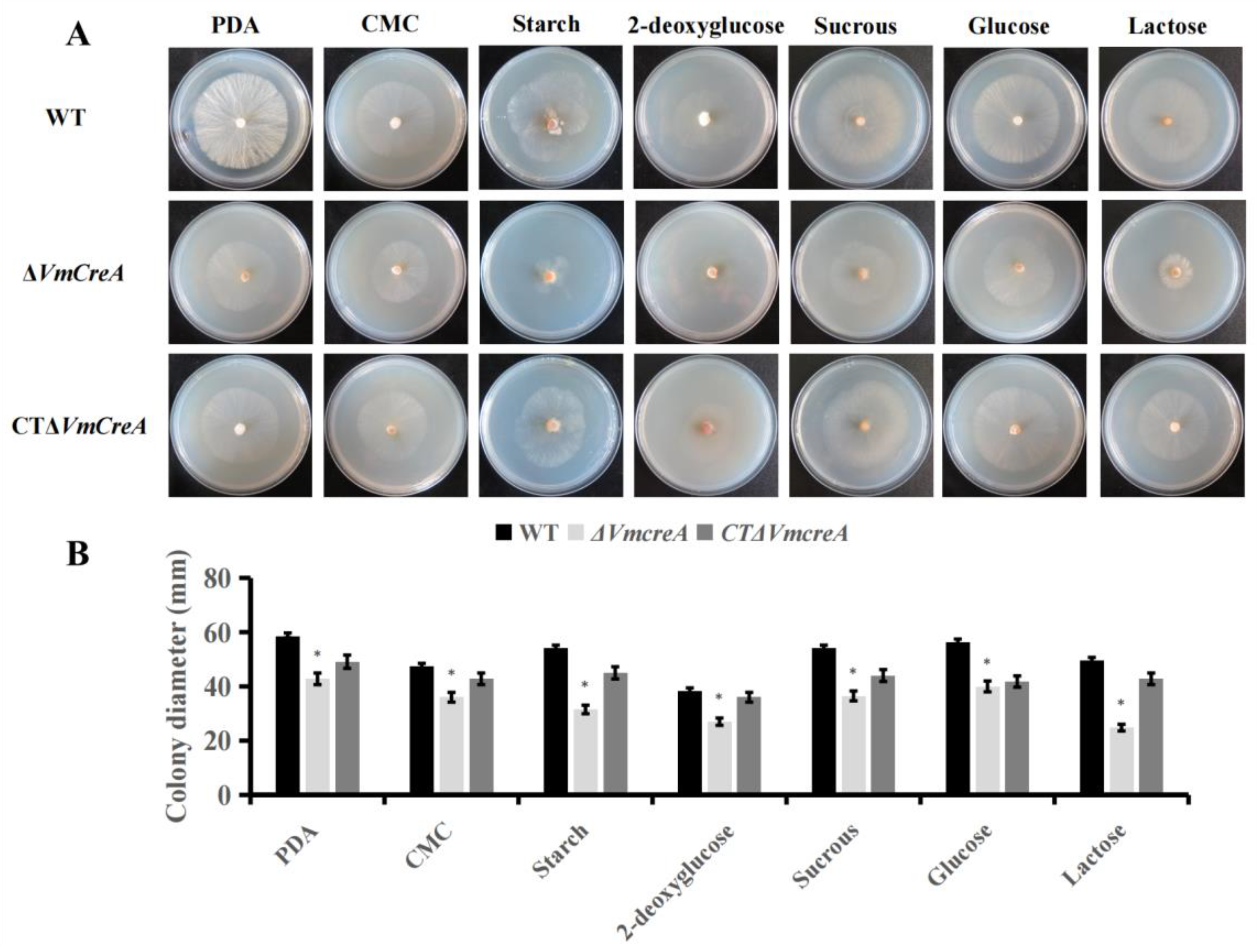
The results of this study revealed that the deletion of VmCreA dramatically affected the colony morphology of V. mali. For the wild-type strains, the ΔVmCreA mutants and complementary transformant were all cultured on PDA medium. It was observed that the mycelial growth rate of ΔVmCreA was slower when compared with the WT and CTΔVmCreA (Figure 3A). In addition, all of the strains were inoculated on minimal media (MM) with different substrates as the only carbon sources, including glucose, lactose, sucrose, starch, carboxymethy cellulose (CMC), and 2-deoxyglucose. Then, after five days, the colony diameter of ΔVmCreA was found to be lower. Therefore, based on the above results, it was suggested that VmCreA was necessary for the vegetative growth and the utilization of different carbon sources in V. mali (Figure 3B). However, the effects of VmCreA on fungal spores in V. mali could not be determined.

2.4. VmCreA Participates in the CCR of V. mali

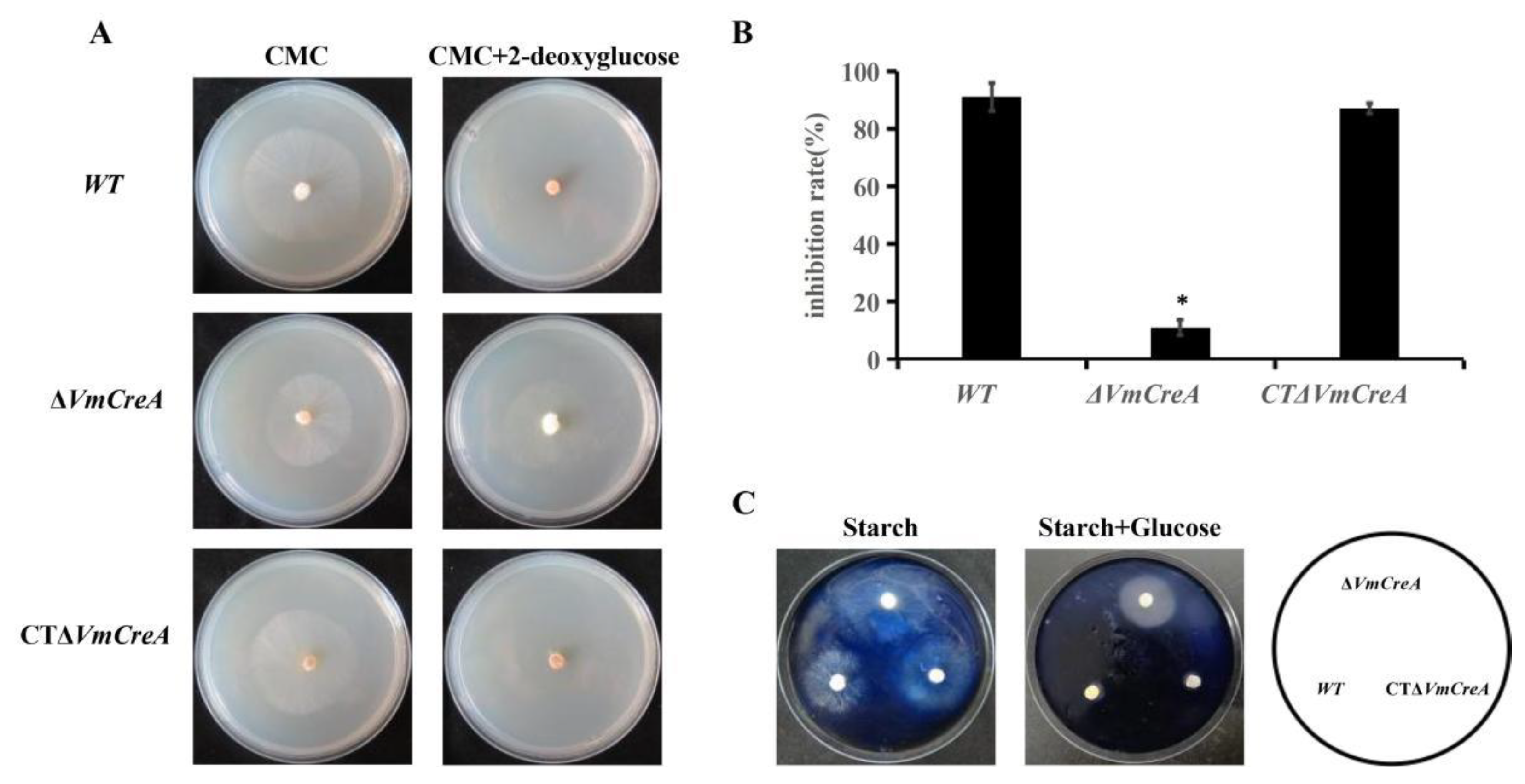
This study conducted further experiments to verify whether VmCreA is involved in the regulation of CCR. The growth-inhibition rates of the WT, ΔVmCreA, and CTΔVmCreA, which had been inoculated on MM with CMC were found to be generally consistent. However, when grown on medium with 2-deoxyglucose and CMC as the common carbon sources, ΔVmCreA had a growth rate consistent with that achieved with a single carbon source. It was also found that the growth rates of the WT and CTΔVmCreA were significantly inhibited under certain conditions, as shown in Figure 4A. In such cases, the 2-deoxyglucose could be identified but had not been utilized, indicating that the WT utilized neither 2-deoxyglucose nor CMC, and growth was significantly inhibited. Meanwhile, although there was a lack of CCR, ΔVmCreA was still able to grow by utilizing CMC on a medium containing 2-deoxyglucose (Figure 4B). It was observed that when starch was used as the only carbon source, none of the strains were stained blue by the iodine liquid, indicating that the strains had all used the starch for growth. However, when both starch and glucose were used as the common carbon sources, the ΔVmCreA was not stained blue by the iodine liquid. Those findings indicated that the ΔVmCreA has a CCR defect and can use both glucose and starch. Therefore, it could be concluded that the VmCreA is involved in regulating the CCR of V. mali (Figure 4C).

2.5. Stress Responses of the V. mali Are Influenced by VmCreA

This study observed the responses of the strains to osmotic and cell wall stress agents in order to determine the influencing effects of VmCreA on the regulation responses of the V. mali for cell wall homeostasis. The mycelial growth rates of the WT, ΔVmCreA, and CTΔVmCreA were all inhibited under the osmotic stress. However, the ΔVmCreA was found to be more sensitive to the cell wall stress induced by CFW and SDS than the WT and CTΔVmCreA strains. Therefore, based on those findings, it was concluded that the VmCreA had influenced the responses of the V. mali to cell wall stress agents (Figure 5).

2.6. VmCreA Is Required for V. mali Pathogenicity

Mycelial plugs of the various strains were inoculated onto apple twigs and fruit. Then, following a culturing period of seven days, it was observed that the lengths of the lesions on the apple fruit with ΔVmCreA inoculations had decreased by approximately 53% compared to those with the WT (Figure 6A). It was also found that the lengths of the lesions on the apple twigs were reduced by approximately 57% (Figure 6B). In addition, the pathogenicity of the complement mutants had returned to the original levels. The above results indicated that the VmCreA is required for V. mali pathogenicity.

2.7. VmCreA Regulates Hydrolases Production

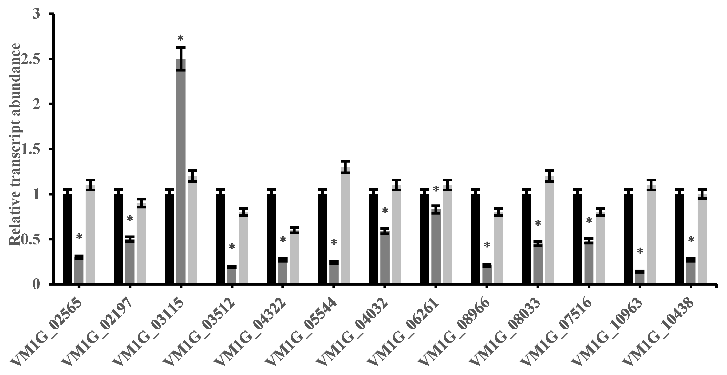
In order to further understand the role of VmCreA in the synthesis of the V. mali cell wall-hydrolase, the expressions of 13 genes were detected using qRT-PCR. It was discovered that the expressions of 12 genes in the ΔVmCreA mutant were inhibited when compared to the WT. The inhibited genes included VM1G_02565 (EndopolygalacturonaseD), VM1G_03512 (Glucoamylase), and VM1G_10963 (Pectin lyase B). However, the VM1G_03115 encoding of Aspartic protease was found to be significantly up-regulated in the ΔVmCreA mutant, as shown in Figure 7.

3. Discussion

Carbon catabolite repression (CCR) is widely found in various fungi. CCR is a process used to preferentially utilize preferred carbon sources from the environment. Environmental signals can stimulate CCR to regulate the growth, development, and pathogenic abilities of fungi [16]. The Valsa canker disease in apples caused by V. mali is an important biological disaster for apple crops. At present, there appears to be a lack of systematic research on the role of CCR in the pathogenicity of this type of fungus.
In this study, the experimental results demonstrated that VmCreA is a key regulator of V. mali CCR. CreA is a protein containing two C2H2-type zinc finger domains, and its zinc finger DNA-binding domain is highly homologous to that of the stress-regulator genes Mig1 in S. cerevisiae [11]. When glucose was limited by S. cerevisiae, Mig1 promotes the genes that encode the enzymes that utilize other secondary carbon sources to relieve CCR [17]. However, the glucose repressor CRE1 from S. sclerotiorum is functionally related to CREA from A. nidulans but not to the Mig proteins from S. cerevisiae [18]. In filamentous fungi, CreA has been identified as the key regulator of the carbon catabolite repression (CCR) responsible for efficiently utilizing carbon sources in the environment that can bind to the conserved 5′-SYGGRG-3′ on the promoter region of related genes [19]. In previous studies involving transcriptome sequencing analyses, it was found that the CreA gene (VM1G_03855) was up-regulated 2.5-fold (12 days versus 3 days) during the hyphal growth stage [13]. Since CCR-binding sites (CCCGC and CTCCAG) were found in the upstream of the VmCreA, it was inferred that the VmCreA may have self-inhibitory effects at certain transcription levels. This study conducted analyses of the relative expression levels of VmCreA at different glucose induction times using qRT-PCR. The results showed that VmCreA expression was relatively high within one hour of the glucose addition and had significantly decreased thereafter. Therefore, based on the analysis results, it could be inferred that the VmCreA expression was inhibited by the glucose. In other words, there may have been self-inhibitory effects (Figure S2).
It has been observed that CreA is relatively conserved in filamentous fungi. Sequence alignments have shown some highly conserved regions throughout various species, and the V. mali also contained the typical CCR nuclear penetration signal “PNSRRG”. It has also been confirmed that MoCreA is mainly localized in the nucleus [4]. Therefore, it was necessary in this study to verify whether the same features also applied to the VmCreA in V. mali. Mycelia basically give fungi their shape and size characteristics. This study discovered that the growth of ΔCreA mycelium was impaired, and its growth rate was significantly slowed down on MM with different substrates as the only carbon sources. The reduced number of mycelia indicated that the CreA was required for the growth of the V. mali. The growth of the ΔVmCreA was inhibited by CMC, starch, and lactose, indicating that VmCreA can regulate the expression of related enzymes using these carbon sources, and its knockdown affects the utilization of these carbon sources; further studies should confirm this aspect. However, it was reported that the disruption of CreA in T. reesei inhibits hyphal growth and leads to a decrease in cellulase production [20]. Of further interest is a deletion removing the CreA gene in A. nidulans, which allows limited germination of the spore but not colony formation; this result is the opposite of the result in the present study [8].
Osmotic stress has been shown to positively affect the morphology and conidiation of ΔCreA in A. flavus [21]. However, this was not found to be the case in V. mali. The growth of ΔCreA was significantly impaired under the cell wall stress conditions induced by SDS and CFW, which was similar to the observations in A. flavus [12], where evidence was found that CreA could be involved in cell wall integrity.
Hydrolases, such as pectinases, cutinases, and amylases, play important roles in fungal virulence, as they catalyze the production of secondary metabolites [22]. In this study, the hydrolases exhibited dramatic decreases in transcription in the ΔVmCreA mutant, and the down-regulated expressions of the hydrolase genes may have negatively affected the V. mali virulence.
The infection process of Valsa canker disease in apples is a rather complex interaction process that includes cell wall hydrolases, small molecules such as toxins, and effector proteins [23]. This study’s results revealed that VmCreA influenced the morphology, vegetative growth, stress responses, and pathogenicity of the V. mali. This is the first report regarding the role of CreA in V. mali. However, the regulation mechanism of VmCreA remains limited, with more scientific data still required to verify the findings of this research.

4. Materials and Methods

4.1. Fungal Strains and Culture Manipulations

Valsa mali (Vm) isolate sdau11-175 was used as the wild-type control strain throughout this study (sdau11-175 was collected from Yantai by the Shandong Research Center for Forestry Harmful Biological Control Engineering and Technology in 2011, and it is a high-Pathogenicity variety), and the null mutants were genetically derived from sdau11-175. All of the strains were maintained at 25 °C on potato dextrose agar (PDA). Cultures for the protoplast preparation were cultivated in liquid potato dextrose (PD) and then incubated in a conical bottle at 25 °C and 120 rpm for three days. PEG-mediated transformation of the protoplast was performed to obtain the knockout mutants. The media used in this study included minimal medium (MM); complete medium (CM); and TB3 medium. For testing the utilization of the different carbon sources, the strains were grown on minimal medium (MM) with 2% (W/V) glucose or an equivalent amount of lactose, sucrose, starch, carboxymethy cellulose (CMC), and 2-deoxyglucose, respectively, as the only carbon sources. For this study’s investigation of whether VmCreA is involved in the CCR pathways, the strains were grown on MM containing 2% (W/V) glucose, with or without 1% (W/V) starch.
Each of this study’s experiments contained three replicates for each strain, and all of the experiments were carried out three times.

4.2. Cloning and Sequencing Analysis of the VmCreA

The VmCreA (KUI69101.1) sequence was downloaded from the National Center for Biotechnology Information Resources (NCBI). NCBI Conserved Domain Search (https://www.ncbi.nlm.nih.gov/cdd, accessed on 19 May 2021); Signal-5.0 (https://services.healthtech.dtu.dk/service.php?SignalP-5.0, accessed on 22 May 2021); and Pfam (http://pfam.xfam.org/, accessed on 22 May 2021) online analysis software were utilized to analyze the domain and predict the signal peptide of the amino acid sequence encoded by VmCreA. Then, the CreA protein sequences of Valsa mali var. Pyri (KUI53877.1); Diaporthe helianthi (POS81118.1); Neurospora crassa (XP_961994.1); Daldinia childiae (XP_033439606.1); Fusarium bulbicola (KAF5969685.1); Pyricularia oryzae (XP_003714427.1); Saccharomyces cerevisiae (AAT93178.1); Colletotrichum higginsianum (CCF41975.1); Fusarium oxysporum (SCO85024.1); and Beauveria bassiana (ABL07611.1) were aligned by ClustalW using MEGA 6 software. A neighbor-joining phylogenetic tree was also constructed.

4.3. Targeted Deletion and Complementation of the CreA Gene

In this study, the CreA deletion mutant strain was created using Double-joint PCR [24]. Two flanking fragments of the ORF of VmCreA were amplified using the primer pairs CreA-UF/CreA-UR and CreA-DF/CreA-DR, respectively, and fused to either side of hygromycin B phosphotransferase genes (hph) that were amplified using the primer pairs hph-F/hph-R using Double-joint PCR. The PCR products were co-transformed into protoplasts of the wild-type strain sdau11-175. The positive transformants were verified using diagnostic PCR with the primers YZ-F1/YZ-R1, YZ-F2/YZ-R2, and CreA-F/CreA-R (Table S1) for VmCreA ORF upstream fragments (containing the 5′ flanking sequence of the CreA gene and a part of the hph) and downstream fragments (containing a part of the hph and the 3′ flanking sequence of the CreA gene), respectively.
For the complementation, a 2.36 kb genomic DNA fragment containing the native promoter and ORF region was amplified using the primer pair HB-F/HB-R (Table S1), cloned into the vector pYF11 containing the hygromycin B gene, and then fused in-frame with the eGFP gene. The resulting complementation vector was reintroduced into the ΔVmCreA mutant through protoplast transformation. The complementary transformants were first screened using hygromycin and then confirmed using qRT-PCR.

4.4. Quantitative Reverse Transcription Polymerase Chain Reactions

In this study, the G6PDH (Glyceraldehyde-6-phosphate dehydrogenase) gene was used as an internal control [25]. The mycelia were harvested at 3, 6, and 9 days post-inoculation on PDA medium from all the examined strains. The total RNA was extracted using a TRIzol reagent, and the first strand cDNA was synthesized using a Prime Script TM RT reagent kit with gDNA Eraser (Perfect Real Time) kits. Subsequently, qRT-PCR was performed using a Bio-Rad CFX-96 PCR instrument. The relative expressions of the transcripts were calculated using the 2−ΔΔCT method [26]. The primers for qRT-PCR used in this study are listed in Table S1.

4.5. Observations of the Growth Rates, Conidia, and Morphological Processes

The PDA medium samples were point inoculated with 5 mm of fungus to evaluate the growth rates. The plates were incubated at 25 °C for three days. The conidia production was calculated according to the amount of pycnidia and the growth rates of the wild-type and mutant strains on PDA medium. The samples had been cultured on PDA medium at 25 °C under dark and light conditions for 25 days [27].

4.6. Cell-Wall Integrity Inhibitor Stress Assay

In the present study, PDA medium containing different stress agents was inoculated with 5 mm fungus of wild-type and mutant strains and then incubated at 25 °C for four days. The stress agents included osmotic stress NaCl (sodium chloride, 200 ppm) and Sor (Sorbitol, 1 M); cell wall stress SDS (sodium dodecyl sulphate, 0.01%); CFW (Calcofluor White, 200 ppm); and CR (Congo Red, 300 ppm).

4.7. Pathogenic Determination Assays

For assaying the virulence on the pathogenic hosts, wild-type and deletion mutants were cultured on PDA medium for four days. Then, 5 mm agar plugs were taken from the edge of a colony and inoculated via prick wounds on the fruit and one-year old twigs of Malus domestica “Fuji”. The inoculated tissues were incubated at 25 °C for seven days, and the lengths of the lesions were recorded using a crossing method.

4.8. Statistical Analysis

Statistical and significance analyses were carried out using SPSS software (IBM SPSS Statistics 20), and significance was recognized if the p-values were <0.05. All the assay results were differentiated using a one-way analysis of variance method.
inhibition rate (%) = [(dc dt)/dc] × 100

5. Conclusions

In summary, we constructed VmCreA deletion mutants and found that VmCreA plays crucial roles in the growth, development, pathogenicity, and carbon source utilization. Our research provides clear evidence that testifies to the molecular pathogenic mechanism of VmCreA in V. mali.

Supplementary Materials

The following supporting information can be downloaded at: https://www.mdpi.com/article/10.3390/ijms24119252/s1.

Author Contributions

Conceptualization, J.J. and Y.D.; methodology, X.X.; software, C.Y.; validation, Y.T. and J.J.; formal analysis, J.J.; investigation, X.X.; resources, Y.D.; data curation, J.J. and Y.D.; writing—original draft preparation, J.J.; writing—review and editing, H.L.; visualization, H.L.; supervision, C.L. and H.L.; project administration, H.L. and C.Y.; funding acquisition, H.L. All authors have read and agreed to the published version of the manuscript.

Funding

This research was funded by the National Natural Science Foundation of China (grant number 31770684) and Shandong Province of China (Innovation Project of Forestry Science and Technology) Project (2019L Y003-4).

Institutional Review Board Statement

Not applicable.

Informed Consent Statement

Not applicable.

Data Availability Statement

All the data that support the findings of this study are available in the paper and its Supplementary Materials published online.

Acknowledgments

The authors gratefully acknowledge Xu Li for investigation of Materials.

Conflicts of Interest

The authors declare no conflict of interest.

References

  1. Wang, X.; Zang, R.; Yin, Z.; Kang, Z.; Huang, L. Delimiting cryptic pathogen species causing apple Valsa canker with multilocus data. Ecol. Evol. 2014, 4, 1369–1380. [Google Scholar] [CrossRef] [PubMed]
  2. Gao, K.; Liu, X. Review of researches on the Valsa canker of apple trees. J. Hebei For. Coll. 1995, 10, 87–91. [Google Scholar]
  3. Aldwinckle, H.; Jones, A.L. Compendium of Apple and Pear Diseases; APS Press: Algiers, Algeria, 1990. [Google Scholar]
  4. Hong, Y.; Cai, R.; Guo, J.; Zhong, Z.; Bao, J.; Wang, Z.; Chen, X.; Zhou, J.; Lu, G.-D. Carbon catabolite repressor MoCreA is required for the asexual development and pathogenicity of the rice blast fungus. Fungal Genet. Biol. 2021, 146, 103496. [Google Scholar] [CrossRef]
  5. Gancedo, J.M. Yeast carbon catabolite repression. Microbiol. Mol. Biol. Rev. 1998, 62, 334–361. [Google Scholar] [CrossRef] [PubMed]
  6. Adnan, M.; Zheng, W.; Islam, W.; Arif, M.; Abubakar, Y.S.; Wang, Z.; Lu, G. Carbon catabolite repression in filamentous fungi. Int. J. Mol. Sci. 2017, 19, 48. [Google Scholar] [CrossRef] [PubMed]
  7. Dowzer, C.E.; Kelly, J.M. Cloning of the cre A gene from Aspergillus nidulans: A gene involved in carbon catabolite repression. Curr. Genet. 1989, 15, 457–459. [Google Scholar] [CrossRef]
  8. Dowzer, C.; Kelly, J.M. Analysis of the creA gene, a regulator of carbon catabolite repression in Aspergillus nidulans. Mol. Cell. Biol. 1991, 11, 5701–5709. [Google Scholar]
  9. Ebbole, D.J. Carbon catabolite repression of gene expression and conidiation in Neurospora crassa. Fungal Genet. Biol. 1998, 25, 15–21. [Google Scholar] [CrossRef]
  10. Jin, K.; Luo, Z.; Jiang, X.; Zhang, Y.; Zhou, Y.; Pei, Y. Carbon catabolite repressor gene BbCre1 influences carbon source uptake but does not have a big impact on virulence in Beauveria bassiana. J. Invertebr. Pathol. 2011, 106, 400–406. [Google Scholar] [CrossRef]
  11. Nehlin, J.O.; Ronne, H. Yeast MIG1 repressor is related to the mammalian early growth response and Wilms’ tumour finger proteins. EMBO J. 1990, 9, 2891–2898. [Google Scholar] [CrossRef]
  12. Fasoyin, O.E.; Wang, B.; Qiu, M.; Han, X.; Chung, K.-R.; Wang, S. Carbon catabolite repression gene creA regulates morphology, aflatoxin biosynthesis and virulence in Aspergillus flavus. Fungal Genet. Biol. 2018, 115, 41–51. [Google Scholar] [CrossRef] [PubMed]
  13. Jin, J.; Diao, Y.; Yu, C.; Xiong, X.; Zhao, T.; He, B.; Liu, H. Transcriptome Analysis during Apple Stem Infected by Valsa mali. J. Shandong Agric. Univ. Nat. Sci. Ed. 2021, 52, 187–193. [Google Scholar]
  14. Kosugi, S.; Hasebe, M.; Tomita, M.; Yanagawa, H. Systematic identification of cell cycle-dependent yeast nucleocytoplasmic shuttling proteins by prediction of composite motifs. Proc. Natl. Acad. Sci. USA 2009, 106, 10171–10176. [Google Scholar] [CrossRef] [PubMed]
  15. Ren, J.; Wen, L.; Gao, X.; Jin, C.; Xue, Y.; Yao, X. DOG 1.0: Illustrator of protein domain structures. Cell Res. 2009, 19, 271–273. [Google Scholar] [CrossRef] [PubMed]
  16. Szilágyi, M.; Miskei, M.; Karányi, Z.; Lenkey, B.; Pócsi, I.; Emri, T. Transcriptome changes initiated by carbon starvation in Aspergillus nidulans. Microbiology 2013, 159 Pt 1, 176–190. [Google Scholar] [CrossRef]
  17. Cziferszky, A.; Seiboth, B.; Kubicek, C.P. The Snf1 kinase of the filamentous fungus Hypocrea jecorina phosphorylates regulation-relevant serine residues in the yeast carbon catabolite repressor Mig1 but not in the filamentous fungal counterpart Cre1. Fungal Genet. Biol. 2003, 40, 166–175. [Google Scholar] [CrossRef]
  18. Vautard, G.; Cotton, P.; Fèvre, M. The glucose repressor CRE1 from Sclerotinia sclerotiorum is functionally related to CREA from Aspergillus nidulans but not to the Mig proteins from Saccharomyces cerevisiae. FEBS Lett. 1999, 453, 54–58. [Google Scholar] [CrossRef]
  19. Kulmburg, P.; Mathieu, M.; Dowzer, C.; Kelly, J.; Felenbok, B. Specific binding sites in the alcR and alcA promoters of the ethanol regulon for the CREA repressor mediating carbon cataboiite repression in Aspergillus nidulans. Mol. Microbiol. 1993, 7, 847–857. [Google Scholar] [CrossRef]
  20. Portnoy, T.; Margeot, A.; Linke, R.B.; Atanasova, L.; Fekete, E.; Sándor, E.; Hartl, L.; Karaffa, L.; Druzhinina, I.S.; Seiboth, B.; et al. The CRE1 carbon catabolite repressor of the fungus Trichoderma reesei: A master regulator of carbon assimilation. BMC Genom. 2011, 12, 269. [Google Scholar] [CrossRef]
  21. Duran, R.; Cary, J.W.; Calvo, A.M. Role of the osmotic stress regulatory pathway in morphogenesis and secondary metabolism in filamentous fungi. Toxins 2010, 2, 367–381. [Google Scholar] [CrossRef]
  22. Mellon, J.E.; Cotty, P.J.; Dowd, M.K. Aspergillus flavus hydrolases: Their roles in pathogenesis and substrate utilization. Appl. Microbiol. Biotechnol. 2007, 77, 497–504. [Google Scholar] [CrossRef] [PubMed]
  23. Ke, X.; Yin, Z.; Song, N.; Dai, Q.; Voegele, R.T.; Liu, Y.; Wang, H.; Gao, X.; Kang, Z.; Huang, L. Transcriptome profiling to identify genes involved in pathogenicity of Valsa mali on apple tree. Fungal Genet. Biol. 2014, 68, 31–38. [Google Scholar] [CrossRef] [PubMed]
  24. Yu, J.-H.; Hamari, Z.; Han, K.-H.; Seo, J.-A.; Reyes-Domínguez, Y.; Scazzocchio, C. Double-joint PCR: A PCR-based molecular tool for gene manipulations in filamentous fungi. Fungal Genet. Biol. 2004, 41, 973–981. [Google Scholar] [CrossRef] [PubMed]
  25. Yin, Z.; Ke, X.; Huang, D.; Gao, X.; Voegele, R.T.; Kang, Z.; Huang, L. Validation of reference genes for gene expression analysis in Valsa mali var. mali using real-time quantitative PCR. World J. Microbiol. Biotechnol. 2013, 29, 1563–1571. [Google Scholar] [CrossRef] [PubMed]
  26. Livak, K.J.; Schmittgen, T.D. Analysis of relative gene expression data using real-time quantitative PCR and the 2− ΔΔCT method. Methods 2001, 25, 402–408. [Google Scholar] [CrossRef]
  27. Wu, Y.; Yin, Z.; Xu, L.; Feng, H.; Huang, L. VmPacC is required for acidification and virulence in Valsa mali. Front. Microbiol. 2018, 9, 1981. [Google Scholar] [CrossRef]
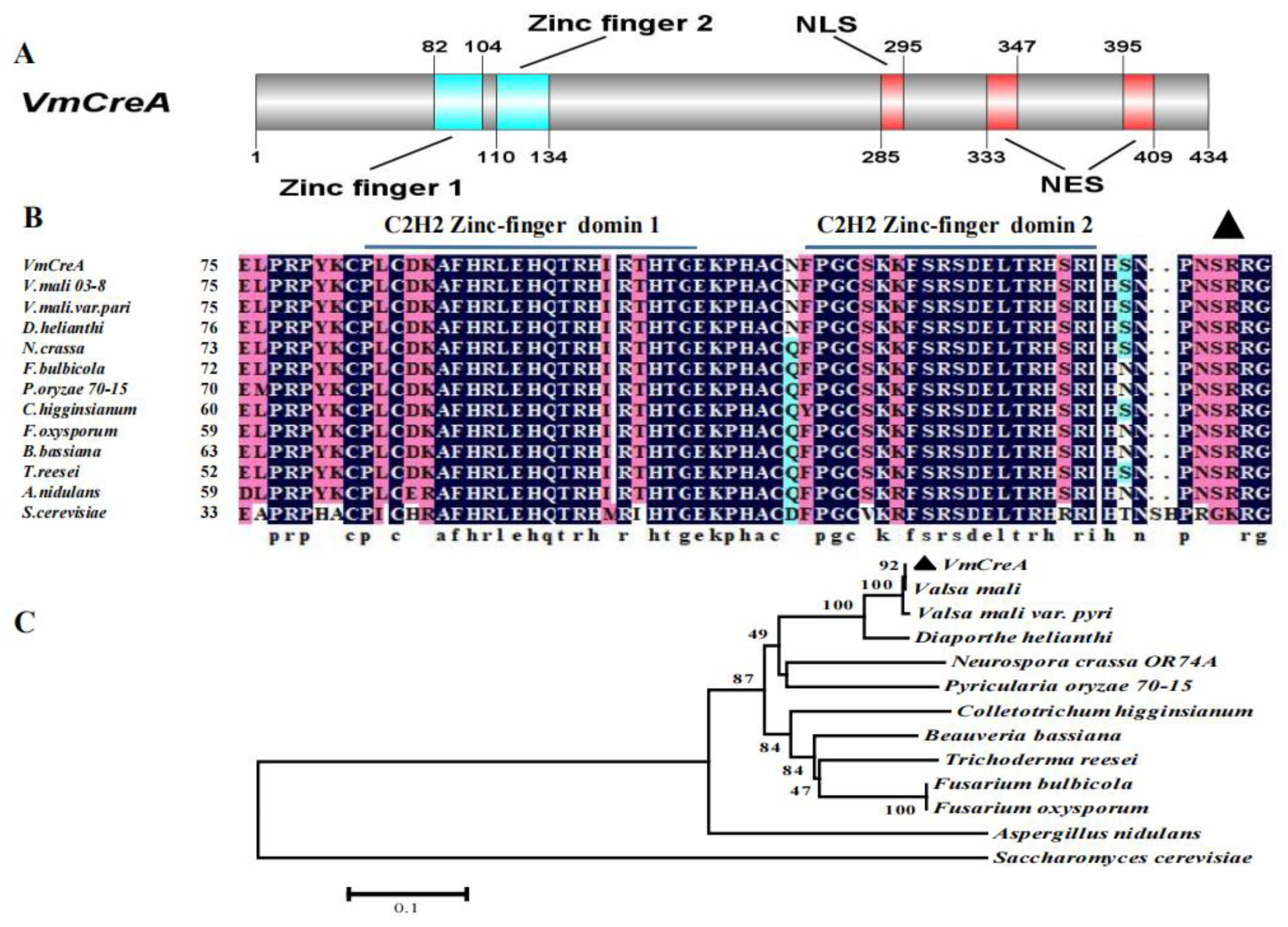
Figure 1. Domain prediction, sequence alignment, and phylogenetic analysis of VmCreA: (A) Domain predictions were performed using Pfam, and nuclear localization signal (NLS) and nuclear export signal (NES) predictions were performed using cNLS Mapper [14]. A schematic diagram of the protein domains and functional motifs was drawn using DOG 2.0 [15]. The numbers denote the corresponding positions of the predicted domains in the protein sequence. (B) Sequence alignment of the zinc-finger domains and the conserved phosphorylation site of VmCreA with its homologs from several other fungi. (C) Phylogenetic relationship of proteins involved in carbon catabolite repression among the V. mali and several other fungi. The scale bar corresponds to the numbers of amino acid substitutions per site.
Ijms 24 09252 g001
Figure 2. Verification of ΔVmCreA and CTΔVmCreA strains: (A) diagrammatic representation of the gene replacement strategy for the construction of the ΔVmCreA and CTΔVmCreA strains; (B) PCR amplification of the segments HPH, UP, and DOWN and of the ligation fragments UP-HPH-DOWN; (C) qRT-PCR verifications of the WT, ΔVmCreA, and CTΔVmCreA strains (The single asterisk indicate significant differences at p < 0.05).
Ijms 24 09252 g002
Figure 3. VmCreA deletion leads to serious defects in vegetative growth: (A) colony morphology of the WT, ΔVmCreA, and CTΔVmCreA strains grown on PDA minimal media (MM) containing various carbon sources, including 2% CMC, 2% starch, 2% 2-deoxyglucose, 2% sucrose, 2% glucose, and 2% lactose; (B) bar graph representing SD from three independent experiments with three replicates, in which the asterisks indicate statistically significant differences (The single asterisk indicate significant differences at p < 0.05).
Ijms 24 09252 g003
Figure 4. Involvement of VmCreA in the regulation of the CCR of V. mali: (A) mycelia growth of VmCreA knockout and complement mutants on minimal media (MM) with CMC and 2-deoxyglucose as the carbon sources; (B) inhibition of the growth rates of the strains in the panel (The single asterisk indicate significant differences at p < 0.05); (C) growth rates with 1% glucose and 1% starch as the carbon sources for three days and the iodine-staining results.
Ijms 24 09252 g004
Figure 5. Inhibition of the growth rates of the V. mali strains under different stress conditions: (A) colony morphology of the WT, ΔVmCreA, and CTΔVmCreA strains grown on PDA, or PDA containing 100 μg/mL CFW, 100 μg/mL CR, 0.01% SDS, 0.5 mol/L NaCl, and 1 mol/L at 25 °C for four days, respectively; (B) inhibition of the growth rates of the strains in the panel (The single asterisk indicate significant differences at p < 0.05).
Ijms 24 09252 g005
Figure 6. Pathogenicity assay of V. mali, where the bars represent the SD from three independent experiments with three replicates (The single asterisk indicate significant differences at p < 0.05): (A) morphology of the V. mali WT, ΔVmCreA, and CTΔVmCreA strains on apple fruit after seven days of inoculation; (B) morphology of the V. mali WT, ΔVmCreA, and CTΔVmCreA strains on apple twigs after seven days of inoculation.
Ijms 24 09252 g006
Figure 7. Results of the qRT-PCR analysis of the hydrolase-related genes: VM1G_02565 (Endopolygalacturonase D); VM1G_02197 (Endo-xylogalacturonan hydrolase A); VM1G_03115 (Aspartic protease pep1); VM1G_03512 (Glucoamylase); VM1G_04322 (Exopolygalacturonase B); VM1G_05544 (Cutinase); VM1G_04032 (Cutinase transcription factor 1 alpha); VM1G_06261 (Pectin lyase A); VM1G_08966 (Pectinesterase A): VM1G_08033 (Endopolygalacturonase D); VM1G_07516 (Rhamnogalacturonate lyase B); VM1G_10963 (Pectin lyase B); and VM1G_10438 (Pectinesterase). (The single asterisk indicate significant differences at p < 0.05).
Ijms 24 09252 g007
Disclaimer/Publisher’s Note: The statements, opinions and data contained in all publications are solely those of the individual author(s) and contributor(s) and not of MDPI and/or the editor(s). MDPI and/or the editor(s) disclaim responsibility for any injury to people or property resulting from any ideas, methods, instructions or products referred to in the content.