Article

Intramolecular Hydrogen Bonding in N6-Substituted 2-Chloroadenosines: Evidence from NMR Spectroscopy

by
Maria Ya. Berzina
1,*,
Barbara Z. Eletskaya
1,
Alexei L. Kayushin
1,
Elena V. Dorofeeva
1,
Olga I. Lutonina
1,
Ilya V. Fateev
1,
Olga N. Zhavoronkova
1,
Arthur R. Bashorin
1,
Alexandra O. Arnautova
1,
Olga S. Smirnova
1,
Konstantin V. Antonov
1,
Alexander S. Paramonov
1,
Maxim A. Dubinnyi
1,2,
Roman S. Esipov
1,
Anatoly I. Miroshnikov
1 and
Irina D. Konstantinova
1,*
1
Shemyakin and Ovchinnikov Institute of Bioorganic Chemistry, Russian Academy of Sciences, Miklukho-Maklaya St. 16/10, 117997 Moscow, Russia
2
Department of Biological and Medical Physics, Moscow Institute of Physics and Technology (State University), 9 Institutskiy per., Dolgoprudny, 141700 Moscow, Russia
*
Authors to whom correspondence should be addressed.
Int. J. Mol. Sci. 2023, 24(11), 9697; https://doi.org/10.3390/ijms24119697
Submission received: 16 May 2023 / Revised: 30 May 2023 / Accepted: 31 May 2023 / Published: 2 June 2023
(This article belongs to the Special Issue Advances in NMR Spectroscopy for Bioactive Small Molecules)

Abstract

:
Two forms were found in the NMR spectra of N6-substituted 2-chloroadenosines. The proportion of the mini-form was 11–32% of the main form. It was characterized by a separate set of signals in COSY, 15N-HMBC and other NMR spectra. We assumed that the mini-form arises due to the formation of an intramolecular hydrogen bond between the N7 atom of purine and the N6–CH proton of the substituent. The 1H,15N-HMBC spectrum confirmed the presence of a hydrogen bond in the mini-form of the nucleoside and its absence in the main form. Compounds incapable of forming such a hydrogen bond were synthesized. In these compounds, either the N7 atom of the purine or the N6–CH proton of the substituent was absent. The mini-form was not found in the NMR spectra of these nucleosides, confirming the importance of the intramolecular hydrogen bond in its formation.

1. Introduction

Interest in the synthesis of new substituted adenosines remains high. Among these nucleosides, effective antimetabolites [1,2] and adenosine receptor agonists [3,4,5] have been found. NMR spectroscopy is a powerful tool for studying the adenosine derivatives. Among all NMR methods, two-dimensional heteronuclear NMR spectroscopy is of particular importance.
Substituents in the purine heterocycle can cause different forms to appear in the NMR spectra. Several theories have been put forward in the literature to explain the presence of different forms of adenine or adenosine derivatives.
N6-substituted adenines and adenosine nucleosides are characterized by the phenomenon of amino–imino tautomerism (Figure 1) [6,7,8,9,10]. It has been reported that the amino form of adenine is absolutely dominant in solution; however, additional substitution can shift the tautomeric amino–imino equilibrium [11]. The introduction of an electron-withdrawing substituent at the C2 position of the purine shifts the equilibrium towards the amino form.
The presence of the second form in NMR was detected for kinetin and its derivatives [11,12]. Novotna et al. suggested that 2-chloro-N6-furfuryl-adenosine exists in two forms: amino and imino tautomers at room temperature in a solution. According to X-ray crystallography data, in the solid state only the amino form is presented [12] (Figure 2).
Martin and Reese suggested that the second form in the NMR spectra emerged because of the hindered rotation. It was registered on cooling the 6-methylamino- and 6-dimethyladenosine solutions [13]. Engel et al. found that two distinct 6-methylamino isomers (rotamers) are associated with the syn and anti conformations of substituent group relative to the N1 of the purine cycle. The main form was syn (Figure 3) [14].
Syn-rotamer predominance was proposed to appear because of intramolecular hydrogen bonding between the hydrogen atom of the exocyclic amino group (N6) and N7 (Figure 3) [15]. Rotamer stabilization arises from two competing hydrogen bonds and a steric effect; syn-conformation is favored by N6–H∙∙∙N7 bonding, whereas the anti-form is stabilized by N6–H∙∙∙Nl bonding, but is destabilized by the steric interaction of the methyl group.
The nature of the substituent at the C2 position affects the presence of the mini-form in NMR. In the NMR spectra of substituted adenosines, the second form is usually registered as broadened signals at N6H–CH [16,17,18,19,20], or not registered at all. The NMR spectra of the purines with an electron-withdrawing substituent (Cl, F) at position 2 show the presence of the second form at room temperature [11], and the authors often do not discuss the second form, but it can be seen in NMR spectra in the Supplementary Information [21,22,23,24,25,26]. Various C2-substituted purines with optically active substituents at C6 were obtained. The presence of the R or S enantiomers did not affect the amount of the second form [21].
At low temperatures (from 273 to 248 K), the mini-form appeared in both C2–H and C2–NH2-substituted purines [11]. NMR spectra are usually taken at 300 or 303 K, and the second form was not detected at such temperatures. In addition, the conditions for recording the spectra differed (CDCl3 instead of DMSO-d6, 100 °C rather than 30 °C) [25,27,28,29], and the relative amount of the mini-form was higher in DMSO than in CDCl3 [30]. In 7-deazakinetin, the second form did not appear in NMR even upon cooling [11]. The N7 atom made a key contribution to the restricted rotation around the C6–N6 bond.
In summary, several models of the hindered rotation structure in C6-substituted purines and the related second form in NMR are described in the literature.
We have recently synthesized a number of 2-chloradenosine analogs containing chiral amino acid amides and their analogs at the C6 position of the purine heterocycle [5]. When analyzing the NMR spectra of nucleosides, it was found that the substances existed in two forms. We observed double signals of the alpha proton of the amino acid residue, and protons of the nearest environment. This phenomenon did not depend on the amino acid chirality, and was observed in both L- and D-amino acids as well as in optically inactive glycine, beta-alanine and ε-lysine. The difference in the chemical shifts of the alpha-proton signals of the amino acid residue was significant (more than 0.8 ppm).
We decided to investigate whether the mini-form can be formed due to the appearance of a hindered structure with an intramolecular hydrogen bond. Our aim was to determine the localization of this hydrogen bond, and confirm our theory with two-dimensional heteronuclear NMR spectroscopy data.

2. Results

The NMR spectra analysis of acylated nucleosides 1a12a and ribosides 1b12b [5] revealed the presence of a significant amount of the nucleoside second form. The content of the mini-form component for nucleosides 1a,b12a,b varied from 11 to 32% of the main form (Figure 4). According to HPLC and LC-MS, all products were homogeneous.
The signal of the N6CH proton was significantly shifted downfield (see Table 1), as well as N6CH carbon and N6H nitrogen signals. Heavy atoms of the purine heterocycle were also shifted downfield (N7, C4) or upfield (C8, C5, C2). The chemical shifts of the amide group protons were shifted upfield. They are not shown in the table, because it is difficult to calculate the difference in chemical shifts between the two forms of the dual set of signals. The exception is the N6H proton. It can be shifted both upfield and downfield.
The NMR spectra set of valine derivative 3b was chosen to represent changes in chemical shifts because of the considerable amount of mini-form in the NMR spectra (See Figures S5–S10 in the Supplementary Materials). The 1D and 2D NMR spectra were recorded again after preparing the sample with an increased concentration of valine derivative 3b (770 mg/mL) to improve the signal-to-noise value. The mini-form had the same set of signals as the main form in COSY, 1H,13C-HMBC and 1H,15N-HMBC spectra (See Figures S11–S16 in the Supplementary Materials). Differences in the chemical shifts of main and mini-form atoms are shown in Figure 5.
The chemical shift of the αCH proton of the valine residue changed by 0.79 ppm according to the NMR 1H spectrum (Figure 6). The chemical shift of the CH proton of the main form 3b was 4.54 ppm, while the signal of the mini-form was shifted downfield to 5.33 ppm. The further the protons were from the chiral center of the amino acid, the less this effect was pronounced (Figure 5).
The dual set of signals is also presented for the amine and amide group protons (Figure 7). The separate set of signals from the nucleoside mini-form was also registered in 13C NMR spectra (see Figure 8) and COSY spectra (Figure 9).
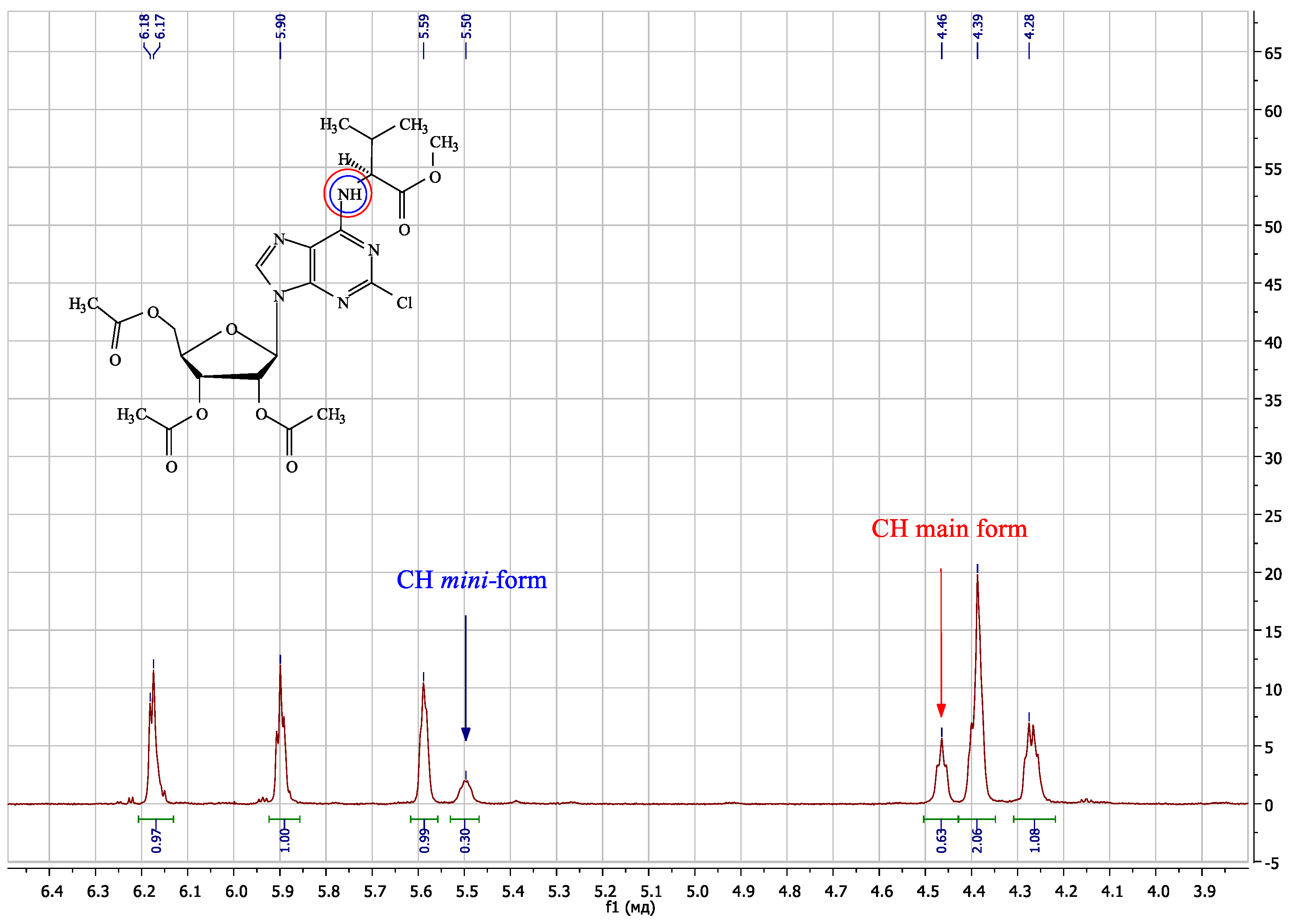
The mini-form was observed not only for ribosides 1b12b, but also for protected nucleosides 1a12a (Figure 10, a fragment of the 1H NMR spectrum of valine methyl ester triacetate 3a is shown; see Figures S1–S4 in the Supplementary Materials). The appearance of the nucleoside mini-form did not require an amide group or a methyl ester.
We studied the temperature effect on the mini-form formation (Figure 11). With a temperature increase from 293 to 318 K, the signals of the same protons of the main and mini-form began to broaden. This broadening is evidence of a chemical exchange process well known in NMR spectroscopy [31]. This NMR evidence of the chemical exchange provided an independent proof that we have two forms of the same substance; the mini-form interchanged with the main form during the NMR experiment. The 1H signals of the main form and the mini-form did not merge into each other, but that would occur at higher temperatures not available on our NMR equipment. In our experiments at 293–318 K, the content of the second form in NMR did not change.
The N6H proton of each form had a cross-peak with the N6CH proton in the COSY NMR spectrum. This discards the amino–imino tautomerism. We suggest that hindered rotation appeared. In the main form, the amino acid substituent rotated freely around the C6–N6 bond, while in the mini-form the structure was in a hindered state (Figure 12). We assumed that the hindered structure was caused by the intramolecular hydrogen bond formation. The hydrogen bond was formed between the N6CH proton and the N7 purine atom, with a stable six-membered ring formed. The proposed hindered structure is shown in Figure 12.
The presence of a hydrogen bond can be detected by the NMR spectrum, both for purine heterocycle [32] and the amino acid [33]. This should result in a cross peak between the N6CH proton and the purine N7 in the 1H,15N-HMBC spectrum. This peak was observed in the spectrum of the 3b mini-form, with no peak for the main form (Figure 13). This proves the presence of the hydrogen bond.
It was necessary to prove the presence of this hydrogen bond to finally confirm our theory. Several new nucleosides that have been synthesized are incapable of forming N6CH∙∙∙N7 hydrogen bonding, either because of the absence of the nitrogen atom in the N7 position (8-aza-7-deaza-adenosine derivative 13) or because of the N6CH proton’s absence (tert-butylamino derivative 14) (Figure 14).
The synthesis of 8-aza-7-deaza-adenosine derivative 13 was carried out according to Scheme 1. The starting 2,6-dichloroallopurinol 15 reacted with L-valine methyl ester in the presence of 2,4,6-collidine. Heterocycle 16 was ribosylated by a transglycosylation reaction with uridine phosphorylase (UP) and purine nucleoside phosphorylase (PNP) [34] to give product 13.
The synthesis of tert-butylamino derivative 14 was carried out according to Scheme 2. The starting 2,6-dichloro-9-(2′,3′,5′-tri-O-acetyl-β-D-ribofuranosyl)purine 17 was obtained according to the procedure published earlier [5]. The reaction was carried out in acetonitrile in the presence of triethylamine. To remove the acetyl protection groups from the ribose, product 18 was treated with a saturated solution of ammonia in methanol at 4 °C to obtain the corresponding nucleoside 14.
The nucleosides 13 and 14 were characterized by 1H, 13C, COSY, 1H,13C HSQC, 1H,13C HMBC, 1H,15N HSQC and 1H,15N HMBC NMR spectra. NMR spectra are provided in the Supplementary Materials (Figures S17–S33). No second form of nucleosides was observed in NMR (Figure 15 and Figure 16).
The theory of the appearance of the mini-form by amino–imino tautomerism turned out to be untenable for our compounds. We have explained the appearance of the second form in the NMR spectra of N6-substituted 2-chloroadenosine nucleosides by the formation of an intramolecular hydrogen bond between the proton of the N6CH substituent and the N7 atom of the purine base. The content of the mini-form was independent of temperature. The mini-form was not observed in the NMR spectra of compounds lacking either the purine N7 atom or the N6CH proton of the substituent.

3. Discussion

There are several hypotheses explaining the presence of the mini-form in the NMR spectra of purine nucleosides. The first is that the appearance of a second form in the NMR spectra of 6-substituted adenosines is attributed to amino–imino tautomerism [12] (tautomerism hypothesis). For the second, the mini-form is attributed to the hindered rotation [11,13,14,15]. We propose here that the hindered structure is stabilized by the non-conventional intramolecular CH∙∙∙N7 hydrogen bond formation (H-bond hypothesis) (Figure 17).
NMR chemical shifts exhibit high sensitivity to any alterations in the chemical or spatial structure of a molecule. Our initial assumption was that analyzing the NMR data would uncover consistent patterns in the chemical shift variations of the atoms implicated in the hindered structure formation. Unfortunately, the chemical shifts presented in Table 1 do not provide definitive conclusions, making it challenging to favor one hypothesis over another.
In our research involving various 2-chloro-C6-substituted purine ribonucleosides [5], we observed cross-peaks between the N6H and CH proton signals in the COSY spectrum in both main and mini-form in all compounds considered (Figure 9, for compound 3b), leading us to doubt the tautomerism hypothesis. On the other hand, the H-bond hypothesis assumes COSY cross-peak presence in both main and mini forms, as seen in our experiments.
We could keep the tautomerism hypothesis in consideration by examining the possibility of an amide-like C6–N6 partial double bond instead of an imino double bond. However, we found this explanation to be unsuitable. Despite the fact that it would account for the stable planar hindered structure, the chemical shift of the N6H proton did not exhibit significant changes (Table 1). Notably, the N6H proton exhibited both upfield and downfield shifts across different compounds.
Moreover, amino–imino tautomerism should drastically influence the 15N chemical shift of the N6H nitrogen. We would expect significant chemical shift changes of around one hundred ppm on the basis of available experimental data and quantum-theory calculations [35,36]. However, contrary to these expectations, our experimental data revealed only 4–20 ppm changes in the N6H nitrogen chemical shift between the main and mini forms (Table 1). Hence, we must reject the amino–imino tautomerism hypothesis that was previously proposed [12], as it is not consistent with the experimental data obtained in the present work.
To further explore the H-bond hypothesis, we obtained the 15N-HMBC spectrum at an increased concentration of the substance 3b (770 mg/mL in DMSO-d6). This spectrum revealed a cross-peak between the purine N7 atom and the N6CH proton of mini-form (Figure 13), indicating the presence of long-range through-bond interactions (Figure 17). Notably, this cross-peak was not observed in the main form. In the case of the tautomerism hypothesis this cross peak could be explained as five bonds correlation only (Figure 17, bold lines), and we could not understand its absence in the main form. Really, it would be predominantly observed in the main form, which constitutes approximately 80% of the total. Hence, that cross-peak makes the H-bond hypothesis highly preferable in comparison with the tautomerism hypothesis.
In general, cross-peaks in multidimensional NMR spectra appear either because of spin–spin coupling between two nuclei (through-bond connectivities) or because of through-space cross-relaxation effects (NOESY or ROESY interactions). The 1H,15N-HMBC NMR experiment used in the present work revealed cross-peaks that appeared because of multiple-bond long range spin–spin couplings from 1H to 15N nuclei. Similar long-range couplings have been exploited in especially designed NMR techniques used for hydrogen bond detection in proteins [33] and nucleic acid base pairs [32]. Moreover, exactly the same 1H,15N-HMBC experiment has been employed to confirm intramolecular O–H···N [37] or N–H···N [38] hydrogen bonding. Several reviews have extensively discussed numerous examples of different hydrogen bonds verified by NMR [39,40]. However, to the best of our knowledge, the utilization of the 1H,15N-HMBC NMR experiment for the detection of non-conventional CH∙∙∙N7 bonds has not been previously reported.
Additionally, we observed a distinct hydrogen bond between 5′-OH and purine N3 [41] in the spectrum (Figure 13, cross-peak {5.26, 221.56}). This spatial proximity and the presence of a hydrogen bond suggest that such bond types are detectable on this 2D HMBC spectrum when the substance is at an elevated concentration.
The presence of the mini-form in various structures was found irrespective of substituent chirality, including L- and D-amino acids (4b and 5b), non-chiral compounds (11b and 12b) and bulky substituent (8b), and suggests its inherent nature. In the absence of the N6CH proton, the formation of a mini-form does not occur (compound 14, Figure 16), underscoring its critical role in the occurrence of hindered rotation. We observed significant downfield chemical shift changes, predominantly for the N6CH proton, providing further evidence of its involvement in the formation of the hindered rotation conformation. Furthermore, the absence of the N7 purine atom in compound 13 corresponds to the absence of the mini-form (Figure 15), further supporting the H-bond hypothesis and rejecting the tautomerism hypothesis.
The formation of the mini-form can be attributed to restricted rotation, which arises not from steric factors but rather from the presence of a stabilizing hydrogen bond. The main form appears to represent a position of the N6-substituent, where no hydrogen bond is formed. Future investigations will delve into the study of the intramolecular hydrogen bond formation in different solvents and under cooling conditions.
In conclusion, the full set of experimental data presented provides unambiguous evidence for the presence of a non-conventional CH∙∙∙N hydrogen bond in 2-chloro-C6-substituted purine ribosides. Alternative explanations involving amino–imino tautomerism have been ruled out due to their inconsistency with the NMR data obtained in this study.

4. Materials and Methods

All solvents and chemicals were used as purchased without further purification. Valine derivative 3b was synthesized according to a previously developed procedure [5]. The progress of reactions was monitored on Silufol precoated silica gel plates (with fluorescence indicator UV254) using methanol/chloroform as a solvent system. Spots were visualized through irradiation with ultraviolet light (254 nm). Analytical high-performance liquid chromatography (HPLC) was performed on a Waters system (Waters 1525, Waters 2489, Breeze 2) using Nova Pak C18 column, 4.6 × 150 mm, 5 µm, flow rate 1 mL/min, eluent A—H2O/0.1% TFA, eluent B—70% CH3CN/H2O/0.1% TFA, gradient: 0–100% B over 20 min), detection at 254 nm. Column chromatography was performed on Silica gel 60 or C18-reversed-phase silica gel (Fluka, Buchs, Switzerland).
NMR spectra were acquired on a Bruker Avance I 700 MHz or Bruker Avance III 800 MHz NMR in DMSO-d6 at 303 K. To study the temperature dependence, the spectra were taken at 293, 298, 303, 313 and 318 K. Chemical shifts in ppm (δ) were measured relative to the residual solvent signals as internal standards (2.50). Coupling constants (J) were measured in Hz. Liquid chromatography mass spectrometry was performed using an Agilent 6210 TOF LC–MS system (Agilent Technologies, Santa Clara, CA, USA).
9-β-D-ribofuranosyl-2-chloro-6-(Nα-L-valinylamido)-purine (3b)
Nucleoside 3b was synthesized according to a previously developed procedure [5].
Main form: 1H NMR (800 MHz, DMSO-d6, J, Hz, 303 K): δ 8.42 (s, 1 H, H 8), 7.67 (m, 0.81 H, NH), 7.67 and 7.19 (2 s, 0.77 and 0.91 H, NH2), 5.90 (d, J = 5.8 Hz, 1 H, H1′), 5.67 (d, J = 6.2 Hz, 1 H, OH2′), 5.38 (s, 1 H, OH3′), 5.25 (m, 1 H, OH5′), 4.59 (m, 1 H, H2′), 4.54 (m, 0.77 H, αCH-Val), 4.23 (m, 1 H, H3′), 4.04 (m, 1 H, H4′), 3.72 (m, 1 H, H5′a), 3.63 (m, 1 H, H5′b), 2.16 (m, 1 H, βCH-Val), 0.90 (m, 6 H, CH3-Val). 13C NMR (176 MHz, DMSO-d6, 303 K): δ 172.75 (CONH2), 155.26 (C6), 153.56 (C2), 150.06 (C4), 140.85 (C8), 118.95 (C5), 88.36 (C1′), 86.25 (C4′), 74.38 (C2′), 70.92 (C3′), 61.85 (C5′), 59.57 (αCH-Val), 30.92 (βCH-Val), 19.54 (CH3-Val), 18.46 ppm (CH3-Val). 15N NMR (71 MHz, DMSO-d6, 303 K): δ 237.28 (N7), 227.62 (N1), 221.20 (N3) 171.28 (N9), 107.95 (NH2), 93.53 ppm (NH).
Mini-form: 1H NMR (800 MHz, DMSO-d6, J, Hz, 303 K): δ 8.42 (s, 1 H, H 8), 7.78 (m, 0.19 H, NH), 7.53 (s, 0.17 H, NH2), 5.90 (d, J = 5.8 Hz, 1 H, H1′), 5.67 (d, J = 6.2 Hz, 1 H, OH2′), 5.38 (s, 1 H, OH3′), 5.33 (m, 0.20 H, αCH-Val), 5.25 (m, 1 H, OH5′), 4.59 (m, 1 H, H2′), 4.23 (m, 1 H, H3′), 4.04 (m, 1 H, H4′), 3.72 (m, 1 H, H5′a), 3.63 (m, 1 H, H5′b), 2.16 (m, 1 H, βCH-Val), 0.90 (m, 6 H, CH3-Val). 13C NMR (176 MHz, DMSO-d6, 303 K): δ 173.95 (CONH2), 155.92 (C6) 153.25 (C2), 152.31 (C4), 140.31 (C8), 117.97 (C5), 88.14 (C1′), 74.25 (C2′), 61.67 (αCH-Val), 31.04 (βCH-Val), 19.39 (CH3-Val), 17.94 ppm (CH3-Val). 15N NMR (71 MHz, DMSO-d6, 303 K): δ 242.57 (N7), 227.62 (N1), 221.20 (N3) 171.28 (N9), 107.92 (NH2), 101.12 ppm (NH).
6-chloro-4-(Nα-L-valinyl)-pyrazolo[3,4-d]pyrimidine methyl ester (16)
To a solution of 150.0 mg (0.794 mmol) of 4,6-dichloro-1H-pyrazolo[3,4-d] and 234.0 mg (1.40 mmol) of L-valine methyl ester hydrochloride in 3 mL of dry DMF was added 300 µL (275.1 mg, 2.273 mmol) of 2,4,6-collidine at room temperature. The mixture was incubated at 50 °C for 24 h. The reaction progress was monitored by HPLC. The solvent was removed in vacuo, and the residue was dissolved in a minimal volume of water. The desired product was isolated by flash column chromatography on C18 silica gel (10 × 1.5 cm, elution by EtOH in H2O, 10%). Then, 87.9 mg (39.0%) of the product was obtained as a white solid with a purity of 97.06%. 1H NMR (700 MHz, DMSO-d6, J, Hz, 303 K): δ 13.60 (s, 0.89 H, N1–H), 8.81 (d, J = 7.4 Hz, 1 H, NH-Val), 8.35 (s, 1 H, H3), 4.62 (m, 1 H, αCH-Val), 3.69 (s, 3 H, −OCH3), 2.21 (m, 1 H, βCH-Val), 1.02 (d, J = 6.2 Hz, 3 H, CH3), 0.98 ppm (d, J = 6.3 Hz, 3 H, CH3). 13C NMR (176 MHz, DMSO-d6, 303 K): δ 171.73 (COOCH3), 156.58 (C4), 155.35 (C6 or C7a), 133.14 (C3), 98.65 (C4a), 58.87 (αCH-Val), 51.70 (–OCH3), 29.99 (βCH-Val), 18.81 (CH3), 18.74 ppm (CH3). 15N NMR (71 MHz, DMSO-d6, 303 K): δ 221.5 (N5), 190.8 (N1), 101.9 ppm (NH-Val). HRMS (ESI) C11H14N5O2Cl [M − H]: 282.0758 calcd., 282.0768 found.
6-chloro-1-β-D-ribofuranosyl-4-(Nα-L-valinyl)-pyrazolo[3,4-d]pyrimidine methyl ester (13)
To a solution of 41 mg of uridine (0.168 mmol), 30 mg of 6-chloro-4-(Nα-L-valinyl)-pyrazolo[3,4-d]pyrimidine methyl ester 16 (0.106 mmol), and 148 mg of KH2PO4 (1.109 mmol) in 212 mL of distilled water was added 3.0 µL (4.2 units) of E. coli PNP and 2.4 µL (4.1 units) of E.coli UP. The pH was adjusted up to 7.0 with 5 M KOH solution and the reaction mixture was incubated at 50 °C. The reaction progress was monitored with HPLC. When conversion reached the highest value, the reaction was terminated by the addition of ethanol (40%, v/v). The reaction mixture was evaporated up to 5 mL, and the desired product was isolated using reversed-phase column chromatography (silica gel C18, Merck), column 2.5×3.0 cm (elution by EtOH in H2O, 40%). Then, 30.4 mg (69.0%) of the product was obtained as a white solid with a purity of 97.86%. 1H NMR (700 MHz, DMSO-d6, J, Hz, 303 K): δ 9.00 (d, J = 7.2 Hz, 1 H, NH-Val), 8.45 (s, 1 H, H3), 6.02 (d, J = 4.6 Hz, 1 H, H1′), 5.39 (m, 1 H, OH2′), 5.15 (m, 1 H, OH3′), 4.70 (m, 1 H, OH5′), 4.64 (m, 1 H, αCH-Val), 4.58 (m, 1 H, H2′), 4.20 (m, 1 H, H3′), 3.91 (m, 1 H, H4′), 3.69 (s, 3 H, OCH3), 3.56 (m, 1 H, H5′a), 3.43 (m, 1 H, H5′b), 2.22 (m, 1 H, βCH-Val), 1.03 (d, J = 6.6 Hz, 3 H, CH3), 0.98 ppm (d, J = 6.8 Hz, 3 H, CH3). 13C NMR (176 MHz, DMSO-d6, 303 K): δ 171.53 (COOCH3), 157.03 (C6), 156.60 (C4), 154.72 (C7a), 133.85 (C3), 99.55 (C4a), 88.10 (C1′), 85.15 (C4′), 72.95 (C2′), 70.68 (C3′), 62.13 (C5′), 58.96 (αCH-Val), 51.78 (OCH3), 30.01 (βCH-Val), 18.79 (CH3), 18.68 ppm (CH3). 15N NMR (71 MHz, DMSO-d6, 303 K): δ 305.3 (N2), 222.8 (N5), 199.5 (N1), 102.5 ppm (NH-Val). HRMS (ESI) C16H22N5O6Cl [M − H]: 414.1180 calcd., 414.1194 found.
9-(2′,3′,5′-Tri-O-acetyl-β-D-ribofuranosyl)-2-chloro-6-tert-butylamino-purine (18)
To a solution of 30.0 mg (0.067 mmol) of 2,6-dichloro-9-(2,3,5-tri-O-acetyl-β-D-ribofuranosyl)purine and 20 µL (14 mg, 0.188 mmol) of tert-butylamine (Sigma-Aldrich) in 1 mL of dry DMF was added 50 µL (36 mg, 0.360 mmol of triethylamine. The mixture was stirred at room temperature for 18 h, and the progress was monitored with thin-layer chromatography (TLC) on silica gel (chloroform–methanol, 30:1). The solvent was removed in vacuo, and the residue was dissolved in a minimal volume of chloroform, and the desired product was isolated using column chromatography on silica gel (10 × 1.5 cm, with elution with MeOH gradient in CHCl3, 0–20%). Then, 32.1 mg (98.7%) of the product was obtained as a yellow solid with a purity of 98.47%. 1H NMR (700 MHz, DMSO-d6, J, Hz, 303 K): δ 8.39 (s, 1 H, H8), 7.57 (br.s, 0.76 H, NH), 6.15 (d, J = 5.5, 1 H, H1′),5.90 (t, J = 5.7 Hz, 1 H, H2′), 5.58 (m, 1 H, H3′), 4.39 (m, 2 H, H5′a and H4′), 4.27 (m, 1 H, H5′b), 2.13 (m, 3 H, CH3CO-3′), 2.05 (m, 3 H, CH3CO-2′), 2.03 (m, 3 H, CH3CO-5′), 1.49 ppm (s, 9 H, CH3). 13C NMR (176 MHz, DMSO-d6, 303 K): δ 169.88 (O–CO5′), 169.29 (O–CO3′), 169.13 (O–CO2′), 154.68 (C6), 152.43 (C2), 139.72 (C8), 85.27 (C1′), 79.47 (C4′), 71.97 (C2′), 69.86 (C3′), 62.60 (C5′), 52.08 (C(CH3)3), 28.46 (C(CH3)3), 20.35 (CH3–CO5′), 20.24 (CH3–CO3′), 20.08 ppm (CH3–CO2′). 15N NMR (71 MHz, DMSO-d6, 303 K): δ 243.0 (N7), 167.3 (N9), 114.2 ppm (NH). HRMS (ESI) C20H26N5O7Cl [M − H]: 482.1442 calcd., 482.1438 found.
9-β-D-Ribofuranosyl-2-chloro-6-tert-butylamino-purine (14)
A total of 24 mg (0.066 mmol) of nucleoside triacetate 18 was dissolved in 5 mL of absolute methanol and cooled in an ice bath. The solution was saturated with ammonia and kept for 24 h at +4 °C. The reaction progress was controlled by TLC on silica gel (chloroform–methanol 10:1). After reaction completion, the solvent was evaporated, the residue was dissolved in a minimal volume of water, and the desired product was isolated with column chromatography on C18-reversed-phase silica gel (12 × 2 cm, elution with EtOH gradient in water, 10–80%). Then, 15.3 mg (86.5%) of the product was obtained as a yellow solid with a purity of 98.80%. 1H NMR (700 MHz, DMSO-d6, J, Hz, 303 K): δ 8.39 (s, 1 H, H8), 7.46 (br.s, 0.87 H, NH), 5.83 (d, J = 5.9 Hz, 1 H, H1′), 5.46 (m, 1 H, OH1′), 5.19 (m, 1 H, OH2′), 5.04 (t, J = 5.6 Hz, 1 H, OH5′),4.52 (m, 1 H, H2′), 4.14 (m, 1 H, H3′), 3.95 (m, 1 H, H4′), 3.66 (m, 1 H, H5′a), 3.57 (m, 1 H, H5′b), 1.50 ppm (s, 9 H, CH3).13C NMR (176 MHz, DMSO-d6, 303 K): δ 154.67 (C6), 152.16 (C2), 149.23 (C4), 139.54 (C8), 118.86 (C5), 87.31 (C1′), 85.60 (C4′), 73.55 (C2′), 70.25 (C3′), 61.26 (C5′), 52.02 (NHCH), 28.52 ppm (CH3). 15N NMR (71 MHz, DMSO-d6, 303 K): δ 241.1 (N7), 218.9 (N3), 170.9 (N9), 113.4 ppm (NH). HRMS (ESI) C14H20N5O4Cl [M − H]: 356.1125 calcd., 356.1120 found.

5. Conclusions

During the examination of the NMR spectra of N6-substituted 2-chloroadenosines, a second form was discovered. This form, present at 11–32% of the main form, was identified by a unique set of signals in the 2D NMR spectra. The mini-form’s temperature dependence was investigated and found to remain constant in quantity, but the peak shape and chemical shift were affected. Our hypothesis proposes that the presence of the mini-form is due to restricted rotation caused by the formation of an intramolecular hydrogen bond between the N7 atom of purine and the N6CH proton of the substituent. This was confirmed by the key cross-peak in the 1H,15N-HMBC NMR spectrum from the CH donor to the N7 acceptor presented in the mini-form but not in the main form. We synthesized compounds that lacked either the donor (CH) or acceptor (N7) of the proposed N6CH∙∙∙N7 hydrogen bond. The mini-form was absent in the NMR spectra of these compounds, supporting the importance of the intramolecular hydrogen bond in its formation. The full set of experimental NMR data has provided unambiguous evidence for the presence of a non-conventional CH∙∙∙N hydrogen bond in 2-chloro-C6-substituted purine ribosides. This type of hydrogen bond was discovered using 1H,15N-HMBC NMR spectrum in a purine nucleoside in a first time.

Supplementary Materials

The following supporting information can be downloaded at: https://www.mdpi.com/article/10.3390/ijms24119697/s1.

Author Contributions

Conceptualization, M.Y.B. and I.D.K.; methodology, M.Y.B., B.Z.E., I.V.F. and M.A.D.; formal analysis, M.Y.B., A.L.K., A.S.P. and K.V.A.; validation, I.V.F. and M.Y.B.; investigation, M.Y.B., B.Z.E., I.V.F., M.A.D., O.I.L., O.N.Z., A.R.B., A.O.A., E.V.D., O.S.S. and K.V.A.; resources, M.Y.B. and R.S.E.; writing—original draft, M.Y.B., M.A.D. and I.D.K., writing—review and editing, M.Y.B., M.A.D. and I.D.K.; supervision, A.I.M., R.S.E. and I.D.K. All authors have read and agreed to the published version of the manuscript.

Funding

This research was funded by the Russian Science Foundation (Project No. 21-13-00429).

Institutional Review Board Statement

Not applicable.

Informed Consent Statement

Not applicable.

Data Availability Statement

Data are contained within the article or Supplementary Materials.

Acknowledgments

The authors would like to thank T.A. Balashova (Shemyakin and Ovchinnikov Institute of Bioorganic Chemistry, Russian Academy of Sciences) for assistance in the analysis of the NMR spectra.

Conflicts of Interest

The authors declare no conflict of interest.

References

  1. Robak, T.; Lech-Maranda, E.; Korycka, A.; Robak, E. Purine nucleoside analogs as immunosuppressive and antineoplastic agents: Mechanism of action and clinical activity. Curr. Med. Chem. 2006, 13, 3165–3189. [Google Scholar] [CrossRef] [PubMed]
  2. Robak, P.; Robak, T. Older and new purine nucleoside analogs for patients with acute leukemias. Cancer Treat. Rev. 2013, 39, 851–861. [Google Scholar] [CrossRef] [PubMed]
  3. Aurelio, L.; Baltos, J.A.; Ford, L.; Nguyen, A.T.N.; Jorg, M.; Devine, S.M.; Valant, C.; White, P.J.; Christopoulos, A.; May, L.T.; et al. A Structure-Activity Relationship Study of Bitopic N-6-Substituted Adenosine Derivatives as Biased Adenosine A(1) Receptor Agonists. J. Med. Chem. 2018, 61, 2087–2103. [Google Scholar] [CrossRef] [PubMed]
  4. Jacobson, K.A.; Gao, Z.-G.; Paoletta, S.; Kiselev, E.; Chakraborty, S.; Jayasekara, P.S.; Balasubramanian, R.; Tosh, D.K. John Daly Lecture: Structure-guided Drug Design for Adenosine and P2Y Receptors. Comput. Struct. Biotechnol. J. 2015, 13, 286–298. [Google Scholar] [CrossRef] [PubMed] [Green Version]
  5. Berzina, M.Y.; Eletskaya, B.Z.; Kayushin, A.L.; Dorofeeva, E.V.; Lutonina, O.I.; Fateev, I.V.; Paramonov, A.S.; Kostromina, M.A.; Zayats, E.A.; Abramchik, Y.A.; et al. Synthesis of 2-chloropurine ribosides with chiral amino acid amides at C6 and their evaluation as A1 adenosine receptor agonists. Bioorg. Chem. 2022, 126, 105878. [Google Scholar] [CrossRef]
  6. Dračínský, M.; Pohl, R. NMR studies of purines. Annu. Rep. NMR Spectrosc. 2014, 82, 59–113. [Google Scholar] [CrossRef]
  7. Bakkestuen, A.K.; Gundersen, L.-L.; Petersen, D.; Utenova, B.T.; Vik, A. Synthesis and antimycobacterial activity of agelasine E and analogs. Org. Biomol. Chem. 2005, 3, 1025–1033. [Google Scholar] [CrossRef]
  8. Roggen, H.; Gundersen, L.L. Synthetic Studies Directed towards Agelasine Analogs–Synthesis, Tautomerism, and Alkylation of 2-Substituted N-Methoxy-9-methyl-9H-purin-6-amines. Eur. J. Org. Chem. 2008, 30, 5099–5106. [Google Scholar] [CrossRef]
  9. Fujii, T.; Saito, T.; Itaya, T.; Kizu, K.; Kumazawa, Y.; Nakajima, S. Purines. XXIX. Syntheses of 9-Alkyl-2-deuterio-N6-methoxyadenines and 2-Deuterio-N6, 9-dimethyladenine: Tautomerism in 9-Substituted N6-Alkoxyadenines. Chem. Pharm. Bull. 1987, 35, 4482–4493. [Google Scholar] [CrossRef] [Green Version]
  10. Morozov, Y.V.; Savin, F.; Chekhov, V.; Budowsky, E.; Yakovlev, D.Y. Photochemistry of N6-methoxyadenosine and of N4-hydroxycytidine and its methyl derivatives I: Spectroscopic and quantum chemical investigation of ionic and tautomeric forms: Syn-anti isomerization. J. Photochem. 1982, 20, 229–252. [Google Scholar] [CrossRef]
  11. Baranowski, D.; Framski, G.; Wyszko, E.; Ostrowski, T. Studies on structure of kinetin riboside and its analogues by variable-temperature NMR. J. Mol. Struct. 2019, 1195, 110–118. [Google Scholar] [CrossRef]
  12. Novotná, R.; Trávníček, Z.; Popa, I. X-ray crystallographic and NMR study of the tautomerism in kinetin, kinetin riboside and their derivatives: A comparison between the solid state and solution. J. Mol. Struct. 2010, 963, 202–210. [Google Scholar] [CrossRef]
  13. Martin, D.; Reese, C. Restricted rotation in methylated derivatives of adenosine and cytidine. Chem. Commun. 1967, 1275–1276. [Google Scholar] [CrossRef]
  14. Engel, J.D.; Von Hippel, P.H. Effects of methylation on the stability of nucleic acid conformations. Monomer level. Biochemistry 1974, 13, 4143–4158. [Google Scholar] [CrossRef]
  15. Dodin, G.; Dreyfus, M.; Dubois, J.-E. Restricted rotation in 6-methylaminopurine and analogues. Intramolecular hydrogen bonding. J. Chem. Soc. Perkin Trans. 2 1979, 438–442. [Google Scholar] [CrossRef]
  16. Vícha, J.; Malon, M.; Veselá, P.; Humpa, O.; Strnad, M.; Marek, R. 1H-, 13C-, and 15N-NMR chemical shifts for selected glucosides and ribosides of aromatic cytokinins. Magn. Reson. Chem. 2010, 48, 318–322. [Google Scholar] [CrossRef]
  17. Casati, S.; Manzocchi, A.; Ottria, R.; Ciuffreda, P. 1H, 13C and 15N NMR assignments for N6-isopentenyladenosine/inosine analogues. Magn. Reson. Chem. 2010, 48, 745–748. [Google Scholar] [CrossRef] [PubMed]
  18. Drenichev, M.S.; Oslovsky, V.E.; Sun, L.; Tijsma, A.; Kurochkin, N.N.; Tararov, V.I.; Chizhov, A.O.; Neyts, J.; Pannecouque, C.; Leyssen, P. Modification of the length and structure of the linker of N6-benzyladenosine modulates its selective antiviral activity against enterovirus 71. Eur. J. Med. Chem. 2016, 111, 84–94. [Google Scholar] [CrossRef]
  19. Casati, S.; Manzocchi, A.; Ottria, R.; Ciuffreda, P. 1H, 13C and 15N NMR spectral assignments of adenosine derivatives with different amino substituents at C6-position. Magn. Reson. Chem. 2011, 49, 279–283. [Google Scholar] [CrossRef]
  20. Oslovsky, V.E.; Drenichev, M.S.; Alexeev, C.S.; Solyev, P.N.; Esipov, R.S.; Mikhailov, S.N. Synthesis of Cytokinins via Enzymatic Arsenolysis of Purine Nucleosides. Curr. Protoc. Nucleic Acid Chem. 2018, 75, e61. [Google Scholar] [CrossRef]
  21. Savelieva, E.M.; Zenchenko, A.A.; Drenichev, M.S.; Kozlova, A.A.; Kurochkin, N.N.; Arkhipov, D.V.; Chizhov, A.O.; Oslovsky, V.E.; Romanov, G.A. In Planta, In Vitro and In Silico Studies of Chiral N 6-Benzyladenine Derivatives: Discovery of Receptor-Specific S-Enantiomers with Cytokinin or Anticytokinin Activities. Int. J. Mol. Sci. 2022, 23, 11334. [Google Scholar] [CrossRef] [PubMed]
  22. Devine, S.M.; Scammells, P.J. Synthesis and Utility of 2-Halo-O6-(benzotriazol-1-yl)-Functionalized Purine Nucleosides. Eur. J. Org. Chem. 2011, 6, 1092–1098. [Google Scholar] [CrossRef]
  23. Savelieva, E.M.; Oslovsky, V.E.; Karlov, D.S.; Kurochkin, N.N.; Getman, I.A.; Lomin, S.N.; Sidorov, G.V.; Mikhailov, S.N.; Osolodkin, D.I.; Romanov, G.A. Cytokinin activity of N6-benzyladenine derivatives assayed by interaction with the receptors in planta, in vitro, and in silico. Phytochemistry 2018, 149, 161–177. [Google Scholar] [CrossRef] [PubMed]
  24. Panda, A.; Satpati, S.; Dixit, A.; Pal, S. Novel homologated-apio adenosine derivatives as A3 adenosine receptor agonists: Design, synthesis and molecular docking studies. RSC Adv. 2016, 6, 11233–11239. [Google Scholar] [CrossRef]
  25. Niu, H.-Y.; Bai, S.-X.; Wu, S.; Qu, G.-R.; Guo, H.-M. Synthesis of Chiral N-(Purin-6-yl)amino Acid Derivatives by using Natural Amino Acids as Starting Materials. Asian J. Org. Chem. 2012, 1, 238–244. [Google Scholar] [CrossRef]
  26. Eletskaya, B.Z.; Berzina, M.Y.; Fateev, I.V.; Kayushin, A.L.; Dorofeeva, E.V.; Lutonina, O.I.; Zorina, E.A.; Antonov, K.V.; Paramonov, A.S.; Muzyka, I.S.; et al. Enzymatic Synthesis of 2-Chloropurine Arabinonucleosides with Chiral Amino Acid Amides at the C6 Position and an Evaluation of Antiproliferative Activity In Vitro. Int. J. Mol. Sci. 2023, 24, 6223. [Google Scholar] [CrossRef]
  27. Krasnov, V.P.; Musiyak, V.V.; Vozdvizhenskaya, O.A.; Galegov, G.A.; Andronova, V.L.; Gruzdev, D.A.; Chulakov, E.N.; Vigorov, A.Y.; Ezhikova, M.A.; Kodess, M.I. N-[ω-(Purin-6-yl) aminoalkanoyl] Derivatives of Chiral Heterocyclic Amines as Promising Anti-Herpesvirus Agents. Eur. J. Org. Chem. 2019, 2019, 4811–4821. [Google Scholar] [CrossRef]
  28. Ivanov, A.V.; Smirnova, O.A.; Golubeva, N.A.; Ivanov, M.A.; Tunitskaya, V.L.; Shipitsyn, A.V.; Alexandrova, L.A. Base-Modified Ribonucleosides as Potential Anti-Hepatitis C virus Agents. Nucleic Acids Symp. Ser. 2008, 52, 619–620. [Google Scholar] [CrossRef]
  29. Γoлубева, Н.; Иванoв, А.; Иванoв, М.; Батюнина, О.; Шипицын, А.; Туницкая, В.; Александрoва, Л. Нoвые аналoги нуклеoзидoв и их 5′-трифoсфаты: синтез и биoлoгические свoйства. Вестник Мoскoвскoгo Университета. Серия 2 Химия 2008, 49, 112–116. [Google Scholar]
  30. Reitz, A.B.; Graden, D.W.; Jordan, A.D., Jr.; Maryanoff, B.E. Conformational study of N-substituted adenines by dynamic proton NMR: Relatively high barrier to rotation about C6-N6 in N3,N6-disubstituted adenines. J. Org. Chem. 1990, 55, 5761–5766. [Google Scholar] [CrossRef]
  31. Department of Chemistry and Biochemistry. NMR Facility. NMR Theory and Techniques. Available online: https://nmr.chem.ucsb.edu/education/part3.html (accessed on 12 April 2023).
  32. Dingley, A.J.; Grzesiek, S. Direct Observation of Hydrogen Bonds in Nucleic Acid Base Pairs by Internucleotide 2JNN Couplings. J. Am. Chem. Soc. 1998, 120, 8293–8297. [Google Scholar] [CrossRef]
  33. Meissner, A.; Sørensen, O.W. New Techniques for the Measurement of C′N and C′HNJ Coupling Constants across Hydrogen Bonds in Proteins. J. Magn. Reson. 2000, 143, 387–390. [Google Scholar] [CrossRef] [PubMed]
  34. Esipov, R.S.; Gurevich, A.I.; Chuvikovsky, D.V.; Chupova, L.A.; Muravyova, T.I.; Miroshnikov, A.I. Overexpression of Escherichia coli genes encoding nucleoside phosphorylases in the pET/bl21(DE3) system yields. Act. Recomb. Enzym. Protein Expr. Purif. 2002, 24, 56–60. [Google Scholar] [CrossRef] [PubMed]
  35. Semenov, V.A.; Samultsev, D.O.; Rulev, A.Y.; Krivdin, L.B. Theoretical and experimental 15N NMR study of enamine–imine tautomerism of 4-trifluoromethyl[b]benzo-1,4-diazepine system. Magn. Reson. Chem. 2015, 53, 1031–1034. [Google Scholar] [CrossRef] [PubMed]
  36. Tóth, G.; Szöllősy, Á.; Hermecz, I.; Horváth, Á.; Mészáros, Z. Tautomerism of 9-formyltetrahydro-4 H-pyrido [1, 2-a] pyrimidin-4-ones and their ring homologues: A 1H, 13C, and 15N nuclear magnetic resonance study. J. Chem. Soc. Perkin Trans. 2 1985, 12, 1881–1885. [Google Scholar] [CrossRef]
  37. Huang, X.S.; Liu, X.; Constantine, K.L.; Leet, J.E.; Roongta, V. Observation of O–H∙∙∙N scalar coupling across a hydrogen bond in nocathiacin I. Magn. Reson. Chem. 2007, 45, 447–450. [Google Scholar] [CrossRef]
  38. Jansma, A.; Zhang, Q.; Li, B.; Ding, Q.; Uno, T.; Bursulaya, B.; Liu, Y.; Furet, P.; Gray, N.S.; Geierstanger, B.H. Verification of a Designed Intramolecular Hydrogen Bond in a Drug Scaffold by Nuclear Magnetic Resonance Spectroscopy. J. Med. Chem. 2007, 50, 5875–5877. [Google Scholar] [CrossRef]
  39. Muller, R. Encyclopedia of Nuclear Magnetic Resonance; Wiley: New York, NY, USA, 1996. [Google Scholar] [CrossRef]
  40. Alkorta, I.; Elguero, J.; Denisov, G.S. A review with comprehensive data on experimental indirect scalar NMR spin–spin coupling constants across hydrogen bonds. Magn. Reson. Chem. 2008, 46, 599–624. [Google Scholar] [CrossRef]
  41. Plochocka, D.; Rabczenko, A.; Davies, D.B. Intramolecular hydrogen bonding and molecular conformations of nucleosides: N (6)-Dimethyl-2′, 3′-isopropylidene adenosine. Biochim. Biophys. Acta (BBA)-Nucleic Acids Protein Synth. 1997, 476, 1–15. [Google Scholar] [CrossRef]
Figure 1. The amino and imino tautomers of 6-methoxyamino-purines (data from refs. [6,7,8]).
Ijms 24 09697 g001
Figure 2. The proposed tautomeric forms of 2-chloro-N6-(5-methylfurfuryl)adenosine resulting from X-ray crystallographic and NMR spectroscopic studies (data from ref. [12]).
Ijms 24 09697 g002
Figure 3. Proposed intramolecular hydrogen bonding in N6-substituted purines (data from refs. [14,15]).
Ijms 24 09697 g003
Figure 4. The amount of mini-form in nucleoside NMR spectra.
Ijms 24 09697 g004
Figure 5. The differences in the chemical shifts for mini-form and main form atoms of nucleoside 3b.
Ijms 24 09697 g005
Figure 6. Fragment of valine derivative 3b 1H NMR spectrum. The N6CH proton chemical shifts are shown.
Ijms 24 09697 g006
Figure 7. Fragment of valine derivative 3b 1H NMR spectrum. The NH and NH2 groups protons chemical shifts are shown.
Ijms 24 09697 g007
Figure 8. Fragment of valine derivative 3b 13C NMR spectrum.
Ijms 24 09697 g008
Figure 9. Fragment of valine derivative 3b 1H COSY spectrum. Red lines are for the main form, blue lines are for the mini form.
Ijms 24 09697 g009
Figure 10. Fragment of valine derivative triacetate 3a 1H NMR spectrum.
Ijms 24 09697 g010
Figure 11. Temperature dependence of the shape and chemical shift of N6H signal in 3b mini-form 1H NMR. Temperatures are given in Kelvin.
Ijms 24 09697 g011
Figure 12. Chemical shifts of the main component (A) and the mini-form with N6CH∙∙∙N7 hydrogen bond (B) of 3b, and differences between them (C). Only the chemical shifts that differ between the forms are shown. Full assignment is given in the Supplementary Materials (Figure S34).
Ijms 24 09697 g012
Figure 13. The fragment of 1H,15N-HMBC spectrum of the valine derivative 3b (293 K).
Ijms 24 09697 g013
Figure 14. Structures of nucleosides incapable of forming the N6CH∙∙∙N7 intramolecular hydrogen bond. Red arrows show free rotation of C6 substituent.
Ijms 24 09697 g014
Scheme 1. Synthesis of 8-aza-7-deazaadenosine derivative 13.
Ijms 24 09697 sch001
Scheme 2. Synthesis of tert-butylamine derivative 14.
Ijms 24 09697 sch002
Figure 15. The allopurinol nucleoside 13 1H NMR spectrum (303 K).
Ijms 24 09697 g015
Figure 16. The nucleoside 14 1H NMR spectrum (303 K).
Ijms 24 09697 g016
Figure 17. Two hypotheses of the occurrence of the mini-form.
Ijms 24 09697 g017
Table 1. The chemical shifts difference between the mini-form and the main form. Shading has been introduced for clarity. Violet background color indicates the downfield shift, and rose indicates an upfield shift.
CompoundAmino Acid
Residue
Δδ N6CH, ppmΔδ N6H, ppmΔδ C8H, ppmΔδ N6CH, ppmΔδ N6H, ppmΔδ N7, ppmΔδ C4, ppm
1aGly0.42−0.20−0.062.46− *
2aL-Ala0.72−0.17−0.072.46.6
3aL-Val1.04−0.13−0.071.224.1
4aL-Ser0.73−0.05−0.072.577.91.41.91
5aD-Ser0.73−0.05−0.082.67.51.21.88
6aL-Thr0.790.03−0.092.456.91.97
7aL-Met0.72−0.19−0.072.136.4
8aL-Tyr0.83−0.10−0.062.966.61.41.91
9aL-Lys0.75−0.16−0.072.437.61.80
10aL-S-Me-Cys0.86−0.14−0.042.937.11.83
11aNε-Lys0.40−0.04−0.082.14
12aβ-Ala0.40−0.04−0.082.14
1bGly0.47−0.39−0.032.5420.52.21
2bL-Ala0.85−0.18−0.031.116.52.28
3bL-Val0.790.112.17.62.42.23
4bL-Ser0.740.07−0.032.278.02.36
5bD-Ser0.740.08−0.032.388.11.42.29
6bL-Thr0.820.07−0.032.267.82.21
7bL-Met0.71−0.019.857.52.28
8bL-Tyr0.730.162.278.52.04
9bL-Lys0.79−0.17−0.052.351.85
10bL-S-Me-Cys0.780.112.468.81.97
11bNε-Lys0.72−0.022.491.85
12bβ-Ala0.41−0.090.032.557.1
* Signal of mini-form is overlapped by the main form signal.
Disclaimer/Publisher’s Note: The statements, opinions and data contained in all publications are solely those of the individual author(s) and contributor(s) and not of MDPI and/or the editor(s). MDPI and/or the editor(s) disclaim responsibility for any injury to people or property resulting from any ideas, methods, instructions or products referred to in the content.