Article

Analysis of WAK Genes in Nine Cruciferous Species with a Focus on Brassica napus L.

1
Key Laboratory of Plant Secondary Metabolism and Regulation of Zhejiang Province, College of Life Sciences and Medicine, Zhejiang Sci-Tech University, Hangzhou 310018, China
2
UWA School of Agriculture and Environment and The UWA Institute of Agriculture, Faculty of Science, The University of Western Australia, Perth, WA 6009, Australia
3
Leshan Academy of Agricultural Sciences, Leshan 614000, China
*
Author to whom correspondence should be addressed.
Int. J. Mol. Sci. 2023, 24(17), 13601; https://doi.org/10.3390/ijms241713601
Submission received: 9 July 2023 / Revised: 23 August 2023 / Accepted: 30 August 2023 / Published: 2 September 2023
(This article belongs to the Section Molecular Plant Sciences)

Abstract

:
The wall-associated kinase family contributes to plant cell elongation and pathogen recognition. Nine Cruciferous species were studied for identification and molecular evolution of the WAK gene family. Firstly, 178 WAK genes were identified. A phylogenetic tree was constructed of the Cruciferous WAK proteins into four categories, of which the Brassica rapa, Brassica oleracea and Brassica napus genes in the U’s triangle were more closely related. The WAK gene family was unevenly distributed in B. napus chromosomal imaging, with the largest number of BnWAK genes located on chromosome C08. In the expression analysis, the expression patterns of the WAK gene family varied under different stress treatments, and some members of BnWAKs were significantly different under stress treatments. This study lays a foundation for further revealing the functional mechanisms of the WAK gene family in Brassica napus.

1. Introduction

During the latter part of the 1990s, the wall-associated kinase (WAK) was identified in Arabidopsis as a constituent of a diminutive gene cluster situated on chromosome 1 [1]. In plants, WAK is one of the genes associated with the cell wall, which plays an essential role in monitoring and interacting with the extracellular environment [2,3]. The WAK gene family belongs to distinct subgroups within the receptor-like protein kinase (RLK) superfamily. They share a well-conserved Ser/Thr domain and numerous epidermal growth factor (EGF) repeats [4,5,6,7]. WAKs bind and respond to pectin with their unique EGF repeats, and evidence supports the conjecture that the interaction with negatively charged pectin is dependent upon the positively charged conserved lysine residues of EGF repeats [1,3,5,8]. However, it is still questionable how WAKs bind to pectin, and this interaction may be related to sugar modification, which carries predicted N- and O-linked glycosylation signals [1].
Found in both dicots and monocots, there have been more and more reports of related WAK genes in plants [2,5]. In addition to their role in cell expansion, WAKs play an essential role in cell communication, as well as contributing to plant immunity in various ways [7,9]. WAKs have only recently gained prominence in plant immunity as vital players [10]. In some cases, these receptors are localized on the cell surface and recognize invasion molecules including effectors or damage-associated molecular patterns, whereas in others, the cell wall is modified and strengthened, thereby increasing the synthesis of cellulose and a plant antitoxin and oxidative burst, thereby limiting the intrusion of pathogens [3,11,12]. Various cereal crops have been reported to be tolerant to fungal and bacterial diseases through WAKs, including ZmWAK (qHSR1) [13], ZmWAK-RLK1 (Htn1) [14] and OsWAK (Xa4) [15]. These genes are resistant to maize head smut, northern maize leaf blight (NCLB) and rice bacterial blight. Moreover, the WAKs of dicotyledons play an essential role in immunity as well. Through its complexation with the chitin receptors, a pivotal role is played by GhWAK7A in the response of cotton to fungal wilt pathogens [16]. For cotton to be more resistant to Verticillium wilt, GhWAKL is crucial [17].
A few researchers have studied the molecular functions of WAK genes in recent years, but a report on their molecular evolution is unavailable. There are many different types of crops in the world, such as rice, wheat, corn, rapeseed, cabbage and soybeans [18,19,20,21,22,23,24,25]. And the Brassicaceae family includes various economic plants for edible or ornamental purposes [26]. As currently delimited, the Brassicaceae includes 349 genera and 4060 species [27]. Brassicaceae vegetables are considered to be a significant part of the human diet due to their phytochemical content [26]. As a crucial member of Brassicaceae, Brassica napus. L. is a crucial oil crop, considered the third vegetable oil resource only after soybeans and palm oil, and produces 13% of total edible oil around the world [28,29,30,31]. However, its productivity has been reduced with many environmental adversities [32], such as salt [26], pathogens [33,34] and heat [35]. A crucial component of plant immunity, WAKs are essential to the quality and yield of B. napus. The study of the WAK gene family will provide insight into genetic breeding of oilseed resistance as well as provide a basis for a functional analysis of WAK genes.

2. Results

2.1. Analysis of the WAK Protein Phylogeny and Characterization

It has been found that B. napus has a total of 36 WAK genes, and each protein contains at least one WAK domain (Table S1). Among the BnWAK genes, the length ranges from 3022 bp (BnWAK5) to 661 bp (BnWAK21), and the length of the corresponding proteins ranges from 990 (BnWAK5) to 216 (BnWAK21) aa residues. Among the WAK proteins, the prediction of molecular weight varies between 109.7 kDa (BnWAK5) and 23.7 kDa (BnWAK21), while the theoretical pI varies between 4.96 (BnWAK18) and 7.91 (BnWAK5) (Table S1). According to the predicted subcellular localization, all BnWAK proteins are located extracellularly, consistent with the physiological function of cell receptors. The WAK gene family was further analyzed in eight other species in the Cruciferae family and 16 were found in Arabidopsis helleri, 16 in Arabidopsis lyrata, 16 in Arabidopsis thaliana, 7 in Arabis alpina, 19 in Brassica juncea, 26 in Brassica oleracea, 13 in Brassica rapa and 33 in Camelina sativa (Table S1).
The evolutionary relationship between WAK genes in B. napus and other Cruciferous plants was investigated by constructing a rooted phylogenetic tree of WAKs, including B. napus (36), A. helleri (16), A. lyrata (16), A. thaliana (16), A. alpina (7), B. juncea (19), B. oleracea (26), B. rapa (13) and C. sativa (33). Based on a phylogenetic analysis, WAK proteins clustered spontaneously into nine clades, and WAK was unevenly distributed in these clades (Figure 1). A total of 8 to 50 members were found within these clades, with clade VB containing the highest number of WAK genes. Among these plant species, clade I contained the lowest number of WAK family genes. B. napus, B. juncea, B. oleracea and B. rapa are typically clustered together, even within the same subfamily. In the IVb subfamily, for example, all these plants were grouped together.

2.2. Gene Organization and Conserved Motif of the WAK Gene Family in B. napus

The structure and motif distribution of WAK genes were analyzed to investigate their characteristics (Figure 2). Utilizing the protein sequences of nine Cruciferous species, conserved motifs were identified through the MEME program, and 10 conserved motifs were identified in total (Figure 2b). A MEME analysis showed that WAK genes had minor differences in motif composition and quantity, with conserved motifs ranging from 1 to 43. There is a large presence of motif 9 and motif 7 among all WAK proteins, which were annotated as core motifs (Figure 2b). While motif distributions vary, they tend to be highly consistent within the same group (Figure 2b). As an example, motif 9 was primarily present among members of a clade within subfamily Vb, whereas motif 9 was present among all members of group Ia. Group Ⅲ comprised ten motifs, while subgroup Ⅳb contained conserved motifs 7, 8 and 9.
Additionally, it was revealed that WAK genes were composed of more exons based on the predicted gene structure (Figure 2d). GFF annotation results indicated that the relative number of exons varied from 3 to 36 in these WAKs. In addition, 56 WAKs contain introns. These 56 genes are widely present in all groups. The majority of family members contain between 3 and 10 exons, while BoWAK24 consists of 36 exons and CsaWAK3 consists of 25 exons. Most members of the same subfamily tend to be more similar in terms of the structure of their exons and introns as well as their intron phase distribution. As a result, the validity of classifications based on the phylogenetic analysis can be further supported.

2.3. Chromosomal Localization and Collinearity Analysis of BnWAKs

According to information about physical location, the BnWAK genes were located on the 12 chromosomes of B. napus (Figure 3). BnWAK genes are evenly distributed across chromosomes. There were 17 BnWAKs on the A subgenome and 19 BnWAKs on the C subgenome. It was found that there were 1 to 7 BnWAK genes on each chromosome, with the densest distributions occurring on ChrC08 (7), then ChrA08 (5) and ChrC06 (4). There are also some regions, including ChrC02, ChrC03 and ChrC05, with only one BnWAK gene. Out of 36 BnWAK genes, the exact chromosomal positions of the 5 genes located on A06_random, A09_random and C05_random were unknown. In B. napus, the quantity of WAK genes is not significantly correlated with chromosome length.
The MCScanx gene duplication analysis was performed to explore WAK genes’ expansion in B. napus. The gene duplication analysis detected 18 pairs of gene duplication events in total (Figure 3c). Among these pairs, one was a tandem duplication (BnWAK22/BnWAK35), located on ChrC06, while the remainders were segmental duplication genes, mostly located on ChrC08 (5/7), ChrA07 (3/3), ChrA08 (3/5) and ChrA09 (2/2). The evolution of BnWAK genes, particularly those located on ChrC08, ChrA07, ChrA08 and ChrA09, was significantly influenced by segmental duplication. Ka/Ks was applied to 18 gene pairs in order to analyze the forces that drove gene evolution. The ratios of Ka/Ks for the 18 replicated gene pairs indicated that BnWAK development was significantly impacted by purified selection.

2.4. Cis-Acting Element Analysis of WAK Promoters in B. napus

To analyze the potential regulation and functions of WAK genes, cis-acting elements (CREs) were analyzed on promoter sequences (Figure 4). In the promoter region, abundant CREs were found, such as stress-responsive elements and hormone-responsive elements. Most of these elements were light-responsive elements, suggesting that light signals are significant regulators of plant growth. Additionally, some elements associated with endogenous hormones including MeJA, gibberellin, auxin, abscisic acid, low temperature, drought, defense and stress, and other environment-related elements, and meristem response elements were also presented. Moreover, MYB, the largest transcription factor family in plants, has also been observed to possess CREs in large quantities as a result of various stresses encountered during the plant development.
In total, 853 CREs, which were related to response and hormonal stress, were selected to predict the underlying mechanisms of BnWAK genes under abiotic and hormonal treatment (Figure 4). A total of 132 methyl jasmonate (MeJA) CREs were found among several hormone-associated CREs, suggesting that this hormone is critical to plant growth. Additionally, the remaining hormone CREs were abscisic acid (ABA, 61), indole-3-acetic acid (IAA, 16), gibberellin (GA, 24) and salicylic acid (SA, 19). Multiple hormone response elements in gene promoter regions are generally associated with heightened hormone sensitivity. The balance of hormones may be regulated with this element, e.g., BnWAK3 contains 12 MeJA elements, BnWAK26 includes 10 MeJA elements and BnWAK30 contains 10 MeJA elements. In addition, a selection of CREs associated with the growth and development of plants, such as those pertaining to cell division organization and circadian rhythms, were also chosen. This implies that the genes in which these cis-regulatory elements are located tend to have specific functions and that these genes are relatively conserved in plants.

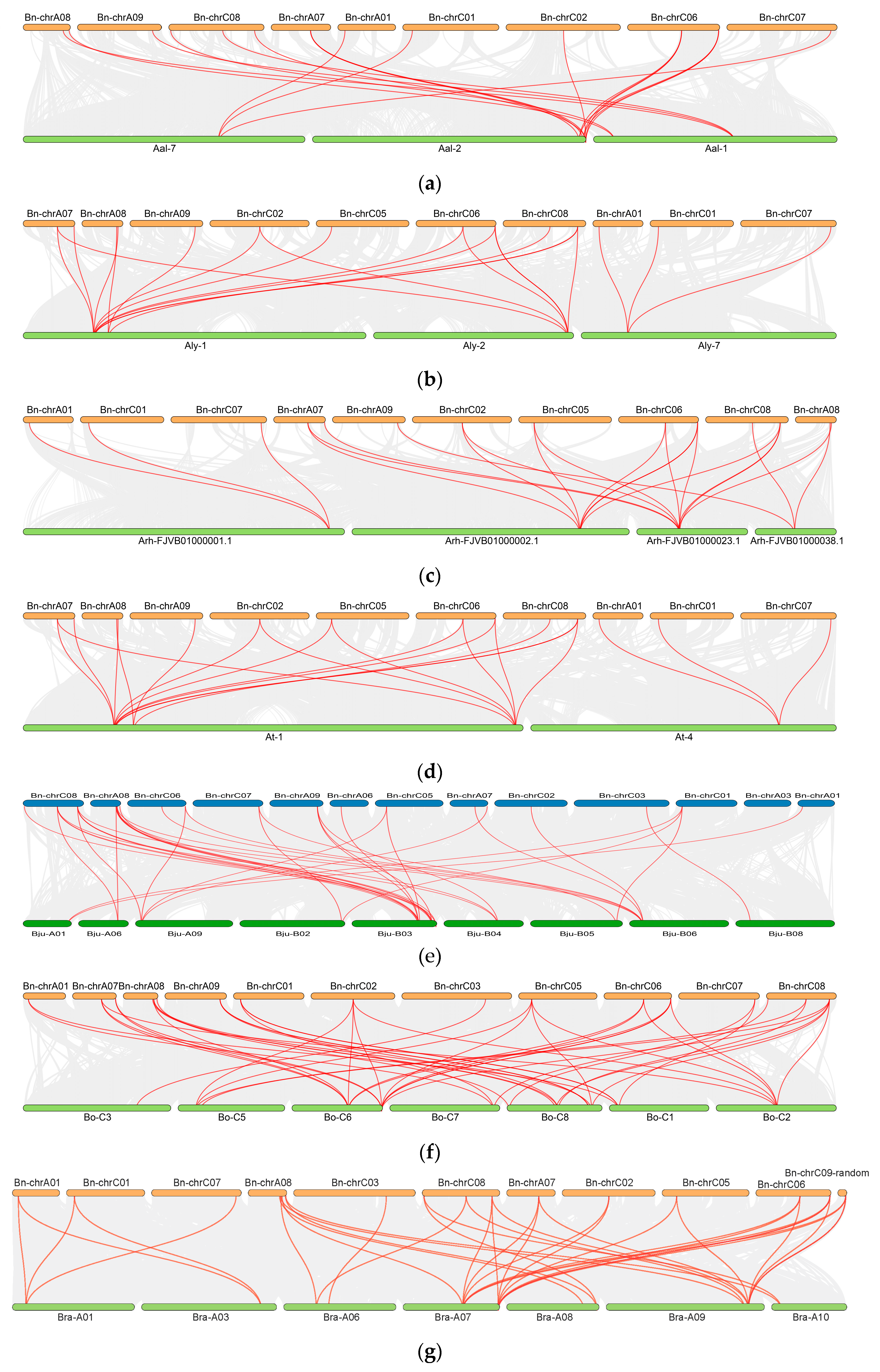
2.5. Interspecies Syntenic Analysis in Cruciferous Species

To investigate the homology and evolutionary relationships between WAK genes, nine Cruciferous species (A. thaliana, A. lyrate, A. helleri, A. alpina, C. sativa, B. juncea, B. rapa, B. carinata and B. oleracea) and B. napus were subjected to a synteny analysis (Figure 5). The results showed that BnWAK genes were homologous in all nine species, with 20 pairs with A. alpina (Figure 5a), 22 pairs with A. lyrate (Figure 5b), 25 pairs with A. helleri (Figure 5c), 23 pairs with A. thaliana (Figure 5d), 41 pairs with B. juncea (Figure 5e), 50 pairs with B. oleracea (Figure 5f), 36 pairs with B. rapa (Figure 5g), 65 pairs with B. carinata (Figure 5h) and 56 pairs with C. sativa (Figure 5i), suggesting that these WAK gene pairs may have come from a common ancestor and had the same function.
In the case of the crosstalk between B. napus and B. carinata, individual homologous genes displayed a purity of one-to-many or many-to-one. Several WAK genes seem to have contributed to the evolution of plants with U’s triangular. Several proteins of unknown function in B. napus can be predicted based on these results.

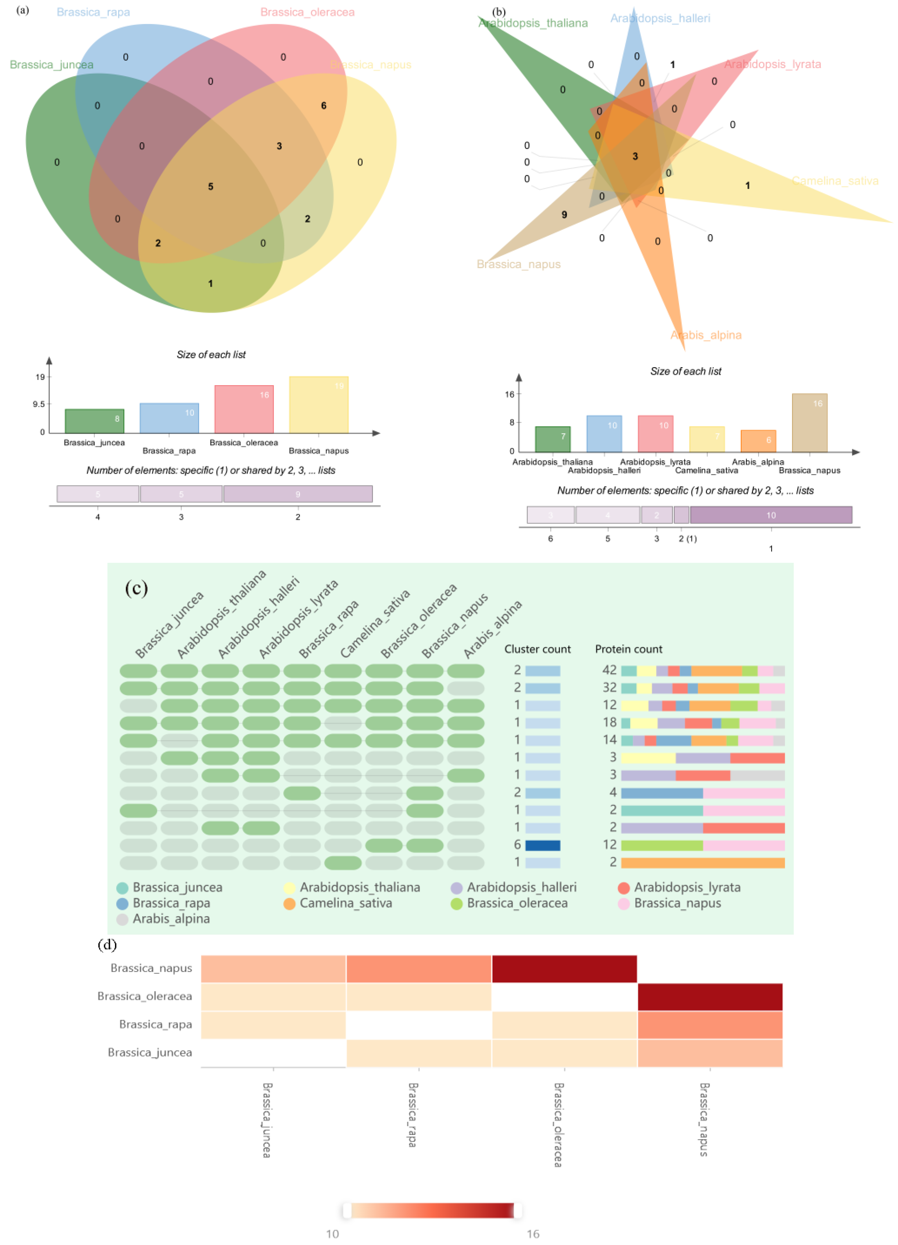
2.6. Orthologous Gene Clustering of WAK Gene Family in Five Cruciferous Species

To explore the evolutionary relationships of WAK genes among nine species of Cruciferous plants—A. thaliana, A. lyrate, A. helleri, A. alpina, C. sativa, B. juncea, B. rapa, B. carinata, B. oleracea and B. napus—we performed a direct lineage homology analysis on the OrthoVenn2 web platform. The direct lineage clusters of the five species identified are presented in Figure 6, with 178 WAK proteins in the five Cruciferous species clustered into three homology groups. In total, 11 B. juncea WAKs, 2 A. halleri WAKs, 2 A. lyrate WAKs, 2 A. alpina WAKs, 10 BnWAKs, 7 B. oleracea WAKs, 4 C. sativa WAKs and 2 B. rapa WAKs did not cluster in any direct lineage cluster and were identified as singletons.
In addition, as exhibited in Figure 6a between four Brassica plants, there were five core gene clusters (31 WAKs) among the four species, one gene cluster (4 WAKs) between B. juncea and B. napus, two gene clusters (4 WAKs) between B. rapa and B. napus, six gene clusters (12 WAKs) between B. oleracea and B. napus and in total we identified one Brassica-specific gene cluster (13 WAKs) among the three Brassica species. The absence of B. rapa- and B. oleracea-specific gene clusters in B. napus suggests that gene loss occurs during hybridization. Additionally, Figure 6d presents the relationship among B. napus, B. rapa, B. oleracea and B. juncea, especially between B. napus and B. oleracea.
B. napus was compared with four Arabidopsis plants and C. sativa. As shown in Figure 6b, there were three core gene clusters among those plants. It is worthwhile to mention that the Arabidopsis plants have no specific core gene clusters, except for C. sativa and B. napus, which have specific gene clusters. This result demonstrates that the Cruciferous plants have retained their affinities in evolution while also having evolutionary specificity among the subfamilies. A detailed list of direct gene clusters and single examples of gene clusters is given in Table S2.

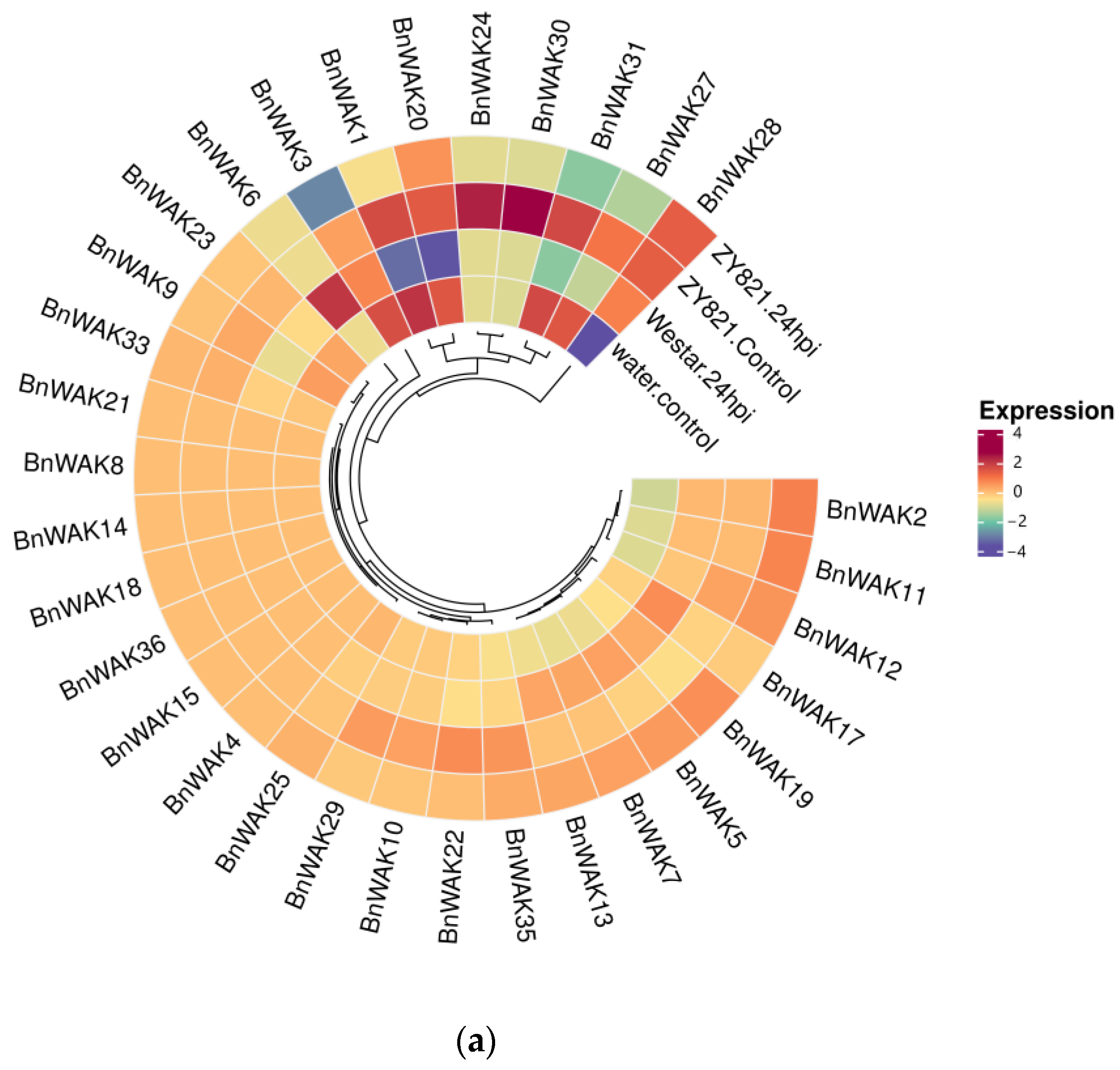
2.7. Expression Pattern of BnWAK Gene in Response to Different Stresses

Based on transcriptome data of B. napus under different biotic stresses published on NCBI, the expression of BnWAKs under different biotic stresses was analyzed. The results showed that BnWAKs expressed differently under different stress treatments (Figure 7).
An analysis of transcriptome data from two varieties of B. napus, tolerant and susceptible, revealed significant differences in the expression of some WAK proteins between the two varieties following Sclerotinia sclerotiorum infection. The susceptible varieties BnWAK24, 30 and 31 showed some increase after infection with S. sclerotiorum, while BnWAK2, 11, 19 and 5 showed some decrease after infection. Among the tolerant species, BnWAK1, 3, 9, 20, 23, 31 and 27 showed some increase, while BnWAK2, 28, 11 and 12 showed some decrease, especially BnWAK28 (Figure 7a). Not coincidentally, powdery mildew caused with Cruciferous powdery mildew is an epidemic disease of B. napus growing worldwide. An analysis of the foliar transcriptome of immune varieties ‘White Flower’ and susceptible varieties ‘Zhong Shuang 11’ showed the highest expression of BnWAK5 in the immune variety and BnWAK35, 32, 25, 29 and 10 in the susceptible variety (Figure 7b).
The WAK family also has an active expression during the abiotic stress response. As the frequency of heat and droughts increases with global warming, the analysis of transcriptome data from B. napus under heat and drought stresses showed that the expression of BnWAK34, 13 and 12 increased to some extent under heat treatment, with BnWAK34 being the most significant. The expression of BnWAK1 and 23 was downregulated. The situation was different under drought treatment, where the expression of BnWAK3 and 30 was increased (Figure 8). This suggests that the BnWAK genes are also closely related to the response to a high temperature and drought.

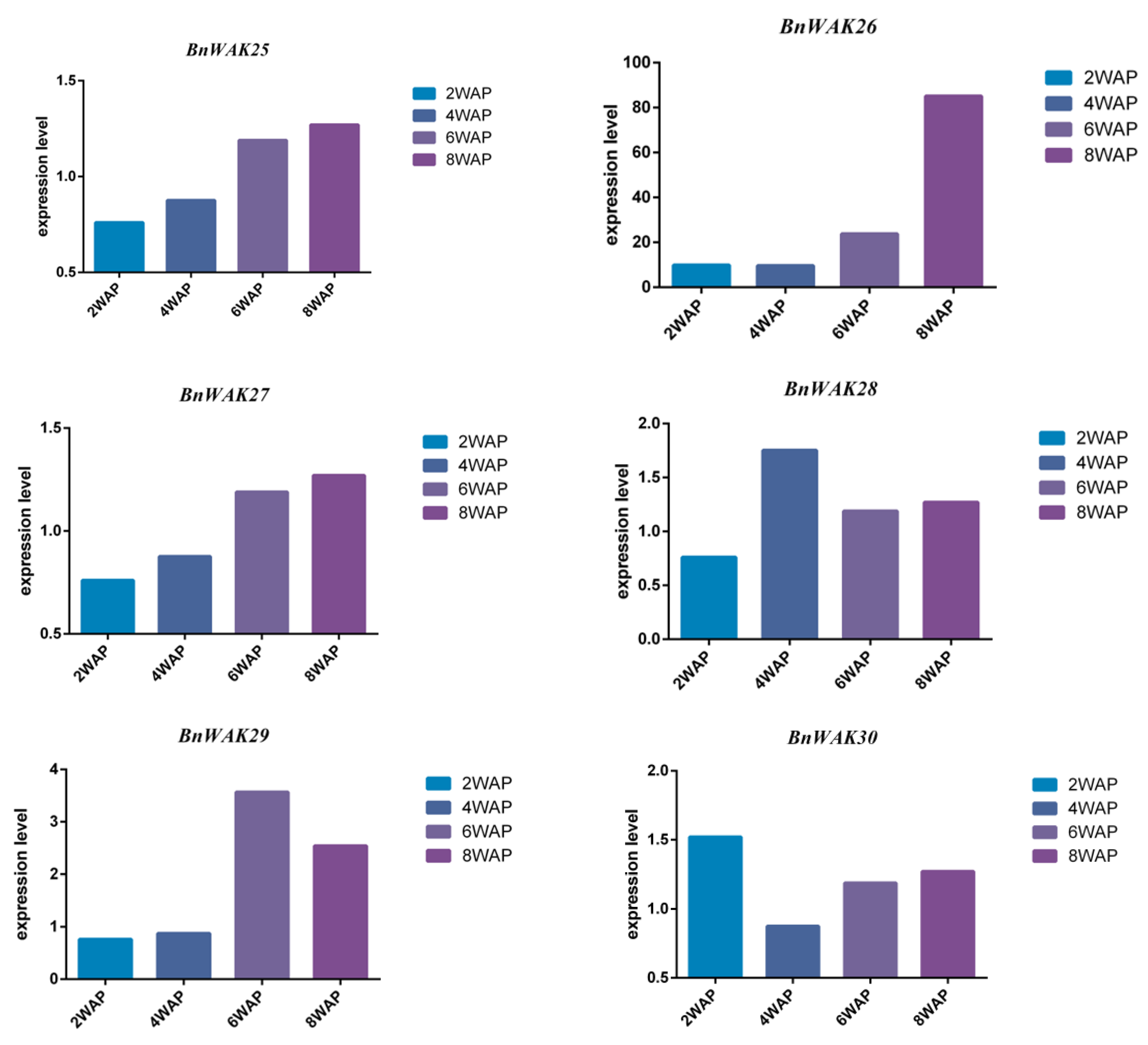
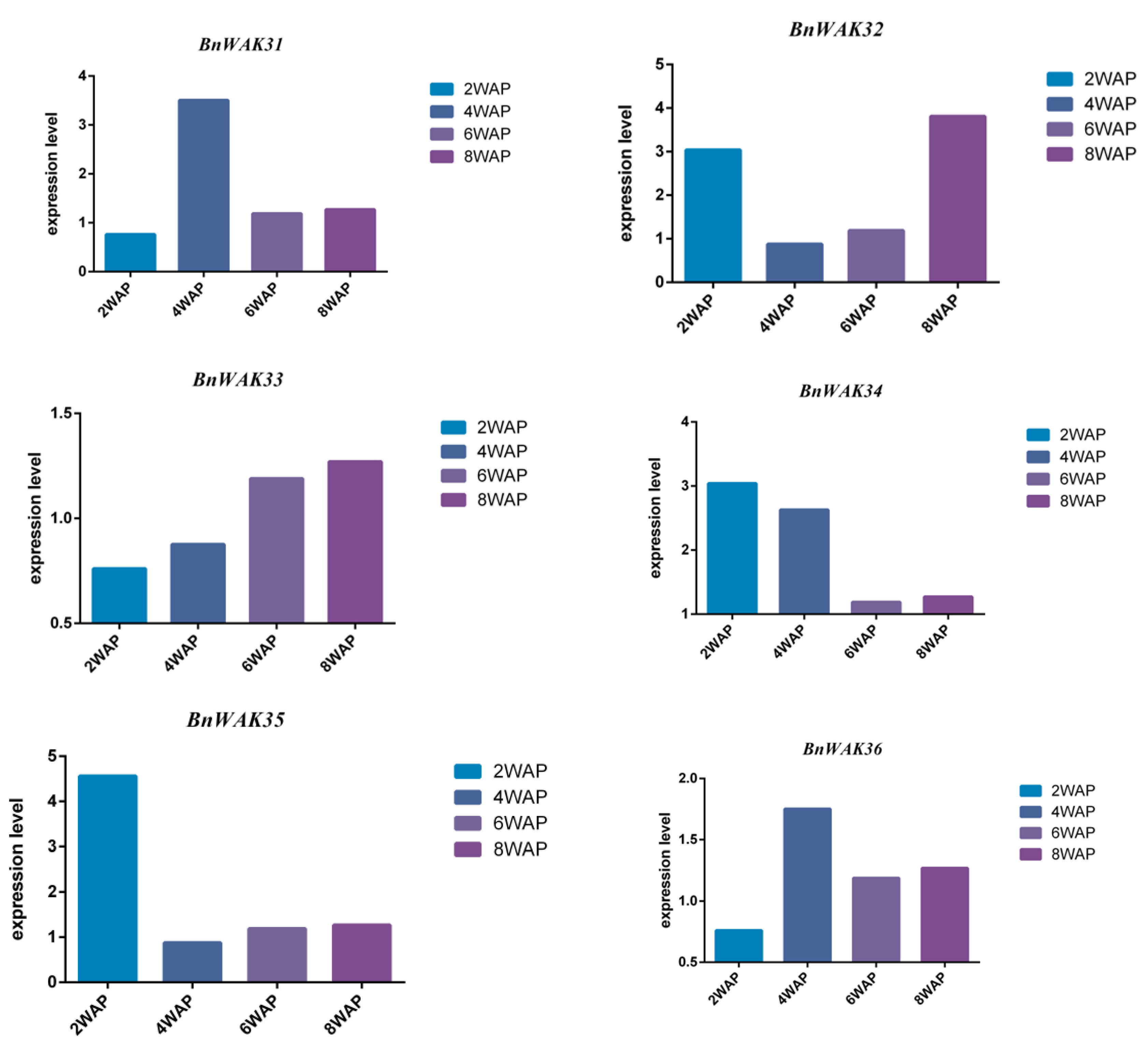
2.8. Expression Pattern of BnWAK Gene in Oil Accumulation

All BnWAK genes were analyzed using an RNA-seq analysis at 2, 4, 6 and 8 weeks after pollination to determine their potential functions during fatty acid biosynthesis. The results indicated that 36 BnWAK genes tended to change their expression during fatty acid biosynthesis (Figure 9). Especially, the expression of BnWAK6, 7 and 12 were significantly upregulated between 2 and 4 weeks and dramatically downregulated after 4 weeks after pollination, which exhibited a tight relationship with the biosynthesis of fatty acids. In contrast, the expression of BnWAK10, 26 and 29 was at a low level between 2 and 4 weeks, but showed a significant raise after 6 weeks, which suggested the connection with the degradation of fatty acids. After pollination, the expression of several genes, such as BnWAK4, 9, 14, 18, 20, 25, 27 and 33, was always upregulated, indicating that these genes may be related to seed development. As a whole, different BnWAK genes exhibited different expression patterns during fatty acid biosynthesis, even duplicated genes.

3. Discussion

Plants are capable of responding to various stresses and can modulate various developmental processes, including photomorphogenesis and circadian rhythms [36]. WAK genes have been demonstrated to play an imperative role in the response pathway of plants sensing pathogen invasion to regulate cell expansion [10,34]. In this study, a total of 178 WAK genes were identified from nine Cruciferous species including A. thaliana, A. lyrate, A. helleri, A. alpina, C. sativa, B. juncea, B. napus, B. rapa and B. oleracea. Previous studies have found 130 members of the WAK gene family in rice [37], 23 in roses [38], 29 in cotton [16,17] and 11 in walnuts [6].
A phylogenetic analysis of WAK family genes revealed that the number of WAK genes varied greatly among Cruciferous species, and the expansion of the WAK genes followed the duplication of the genome (Figure 1). Furthermore, the evolution of WAK genes displayed a clear species bias, with the duplication of species genes occurring after the divergence of species. According to the theory of the triangle of U, B. napus was derived from natural interspecific crosses between kale and cabbage [39]. For example, the genome size of B. napus was nearly twice that of B. rapa and B. oleracea, which may have led to the numbers of WAK members in B. napus being nearly twice those of B. rapa and B. oleracea. However, the WAK numbers in the Arabidopsis are not much different.
Evidence for the evolution of species or genes can be found in the gene structure and motif distribution (Figure 2). Accordingly, members of the same subfamily exhibited consistent exon/intron and motif distribution patterns, suggesting a similar role in functions [40,41]. According to Figure 2b, the classification based on the phylogenetic tree was further confirmed with the gene structure and motif distribution analysis. Furthermore, the structure and number of introns of the WAK genes were well conserved among the nine Cruciferous species. This is consistent with previous studies [18]. The number of thirty-six BnWAK genes on chromosomes ranged from one to eight, and most of these genes were randomly distributed on chromosomes C02, C03, C05, C06, C08, A06, A07, A08 and A09, with five genes unable to be localized on chromosomes. Subcellular localization predictions for the BnWAK gene family revealed that most of the genes in this gene family were localized to be expressed in the extracellular space, suggesting that the BnWAK family is important for the plant immunity of B. napus.
In response to a variety of stresses, cis-elements contribute to regulate and function certain genes [40,42]. The cis-acting element analysis revealed that BnWAK genes contain a significant number of elements with various functions (Figure 4). As a result, BnWAKs may play an influential role in the tolerance to environmental stress and the response to light during the life of plants. Evidence indicates that WAK genes contribute to plant immunity in multiple ways, including defense against pathogens with diverse lifestyles [3]. This may partly explain why BnWAK promoter regions contain so many cis-elements (Figure 4). It remains to be determined how the BnWAK genes regulate different stress-responsive functions as cis-elements exist in the upstream sequences.
The growth and yield of B. napus can be significantly affected by both biotic and abiotic stresses [33], such as heat, droughts and pathogen infection. Previous studies have demonstrated that WAK is a critical member for the disease resistance of fungus and bacteria in several cereal crop species. There have been extensive studies of WAK in several plant species under varying stresses, including rice [15,43,44], maize [13,14] and blue carpet [45]. In rice, WAK genes are not only responsible for the somatic regulation of elongation growth [46] but also play an instrumental role in resistance to various stresses [14,15]. For instance, OsWAK11 is the link between changes in cell wall pectin methylation and the Brassinosteroid (BR) signaling pathway, which together regulate adaptive changes in the cell growth rate [43]. OsWAK14, OsWAK91 and OsWAK92 are important members of the rice blast resistance [44]. For maize, the maize WAK gene ZmWAK-RLK1 (Htn1) is involved in resistance to northern corn leaf blight (NCLB) by affecting benzoxazin (BX) secondary metabolites [43]. Not coincidentally, ZmWAK (qHSR1) prevented the endophytic nutritional growth of S. reilianum and conferred resistance to maize silky black ears disease [13]. It was also observed that B. napus WAK genes were expressed under various stresses in our study. The expression levels of these genes differed under different stresses, indicating that they were widely expressed under different conditions. The results of our study indicated that several genes are associated with resistance to S. sclerotiorum infection, including BnWAK28, BnWAK6, BnWAK24 and BnWAK30. Moreover, BnWAK5 appears to contribute to resistance to powdery mildew. Furthermore, heat stress increased the expression of BnWAK34, 13 and 12, and inhibited the expression of BnWAK1 and 23. Additionally, drought treatment induced the expression of BnWAK3 and 30 (Figure 8). The results indicate that BnWAK genes have an influential role in the biotic and abiotic stress response.
Furthermore, as an oilseed crop, it is crucial to increase oil production or alter fatty acid fractions [47]. And fatty acid biosynthesis occurs mainly between 2 and 4 weeks after pollination, while degradation occurs mainly after 6 weeks [47]. Additionally, we examined the expression of BnWAKs during different stages of fatty acid biosynthesis. Interestingly, we found that BnWAKs tend to change over time (Figure 9). A significant increase in the expression of several genes was observed during the fatty acid synthesis stage, including BnWAK2, BnWAK5, BnWAK7 and BnWAK12. In light of these findings, it appears that genes in the BnWAK family may have a role in fatty acid biosynthesis. In future work, stress-related WAK genes associated with fatty acid synthesis in B. napus will be used to breed more stress-resistant and high-yielding varieties, and to explore the mechanisms behind these genes. In future work, stress-related WAK genes and fatty acid synthesis in B. napus will be used to breed more stress-tolerant and high-yielding varieties and to explore the stress tolerance mechanisms behind these genes.

4. Materials and Methods

4.1. Growth Conditions and Plant Materials

B. napus (Zheda 630) seeds were collected from the Institute of Crop Science and Zhejiang Key Laboratory of Crop Germplasm, Zhejiang University, Hangzhou, China. The seeds were germinated in a saturated seedling sponge and subsequently placed in a light-deprived environment, ensuring their constant moisture. After the 4-day germination phase, B. napus seedlings were transplanted into a hydroponic tank filled with a Hoagland nutrient solution, maintained under controlled conditions of 24/20 °C (day/night), a photoperiod of 14/10 h (light/night) and a relative humidity of 60–70%. The seedlings were exposed to an active photon flux below 200 μmol m−2 s−1 [48]. Drought treatment was inflicted by applying the commonly employed 15% PEG6000 solution. After 35 days, B. napus seedlings were stressed with 15% PEG6000 at 24 h and 48 h. There were three treatments in total, i.e., CK (0 h), 24 hours (24 h) and 48 hours (48 h).

4.2. Identification and Analysis of WAK Family Members in Nine Cruciferous Species

Data from Ensemble (http://plants.ensembl.org/index.html (accessed on 1 January 2022) were obtained for A. thaliana, A. lyrata, A. halleri, A. alpina, C. sativa, B. juncea, B. napus, B. rapa and B. oleracea genomes. From the Pfam database, the term WAK-domain search model accession (PF08488) [46] was used to identify potential WAK genes with the e-value set to 10−3 as the criterion (https://www.ebi.ac.uk/Tools/hmmer/ (accessed on 1 January 2022)) [49,50]. To identify members of the WAK gene family, candidate sequences were searched in the SMART and NCBI CDD databases [51,52,53].

4.3. Alignment of Multiple Sequences and Construction of Phylogenetic Trees

A phylogenetic analysis of the WAK protein family in nine Cruciferous species was performed. The WAK protein sequences identified from other species and B. napus were compared across multiple sequences using Clustal W available in MEGA11 [54,55]. A maximum likelihood phylogenetic tree was constructed with MEGA11 for the WAK from nine plants, with a bootstrap value of 1000. This tree was used as the optimal model after computational simulation.

4.4. Genomic Information and Analysis of Physiological and Biochemical Properties

Based on obtained BnWAK gene sequence information, BnWAKs were localized to their corresponding positions on chromosomes using TBtools [56,57]. Information on the theoretical isoelectric point, molecular mass, amino acid length and other characteristics of BnWAKs was analyzed and predicted using the ExPASy website (https://www.expasy.org/ (accessed on 2 January 2022)) [58,59]. Using the WoLF PSORT (http://wolfpsort.hgc.jp/ (accessed on 2 January 2022)) and CELLO v2.5 (http://cello.life.nctu.edu.tw/ (accessed on 2 January 2022)) online tools, the subcellular localization was analyzed [60,61,62].

4.5. Analysis of WAK Family Protein Sequences for Gene Structure, Motifs and Gene Promoter Cis-Acting Elements

MEME was used to analyze conserved motifs in the WAK gene family (https://meme-suite.org/meme/ (accessed on 3 January 2022)) [40,59]. Perl scripts were used to obtain information related to the gene structure, such as exons and introns, from GFF files. Data visualization was performed using TBtools [53,57]. A Perl script was utilized to extract the 1500 bp DNA sequences upstream of the identified WAK genes. To identify cis-regulatory components (CREs) and select those involved in abiotic and hormonal stress, the Plant CARE database (http://bioinformatics.psb.ugent.be/webtools/plantcare/html/ (accessed on 4 January 2022)) was utilized.

4.6. Collinearity Analysis and Homologous Gene Analysis

The collinearity analysis was performed on the genomes of B. napus with A. thaliana, A. halleri, A. lyrate, A. alpina, C. sativa, B. juncea, B. rapa, B. oleracea and B. carinata using McscanX [61,62] and mapped using TBtools [62] software. The direct homologous gene analysis was performed using OrthoVenn2 (https://orthovenn2.bioinfotoolkits.net/home (accessed on 4 January 2022)) on B. napus with B. juncea, B. rapa and B. oleracea [63].

4.7. Expression and Stress Response Expression Profiling of BnWAK

To investigate expression (under stress and non-stressed conditions) of the BnWAKs, RNA-Seq data were downloaded from previously published data that were downloaded from the NCBI database (GSE77637, GSE81545, GSE156029 and GSE188377) [34,64,65]. GSE77637 was analyzed to investigate the expression of BnWAKs from B. napus at different developmental stages [31]. GSE81545 was used to explore the expression of BnWAKs from B. napus after Sclerotinia sclerotiorum infection [34]. GSE188377 was used to investigate the expression of BnWAKs from B. napus after Erysiphe cruciferarum infection [65]. GSE156029 was applied to explore BnWAK expression under a drought and heat stress. After log10 (FPKM + 1 × 10⁻⁵) normalization, heatmaps were generated with OmicStudio tools (https://www.omicstudio.cn (accessed on 7 January 2022)) [66,67].
The total RNA of B. napus (Zheda 630) under 15% PEG6000 treatment at different times was isolated with a FastPure Plant Total RNA Isolation Kit (Vazyme, Nanjing, China). Zero hours was used as a reference, ACT2 was used as the internal reference gene and Primer-BLAST (https://blast.ncbi.nlm.nih.gov/Blast.cgi (accessed on 7 January 2022)) was used to design the primers. The relevant primers are shown in Table S3. Six samples were taken at each time point. Three replicates of each sample were presented using ChamQ Universal SYBR qPCR Master Mix (Vazyme). The reaction conditions are according to the instructions of ChamQ Universal SYBR qPCR Master Mix (Vazyme); more information is presented in Table S4. The 2−ΔΔCt method was used to analyze the results [68]. The qRT-PCR analysis involved eight selected BnWAKs, including BnWAK2, BnWAK5, BnWAK11, BnWAK12, BnWAK15, BnWAK26, BnWAK27 and BnWAK35.

Supplementary Materials

The following supporting information can be downloaded at: https://www.mdpi.com/article/10.3390/ijms241713601/s1.

Author Contributions

Conceptualization, L.X. and Z.X.; methodology, L.X., Z.X. and Y.D.; formal analysis, Z.X., Y.D. and M.X.; software, H.L., Y.D. and M.X.; validation, L.X., Y.D. and Z.Z.; writing—original draft preparation, Z.X. and Y.D.; writing—review and editing, Z.X., H.L., M.X. and Z.Z.; visualization, Z.X., Y.D., H.L. and M.X.; supervision, Z.Z. and L.X.; project administration, Z.Z. and L.X.; funding acquisition, L.X. All authors have read and agreed to the published version of the manuscript.

Funding

This research was funded by the Science and Technology Program of Zhejiang Province (2022C02034), the Agriculture and Rural Affairs Department of Zhejiang Province (2022SNJF010), the China Scholarship Council (CSC) and the Fundamental Research Funds of Zhejiang Sci-Tech University (23042097-Y).

Institutional Review Board Statement

Not applicable.

Informed Consent Statement

Not applicable.

Data Availability Statement

Data available on request from the authors.

Conflicts of Interest

The authors declare no conflict of interest.

References

  1. He, Z.H.; Cheeseman, I.; He, D.; Kohorn, B.D. A cluster of five cell wall-associated receptor kinase genes, Wak1-5, are expressed in specific organs of Arabidopsis. Plant Mol. Biol. 1999, 39, 1189–1196. [Google Scholar] [CrossRef]
  2. Decreux, A.; Messiaen, J. Wall-associated kinase WAK1 interacts with cell wall pectins in a calcium-induced conformation. Plant Cell Physiol. 2005, 46, 268–278. [Google Scholar] [CrossRef] [PubMed]
  3. Stephens, C.; Hammond-Kosack, K.E.; Kanyuka, K. WAKsing plant immunity, waning diseases. J. Exp. Bot. 2022, 73, 22–37. [Google Scholar] [CrossRef]
  4. Kohorn, B.D. WAKs; cell wall associated kinases. Curr. Opin. Cell Biol. 2001, 13, 529–533. [Google Scholar] [CrossRef] [PubMed]
  5. Decreux, A.; Thomas, A.; Spies, B.; Brasseur, R.; Van Cutsem, P.; Messiaen, J. In vitro characterization of the homogalacturonan-binding domain of the wall-associated kinase WAK1 using site-directed mutagenesis. Phytochemistry 2006, 67, 1068–1079. [Google Scholar] [CrossRef]
  6. Li, M.; Ma, J.; Liu, H.; Ou, M.; Ye, H.; Zhao, P. Identification and Characterization of Wall-Associated Kinase (WAK) and WAK-like (WAKL) Gene Family in Juglans regia and Its Wild Related Species Juglans mandshurica. Genes 2022, 13, 134. [Google Scholar] [CrossRef]
  7. Kohorn, B.D. Cell wall-associated kinases and pectin perception. J. Exp. Bot. 2016, 67, 489–494. [Google Scholar] [CrossRef] [PubMed]
  8. Kohorn, B.D.; Kohorn, S.L.; Todorova, T.; Baptiste, G.; Stansky, K.; McCullough, M. A dominant allele of Arabidopsis pectin-binding wall-associated kinase induces a stress response suppressed by MPK6 but not MPK3 mutations. Mol. Plant 2012, 5, 841–851. [Google Scholar] [CrossRef]
  9. Ferrari, S.; Savatin, D.V.; Sicilia, F.; Gramegna, G.; Cervone, F.; Lorenzo, G.D. Oligogalacturonides: Plant damage-associated molecular patterns and regulators of growth and development. Front. Plant Sci. 2013, 4, 49. [Google Scholar] [CrossRef] [PubMed]
  10. Kanyuka, K.; Rudd, J.J. Cell surface immune receptors: The guardians of the plant’s extracellular spaces. Curr. Opin. Plant Biol. 2019, 50, 1–8. [Google Scholar] [CrossRef]
  11. Li, H.; Zheng, X.; Tao, L.; Yang, Y.; Gao, L.; Xiong, J. Aeration Increases Cadmium (Cd) Retention by Enhancing Iron Plaque Formation and Regulating Pectin Synthesis in the Roots of Rice (Oryza sativa) Seedlings. Rice 2019, 12, 28. [Google Scholar] [CrossRef]
  12. Xiong, J.; Yang, Y.; Fu, G.; Tao, L. Novel roles of hydrogen peroxide (H2O2) in regulating pectin synthesis and demethylesterification in the cell wall of rice (Oryza sativa) root tips. New Phytol. 2015, 206, 118–126. [Google Scholar] [CrossRef] [PubMed]
  13. Zuo, W.; Chao, Q.; Zhang, N.; Ye, J.; Tan, G.; Li, B.; Xing, Y.; Zhang, B.; Liu, H.; Fengler, K.A.; et al. A maize wall-associated kinase confers quantitative resistance to head smut. Nat. Genet. 2015, 47, 151–157. [Google Scholar] [CrossRef] [PubMed]
  14. Yang, P.; Praz, C.; Li, B.; Singla, J.; Robert, C.; Kessel, B.; Scheuermann, D.; Lüthi, L.; Ouzunova, M.; Erb, M.; et al. Fungal resistance mediated by maize wall-associated kinase ZmWAK-RLK1 correlates with reduced benzoxazinoid content. New Phytol. 2019, 221, 976–987. [Google Scholar] [CrossRef]
  15. Hu, K.; Cao, J.; Zhang, J.; Xia, F.; Ke, Y.; Zhang, H.; Xie, W.; Liu, H.; Cui, Y.; Cao, Y.; et al. Improvement of multiple agronomic traits by a disease resistance gene via cell wall reinforcement. Nat. Plants 2017, 3, 17009. [Google Scholar] [CrossRef] [PubMed]
  16. Wang, P.; Zhou, L.; Jamieson, P.; Zhang, L.; Zhao, Z.; Babilonia, K.; Shao, W.; Wu, L.; Mustafa, R.; Amin, I.; et al. The Cotton Wall-Associated Kinase GhWAK7A Mediates Responses to Fungal Wilt Pathogens by Complexing with the Chitin Sensory Receptors. Plant Cell 2020, 32, 3978–4001. [Google Scholar] [CrossRef] [PubMed]
  17. Feng, H.; Li, C.; Zhou, J.; Yuan, Y.; Feng, Z.; Shi, Y.; Zhao, L.; Zhang, Y.; Wei, F.; Zhu, H. A cotton WAKL protein interacted with a DnaJ protein and was involved in defense against Verticillium dahliae. Int. J. Biol. Macromol. 2021, 167, 633–643. [Google Scholar] [CrossRef]
  18. Li, H.; Zhang, H.; Yang, Y.; Fu, G.; Tao, L.; Xiong, J. Effects and oxygen-regulated mechanisms of water management on cadmium (Cd) accumulation in rice (Oryza sativa). Sci. Total Environ. 2022, 846, 157484. [Google Scholar] [CrossRef]
  19. Gao, Y.; Feng, B.; Gao, C.; Zhang, H.; Wen, F.; Tao, L.; Fu, G.; Xiong, J. The Evolution and Functional Roles of miR408 and Its Targets in Plants. Int. J. Mol. Sci. 2022, 23, 530. [Google Scholar] [CrossRef]
  20. Yang, Y.; Xiong, J.; Tao, L.; Cao, Z.; Tang, W.; Zhang, J.; Yu, X.; Fu, G.; Zhang, X.; Lu, Y. Regulatory mechanisms of nitrogen (N) on cadmium (Cd) uptake and accumulation in plants: A review. Sci. Total Environ. 2020, 708, 135186. [Google Scholar] [CrossRef] [PubMed]
  21. Gao, L.; Chang, J.; Chen, R.; Li, H.; Lu, H.; Tao, L.; Xiong, J. Comparison on cellular mechanisms of iron and cadmium accumulation in rice: Prospects for cultivating Fe-rich but Cd-free rice. Rice 2016, 9, 39. [Google Scholar] [CrossRef]
  22. Zhao, T.; Xu, X.; Wang, M.; Li, C.; Li, C.; Zhao, R.; Zhu, S.; He, Q.; Chen, J. Identification and profiling of upland cotton microRNAs at fiber initiation stage under exogenous IAA application. BMC Genom. 2019, 20, 421. [Google Scholar] [CrossRef]
  23. Li, C.; He, Q.; Zhang, F.; Yu, J.; Li, C.; Zhao, T.; Zhang, Y.; Xie, Q.; Su, B.; Mei, L.; et al. Melatonin enhances cotton immunity to Verticillium wilt via manipulating lignin and gossypol biosynthesis. Plant J. Cell Mol. Biol. 2019, 100, 784–800. [Google Scholar] [CrossRef]
  24. Gao, C.; Zhang, X.; Li, H.; Ussi, A.; Gao, Y.; Xiong, J. Roles of lncRNAs in Rice: Advances and Challenges. Rice Sci. 2020, 27, 3. [Google Scholar] [CrossRef]
  25. Yang, Y.; Xiong, J.; Chen, R.; Fu, G.; Chen, T.; Tao, L. Excessive nitrate enhances cadmium (Cd) uptake by up-regulating the expression of OsIRT1 in rice (Oryza sativa). Environ. Exp. Bot. 2016, 122, 141–149. [Google Scholar] [CrossRef]
  26. Zhang, N.; Jing, P. Anthocyanins in Brassicaceae: Composition, stability, bioavailability, and potential health benefits. Crit. Rev. Food Sci. Nutr. 2022, 62, 2205–2220. [Google Scholar] [CrossRef] [PubMed]
  27. Chen, H.; German, D.A.; Al-Shehbaz, I.A.; Yue, J.; Sun, H. Phylogeny of Euclidieae (Brassicaceae) based on plastome and nuclear ribosomal DNA data. Mol. Phylogenet. Evol. 2020, 153, 106940. [Google Scholar] [CrossRef]
  28. Cardoza, V.; Stewart, C.N., Jr. Canola (Brassica napus L.). Methods Mol. Biol. 2006, 343, 257–266. [Google Scholar] [CrossRef]
  29. Tang, S.; Zhao, H.; Lu, S.; Yu, L.; Zhang, G.; Zhang, Y.; Yang, Q.Y.; Zhou, Y.; Wang, X.; Ma, W.; et al. Genome- and transcriptome-wide association studies provide insights into the genetic basis of natural variation of seed oil content in Brassica napus. Mol. Plant 2021, 14, 470–487. [Google Scholar] [CrossRef]
  30. Tan, Z.; Xie, Z.; Dai, L.; Zhang, Y.; Zhao, H.; Tang, S.; Wan, L.; Yao, X.; Guo, L.; Hong, D. Genome- and transcriptome-wide association studies reveal the genetic basis and the breeding history of seed glucosinolate content in Brassica napus. Plant Biotechnol. J. 2022, 20, 211–225. [Google Scholar] [CrossRef] [PubMed]
  31. Wan, H.; Cui, Y.; Ding, Y.; Mei, J.; Dong, H.; Zhang, W.; Wu, S.; Liang, Y.; Zhang, C.; Li, J.; et al. Time-Series Analyses of Transcriptomes and Proteomes Reveal Molecular Networks Underlying Oil Accumulation in Canola. Front. Plant Sci. 2017, 7, 2007. [Google Scholar] [CrossRef]
  32. Raza, A.; Razzaq, A.; Mehmood, S.S.; Hussain, M.A.; Wei, S.; He, H.; Zaman, Q.U.; Xuekun, Z.; Hasanuzzaman, M. Omics: The way forward to enhance abiotic stress tolerance in Brassica napus L. GM Crops Food 2021, 12, 251–281. [Google Scholar] [CrossRef] [PubMed]
  33. Hwang, S.F.; Strelkov, S.E.; Feng, J.; Gossen, B.D.; Howard, R.J. Plasmodiophora brassicae: A review of an emerging pathogen of the Canadian canola (Brassica napus) crop. Mol. Plant Pathol. 2012, 13, 105–113. [Google Scholar] [CrossRef] [PubMed]
  34. Girard, I.J.; Tong, C.; Becker, M.G.; Mao, X.; Huang, J.; de Kievit, T.; Fernando, W.; Liu, S.; Belmonte, M.F. RNA sequencing of Brassica napus reveals cellular redox control of Sclerotinia infection. J. Exp. Bot. 2017, 68, 5079–5091. [Google Scholar] [CrossRef]
  35. Huang, R.; Liu, Z.; Xing, M.; Yang, Y.; Wu, X.; Liu, H.; Liang, W. Heat Stress Suppresses Brassica napus Seed Oil Accumulation by Inhibition of Photosynthesis and BnWRI1 Pathway. Plant Cell Physiol. 2019, 60, 1457–1470. [Google Scholar] [CrossRef] [PubMed]
  36. Casal, J.J.; Fankhauser, C.; Coupland, G.; Blázquez, M.A. Signalling for developmental plasticity. Trends Plant Sci. 2004, 9, 309–314. [Google Scholar] [CrossRef]
  37. de Oliveira, L.F.V.; Christoff, A.P.; de Lima, J.C.; de Ross, B.C.F.; Sachetto-Martins, G.; Margis-Pinheiro, M.; Margis, R. The Wall-associated Kinase gene family in rice genomes. Plant Sci. 2014, 229, 181–192. [Google Scholar] [CrossRef]
  38. Liu, X.; Wang, Z.; Tian, Y.; Zhang, S.; Li, D.; Dong, W.; Zhang, C.; Zhang, Z. Characterization of wall-associated kinase/wall-associated kinase-like (WAK/WAKL) family in rose (Rosa chinensis) reveals the role of RcWAK4 in Botrytis resistance. BMC Plant Biol. 2021, 21, 526. [Google Scholar] [CrossRef]
  39. Koh, J.; Barbulescu, D.M.; Norton, S.; Redden, B.; Salisbury, P.A.; Kaur, S.; Cogan, N.; Slater, A.T. A multiplex PCR for rapid identification of Brassica species in the triangle of U. Plant Methods 2017, 13, 49. [Google Scholar] [CrossRef]
  40. Han, Y.; Hou, Z.; He, Q.; Zhang, X.; Yan, K.; Han, R.; Liang, Z. Genome-Wide Characterization and Expression Analysis of bZIP Gene Family Under Abiotic Stress in Glycyrrhiza uralensis. Front. Genet. 2021, 12, 754237. [Google Scholar] [CrossRef]
  41. Chen, M.M.; Zhang, M.; Liang, Z.S.; He, Q.L. Characterization and Comparative Analysis of Chloroplast Genomes in Five Uncaria Species Endemic to China. Int. J. Mol. Sci. 2022, 23, 11617. [Google Scholar] [CrossRef]
  42. Niu, T.; Wang, X.; Abbas, M.; Shen, J.; Liu, R.; Wang, Z.; Liu, A. Expansion of constans-like genes in sunflower confers putative neofunctionalization in the adaptation to abiotic stresses. Ind. Crops Prod. 2022, 176, 114400. [Google Scholar] [CrossRef]
  43. Yue, Z.L.; Liu, N.; Deng, Z.P.; Zhang, Y.; Wu, Z.M.; Zhao, J.L.; Sun, Y.; Wang, Z.Y.; Zhang, S.W. The receptor kinase OsWAK11 monitors cell wall pectin changes to fine-tune brassinosteroid signaling and regulate cell elongation in rice. Curr. Biol. CB 2022, 32, 2454–2466.e7. [Google Scholar] [CrossRef]
  44. Delteil, A.; Gobbato, E.; Cayrol, B.; Estevan, J.; Michel-Romiti, C.; Dievart, A.; Kroj, T.; Morel, J.B. Several wall-associated kinases participate positively and negatively in basal defense against rice blast fungus. BMC Plant Biol. 2016, 16, 17. [Google Scholar] [CrossRef]
  45. Chen, P.; Giarola, V.; Bartels, D. The Craterostigma plantagineum protein kinase CpWAK1 interacts with pectin and integrates different environmental signals in the cell wall. Planta 2021, 253, 92. [Google Scholar] [CrossRef] [PubMed]
  46. Wagner, T.A.; Kohorn, B.D. Wall-associated kinases are expressed throughout plant development and are required for cell expansion. Plant Cell 2001, 13, 303–318. [Google Scholar] [CrossRef]
  47. Huang, K.L.; Tian, J.; Wang, H.; Fu, Y.F.; Li, Y.; Zheng, Y.; Li, X.B. Fatty acid export protein BnFAX6 functions in lipid synthesis and axillary bud growth in Brassica napus. Plant Physiol. 2021, 186, 2064–2077. [Google Scholar] [CrossRef] [PubMed]
  48. Xu, Z.; Pan, J.; Ullah, N.; Duan, Y.; Hao, R.; Li, J.; Huang, Q.; Xu, L. 5-Aminolevulinic acid mitigates the chromium-induced changes in Helianthus annuus L. as revealed by plant defense system enhancement. Plant Physiol. Biochem. PPB 2023, 198, 107701. [Google Scholar] [CrossRef] [PubMed]
  49. Potter, S.C.; Luciani, A.; Eddy, S.R.; Park, Y.; Lopez, R.; Finn, R.D. HMMER web server: 2018 update. Nucleic Acids Res. 2018, 46, W200–W204. [Google Scholar] [CrossRef] [PubMed]
  50. Zhang, H.; Xu, J.; Chen, H.; Jin, W.; Liang, Z. Characterization of NAC family genes in Salvia miltiorrhiza and NAC2 potentially involved in the biosynthesis of tanshinones. Phytochemistry 2021, 191, 112932. [Google Scholar] [CrossRef]
  51. Lu, S.; Wang, J.; Chitsaz, F.; Derbyshire, M.K.; Geer, R.C.; Gonzales, N.R.; Gwadz, M.; Hurwitz, D.I.; Marchler, G.H.; Song, J.S.; et al. CDD/SPARCLE: The conserved domain database in 2020. Nucleic Acids Res. 2020, 48, D265–D268. [Google Scholar] [CrossRef] [PubMed]
  52. Letunic, I.; Khedkar, S.; Bork, P. SMART: Recent updates, new developments and status in 2020. Nucleic Acids Res. 2021, 49, D458–D460. [Google Scholar] [CrossRef]
  53. Chen, X.; Mao, Y.; Chai, W.; Yan, K.; Liang, Z.; Xia, P. Genome-wide identification and expression analysis of MYB gene family under nitrogen stress in Panax notoginseng. Protoplasma 2023, 260, 189–205. [Google Scholar] [CrossRef]
  54. Tamura, K.; Stecher, G.; Kumar, S. MEGA11: Molecular Evolutionary Genetics Analysis Version 11. Mol. Biol. Evol. 2021, 38, 3022–3027. [Google Scholar] [CrossRef]
  55. Larkin, M.A.; Blackshields, G.; Brown, N.P.; Chenna, R.; McGettigan, P.A.; McWilliam, H.; Valentin, F.; Wallace, I.M.; Wilm, A.; Lopez, R.; et al. Clustal W and Clustal X version 2.0. Bioinformatics 2007, 23, 2947–2948. [Google Scholar] [CrossRef]
  56. Chen, C.; Chen, H.; Zhang, Y.; Thomas, H.R.; Frank, M.H.; He, Y.; Xia, R. TBtools: An Integrative Toolkit Developed for Interactive Analyses of Big Biological Data. Mol. Plant 2020, 13, 1194–1202. [Google Scholar] [CrossRef] [PubMed]
  57. Chen, X.; Chang, Q.; Xia, P.; Liang, Z.; Yan, K. The complete chloroplast genome of Clematis henryi var. ernate (Ranunculaceae). Mitochondrial DNA. Part B Resour. 2021, 6, 1319–1320. [Google Scholar] [CrossRef]
  58. Duvaud, S.; Gabella, C.; Lisacek, F.; Stockinger, H.; Ioannidis, V.; Durinx, C. Expasy, the Swiss Bioinformatics Resource Portal, as designed by its users. Nucleic Acids Res. 2021, 49, W216–W227. [Google Scholar] [CrossRef]
  59. Feng, T.; Jiang, Y.; Jia, Q.; Han, R.; Wang, D.; Zhang, X.; Liang, Z. Transcriptome Analysis of Different Sections of Rhizome in Polygonatum sibiricum Red. and Mining Putative Genes Participate in Polysaccharide Biosynthesis. Biochem. Genet. 2022, 60, 1547–1566. [Google Scholar] [CrossRef]
  60. Horton, P.; Park, K.J.; Obayashi, T.; Fujita, N.; Harada, H.; Adams-Collier, C.J.; Nakai, K. WoLF PSORT: Protein localization predictor. Nucleic Acids Res. 2007, 35, W585–W587. [Google Scholar] [CrossRef]
  61. Wang, Y.; Tang, H.; Debarry, J.D.; Tan, X.; Li, J.; Wang, X.; Lee, T.H.; Jin, H.; Marler, B.; Guo, H.; et al. MCScanX: A toolkit for detection and evolutionary analysis of gene synteny and collinearity. Nucleic Acids Res. 2012, 40, e49. [Google Scholar] [CrossRef]
  62. Xia, P.; Hu, W.; Zheng, Y.; Wang, Y.; Yan, K.; Liang, Z. Structural and interactions analysis of a transcription factor PnMYB2 in Panax notoginseng. J. Plant Physiol. 2022, 275, 153756. [Google Scholar] [CrossRef]
  63. Xu, L.; Dong, Z.; Fang, L.; Luo, Y.; Wei, Z.; Guo, H.; Zhang, G.; Gu, Y.Q.; Coleman-Derr, D.; Xia, Q.; et al. OrthoVenn2: A web server for whole-genome comparison and annotation of orthologous clusters across multiple species. Nucleic Acids Res. 2019, 47, W52–W58. [Google Scholar] [CrossRef]
  64. Yan, G.; Zhang, M.; Guan, W.; Zhang, F.; Dai, W.; Yuan, L.; Gao, G.; Xu, K.; Chen, B.; Li, L.; et al. Genome-Wide Identification and Functional Characterization of Stress Related Glyoxalase Genes in Brassica napus L. Int. J. Mol. Sci. 2023, 24, 2130. [Google Scholar] [CrossRef]
  65. Zhang, M.; Gong, Q.; Su, X.; Cheng, Y.; Wu, H.; Huang, Z.; Xu, A.; Dong, J.; Yu, C. Microscopic and Transcriptomic Comparison of Powdery Mildew Resistance in the Progenies of Brassica carinata × B. napus. Int. J. Mol. Sci. 2022, 23, 9961. [Google Scholar] [CrossRef]
  66. Zheng, H.; Zhao, H.; Zhang, X.; Liang, Z.; He, Q. Systematic Identification and Validation of Suitable Reference Genes for the Normalization of Gene Expression in Prunella vulgaris under Different Organs and Spike Development Stages. Genes 2022, 13, 1947. [Google Scholar] [CrossRef] [PubMed]
  67. Ding, K.; Pei, T.; Bai, Z.; Jia, Y.; Ma, P.; Liang, Z. SmMYB36, a Novel R2R3-MYB Transcription Factor, Enhances Tanshinone Accumulation and Decreases Phenolic Acid Content in Salvia miltiorrhiza Hairy Roots. Sci. Rep. 2017, 7, 5104. [Google Scholar] [CrossRef]
  68. Livak, K.J.; Schmittgen, T.D. Analysis of Relative Gene Expression Data Using 677 Real-Time Quantitative PCR and the 2−ΔΔCT Method. Methods 2001, 25, 402–408. [Google Scholar] [CrossRef] [PubMed]
Figure 1. Phylogenetic analysis of 9 Cruciferous species’ WAK members. According to the alignment of the WAK domain, a phylogenetic tree was constructed using the neighbor-joining (NJ) method with the default parameters and 1000 bootstrap replicates. Based on the results of phylogenetic analysis, the WAK members of 9 cruciferous species were divided into 9 subfamilies, denoted by the Roman numerals from Ia to IVb.
Ijms 24 13601 g001
Figure 2. An ML phylogenetic tree with conserved motifs, gene structures and conserved domains of WAK proteins. (a). ML phylogenetic tree based on the WAK proteins’ domain sequence. (b). Ten conserved motifs of 178 WAK proteins from 9 Cruciferous species. The MEME program identified 10 conserved motifs, each characterized by a distinct color. Motif 9 constitutes key components of the WAK structural domain. (c). Conversed structural domains of 178 WAK genes for 9 Cruciferous species. WAK conserved structural domains are mapped directly to the gene structure. (d). Exon/intron structure of 178 WAK genes for 9 Cruciferous species. The green region represents the UTR, the yellow region represents the CDS and the gray lines indicate the exon regions, where exons partition most of the WAK conserved structural domains.
Ijms 24 13601 g002
Figure 3. Chromosomal distribution and gene duplication events of BnWAKs. (a). BnWAKs’ position on the chromosome. The chromosome, in this instance, is divided into units of 0.1MB each. The density of the region is represented by the color variation, where blue indicates low, yellow indicates medium and red indicates high density. The physical location of the BnWAKs is represented by blue lines on the chromosome. (b). The quantity of BnWAKs on chromosomes. Each chromosome is represented by different colors. There are three genes that are not located on the chromosome. Therefore, large wedges imply a greater number of BnWAK genes. (c). A genome-wide analysis of gene synteny in B. napus. Synteny relationships are depicted by gray lines for each gene in B. napus, while replication events are represented by red lines.
Ijms 24 13601 g003aIjms 24 13601 g003b
Figure 4. Cis-regulatory elements in the promoter region of the BnWAK genes. The prediction of cis-regulatory elements for 1.5 kb sequences upstream of BnWAKs. And some of the stress response elements and hormone response elements are finally shown.
Ijms 24 13601 g004
Figure 5. Synteny analyses of 8 dicotyledonous for WAK genes. The syntenic analysis between B. napus and the other eight dicotyledonous including A. thaliana, A. lyrata, A. halleri, A. alpina, C. sativa, B. carinata, B. juncea, B. rapa and B. oleracea. Collinear blocks are presented with gray lines shown in the background, while red lines highlight WAK gene pairs with syntenic relationships. (a) The syntenic analysis between B. napus and A. alpina (b) The syntenic analysis between B. napus and A. lyrata (c) The syntenic analysis between B. napus and A. halleri (d) The syntenic analysis between B. napus and A. thaliana (e) The syntenic analysis between B. napus and B. juncea (f) The syntenic analysis between B. napus and B. oleracea (g) The syntenic analysis between B. napus and B. rapa (h) The syntenic analysis between B. napus and B. carinata (i) The syntenic analysis between B. napus and C. sativa.
Ijms 24 13601 g005aIjms 24 13601 g005b
Figure 6. Orthologous gene clustering analysis. The orthologous gene clusters between the WAK gene family in A. thaliana, A. lyrata, A. halleri, A. alpina, C. sativa, B. napus, B. juncea, B. rapa and B. oleracea were identified and visualized using the OrthoVenn2 web platform. (a) Orthologous gene clustering analysis among the WAK gene family in B. napus, B. juncea, B. rapa and B. oleracea (b) Orthologous gene clustering analysis among the WAK gene family in B. napus, A. thaliana, A. lyrata, A. halleri, A. alpina, and C. sativa. (c) Orthologous gene clustering analysis among the WAK gene family in 9 Cruciferous species (d) Pairwise heatmap among the WAK gene family in B. napus, B. juncea, B. rapa and B. oleracea.
Ijms 24 13601 g006
Figure 7. Analysis of BnWAK genes’ expression in B. napus leaves under biotic stress. (a). Expression levels of genome-wide WAK genes in both susceptible (Westar) and tolerant (ZY821) genotypes of B. napus leaves infected with Sclerotinia sclerotiorum. (b). Expression levels of genome-wide WAK genes in both susceptible (Zhongshuang11) and tolerant (White flower) genotypes of B. napus leaves infected with Erysiphe cruciferarum.
Ijms 24 13601 g007aIjms 24 13601 g007b
Figure 8. Analysis of BnWAK genes’ expression in B. napus under abiotic stress. (a). Expression of genome-wide BnWAK genes in B. napus leaves under drought and heat treatment. (b). qRT-PCR was used to measure drought-induced expression of selected BnWAK genes. Final results are exhibited as mean ± standard deviation. The labels 0 h (CK), 24 h and 48 h indicate the specific time points (in hours) at which the samples were collected for expression analysis following the stress treatment. LSD test was employed to determine significant differences among them, where distinct letters indicate a significant difference (p < 0.05).
Ijms 24 13601 g008
Figure 9. Expression analysis of BnWAK genes during fatty acid biosynthesis. Expression levels of genome-wide WAK genes in developing seeds of B. napus at 2, 4, 6 and 8 weeks after pollination.
Ijms 24 13601 g009aIjms 24 13601 g009b
Disclaimer/Publisher’s Note: The statements, opinions and data contained in all publications are solely those of the individual author(s) and contributor(s) and not of MDPI and/or the editor(s). MDPI and/or the editor(s) disclaim responsibility for any injury to people or property resulting from any ideas, methods, instructions or products referred to in the content.

