Article

Exploration of the Main Antibiofilm Substance of Lactobacillus plantarum ATCC 14917 and Its Effect against Streptococcus mutans

1
Hospital of Stomatology, Sun Yat-sen University, Guangzhou 510055, China
2
Guangdong Provincial Key Laboratory of Stomatology, Sun Yat-sen University, Guangzhou 510055, China
3
School of Pharmaceutical Sciences, Sun Yat-sen University, Guangzhou 510006, China
*
Author to whom correspondence should be addressed.
Int. J. Mol. Sci. 2023, 24(3), 1986; https://doi.org/10.3390/ijms24031986
Submission received: 18 December 2022 / Revised: 11 January 2023 / Accepted: 17 January 2023 / Published: 19 January 2023
(This article belongs to the Section Molecular Microbiology)

Abstract

:
Dental plaque, a complex biofilm system established by cariogenic bacteria such as Streptococcus mutans (S. mutans), is the initiator of dental caries. Studies have found that the cell-free supernatant (CFS) of Lactobacilli could inhibit S. mutans biofilm formation. However, the main antibiofilm substance of the Lactobacilli CFS that acts against S. mutans is unclear. The present study found that the CFS of Lactobacillus plantarum (L. plantarum) ATCC 14917 had the strongest antibiofilm effect among the five tested oral Lactobacilli. Further bioassay-guided isolation was performed to identify the main antibiofilm substance. The antibiofilm effect of the end product, named 1-1-4-3, was observed and the structure of it was elucidated by using Q-TOF MS, 2D NMR and HPLC. The results showed that several components in the CFS had an antibiofilm effect; however, the effect of 1-1-4-3 was the strongest, as it could reduce the generation of exopolysaccharides and make the biofilm looser and thinner. After structure elucidation and validation, 1-1-4-3 was identified as a mixture of lactic acid (LA) and valine. Additionally, LA was shown to be the main antibiofilm substance in 1-1-4-3. In summary, this study found that the antibiofilm effect of the L. plantarum CFS against S. mutans was attributable to the comprehensive effect of multiple components, among which LA played a dominant role.

1. Introduction

Dental caries is a widely prevalent oral disease that seriously endangers human public health, with 2.5 billion people affected globally [1]. The initiator of dental caries is dental plaque, a complex biofilm system consisting of a collection of microbial communities enclosed by a matrix of exopolysaccharides (EPSs). The particular conditions of biofilm protect microbes from environmental damage factors, including host immune cells and drug treatments, and also impede the diffusion of acid, resulting in the demineralization of tooth enamel and, eventually, the occurrence of dental caries [2,3]. It has been reported that the pH of carious lesions could decrease down to a pH below 4.0, which is far below the pH threshold of enamel demineralization (5.5) [4,5].
Lactobacilli was the first caries-related microorganism discovered. The research on the relationship between Lactobacilli and caries can be traced back to the first half of the 20th century, when Lactobacilli was considered to be the main cariogenic bacteria [6]. Studies have found that the quantity of Lactobacilli in saliva and carious lesions is positively correlated to the decayed-missing-filled tooth index of permanent teeth [7]. Additionally, the detection rate of oral Lactobacilli in kindergarten children with caries was found to be significantly higher than that in caries-free children [8,9]. However, in recent decades, the role of Lactobacilli in dental caries has been doubted. It has been considered that Lactobacilli is not the initiating factor of caries, but rather an opportunistic invader of existing carious lesions due to their low pH milieu, which is beneficial to its survival [6]. It has also been reported that oral Lactobacilli can exert an antimicrobial effect against Streptococcus mutans (S. mutans), the main cariogenic bacteria, which has the ability to synthesize EPSs, acidogenicity and acid resistance. Studies have found that Lactobacillus plantarum (L. plantarum), Lactobacillus casei (L. casei), Lactobacillus salivarius (L. salivarius), Lactobacillus gasseri (L. gasseri) and Lactobacillus fermentum (L. fermentum) are all able to inhibit the biofilm formation of S. mutans [10,11,12]. As lactic acid bacteria (LAB), both Lactobacilli and S. mutans can produce lactic acid (LA) during sugar metabolism [13,14]. To figure out the role of Lactobacilli in dental caries, it is necessary to explore the mechanism of interaction between Lactobacilli and S. mutans.
Currently, the mechanism of inhibition of Lactobacilli against S. mutans is not yet clear. According to previous studies, the mechanism mainly includes the production of substances that can inhibit the growth of S. mutans [15,16,17], the coaggregation and competitive inhibition of adherence to the tooth surface [18,19], and the regulation of the expression of virulence-related genes in S. mutans [10,20]. A cell-free supernatant (CFS) of the culture broth of a microorganism is a liquid containing the metabolites generated during microbial growth and the residual nutrients of the medium used [14]. Studies found in the literature show that the CFS of Lactobacilli could exert an antimicrobial effect, and the antimicrobial compounds in the CFS mainly include LA, acetic acids, hydrogen peroxide, long-chain fatty acids and their esters, and proteinaceous compounds [14,21]. It was reported that the CFS of Lactobacilli contains biosurfactant-like substances that can inhibit the biofilm formation of S. mutans [22]. Additionally, the Lactobacilli CFS inhibited the growth and biofilm formation of S. mutans via bacteriocin-like substances [23]. Busarcevic et al. [24] extracted and purified a bacteriocin from the L. salivarius CFS, named LS1, that inhibited the growth of S. mutans.
Nevertheless, research about the inhibitive effect of the oral Lactobacilli CFS against S. mutans has not carried out with regard to the in-depth separation, purification or identification of a main antibiofilm component. In order to clarify how S. mutans and Lactobacilli interact with or compete against each other in carious lesions, and the distinct roles they contribute to dental caries, it is necessary to explore and determine the main antibiofilm substance against S. mutans generated by oral Lactobacilli. Therefore, this study aimed to extract and purify the main component in the CFS of oral Lactobacilli that can inhibit the biofilm formation of S. mutans, and to further clarify its properties and structures.

2. Results

2.1. Antibiofilm Effect of Cell-Free Supernatant (CFS) of Lactobacilli against Streptococcus mutans (S. mutans)

In this study, Lactobacilli in the late logarithmic growth phase (Figure S1) was incubated for acquirement of CFS. As shown in Figure 1A, all original CFSs could inhibit 98% of biofilm formation of S. mutans at a concentration of over 50% (v/v). When the CFSs were diluted to the concentration of 12.5%, they performed different antibiofilm activities, among which the effect of the L. plantarum CFS was the strongest with a biofilm formation of only 44.4% (p ≤ 0.05).
As shown in the literature, the antimicrobial compounds in the CFS of Lactobacilli include organic acid and hydrogen peroxide. To preliminarily examine the properties of the main antimicrobial substance, the CFS was treated with catalase, and its pH was neutralized. After being treated with catalase, there was little change in the antibiofilm efficiency of the tested CFS (Figure 1B). Only the CFS of L. salivarius showed a reduced antibiofilm effect at the concentration of 25% after being treated with catalase (Figure S2). These results indicated that hydrogen peroxide was not the main antibiofilm substance in the CFS of tested Lactobacilli. In addition, this study found that the pH of the tested CFS ranged from 3.7 to 4.1. After the pH was adjusted to 6.5, all tested CFSs lost their antibiofilm ability (Figure 1C). In order to figure out whether the antibiofilm effect was attributed to the low-pH environment or to some substance that needed to function under acidic conditions, we examined the antibiofilm effect of a brain heart infusion (BHI) medium with the same pH as the corresponding original CFS (Figure 1D). The results showed that all tested BHIs had a certain antibiofilm effect; however, the ratios of biofilm formation were over 80% in all groups. These results indicated that the main antibiofilm component might be either a weak acid or a pH-dependent bioactive substance. As the CFS of L. plantarum showed the strongest antibiofilm activity among the tested CFSs, it was selected to be studied further to determine the main antibiofilm substance within it.

2.2. Isolation of the Main Antibiofilm Component of Lactobacillus plantarum (L. plantarum) CFS

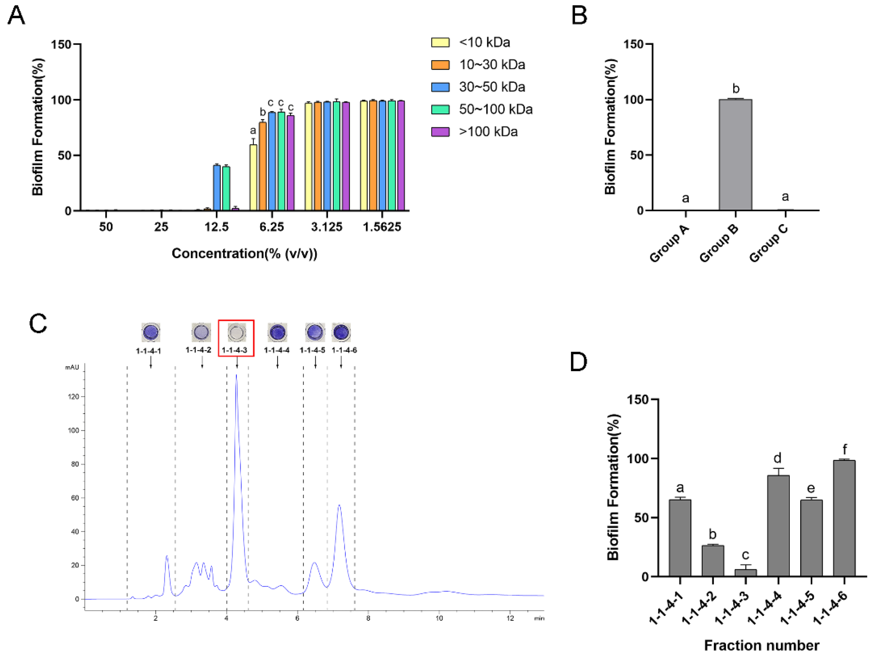
After ultrafiltration, the CFS of L. plantarum was divided into five fractions with different molecular weights, including less than 10 kDa, 10~30 kDa, 30~50 kDa, 50~100 kDa and greater than 100 kDa. The antibiofilm ability of each fraction is shown in Figure 2A. The results illustrate that all fractions could inhibit over 99.5% of biofilm formation at the concentration of 25%. When the fractions were diluted to the concentration of 6.25%, the difference in the inhibitive effect appeared. As shown in the results, the fraction with a molecular weight of less than 10 kDa showed the strongest effect, with a biofilm formation of 59.7% (p ≤ 0.05). Therefore, the fraction weighing less than 10 kDa was selected for further separation.
As the pH environment of the eluent cannot be well controlled in the subsequent separation, to further study the influence of pH, we tested the antibiofilm ability of the fraction weighing less than 10 kDa with different treatments. As shown in Figure 2B, in Group A, the fraction weighing less than 10 kDa in the CFS can inhibit 99.7% of biofilm formation at its original pH value (pH = 3.7). However, it lost its inhibitive effect when the pH was adjusted to 6.5 (Group B). Interestingly, when the pH was returned to the original value (Group C), the antibiofilm effect also recovered, with a 99.6% inhibition of biofilm formation, which was not significantly different from Group A (p > 0.05). These results indicate that the influence of pH on the antibiofilm ability of the active substance was reversible.
During the further separation, the antibiofilm activity of each fraction was determined. The high-performance liquid chromatography (HPLC) chromatogram and antibiofilm effect of each fraction that was examined before the final separation are shown in Figures S3–S6. The fractions with the strongest effect were further separated. Fr. 1-1-4-3 was finally isolated as the main antibiofilm component, as it had the strongest antibiofilm effect with a narrow and symmetrical peak shape, as shown in the final separation (Figure 2C,D).

2.3. Antibiofilm Effect of Component 1-1-4-3

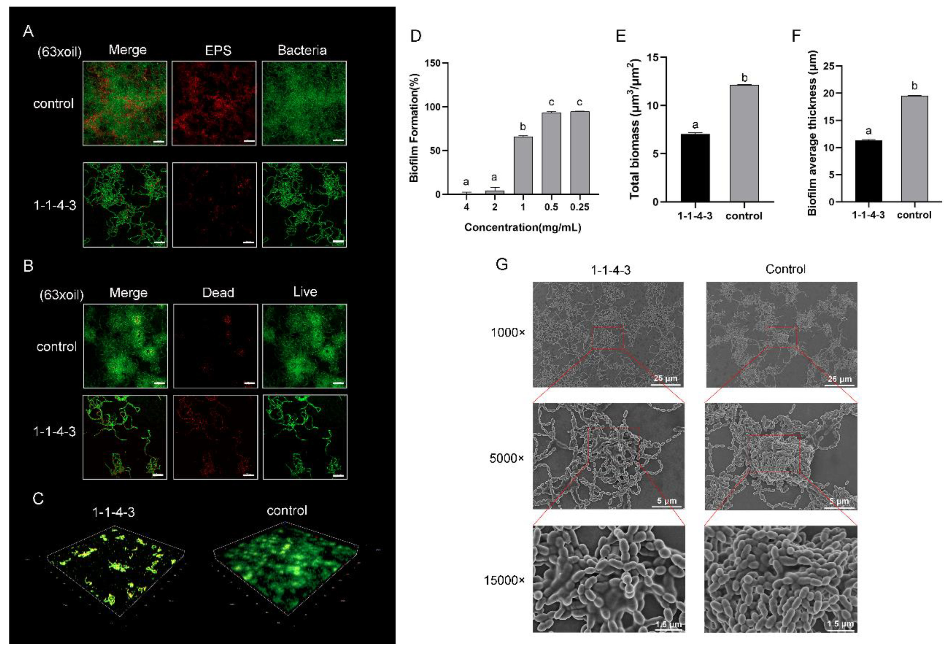
The results showed that the antibiofilm effect of 1-1-4-3 was dose-dependent, with a biofilm formation of 4.2% and 65.9% at a concentration of 2 mg/mL and 1 mg/mL, respectively (Figure 3D). To further study the impact of 1-1-4-3 on the biofilm formation of S. mutans, we observed the biofilm in the presence or absence of 1-1-4-3 with confocal laser scanning microscopy (CLSM) and scanning electron microscopy (SEM). In Figure 3A, the green signal represents bacteria and the red signal represents the distribution of EPSs. It is obvious that after being treated with 1-1-4-3, the EPSs in the biofilm reduced significantly, showing a loose connection among the bacteria. Furthermore, in Figure 3B, the green signal represents live bacteria and the red represents dead bacteria. It was found that after being treated with 1-1-4-3, the total amount of bacteria decreased and the biofilm became obviously looser than that in the control group. Clumps of biofilm decreased and the biofilm became thinner, with weaker connections among bacterium and an apparent reduction in the total biofilm biomass (p ≤ 0.05) (Figure 3C,E). The average thickness of the biofilm exposed to 1-1-4-3 was 11.3 μm, whereas the control was 19.6 μm (p ≤ 0.05) (Figure 3F). In the images captured by SEM (Figure 3G), it can be seen that the biofilm treated with 1-1-4-3 became flat, and the clumps of biofilm were loose and weak.

2.4. Characterization and Validation of Component 1-1-4-3

2.4.1. 1H and 13C Nuclear Magnetic Resonance (NMR) and Quadrupole Time-of-Flight Mass Spectrometry (Q-TOF-MS) Data for Component 1-1-4-3

1H NMR and 13C NMR data for 1-1-4-3 are shown in Table 1. Positive HR-ESI-MS showed a signal at m/z 118.0863 (calcd. for C5H12NO2+ [M + H]+ 118.0863) and negative HR-ESI-MS showed a signal at m/z 89.0244 (calcd. for C3H5O3 [M – H] 89.0244) (Figures S7 and S8).

2.4.2. Analysis of NMR and Q-TOF-MS Data

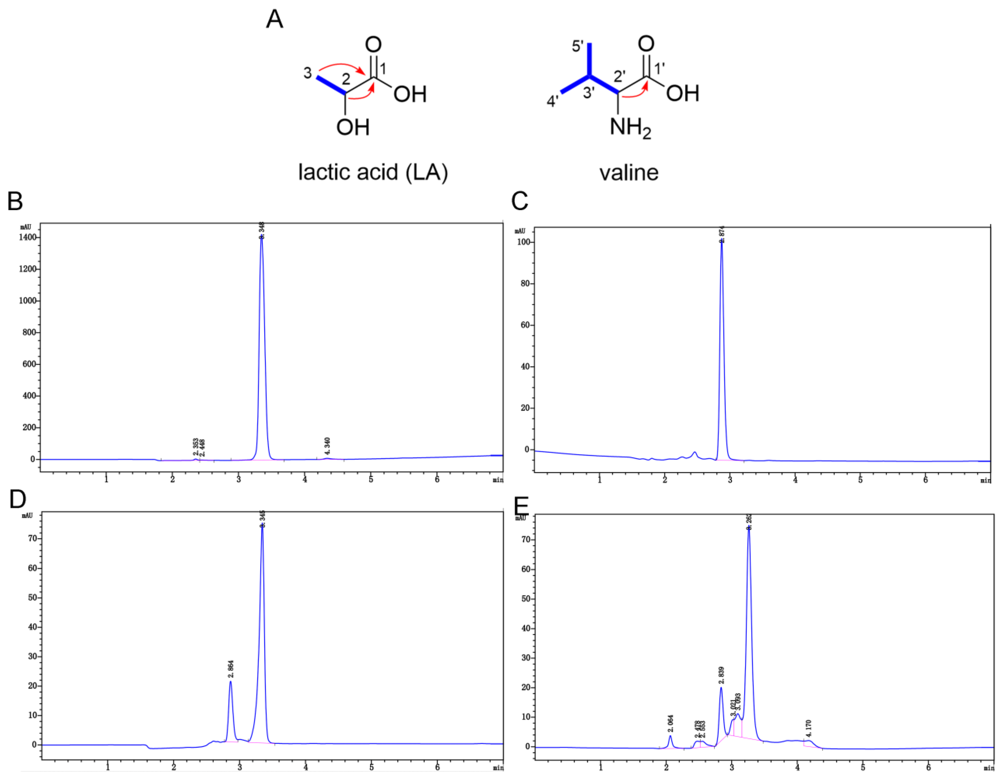
The 1H NMR and 13C NMR spectra of 1-1-4-3 showed two groups of resonance signals with a ratio of 2:1, which indicated that 1-1-4-3 was a mixture (Figures S9 and S10). Next, the two groups of high and low signals were analyzed. One secondary methyl (δH 1.22, d, (J = 6.9 Hz); δC 21.0 (CH3)), an oxygenated methine (δH 4.02, q, (J = 6.9 Hz); δC 66.3 (CH)) and one carbonyl (δC 176.9 (C)) were distinguished in the group with high signals in the 1H NMR, 13C NMR and DEPT spectra (Figures S11 and S12). The 1H–1H COSY correlation between the protons at δH 1.22 (H-3) and the proton at δH 4.02 (H-2) (Figure S13), as well as the HMBC correlations between H-3 and H-2 and the carbonyl (δC 176.9, C-1) (Figure S14), suggest that the secondary methyl, the oxygenated methine and the carbonyl were connected to form the structure of LA (Figure 4A). The quasimolecular ion peak at m/z 89.0244 (calcd. for C3H5O3 [M − H] 89.0244) in the HR-ESI-MS further supported the presence of LA as a major constituent in 1-1-4-3. As for the low-signal group, the 1H NMR, 13C NMR and DEPT spectra displayed characteristic signals for two secondary methyls (δH 0.89 and 0.93, each d, (J = 7.0 Hz); δC 18.1 and 19.3 (each CH3)); two methines, including a nitrogenated one (δH 3.12, d, (J = 3.6 Hz); δC 59.9 (CH)); and a carbonyl (δC 170.0 (C)). As shown in Figure 4A, the observed 1H–1H COSY correlations between H-2′/H-3′ and H-3′/H-4′(H-5′) indicated the moiety of CH-2′–CH-3′(–CH3-5′)–CH3-4′, which was linked to the carbonyl (δC 170.0, C-1′) by the key HMBC correlation between H-2′ and C-1′. Therefore, the component with low content was determined to be valine, whose molecular weight was also consistent with the HR-ESI-MS ion peak at m/z 118.0863 (calcd. for C5H12NO2+ [M + H]+ 118.0863). In addition, the 1D NMR and HPLC analysis of the authentic samples, LA and valine, as well as an analysis of their mixture (LA and valine at a ratio of 2:1), were also performed. By comparing the 1D NMR data (Table 1 and Figures S15–S22) and HPLC spectra (Figure 4B–E) of 1-1-4-3 with those of LA, valine, and their mixture, it was further confirmed that 1-1-4-3 contained LA and valine.

2.5. Verification of the Role of Lactic Acid (LA) in the Antibiofilm Activity

2.5.1. Antibiofilm Effect of Component 1-1-4-3, LA (d/l-), Valine (d/l-) and the Mixture of LA and Valine

As for the antibiofilm effect (Figure 5A), LA (d/l-) showed a strong antibiofilm ability, with over 97% of biofilm formation inhibited at the concentration of 1 mg/mL. However, valine (d/l-) did not show an inhibitive effect on biofilm formation at all tested concentrations. The mixture of LA and valine showed a weaker antibiofilm effect compared with that of LA (d/l-), and its effect was similar to that of 1-1-4-3 (p > 0.05). These results indicate that LA was the antibiofilm component within 1-1-4-3. Moreover, the effect between the two chiral LAs was not significantly different, and neither was the effect among the four kinds of mixtures (p > 0.05).

2.5.2. The Antibiofilm Activity of LA and Low-pH Environment

In order to figure out the role of LA, we compared the antibiofilm activity of LA and of BHI broth with 1% (w/v) sucrose (1% BHIS) that was at the same pH as the LA at each concentration. The pH of LA at the concentrations of 0.5, 1, 2 and 4 mg/mL was 3.6, 3.5, 3.4 and 3.2, respectively. As shown in Figure 5B, the 1% BHIS with the same pH had a certain antibiofilm effect, which indicated that the low-pH environment did have an effect on the antibiofilm activity. However, d/l- LA at 1 and 2 mg/mL had a significantly stronger effect than that of the 1% BHIS with the same pH (p ≤ 0.05). The results indicate that in the CFS of L. plantarum, the low-pH environment played a certain role in the antibiofilm activity; however, LA also displayed strong antibiofilm activity in its undissociated form. In addition, to figure out the mechanism of inhibition of LA on the biofilm formation of S. mutans, we examined the minimal inhibitory concentration (MIC) of LA. The results showed that the MIC of LA (d/l-) was 1 mg/mL. Additionally, the minimum biofilm inhibitory concentration to inhibit 99% biofilm (MBIC99) of d/l- LA was 1.30 and 1.03 mg/mL. The MIC was close to the MBIC99, which indicates that the inhibition mechanism of the L. plantarum CFS on the biofilm formation of S. mutans may be mainly attributed to the inhibition of the growth of S. mutans by LA.

3. Discussion

Previous studies have reported that Lactobacilli is dominant in dentine caries [25]. Additionally, the detection of oral Lactobacilli together with S. mutans has been correlated to the severity of dental caries [8]. However, as described above, the CFS of Lactobacilli was found to be inhibitive against S. mutans, the main cariogenic bacteria. The pathogenic mechanism of Lactobacilli in promoting caries and the interaction between Lactobacilli and S. mutans remain unclear.
The current study found that hydrogen peroxide played a small role in the antibiofilm activity of the five selected CFSs. The results were consistent with those of a previous study [10], which might indicate that the amount of hydrogen peroxide produced by the selected Lactobacilli was small, and, thus, hydrogen peroxide was not the main antibiofilm substance against S. mutans in the CFS. Since hydrogen peroxide is naturally unstable, further experiments can be carried out to quantify hydrogen peroxide in the five tested CFSs and clarify the role of hydrogen peroxide. This study found that the active components of the five tested CFSs needed to exert an inhibitive effect under an acidic condition. According to previous studies, organic acid in the CFS of Lactobacilli can obviously improve the effect of antibiofilm substances [14]. Interestingly, the influence of pH on the crude extract was reversible. These results indicate that the main active component might be a weak acid that causes an ionization equilibrium [26], or is a pH-dependent substance.
In oral microbiology, there is a lack of studies on the bioassay-guided isolation of active metabolites in regard to the interaction between bacteria. There are many methods for the extraction of active components in natural products, including extraction, sedimentation, dialysis, HPLC, etc. [27,28,29]. In the present study, the main antibiofilm component against S. mutans was screened from the CFS of L. plantarum by ultrafiltration, ethanol precipitation and HPLC. Ultrafiltration processes offer several advantages in comparison to the traditional conventional process: retention of characteristics and the structure of components in natural products, high selectivity, simplicity in continuous operations, integration, and scaling up [30]. In addition, this study combined the use of normal- and reversed-phase HPLC. The separation efficiency of HPLC under gradient conditions is best described by the peak capacity, which represents the maximum number of components that could be theoretically separated on a given liquid chromatography column within a gradient time [31]. The combination of normal- and reversed-phase HPLC produces a high peak capacity and better orthogonality, and can dramatically improve the separation power of chromatography, according to the mathematical model established by Giddings [32]. Moreover, extraction was used less in this experiment, which can reduce the inflammation, toxicity and environmental pollution of organic solvents [33].
During the separation, it was found that, except for the fraction weighing less than 10 kDa, the fractions exerted a certain antibiofilm effect. According to the literature, the antibiofilm components weighing more than 10 kDa in the Lactobacilli CFS mainly include proteinaceous compounds [14]. These results indicate that the antibiofilm substances in the CFS of L. plantarum might contain macromolecular proteinaceous compounds. In addition, during the separation via reversed-phase HPLC, some components with a late peak also had certain antibiofilm activity, and the polarity of such components was low. According to previous studies, the CFS might contain long-chain fatty acids [14,21]. Furthermore, it was found that the elution times of the most active component in the normal- and reversed-phase HPLC were close. Therefore, the active component had a certain solubility in both water and organic solvents. These results indicate that the active component might contain amphiphilic bacteriocins or biosurfactants and organic acids soluble in water and organic solvents.
In this study, the fractions with the strongest antibiofilm effect were further separated. Additionally, the antibiofilm effect of the end product, 1-1-4-3, was examined via CLSM and SEM. The results indicate that 1-1-4-3 can reduce the EPS and clustered biofilm formation while causing the biofilm to be scattered and sparse. According to the literature, the L. casei CFS can reduce the number of EPSs and proportion of water-insoluble glucan in the biofilm of S. mutans and reduce the surface roughness of the biofilm [34]. Through the structural identification using 2D NMR and MS, as well as through the HPLC analysis and activity verification, it was found that 1-1-4-3 was a mixture of LA and valine with a ratio of 2:1, and the active component was LA. The problem of proton overlapping resonance in 1D NMR was overcome by 2D NMR, which is more conducive for conducting a structural analysis of complex compounds [35].
This study found that the effect of LA was obviously stronger than that of the other components, and the active components exerted their effects under an acidic environment. In addition, LA was the main organic acid in the Lactobacilli CFS [14]. These results indicate that LA plays a dominant role in the antibiofilm effect of the L. plantarum CFS. Chen et al. [36] found that the concentration of LA in the CFS of L. plantarum can reach 23.3~27 mg/mL, which might explain why, after a series of dilution, the tested CFS still had a strong antibiofilm activity against S. mutans. It was reported that Lactobacilli can produce both LA isomers (d- and l-) or the racemic blend) [14]. In this study, the antibiofilm effect of d/l- LA was not significantly different, which proved that both chiral LAs in the CFS had an antibiofilm activity effect.
According to the literature, despite the fact that S. mutans can produce and resist acid, its resistance against acid is limited. The current study found that both LA and BHIS with the same pH as LA had an antibiofilm effect, but the effect of LA was significantly stronger. It has been reported that a low-pH environment can inhibit the growth of S. mutans, and that its virulence is related to biofilm formation [37]. Cells of S. mutans grew at a slow rate in a medium with a pH of 5.3 and lost the ability to perform glycolysis when the pH dropped to 3.45 [5]. Meanwhile, according to previous studies, LA is the main end product of S. mutans metabolism under carbon excess. Additionally, excess undissociated LA could inhibit the growth of S. mutans [38]. Excessive LA may change the osmotic stresses in S. mutans cells, which leads to the inhibition of the activity of lactate dehydrogenase, resulting in the impairment of the ability of S. mutans to metabolize exogenous carbohydrates [39,40]. It was considered that only undissociated LA causes inhibition, not the lactate anions, because only the protonated species can be transported across the cytoplasmic membrane [38]. The phenomenon of the stronger effect of LA compared with the BHIS with the same pH might be explained by the fact that an LA solution can carry more hydrogen ions in the undissociated form. Furthermore, the undissociated LA can cross the membrane, resulting in the decrease in internal pH values and the impairment of the carbohydrate metabolism. In view of the antimicrobial mechanism of LA reported previously, the authors thought that the inhibition mechanism of the L. plantarum CFS on the biofilm formation of S. mutans may be mainly attributed to its inhibition of the growth of S. mutans due to LA.
Considering that the detection of Lactobacilli has been associated with caries severity [8] and Lactobacilli can reduce the quantity of S. mutans in the oral cavity [41], it is speculated that the activity of L. plantarum in carious lesions may be described as follows: when the concentration of LA is below the tolerance threshold of S. mutans, L. plantarum produces LA together with S. mutans, promoting the progression of caries, whereas when the concentration of LA is above the tolerance threshold of S. mutans in a low-pH environment, LA and other substances in the CFS produced by L. plantarum inhibit the growth and biofilm formation of S. mutans, competing for dominance in the carious lesions.
The findings of this study suggest that the interaction between Lactobacilli and S. mutans might promote dental caries due to excess LA. Therefore, the administration of Lactobacilli supplements as probiotics may need to be weighed against the possibility that they contribute to dental caries. However, the tested L. plantarum CFS contained other antibiofilm components which are possibly pH sensitive. In further studies, such components could be further separated to develop caries prevention agents with pH sensitivity and low toxicity that maintain the balance of the microbiome in the neutral environment.
In conclusion, this study found that the antibiofilm effect of the L. plantarum CFS against S. mutans was attributable to the comprehensive effect of multiple components; however, LA plays a dominant role in the antibiofilm activity.

4. Materials and Methods

4.1. Bacterial Strains, Culture Conditions and Growth Curves Determination

Three Lactobacilli strains, L. plantarum ATCC 14917, L. casei ATCC 393 and L. gasseri ATCC 33323, and S. mutans UA 159 were acquired from Guangdong Microbial Culture Collection Center (GDMCC; Guangzhou, China). The other two Lactobacilli strains, L. fermentum ATCC 14931 and L. salivarius ATCC 11741, were acquired from China General Microbiological Culture Collection Center (CGMCC; Beijing, China). The Lactobacilli strains and S. mutans were cultured in de Man–Rogosa–Sharpe (MRS) broth and BHI broth, respectively, at 37 °C under microaerophilic conditions (6% O2, 3.6% H2, 3.6% CO2, 86.8% N2). Growth curves of Lactobacilli and S. mutans were determined by counting viable cells seeded on the corresponding agar medium every two hours.

4.2. Preparation of CFS

The CFS of each Lactobacilli was prepared according to the modified protocol of Lin [23]. Briefly, bacteria at the late logarithmic growth phase were adjusted to 1 × 10⁷ CFU/mL and then incubated microaerophilically at 37 °C for 48 h. Subsequently, the spent culture was centrifuged (5000× g, 15 min, 4 °C), which was followed by filtration through a 0.22 μm filter (Merck Millipore, Boston, MA, USA) to acquire the CFS. Considering that the concentration of the CFS in the natural niche of biofilm should be 100% (v/v) or possibly even higher due to local diffusion obstruction, in this study, the CFS was concentrated at a final concentration of 200% (v/v) by freeze-drying.
The prepared CFS was divided into three portions, as described previously [10]. One portion was the original concentrated CFS, and its pH was tested. The other two portions were treated to eliminate the effect of pH and hydrogen peroxide. The effect of pH was neutralized by adjusting the pH value of the CFS to 6.5 ± 0.1 with 1 M of NaOH. Additionally, the effect of hydrogen peroxide was eliminated by treating the CFS with 0.5 mg/mL of catalase (Solarbio, Beijing, China). Furthermore, to study the role of the low-pH environment, we prepared the BHI with the same pH as the original concentrated CFS, which was adjusted with 1 M of HCl. The treated and untreated CFSs were stored at −20 °C.

4.3. Antibiofilm Assay of CFS on S. mutans

A previously described, a crystal-violet-based microtiter plate assay was performed to determine the inhibitive effect of the CFS on the biofilm formation of S. mutans [42]. Briefly, S. mutans, at a final concentration of 1.0 × 10⁶ CFU/mL in 2% BHIS, was cultured in 96-well flat-bottom microplates with or without the addition of the prepared CFS or BHI at the same volume, ensuring that the final volume of each well was 200 μL. The final concentrations of the tested CFSs were in the range of 6.25% to 200% (v/v). After incubation at 37 °C for 24 h, the biofilm was washed twice with phosphate-buffered saline (PBS) and fixed with methanol for 15 min. Later, the wells were air-dried for 30 min and the fixed biofilm was then stained with 0.1% crystal violet solution for 15 min. After that, the excess dye was removed and the biofilm was rinsed with water. The crystal violet stains were solubilized in 96% ethanol, and were shaken vigorously for 1 h until completely dissolved. Lastly, the optical densities were measured at the wavelength of 595 nm using a microplate reader. S. mutans was treated with MRS broth as the negative control according to the same procedure as the CFS. Biofilm formation was expressed as a ratio of crystal-violet-stained biofilms relative to the negative control.

4.4. Ultrafiltration of L. plantarum CFS and Further Investigation of the Influence of pH

The original CFS of L. plantarum was fractionated with Amicon® Ultra-15 Centrifugal Filter Devices with different cutoffs (MWCO), including 10 K, 30 K, 50 K and 100 K (10,000 MWCO, 30,000 MWCO, 50,000 MWCO and 100,000 MWCO) (Merck KGaA, Darmstadt, Germany), according to the manufacturer’s recommendations. Briefly, 15 mL of the CFS was centrifuged at 4000× g at 4 °C to reach the volume of 150~300 μL. The sample held by the filter was collected, whereas the eluted sample was subjected to further centrifuging by a device with a lower cutoff. The effect of all collected fractions on the biofilm formation of S. mutans was determined by using a crystal violet staining assay.
To further test the impact of pH on the antibiofilm ability of the fraction screened by ultrafiltration, three groups (A~C) were set for the assay. Group A: the fraction screened by ultrafiltration was untreated. Group B: the pH value of the fraction was adjusted to 6.5 with 1 M of NaOH. Group C: the pH value of the fraction was first adjusted to 6.5 with 1 M of NaOH, and then was adjusted back to the original value with 1 M of HCl.

4.5. Bioassay-Guided Isolation of the Main Antibiofilm Component

The active fraction was further separated by HPLC in the following process. Reversed- and normal-phase HPLC were both performed on an Agilent 1260 System (Agilent Technologies Inc., Palo Alto, CA, USA). Solvent mixtures are reported as %v/v, unless otherwise stated. Each fraction obtained by HPLC was subjected to the antibiofilm bioassay, and the fraction with the strongest effect was further separated.
Briefly, the fraction screened out with ultrafiltration was concentrated via freeze-drying. Then, the sample was chromatographed over a ZORBAX SB-C18 column (5 μm, 4.6 mm × 150 mm, Agilent Technologies Inc., Palo Alto, CA, USA) using the eluent of acetonitrile (MeCN) and H2O that was supplemented with 0.1% trifluoroacetic acid (TFA) to obtain 12 fractions (Frs. 1~12). The procedure for the linear gradient of MeCN supplemented with 0.1% TFA was as follows: 0~5 min, 5%; 8~20 min, 10%; 23~33 min, 15%; 36~46 min, 20%; 49~59 min, 30%; 69~79 min, 100%; and 84 min, 5%. Fr. 1 with the strongest activity was then further subjected to an AccucoreTM HILIC column (2.6 μm, 2.1 mm × 150 mm, Thermo Fisher Scientific Inc., Waltham, MA, USA), where the same eluent described above was used to obtain 12 fractions (Frs. 1-1~1-12), and the linear gradient of MeCN supplemented with 0.1% TFA was as follows: 0~30 min, 5~30%. Fr. 1-1 with activity was pooled and lyophilized, and was then dissolved in cold anhydrous ethanol and kept at −20 °C overnight to precipitate polysaccharides, according to previous studies with some modifications [43]. The results of the bioassay indicated that the supernatant of Fr. 1-1 showed a strong antibiofilm effect, whereas its precipitate had no antibiofilm ability. Then, the supernatant of Fr. 1-1 was lyophilized and further separated by using an Eclipse XDB-C18 column (5 μm, 4.6 mm × 250 mm, Agilent Technologies Inc., Palo Alto, CA, USA), and was eluted with 5% isopropanol in methanol absolute/ultrapure water (95:5) to obtain 11 fractions (Frs. 1-1-1~1-1-12). Fr. 1-1-4 with activity was then purified by using a ZORBAX NH2 column (5 µm, 4.6 mm × 250 mm, Agilent Technologies Inc., Palo Alto, CA, USA) and eluted with ethanol/n-hexane (70:30) to obtain 6 fractions (Frs. 1-4-1~1-4-6). The results showed that Fr. 1-1-4-3 had the strongest antibiofilm activity with a narrow and symmetrical peak shape; thus, it was selected for further study.

4.6. Examination of Antibiofilm Effect of Component 1-1-4-3

The effect of the antibiofilm activity of 1-1-4-3 at different doses was tested by using the crystal violet staining assay described above. Meanwhile, the morphological changes of the S. mutans biofilm were observed with SEM and CLSM. Briefly, S. mutans, at a final concentration of 1.0 × 10⁶ CFU/mL in 1% BHIS, was cultured on sterile glass coverslips or confocal dishes with or without the addition of 1-1-4-3. For SEM observation, after incubation on sterile glass coverslips for 24 h, the biofilm was then fixed with 2.5% glutaraldehyde for 6 h, followed by dehydration using graded ethanol (50~100%). Subsequently, the biofilm was successively treated with a solution of t-butanol/ethanol (1:1) and a solution of t-butanol (each step for 15 min), and was then freeze-dried. Samples were sputtered with gold and observed via SEM (Zeiss, Oberkochen, Germany). For the CLSM observation, after incubation on confocal dishes for 24 h, the biofilm was rinsed with PBS three times and was then stained with SYTO 9 and propidium iodide using the LIVE/DEAD BacLight Bacterial Viability Kit (L7012, Invitrogen, Carlsbad, CA, USA), according to the manufacturer’s instructions. After that, the LSM 980 with Airyscan 2 (Zeiss, Oberkochen, Germany) was applied to observe the biofilm and capture images, which were then analyzed by using COMSTAT [44]. In addition, the distribution of EPSs in biofilm was observed with the Alexa Fluor 647 Dextran conjugate (Invitrogen Corp., Carlsbad, CA, USA) and SYTO 9. Briefly, with or without the addition of 1-1-4-3, S. mutans was seeded in confocal dishes containing the culture medium with 2.5 μM of the Alexa Fluor 647 Dextran conjugate. After incubating for 24 h, 2.5 μM SYTO-9 was applied to label S. mutans cells, as previously described. The distribution of EPSs in the biofilm was observed by using the LSM 980 with Airyscan 2, as described above.

4.7. Structure Elucidation of Component 1-1-4-3

4.7.1. NMR and Q-TOF-MS Analysis of Component 1-1-4-3

Next, 1D and 2D NMR spectra were recorded on Bruker AVANCE III HD 600 MHz (Bruker, Karlsruhe, Germany) spectrometers at 300 K with TMS as the internal standard. Chemical shifts are expressed in ppm, with reference to the residual solvent signals (DMSO-d6 with δH 2.50 and δC 39.6). As for the mass spectrometry (MS) analysis, the positive and negative electrospray ionization (ESI) MS data were measured with Q-TOF-MS (Bruker maXis impact II, Bruker Daltonics, Karlsruhe, Germany). The MS raw data were obtained in the format from the Bruker Compass Data Analysis Viewer version 4.1 (Bruker Daltonics, Karlsruhe, Germany). According to the analysis of the 2D NMR and ESI-MS data of 1-1-4-3 (see Section 2.4), it was found that 1-1-4-3 was a mixture of LA (C3H6O3) and valine (C5H12NO2) at a ratio of about 2:1.

4.7.2. NMR Analysis of Valine and LA Standards and HPLC Analysis of Component 1-1-4-3 and Its Standards

To further confirm the above results, the authentic samples of valine, LA (Macklin Inc., Shanghai, China) and their mixture were subjected to a 1D NMR analysis, according to the processes described in Section 4.7.1. In addition, the HPLC analyses of 1-1-4-3, LA, valine and the 2:1 mixture of LA and valine were performed on an Agilent 1260 System using an Eclipse XDB-C18 column with the mobile phase consisting of 95% eluent A (ultrapure water) and 5% eluent B (5% isopropanol in methanol). The flow rate was 1.0 mL/min and the elution peak was detected at 220 nm.

4.8. Exploration of the Role of LA in the Antibiofilm Activity

4.8.1. Comparison of the Antibiofilm Effect of Component 1-1-4-3, LA (d/l-), Valine (d/l-) and the Mixture of LA and Valine

1-1-4-3, LA (d/l-), valine (d/l-) and a molar concentration of the 2:1 mixture of LA and valine were dissolved in 1% BHIS at a pH of 3.70 ± 0.05 until a series of gradient concentrations was established (0.25~4 mg/mL), and their antibiofilm activity was tested by using the crystal violet staining assay. Bacteria exposed to 1% BHIS with the same pH value were set as the negative control.

4.8.2. Examination of the Antibiofilm Activity of LA and Low pH

To simulate the pH environment of the L. plantarum CFS, LA was diluted in 1% BHIS at a pH of 3.70 ± 0.05 until a series of gradient concentrations was established (0.5~4 mg/mL); then, the pH value of LA at each concentration was tested. In order to figure out the role of low pH, we prepared 1% BHIS with the same pH as LA at each concentration, whose pH was adjusted by using 1 M of HCl. The antibiofilm activity of LA at different concentrations and 1% BHIS with the same pH as LA at each concentration were then tested via the crystal violet assay. Bacteria exposed to 1% BHIS at the pH of 3.70 ± 0.05 were set as the negative control. The MBIC99 of LA was determined by constructing a dose–response curve via nonlinear fitting and calculating the concentration of IC99 via the GraphPad Prism version 8.0.2 software.

4.8.3. Examination of the MIC of LA

The MIC of LA was measured by using the standard broth microdilution method, following the recommendations of the Clinical and Laboratory Standards Institute (CLSI) with some modifications [45]. Briefly, bacteria were diluted to a final concentration of 1.0×10⁶ CFU/mL and treated with LA at a series of gradient concentrations (0.5~4 mg/mL). Nontreated bacteria with broth were set as the negative control. The experiments were performed in 96-well microtiter plates. The MIC was determined as the lowest concentration of drugs that inhibited bacterial growth via visual inspection after 24 h of incubation.

4.9. Statistical Analysis

All the experiments were performed in triplicate, and the data are presented as the mean ± standard deviation. Statistical analysis was carried out using the GraphPad Prism version 8.0.2 software via a one-way analysis of variance (ANOVA) or unpaired t-test. Statistical significance was defined as p ≤ 0.05.

Supplementary Materials

The following supporting information can be downloaded at: https://www.mdpi.com/article/10.3390/ijms24031986/s1.

Author Contributions

Conceptualization, J.L. and H.L.; Data curation, J.L.; Formal analysis, J.L., Y.Z. and G.T.; Funding acquisition, H.L.; Investigation, J.L. and R.W.; Methodology, J.L., Y.Z., R.W. and H.L.; Project administration, H.L.; Supervision, H.L.; Validation, J.L. and Y.Z.; Visualization, J.L. and G.T.; Writing—original draft, J.L.; Writing—review and editing, Y.Z., G.T., R.W. and H.L. All authors have read and agreed to the published version of the manuscript.

Funding

This research was funded by the National Natural Science Foundation of China, grant number 81970928.

Institutional Review Board Statement

Not applicable.

Informed Consent Statement

Not applicable.

Data Availability Statement

The data presented in this study are available on request from the corresponding author.

Conflicts of Interest

The authors declare no conflict of interest.

References

  1. Kassebaum, N.J.; Smith, A.; Bernabe, E.; Fleming, T.D.; Reynolds, A.E.; Vos, T.; Murray, C.; Marcenes, W. Global, Regional, and National Prevalence, Incidence, and Disability-Adjusted Life Years for Oral Conditions for 195 Countries, 1990–2015: A Systematic Analysis for the Global Burden of Diseases, Injuries, and Risk Factors. J. Dent. Res. 2017, 96, 380–387. [Google Scholar] [CrossRef] [PubMed]
  2. Folliero, V.; Dell’Annunziata, F.; Roscetto, E.; Amato, A.; Gasparro, R.; Zannella, C.; Casolaro, V.; De Filippis, A.; Catania, M.R.; Franci, G.; et al. Rhein: A novel antibacterial compound against Streptococcus mutans infection. Microbiol. Res. 2022, 261, 127062. [Google Scholar] [CrossRef] [PubMed]
  3. Zijnge, V.; van Leeuwen, M.B.; Degener, J.E.; Abbas, F.; Thurnheer, T.; Gmur, R.; Harmsen, H.J. Oral biofilm architecture on natural teeth. PLoS ONE 2010, 5, e9321. [Google Scholar] [CrossRef] [PubMed] [Green Version]
  4. Svensäter, G.; Borgström, M.; Bowden, G.H.W.; Edwardsson, S.; Malmö, U.; Faculty, O.O. The Acid-Tolerant Microbiota Associated with Plaque from Initial Caries and Healthy Tooth Surfaces. Caries Res. 2003, 37, 395–403. [Google Scholar] [CrossRef] [PubMed]
  5. Belli, W.A.; Marquis, R.E. Adaptation of Streptococcus mutans and Enterococcus hirae to acid stress in continuous culture. Appl. Environ. Microbiol. 1991, 57, 1134–1138. [Google Scholar] [CrossRef] [PubMed] [Green Version]
  6. Caufield, P.W.; Schon, C.N.; Saraithong, P.; Li, Y.; Argimon, S. Oral Lactobacilli and Dental Caries: A Model for Niche Adaptation in Humans. J. Dent. Res. 2015, 94, 110S–118S. [Google Scholar] [CrossRef]
  7. Latifi-Xhemajli, B.; Veronneau, J.; Begzati, A.; Bytyci, A.; Kutllovci, T.; Rexhepi, A. Association between salivary level of infection with Streptococcus mutans/Lactobacilli and caries-risk factors in mothers. Eur. J. Paediatr. Dent. 2016, 17, 70–74. [Google Scholar]
  8. Lapirattanakul, J.; Nomura, R.; Okawa, R.; Morimoto, S.; Tantivitayakul, P.; Maudcheingka, T.; Nakano, K.; Matsumoto-Nakano, M. Oral Lactobacilli Related to Caries Status of Children with Primary Dentition. Caries Res. 2020, 54, 194–204. [Google Scholar] [CrossRef]
  9. Ledder, R.G.; Kampoo, K.; Teanpaisan, R.; McBain, A.J. Oral Microbiota in Severe Early Childhood Caries in Thai Children and Their Families: A Pilot Study. Front. Microbiol. 2018, 9, 2420. [Google Scholar] [CrossRef]
  10. Wasfi, R.; Abd El-Rahman, O.A.; Zafer, M.M.; Ashour, H.M. ProbioticLactobacillus sp. inhibit growth, biofilm formation and gene expression of caries-inducingStreptococcus mutans. J. Cell. Mol. Med. 2018, 22, 1972–1983. [Google Scholar] [CrossRef] [Green Version]
  11. Samot, J.; Badet, C. Antibacterial activity of probiotic candidates for oral health. Anaerobe 2013, 19, 34–38. [Google Scholar] [CrossRef] [PubMed]
  12. Teanpaisan, R.; Piwat, S.; Dahlén, G.; Institutionen, F.O.; Institute, O.O.; Sahlgrenska, A.; Göteborgs, U.; Gothenburg, U.; Sahlgrenska, A. Inhibitory effect of oral Lactobacillus against oral pathogens. Lett. Appl. Microbiol. 2011, 53, 452–459. [Google Scholar] [CrossRef]
  13. Iwami, Y.; Hata, S.; Takahashi, N.; Yamada, T. Difference in amounts between titratable acid and total carboxylic acids produced by oral streptococci during sugar metabolism. J. Dent. Res. 1989, 68, 16–19. [Google Scholar] [CrossRef] [PubMed]
  14. Mani-Lopez, E.; Arrioja-Breton, D.; Lopez-Malo, A. The impacts of antimicrobial and antifungal activity of cell-free supernatants from lactic acid bacteria in vitro and foods. Compr. Rev. Food. Sci. Food Saf. 2022, 21, 604–641. [Google Scholar] [CrossRef]
  15. Wattanarat, O.; Nirunsittirat, A.; Piwat, S.; Manmontri, C.; Teanpaisan, R.; Pahumunto, N.; Makeudom, A.; Sastraruji, T.; Krisanaprakornkit, S. Significant elevation of salivary human neutrophil peptides 1-3 levels by probiotic milk in preschool children with severe early childhood caries: A randomized controlled trial. Clin. Oral Investig. 2021, 25, 2891–2903. [Google Scholar] [CrossRef] [PubMed]
  16. Wannun, P.; Piwat, S.; Teanpaisan, R. Purification and characterization of bacteriocin produced by oral Lactobacillus paracasei SD1. Anaerobe 2014, 27, 17–21. [Google Scholar] [CrossRef]
  17. Yang, K.M.; Kim, J.S.; Kim, H.S.; Kim, Y.Y.; Oh, J.K.; Jung, H.W.; Park, D.S.; Bae, K.H. Lactobacillus reuteri AN417 cell-free culture supernatant as a novel antibacterial agent targeting oral pathogenic bacteria. Sci. Rep. 2021, 11, 1631. [Google Scholar] [CrossRef] [PubMed]
  18. Rossoni, R.D.; Velloso, M.D.S.; de Barros, P.P.; de Alvarenga, J.A.; Santos, J.D.D.; Santos Prado, A.C.C.D.; Ribeiro, F.D.C.; Anbinder, A.L.; Junqueira, J.C. Inhibitory effect of probiotic Lactobacillus supernatants from the oral cavity on Streptococcus mutans biofilms. Microb. Pathog. 2018, 123, 361–367. [Google Scholar] [CrossRef] [Green Version]
  19. Fang, F.; Xu, J.; Li, Q.; Xia, X.; Du, G. Characterization of a Lactobacillus brevis strain with potential oral probiotic properties. BMC Microbiol. 2018, 18, 221. [Google Scholar] [CrossRef]
  20. Jeong, D.; Kim, D.H.; Song, K.Y.; Seo, K.H. Antimicrobial and anti-biofilm activities of Lactobacillus kefiranofaciens DD2 against oral pathogens. J. Oral Microbiol. 2018, 10, 1472985. [Google Scholar] [CrossRef] [Green Version]
  21. van Zyl, W.F.; Deane, S.M.; Dicks, L.M.T. Molecular insights into probiotic mechanisms of action employed against intestinal pathogenic bacteria. Gut Microbes 2020, 12, 1831339. [Google Scholar] [CrossRef] [PubMed]
  22. Elshikh, M.; Marchant, R.; Banat, I.M. Biosurfactants: Promising bioactive molecules for oral-related health applications. FEMS Microbiol. Lett. 2016, 363, w213. [Google Scholar] [CrossRef] [PubMed] [Green Version]
  23. Lin, X.; Chen, X.; Chen, Y.; Jiang, W.; Chen, H. The effect of five probiotic lactobacilli strains on the growth and biofilm formation of Streptococcus mutans. Oral Dis. 2015, 21, e128–e134. [Google Scholar] [CrossRef]
  24. Busarcevic, M.; Kojic, M.; Dalgalarrondo, M.; Chobert, J.M.; Haertle, T.; Topisirovic, L. Purification of bacteriocin LS1 produced by human oral isolate Lactobacillus salivarius BGHO1. Oral Microbiol. Immunol. 2008, 23, 254–258. [Google Scholar] [CrossRef] [PubMed]
  25. Munson, M.A.; Banerjee, A.; Watson, T.F.; Wade, W.G. Molecular analysis of the microflora associated with dental caries. J. Clin. Microbiol. 2004, 42, 3023–3029. [Google Scholar] [CrossRef] [Green Version]
  26. Story, D.A. Bench-to-bedside review: A brief history of clinical acid-base. Crit. Care 2004, 8, 253–258. [Google Scholar] [CrossRef] [PubMed] [Green Version]
  27. Yu, H.F.; Ding, C.F.; Zhang, L.C.; Wei, X.; Cheng, G.G.; Liu, Y.P.; Zhang, R.P.; Luo, X.D. Alstoscholarisine K, an Antimicrobial Indole from Gall-Induced Leaves of Alstonia scholaris. Org. Lett. 2021, 23, 5782–5786. [Google Scholar] [CrossRef]
  28. Annang, F.; Perez-Moreno, G.; Gonzalez-Menendez, V.; Lacret, R.; Perez-Victoria, I.; Martin, J.; Cantizani, J.; de Pedro, N.; Choquesillo-Lazarte, D.; Ruiz-Perez, L.M.; et al. Strasseriolides A-D, A Family of Antiplasmodial Macrolides Isolated from the Fungus Strasseria geniculata CF-247251. Org. Lett. 2020, 22, 6709–6713. [Google Scholar] [CrossRef]
  29. Chen, M.; Wang, R.; Zhao, W.; Yu, L.; Zhang, C.; Chang, S.; Li, Y.; Zhang, T.; Xing, J.; Gan, M.; et al. Isocoumarindole A, a Chlorinated Isocoumarin and Indole Alkaloid Hybrid Metabolite from an Endolichenic Fungus Aspergillus sp. Org. Lett. 2019, 21, 1530–1533. [Google Scholar] [CrossRef]
  30. Vollet, M.G.; Belleville, M.P.; Lacour, S.; Dupas, H.M. Membrane Fractionation of Protein Hydrolysates from By-Products: Recovery of Valuable Compounds from Spent Yeasts. Membranes 2020, 11, 23. [Google Scholar] [CrossRef]
  31. Gilar, M.; Olivova, P.; Daly, A.E.; Gebler, J.C. Orthogonality of Separation in Two-Dimensional Liquid Chromatography. Anal. Chem. 2005, 77, 6426–6434. [Google Scholar] [CrossRef] [PubMed]
  32. Giddings, J.C. Sample dimensionality: A predictor of order-disorder in component peak distribution in multidimensional separation. J. Chromatogr. A 1995, 703, 3–15. [Google Scholar] [CrossRef]
  33. Boateng, I.D. Evaluating the status quo of deep eutectic solvent in food chemistry. Potentials and limitations. Food Chem. 2023, 406, 135079. [Google Scholar] [CrossRef] [PubMed]
  34. Wu, J.; Jiang, X.; Yang, Q.; Zhang, Y.; Wang, C.; Huang, R. Inhibition of Streptococcus mutans Biofilm Formation by the Joint Action of Oxyresveratrol and Lactobacillus casei. Appl. Environ. Microbiol. 2022, 88, e243621. [Google Scholar] [CrossRef]
  35. Hansen, A.L.; Kupče, E.; Li, D.; Bruschweiler-Li, L.; Wang, C.; Brüschweiler, R. 2D NMR-Based Metabolomics with HSQC/TOCSY NOAH Supersequences. Anal. Chem. 2021, 93, 6112–6119. [Google Scholar] [CrossRef]
  36. Chen, C.C.; Lai, C.C.; Huang, H.L.; Su, Y.T.; Chiu, Y.H.; Toh, H.S.; Chiang, S.R.; Chuang, Y.C.; Lu, Y.C.; Tang, H.J. Antimicrobial ability and mechanism analysis of Lactobacillus species against carbapenemase-producing Enterobacteriaceae. J. Microbiol. Immunol. Infect. 2021, 54, 447–456. [Google Scholar] [CrossRef]
  37. Cao, Y.; Zhou, Y.; Chen, D.; Wu, R.; Guo, L.; Lin, H. Proteomic and metabolic characterization of membrane vesicles derived from Streptococcus mutans at different pH values. Appl. Microbiol. Biotechnol. 2020, 104, 9733–9748. [Google Scholar] [CrossRef] [PubMed]
  38. Assinder, S.J.; Eynstone, L.V.J.; Shellis, R.P.; Dibdin, G.H. Inhibition of acid production in Streptococcus mutans R9: Inhibition constants and reversibility. FEMS Microbiol. Lett. 1995, 134, 287–292. [Google Scholar] [CrossRef]
  39. Ma, Q.; Pan, Y.; Chen, Y.; Yu, S.; Huang, J.; Liu, Y.; Gong, T.; Zhang, Q.; Sun, Q.; Zou, J.; et al. Acetylation of Lactate Dehydrogenase Negatively Regulates the Acidogenicity of Streptococcus mutans. mBio 2022, 13, e201322. [Google Scholar] [CrossRef]
  40. Zou, H.; Wu, Z.; Xian, M.; Liu, H.; Cheng, T.; Cao, Y. Not only osmoprotectant: Betaine increased lactate dehydrogenase activity and L-lactate production in lactobacilli. Bioresour. Technol. 2013, 148, 591–595. [Google Scholar] [CrossRef]
  41. Sivamaruthi, B.S.; Kesika, P.; Chaiyasut, C. A Review of the Role of Probiotic Supplementation in Dental Caries. Probiotics Antimicrob. Proteins 2020, 12, 1300–1309. [Google Scholar] [CrossRef] [PubMed]
  42. O’Toole, G.A.; Kolter, R. Initiation of biofilm formation in Pseudomonas fluorescens WCS365 proceeds via multiple, convergent signalling pathways: A genetic analysis. Mol. Microbiol. 1998, 28, 449–461. [Google Scholar] [CrossRef]
  43. Wang, Z.; Xue, R.; Cui, J.; Wang, J.; Fan, W.; Zhang, H.; Zhan, X. Antibacterial activity of a polysaccharide produced from Chaetomium globosum CGMCC 6882. Int. J. Biol. Macromol. 2019, 125, 376–382. [Google Scholar] [CrossRef] [PubMed]
  44. Chen, Y.; Cui, G.; Cui, Y.; Chen, D.; Lin, H. Small molecule targeting amyloid fibrils inhibits Streptococcus mutans biofilm formation. AMB Express 2021, 11, 171. [Google Scholar] [CrossRef] [PubMed]
  45. CLSI Methods for Dilution Antimicrobial Susceptibility Tests for Bacteria That Grow Aerobically; Approved Standard—Ninth Edition; CLSI Document: Wayne, PA, USA, 2012.
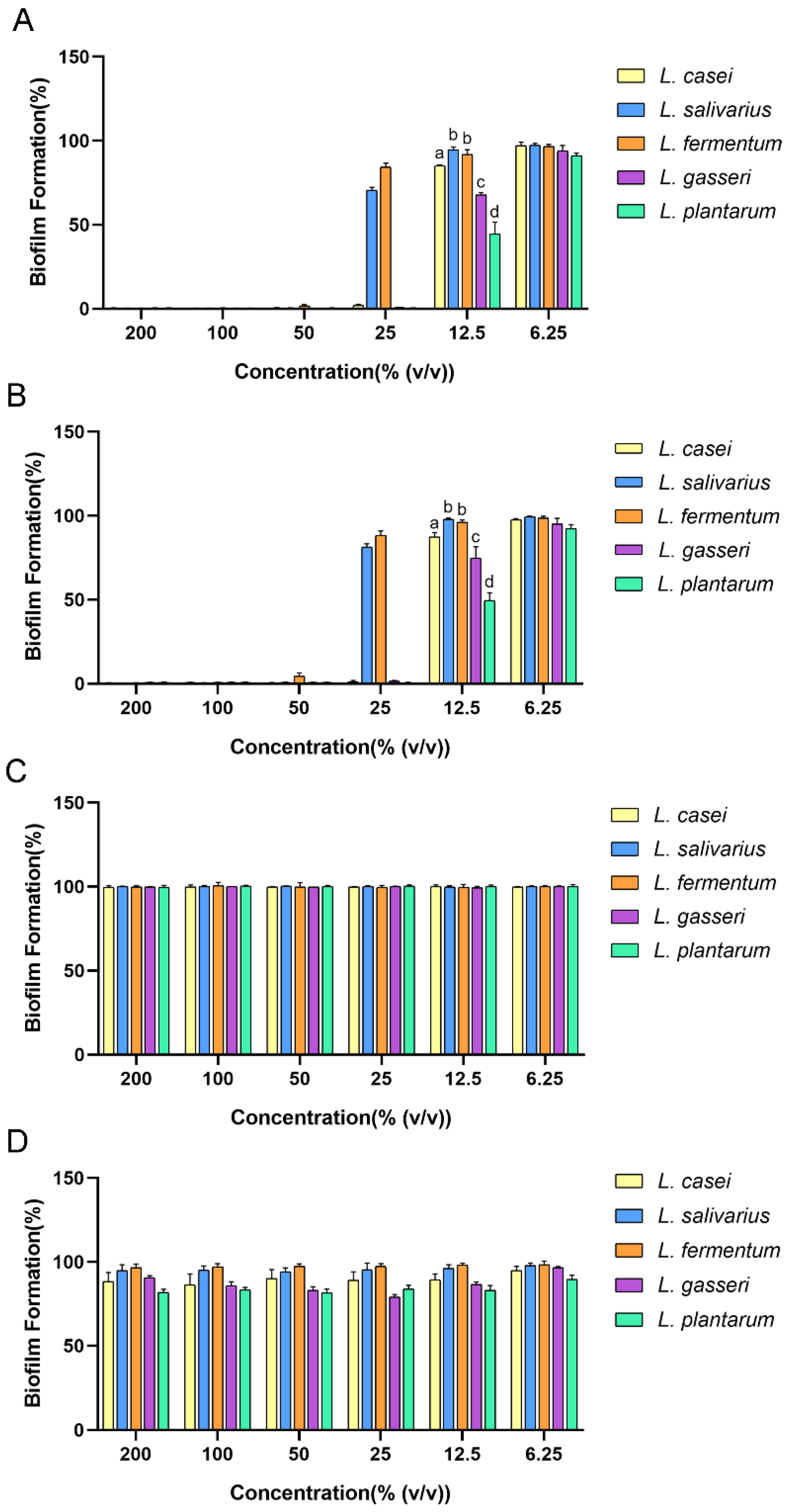
Figure 1. Antibiofilm effect of cell-free supernatant (CFS) of five Lactobacilli CFSs against Streptococcus mutans (S. mutans): (A) original CFS; (B) CFS treated with catalase; (C) CFS with pH adjusted to 6.5; (D) BHI with the same pH value as original CFS. The data are presented as the means ± SD. Different superscript letters for different values denote statistically significant differences (p ≤ 0.05).
Ijms 24 01986 g001
Figure 2. Isolation of the main antibiofilm component of Lactobacillus plantarum (L. plantarum) CFS. (A) Antibiofilm effect of fractions of L. plantarum CFS with different molecular weights. (B) The effect of pH on the antibiofilm ability of the fraction weighing less than 10 kDa of L. plantarum CFS. Group A: the fraction weighing less than 10 kDa. Group B: the fraction weighing less than 10 kDa with pH adjusted to 6.5. Group C: the fraction weighing less than 10 kDa with pH adjusted to 6.5 and then returned to the original value. (C) High-performance liquid chromatography (HPLC) chromatogram of Fr. 1-1-4. Arrows indicate the peak fractions, with the results of crystal violet staining above. (D) Antibiofilm effect of the six isolated fractions (Frs. 1-1-4-1~1-1-4-6). The data are presented as the means ± SD. Different superscript letters for different values denote statistically significant differences (p ≤ 0.05).
Ijms 24 01986 g002
Figure 3. Antibiofilm effect of component 1-1-4-3. (AC) Confocal laser scanning microscopy (CLSM) images of biofilm of S. mutans in the absence and presence of 1-1-4-3: (A) Green signal indicates microorganisms, whereas red signal indicates exopolysaccharides (EPSs). (B) Green signal represents live bacteria and red represents dead bacteria; (C) 3D structure of the biofilm. Scale bar: 10 µm. (D) Effect of dose on the antibiofilm ability of 1-1-4-3. (E,F) Total biomass and biofilm average thickness of S. mutans biofilm in 1-1-4-3 and control group. (G) Scanning electron microscopy (SEM) images of biofilm of S. mutans in the absence and presence of 1-1-4-3. Microstructure was observed and red boxes at low magnification were enlarged to a higher magnification. Different superscript letters for different values denote statistically significant differences (p ≤ 0.05).
Ijms 24 01986 g003
Figure 4. Characterization and validation of component 1-1-4-3. (A) The1H–1H COSY (blue bold) and key HMBC (red arrow) correlations of 1-1-4-3. (B~E) HPLC spectrum of lactic acid (LA) (B), valine (C), mixture of LA and valine (D), and 1-1-4-3 (E).
Ijms 24 01986 g004
Figure 5. Verification of the role of LA in antibiofilm activity. (A) Antibiofilm effect of 1-1-4-3, valine (d/l-), LA (d/l-) and the mixture of valine and LA. (B) Antibiofilm effect of LA and BHIS at the same pH as LA at each concentration. The data are presented as the means ± SD. Different superscript letters for different values denote statistically significant differences (p ≤ 0.05).
Ijms 24 01986 g005
Table 1. NMR spectroscopic data of component 1-1-4-3, LA + valine, LA, and valine in DMSO-d6 at 600 (1H) and 150 (13C) MHz.
Position1-1-4-3LA + ValineLAValine
δC, TypeδH, Mult. (J in Hz)δC, TypeδH, Mult. (J in Hz)δC, TypeδH, Mult. (J in Hz)δC, TypeδH, Mult. (J in Hz)
121.0, CH31.22, d (6.9)20.9, CH31.23, d (6.9)20.9, CH31.23, d (6.8)
266.3, CH4.02, q (6.8)66.2, CH4.03, q (6.8)66.3, CH4.04, q (6.8)
3176.9, C 176.8, C 176.8, C
1′18.1, CH30.89, d (7.0)18.2, CH30.91, d (7.0) 18.0, CH30.87, d (7.0)
2′29.5, CH2.16–2.09, m29.5, CH2.17–2.10, m 29.5, CH2.19–2.06, m
3′19.3, CH30.93, d (7.0)19.0, CH30.94, d (7.0) 19.5, CH30.91, d (7.0)
4′59.9, CH3.12, d (3.6)59.4, CH3.28, d (3.9) 60.2, CH3.00, d (3.5)
5′170.0, C 170.3, C 170.4, C
Disclaimer/Publisher’s Note: The statements, opinions and data contained in all publications are solely those of the individual author(s) and contributor(s) and not of MDPI and/or the editor(s). MDPI and/or the editor(s) disclaim responsibility for any injury to people or property resulting from any ideas, methods, instructions or products referred to in the content.