Article

Genome-Wide Meta-Analysis Identifies Multiple Novel Rare Variants to Predict Common Human Infectious Diseases Risk

by
Andrea Gelemanović
1,†,
Tatjana Ćatipović Ardalić
2,†,
Ajka Pribisalić
1,
Caroline Hayward
3,
Ivana Kolčić
1,4 and
Ozren Polašek
1,4,*
1
Department of Public Health, University of Split School of Medicine, 21000 Split, Croatia
2
Department of Pediatrics, University Hospital of Split, 21000 Split, Croatia
3
Medical Research Council Human Genetics Unit, Institute of Genetics and Cancer, University of Edinburgh, Western General Hospital, Edinburgh EH4 2XU, UK
4
Department of General Courses, Algebra University College, 10000 Zagreb, Croatia
*
Author to whom correspondence should be addressed.
These authors contributed equally to this work.
Int. J. Mol. Sci. 2023, 24(8), 7006; https://doi.org/10.3390/ijms24087006
Submission received: 20 March 2023 / Revised: 2 April 2023 / Accepted: 5 April 2023 / Published: 10 April 2023

Abstract

:
Infectious diseases still threaten global human health, and host genetic factors have been indicated as determining risk factors for observed variations in disease susceptibility, severity, and outcome. We performed a genome-wide meta-analysis on 4624 subjects from the 10,001 Dalmatians cohort, with 14 infection-related traits. Despite a rather small number of cases in some instances, we detected 29 infection-related genetic associations, mostly belonging to rare variants. Notably, the list included the genes CD28, INPP5D, ITPKB, MACROD2, and RSF1, all of which have known roles in the immune response. Expanding our knowledge on rare variants could contribute to the development of genetic panels that could assist in predicting an individual’s life-long susceptibility to major infectious diseases. In addition, longitudinal biobanks are an interesting source of information for identifying the host genetic variants involved in infectious disease susceptibility and severity. Since infectious diseases continue to act as a selective pressure on our genomes, there is a constant need for a large consortium of biobanks with access to genetic and environmental data to further elucidate the complex mechanisms behind host–pathogen interactions and infectious disease susceptibility.

1. Introduction

Despite the extensive research on infectious diseases, we still face major limitations in understanding their pathogenesis. Uncertainties arise from a pathogen’s adaptability to modify or adapt to a new environment, different mechanisms of host–pathogen interactions, host genetics and possibly even random effects arising from interactions between a host and pathogen [1,2,3]. Recent studies often focus on the host’s genetic profile, which is suggested to be a determining factor in variations in disease occurrence and treatment outcome [4].
Previous studies often used candidate genes that are involved in the innate or adaptive immune response and play a role in a spectrum of infectious disease-associated outcomes [5]. Such studies achieved numerous positive results, with IL4, TLR2 and CCL5 validated in a large-scale systematic review and meta-analysis of respiratory infectious diseases [6]. The same study suggested numerous methodological barriers present in primary studies, suggesting that improvements are needed before translatable knowledge can be developed [6].
One frequently used study design in this situation is a hypothesis-free genome-wide association study (GWAS). The advantage of this design includes the possibility of discovering genes that were not previously implicated in disease pathogenesis, and thus understanding a much broader set of predictors for infectious diseases [7,8,9]. This study design can be especially effective in isolated populations, where allele frequencies might have drifted [10,11], as well as in the meta-analytic design [12,13], where the analysis of rare variants can also help to provide a better understanding of disease mechanisms [14]. Although the most common GWAS approach is still a univariate model where a single phenotype is associated with genetic variants, multivariate GWAS approaches that rely on associating several highly correlated phenotypes with genetic variants are recently gaining attention and could be used in addition to univariate models to increase their power [15,16,17]. In addition, to better understand better a particular genetic variant carries the risk for disease prediction, instead of the traditional GWAS approach, which focuses on each single genetic variant, there is an increased trend toward the use of various machine learning approaches, which is well suited for high-dimensional data and allows for the detection of the epistatic or non-linear effects among genetic variants with a particular phenotype [18]. However, there are only a few univariate GWA studies available for bacterial and viral infectious diseases, most of which are for tuberculosis, with modest replications between them [6,19,20,21,22,23,24,25,26,27]. The emergence of COVID-19 prompted even more research interest [28,29], promising possible new diagnostic and treatment options based on these discoveries [30,31,32,33]. Therefore, the aim of this study was to apply genome-wide meta-analysis to gain insight into bacterial and viral infectious disease pathogenesis and the possible role of host genetics in disease development.

2. Results

A total of 4624 participants were included in this study. No specific inclusion or exclusion criteria were established, except for age > 18 years. A substantial number of differences between the sub-cohorts were observed, both in the infection-related traits and in confounding variables, e.g., there were more women in the Korčula sub-cohort, slightly younger participants in the Split sub-cohort and participants in the Vis sub-cohort had in general a lower socioeconomic status and fewer years of schooling (Table 1). Due to the observed differences, demographic characteristics were used as confounding variables in the GWAS models.
The genome-wide meta-analysis of all three sub-cohorts showed 28 Bonferroni corrected genome-wide significant associations (p < 3.57 × 10⁻⁹) with hepatitis (six loci), meningitis (eight loci), systemic infections (seven loci) and tuberculosis (seven loci), while one locus showed suggestive association (p < 5 × 10⁻⁸) with pneumonia (Table 2). Manhattan and QQ plots for these traits are shown in Figure S1, and the regional LD plots for each identified locus are shown in Figure S2.
The identified variants explained only a small proportion of the phenotype variance, ranging from 0.058% for hepatitis to 0.113% for meningitis (Table S6). Out of these identified variants, 16 (55%) are rare variants with an MAF below 1%, and we show the number of individuals with certain genotypes in Table S1. Some of the associations may be misleading, as there are a few situations where the controls have both rare alleles and some cases have none (minor homozygotes, e.g., rs78111295 (FAT4), rs116306652 (CACNA1E), rs116886525 (APOA5), rs17587821 (ITPKB), rs117768315 (PCED1B) and rs570545343 (NLN)).
Gene descriptions and their involvement in biological pathways are described in Tables S2 and S3, respectively, highlighting the transcriptome-associated pathways as the most significantly enriched pathways with systemic infections and cell cycle processes for hepatitis. In addition, these genes seem to have a role in the immune response or other cell functions related to infection susceptibility. We also checked for the association of identified loci with other traits from previously published GWA analyses, and the results are shown in Table S4, where these SNPs have been implicated in various other complex traits and diseases (e.g., atopic dermatitis, rheumatoid arthritis, Alzheimer’s disease, coronary artery disease and bone mineral density). A graphic summary of the significantly associated loci with functional enrichment and association with other complex traits is depicted in Figure 1. At the same time, detailed protein–protein interaction (PPI) partners are shown in Figure S3 for each infection-related trait for which significant GWAS loci were identified.
We investigated whether any of the previously implied variants from a meta-analysis of candidate gene studies or GWA studies on susceptibility to various bacterial and viral infectious diseases were replicated in our analyses. COVID-19 studies were excluded for this stage of the analysis, considering that for the purpose of this study, we did not have information on the COVID-19 status of the participants. The replication results are summarized in Table S5, where we show all the replicated variants with a p-value below 0.05.
We did not detect any significant results for the self-reported common cold or influenza frequency, probably due to the relatively small sample sizes (Table S7).
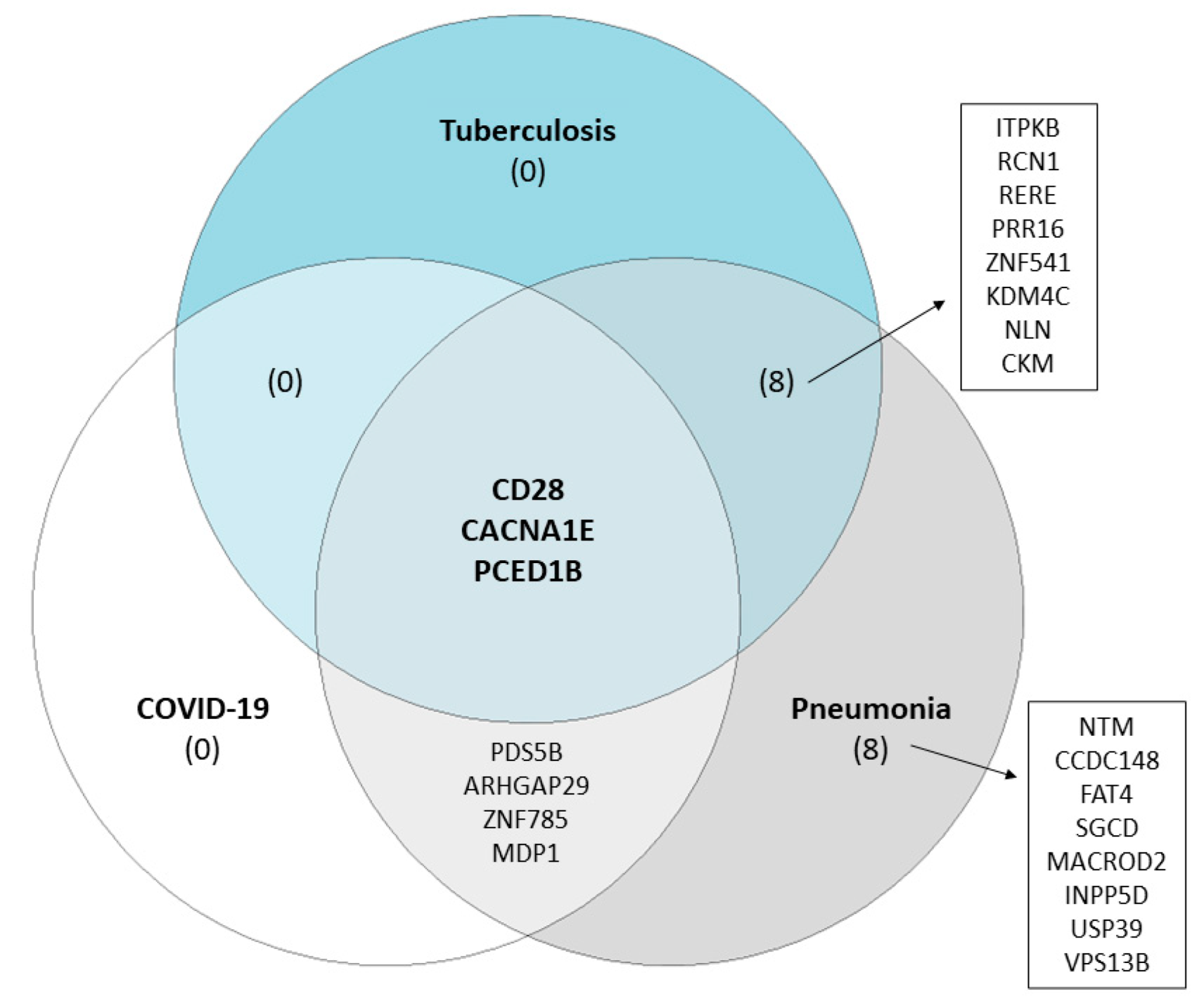
To evaluate the potential effect of these candidate rare variants, we first searched the GEO database to find publicly available RNA-seq datasets to identify differentially expressed genes (DEGs) in any of our infectious diseases for which we identified candidate SNPs—hepatitis, meningitis, tuberculosis or pneumonia. Only two RNA-seq datasets were identified as relevant based on our search criteria under the accession numbers GSE196399 (patients with severe community-acquired pneumonia vs healthy controls) and GSE94438 (tuberculosis patients vs household contacts). We supplemented this with an additional RNA-seq dataset focusing on transcriptome changes in COVID-19 patients (GSE223885). Of the 29 candidate GWAS loci, 23 were identified as DEGs in pneumonia cases, 11 in tuberculosis cases, and 7 in COVID-19 cases (Table S8). Only three genes overlap between the three studies, with CACNA1E being overexpressed, whereas CD28 and PCED1B had lower expression levels in patients with infectious disease (Figure 2). As none of the candidate GWAS variants showed eQTL in whole blood based on the GTEx data, we carried out evaluations at the gene level and identified several other SNPs in the CACNA1E, CD28 and PCED1B gene, all demonstrating lower expression in individuals with minor homozygote genotypes.

3. Discussion

The results of this study suggest that GWAS in biobanks may serve as a potential approach for hypothesis generation in infectious disease susceptibility. Furthermore, these results contribute to the hypothesis that the host genetic susceptibility to bacterial and viral infections in adults is polygenic and comprises common variants with low explained variance and/or “unfortunate” combinations of multiple rare variants [4,34].
Despite the small number of cases, we found several significant associations with infectious disease susceptibility, where pathway analysis revealed significant enrichment in genes and pathways involved in immune response as expected, but also in genes outside of the immune system. Most genes associated with hepatitis have been previously implicated in the pathogenesis of various liver diseases. For example, TGIF1 (TGFB-induced factor homeobox 1) is shown to be up-regulated in patients with hepatitis C virus infection [35,36], but down-regulated in fibrotic liver patients [37]. A study of patients with hepatitis B virus (HBV)-associated hepatocellular carcinoma showed that the FAT4 (FAT atypical cadherin 4) gene might act as a tumor suppressor gene, which is inactivated in cases with hepatocellular carcinoma, but also in various other human cancers [38,39]. Although the results should be taken with care, one study showed the association of hepatitis with gastric cancer [40], and the link between the two might involve the Rho GTPase activator ARHGAP29, which has been previously associated with gastric cancer [41], and associated with hepatitis in this study. Most interestingly, hepadnavirus integrates into the host genome within the gene NTM (neurotrimin) 1 h after infection [42].
Several genes that were significantly associated with meningitis in this study are involved in various neurological disorders, which is in line with ongoing and controversial studies showing that various bacterial and viral pathogens are risk factors for some neurodegenerative diseases, such as Alzheimer’s disease, Parkinson’s disease, multiple sclerosis, amyotrophic lateral sclerosis, and autism spectrum disorders [43,44,45]. Here, we report APOA5 to be associated with meningitis, while previous studies implicated various other apolipoproteins, such as APOA1 and APOE, are involved in meningitis [46,47], as well as various bacterial or respiratory infections [47,48,49], both in human and mouse studies. Additionally, the APOA1–APOC3–APOA4 gene cluster has been associated with the risk of Alzheimer’s disease [50]. CACNA1E, as identified in this study, and other voltage-gated calcium channel subunit genes have been previously associated with various neuropsychiatric disorders, such as schizophrenia, autism, and bipolar disorder [51], but also linked to the development of cortical lesions in patients with multiple sclerosis [52]. Previously performed GWA studies on other common traits also showed a significant association of the loci identified in this study with Alzheimer’s disease and schizophrenia (Table S4). We also show the association of meningitis with two genes with well-known functions in the immune response. CD28, a component of the immune system that is involved in T-cell activation, induction of cell proliferation and cytokine production and promotion of T-cell survival, was used in several animal and in vitro studies to show that CD28-deficient mice develop experimental autoimmune meningitis [53] or that the blockade of CD28 improves experimental autoimmune encephalomyelitis [54]. ITPKB (inositol-trisphosphate 3-kinase B), a well-known gene associated with the immune response, showed in animal studies its significant role in the thymocyte differentiation function of peripheral T-cells [55,56]. Functional enrichment analysis of the loci in this study associated with meningitis also showed significant enrichment in the immune response network and pathways associated with T-cell signaling (Table S3).
A single gene associated with pneumonia was APOBEC1, a member of the cytidine deaminases family, which was previously reported to play a central role in innate and adaptive immunity interplay [57], or in the delayed development of pneumocystis pneumonia in AIDS patients [58] and clearance of pneumococcal pneumonia [59].
We also identified several genes that might contribute to susceptibility to systemic infections in this study, most of which seem to have a distinct role in the immune response. RSF1 (human remodeling and spacing factor (1)) was shown to interact with SP100, which is known to have an important role in regulating the immune response to intracellular pathogens [60]. In addition, one study showed that the HBV viral protein pX interacts with RSF1 and as a result, HBV transcription is increased [61], suggesting that this gene might have a role in the life cycle of viruses whose expression is regulated at the transcription level. The role of MACROD2 (MACRO domain containing (2)) could be explained through its interaction with ARTD10, which is inducible by inflammatory and immunogenic stimuli and enhances the NF-κβ signaling pathway to increase inflammation, the innate immune response, cell survival and proliferation [62]. INPPD5, also known as SHIP1, was previously analyzed in numerous studies, and is also implicated to have a role in immune response [63]. The role of this gene is closely linked to IL10 and several important pathways [64], resulting in modifying effects in the case of the clearance of S. aureus and S. pneumoniae [65], as well as in the pathogenesis of Pseudomonas aeruginosa [66], Epstein–Barr virus [67], cytomegalovirus [68,69,70], or even immunity to helminth infection in mice [71].
We found a significant association between tuberculosis and KDM4C, a lysine demethylase that was also shown to be significantly associated with non-small cell lung carcinoma [72]. Another gene associated with the risk of tuberculosis was MDP1, magnesium-dependent phosphatase 1, which has also been proposed to have a role in gastric cancer [73]. This could be part of the larger pathway that explains the relationship between tuberculosis and carcinogenesis involving the CCL7-CCL2-CCR2 axis, which has a role in the immune response, tumor regulation and the WT1 gene associated with the susceptibility to tuberculosis [6]. In line with this, the KEGG pathway revealed significant enrichment in the viral carcinogenesis pathway (Table S3). Lastly, we found that PCED1B is associated with tuberculosis, in line with another recent study [74].
As most of the significantly associated variants identified in this study are rare, this finding could lead to the prediction of individualized infection disease risk based on the accumulation of such rare variants. Infections are amongst the strongest known selective pressures on our genomes [75], acting as a purifying selective pressure during childhood and historically removing the most susceptible individuals from the population [76]. On the contrary, recent generations are exposed to mandatory vaccinations, improved sanitary conditions, better pathogen control and more effective clinical treatment, so the disease-associated deleterious variants are retained and accumulate in populations. Under the assumption that the (re-)emerging epidemics will utilize similar susceptibility and pathogenesis mechanisms, identifying rare variants with strong clinical effects could become an invaluable tool for predicting an individual’s risk of being infected with specific pathogens [77].
The results of this study were obtained based on very small sample sizes for each disease in a very heterogeneous population sample, thus requiring replication in independent populations. This could partially be explained by the response bias to the voluntary participation in the 10,001 Dalmatians biobank. However, the differences might also emerge because the two sub-cohorts are isolated island populations, while the third cohort is a more heterogeneous population on the mainland. Therefore, we consider this report the initial stage for a larger scale, cross-ethnical meta-analysis to measure the effects of these and other rare variants associated with infectious disease risk. Besides the small sample size representing a major limitation, this study may also suffer from the imprecision of the traits, which were registered based on clinical examinations and hospital admittance records, or via surveys and thus relying on patient memory. The use of electronic health records with diagnosed and validated pathogens is a possible step forward, especially in biobanks that have good links with the clinical records, in order to overcome the major issues of genetic studies in the field of infectious disease susceptibility regarding sample size, harmonization of study protocols, patient characterization and improvement in the use of appropriate analytic methods [6]. Due to the pathophysiological differences between bacterial and viral infections and the limitations mentioned above (small sample sizes and trait imprecision), we opted not to include multivariate GWAS models in this study. However, future studies should build upon these initial efforts, and larger biobanks should incorporate both univariate and multivariate GWAS approaches to progress our understanding of the role of host genetic factors in host–pathogen interaction. In addition, the mechanisms described here may be population-specific, thus having a limited role in the general population. These population-specific differences are already noticeable in the demographic characteristics, with significant differences among cohorts. Due to the nature of isolated island populations, there may also be a genetic difference, which might represent bias; however, we tried to overcome this by using a meta-analysis approach and a more stringent adjusted p-value threshold. Nevertheless, some of the obtained rare variants in this study may provide interesting targets for future functional studies if replicated in other populations with larger sample sizes and clinically verified traits.
A potential way forward in understanding the host genetics involvement in infectious diseases susceptibility and pathophysiology could be the continuous collection of samples and data in observational studies, which could build upon the existing initiatives across Europe and elsewhere. Although the COVID-19 pandemic showed us that we are still far from fully understanding complex host–pathogen interactions, elucidating the involvement of host factors in infectious disease pathogenesis should be labelled as a high research priority, aiming to contribute to a better understanding and a possibility for the quicker translation of knowledge into clinical care.

4. Materials and Methods

4.1. Study Populations

This study is based on the Croatian biobank 10,001 Dalmatians [78,79], the most comprehensive research resource for investigating genetic, environmental and social determinants of health and disease in Croatia. The study included various measurements with over 250 disease-related quantitative traits, including medical examination, anthropometry, clinically relevant analyses, cognition, smell, taste and hearing thresholds, diet, and general lifestyle. Here, we used the data from three sub-cohorts for which we had bacterial and viral infections disease data—Korčula (N = 2833), Vis (N = 1039) and Split (N = 1012). While the first two are isolated island populations, the third originates from the second-largest city in Croatia. The data were collected in several phases, starting in 2003 up to 2014. Additionally, a follow-up postal survey was sent to participants from the sub-cohorts of Korčula and Vis in 2016, with 1082 responses (28% response rate).
The project has been approved by the Ethical Boards of the Medical Schools at the University of Zagreb and the University of Split. All participants were given information on the study aims and goals and provided informed consent before entering the study. In addition, all the methods and activities related to human subjects were performed following the relevant guidelines. Because the study took place in isolated island communities, where the identification of subjects might be easier than in the general population, a stricter personal information protection plan was utilized. This meant that we devised separate collection, storage, and management protocols of all the personal records, with exclusive access to these data only granted to the PI, who enrolled all the participants and had exclusive access to these data.

4.2. Genotyping and SNP Imputation

DNA was extracted from venous blood leukocytes using Nucleon BACC3 kits (Tepnel, Manchester, UK). Genotyping employed Illumina HumanHap300 v1 (Vis: 317,509 single-nucleotide polymorphisms, SNPs) and Illumina HumanHap370CNV-Quad (Korčula phase 1: 346,034 SNPs, Korčula phase 2: 719,487 SNPs; Split phase 1: 320,406 SNPs, Split phase 2: 646,888 SNPs). Illumina GenomeStudio Software v3.0 (Vis, Korčula) or v3.1 (Split) was used for genotype calling. Participants with less than 95% (Vis) or 97% (Korčula, Split) genotyping rates were removed from further analysis. SNPs with a call rate below 98%, minor allele frequency (MAF) below 1% and 0.01% for Exome Chip Markers (Korčula, Split) and p-value for Fisher’s exact test of the Hardy–Weinberg equilibrium (HWE) below 10–6 were also removed to remove low-quality SNPs. In addition, samples with excessive autosomal heterozygosity or gender inconsistency (based on the sex chromosomes genotypes) were also removed. This left us with a final sample size of 960 in Vis, 2700 in Korčula and 966 in Split, contributing to the total sample size of 4624 participants in this study.
Genotype data were phased using SHAPEIT v2.r873 [80] and the duohmm function [81], and imputed to the reference panel HRC v1.1 [82] using the Sanger Imputation Service. Both the phasing and imputation steps were performed separately for each genotyping platform and sub-cohort. SNPs with an imputation quality score (INFO) below 0.4 and monomorphic variants were excluded, leaving 12,468,939 SNPs for Vis, 12,382,856 SNPs for Korčula and 11,400,586 SNPs for Split available for downstream analysis.

4.3. Trait Definition

A total of 14 traits were used in these analyses, defined through an extensive questionnaire that catalogued the participants’ history of diseases and hospitalizations and the available medical records on these events. The following three groups of variables were identified: binary variables, binary-derived variables, and ordinal variables. The binary group was based on specific diagnoses in the subjects’ medical histories. The diagnoses investigated included tuberculosis, pneumonia, hepatitis, or meningitis. The second group consisted of derived traits that were of a broader scope, including various respiratory infections (pooled pneumonia and tuberculosis), gastrointestinal infections (pooled hepatitis and other gastrointestinal diseases), systemic infections, bacterial infections (respiratory infections and bacterial meningitis), and viral infections (hepatitis and viral meningitis). We also included appendectomy and tonsillectomy in the second group, as they are associated with infection-related traits.
The final group consists of an estimate of the infectious burden and two traits added in the follow-up survey—annual common cold frequency and influenza frequency over the last ten years. The infectious burden was defined as an individual’s burden of infectious diseases up to the date of the questionnaire and was calculated for each participant (cases coded as 1, controls coded as 0) using the following equation:
Infectious burden = (tuberculosis × 1) + (pneumonia × 1) + (hepatitis × 1) + (meningitis × 1) + (respiratory infections (tuberculosis and pneumonia cases excluded) × 1) + (gastrointestinal infections (hepatitis cases excluded) × 1) + (systemic infections × 1) + (appendectomy × 0.5) + (tonsillectomy × 0.5)
The cases of appendectomy and tonsillectomy were multiplied by 0.5, as their origin can be very heterogeneous. If a subject had the same infectious disease two or more times, their infectious burden score was multiplied by 2, to give more strength to individuals with recurrent infections, which were assumed to happen in highly susceptible individuals. The total score of infectious burden divided participants into groups coded from 0 to 4. Most participants had none or one infectious disease event, while a small number had two or more different infectious diseases during their life. Cold and influenza frequency divided participants into groups coded from 0 to 3 based on the following possible answers of having a cold/influenza: several times per year/every year in the last ten years, once a year/several times in the last ten years, less than once a year/once in the last ten years, never or almost never/never.

4.4. Statistical Analyses

4.4.1. Genome-Wide Association Analyses

Each infection-related trait was firstly adjusted for age, gender, the first three principal components, years of schooling (ranging from 0 to 26, where elementary school is defined as 0–8, high school as 9–12, and higher education and university as higher than 13 years), socioeconomic status (measured as the total sum of the items a subject had in possession, such as plumbing, heating, phone, computer, etc., ranging from 0 to 16), number of reported different infections (in order to penalize for multiple infections), and for all respiratory infection traits (pneumonia, respiratory infections, cold frequency and influenza frequency), we additionally adjusted the model to penalize for recurrent pneumonia cases. The variables years of schooling and material status were transformed into categorical variables (3 categories for years of schooling and 4 categories for material status), in line with our previous studies [83,84,85]. Missing data were imputed based on the median value depending on the cohort, gender, and age group. For the latter purpose, participants were divided into the following age groups: 18–39, 40–64, and ≥65. To account for the population structure and familial relatedness, we calculated the kinship matrix using the ibs function from the GenABEL R package [86].
We used a logistic regression model for binary and binary-derived variables and a linear regression model for ordinal variables [87]. As the analyses were performed on imputed genetic data, we used imputed allelic dosages. All the models assumed an additive allelic effect, and the environmental residuals used for the association analysis were derived from the polygenic function in the GenABEL R package [86]. All association analyses were performed using RegScan v0.2 software [88]. Summary statistics (effect sizes, standard errors, and p-values) of each sub-cohorts were adjusted by the genomic control method based on GRAMMAR-gamma factors [89] (genomic inflation factor lambda and unbiased estimate of the regression coefficient) from the polygenic function in the GenABEL R package in order to account for the relatedness and population stratification.

4.4.2. Meta-Analyses

Before meta-analysis, GWAS summary statistics were cleaned using the standardized QC protocol for EasyQC software v9.2 [90]. SNPs with mismatching alleles across all three sub-cohorts were excluded. We performed a fixed effect inverse-variance meta-analysis using METAL software version 2011-03-25 [91]. The genome-wide significance threshold was defined as 3.57 × 10⁻⁹ (standardized GWA threshold 5 × 10⁻⁸ divided by the number of traits analyzed) after Bonferroni correction for testing several infection-related traits, as some are highly correlated (e.g., respiratory infections and pneumonia), while the suggestive threshold was set to 5 × 10⁻⁸.

4.4.3. Proportion of Phenotypic Variance Explained by SNPs

This was calculated for each of the identified SNPs according to the formula below (β is the β coefficient, MAF is the minor allele frequency and phvar is the phenotype variance) [92].
Phenotypic variance = β2 × ((2 × MAF × (1 − MAF))/phvar)
Phenotype variance was calculated based on the sample residuals adjusted for covariates. To obtain the total proportion of phenotypic variance explained by all the identified SNPs for each infection-related trait, the individual variance in each SNP was summed.

4.4.4. SNP Function Annotation

SNPs within a 500 kb window around the leading SNP were extracted to annotate the region. Several tools were used, including the online tool HaploReg v4.1 (http://archive.broadinstitute.org/mammals/haploreg/haploreg.php accessed on 1 September 2022) [93] and Bioconductor biomaRt R package v2.36.1 [94,95].
To check whether the leading significant SNPs were previously associated with unrelated traits, we used the online tool PhenoScanner v1.1 (http://www.phenoscanner.medschl.cam.ac.uk/ accessed on 1 September 2022) [96] to examine the published GWAS associations. We reported the identified associations using a p-value threshold of 0.05 and queried for proxy SNPs with an r2 of 1.

4.4.5. Pathway Analysis

We performed pathway analysis using the online tool STRING v10.5 (https://string-db.org/ accessed on 1 March 2023) [97] for significant infection-related traits using the genes associated with top leading SNPs. To create a network, the following default and suggested STRING settings were used: minimum required interaction score of medium confidence (0.4) and the maximum number of interactions used to limit the output to the ten best-scoring hits (both for direct and indirect interactions). We used functional enrichments for Gene Ontologies (GO) and Kegg pathways, also using STRING v10.5 with option analysis, and we reported the most significant pathways with an FDR below 1% and with a minimum of 5 genes from the network in the pathway. Networks were visualized using Cytoscape v3.7.2 [98] with GeneMANIA plugin v3.5.1 [99].

4.4.6. Validation with RNA-Seq Data

The Gene Expression Omnibus database (GEO; https://www.ncbi.nlm.nih.gov/geo/ accessed on 1 March 2023) was searched for relevant publicly available RNA-seq data using the following search criteria: (1) RNA-seq data performed on human whole-blood samples; (2) studies that investigated pneumonia, hepatitis, meningitis, or tuberculosis; (3) studies published in the last year; (4) minimum ten samples per group. In addition, we searched for RNA-seq studies on COVID-19 with the same restriction search criteria. Differential gene expression analyses were performed using R package DESeq2 v1.28.1 [100]. From each included RNA-seq study, raw counts were first pre-filtered to remove genes with less than 10 read counts, normalized via DESeq2 variance stabilizing transformation and differentially expressed genes (DEGs) were identified if the false discovery rate (FDR) adjusted p-value was less than 0.05. Finally, the GTEx database (https://gtexportal.org/home/ accessed on 5 March 2023) was used to identify the potential eQTLs in whole blood.

5. Conclusions

This study identified several novel rare variants associated with susceptibility to hepatitis, meningitis, tuberculosis, and systemic infections. In turn, this demonstrates that biobanks can present a valuable resource for host genetic studies in identifying susceptibility variants to infectious diseases, despite imprecise traits and small sample sizes, which were also the main limitations of this study. Thus, we propose creating a large consortium of biobanks with access to genetic data and data on infectious diseases, ideally linked with electronic health records, to develop an extensive resource for investigating host genetics as a risk factor for susceptibility to infections. Data collection and identification of possible bias and confounders in various cohorts should be standardized across the consortium to allow for proper future analysis, and more emphasis should be given on the identification of appropriate controls (e.g., susceptible but unexposed controls, controls with undiagnosed, latent or mild infection and information on vaccination status). A harmonized consortium could identify highly penetrant rare variants that have accumulated in the modern human population. In conclusion, these efforts could elucidate a fine interplay between monogenic and polygenic effects and develop a new frontier for understanding and predicting emerging infections and epidemics.

Supplementary Materials

The following supporting information can be downloaded at: https://www.mdpi.com/article/10.3390/ijms24087006/s1.

Author Contributions

A.G. and O.P. conceived the study idea; T.Ć.A., C.H., I.K. and A.P. further developed the idea and participated in the study design and data collection; A.G. performed the data analysis; A.G., T.Ć.A. and O.P. drafted the manuscript; all authors reviewed, edited, and approved the final manuscript version. All authors have read and agreed to the published version of the manuscript.

Funding

This research was funded by the Medical Research Council UK, the FP7 project PREPARE (602525), the Croatian National Centre of Research Excellence in Personalized Healthcare grant (number KK.01.1.1.01.0010), and the Centre of Competence in Molecular Diagnostics (KK.01.2.2.03.0006). CH was supported by an MRC Human Genetics Unit programme grant ‘Quantitative traits in health and disease’ (U. MC_UU_00007/10). Funding bodies had no influence on the collection, analysis, or interpretation of the data, writing or decision where to submit the manuscript for publication.

Institutional Review Board Statement

The study was conducted in accordance with the Declaration of Helsinki, and approved by the Ethical Boards of the Medical Schools at the University of Zagreb and the University of Split.

Informed Consent Statement

All participants were given information on the study aims and goals, and provided informed consent prior to entering the study. In addition, all methods and activities related to human subjects were performed in accordance with the relevant guidelines. Because the study took place in the isolated island communities, where the identification of subjects might be easier than in the general population, a stricter personal information protection plan was utilized. This meant that we devised completely separate collection and storage processes and management of all personal records, with exclusive access to these data only granted to the PI, who enrolled all the participants and had sole access to these data.

Data Availability Statement

The data analyzed during the current study are available upon reasonable requests to the corresponding author.

Conflicts of Interest

The authors declare no conflict of interest.

References

  1. Fauci, A.S.; Morens, D.M. The Perpetual Challenge of Infectious Diseases. N. Engl. J. Med. 2012, 366, 454–461. [Google Scholar] [CrossRef] [PubMed] [Green Version]
  2. Cohen, M. Changing patterns of infectious disease. Nature 2000, 406, 762–767. [Google Scholar] [CrossRef]
  3. Karlsson, E.K.; Kwiatkowski, D.P.; Sabeti, P.C. Natural selection and infectious disease in human populations. Nat. Rev. Genet. 2014, 15, 379–393. [Google Scholar] [CrossRef] [PubMed]
  4. Casanova, J.L.; Abel, L. The genetic theory of infectious diseases: A brief history and selected illustrations. Annu. Rev. Genom. Hum. Genet. 2013, 14, 215–243. [Google Scholar] [CrossRef] [Green Version]
  5. Burgner, D.; Jamieson, S.E.; Blackwell, J.M. Genetic susceptibility to infectious diseases: Big is beautiful, but will bigger be even better? Lancet Infect. Dis. 2006, 6, 653–663. [Google Scholar] [CrossRef] [Green Version]
  6. Patarcic, I.; Gelemanovic, A.; Kirin, M.; Kolcic, I.; Theodoratou, E.; Baillie, K.J.; de Jong, M.D.; Rudan, I.; Campbell, H.; Polasek, O. The role of host genetic factors in respiratory tract infectious diseases: Systematic review, meta-analyses and field synopsis. Sci. Rep. 2015, 5, 16119. [Google Scholar] [CrossRef] [PubMed] [Green Version]
  7. Chimusa, E.R.; Zaitlen, N.; Daya, M.; Moller, M.; van Helden, P.D.; Mulder, N.J.; Price, A.L.; Hoal, E.G. Genome-wide association study of ancestry-specific TB risk in the South African Coloured population. Hum. Mol. Genet. 2014, 23, 796–809. [Google Scholar] [CrossRef] [Green Version]
  8. Thye, T.; Owusu-Dabo, E.; Vannberg, F.O.; van Crevel, R.; Curtis, J.; Sahiratmadja, E.; Balabanova, Y.; Ehmen, C.; Muntau, B.; Ruge, G.; et al. Common variants at 11p13 are associated with susceptibility to tuberculosis. Nat. Genet. 2012, 44, 257–259. [Google Scholar] [CrossRef] [Green Version]
  9. Thye, T.; Vannberg, F.O.; Wong, S.H.; Owusu-Dabo, E.; Osei, I.; Gyapong, J.; Sirugo, G.; Sisay-Joof, F.; Enimil, A.; Chinbuah, M.A.; et al. Genome-wide association analyses identifies a susceptibility locus for tuberculosis on chromosome 18q11.2. Nat. Genet. 2010, 42, 739–741. [Google Scholar] [CrossRef]
  10. Polasek, O. Future of biobanks—Bigger, longer, and more dimensional. Croat. Med. J. 2013, 54, 496–500. [Google Scholar] [CrossRef] [Green Version]
  11. Vitart, V.; Biloglav, Z.; Hayward, C.; Janicijevic, B.; Smolej-Narancic, N.; Barac, L.; Pericic, M.; Klaric, I.M.; Skaric-Juric, T.; Barbalic, M.; et al. 3000 years of solitude: Extreme differentiation in the island isolates of Dalmatia, Croatia. Eur. J. Hum. Genet. EJHG 2006, 14, 478–487. [Google Scholar] [CrossRef] [Green Version]
  12. Pattaro, C.; Kottgen, A.; Teumer, A.; Garnaas, M.; Boger, C.A.; Fuchsberger, C.; Olden, M.; Chen, M.H.; Tin, A.; Taliun, D.; et al. Genome-wide association and functional follow-up reveals new loci for kidney function. PLoS Genet. 2012, 8, e1002584. [Google Scholar] [CrossRef] [PubMed] [Green Version]
  13. Okada, Y.; Sim, X.; Go, M.J.; Wu, J.Y.; Gu, D.; Takeuchi, F.; Takahashi, A.; Maeda, S.; Tsunoda, T.; Chen, P.; et al. Meta-analysis identifies multiple loci associated with kidney function-related traits in east Asian populations. Nat. Genet. 2012, 44, 904–909. [Google Scholar] [CrossRef] [PubMed] [Green Version]
  14. Peloso, G.M.; Auer, P.L.; Bis, J.C.; Voorman, A.; Morrison, A.C.; Stitziel, N.O.; Brody, J.A.; Khetarpal, S.A.; Crosby, J.R.; Fornage, M.; et al. Association of low-frequency and rare coding-sequence variants with blood lipids and coronary heart disease in 56,000 whites and blacks. Am. J. Hum. Genet. 2014, 94, 223–232. [Google Scholar] [CrossRef] [PubMed] [Green Version]
  15. Ruotsalainen, S.E.; Partanen, J.J.; Cichonska, A.; Lin, J.; Benner, C.; Surakka, I.; Reeve, M.P.; Palta, P.; Salmi, M.; Jalkanen, S.; et al. An expanded analysis framework for multivariate GWAS connects inflammatory biomarkers to functional variants and disease. Eur. J. Hum. Genet. EJHG 2021, 29, 309–324. [Google Scholar] [CrossRef]
  16. Mallard, T.T.; Linner, R.K.; Grotzinger, A.D.; Sanchez-Roige, S.; Seidlitz, J.; Okbay, A.; de Vlaming, R.; Meddens, S.F.W.; Palmer, A.A.; Davis, L.K.; et al. Multivariate GWAS of psychiatric disorders and their cardinal symptoms reveal two dimensions of cross-cutting genetic liabilities. Cell Genom. 2022, 2, 100140. [Google Scholar] [CrossRef] [PubMed]
  17. Karlsson Linner, R.; Mallard, T.T.; Barr, P.B.; Sanchez-Roige, S.; Madole, J.W.; Driver, M.N.; Poore, H.E.; de Vlaming, R.; Grotzinger, A.D.; Tielbeek, J.J.; et al. Multivariate analysis of 1.5 million people identifies genetic associations with traits related to self-regulation and addiction. Nat. Neurosci. 2021, 24, 1367–1376. [Google Scholar] [CrossRef]
  18. Nicholls, H.L.; John, C.R.; Watson, D.S.; Munroe, P.B.; Barnes, M.R.; Cabrera, C.P. Reaching the End-Game for GWAS: Machine Learning Approaches for the Prioritization of Complex Disease Loci. Front. Genet. 2020, 11, 350. [Google Scholar] [CrossRef]
  19. Tian, C.; Hromatka, B.S.; Kiefer, A.K.; Eriksson, N.; Noble, S.M.; Tung, J.Y.; Hinds, D.A. Genome-wide association and HLA region fine-mapping studies identify susceptibility loci for multiple common infections. Nat. Commun. 2017, 8, 599. [Google Scholar] [CrossRef] [Green Version]
  20. Kleinstein, S.E.; Shea, P.R.; Allen, A.S.; Koelle, D.M.; Wald, A.; Goldstein, D.B. Genome-wide association study (GWAS) of human host factors influencing viral severity of herpes simplex virus type 2 (HSV-2). Genes Immun. 2019, 20, 112–120. [Google Scholar] [CrossRef]
  21. Moreau, K.; Clemenceau, A.; Le Moing, V.; Messika-Zeitoun, D.; Andersen, P.S.; Bruun, N.E.; Skov, R.L.; Couzon, F.; Bouchiat, C.; Erpelding, M.L.; et al. Human Genetic Susceptibility to Native Valve Staphylococcus aureus Endocarditis in Patients with S. aureus Bacteremia: Genome-Wide Association Study. Front. Microbiol. 2018, 9, 640. [Google Scholar] [CrossRef] [PubMed] [Green Version]
  22. Bhattacharyya, C.; Majumder, P.P.; Pandit, B. An exome wide association study of pulmonary tuberculosis patients and their asymptomatic household contacts. Infect. Genet. Evol. J. Mol. Epidemiol. Evol. Genet. Infect. Dis. 2019, 71, 76–81. [Google Scholar] [CrossRef] [PubMed]
  23. Luo, Y.; Suliman, S.; Asgari, S.; Amariuta, T.; Baglaenko, Y.; Martinez-Bonet, M.; Ishigaki, K.; Gutierrez-Arcelus, M.; Calderon, R.; Lecca, L.; et al. Early progression to active tuberculosis is a highly heritable trait driven by 3q23 in Peruvians. Nat. Commun. 2019, 10, 3765. [Google Scholar] [CrossRef] [PubMed] [Green Version]
  24. Liu, S.; Huang, S.; Chen, F.; Zhao, L.; Yuan, Y.; Francis, S.S.; Fang, L.; Li, Z.; Lin, L.; Liu, R.; et al. Genomic Analyses from Non-invasive Prenatal Testing Reveal Genetic Associations, Patterns of Viral Infections, and Chinese Population History. Cell 2018, 175, 347–359.e14. [Google Scholar] [CrossRef] [Green Version]
  25. Lees, J.A.; Ferwerda, B.; Kremer, P.H.C.; Wheeler, N.E.; Seron, M.V.; Croucher, N.J.; Gladstone, R.A.; Bootsma, H.J.; Rots, N.Y.; Wijmega-Monsuur, A.J.; et al. Joint sequencing of human and pathogen genomes reveals the genetics of pneumococcal meningitis. Nat. Commun. 2019, 10, 2176. [Google Scholar] [CrossRef] [Green Version]
  26. Omae, Y.; Toyo-Oka, L.; Yanai, H.; Nedsuwan, S.; Wattanapokayakit, S.; Satproedprai, N.; Smittipat, N.; Palittapongarnpim, P.; Sawanpanyalert, P.; Inunchot, W.; et al. Pathogen lineage-based genome-wide association study identified CD53 as susceptible locus in tuberculosis. J. Hum. Genet. 2017, 62, 1015–1022. [Google Scholar] [CrossRef]
  27. Zheng, R.; Li, Z.; He, F.; Liu, H.; Chen, J.; Xie, X.; Zhou, J.; Chen, H.; Wu, X.; Wu, J.; et al. Genome-wide association study identifies two risk loci for tuberculosis in Han Chinese. Nat. Commun. 2018, 9, 4072. [Google Scholar] [CrossRef] [Green Version]
  28. Rudan, I. Answering the initial 20 questions on COVID-19 (January–February 2020). J. Glob. Health 2020, 10, 010106. [Google Scholar] [CrossRef]
  29. Calisher, C.H. Good news or bad news? The coronavirus pandemic has sickened and killed only a relatively few people but has affected us all. Croat. Med. J. 2020, 61, 296–299. [Google Scholar] [CrossRef]
  30. Ghosh, D.; Bernstein, J.A.; Mersha, T.B. COVID-19 pandemic: The African paradox. J. Glob. Health 2020, 10, e020347. [Google Scholar] [CrossRef]
  31. Liu, S.; Shen, J.; Fang, S.; Li, K.; Liu, J.; Yang, L.; Hu, C.D.; Wan, J. Genetic Spectrum and Distinct Evolution Patterns of SARS-CoV-2. Front. Microbiol. 2020, 11, 593548. [Google Scholar] [CrossRef]
  32. Anastassopoulou, C.; Gkizarioti, Z.; Patrinos, G.P.; Tsakris, A. Human genetic factors associated with susceptibility to SARS-CoV-2 infection and COVID-19 disease severity. Hum. Genom. 2020, 14, 40. [Google Scholar] [CrossRef]
  33. Choudhary, S.; Sreenivasulu, K.; Mitra, P.; Misra, S.; Sharma, P. Role of Genetic Variants and Gene Expression in the Susceptibility and Severity of COVID-19. Ann. Lab. Med. 2021, 41, 129–138. [Google Scholar] [CrossRef]
  34. Picard, C.; Casanova, J.L.; Abel, L. Mendelian traits that confer predisposition or resistance to specific infections in humans. Curr. Opin. Immunol. 2006, 18, 383–390. [Google Scholar] [CrossRef] [PubMed]
  35. Honda, M.; Yamashita, T.; Arai, K.; Sakai, Y.; Sakai, A.; Nakamura, M.; Mizukoshi, E.; Kaneko, S. Peretinoin, an acyclic retinoid, improves the hepatic gene signature of chronic hepatitis C following curative therapy of hepatocellular carcinoma. BMC Cancer 2013, 13, 191. [Google Scholar] [CrossRef] [Green Version]
  36. Ramachandran, S.; Ilias Basha, H.; Sarma, N.J.; Lin, Y.; Crippin, J.S.; Chapman, W.C.; Mohanakumar, T. Hepatitis C virus induced miR200c down modulates FAP-1, a negative regulator of Src signaling and promotes hepatic fibrosis. PLoS ONE 2013, 8, e70744. [Google Scholar] [CrossRef] [PubMed] [Green Version]
  37. Dawood, R.M.; El-Meguid, M.A.; Ibrahim, M.K.; Bader El Din, N.G.; Barakat, A.; El-Wakeel, K.; Alla, M.; Wu, G.Y.; El Awady, M.K. Dysregulation of fibrosis related genes in HCV induced liver disease. Gene 2018, 664, 58–69. [Google Scholar] [CrossRef] [PubMed]
  38. Shi, J.Y.; Xing, Q.; Duan, M.; Wang, Z.C.; Yang, L.X.; Zhao, Y.J.; Wang, X.Y.; Liu, Y.; Deng, M.; Ding, Z.B.; et al. Inferring the progression of multifocal liver cancer from spatial and temporal genomic heterogeneity. Oncotarget 2016, 7, 2867–2877. [Google Scholar] [CrossRef] [Green Version]
  39. Huang, F.Y.; Wong, D.K.; Tsui, V.W.; Seto, W.K.; Mak, L.Y.; Cheung, T.T.; Lai, K.K.; Yuen, M.F. Targeted genomic profiling identifies frequent deleterious mutations in FAT4 and TP53 genes in HBV-associated hepatocellular carcinoma. BMC Cancer 2019, 19, 789. [Google Scholar] [CrossRef] [Green Version]
  40. Wei, X.L.; Qiu, M.Z.; Jin, Y.; Huang, Y.X.; Wang, R.Y.; Chen, W.W.; Wang, D.S.; Wang, F.; Luo, H.Y.; Zhang, D.S.; et al. Hepatitis B virus infection is associated with gastric cancer in China: An endemic area of both diseases. Brit. J. Cancer 2015, 112, 1283–1290. [Google Scholar] [CrossRef] [Green Version]
  41. Qiao, Y.T.; Chen, J.X.; Lim, Y.B.; Finch-Edmondson, M.L.; Seshachalam, V.P.; Qin, L.; Jiang, T.; Low, B.C.; Singh, H.; Lim, C.T.; et al. YAP Regulates Actin Dynamics through ARHGAP29 and Promotes Metastasis. Cell Rep. 2017, 19, 1495–1502. [Google Scholar] [CrossRef] [PubMed] [Green Version]
  42. Chauhan, R.; Churchill, N.D.; Mulrooney-Cousins, P.M.; Michalak, T.I. Initial sites of hepadnavirus integration into host genome in human hepatocytes and in the woodchuck model of hepatitis B-associated hepatocellular carcinoma. Oncogenesis 2017, 6, e317. [Google Scholar] [CrossRef] [PubMed] [Green Version]
  43. Starakis, I.; Panos, G.; Koutras, A.; Mazokopakis, E.E. Pathogens and chronic or long-term neurologic disorders. Cardiovasc. Hematol. Disord. Drug Targets 2011, 11, 40–52. [Google Scholar] [CrossRef] [PubMed]
  44. Karim, S.; Mirza, Z.; Kamal, M.A.; Abuzenadah, A.M.; Azhar, E.I.; Al-Qahtani, M.H.; Damanhouri, G.A.; Ahmad, F.; Gan, S.H.; Sohrab, S.S. The role of viruses in neurodegenerative and neurobehavioral diseases. CNS Neurol. Disord. Drug Targets 2014, 13, 1213–1223. [Google Scholar] [CrossRef]
  45. Alam, M.Z.; Alam, Q.; Kamal, M.A.; Jiman-Fatani, A.A.; Azhar, E.I.; Khan, M.A.; Haque, A. Infectious Agents and Neurodegenerative Diseases: Exploring the Links. Curr. Top. Med. Chem. 2017, 17, 1390–1399. [Google Scholar] [CrossRef] [PubMed]
  46. Song, H.; Seishima, M.; Saito, K.; Maeda, S.; Takemura, M.; Noma, A.; Kondo, A.; Manabe, M.; Urakami, K.; Nakashima, K. Apo A-I and apo E concentrations in cerebrospinal fluids of patients with acute meningitis. Ann. Clin. Biochem. 1998, 35 Pt 3, 408–414. [Google Scholar] [CrossRef]
  47. Wang, C.; Wang, Y.; Wang, A.; Fu, P.; Yang, Y. The diagnostic value of apolipoprotein E in pediatric patients with invasive bacterial infections. Clin. Biochem. 2012, 45, 215–218. [Google Scholar] [CrossRef]
  48. Yao, X.; Gordon, E.M.; Figueroa, D.M.; Barochia, A.V.; Levine, S.J. Emerging Roles of Apolipoprotein E and Apolipoprotein A-I in the Pathogenesis and Treatment of Lung Disease. Am. J. Respir. Cell Mol. Biol. 2016, 55, 159–169. [Google Scholar] [CrossRef] [Green Version]
  49. Li, L.; Thompson, P.A.; Kitchens, R.L. Infection induces a positive acute phase apolipoprotein E response from a negative acute phase gene: Role of hepatic LDL receptors. J. Lipid Res. 2008, 49, 1782–1793. [Google Scholar] [CrossRef] [Green Version]
  50. Lin, Q.; Cao, Y.; Gao, J. Decreased expression of the APOA1-APOC3-APOA4 gene cluster is associated with risk of Alzheimer’s disease. Drug Des. Dev. Ther. 2015, 9, 5421–5431. [Google Scholar] [CrossRef] [Green Version]
  51. Heyes, S.; Pratt, W.S.; Rees, E.; Dahimene, S.; Ferron, L.; Owen, M.J.; Dolphin, A.C. Genetic disruption of voltage-gated calcium channels in psychiatric and neurological disorders. Prog. Neurobiol. 2015, 134, 36–54. [Google Scholar] [CrossRef] [PubMed] [Green Version]
  52. Fischer, M.T.; Wimmer, I.; Hoftberger, R.; Gerlach, S.; Haider, L.; Zrzavy, T.; Hametner, S.; Mahad, D.; Binder, C.J.; Krumbholz, M.; et al. Disease-specific molecular events in cortical multiple sclerosis lesions. Brain J. Neurol. 2013, 136 Pt 6, 1799–1815. [Google Scholar] [CrossRef] [PubMed]
  53. Perrin, P.J.; Lavi, E.; Rumbley, C.A.; Zekavat, S.A.; Phillips, S.M. Experimental autoimmune meningitis: A novel neurological disease in CD28-deficient mice. Clin. Immunol. 1999, 91, 41–49. [Google Scholar] [CrossRef] [PubMed]
  54. Perrin, P.J.; June, C.H.; Maldonado, J.H.; Ratts, R.B.; Racke, M.K. Blockade of CD28 during in vitro activation of encephalitogenic T cells or after disease onset ameliorates experimental autoimmune encephalomyelitis. J. Immunol. 1999, 163, 1704–1710. [Google Scholar] [CrossRef]
  55. Pouillon, V.; Marechal, Y.; Frippiat, C.; Erneux, C.; Schurmans, S. Inositol 1,4,5-trisphosphate 3-kinase B (Itpkb) controls survival, proliferation and cytokine production in mouse peripheral T cells. Adv. Biol. Regul. 2013, 53, 39–50. [Google Scholar] [CrossRef]
  56. Westernberg, L.; Conche, C.; Huang, Y.H.; Rigaud, S.; Deng, Y.; Siegemund, S.; Mukherjee, S.; Nosaka, L.; Das, J.; Sauer, K. Non-canonical antagonism of PI3K by the kinase Itpkb delays thymocyte beta-selection and renders it Notch-dependent. eLife 2016, 5, e10786. [Google Scholar] [CrossRef]
  57. Moris, A.; Murray, S.; Cardinaud, S. AID and APOBECs span the gap between innate and adaptive immunity. Front. Microbiol. 2014, 5, 534. [Google Scholar] [CrossRef] [Green Version]
  58. An, P.; Penugonda, S.; Thorball, C.W.; Bartha, I.; Goedert, J.J.; Donfield, S.; Buchbinder, S.; Binns-Roemer, E.; Kirk, G.D.; Zhang, W.; et al. Role of APOBEC3F Gene Variation in HIV-1 Disease Progression and Pneumocystis Pneumonia. PLoS Genet. 2016, 12, e1005921. [Google Scholar] [CrossRef] [Green Version]
  59. Yamamoto, N.; Kerfoot, S.M.; Hutchinson, A.T.; Dela Cruz, C.S.; Nakazawa, N.; Szczepanik, M.; Majewska-Szczepanik, M.; Nazimek, K.; Ohana, N.; Bryniarski, K.; et al. Expression of activation-induced cytidine deaminase enhances the clearance of pneumococcal pneumonia: Evidence of a subpopulation of protective anti-pneumococcal B1a cells. Immunology 2016, 147, 97–113. [Google Scholar] [CrossRef] [Green Version]
  60. Cai, L.; Wang, Y.; Wang, J.F.; Chou, K.C. Identification of proteins interacting with human SP110 during the process of viral infections. Med. Chem. 2011, 7, 121–126. [Google Scholar] [CrossRef]
  61. Shamay, M.; Barak, O.; Doitsh, G.; Ben-Dor, I.; Shaul, Y. Hepatitis B virus pX interacts with HBXAP, a PHD finger protein to coactivate transcription. J. Biol. Chem. 2002, 277, 9982–9988. [Google Scholar] [CrossRef] [PubMed] [Green Version]
  62. Khong, J.J.; Burdon, K.P.; Lu, Y.; Leonardos, L.; Laurie, K.J.; Walsh, J.P.; Gajdatsy, A.D.; Ebeling, P.R.; McNab, A.A.; Hardy, T.G.; et al. Association of Polymorphisms in MACRO Domain Containing 2 with Thyroid-Associated Orbitopathy. Investig. Ophthalmol. Vis. Sci. 2016, 57, 3129–3137. [Google Scholar] [CrossRef] [Green Version]
  63. Chamberlain, T.C.; Cheung, S.T.; Yoon, J.S.J.; Ming-Lum, A.; Gardill, B.R.; Shakibakho, S.; Dzananovic, E.; Ban, F.; Samiea, A.; Jawanda, K.; et al. Interleukin-10 and Small Molecule SHIP1 Allosteric Regulators Trigger Anti-inflammatory Effects through SHIP1/STAT3 Complexes. iScience 2020, 23, 101433. [Google Scholar] [CrossRef] [PubMed]
  64. Samiea, A.; Yoon, J.S.J.; Cheung, S.T.; Chamberlain, T.C.; Mui, A.L. Interleukin-10 contributes to PGE2 signalling through upregulation of EP4 via SHIP1 and STAT3. PLoS ONE 2020, 15, e0230427. [Google Scholar] [CrossRef] [PubMed]
  65. Li, S.; Zhang, H.; Xiao, J.; Yuan, T.; Shu, Z.; Min, Y.; Xu, W.; Yin, Y.; Zhang, X. Streptococcus pneumoniae Endopeptidase O Promotes the Clearance of Staphylococcus aureus and Streptococcus pneumoniae via SH2 Domain-Containing Inositol Phosphatase 1-Mediated Complement Receptor 3 Upregulation. Front. Cell. Infect. Microbiol. 2020, 10, 358. [Google Scholar] [CrossRef]
  66. Qin, S.; Li, J.; Zhou, C.; Privratsky, B.; Schettler, J.; Deng, X.; Xia, Z.; Zeng, Y.; Wu, H.; Wu, M. SHIP-1 Regulates Phagocytosis and M2 Polarization Through the PI3K/Akt-STAT5-Trib1 Circuit in Pseudomonas aeruginosa Infection. Front. Immunol. 2020, 11, 307. [Google Scholar] [CrossRef] [Green Version]
  67. Chen, Y.; Fachko, D.; Ivanov, N.S.; Skinner, C.M.; Skalsky, R.L. Epstein-Barr virus microRNAs regulate B cell receptor signal transduction and lytic reactivation. PLoS Pathog. 2019, 15, e1007535. [Google Scholar] [CrossRef] [PubMed] [Green Version]
  68. Cojohari, O.; Mahmud, J.; Altman, A.M.; Peppenelli, M.A.; Miller, M.J.; Chan, G.C. Human Cytomegalovirus Mediates Unique Monocyte-to-Macrophage Differentiation through the PI3K/SHIP1/Akt Signaling Network. Viruses 2020, 12, 652. [Google Scholar] [CrossRef]
  69. Mahmud, J.; Miller, M.J.; Altman, A.M.; Chan, G.C. Human Cytomegalovirus Glycoprotein-Initiated Signaling Mediates the Aberrant Activation of Akt. J. Virol. 2020, 94, e00167-20. [Google Scholar] [CrossRef]
  70. Cojohari, O.; Peppenelli, M.A.; Chan, G.C. Human Cytomegalovirus Induces an Atypical Activation of Akt To Stimulate the Survival of Short-Lived Monocytes. J. Virol. 2016, 90, 6443–6452. [Google Scholar] [CrossRef] [Green Version]
  71. Gold, M.J.; Antignano, F.; Hughes, M.R.; Zaph, C.; McNagny, K.M. Dendritic-cell expression of Ship1 regulates Th2 immunity to helminth infection in mice. Eur. J. Immunol. 2016, 46, 122–130. [Google Scholar] [CrossRef] [PubMed] [Green Version]
  72. Soini, Y.; Kosma, V.M.; Pirinen, R. KDM4A, KDM4B and KDM4C in non-small cell lung cancer. Int. J. Clin. Exp. Pathol. 2015, 8, 12922–12928. [Google Scholar] [PubMed]
  73. Zhu, J.; Deng, L.; Chen, B.; Huang, W.; Lin, X.; Chen, G.; Tzeng, C.M.; Ying, M.; Lu, Z. Magnesium-dependent Phosphatase (MDP) 1 is a Potential Suppressor of Gastric Cancer. Curr. Cancer Drug Targets 2019, 19, 817–827. [Google Scholar] [CrossRef] [PubMed]
  74. Li, M.; Cui, J.; Niu, W.; Huang, J.; Feng, T.; Sun, B.; Yao, H. Long non-coding PCED1B-AS1 regulates macrophage apoptosis and autophagy by sponging miR-155 in active tuberculosis. Biochem. Biophys. Res. Commun. 2019, 509, 803–809. [Google Scholar] [CrossRef] [PubMed]
  75. Hill, A.V.S. Evolution, revolution and heresy in the genetics of infectious disease susceptibility. Philos. Trans. R. Soc. B 2012, 367, 840–849. [Google Scholar] [CrossRef] [Green Version]
  76. Chapman, S.J.; Hill, A.V. Human genetic susceptibility to infectious disease. Nat. Rev. Genet. 2012, 13, 175–188. [Google Scholar] [CrossRef] [PubMed]
  77. Alcais, A.; Quintana-Murci, L.; Thaler, D.S.; Schurr, E.; Abel, L.; Casanova, J.L. Life-threatening infectious diseases of childhood: Single-gene inborn errors of immunity? Ann. N. Y. Acad. Sci. 2010, 1214, 18–33. [Google Scholar] [CrossRef]
  78. Rudan, I.; Marusic, A.; Jankovic, S.; Rotim, K.; Boban, M.; Lauc, G.; Grkovic, I.; Dogas, Z.; Zemunik, T.; Vatavuk, Z.; et al. “10001 Dalmatians”: Croatia launches its national biobank. Croat. Med. J. 2009, 50, 4–6. [Google Scholar] [CrossRef] [Green Version]
  79. Joshi, P.K.; Esko, T.; Mattsson, H.; Eklund, N.; Gandin, I.; Nutile, T.; Jackson, A.U.; Schurmann, C.; Smith, A.V.; Zhang, W.; et al. Directional dominance on stature and cognition in diverse human populations. Nature 2015, 523, 459–462. [Google Scholar] [CrossRef] [PubMed] [Green Version]
  80. Delaneau, O.; Zagury, J.F.; Marchini, J. Improved whole-chromosome phasing for disease and population genetic studies. Nat. Methods 2013, 10, 5–6. [Google Scholar] [CrossRef] [PubMed]
  81. O’Connell, J.; Gurdasani, D.; Delaneau, O.; Pirastu, N.; Ulivi, S.; Cocca, M.; Traglia, M.; Huang, J.; Huffman, J.E.; Rudan, I.; et al. A general approach for haplotype phasing across the full spectrum of relatedness. PLoS Genet. 2014, 10, e1004234. [Google Scholar] [CrossRef] [PubMed] [Green Version]
  82. McCarthy, S.; Das, S.; Kretzschmar, W.; Delaneau, O.; Wood, A.R.; Teumer, A.; Kang, H.M.; Fuchsberger, C.; Danecek, P.; Sharp, K.; et al. A reference panel of 64,976 haplotypes for genotype imputation. Nat. Genet. 2016, 48, 1279–1283. [Google Scholar] [PubMed] [Green Version]
  83. Cheng, C.Y.; Schache, M.; Ikram, M.K.; Young, T.L.; Guggenheim, J.A.; Vitart, V.; MacGregor, S.; Verhoeven, V.J.; Barathi, V.A.; Liao, J.; et al. Nine loci for ocular axial length identified through genome-wide association studies, including shared loci with refractive error. Am. J. Hum. Genet. 2013, 93, 264–277. [Google Scholar] [CrossRef] [PubMed] [Green Version]
  84. Nettleton, J.A.; McKeown, N.M.; Kanoni, S.; Lemaitre, R.N.; Hivert, M.F.; Ngwa, J.; van Rooij, F.J.; Sonestedt, E.; Wojczynski, M.K.; Ye, Z.; et al. Interactions of dietary whole-grain intake with fasting glucose- and insulin-related genetic loci in individuals of European descent: A meta-analysis of 14 cohort studies. Diabetes Care 2010, 33, 2684–2691. [Google Scholar] [CrossRef] [PubMed] [Green Version]
  85. Johansson, A.; Marroni, F.; Hayward, C.; Franklin, C.S.; Kirichenko, A.V.; Jonasson, I.; Hicks, A.A.; Vitart, V.; Isaacs, A.; Axenovich, T.; et al. Common variants in the JAZF1 gene associated with height identified by linkage and genome-wide association analysis. Hum. Mol. Genet. 2009, 18, 373–380. [Google Scholar] [CrossRef]
  86. Aulchenko, Y.S.; Ripke, S.; Isaacs, A.; van Duijn, C.M. GenABEL: An R library for genome-wide association analysis. Bioinformatics 2007, 23, 1294–1296. [Google Scholar] [CrossRef] [Green Version]
  87. Sikorska, K.; Lesaffre, E.; Groenen, P.F.; Eilers, P.H. GWAS on your notebook: Fast semi-parallel linear and logistic regression for genome-wide association studies. BMC Bioinform. 2013, 14, 166. [Google Scholar] [CrossRef] [PubMed] [Green Version]
  88. Haller, T.; Kals, M.; Esko, T.; Magi, R.; Fischer, K. RegScan: A GWAS tool for quick estimation of allele effects on continuous traits and their combinations. Brief. Bioinform. 2015, 16, 39–44. [Google Scholar] [CrossRef] [PubMed] [Green Version]
  89. Svishcheva, G.R.; Axenovich, T.I.; Belonogova, N.M.; van Duijn, C.M.; Aulchenko, Y.S. Rapid variance components-based method for whole-genome association analysis. Nat. Genet. 2012, 44, 1166–1170. [Google Scholar] [CrossRef] [PubMed]
  90. Winkler, T.W.; Day, F.R.; Croteau-Chonka, D.C.; Wood, A.R.; Locke, A.E.; Magi, R.; Ferreira, T.; Fall, T.; Graff, M.; Justice, A.E.; et al. Quality control and conduct of genome-wide association meta-analyses. Nat. Protoc. 2014, 9, 1192–1212. [Google Scholar] [CrossRef] [PubMed] [Green Version]
  91. Willer, C.J.; Li, Y.; Abecasis, G.R. METAL: Fast and efficient meta-analysis of genomewide association scans. Bioinformatics 2010, 26, 2190–2191. [Google Scholar] [CrossRef] [PubMed] [Green Version]
  92. Tin, A.; Marten, J.; Halperin Kuhns, V.L.; Li, Y.; Wuttke, M.; Kirsten, H.; Sieber, K.B.; Qiu, C.; Gorski, M.; Yu, Z.; et al. Target genes, variants, tissues and transcriptional pathways influencing human serum urate levels. Nat. Genet. 2019, 51, 1459–1474. [Google Scholar] [CrossRef] [Green Version]
  93. Ward, L.D.; Kellis, M. HaploReg: A resource for exploring chromatin states, conservation, and regulatory motif alterations within sets of genetically linked variants. Nucleic Acids Res. 2012, 40, D930–D934. [Google Scholar] [CrossRef] [Green Version]
  94. Durinck, S.; Spellman, P.T.; Birney, E.; Huber, W. Mapping identifiers for the integration of genomic datasets with the R/Bioconductor package biomaRt. Nat. Protoc. 2009, 4, 1184–1191. [Google Scholar] [CrossRef] [Green Version]
  95. Durinck, S.; Moreau, Y.; Kasprzyk, A.; Davis, S.; De Moor, B.; Brazma, A.; Huber, W. BioMart and Bioconductor: A powerful link between biological databases and microarray data analysis. Bioinformatics 2005, 21, 3439–3440. [Google Scholar] [CrossRef] [PubMed] [Green Version]
  96. Staley, J.R.; Blackshaw, J.; Kamat, M.A.; Ellis, S.; Surendran, P.; Sun, B.B.; Paul, D.S.; Freitag, D.; Burgess, S.; Danesh, J.; et al. PhenoScanner: A database of human genotype-phenotype associations. Bioinformatics 2016, 32, 3207–3209. [Google Scholar] [CrossRef] [PubMed] [Green Version]
  97. Szklarczyk, D.; Franceschini, A.; Wyder, S.; Forslund, K.; Heller, D.; Huerta-Cepas, J.; Simonovic, M.; Roth, A.; Santos, A.; Tsafou, K.P.; et al. STRING v10: Protein-protein interaction networks, integrated over the tree of life. Nucleic Acids Res. 2015, 43, D447–D452. [Google Scholar] [CrossRef] [PubMed]
  98. Shannon, P.; Markiel, A.; Ozier, O.; Baliga, N.S.; Wang, J.T.; Ramage, D.; Amin, N.; Schwikowski, B.; Ideker, T. Cytoscape: A software environment for integrated models of biomolecular interaction networks. Genome Res. 2003, 13, 2498–2504. [Google Scholar] [CrossRef] [PubMed]
  99. Montojo, J.; Zuberi, K.; Rodriguez, H.; Kazi, F.; Wright, G.; Donaldson, S.L.; Morris, Q.; Bader, G.D. GeneMANIA Cytoscape plugin: Fast gene function predictions on the desktop. Bioinformatics 2010, 26, 2927–2928. [Google Scholar] [CrossRef] [Green Version]
  100. Love, M.I.; Huber, W.; Anders, S. Moderated estimation of fold change and dispersion for RNA-seq data with DESeq2. Genome Biol. 2014, 15, 550. [Google Scholar] [CrossRef] [PubMed] [Green Version]
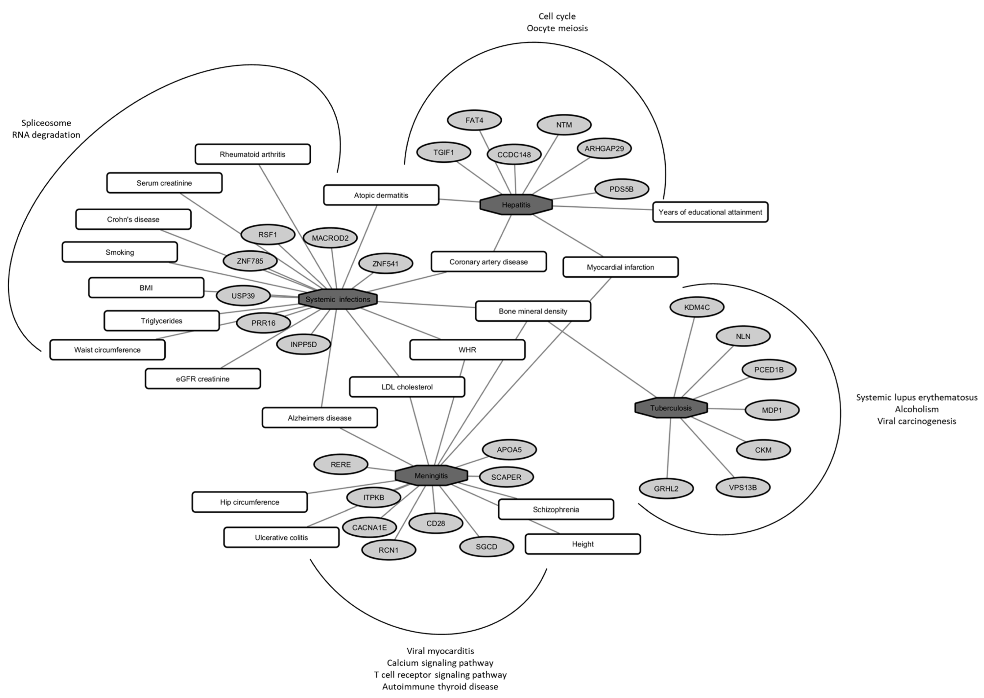
Figure 1. Pleiotropic network of identified loci (light grey oval shaped) with various infectious traits (dark grey octagon shaped) after a genome-wide meta-analysis of 4624 participants from the 10,001 Dalmatians biobank. Associations with various complex traits from other published GWA scans are shown in rounded rectangles, and KEGG pathway enrichment with an FDR cutoff of 1% for each of the infectious traits is depicted outside of the curved line (WHR—waist-to-hip ratio; BMI—body mass index; LDL—low-density lipoprotein; eGFR—estimated glomerular filtration rate).
Ijms 24 07006 g001
Figure 2. Validation of GWAS candidate loci using differentially expressed genes (FDR < 0.05) from publicly available RNA-seq studies (GEO accession IDs: pneumonia GSE196399, tuberculosis GSE94438 and COVID-19 GSE223885).
Ijms 24 07006 g002
Table 1. Demographic and phenotypic characteristics of study populations.
VisKorčulaSplitTOTALp *
N96026989664624
Gender, women; n (%)558 (58.12)1712 (63.45)587 (60.77)2857 (61.79)0.010 KV
Age in years, median (IQR)56 (24.00)55 (23.00)52 (21.00)55 (22.25)<0.001 KV, KS, VS
Years of schooling; n (%)
Elementary school [0–8]390 (40.63)711 (26.35)62 (6.42)1163 (25.15)<0.001 KV, KS, VS
High school [9–12]413 (43.02)1461 (54.15)473 (48.96)2347 (50.76)
University [≥13]157 (16.35)526 (19.50)431 (44.62)1114 (24.09)
Socioeconomic status; n (%)
1st quartile [0–8]350 (36.46)662 (24.54)134 (13.87)1146 (24.78)<0.001 KV, KS, VS
2nd quartile [9–10]254 (26.46)769 (28.50)200 (20.70)1223 (26.45)
3rd quartile [11–12]224 (23.33)737 (27.32)301 (31.16)1262 (27.29)
4th quartile [≥13]132 (13.75)530 (19.64)331 (34.27)993 (21.48)
Infectious disease count; n (%)
None576 (60.00)1677 (62.16)622 (64.39)2875 (62.18)0.114
One293 (30.52)752 (27.87)277 (28.68)1322 (28.59)
Two77 (8.02)231 (8.56)61 (6.31)369 (7.98)
Three11 (1.15)33 (1.22)6 (0.62)50 (1.08)
Four3 (0.31)5 (0.19)NA8 (0.17)
Recurrent pneumonia cases; n (%)9 (0.94)16 (0.59)1 (0.10)26 (0.56)0.047 VS
Infectious traits; n (%)
Tuberculosis 12 (1.25)16 (0.59)6 (0.62)34 (0.74)0.110
Pneumonia 105 (10.94)235 (8.71)27 (2.80)367 (7.94)<0.001 KS, VS
Hepatitis 26 (2.71)30 (1.11)20 (2.07)76 (1.64)0.002 KV
Meningitis 7 (0.73)16 (0.59)7 (0.73)30 (0.65)0.854
Respiratory infections 161 (16.77)535 (19.83)111 (11.49)807 (17.45)<0.001 KS, VS
Gastrointestinal infections 39 (4.06)55 (2.04)33 (3.42)127 (2.75)0.002 KV
Systemic infections 26 (2.71)36 (1.33)25 (2.59)87 (1.88)0.005 KV, KS
Bacterial infections 186 (19.38)571 (21.16)134 (13.87)891 (19.27)<0.001 KS, VS
Viral infections 74 (7.71)66 (2.45)42 (4.35)182 (3.94)<0.001 KV, KS, VS
Appendectomy 69 (7.19)169 (6.26)74 (7.66)312 (6.75)0.273
Tonsillectomy 168 (17.50)491 (18.20)160 (16.56)819 (17.71)0.517
Infectious burden
0576 (60.00)1677 (62.16)622 (64.39)2875 (62.18)0.001 KS, VS
0.5151 (15.73)385 (14.27)168 (17.39)704 (15.22)
1149 (15.52)388 (14.38)116 (12.01)653 (14.12)
1.541 (4.27)164 (6.08)38 (3.93)243 (5.25)
226 (2.71)48 (1.78)19 (1.97)93 (2.01)
2.58 (0.83)22 (0.81)2 (0.21)32 (0.69)
38 (0.83)7 (0.26)1 (0.10)16 (0.35)
3.51 (0.11)3 (0.11)NA4 (0.09)
4NA4 (0.15)NA4 (0.09)
Annual cold frequency, survey response244771NA1015
Several times per year67 (27.46)160 (20.75)NA227 (22.36)0.274
Once a year83 (34.01)305 (39.56)NA388 (38.23)
Less than once a year67 (27.46)229 (29.70)NA296 (29.16)
Never or almost never27 (11.07)77 (9.99)NA104 (10.25)
Influenza frequency in the last 10 years, survey response197655NA852
Every year9 (4.57)16 (2.44)NA25 (2.93)0.857
Several times48 (24.37)178 (27.18)NA226 (26.53)
Once57 (28.93)180 (27.48)NA237 (27.82)
Never83 (42.13)281 (42.90)NA364 (42.72)
* post-hoc pairwise comparisons (K—Korčula; V—Vis; S—Split). Abbreviations: IQR—interquartile range; NA—not applicable.
Table 2. Significant results after GWA meta-analyses (filtered for the top leading SNP in ±500 kb window, Bonferroni corrected associations were significant when p < 3.57 × 10⁻⁹ or suggestive when p < 5 × 10⁻⁸, SNPs were considered not to suffer from heterogeneity based on I2 statistics when p > 0.05, with same effect direction, variants annotated to protein-coding genes and variants present in meta-analysis for all three sub-cohorts (N = 4624)).
TraitSNPLocationAlleles *EAFOR (95% CI)pGeneVariant Type
Hepatitisrs18829090211:131868501G/A0.0061.13 (1.09–1.16)9.05 × 10⁻¹²NTMIntron
rs729360922:159097061A/G0.0041.16 (1.11–1.20)8.68 × 10⁻¹¹CCDC148Intron
rs1707773613:33251708A/G0.0171.06 (1.04–1.08)3.19 × 10⁻¹⁰PDS5BIntron
rs344479531:94730687A/C0.0061.12 (1.08–1.15)7.99 × 10⁻¹⁰ARHGAP29Intron
rs781112954:126203327T/C0.0311.04 (1.03–1.06)1.21 × 10⁻⁹FAT4Upstream
rs14560718018:3452913T/C0.0041.15 (1.10–1.19)2.97 × 10⁻⁹TGIF1Intron
Meningitisrs133581885:155325029G/A0.0051.10 (1.08–1.13)8.46 × 10⁻¹³SGCDIntron
rs175878211:226820605C/T0.0131.06 (1.04–1.08)4.14 × 10⁻¹²ITPKB3′-UTR
rs1892576882:204544219T/C0.0131.06 (1.04–1.07)1.31 × 10⁻¹¹CD28Upstream
rs18853087115:76781806T/C0.0061.08 (1.06–1.10)7.57 × 10⁻¹¹SCAPERIntron
rs6187881411:32119462C/T0.0261.05 (1.03–1.06)1.41 × 10⁻¹⁰RCN1Intron
rs11688652511:116671391T/C0.0061.09 (1.06–1.12)2.29 × 10⁻¹⁰APOA5Upstream
rs356087921:8450425G/A0.0231.04 (1.03–1.05)4.87 × 10⁻¹⁰REREIntron
rs1163066521:181673900T/G0.0141.05 (1.04–1.07)1.46 × 10⁻⁹CACNA1EIntron
Pneumoniars18762419412:7775204C/T0.0091.15 (1.10–1.20)2.15 × 10⁻⁸APOBEC1Downstream
Systemic infectionsrs14607272511:77519509T/C0.0081.13 (1.09–1.16)7.50 × 10⁻¹³RSF1Intron
rs14244188920:16052044T/G0.0061.13 (1.09–1.17)1.63 × 10⁻¹⁰MACROD2Downstream
rs769313435:119876729G/A0.0051.14 (1.10–1.18)3.70 × 10⁻¹⁰PRR16Intron
rs5821908719:48061409C/T0.0041.20 (1.14–1.26)7.57 × 10⁻¹⁰ZNF541Upstream
rs1383369762:234090697A/G0.0101.10 (1.07–1.12)1.26 × 10⁻⁹INPP5DIntron
rs1924371302:85840200T/C0.0041.21 (1.15–1.27)1.34 × 10⁻⁹USP39Intron
rs656519316:30600260C/T0.0281.05 (1.04–1.07)2.38 × 10⁻⁹ZNF785Upstream
Tuberculosisrs5545962379:6953063G/T0.0061.09 (1.07–1.12)2.06 × 10⁻¹¹KDM4CIntron
rs14525489414:24683304A/C0.0111.07 (1.05–1.09)1.17 × 10⁻¹⁰MDP1Missense
rs5705453435:65171990A/G0.0151.06 (1.04–1.07)1.24 × 10⁻¹⁰NLNDownstream
rs11776831512:47562000A/G0.0101.06 (1.04–1.07)1.48 × 10⁻¹⁰PCED1BIntron
rs18232041119:45821257A/G0.0111.08 (1.05–1.10)9.69 × 10⁻¹⁰CKMIntron
rs1405116998:102628803C/A0.0081.07 (1.05–1.10)2.37 × 10⁻⁹GRHL2Intron
rs1407824488:100743166C/T0.0041.10 (1.07–1.13)2.99 × 10⁻⁹VPS13BIntron
* Alleles: risk allele (effect allele)/other allele (non-effect allele). Abbreviations: EAF—effect allele frequency.
Disclaimer/Publisher’s Note: The statements, opinions and data contained in all publications are solely those of the individual author(s) and contributor(s) and not of MDPI and/or the editor(s). MDPI and/or the editor(s) disclaim responsibility for any injury to people or property resulting from any ideas, methods, instructions or products referred to in the content.