Article

Elevated Midkine Serum Levels Are Associated with Long-Term Survival in Critically Ill Patients

1
Department for Gastroenterology, Metabolic Disorders and Intensive Care Medicine, RWTH-University Hospital Aachen, Pauwelsstraße 30, 52074 Aachen, Germany
2
Institute of Laboratory Medicine, Western Palatinate Hospital, 67655 Kaiserslautern, Germany
3
Department of Hepatology and Gastroenterology, Charité-Universitätsmedizin Berlin, Campus Virchow-Klinikum (CVK) and Campus Charité Mitte (CCM), Augustenburger Platz 1, 13353 Berlin, Germany
4
Institute of Molecular Pathobiochemistry, Experimental Gene Therapy and Clinical Chemistry (IFMPEGKC), RWTH-University Hospital Aachen, Pauwelsstraße 30, 52074 Aachen, Germany
*
Author to whom correspondence should be addressed.
Int. J. Mol. Sci. 2024, 25(1), 454; https://doi.org/10.3390/ijms25010454
Submission received: 13 November 2023 / Revised: 22 December 2023 / Accepted: 27 December 2023 / Published: 29 December 2023
(This article belongs to the Special Issue Sepsis and Septic Shock: From Molecular Mechanisms to Novel Therapies)

Abstract

:
Midkine (Mdk) is a multifunctional protein involved in inflammatory processes. Hence, circulating Mdk is increased in sepsis and has been previously suggested as a potential biomarker in these patients. The aim of this study was to elucidate the role of Mdk serum concentrations in critical illness and sepsis and to verify its value as a prognostic biomarker. Thus, we analyzed the Mdk serum concentrations of 192 critically ill patients on admission to the medical intensive care unit (ICU). While the serum levels of Mdk at admission were similar in septic and nonseptic critical illness (362 vs. 337 ng/L, p = 0.727), we found several interesting correlations of Mdk to laboratory and clinical markers associated with ischemia or hypoxia, e.g., to renal failure and hepatic injury. Mdk serum concentrations at admission did not differ between various causes of sepsis or other critical illness. Most noticeable, we observed upregulated Mdk serum concentrations at admission in patients surviving in the long-term, which was only seen in nonseptic critical illness but not in sepsis. Our study suggests a relevant role of Mdk in critically ill patients in general and highlights the possible protective features of Mdk in critical illness.

1. Introduction

In internal and critical care medicine, predicting the patient outcome and diagnosis of sepsis and septic shock remain major challenges with ongoing need for research. Mortality is generally high in patients in the intensive care unit, with around 30 to 50% mortality in sepsis [1]. There is still a scarcity of routinely and reliably applicable laboratory markers indicating sepsis and critical illness or their prognosis. Specifically, sepsis is constituted by a life-threatening host immune response leading to severe organ dysfunction [2]. This organ dysfunction is defined and measured using the SOFA (sepsis-related organ failure assessment) score [3]. However, the exact mechanisms behind the uncontrolled infection, the dysregulated immune response and the development of organ dysfunction are still not clearly understood, nor can be exactly measured, and thus warrant further investigation [4,5].
Midkine (Mdk), known as the neurite growth-promoting factor 2, is a multifunctional protein that was first discovered in mouse embryogenesis [6,7]. Along with pleiotrophin (PTN), it is part of a structurally unique family of heparin-binding growth factors [8]. Mdk is a 13 kDa cysteine-rich polypeptide consisting of two domains (N- and C-domains) held together with disulfide bridges [8,9]. Moreover, Mdk is a soluble and secreted protein and could therefore serve as a biomarker for disease [10]. While Mdk gene expression has been detected at many sites, including the gastrointestinal tract, spleen, kidney, lungs and thyroid gland, its strongest relative expression seems to be in the mucosa of the small intestine [10,11,12,13,14,15,16,17]. Despite this Mdk gene expression, there is mostly a lack of any corresponding detectable Mdk protein expression in healthy tissues as opposed to malignant tissue [18,19]. Notably, the only healthy tissue with consistent Mdk protein expression seems to be the kidney [20].
Mdk has been shown to be overexpressed in various disease processes involving inflammation, most prominently in malignant diseases, including at least 20 different cancer types [10,21]. However, the alteration of Mdk has also been described in ischemic disease [22,23,24,25], kidney injury [24,26,27,28,29,30] and autoimmune disease [31,32,33,34,35,36]. Apart from its function as a growth factor, Mdk exerts numerous biological functions in the inflammation process and the recruitment of inflammatory cells [8,12,31,33,37,38,39], as well as the preservation of tissue viability during hypoxic stress [40]. Additionally, a strong antibacterial activity of Mdk has been demonstrated in vitro [8,41]. Hence, its involvement in the emergence and pathophysiology of sepsis can be anticipated. In fact, in a pilot study from 2010 consisting of 38 septic patients, 82 patients with active inflammatory bowel disease (IBD) and 87 healthy subjects, Mdk was increased in septic and IBD patients [42]. Another small study from 2020 involving 26 septic patients demonstrated increased plasma Mdk levels in sepsis survivors compared to non-survivors at day 28 [43]. Apart from the elevation in human sepsis, the inhibition of Mdk has also been demonstrated to ameliorate sepsis-induced lung injury in a mouse model in a recent study from 2021 [44]. Besides these studies, little is known about the involvement of Mdk not only in sepsis but in critical illness in general. Therefore, we conducted a detailed clinical study investigating of the regulation of Mdk in critical illness and sepsis, its association with various clinical markers and organ dysfunction and its potential as a prognostic biomarker.

2. Results

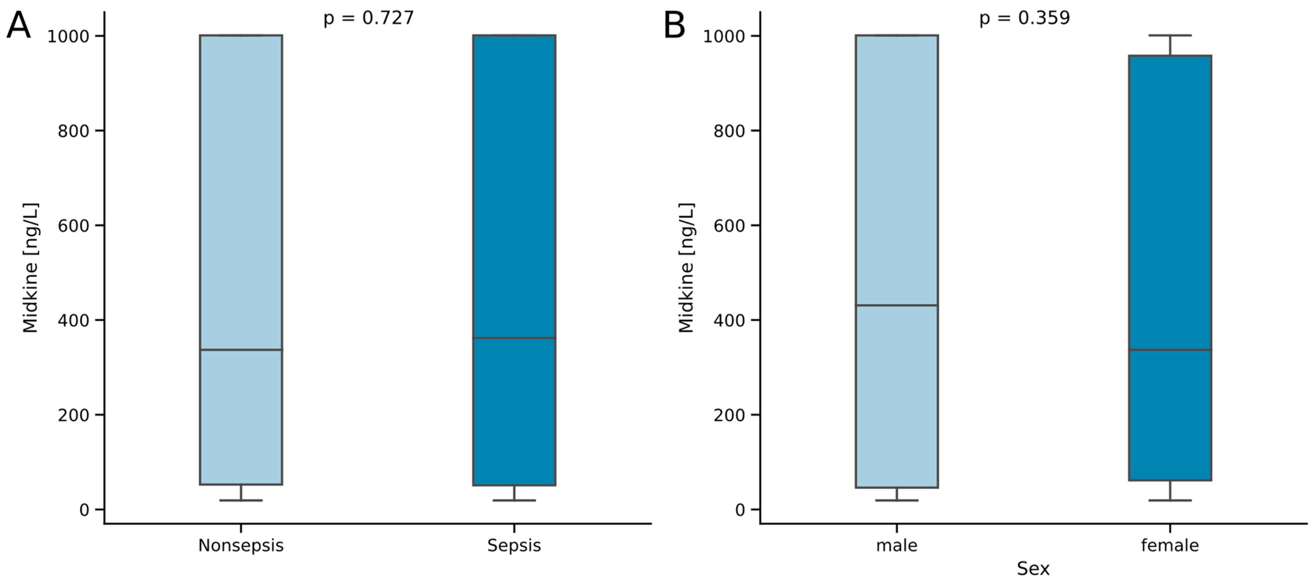
2.1. Midkine Serum Concentrations Do Not Differ between Critically Ill Patients with and without Sepsis

The cohort of this study comprises 125 patients admitted to the medical ICU due to sepsis and 67 patients admitted due to other critical illness. The median age of the cohort was 64.5 years, without statistical difference between septic and nonseptic critically ill patients. No differences were observed between the two study groups for age, sex, comorbidities (measured using the Charlson Comorbidity Index) or mortality. Notably, we also did not observe a significant difference in the levels of Mdk between the two groups (Figure 1A). However, patients with septic disease showed higher scores for disease severity (APACHE II, median of 18 vs. 16 points, p = 0.039) and organ insufficiency (SOFA, median of 11 vs. 7 points, p = 0.006). Coherently, patients with sepsis had higher demands for mechanical ventilation (73.6 vs. 57.5%, p = 0.036), as well as vasopressor therapy (70 vs. 47.4%, p = 0.005), and thus required a longer stay in the ICU (median of 10 vs. 6 days, p < 0.001; Table 1). Concerning other possible influence factors of Mdk serum levels, we did not observe a difference between sexes (Figure 1B) nor a correlation with age or body mass index (BMI) (Table 2).

2.2. Midkine Serum Levels Are Not Associated with Disease Etiology in Critically Ill Patients

In this study, most sepsis patients were treated due to a pulmonary focus (55.2%). Other sites of infection were the abdomen (15.2%) or the urogenital tract (8%). Other sepsis patients (21.6%) were treated due to bloodstream infections, skin infections or an unknown focus of infection. Patients with nonseptic critical illness were treated due to cardiocirculatory disease (19.4%), advanced liver disease (19.4%) or respiratory failure (14.9%), as well as numerous other diseases (46.3%). Further, looking into the potential regulation between those disease etiologies, we observed a higher level of Mdk in pulmonary and other focuses (median of 425 and 431 ng/L, respectively), as compared to abdominal or urogenital infections (median of 105 and 270 ng/L, respectively), although those changes did not reach a level of statistical significance (p = 0.481). Nonseptic patients did not show a regulation of Mdk in different disease etiologies (p = 0.772, Table 3).
To examine the influence of preexisting comorbidities on the serum levels of Mdk, we compared Mdk serum levels in patients with and without various diseases. Here, we could not show any differences in Mdk serum levels for diabetes, liver disease, coronary artery disease, hypertension, chronic alcohol abuse, chronic obstructive lung disease or active malignancy (Table 4).

2.3. Midkine Correlates with Clinically Established Biomarkers of Bacterial Inflammation, Kidney Function, Coagulation Function and Insulin Metabolism

Next, we aimed to evaluate other potential factors regulating Mdk serum concentrations in critical illness. For further investigation, we performed extensive correlation analyses between Mdk serum concentrations and a wide selection of laboratory as well as clinical markers. Concerning peripheral blood counts and inflammatory markers, we observed a positive correlation of medium strength between Mdk and procalcitonin (Spearman’s r = 0.263, p = 0.001). However, such a correlation was not seen for other inflammatory markers such as peripheral leucocyte count or the C-reactive protein (CRP). Furthermore, Mdk also shows weak to medium positive correlations to the markers of kidney dysfunction, i.e., uric acid (Spearman’s r = 0.190, p = 0.018), creatinine (Spearman’s r = 0.197, p = 0.006) and cystatin C (Spearman’s r = 0.231, p = 0.010). For the markers of hepatobiliary injury, we detected a moderate correlation of Mdk to aspartate aminotransferase (AST, Spearman’s r = 0.256, p = 0.001) and alanine aminotransferase (ALT, Spearman’s r = 0.154, p = 0.034), while other markers of liver function such as bilirubin or the internationalized normalized ratio (INR) did not correlate with Mdk. Interestingly, we also observed a moderate correlation with the activated partial thromboplastin time (aPTT, Spearman’s r = 0.330, p < 0.001). While the markers of the cardiocirculatory system or ICU parameters did not correlate to Mdk serum levels, we noted correlations to the markers of metabolism. Here, insulin (Spearman’s r = 0.229, p = 0.031) and C-peptide (Spearman’s r = 0.254, p = 0.016) levels showed a medium positive correlation to Mdk serum levels (Table 3).

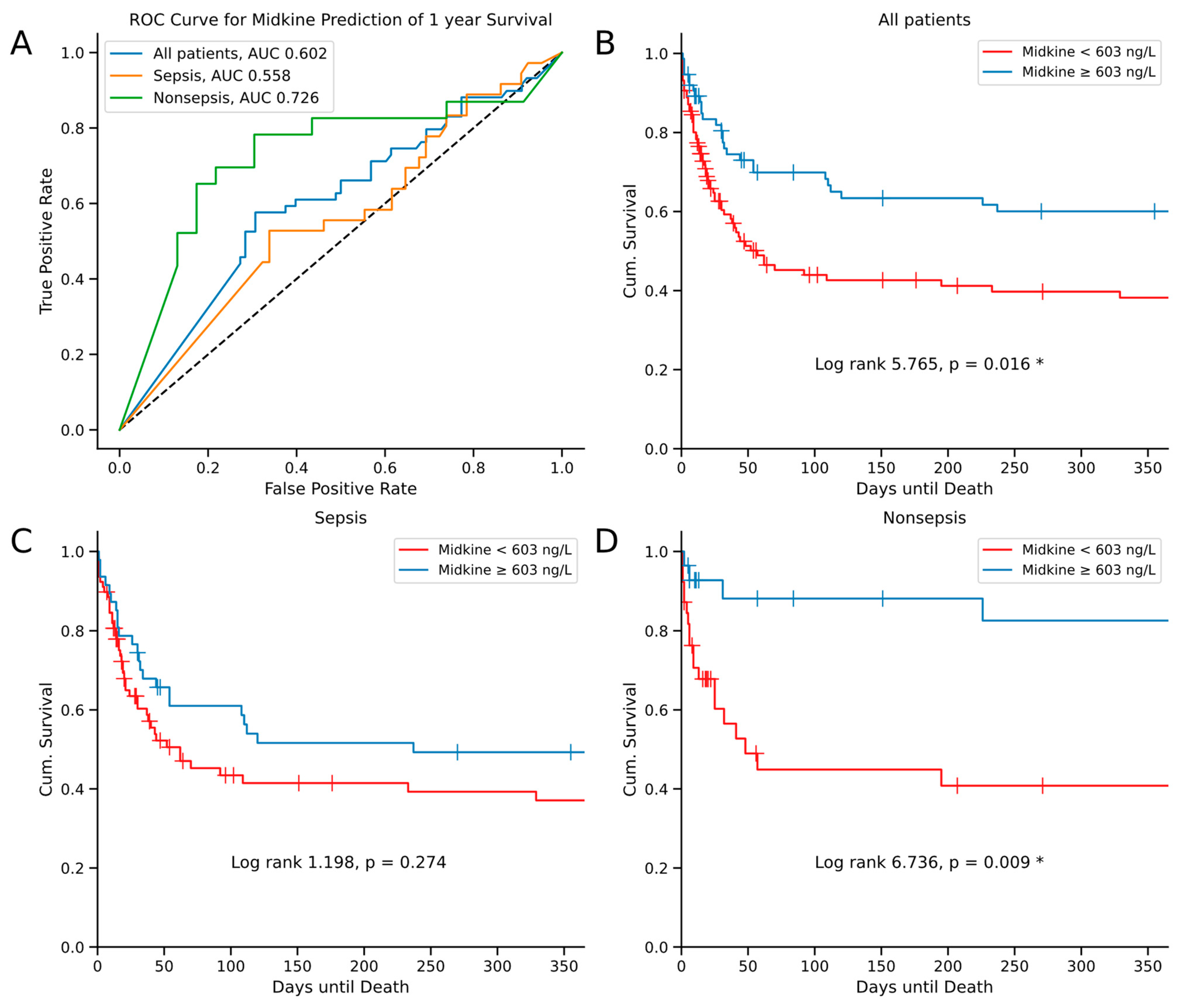
2.4. Midkine Predicts Long-Term Survival in Critically Ill Patients

Next, we focused on elucidating the prognostic value of Mdk serum levels in critically ill patients. First, we looked at the Mdk levels at admission in comparison between surviving and deceased patients at consecutive standardized time points over one year (i.e., 30, 60, 90, 180 and 365 days). Strikingly, we observed increased Mdk serum concentrations (obtained at ICU admission) in surviving patients for all mortality time points, which reached statistical significance including day 90 and later (p = 0.03 at day 90, p = 0.043 at day 180, p = 0.033 at day 365; Figure 2). In a subsequent receiver operating curve (ROC) analysis, Mdk serum levels showed an area under the curve (AUC) of 0.602 for the prediction of survival at one year (Figure 3A). To understand the difference between septic and nonseptic patients, we performed the ROC analysis also for those subgroups. Here, septic patients showed a lower AUROC of 0.558 in comparison to nonseptic patients with an AUROC of 0.726 (Figure 3A). In addition, we conducted a Kaplan–Meier curve analysis with the Youden index as a means to calculate an ideal cut-off value with respect to survival prediction for Mdk serum levels at 603 ng/L at admission to the ICU. First analyzing all patients, the Kaplan–Meier curves showed the largest separation towards the end of the follow-up timeframe at day 365 (log-rank 5.765, p = 0.016; Figure 3B). To further dissect the insights of the ROC analysis, we also conducted separate Kaplan–Meier analyses for our study cohorts of septic and nonseptic patients. In nonseptic patients, we could show an even larger curve separation (log-rank 6.736, p = 0.009; Figure 3D). However, in septic patients, we did not see a statistically significant curve separation (log-rank 1.198, p = 0.274; Figure 3C).

3. Discussion

Previously, peripheral Mdk levels have been shown to be elevated in sepsis and might indicate prognosis in these patients [42,43]. In this study, we investigated the serum levels of Mdk in critical illness and sepsis along with the possible use of Mdk for the prognostication of survival. While being independent of age, sex or BMI, Mdk serum levels did not differ between critically ill patients with and without sepsis. Moreover, we did not observe any changes in Mdk between the different disease categories apart from trends towards higher Mdk in pulmonary and other sepsis. The data suggest a correlation of Mdk serum levels to the markers of bacterial inflammation, kidney function, coagulation function and insulin metabolism. Most interestingly, we reported higher levels of Mdk in patients surviving the ICU. Our findings indicate a prognostic character of Mdk in critical illness, but on the contrary, not in sepsis.
Mdk is a soluble and secreted multifunctional protein, which is most prominently known as a biomarker in cancer research [10]. Mdk has been reported as elevated in sepsis in a small Polish pilot study from 2010 [42]. Our data suggests that the elevation of Mdk seems to be a feature of critical illness, rather than just sepsis alone, as the levels of serum Mdk are similar in the groups of our study (Figure 1A). Encouraging its use as a potential biomarker, we can report the independence of Mdk serum levels from age, sex and BMI (Figure 1B, Table 2). Previous data did not suggest changes in Mdk serum levels regarding the site of infection leading to sepsis [42]. In our ICU cohort, we also did not find any statistically significant differences in circulating Mdk serum levels between the causes of sepsis (Table 3). Moreover, we also did not find differences between the categories of nonseptic disease (Table 3), continuously suggesting that Mdk elevation is a general feature of critical illness. Regarding comorbidities, Mdk is known to be elevated in malignant disease [10,21], ischemic disease [22,23,24,25], kidney injury [24,26,27,28,29,30] and autoimmune disease [31,32,33,34,35,36]. However, when comparing the occurrence of several comorbidities of critically ill patients in our cohort, we did not find alterations in peripheral Mdk serum levels dependent on the comorbidity (Table 4). This indicates overlaying factors influencing the circulating Mdk in acute illness. However, concerning the previously described involvement in the pathogenesis of Mdk in kidney injury, we describe multiple correlations of Mdk to the markers of kidney function, i.e., creatinine and cystatin C (Table 2). As a heparin-binding growth factor, Mdk is known to be increased with heparin administration [45]; likewise, Mdk was also correlated to the length of aPTT (which is increased in heparin administration) in our study but not INR (Table 2). Interestingly, we found positive correlations of Mdk to hepatic transaminases (AST and ALT; Table 2) with the absence of correlation to peripheral bilirubin, supporting the concept of elevation of Mdk in ischemic states and hypoxic stress [22,23,24,25], which is a common feature of severe critical illness. While one study did not find differences in Mdk levels in critically ill patients with and without cardiovascular, respiratory, hematologic or kidney dysfunction [42], another study described differences in Mdk dependent on the severity of acute respiratory syndrome (ARDS) and kidney injury [43]. Our data seems to fit somewhere in between the results of those studies, as we report an association between kidney injury and Mdk serum levels but no correlation to the Horovitz quotient (PaO2/FiO2) for the diagnosis of ARDS (Table 2).
Arguably the most relevant finding of our study is the association of elevated serum Mdk on admission to the ICU to increased survival of critical illness. Currently, there is conflicting evidence regarding the impact of Mdk serum levels on survival in critical illness. On one hand, many studies suggest protective biological effects of Mdk, e.g., the recruitment of inflammatory cells [8,12,31,33,37,38,39], the preservation of tissue viability in hypoxic stress [40] and the antibacterial activity of Mdk in vitro [8,41]. On the other hand, a small Chinese study from 2020 including 26 septic patients, described lower levels of circulating plasma Mdk in survivors at day 28 [43]. There is no evidence available supporting the impact of Mdk on survival in nonseptic critically ill patients. Although we also demonstrated an association of Mdk with mortality in this study, there are a few key differences to be discussed. Firstly, in our study, Mdk seems to have protective effects, as we consistently measured higher Mdk levels on admission in surviving patients for all survival analysis time points. Secondly, the data of this study described an association of Mdk to survival in the long-term, rather than in the short-term (i.e., day 28). Lastly, we found an association of Mdk levels on admission to survival in all critically ill patients, which remarkably was not retained in sepsis, but rather in nonseptic critical illness. This study supports the concept of protective effects of Mdk in critical illness. Moreover, the entry levels of Mdk could reflect the inflammatory state of the disease and therefore impact the survival of patients via the widespread cytoprotective effects of Mdk on inflammation, apoptosis and in hypoxic stress, independent from disease etiology in critical illness [10,46,47,48].
Acknowledging the limitations of our study is important. By conducting a single-center study, we were able to achieve high technical accuracy and reproducibility. Although we investigated Mdk serum levels in the context of a large cohort in a biomarker study, the extensive analyses of patient subgroups lacked the statistical power to reliably detect smaller alterations in biomarker concentrations. Possibly related to this, we detected several correlations of medium strength of Mdk with laboratory and clinical markers (Table 2), the clinical value of which must be carefully evaluated. Furthermore, the lack of Midkine measurements in healthy controls makes comparisons between healthy individuals and critical illness impossible. Moreover, the cut-off of Mdk measurements for the assay we used was 1000 ng/L. A considerable number of measurements were at this upper cut-off and therefore probably in part well above it. Measuring Mdk levels above this cut-off would most likely lead to a deeper understanding of the distribution and regulation of Mdk. In addition, follow-up measurements at later time points during the intensive care treatment would also enhance our understanding of the role of Mdk in critical illness.

4. Materials and Methods

4.1. Study Design

This study was conducted as a retrospective, observational study to elucidate the role of Mdk in critically ill patients in a medical intensive care unit (ICU). For inclusion in this study, written informed consent was attained from the patient, his or her spouse, or legal guardian. We included 192 patients admitted to our medical intensive care unit of the Department of Gastroenterology, Digestive Disease and Intensive Care Medicine. Patients with consent, who were above or equal to the age of 18 years, were included in this study, as described previously [49,50]. We excluded (a) patients with expected short-term (less than 48 h) intensive care treatment, (b) patients admitted from another ICU and (c) patients admitted due to acute poisoning. The diagnosis of sepsis was established using the Third Consensus Definition for Sepsis (Sepsis-3) [2]. Patient comorbidities were assessed using the Charlson Comorbidity Index [51]. For collecting follow-up data concerning survival of patients, we contacted the patient, his or her relatives, or primary care physician. This study was approved by the local ethics committee (EK150/06) of the RWTH Aachen University Hospital and was conducted in accordance with the 1964 Declaration of Helsinki.

4.2. Midkine (Mdk) Measurements

Collection of blood samples was conducted at the time of admission to the intensive care unit. Blood samples were centrifuged at 4 °C for 10 min and were aliquoted into samples of 1 milliliter before being frozen at −80 °C until further use. Mdk concentrations were measured using a commercially available ELISA in accordance with the instructions of the manufacturer (BioVendor—Laboratorni medicina a.s., Karasek 1767/1, 621 00 Brno, Czech Republic). The measurements were performed blinded to clinical or other laboratory data of patients.

4.3. Statistical Analysis

Data was analyzed and graphed using SPSS version 29 (SPSS, Chicago, IL, USA) and the packages NumPy version 1.21.5 [52], Pandas version 1.4.4 [53], Matplotlib version 3.5.2 [54], Seaborn version 0.11.2 [55], Pingouin version 0.5.3 [56], Scikit-learn version 1.0.2 [57] and Lifelines version 0.27.7 [58] in Jupyter Notebooks version 6.5.4 [59] using Python version 3.11 [60]. Data is given as median and range due to the possible skewed distribution of parameters. The two-tailed Mann–Whitney U test or chi-squared test was applied to two groups of unpaired samples, as normal distribution could not be assumed. The Kruskal–Wallis test was applied to more than two groups. A significance level of p = 0.05 was used for all corresponding calculations. The correlation of parameters was assessed using the Spearman’s rank correlation test. The Youden index (as the sum of sensitivity and specificity minus one) was calculated to identify optimal cut-off values for parameters to discriminate prognosis. To evaluate the quality of a predictive marker, receiver operating characteristics (ROC) curves and the corresponding area under the curve (AUC) were generated. Patient survival was depicted by Kaplan–Meier curves followed by a log-rank test for level of significance.

5. Conclusions

Our study shows that Mdk serum concentrations are similar between septic and nonseptic individuals in a large cohort of critically ill patients. Possibly linked to the states of ischemia or hypoxia, we reveal several interesting correlations of Mdk concentrations, e.g., to renal failure and hepatic injury. Most strikingly, this study associates lower Mdk serum concentrations with higher mortality in critical illness, with the strongest influence in nonseptic patients. Possible future research should aim at a deeper understanding and validation of the role of Mdk in nonseptic critical illness.

Author Contributions

Conceptualization, P.H., D.W., F.T. and A.K.; data curation, P.H., D.W., S.A.J., J.K.A., L.B., M.R.P., J.F.B. and T.H.W.; formal analysis, P.H., D.W., S.A.J. and A.K.; methodology, P.H., S.A.J., E.Y., R.W. and A.K.; supervision, J.F.B., K.H., T.H.W., R.W., E.Y., C.T., F.T. and A.K.; writing—original draft, P.H., D.W. and A.K.; writing—review and editing, P.H., S.A.J., D.W., M.R.P., J.K.A., K.H., T.H.W., R.W., F.T. and A.K. All authors have read and agreed to the published version of the manuscript.

Funding

This work was supported by the German Research Foundation (DFG; CRC1382, Project-ID 403224013).

Institutional Review Board Statement

The local ethics committee, in accordance with the ethical standards of the Declaration of Helsinki (reference number EK150/06), approved our study.

Informed Consent Statement

Written informed consent was obtained from the patient, his/her spouse or legal guardian.

Data Availability Statement

The original data sets presented in this study are available on request from the corresponding author.

Conflicts of Interest

The authors declare no conflicts of interest.

References

  1. Fleischmann-Struzek, C.; Mellhammar, L.; Rose, N.; Cassini, A.; Rudd, K.E.; Schlattmann, P.; Allegranzi, B.; Reinhart, K. Incidence and mortality of hospital- and ICU-treated sepsis: Results from an updated and expanded systematic review and meta-analysis. Intensive Care Med. 2020, 46, 1552–1562. [Google Scholar] [CrossRef] [PubMed]
  2. Singer, M.; Deutschman, C.S.; Seymour, C.W.; Shankar-Hari, M.; Annane, D.; Bauer, M.; Bellomo, R.; Bernard, G.R.; Chiche, J.D.; Coopersmith, C.M.; et al. The Third International Consensus Definitions for Sepsis and Septic Shock (Sepsis-3). JAMA 2016, 315, 801–810. [Google Scholar] [CrossRef] [PubMed]
  3. Evans, L.; Rhodes, A.; Alhazzani, W.; Antonelli, M.; Coopersmith, C.M.; French, C.; Machado, F.R.; McIntyre, L.; Ostermann, M.; Prescott, H.C.; et al. Surviving sepsis campaign: International guidelines for management of sepsis and septic shock 2021. Intensive Care Med. 2021, 47, 1181–1247. [Google Scholar] [CrossRef] [PubMed]
  4. Boomer, J.S.; Green, J.M.; Hotchkiss, R.S. The changing immune system in sepsis: Is individualized immuno-modulatory therapy the answer? Virulence 2014, 5, 45–56. [Google Scholar] [CrossRef] [PubMed]
  5. Ryan, T.; Coakley, J.D.; Martin-Loeches, I. Defects in innate and adaptive immunity in patients with sepsis and health care associated infection. Ann. Transl. Med. 2017, 5, 447. [Google Scholar] [CrossRef] [PubMed]
  6. Kadomatsu, K.; Tomomura, M.; Muramatsu, T. cDNA cloning and sequencing of a new gene intensely expressed in early differentiation stages of embryonal carcinoma cells and in mid-gestation period of mouse embryogenesis. Biochem. Biophys. Res. Commun. 1988, 151, 1312–1318. [Google Scholar] [CrossRef] [PubMed]
  7. Winkler, C.; Yao, S. The midkine family of growth factors: Diverse roles in nervous system formation and maintenance. Br. J. Pharmacol. 2014, 171, 905–912. [Google Scholar] [CrossRef] [PubMed]
  8. Muramatsu, T. Midkine and pleiotrophin: Two related proteins involved in development, survival, inflammation and tumorigenesis. J. Biochem. 2002, 132, 359–371. [Google Scholar] [CrossRef]
  9. Muramatsu, T. Structure and function of midkine as the basis of its pharmacological effects. Br. J. Pharmacol. 2014, 171, 814–826. [Google Scholar] [CrossRef]
  10. Jones, D.R. Measuring midkine: The utility of midkine as a biomarker in cancer and other diseases. Br. J. Pharmacol. 2014, 171, 2925–2939. [Google Scholar] [CrossRef]
  11. Tsutsui, J.; Kadomatsu, K.; Matsubara, S.; Nakagawara, A.; Hamanoue, M.; Takao, S.; Shimazu, H.; Ohi, Y.; Muramatsu, T. A new family of heparin-binding growth/differentiation factors: Increased midkine expression in Wilms’ tumor and other human carcinomas. Cancer Res. 1993, 53, 1281–1285. [Google Scholar] [PubMed]
  12. Cohen, S.; Shoshana, O.Y.; Zelman-Toister, E.; Maharshak, N.; Binsky-Ehrenreich, I.; Gordin, M.; Hazan-Halevy, I.; Herishanu, Y.; Shvidel, L.; Haran, M.; et al. The cytokine midkine and its receptor RPTPzeta regulate B cell survival in a pathway induced by CD74. J. Immunol. 2012, 188, 259–269. [Google Scholar] [CrossRef] [PubMed]
  13. Hovanessian, A.G. Midkine, a cytokine that inhibits HIV infection by binding to the cell surface expressed nucleolin. Cell Res. 2006, 16, 174–181. [Google Scholar] [CrossRef] [PubMed]
  14. Inazumi, T.; Tajima, S.; Nishikawa, T.; Kadomatsu, K.; Muramatsu, H.; Muramatsu, T. Expression of the retinoid-inducible polypeptide, midkine, in human epidermal keratinocytes. Arch. Dermatol. Res. 1997, 289, 471–475. [Google Scholar] [CrossRef] [PubMed]
  15. Kerzerho, J.; Adotevi, O.; Castelli, F.A.; Dosset, M.; Bernardeau, K.; Szely, N.; Lang, F.; Tartour, E.; Maillere, B. The angiogenic growth factor and biomarker midkine is a tumor-shared antigen. J. Immunol. 2010, 185, 418–423. [Google Scholar] [CrossRef] [PubMed]
  16. Lee, S.H.; Suh, H.N.; Lee, Y.J.; Seo, B.N.; Ha, J.W.; Han, H.J. Midkine prevented hypoxic injury of mouse embryonic stem cells through activation of Akt and HIF-1alpha via low-density lipoprotein receptor-related protein-1. J. Cell Physiol. 2012, 227, 1731–1739. [Google Scholar] [CrossRef]
  17. Nordin, S.L.; Jovic, S.; Kurut, A.; Andersson, C.; Gela, A.; Bjartell, A.; Morgelin, M.; Olin, A.I.; Lund, M.; Egesten, A. High expression of midkine in the airways of patients with cystic fibrosis. Am. J. Respir. Cell Mol. Biol. 2013, 49, 935–942. [Google Scholar] [CrossRef] [PubMed]
  18. Miyashiro, I.; Kaname, T.; Shin, E.; Wakasugi, E.; Monden, T.; Takatsuka, Y.; Kikkawa, N.; Muramatsu, T.; Monden, M.; Akiyama, T. Midkine expression in human breast cancers: Expression of truncated form. Breast Cancer Res. Treat. 1997, 43, 1–6. [Google Scholar] [CrossRef]
  19. Huang, Y.; Cao, G.; Wang, H.; Wang, Q.; Hou, Y. The expression and location of midkine in gastric carcinomas of Chinese patients. Cell Mol. Immunol. 2007, 4, 135–140. [Google Scholar]
  20. Muramatsu, H.; Shirahama, H.; Yonezawa, S.; Maruta, H.; Muramatsu, T. Midkine, a retinoic acid-inducible growth/differentiation factor: Immunochemical evidence for the function and distribution. Dev. Biol. 1993, 159, 392–402. [Google Scholar] [CrossRef]
  21. Filippou, P.S.; Karagiannis, G.S.; Constantinidou, A. Midkine (MDK) growth factor: A key player in cancer progression and a promising therapeutic target. Oncogene 2020, 39, 2040–2054. [Google Scholar] [CrossRef] [PubMed]
  22. Horiba, M.; Kadomatsu, K.; Yasui, K.; Lee, J.K.; Takenaka, H.; Sumida, A.; Kamiya, K.; Chen, S.; Sakuma, S.; Muramatsu, T.; et al. Midkine plays a protective role against cardiac ischemia/reperfusion injury through a reduction of apoptotic reaction. Circulation 2006, 114, 1713–1720. [Google Scholar] [CrossRef] [PubMed]
  23. Yoshida, Y.; Goto, M.; Tsutsui, J.; Ozawa, M.; Sato, E.; Osame, M.; Muramatsu, T. Midkine is present in the early stage of cerebral infarct. Brain Res. Dev. Brain Res. 1995, 85, 25–30. [Google Scholar] [CrossRef] [PubMed]
  24. Sato, W.; Kadomatsu, K.; Yuzawa, Y.; Muramatsu, H.; Hotta, N.; Matsuo, S.; Muramatsu, T. Midkine is involved in neutrophil infiltration into the tubulointerstitium in ischemic renal injury. J. Immunol. 2001, 167, 3463–3469. [Google Scholar] [CrossRef] [PubMed]
  25. Horiba, M.; Kadomatsu, K.; Nakamura, E.; Muramatsu, H.; Ikematsu, S.; Sakuma, S.; Hayashi, K.; Yuzawa, Y.; Matsuo, S.; Kuzuya, M.; et al. Neointima formation in a restenosis model is suppressed in midkine-deficient mice. J. Clin. Investig. 2000, 105, 489–495. [Google Scholar] [CrossRef] [PubMed]
  26. Kawai, H.; Sato, W.; Yuzawa, Y.; Kosugi, T.; Matsuo, S.; Takei, Y.; Kadomatsu, K.; Muramatsu, T. Lack of the growth factor midkine enhances survival against cisplatin-induced renal damage. Am. J. Pathol. 2004, 165, 1603–1612. [Google Scholar] [CrossRef] [PubMed]
  27. Kosugi, T.; Sato, W. Midkine and the kidney: Health and diseases. Nephrol. Dial. Transplant. 2012, 27, 16–21. [Google Scholar] [CrossRef]
  28. Kato, K.; Kosugi, T.; Sato, W.; Arata-Kawai, H.; Ozaki, T.; Tsuboi, N.; Ito, I.; Tawada, H.; Yuzawa, Y.; Matsuo, S.; et al. Growth factor Midkine is involved in the pathogenesis of renal injury induced by protein overload containing endotoxin. Clin. Exp. Nephrol. 2011, 15, 346–354. [Google Scholar] [CrossRef]
  29. Kosugi, T.; Yuzawa, Y.; Sato, W.; Arata-Kawai, H.; Suzuki, N.; Kato, N.; Matsuo, S.; Kadomatsu, K. Midkine is involved in tubulointerstitial inflammation associated with diabetic nephropathy. Lab. Investig. 2007, 87, 903–913. [Google Scholar] [CrossRef]
  30. Salaru, D.L.; Mertens, P.R.; Bartsch, P. Loss of heparin-binding protein prevents necrotizing glomerulonephritis: First clues hint at plasminogen activator inhibitor-1. Int. Urol. Nephrol. 2013, 45, 1483–1487. [Google Scholar] [CrossRef]
  31. Sonobe, Y.; Li, H.; Jin, S.; Kishida, S.; Kadomatsu, K.; Takeuchi, H.; Mizuno, T.; Suzumura, A. Midkine inhibits inducible regulatory T cell differentiation by suppressing the development of tolerogenic dendritic cells. J. Immunol. 2012, 188, 2602–2611. [Google Scholar] [CrossRef]
  32. Krzystek-Korpacka, M.; Neubauer, K.; Matusiewicz, M. Circulating midkine in Crohn’s disease: Clinical implications. Inflamm. Bowel Dis. 2010, 16, 208–215. [Google Scholar] [CrossRef]
  33. Takada, T.; Toriyama, K.; Muramatsu, H.; Song, X.J.; Torii, S.; Muramatsu, T. Midkine, a retinoic acid-inducible heparin-binding cytokine in inflammatory responses: Chemotactic activity to neutrophils and association with inflammatory synovitis. J. Biochem. 1997, 122, 453–458. [Google Scholar] [CrossRef]
  34. Liu, X.; Mashour, G.A.; Webster, H.F.; Kurtz, A. Basic FGF and FGF receptor 1 are expressed in microglia during experimental autoimmune encephalomyelitis: Temporally distinct expression of midkine and pleiotrophin. Glia 1998, 24, 390–397. [Google Scholar] [CrossRef]
  35. Wang, J.; Takeuchi, H.; Sonobe, Y.; Jin, S.; Mizuno, T.; Miyakawa, S.; Fujiwara, M.; Nakamura, Y.; Kato, T.; Muramatsu, H.; et al. Inhibition of midkine alleviates experimental autoimmune encephalomyelitis through the expansion of regulatory T cell population. Proc. Natl. Acad. Sci. USA 2008, 105, 3915–3920. [Google Scholar] [CrossRef]
  36. Maruyama, K.; Muramatsu, H.; Ishiguro, N.; Muramatsu, T. Midkine, a heparin-binding growth factor, is fundamentally involved in the pathogenesis of rheumatoid arthritis. Arthritis Rheum. 2004, 50, 1420–1429. [Google Scholar] [CrossRef]
  37. Kadomatsu, K.; Kishida, S.; Tsubota, S. The heparin-binding growth factor midkine: The biological activities and candidate receptors. J. Biochem. 2013, 153, 511–521. [Google Scholar] [CrossRef]
  38. Sanino, G.; Bosco, M.; Terrazzano, G. Physiology of Midkine and Its Potential Pathophysiological Role in COVID-19. Front. Physiol. 2020, 11, 616552. [Google Scholar] [CrossRef]
  39. Weckbach, L.T.; Grabmaier, U.; Uhl, A.; Gess, S.; Boehm, F.; Zehrer, A.; Pick, R.; Salvermoser, M.; Czermak, T.; Pircher, J.; et al. Midkine drives cardiac inflammation by promoting neutrophil trafficking and NETosis in myocarditis. J. Exp. Med. 2019, 216, 350–368. [Google Scholar] [CrossRef]
  40. Muramatsu, T. Midkine: A promising molecule for drug development to treat diseases of the central nervous system. Curr. Pharm. Des. 2011, 17, 410–423. [Google Scholar] [CrossRef]
  41. Svensson, S.L.; Pasupuleti, M.; Walse, B.; Malmsten, M.; Morgelin, M.; Sjogren, C.; Olin, A.I.; Collin, M.; Schmidtchen, A.; Palmer, R.; et al. Midkine and pleiotrophin have bactericidal properties: Preserved antibacterial activity in a family of heparin-binding growth factors during evolution. J. Biol. Chem. 2010, 285, 16105–16115. [Google Scholar] [CrossRef]
  42. Krzystek-Korpacka, M.; Mierzchala, M.; Neubauer, K.; Durek, G.; Gamian, A. Midkine, a multifunctional cytokine, in patients with severe sepsis and septic shock: A pilot study. Shock 2011, 35, 471–477. [Google Scholar] [CrossRef]
  43. Chang, W.; Peng, F.; Sun, Q.; Meng, S.S.; Qiu, H.B.; Xu, J.Y. Plasma Midkine Is Associated With 28-Day Mortality and Organ Function in Sepsis. J. Intensive Care Med. 2020, 35, 1290–1296. [Google Scholar] [CrossRef]
  44. Xu, J.Y.; Chang, W.; Sun, Q.; Peng, F.; Yang, Y. Pulmonary midkine inhibition ameliorates sepsis induced lung injury. J. Transl. Med. 2021, 19, 91. [Google Scholar] [CrossRef]
  45. Sugito, S.; Hall, S.; Al-Omary, M.S.; De Malmanche, T.; Robertson, G.; Collins, N.; Boyle, A. Heparin Administration, but Not Myocardial Ischemia or Necrosis, Leads to Midkine Elevation. J. Cardiovasc. Transl. Res. 2020, 13, 741–743. [Google Scholar] [CrossRef]
  46. Yazihan, N. Midkine in inflammatory and toxic conditions. Curr. Drug Deliv. 2013, 10, 54–57. [Google Scholar] [CrossRef]
  47. Kadomatsu, K.; Bencsik, P.; Gorbe, A.; Csonka, C.; Sakamoto, K.; Kishida, S.; Ferdinandy, P. Therapeutic potential of midkine in cardiovascular disease. Br. J. Pharmacol. 2014, 171, 936–944. [Google Scholar] [CrossRef]
  48. Cohen, S.; Shachar, I. Midkine as a regulator of B cell survival in health and disease. Br. J. Pharmacol. 2014, 171, 888–895. [Google Scholar] [CrossRef]
  49. Hohlstein, P.; Brozat, J.F.; Schuler, J.; Abu Jhaisha, S.; Pollmanns, M.R.; Bundgens, L.; Wirtz, T.H.; Yagmur, E.; Hamesch, K.; Weiskirchen, R.; et al. Secreted Frizzled Related Protein 5 (SFRP5) Serum Levels Are Decreased in Critical Illness and Sepsis and Are Associated with Short-Term Mortality. Biomedicines 2023, 11, 313. [Google Scholar] [CrossRef]
  50. Yagmur, E.; Abu Jhaisha, S.; Buendgens, L.; Sapundzhieva, N.; Brozat, J.F.; Hohlstein, P.; Pollmanns, M.R.; Koek, G.H.; Weiskirchen, R.; Trautwein, C.; et al. Clusterin Plasma Concentrations Are Decreased in Sepsis and Inversely Correlated with Established Markers of Inflammation. Diagnostics 2022, 12, 3010. [Google Scholar] [CrossRef]
  51. Charlson, M.E.; Pompei, P.; Ales, K.L.; MacKenzie, C.R. A new method of classifying prognostic comorbidity in longitudinal studies: Development and validation. J. Chronic Dis. 1987, 40, 373–383. [Google Scholar] [CrossRef] [PubMed]
  52. Harris, C.R.; Millman, K.J.; van der Walt, S.J.; Gommers, R.; Virtanen, P.; Cournapeau, D.; Wieser, E.; Taylor, J.; Berg, S.; Smith, N.J.; et al. Array programming with NumPy. Nature 2020, 585, 357–362. [Google Scholar] [CrossRef] [PubMed]
  53. McKinney, W. Data Structures for Statistical Computing in Python. In Proceedings of the 9th Python in Science Conference, Austin, TX, USA, 28 June–3 July 2010. [Google Scholar]
  54. Hunter, J.D. Matplotlib: A 2D Graphics Environment. Comput. Sci. Eng. 2007, 9, 90–95. [Google Scholar] [CrossRef]
  55. Waskom, M.; Botvinnik, O.; O’Kane, D.; Hobson, P.; Lukauskas, S.; Gemperline, D.C.; Augspurger, T.; Halchenko, Y.; Cole, J.B.; Warmenhoven, J.; et al. Mwaskom/Seaborn: v0.8.1 (September 2017); Zenodo: Genève, Switzerland, 2017. [Google Scholar]
  56. Vallat, R. Pingouin: Statistics in Python. J. Open Source Softw. 2018, 3, 1026. [Google Scholar] [CrossRef]
  57. Pedregosa, F.; Varoquaux, G.; Gramfort, A.; Michel, V.; Thirion, B.; Grisel, O.; Blondel, M.; Müller, A.; Nothman, J.; Louppe, G.; et al. Scikit-learn: Machine Learning in Python. J. Mach. Learn. Res. 2011, 12, 2825–2830. [Google Scholar]
  58. Davidson-Pilon, C. lifelines: Survival analysis in Python. J. Open Source Softw. 2019, 4, 1317. [Google Scholar] [CrossRef]
  59. Kluyver, T.; Ragan-Kelley, B.; Pérez, F.; Granger, B.; Bussonnier, M.; Frederic, J.; Kelley, K.; Hamrick, J.; Grout, J.; Corlay, S.; et al. Jupyter Notebooks—A publishing format for reproducible computational workflows. In Proceedings of the 20th International Conference on Electronic Publishing: Players, Agents and Agendas, Göttingen, Germany, 7–9 June 2016; pp. 87–90. [Google Scholar]
  60. Van Rossum, G.; Drake, F.L. Python 3 Reference Manual; CreateSpace: Scotts Valley, CA, USA, 2009. [Google Scholar]
Figure 1. Serum Midkine concentrations in critically ill patients with and without sepsis (A) and comparison between sexes (B). Sample sizes: patients n = 192, nonsepsis n = 67, sepsis n = 125. Significance between groups was assessed using the Mann–Whitney U test. p-values < 0.05 were considered statistically significant.
Ijms 25 00454 g001
Figure 2. Midkine levels in a consecutive survival analysis of critically ill patients treated in the ICU. (AE) Survival status on days 30 through 365. Sample sizes: patients n = 192. * Significance between groups was assessed using the Mann–Whitney U test. p-values < 0.05 were considered statistically significant and were highlighted (“*”).
Ijms 25 00454 g002
Figure 3. Receiver operating characteristic (ROC) curves for the prediction of one-year survival using serum Midkine levels in all patients, sepsis and nonsepsis patients. Black dashed line represents the ROC curve for a random guess (A). Kaplan–Meier curves for Midkine <603 ng/L (red) and ≥603 ng/L (blue) in all patients (B), septic patients (C) and nonseptic patients (D). Censored events are indicated with a crossing vertical line. Cut-off values of the Kaplan–Meier curve were determined using the Youden index for all patients. Sample sizes: patients n = 192, nonsepsis n = 67, sepsis n = 125. * Significance between groups was assessed using the log-rank test. p-values < 0.05 were considered statistically significant and were highlighted (“*”). Abbreviations: AUC: area under curve.
Ijms 25 00454 g003
Table 1. Baseline patient characteristics.
ParameterAll PatientsSepsisNonsepsisp-Value
Number n19212567
Sex (male/female) n113/7978/4735/320.226
Age (years)64.5 (18–89)65 (21–89)63 (18–87)0.663
APACHE II score17 (2–40)18 (3–40)16 (2–37)0.039 *
SOFA score10 (0–18)11 (3–17)7 (0–18)0.006 *
Charlson Comorbidity index4 (0–16)4 (0–16)4 (0–13)0.297
Mechanical ventilation n (%)130 (68.0)92 (73.6)38 (57.5)0.036 *
Vasopressor demand n (%)115 (62.2)84 (70.0)31 (47.7)0.005 *
ICU days n8 (1–137)10 (1–137)6 (2–44)<0.001 *
Death in ICU n (%)52 (27.1)38 (30.4)14 (20.9)0.214
30-day mortality n (%)57 (34.5)41 (36.3)16 (30.8)0.606
1-year mortality n (%)88 (59.9)65 (64.4)23 (50.0)0.142
Midkine (ng/mL)358 (19–1000)362 (19–1000)337 (19–1000)0.727
The median and range (in parentheses) are given unless indicated otherwise. Abbreviations: APACHE: acute physiology and chronic health evaluation; SOFA: sequential organ failure assessment; ICU: intensive care unit. * Significance between sepsis and nonsepsis patients was assessed using the Mann–Whitney U test or chi-squared test, respectively. p-Values < 0.05 were considered statistically significant and were highlighted (“*”).
Table 2. Correlations of clinical and laboratory parameters with Midkine serum concentrations at ICU admission.
Parametersrp-Value
Demographics
Age0.0050.943
Body mass index0.0440.550
Blood count and markers of inflammation
Leukocytes0.0050.945
Hemoglobin0.0770.290
Platelets−0.0240.747
C-reactive Protein0.0840.249
Procalcitonin0.2630.001 *
Interleukin 60.0350.670
Interleukin 100.0540.594
Electrolytes and renal system
Sodium−0.0660.362
Potassium0.0580.428
Urea0.1030.156
Uric acid0.1900.018 *
Creatinine0.1970.006 *
Cystatin C0.2310.010 *
Hepato-pancreatico-biliary system and coagulation
Protein, total−0.0200.803
Albumin−0.0850.339
INR−0.0580.432
aPTT0.330<0.001 *
Bilirubin, total0.0130.862
γGT0.1270.082
AST0.2560.001 *
ALT0.1540.034 *
Lipase0.1030.211
Cardiopulmonary system
NTproBNP0.1310.212
Norepinephrine demand at day 1 (µg/day)0.0550.456
Horovitz quotient (PaO2/FiO2)−0.0470.710
Ventilatory FiO2 demand0.0790.527
Net fluid balance day 1−0.0210.776
Net fluid balance day 3−0.0620.450
Metabolism
Glucose0.0270.712
HbA1c0.0590.582
Insulin0.2290.031 *
C-Peptide0.2540.016 *
HOMA IR0.1610.133
Cholesterol0.0390.633
HDL-cholesterol−0.1280.237
LDL-cholesterol−0.0100.928
Triglycerides0.0840.303
Disease severity parameters
Days on ICU0.0460.526
SOFA day 1−0.0370.749
SOFA day 3−0.0590.668
APACHE-II day 10.0090.909
APACHE-II day 3−0.1040.420
Spearman’s rank correlation test was used to calculate significant correlations of positive and negative nature. p-values < 0.05 were considered statistically significant and were highlighted (“*”). Abbreviations: ICU: intensive care unit; INR: international normalized ratio; γGT: Gamma-glutamyl transpeptidase; ALT/AST: alanine/aspartate aminotransferase; NTproBNP: N-terminal pro B-type natriuretic peptide; FiO2: fraction of inspired oxygen; HbA1c: glycosylated hemoglobin A1; HOMA: homeostatic model assessment; HDL: high-density lipoprotein; LDL: low-density lipoprotein; SOFA: sequential organ failure assessment; APACHE-II: acute physiology and chronic health evaluation II.
Table 3. Disease etiology of the study population and subgroup Midkine concentrations.
Etiology of (Non)Septic Critical IllnessSepsis
n = 125, n (%)
Nonsepsis
n = 67, n (%)
Midkine (ng/L)p
Pulmonary69 (55.2) 425 (19–1000)0.481
Abdominal19 (15.2) 105 (19–1000)
Urogenital10 (8) 270 (22–1000)
Other27 (21.6) 431 (19–1000)
Cardiocirculatory disorder 13 (19.4)365 (19–1000)0.772
Respiratory failure 10 (14.9)300 (19–1000)
Advanced liver disease 13 (19.4)337 (19–1000)
Other 31 (46.3)240 (19–1000)
The absolute numbers and percentages of the respective subgroup (in parentheses) or median and range (in parentheses) are given. Significance between more than two groups was assessed using the Kruskal–Wallis test. p-values < 0.05 were considered statistically significant.
Table 4. Comorbidities and their influence on Midkine levels at ICU admission.
ComorbidityMidkine Concentration in ng/L, Median (Range)p
Diabetes (n = 50)209 (19–1000)0.182
Liver disease (n = 20)225 (19–1000)0.735
Coronary artery disease (n = 63)379 (19–1000)0.617
Hypertension (n = 75)462 (19–1000)0.162
Chronic alcohol abuse (n = 25)337 (19–1000)0.543
Chronic obstructive lung disease (n = 25)425 (19–1000)0.939
Active malignancy (n = 23)151 (19–1000)0.302
The median and range (in parentheses) are given unless indicated otherwise. Significance between groups was assessed using the Mann–Whitney U test. p-values < 0.05 were considered statistically.
Disclaimer/Publisher’s Note: The statements, opinions and data contained in all publications are solely those of the individual author(s) and contributor(s) and not of MDPI and/or the editor(s). MDPI and/or the editor(s) disclaim responsibility for any injury to people or property resulting from any ideas, methods, instructions or products referred to in the content.

