Article

Redox Properties of Bacillus subtilis Ferredoxin:NADP+ Oxidoreductase: Potentiometric Characteristics and Reactions with Pro-Oxidant Xenobiotics

by
Mindaugas Lesanavičius
1,
Daisuke Seo
2,
Gintarė Maurutytė
1 and
Narimantas Čėnas
1,*
1
Department of Xenobiotics Biochemistry, Institute of Biochemistry, Life Sciences Center, Vilnius University, Saulėtekio Av. 7, LT-10257 Vilnius, Lithuania
2
Division of Material Sciences, Graduate School of Natural Science and Technology, Kanazawa University, Kakuma, Kanazawa 920-1192, Japan
*
Author to whom correspondence should be addressed.
Int. J. Mol. Sci. 2024, 25(10), 5373; https://doi.org/10.3390/ijms25105373
Submission received: 28 March 2024 / Revised: 3 May 2024 / Accepted: 10 May 2024 / Published: 14 May 2024
(This article belongs to the Collection Feature Paper Collection in Biochemistry)

Abstract

:
Bacillus subtilis ferredoxin:NADP+ oxidoreductase (BsFNR) is a thioredoxin reductase-type FNR whose redox properties and reactivity with nonphysiological electron acceptors have been scarcely characterized. On the basis of redox reactions with 3-acetylpyridine adenine dinucleotide phosphate, the two-electron reduction midpoint potential of the flavin adenine dinucleotide (FAD) cofactor was estimated to be −0.240 V. Photoreduction using 5-deazaflavin mononucleotide (5-deazaFMN) as a photosensitizer revealed that the difference in the redox potentials between the first and second single-electron transfer steps was 0.024 V. We examined the mechanisms of the reduction of several different groups of non-physiological electron acceptors catalyzed by BsFNR. The reactivity of quinones and aromatic N-oxides toward BsFNR increased when increasing their single-electron reduction midpoint redox potentials. The reactivity of nitroaromatic compounds was lower due to their lower electron self-exchange rate, but it exhibited the same trend. A mixed single- and two-electron reduction reaction was characteristic of quinones, whereas reactions involving nitroaromatics proceeded exclusively via the one-electron reduction reaction. The oxidation of FADH to FAD is the rate-limiting step during the oxidation of fully reduced FAD. The calculated electron transfer distances in the reaction with nitroaromatics were close to those of other FNRs including the plant-type enzymes, thus demonstrating their similar active site accessibility to low-molecular-weight oxidants despite the fundamental differences in their structures.

1. Introduction

Ferredoxin:NADP+ oxidoreductases (FNRs, EC 1.18.1.2) are ubiquitous flavoenzymes found in all domains of life and belong to the class of dehydrogenase–electrontransferases. In oxygenic photosynthesis, monomeric plant-type FNR with a molecular mass of 35–36 kDa transfers a redox equivalent from reduced iron–sulfur protein ferredoxin (Fd) to NADP+, providing NADPH for CO2 assimilation [1,2,3,4,5]. In non-photosynthetic processes, FNR catalyzes the reduction of Fd at the expense of NADPH supplying reduced Fd for nitrate assimilation (roots), biosynthesis of isoprenoids (the malaria parasite Plasmodium falciparum), and so on [5,6,7]. Under iron-limiting conditions, the FMN-containing low-potential electron carriers flavodoxins (Flds) function as the physiological redox partners of FNR in place of Fd [8,9]. However, stronger and more specific interactions occur between FNR and Fd in comparison to those occurring with Fld [10].
FNR from the Gram-positive bacterium Bacillus subtilis (BsFNR) belongs to a novel group of FNRs classified as NADPH-thioredoxin reductase (TrxR)-type FNR based on their high sequence similarity with bacterial TrxRs [3,11]. A feature that distinguishes B. subtilis, as well as other representatives of the TrxR-type FNR, FNRs from Rhodopseudomonas palustris, and Chlorobaculum tepidum, from plant-type FNR is their homodimeric structures (Figure 1) [12,13]. Another distinguishing feature is that the FAD-binding domain comprises two discontinuous segments separated by the segment of the NADPH-binding domain (Figure 1), while in plant-type FNRs, each domain is composed of a single peptide segment [3]. In BsFNR, the FAD-binding domain is composed of residues 1–124 and 250–332, and the NADP(H)-binding domain is composed of residues 125–249. The FAD-binding and NADP(H)-binding domains are connected by two flexible hinge regions that enable the rotation of the domains relative to one another. This is plausible since, in the case of BsFNR, the distance between bound NADP+ and the isoalloxazine ring of FAD is ca. 15 Å in the crystal structure, which is too distant for an efficient hydride transfer. The crystal structure of BsFNR shows two aromatic amino acids, Tyr50 and His324, located over the si and re faces of the isoalloxazine ring of FAD, respectively. These amino acids are almost parallel to the isoalloxazine ring, with the corresponding distance being 3.3–3.6 Å, and form a π-π stacked structure [13].
The mechanism of the catalysis of BsFNR with its physiological substrates has been partially characterized. At pH 7.0 and 10 °C, NADPH reduces the enzyme, with k ≥ 500 s−1, while its reoxidation with NADP+ occurs much more slowly and partially [15]. The reactions are accompanied by the transient formation of charge transfer complexes of NADP+/H with reduced/oxidized FAD, respectively, which absorb at longer wavelengths. The maximal steady-state turnover rates of BsFNR using the non-physiological electron acceptor ferricyanide or Fe4S4-containing B. subtilis ferredoxin (BsFd) are close to 1000 s−1 and 50–100 s−1 based on single-electron transfer, respectively [16,17]. In the reduction of BsFNR with reduced BsFd, the rate-limiting step is the reduction of the FAD semiquinone (FADH) intermediate, with k > 50 s−1 [15]. A possible physiological function of BsFNR is the reduction of BsFd and flavodoxins, which further act as electron donors for cytochrome P450 BioI [18,19], lipid desaturase [20], and NO synthase, which, in this case, consists only of the oxygenase domain [21].
There are other, virtually untouched aspects of BsFNR redox reactions. In particular, B. subtilis, as a soil bacterium, is naturally exposed to redox-active humic compounds, quinones generated by various fungi and bacteria, and nitroaromatic industrial compounds [22,23,24,25]. Like other FNRs, BsFNR is a potential target of these compounds, and it can stimulate their toxic effects by reducing them to free radicals that cause oxidative stress, or, alternatively, it can participate in their reductive detoxification [25,26]. Another aspect is the possible action of BsFNR as a target of redox-cycling drugs, analogous to what was suggested for the malaria parasite Plasmodium falciparum FNR [27]. Although B. subtilis is generally harmless to humans, separate cases of it being pathogenic have been reported [28,29]. On the other hand, TrxR-type FNRs have been found in most firmicutes, including pathogens, and, for example, BsFNR has 70% amino acid sequence homology with TrxR-like FNR from pathogenic Listeria monocytogenes [30]. For these reasons, we employed steady- and pre-steady-state kinetics methods to determine the structure–activity relationships of BsFNR in reactions with a series of redox-cycling quinones, nitroaromatic compounds, and aromatic N-oxides. For the quantitative analysis of the obtained data and comparison with other FNRs, the redox potentials of BsFNR were also determined.

2. Results

2.1. The Determination of Redox Potentials of BsFNR

The potentiometric properties of TrxR-type FNRs are largely unexplored, with the exception of the previously determined standard redox potential ( E 7 0 , the potential of the E-FAD/E-FADH redox couple) and corresponding single-electron transfer potentials (E-FAD/E-FADH and E-FADH/E-FADH redox couples) of R. palustris FNR [31]. The E 7 0 of flavoenzymes can be determined using the Haldane relationship, according to which the ratio of the bimolecular rate constants of forward and reverse reactions yields the equilibrium constant of the reaction (K) [31,32]. This, in turn, is related to the difference in the standard redox potential of the reactants ( E 0 ( V ) = 0.0295 × log K for a two-electron transfer). Because the estimation of the kinetic parameters of the reverse reaction, the reduction of NADP+ by BsFNR, is complicated by BsFd substrate inhibition [15], we examined the enzyme reactions using the NADP(H) analogue 3-acetylpyridine adenine dinucleotide phosphate (APADP(H), E 7 0 = −0.258 V [33]). In this case, K is expressed as the ratio of bimolecular rate constants ( k cat / K m ) of APADPH oxidation and APADP+ reduction. In the forward reaction, APADPH (50–500 µM) was generated in situ using the glucose-6-phosphate/glucose-6-phosphate dehydrogenase reduction system, and the reaction rate was monitored following the reduction of 1.0 mM of ferricyanide. The reaction rates did not depend on ferricyanide concentration in the range of 0.25–1.0 mM. The reaction was characterized by k cat app = 105.0 ± 3.2 s−1 and k cat / K m = 1.5 ± 0.2 × 10⁶ M−1s−1 (on a two-electron basis). The enzymatic reduction of APADP+ (50–500 µM) by 200 µM of NADPH proceeded with k cat app = 35.9 ± 1.0 s−1 and k cat / K m = 3.6 ± 0.4 × 10⁵ M−1s−1. Thus, according to calculations K = 4.1 ± 0.7, and E 7 0 for the enzyme was −0.240 ± 0.002 V.
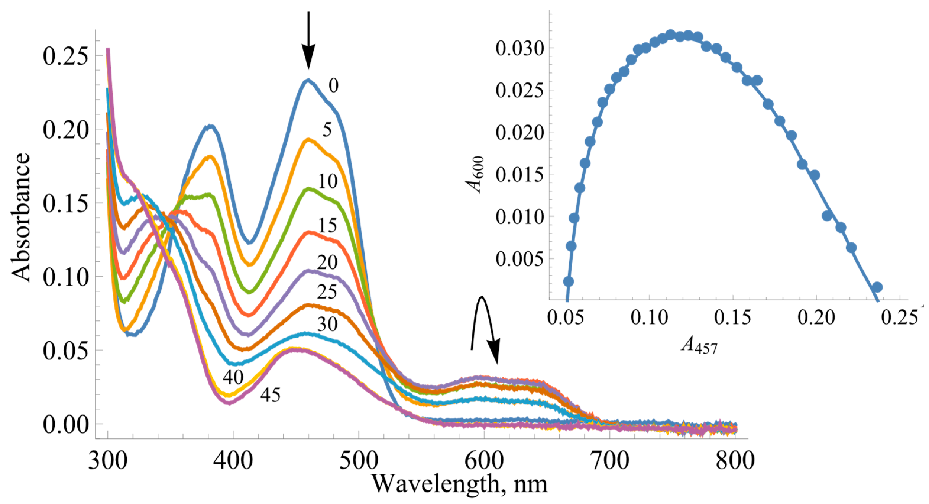
During the photoreduction of BsFNR with 5-deazaFMN and EDTA as a photosensitizer, a neutral (blue) FAD semiquinone (FADH) transiently formed, as evidenced by the characteristic broad absorption band in the 550–650 nm range (Figure 2). The extinction coefficient ε600 for BsFNR FAD is not known; however, the amount of E-FADH can be assessed from the A457 vs. A600 plot (Figure 2 inset) using the value ε600 = 3.6 mM−1cm−1 for the Y50G BsFNR mutant [34]. In this case, the maximal amount of semiquinone stabilized was 44%. The separation between the two single-electron-transfer potentials ( E 7 1 = E 7 E - FAD / E - FADH E 7 E - FADH / E - FADH ) can be calculated based on the semiquinone formation constant Ks (Equations (1) and (2)):
E - FADH max E - FAD tot = K s 2 + K s
Δ E 7 1 V = 0.059 × log K s
where E - FADH max is the maximal concentration of semiquinone and E - FAD tot is the total concentration of enzyme. The calculations yield Ks = 2.572 and E 7 1 = 0.024 V, and, subsequently, E 7 E - FAD / E - FADH = −0.228 V and E 7 E - FADH / E - FADH = −0.252 V.

2.2. Steady-State Kinetics and Oxidant Substrate Specificity of BsFNR

Earlier studies performed on FNRs from other sources indicate that juglone (5-hydroxy-1,4-naphthoquinone) and its derivatives are potent nonphysiological oxidants of these enzymes [31,35]. A series of parallel lines were obtained in double reciprocal plots upon varying the concentration of NADPH with a constant concentration of juglone (Figure 3) and vice versa. This shows that the BsFNR-catalyzed quinone reductase reaction proceeds via a “ping-pong” mechanism.
As calculated according to Equation (9) (presented in the Materials and Methods Section), the kcat value for juglone reduction at an infinite NADPH concentration is equal to 360.2 ± 9.8 s−1, and the values of the bimolecular rate constants ( k cat / K m ) for juglone and NADPH are equal to 4.35 ± 0.23 × 10⁶ M−1s−1 and 1.00 ± 0.07 × 10⁷ M−1s−1, respectively.
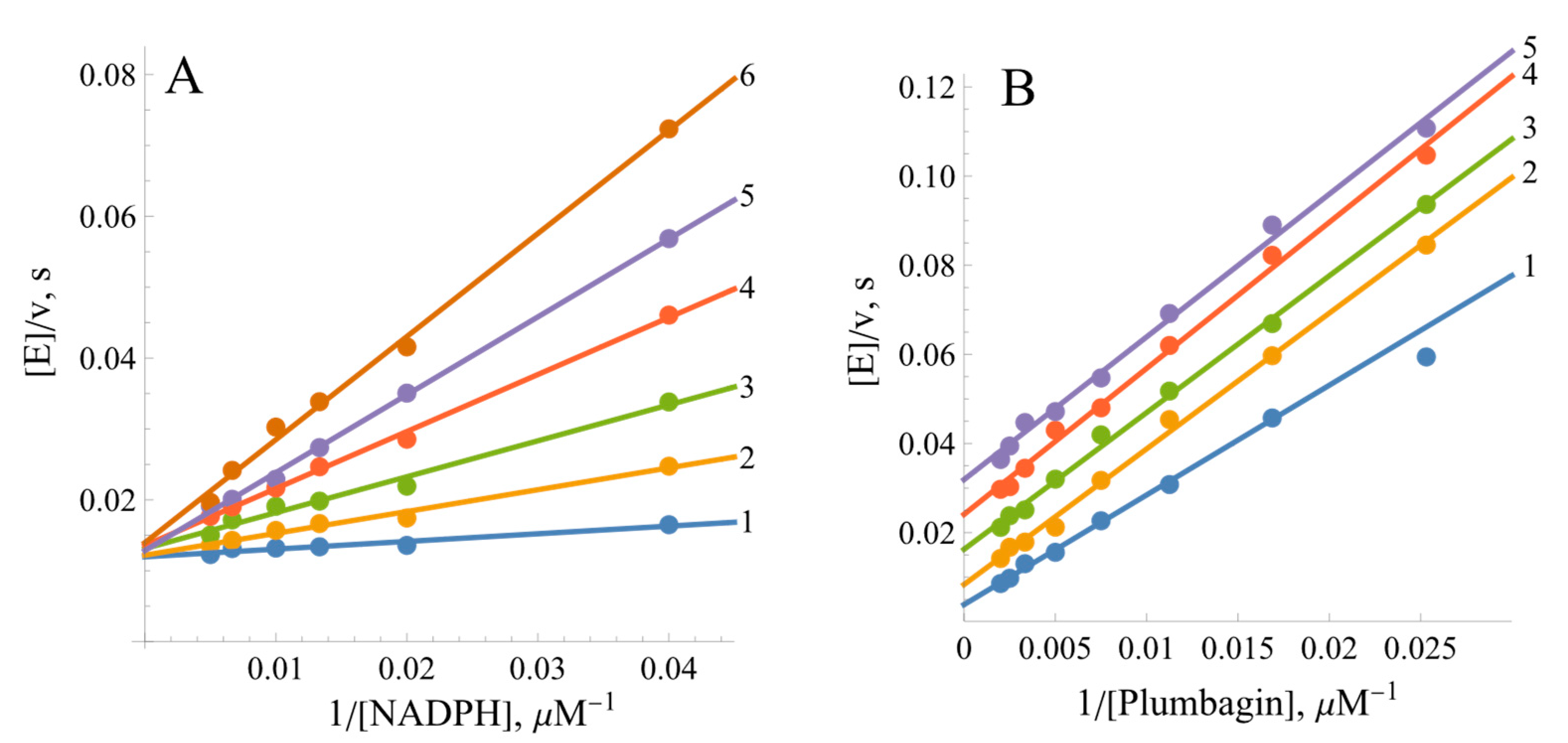
The quinone reductase reaction of BsFNR is inhibited by the reaction product NADP+. At a fixed oxidant concentration, NADP+ acts as a competitive inhibitor towards NADPH, increasing the slopes of the Lineweaver–Burk plots but not affecting the maximal rate of reaction (Figure 4A). Its inhibition constant (Ki), calculated according to Equation (10) (Materials and Methods), is equal to 62.6 ± 10.5 µM. In turn, at a fixed NADPH concentration, NADP+ acts as an uncompetitive inhibitor towards the oxidant, decreasing the maximal reaction rate but not increasing the slopes of the Lineweaver–Burk plots (Figure 4B). Its uncompetitive Ki, calculated according to Equation (11) (shown in the Materials and Methods Section), is equal to 261 ± 27 µM.
A series of quinones (Q), aromatic nitrocompounds (ArNO2), and aromatic N-oxides (ArN→O) with single-electron reduction potentials ( E 7 1 ) from 0.09 to −0.494 V were studied to determine the specificity of BsFNR towards oxidizing substrates. The latter group of compounds, derivatives of 3-amino-1,2,4-benzotriazine-1,4-dioxide (tirapazamine), were studied in connection with their antimicrobial activity [36]. The single-electron acceptors ferricyanide, benzyl viologen, and FeEDTA were also studied. Among the studied forms of ArNO2, there are explosives, such as tetryl and 2,4,6-trinitrotoluene; antibacterial agents, like nitrofurantoin and nifuroxime; and the anticancer agent CB-1954. The apparent maximal reduction rate constants k cat app of the electron acceptors at 100 µM of NADPH and their respective k cat / K m values are given in Table 1.
The log k cat / K m values for ArNO2 exhibit a linear dependence on their E 7 1 values (Table 1, Figure 5), with tetryl (16) being the most potent electron acceptor in that group. On the other hand, the log k cat / K m values for quinones with a similar E 7 1 are about an order of magnitude higher. Moreover, quinones and aromatic N-oxides together exhibit a distinct parabolic dependence on their E 7 1 values (Figure 5). One should also note the single-electron acceptor benzyl viologen (40), with its steady-state kinetic constants being close to those of quinones with similar E 7 1 values.
We found that BsFNR reduces Q and ArNO2 to their radicals. Typically, the single-electron flux for quinone reduction by NAD(P)H-oxidizing enzymes is defined as the ratio of the rate of 1,4-benzoquinone-mediated reduction of the added cytochrome c to the double rate of 1,4-benzoquinone-mediated NAD(P)H enzymatic oxidation at pH < 7.2 [41]. Such an approach is based on the fast reduction of cytochrome c by the 1,4-benzosemiquinone (k ≈ 106 M−1s−1) and its slow reduction by the hydroquinone form. In the case of the BsFNR-catalyzed reduction of 20–100 µM of 1,4-benzoquinone by 100 µM of NADPH, the single-electron flux was equal to 17%. On the other hand, during the reduction of 9,10-phenanthrenequinone, the calculated single-electron flux was equal to 70%, with superoxide dismutase (SOD) (100 U/mL) inhibiting cytochrome c reduction by 43%. The assessment of the single-electron flux for the reduction of ArNO2 is also based on the ArNO2•−-mediated reduction of added cytochrome c. In the presence of 50 µM of NADPH and 100 µM of TNT or p-nitrobenzaldehyde, the rate of BsFNR-catalyzed reduction of 50 µM of cytochrome c was equal to 180–200% of the NADPH oxidation rate, corresponding to 90–100% single-electron flux. SOD inhibited the reactions by 10–30%, reflecting the rapid reoxidation of the radicals with O2 and the participation of the superoxide in the reduction of cytochrome c.
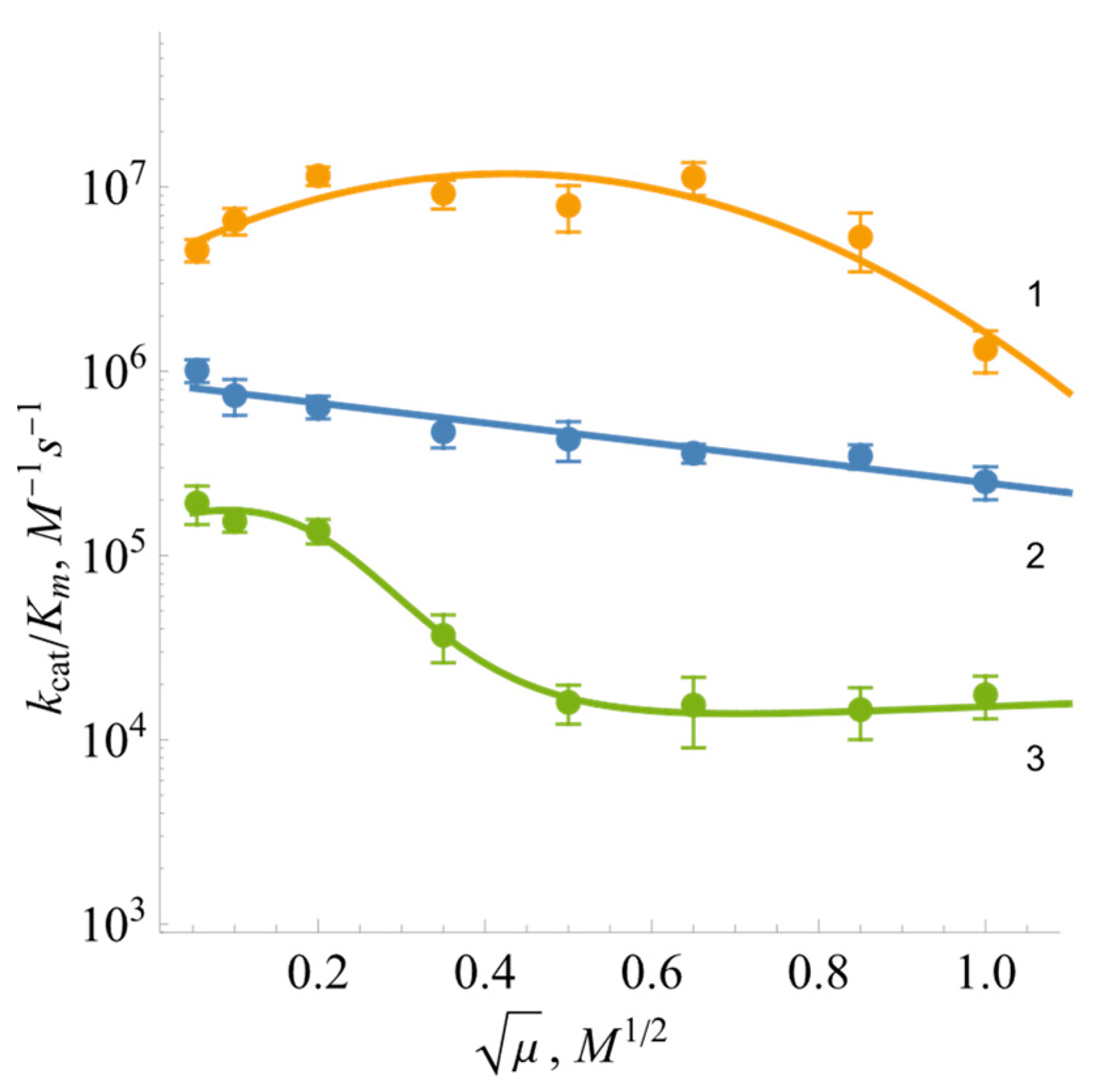
In order to characterize the role of electrostatic interactions in the reduction of oxidants, experiments were performed, with varying ionic strengths, using negatively charged oxidant ferricyanide, positively charged benzyl viologen, and neutral 1,4-naphthoquinone (Figure 6). The log k cat / K m for ferricyanide exhibits a parabolic character with a peak value at I ≈ 0.25 M. The reactivity of uncharged 1,4-napthoquinone decreases slightly upon increasing ionic strength. In general, the log k cat / K m of benzyl viologen decreased upon increasing the ionic strength of the buffer solution (Figure 6).

2.3. Pre-Steady-State Kinetics of BsFNR Oxidation under Multiple Turnover Conditions

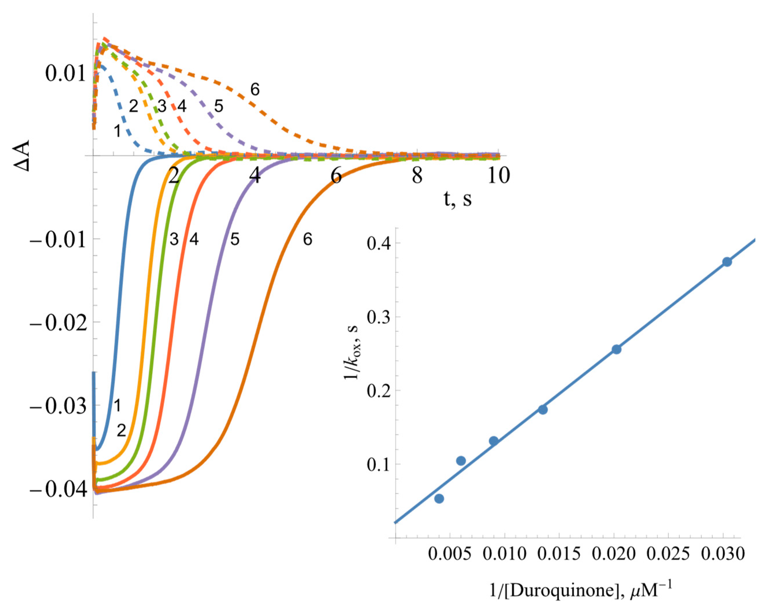
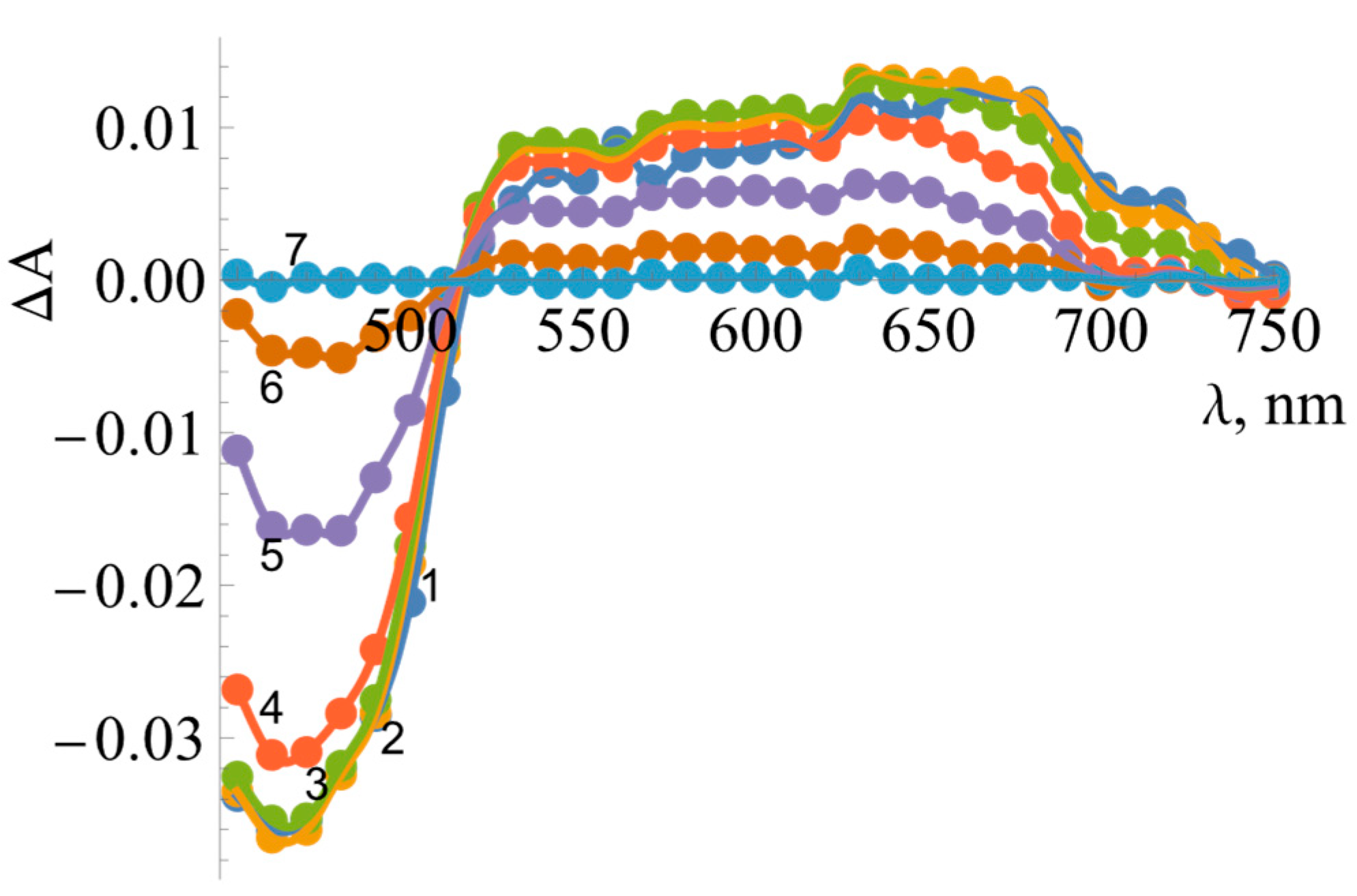
Some insight into the reoxidation mechanism of BsFNR can be gleaned from the spectral changes of the enzyme-bound FAD during the multiple turnovers under aerobic conditions in the presence of NADPH and tetramethyl-1,4-benzoquinone (duroquinone, DQ). DQ does not absorb above 460 nm, and its semiquinone form is rapidly reoxidized by oxygen [37]. The control experiment performed in the absence of DQ showed an initial fast phase of FAD reduction by NADPH, which was then followed by a much slower reoxidation by O2 seen at 460 nm and a transient increase in absorbance at 600 nm (Figure 7A), at the same time scale. The addition of DQ enhanced the reoxidation rate by up to two orders of magnitude (Figure 7B).
The reaction rate constants were calculated according to Equation (3) [42]:
k ox = NADPH 0 E red max × t 1 / 2 ( off )
where [NADPH]0 is the initial concentration of NADPH, [Ered]max is the maximal concentration of the reduced enzyme formed during the turnover, and t1/2(off) is the time interval between the formation of the half-maximum amount of Ered and its decay to the half-maximum value. According to the absorbance difference between the oxidized and reduced enzyme forms, that is, Δε460 = 9.02 mM−1cm−1 [15], the maximal extent of enzyme reduction is 91% (Figure 7A). This yields kox = 0.22 s−1 for the reoxidation of BsFNR by O2, a value that is similar to the enzyme NADPH oxidase activity, 0.17 s−1. Analogous calculations at various DQ concentrations (Figure 8) yield the value of kox(max.) = 48.3 ± 6.7 s−1 (Figure 8 inset), which may reflect the maximum rate of the oxidative half-reaction.
Measurements taken at different wavelengths showed that the intermediate product of reoxidation by DQ has a flat absorption maximum between 600 and 700 nm (Figure 9). The decay of the reaction intermediate begins at about 500 ms after the start of the reaction.

3. Discussion

Since TrxR-type FNRs are relatively poorly studied enzymes, our studies focused on revealing similarities and differences between the redox properties of BsFNR and other groups of FNRs. For the first time, we determined the standard redox potential of BsFNR, i.e., −0.240 V, which is significantly more positive than the E 7 0 of FNRs from other sources, e.g., −0.280 V (P. falciparum [43]) or −0.342 V (spinach [44]). Most importantly, however, it is sufficiently more positive than the potential of its TrxR-type homologue, FNR from R. palustris, with a value of −0.276 V [31]. The amino acids surrounding the isoalloxazine ring of FAD in both enzymes are largely conserved, except for the change of Tyr328 to His324 and Thr329 to Ser325 in BsFNR (Figure 10) [12,13]. The possible effect of the latter substitution on BsFNR E 7 0 is not clear, but the effect of the presence of His324 is similar to the case for flavodoxin from Desulfovibrio vulgaris, where Tyr98His substitution increased the E 7 0 of FMN by 0.07 V [45]. It is believed that the presence of the imidazole group of histidine stabilizes the anionic form of the reduced flavin, i.e., makes its oxidation more difficult. This elevated E 7 0 value agrees with previous data on the inefficient reoxidation of BsFNR by NADP+ [15] and the possible role of this enzyme in Fd/Fld reduction at the expense of NADPH. On the other hand, the FAD semiquinone stability of BsFNR, 44% at equilibrium (Figure 2), is slightly but not drastically higher than that of R. palustris FNR, i.e., 26.5% [31]. The stability of FADH may be due to the presence of Tyr50, Asp57, Ile296, and Thr326, the last of which forms a H-bond with N5 of isoalloxazine, in the active site of BsFNR, corresponding to the Tyr49, Asp56, Ile 300, and Thr330 of R. palustris FNR [46]. The semiquinone-stabilizing effect of the aforementioned amino acid residues has been discussed in our previous work [31].
Due to the possible involvement of BsFNR in reactions with redox-active agents in the soil, further attention was paid to the analysis of reactions of model redox-active compounds of different groups. Here, we found a series of features common to a wide range of FNRs: (i) the “ping-pong” mechanism of reaction and the way of NADP+ inhibition (Figure 3 and Figure 4) are similar to those of other FNRs [31,35]. This result shows that the reductive and oxidative half-reactions occur separately but at the same binding site. The considerable variation in the k cat app values of the reactions with different oxidants (Table 1) suggest that oxidative half-reaction is the rate-limiting step. The competitive inhibition of NADP+ with respect to NADPH and the uncompetitive inhibition with respect to the oxidant is explained by the specific case of the “ping-pong” mechanism, wherein NADP+ strongly competes with NADPH for binding to the oxidized enzyme but binds poorly to the reduced form of the enzyme and/or does not reoxidize it [48]. (ii) Another feature common to various classes of FNRs is the dependence of log k cat / K m of various classes of oxidants on their E 7 1 , i.e., the absence of a pronounced impact of their structural features on reactivity (Figure 5) [31,35]. This may correspond to the “outer sphere” single-electron transfer model [49]. In this case, the rate constant for the electron transfer between reactants (k12) depends on their electron self-exchange rate constants (k11 and k22) and the equilibrium constant of reaction (K):
log K = E 1 V 0.059
k 12 = k 11 × k 22 × K × f
and f is expressed as
log f = log 2   K 4 log k 11 × k 22 Z 2
where Z is the frequency factor, 1011 M−1s−1. According to Equations (5) and (6) for the reaction between an electron donor and a series of homologous electron acceptors with similar k22 values, log k12 will exhibit a parabolic (or linear, in the case of low exothermicity) dependence on ΔE1. We observed this experimentally: the k22 of ArNO2, ≈ 106 M−1s−1, is 100 times lower than that of Q and ArN→O, ≈ 108 M−1s−1 [40,50], leading to a 10-fold lower reactivity of ArNO2 when compared to quinones of similar E 7 1 values (Figure 5). (iii) The transient formation of 500–700 nm absorbing species during enzyme reoxidation is typical for other FNRs [31,35] and consistent with a two-step reoxidation scheme, namely, FADH→FADH→FAD, with FADH oxidation being the rate-limiting step. This means that the obtained k cat / K m of the compounds (Table 1) reflect FADH oxidation. The small absorption increase above 700 nm is uncharacteristic of FADH and indicative of a parallel formation of other reaction intermediates, e.g., the complexes of FADH with NADP(H) observed in adrenodoxin reductase [51,52,53,54].
In parallel, some differences from previously studied FNRs were also observed. The experiments conducted at varied ionic strengths may characterize the region of BsFNR that interacts with charged oxidants. The data on Anabaena PCC7119 and P. falciparum FNRs demonstrate the bell-shaped dependences of log k cat / K m on ionic strength irrespective of the charge of the oxidants [35]. This can be attributed to an interaction with both the negatively charged glutamate and positively charged lysine residues, which are located close to the dimethylbenzene part of the isoalloxazine ring [55,56,57]. However, the data in Figure 6 show that the oxidants interact with the negatively charged amino acid residue(s) of BsFNR. In this case, the potential candidates are Asp285 and, possibly, Asp57 near the isoalloxazine ring of FAD [13]. Most likely, the parallel conformational changes in the protein occur at high ionic strength, decreasing the reactivity of the uncharged 1,4-naphthoquinone (Figure 6). The positively charged fragment of the BsFNR FAD domain, Lys264, Lys317, andArg319 [13], which possibly electrostatically interacts with the negatively charged BsFd [18], may not interact with non-physiological electron acceptors because it is too far from the isoalloxazine ring [15]. However, the most unexpected difference from other FNRs was the mixed one- and two-electron reduction of quinones by BsFNR. Both Anabaena PCC7119 and P. falciparum FNRs reduce both quinones and ArNO2 only in a single-electron fashion [35,58]. The fact that the Glu301Ala mutant of Anabaena FNR reduced 1,4-benzoquinone in a 50% single-electron manner can be explained by its significant destabilization of FADH, 8% at equilibrium [58]. However, BsFNR reduces 1,4-benzoquinone with only 17% of single-electron flux, although the FADH stabilization is 44% (Figure 2). Similarly, another TrxR-type R. palustris FNR with 26.5% FADH stabilization reduces 1,4-benzoquinone with 54% single-electron flux [31]. Since this regularity was observed in the case of two TrxR-type FNRs, it is possible that single- and two-electron reduction flux are not determined by FADH stability alone but also by the specificity of the isoalloxazine surroundings. One of the operative factors could be the unusual π-π stacking between the isoalloxazine ring and the aromatic Tyr50 and His324 groups [16,34]. This feature, including its possible influence on the degradation of xenobiotics, is the subject of our further research.
Finally, the influence of BsFNR structural peculiarities in reactions with redox-active xenobiotics can be quantitatively described. According to Mauk et al., the electron transfer distance in metalloproteins reacting with inorganic complexes under infinite ionic strength (i.e., no electrostatic effects) can be related to the metalloproteins’ electron self-exchange rate constant (k11) (Equation (7)) [59]:
R p Å = 6.3 0.35 ln k 11
This approach has been used to estimate the Rp values in the single-electron oxidation of various flavoenzyme dehydrogenase-electrontransferases [31,35]. However, the values thus obtained should be regarded cautiously due to the possibility of electron transfer distances being overestimated since, in the case of dehydrogenase-electrontransferases, the dimethylbenzene part of the isoalloxazine ring is partly exposed to the solvent. Thus, these data can only be used to compare the “intrinsic” reactivity of different FNRs.
Since there is some uncertainty regarding the mechanism of quinone reduction by BsFNR, we will only analyze the case of ArNO2 reduction. At E 7 1 = 0 , k 12 = k 11 × k 22   (Equation (5)). At E 7 1 of ArNO2 = −0.228 V, i.e., the potential of E-FADH/E-FAD redox couple, and considering that the k22 of ArNO2 is equal to 106 M−1s−1 [50], the value of log k11 calculated according to the data in Figure 5 is equal to 3.11 ± 0.15. According to Equation (7), this yields Rp = 3.8 ± 0.1 Å. For comparison, Rp values calculated in an analogous way for nitroreductase reactions are equal to 4.4 Å (Anabaena FNR), 4.9–5.6 Å (P. falciparum FNR), and 5.4 ± 0.2 Å (R. palustris FNR) [31,35]. This shows that these Rps are close to each other, including for the plant-type FNRs. Complementing the R. palustris FNR data [31], this shows that the active sites of TrxR-type FNRs are not characterized by decreased accessibility to low-molecular-weight oxidants, which may be caused by the shielding of the isoalloxazine ring by the flexible C-terminal region, or by the domain movement of the protein.
In addition to providing fundamental information on the structure and catalysis of this class of FNRs, these data may also have some practical implications, e.g., with respect to using the FNRs of this group in bioelectrocatalysis [60]. In this case, the environment of the redox cofactor and the distance from the surface of the protein usually determine the rate of the corresponding electrochemical reactions.

4. Materials and Methods

4.1. Reagents and Enzymes

B. subtilis ferredoxin:NADP+ oxidoreductase was prepared as previously described, and its concentration was determined spectrophotometrically according to ε457 = 12.3 mM−1cm−1 [16]. NADP(H), 3-acetylpyridineadenine dinucleotide phosphate (APADP+), horse heart cytochrome c, superoxide dismutase, glucose oxidase, catalase, glucose 6-phosphate, glucose-6-phosphate dehydrogenase from Leuconostoc mesenteroides, 5-deaza-FMN, and other commercially available reagents were obtained from Sigma-Aldrich (St. Louis, MO, USA) and used as received.

4.2. Steady-State Kinetics

The steady-state kinetics experiments were performed using a Cary60 UV/Vis spectrophotometer (Agilent Technologies, Santa Clara, CA, USA). All experiments were performed in 0.02 M Hepes/NaOH + 1 mM EDTA buffer solution, with a pH of 7.0, at 25 °C. The kinetic data were fitted to the Michaelis–Menten equation in Mathematica (Wolfram Research, Inc., Mathematica, Version 14.0, Champaign, IL, USA (2024)) (Equation (8)) to yield the steady-state parameters of the reactions, namely, the catalytic constants k cat app and bimolecular reaction rate constants (or catalytic efficiency constants) k cat / K m of the oxidants under a fixed concentration of NADPH:
v E = k cat app S K m + S
where v is the reaction rate, [E] is the concentration of BsFNR, [S] is the concentration of the oxidant, and k cat app represents the number of molecules of NADPH oxidized by a single native molecule of the enzyme per second at saturated concentrations of both substrates. The fitted parameters are equal to the reciprocal intercepts and slopes of Lineweaver–Burk plots, [E]/v vs. 1/[S], respectively. The concentrations of the enzyme used in these experiments were 5–50 nM. The kinetic parameters of the steady-state reactions according to the “ping-pong” mechanism were calculated according to Equation (9):
v E = k cat app Q NADPH K m NADPH Q + K m Q NADPH + NADPH Q
The competitive inhibition constant Ki of NADP+ vs. NADPH was calculated according to Equation (10):
v E = k cat app NADPH K m NADPH 1 + NADP + K i + NADPH
The uncompetitive inhibition constant Ki of NADP+ vs. electron acceptor (Q) was calculated according to Equation (11):
v E = k cat app Q K m Q + Q 1 + NADP + K i
The rates of enzymatic NADPH oxidation in the presence of quinones, nitroaromatics, aromatic N-oxides, or single-electron acceptors were determined using the value Δε340 = 6.2 mM−1cm−1, and they were corrected for the intrinsic NADPH-oxidase activity of BsFNR (0.17 s−1) and/or nonenzymatic NADPH oxidation by high-potential quinones. When 50 µM of cytochrome c was added to the reaction mixture, its quinone- and nitroaromatic-mediated reduction was assessed according to Δε550 = 20 mM−1cm−1. The ferricyanide reduction rate was measured according to Δε420 = 1.03 mM−1cm−1. The rate of BsFNR-catalyzed reduction of APADP+ by NADPH was determined according to Δε363 = 5.6 mM−1cm−1 [33]. APADPH was prepared in situ by reducing APADP+ with 10 mM of glucose 6-phosphate and 0.01 mg/mL of glucose 6-phosphate dehydrogenase, and its concentration was determined according to ε365 = 7.8 mM−1cm−1 [33]. NaCl was used to vary the ionic strength of the buffer solution. The stock solutions of organic compounds were prepared in DMSO; the final concentration of DMSO in reaction mixtures was 1% (v/v). The starting concentration for the oxidants ranged from 100 to 1000 µM, and every compound was measured in a series of measurements with 1.5× dilution for 6–10 different concentrations. Every measurement was performed thrice.

4.3. Presteady-State Kinetics

Pre-steady-state kinetics assays were performed under aerobic conditions using SX20 stopped-flow system (Applied Photophysics, Leatherhead, UK). The reduction of BsFNR by NADPH and its reoxidation by a quinone or oxygen was evaluated at 460 nm and 600 nm. The reaction intermediate spectra were recorded at different wavelengths in the 450–750 nm range over various timepoints. The BsFNR in syringe 1 (10 µM) was mixed with the contents of syringe 2 (100 µM of NADPH or 100 µM of NADPH and 66–500 µM of duroquinone). All measurements were performed in triplicate.

4.4. Photoreduction of BsFNR

The photoreduction of BsFNR (20 µM) was performed under anaerobic conditions in 0.02 M Hepes/NaOH buffer solution (pH 7.0) using 5-deazaFMN (2 µM) and EDTA (8 mM) as photosensitizer and sacrificial reagent, respectively. The reaction mixture contained 10 mM of glucose, 50 nM of glucose oxidase, and 50 nM of catalase. Before the introduction of the concentrated enzyme stock solution, the solution in the sealed spectrophotometer cuvette was flushed with oxygen-free argon for 30 min. The 5-deazaFMN solution and cuvette contents were protected from light before illumination. The cell was subsequently illuminated for one-minute-long intervals using a 100 W incandescent lamp at a distance of 20 cm. The progress of the reaction was followed spectrophotometrically for 1–3 h; each spectrum is an average of two.

Author Contributions

Conceptualization, N.Č.; formal analysis, M.L. and N.Č.; investigation, M.L. and G.M.; methodology, M.L. and N.Č.; resources, D.S. and N.Č.; supervision, N.Č.; visualization, M.L. and G.M.; writing—original draft, M.L. and G.M.; writing—review and editing, M.L., D.S., and N.Č. All authors have read and agreed to the published version of the manuscript.

Funding

M.L., D.S., G.M. and N.Č. did not receive external funding.

Institutional Review Board Statement

Not applicable.

Informed Consent Statement

Not applicable.

Data Availability Statement

The data can be provided by the authors upon reasonable request.

Acknowledgments

We are grateful to J. Šarlauskas (Institute of Biochemistry, Life Sciences Center, Vilnius University) for generously providing noncommercial nitroaromatics and aromatic N-oxides and to V. Miškinienė (Institute of Biochemistry, Life Sciences Center, Vilnius University) for the generous gift of CB-1954. Some of the data presented in this article were used by G.M. for her BSc thesis. Molecular graphics and analyses were performed with UCSF ChimeraX developed by the Resource for Biocomputing, Visualization, and Informatics at the University of California, San Francisco, with support from the National Institutes of Health R01-GM129325 and the Office of Cyber Infrastructure and Computational Biology, National Institute of Allergy and Infectious Diseases.

Conflicts of Interest

The funders had no role in the design of this study; in the collection, analyses, or interpretation of data; in the writing of the manuscript; or in the decision to publish the results.

References

  1. Hanke, G.T.; Kurisu, G.; Kusunoki, M.; Hase, T. Fd:FNR electron transfer complexes: Evolutionary refinement of structural interactions. Photosynth. Res. 2004, 81, 317–327. [Google Scholar] [CrossRef] [PubMed]
  2. Ceccarelli, E.A.; Arakaki, A.K.; Cortez, N.; Carrillo, N. Functional plasticity and catalytic efficiency in plant and bacterial ferredoxin-NADP(H) reductases. Biochim. Biophys. Acta—Proteins Proteom. 2004, 1698, 155–165. [Google Scholar] [CrossRef]
  3. Aliverti, A.; Pandini, V.; Pennati, A.; de Rosa, M.; Zanetti, G. Structural and functional diversity of ferredoxin-NADP+ reductases. Arch. Biochem. Biophys. 2008, 474, 283–291. [Google Scholar] [CrossRef] [PubMed]
  4. Karplus, P.A.; Daniels, M.J.; Herriott, J.R. Atomic structure of ferredoxin-NADP+ reductase: Prototype for a structurally novel flavoenzyme family. Science 1991, 251, 60–66. [Google Scholar] [CrossRef] [PubMed]
  5. Iyanagi, T. Roles of ferredoxin-NADP+ oxidoreductase and flavodoxin in NAD(P)H-dependent electron transfer systems. Antioxidants 2022, 11, 2143. [Google Scholar] [CrossRef] [PubMed]
  6. Onda, Y.; Matsumura, T.; Kimata-Ariga, Y.; Sakakibara, H.; Sugiyama, T.; Hase, T. Differential interaction of maize root ferredoxin:NADP+ oxidoreductase with photosynthetic and non-photosynthetic ferredoxin isoproteins. Plant Physiol. 2000, 123, 1037–1046. [Google Scholar] [CrossRef] [PubMed]
  7. Röhrich, R.C.; Englert, N.; Troschke, K.; Reichenberg, A.; Hintz, M.; Seeber, F.; Balconi, E.; Aliverti, A.; Zanetti, G.; Köhler, U.; et al. Reconstitution of an apicoplast-localised electron transfer pathway involved in the isoprenoid biosynthesis of Plasmodium falciparum. FEBS Lett. 2005, 579, 6433–6438. [Google Scholar] [CrossRef] [PubMed]
  8. Nogués, I.; Tejero, J.; Hurley, J.K.; Paladini, D.; Frago, S.; Tollin, G.; Mayhew, S.G.; Gómez-Moreno, C.; Ceccarelli, E.A.; Carrillo, N.; et al. Role of the C-terminal tyrosine of ferredoxin-nicotinamide adenine dinucleotide phosphate reductase in the electron transfer processes with its protein partners ferredoxin and flavodoxin. Biochemistry 2004, 43, 6127–6137. [Google Scholar] [CrossRef]
  9. Medina, M.; Gomez-Moreno, C.; Tollin, G. Effects of chemical modification of Anabaena flavodoxin and ferredoxin-NADP+ reductase on the kinetics of interprotein electron transfer reactions. Eur. J. Biochem. 1992, 210, 577–583. [Google Scholar] [CrossRef]
  10. Marcuello, C.; de Miguel, R.; Martínez-Júlvez, M.; Gómez-Moreno, C.; Lostao, A. Mechanostability of the single-electron-transfer complexes of Anabaena ferredoxin-NADP+ reductase. ChemPhysChem 2015, 16, 3161–3169. [Google Scholar] [CrossRef]
  11. Hammerstad, M.; Hersleth, H.-P. Overview of structurally homologous flavoprotein oxidoreductases containing the low Mr thioredoxin reductase-like fold—A functionally diverse group. Arch. Biochem. Biophys. 2021, 702, 108826. [Google Scholar] [CrossRef] [PubMed]
  12. Seo, D.; Muraki, N.; Kurisu, G. Kinetic and structural insight into a role of the re-face Tyr328 residue of the homodimer type ferredoxin-NADP+ oxidoreductase from Rhodopseudomonas palustris in the reaction with NADP+/NADPH. Biochim. Biophys. Acta—Bioenerg. 2020, 1861, 148140. [Google Scholar] [CrossRef] [PubMed]
  13. Komori, H.; Seo, D.; Sakurai, T.; Higuchi, Y. Crystal structure analysis of Bacillus subtilis ferredoxin-NADP+ oxidoreductase and the structural basis for its substrate selectivity. Protein Sci. 2010, 19, 2279–2290. [Google Scholar] [CrossRef] [PubMed]
  14. Meng, E.C.; Goddard, T.D.; Pettersen, E.F.; Couch, G.S.; Pearson, Z.J.; Morris, J.H.; Ferrin, T.E. ChimeraX: Tools for structure building and analysis. Protein Sci. 2023, 32, e4792. [Google Scholar] [CrossRef] [PubMed]
  15. Seo, D.; Soeta, T.; Sakurai, H.; Sétif, P.; Sakurai, T. Pre-steady-state kinetic studies of redox reactions catalysed by Bacillus subtilis ferredoxin-NADP+ oxidoreductase with NADP+/NADPH and ferredoxin. Biochim. Biophys. Acta—Bioenerg. 2016, 1857, 678–687. [Google Scholar] [CrossRef] [PubMed]
  16. Seo, D.; Asano, T.; Komori, H.; Sakurai, T. Role of the C-terminal extension stacked on the re-face of the isoalloxazine ring moiety of the flavin adenine dinucleotide prosthetic group in ferredoxin-NADP+ oxidoreductase from Bacillus subtilis. Plant Physiol. Bioch. 2014, 81, 143–148. [Google Scholar] [CrossRef] [PubMed]
  17. Seo, D.; Naito, H.; Nishimura, E.; Sakurai, T. Replacement of Tyr50 stacked on the si-face of the isoalloxazine ring of the flavin adenine dinucleotide prosthetic group modulates Bacillus subtilis ferredoxin-NADP+ oxidoreductase activity toward NADPH. Photosynth. Res. 2015, 125, 321–328. [Google Scholar] [CrossRef] [PubMed]
  18. Green, A.J.; Munro, A.W.; Cheesman, M.R.; Reid, G.A.; von Wachenfeldt, C.; Chapman, S.K. Expression, purification and characterisation of a Bacillus subtilis ferredoxin: A potential electron transfer donor to cytochrome P450 BioI. J. Inorg. Biochem. 2003, 93, 92–99. [Google Scholar] [CrossRef] [PubMed]
  19. Lawson, R.J.; von Wachenfeldt, C.; Haq, I.; Perkins, J.; Munro, A.W. Expression and characterization of the two flavodoxin proteins of Bacillus subtilis, YkuN and YkuP: Biophysical properties and interactions with cytochrome P450 BioI. Biochemistry 2004, 43, 12390–12409. [Google Scholar] [CrossRef]
  20. Chazarreta-Cifre, L.; Martiarena, L.; de Mendoza, D.; Altabe, S.G. Role of ferredoxin and flavodoxins in Bacillus subtilis fatty acid desaturation. J. Bacteriol. 2011, 193, 4043–4048. [Google Scholar] [CrossRef]
  21. Holden, J.K.; Lim, N.; Poulos, T.L. Identification of redox partners and development of a novel chimeric bacterial nitric oxide synthase for structure activity analyses. J. Biol. Chem. 2014, 289, 29437–29445. [Google Scholar] [CrossRef]
  22. Liebeke, M.; Pöther, D.C.; Van Duy, N.; Albrecht, D.; Becher, D.; Hochgräfe, F.; Lalk, M.; Hecker, M.; Antelmann, H. Depletion of thiol-containing proteins in response to quinones in Bacillus subtilis. Mol. Microbiol. 2008, 69, 1513–1529. [Google Scholar] [CrossRef] [PubMed]
  23. Tauber, J.P.; Schroeckh, V.; Shelest, E.; Brakhage, A.A.; Hoffmeister, D. Bacteria induce pigment formation in the basidiomycete Serpula lacrymans. Environ. Microbiol. 2016, 18, 5218–5227. [Google Scholar] [CrossRef] [PubMed]
  24. Imlay, J.A. Where in the world do bacteria experience oxidative stress? Environ. Microbiol. 2019, 21, 521–530. [Google Scholar] [CrossRef] [PubMed]
  25. Kapley, A.; Prasad, S.; Purohit, H.J. Changes in microbial diversity in fed-batch reactor operation with wastewater containing nitroaromatic residues. Bioresource Technol. 2007, 98, 2479–2484. [Google Scholar] [CrossRef]
  26. Ni, H.; Yao, L.; Li, N.; Cao, Q.; Dai, C.; Zhang, J.; He, Q.; He, J. Biodegradation of pendimethalin by Bacillus subtilis Y3. J. Environ. Sci.—China 2016, 41, 121–127. [Google Scholar] [CrossRef]
  27. Cichocki, B.A.; Donzel, M.; Heimsch, K.C.; Lesanavičius, M.; Feng, L.; Montagut, E.J.; Becker, K.; Aliverti, A.; Elhabiri, M.; Čėnas, N.; et al. Plasmodium falciparum ferredoxin-NADP+ reductase-catalyzed redox cycling of plasmodione generates both predicted key drug metabolites: Implication for antimalarial drug development. ACS Infect. Dis. 2021, 7, 1996–2012. [Google Scholar] [CrossRef]
  28. Lampropoulos, P.K.; Gkentzi, D.; Tzifas, S.; Dimitriou, G. Neonatal sepsis due to Bacillus subtilis. Cureus 2021, 13, e17692. [Google Scholar] [CrossRef]
  29. Tokano, M.; Tarumoto, N.; Imai, K.; Sakai, J.; Maeda, T.; Kawamura, T.; Seo, K.; Takahashi, K.; Yamamoto, T.; Maesaki, S. Bacterial meningitis caused by Bacillus subtilis var. natto. Internal Med. 2023, 62, 1989–1993. [Google Scholar] [CrossRef]
  30. Elfmann, C.; Zhu, B.; Stülke, J.; Halbedel, S. ListiWiki: A database for the foodborne pathogen Listeria monocytogenes. Int. J. Med. Microbiol. 2023, 313, 151591. [Google Scholar] [CrossRef]
  31. Lesanavičius, M.; Seo, D.; Čėnas, N. Thioredoxin reductase-type ferredoxin:NADP+ oxidoreductase of Rhodopseudomonas palustris: Potentiometric characteristics and reactions with nonphysiological oxidants. Antioxidants 2022, 11, 1000. [Google Scholar] [CrossRef] [PubMed]
  32. Cheng, Z.; Arscott, L.D.; Ballou, D.P.; Williams, C.H. The relationship of the redox potentials of thioredoxin and thioredoxin reductase from Drosophila melanogaster to the enzymatic mechanism: Reduced thioredoxin is the reductant of glutathione in drosophila. Biochemistry 2007, 46, 7875–7885. [Google Scholar] [CrossRef] [PubMed]
  33. Kaplan, N.O.; Ciotti, M.M. Chemistry and properties of the 3-acetylpyridine analogue of diphosphopyridine nucleotide. J. Biol. Chem. 1956, 221, 823–832. [Google Scholar] [CrossRef]
  34. Seo, D. The role of the si-face tyrosine of a homodimeric ferredoxin-NADP+ oxidoreductase from Bacillus subtilis during complex formation and redox equivalent transfer with NADP+/H and ferredoxin. Antioxidants 2023, 12, 1741. [Google Scholar] [CrossRef] [PubMed]
  35. Lesanavičius, M.; Aliverti, A.; Šarlauskas, J.; Čėnas, N. Reactions of Plasmodium falciparum ferredoxin:NADP+ oxidoreductase with redox cycling xenobiotics: A mechanistic study. Int. J. Mol. Sci. 2020, 21, 3234. [Google Scholar] [CrossRef] [PubMed]
  36. Shah, Z.; Mahbuba, R.; Turcotte, B. The anticancer drug tirapazamine has antimicrobial activity against Escherichia coli, Staphylococcus aureus and Clostridium difficile. FEMS Microbiol. Lett. 2013, 347, 61–69. [Google Scholar] [CrossRef] [PubMed]
  37. Wardman, P. Reduction potentials of one-electron couples involving free radicals in aqueous solution. J. Phys. Chem. Ref. Data 1989, 18, 1637–1755. [Google Scholar] [CrossRef]
  38. Song, Y.; Buettner, G.R. Thermodynamic and kinetic considerations for the reaction of semiquinone radicals to form superoxide and hydrogen peroxide. Free Radical Bio. Med. 2010, 49, 919–962. [Google Scholar] [CrossRef]
  39. Čėnas, N.; Nemeikaitė-Čėnienė, A.; Kosychova, L. Single- and two-electron reduction of nitroaromatic compounds by flavoenzymes: Mechanisms and implications for cytotoxicity. Int. J. Mol. Sci. 2021, 22, 8534. [Google Scholar] [CrossRef]
  40. Nemeikaitė-Čėnienė, A.; Šarlauskas, J.; Jonušienė, V.; Marozienė, A.; Misevičienė, L.; Yantsevich, A.V.; Čėnas, N. Kinetics of flavoenzyme-catalyzed reduction of tirapazamine derivatives: Implications for their prooxidant cytotoxicity. Int. J. Mol. Sci. 2019, 20, 4602. [Google Scholar] [CrossRef]
  41. Iyanagi, T.; Yamazaki, I. One-electron-transfer reactions in biochemical systems V. Difference in the mechanism of quinone reduction by the NADH dehydrogenase and the NAD(P)H dehydrogenase (DT-diaphorase). Biochim. Biophys. Acta—Bioenerg. 1970, 216, 282–294. [Google Scholar] [CrossRef] [PubMed]
  42. Chance, B. A simple relationship for a calculation of the “on” velocity constant in enzyme reactions. Arch. Biochem. Biophys. 1957, 71, 130–136. [Google Scholar] [CrossRef] [PubMed]
  43. Balconi, E.; Pennati, A.; Crobu, D.; Pandini, V.; Cerutti, R.; Zanetti, G.; Aliverti, A. The ferredoxin-NADP+ reductase/ferredoxin electron transfer system of Plasmodium falciparum. FEBS J. 2009, 276, 3825–3836. [Google Scholar] [CrossRef]
  44. Batie, C.J.; Kamin, H. The relation of pH and oxidation-reduction potential to the association state of the ferredoxin•ferredoxin:NADP+ reductase complex. J. Biol. Chem. 1981, 256, 7756–7763. [Google Scholar] [CrossRef]
  45. Swenson, R.P.; Krey, G.D. Site-directed mutagenesis of tyrosine-98 in the flavodoxin from Desulfovibrio vulgaris (hildenborough): Regulation of oxidation-reduction properties of the bound FMN cofactor by aromatic, solvent, and electrostatic interactions. Biochemistry 1994, 33, 8505–8514. [Google Scholar] [CrossRef] [PubMed]
  46. Seo, D.; Okabe, S.; Yanase, M.; Kataoka, K.; Sakurai, T. Studies of interaction of homo-dimeric ferredoxin-NAD(P)+ oxidoreductases of Bacillus subtilis and Rhodopseudomonas palustris, that are closely related to thioredoxin reductases in amino acid sequence, with ferredoxins and pyridine nucleotide coenzymes. Biochim. Biophys. Acta—Proteins Proteom. 2009, 1794, 594–601. [Google Scholar] [CrossRef] [PubMed]
  47. Larkin, M.A.; Blackshields, G.; Brown, N.P.; Chenna, R.; Mcgettigan, P.A.; McWilliam, H.; Valentin, F.; Wallace, I.M.; Wilm, A.; Lopez, R.; et al. Clustal W and Clustal X version 2.0. Bioinformatics 2007, 23, 2947–2948. [Google Scholar] [CrossRef] [PubMed]
  48. Valiauga, B.; Williams, E.M.; Ackerley, D.F.; Čėnas, N. Reduction of quinones and nitroaromatic compounds by Escherichia coli nitroreductase A (NfsA): Characterization of kinetics and substrate specificity. Arch. Biochem. Biophys. 2017, 614, 14–22. [Google Scholar] [CrossRef] [PubMed]
  49. Marcus, R.A.; Sutin, N. Electron transfers in chemistry and biology. Biochim. Biophys. Acta—Rev. Bioenerg. 1985, 811, 265–322. [Google Scholar] [CrossRef]
  50. Wardman, P.; Dennis, M.F.; Everett, S.A.; Patel, K.B.; Stratford, M.R.L.; Tracy, M. Radicals from one-electron reduction of nitro compounds, aromatic N-oxides and quinones: The kinetic basis for hypoxia-selective, bioreductive drugs. Biochem. Soc. Symp. 1995, 61, 171–194. [Google Scholar] [CrossRef]
  51. Edmondson, D.E.; Tollin, G. Semiquinone formation in flavo- and metalloflavoproteins. In Topics in Current Chemistry; Springer: Berlin, Heidelberg, 1983; Volume 108, pp. 109–138. [Google Scholar]
  52. Tejero, J.; Peregrina, J.R.; Martínez-Júlvez, M.; Gutiérrez, A.; Gómez-Moreno, C.; Scrutton, N.S.; Medina, M. Catalytic mechanism of hydride transfer between NADP+/H and ferredoxin-NADP+ reductase from Anabaena PCC 7119. Arch. Biochem. Biophys. 2007, 459, 79–90. [Google Scholar] [CrossRef] [PubMed]
  53. Kitagawa, T.; Sakamoto, H.; Sugiyama, T.; Yamano, T. Formation of the semiquinone form in the anaerobic reduction of adrenodoxin reductase by NADPH. Resonance Raman, EPR, and optical spectroscopic evidence. J. Biol. Chem. 1982, 257, 12075–12080. [Google Scholar] [CrossRef]
  54. Sakamoto, H.; Ohta, M.; Miura, R.; Sugiyama, T.; Yamano, T.; Miyake, Y. Studies on the reaction mechanism of NADPH-adrenodoxin reductase with NADPH. J. Biochem. 1982, 92, 1941–1950. [Google Scholar] [CrossRef]
  55. Aliverti, A.; Corrado, M.E.; Zanetti, G. Involvement of lysine-88 of spinach ferredoxin-NADP+ reductase in the interaction with ferredoxin. FEBS Lett. 1994, 343, 247–250. [Google Scholar] [CrossRef] [PubMed]
  56. Aliverti, A.; Deng, Z.; Ravasi, D.; Piubelli, L.; Karplus, P.A.; Zanetti, G. Probing the function of the invariant glutamyl residue 312 in spinach ferredoxin-NADP+ reductase. J. Biol. Chem. 1998, 273, 34008–34015. [Google Scholar] [CrossRef]
  57. Hurley, J.K.; Hazzard, J.T.; Martínez-Júlvez, M.; Medina, M.; Gómez-Moreno, C.; Tollin, G. Electrostatic forces involved in orienting Anabaena ferredoxin during binding to Anabaena ferredoxin:NADP+ reductase: Site-specific mutagenesis, transient kinetic measurements, and electrostatic surface potentials. Protein Sci. 1999, 8, 1614–1622. [Google Scholar] [CrossRef] [PubMed]
  58. Anusevičius, Ž.; Misevičienė, L.; Medina, M.; Martinez-Julvez, M.; Gomez-Moreno, C.; Čėnas, N. FAD semiquinone stability regulates single- and two-electron reduction of quinones by Anabaena PCC7119 ferredoxin:NADP+ reductase and its Glu301Ala mutant. Arch. Biochem. Biophys. 2005, 437, 144–150. [Google Scholar] [CrossRef] [PubMed]
  59. Mauk, A.G.; Scott, R.A.; Gray, H.B. Distances of electron transfer to and from metalloprotein redox sites in reactions with inorganic complexes. J. Am. Chem. Soc. 1980, 102, 4360–4363. [Google Scholar] [CrossRef]
  60. Herold, R.A.; Reinbold, R.; Schofield, C.J.; Armstrong, F.A. NADP(H)-dependent biocatalysis without adding NADP(H). Proc. Natl. Acad. Sci. USA 2023, 120, e2214123120. [Google Scholar] [CrossRef]
Figure 1. The 3D structure of a BsFNR dimer (PDB id: 3LZX). FAD-binding domain is colored yellow, while the NADP(H)-binding domain is colored red. The box denotes amino acids comprising each domain in a monomer. The bound FAD is shown in purple. The molecular graphics were made using UCSF ChimeraX [14].
Ijms 25 05373 g001
Figure 2. The determination of the redox potentials of BsFNR. The photoreduction of 20 µM of BsFNR at different times of illumination, with the arrows indicating the changes in the spectra. The numbers next to the curves correspond to the total duration of illumination in minutes. Inset shows the interdependence of absorbance changes at 457 and 600 nm during the photoreduction of 20 µM of BsFNR.
Ijms 25 05373 g002
Figure 3. Lineweaver–Burk plots of the steady-state kinetics of the oxidation of NADPH catalyzed by BsFNR with varied concentrations of juglone (5-hydroxy-1,4-naphthoquinone): 1—200 µM of juglone, 2—133 µM of juglone, 3—89 µM of juglone, 4—59 µM of juglone, 5—39 µM of juglone, 6—26 µM of juglone, and 7—17 µM of juglone.
Ijms 25 05373 g003
Figure 4. Inhibition of BsFNR-catalyzed reactions by the reaction product NADP+. (A) Competitive inhibition at varied concentrations of NADPH in the presence of 200 µM of plumbagin (5-hydroxy-2-methyl-1,4-naphthoquinone): 1—no NADP+, 2—100 µM of NADP+, 3—250 µM of NADP+, 4—500 µM of NADP+, 5—750 µM of NADP+, and 6—1000 µM of NADP+. (B) Uncompetitive inhibition at varied plumbagin concentrations in the presence of 100 µM of NADPH: 1—no NADP+, 2—500 µM of NADP+, 3—1000 µM of NADP+, 4—2000 µM of NADP+, and 5—3000 µM of NADP+.
Ijms 25 05373 g004
Figure 5. Dependence of the reactivity of quinones, nitroaromatic compounds, aromatic N-oxides, and benzyl viologen on their single-electron reduction midpoint potentials (log10 scale). Numbers and reduction potentials of the compounds are given in Table 1.
Ijms 25 05373 g005
Figure 6. The dependence of log kcat/Km of BsFNR-catalyzed reaction on the ionic strength of the buffer: 1—potassium ferricyanide, 2—1,4-naphthoquinone, and 3—benzyl viologen.
Ijms 25 05373 g006
Figure 7. The absorbance changes at 460 nm (1, orange) and 600 nm (2, blue) during the reduction of BsFNR (5 µM) by 50 µM of NADPH and its subsequent reoxidation by O2 (A) or 250 µM of duroquinone (B) (concentrations were reported after mixing).
Ijms 25 05373 g007
Figure 8. The kinetics of BsFNR (5 µM) reduction and reoxidation under multiple turnover conditions with a varying concentration of duroquinone in the presence of 50 µM of NADPH followed at 460 nm (solid curves) and 600 nm (dashed curves): 1—250 µM of duroquinone, 2—166 µM of duroquinone, 3—111 µM of duroquinone, 4—74 µM of duroquinone, 5—49 µM of duroquinone, and 6—33 µM of duroquinone (all concentrations were reported after mixing). The inset shows the Lineweaver—Burk plot for the dependence of the apparent first-order rate constant of the reoxidation of reduced FAD on the concentration of duroquinone.
Ijms 25 05373 g008
Figure 9. The spectra of reaction intermediates formed during the turnover of 5 µM of BsFNR in the presence of 50 µM of NADPH and 250 µM of duroquinone (concentrations were reported after mixing). Differences in absorbance are shown at several timepoints over the 450–750 nm range. Spectra correspond to 50 ms (1, blue), 100 ms (2, orange), 250 ms (3, green), 500 ms (4, red), 750 ms (5, purple), 1 s (6, brown), and 5 s (7, teal) after mixing.
Ijms 25 05373 g009
Figure 10. The partially aligned amino acid sequences of BsFNR, R. palustris FNR (RPA3954) and Chlorobaculum tepidum FNR (CT1512). The residues stacked on the si-face (Tyr50 in BsFNR) and re-face (His324 in BsFNR) of the isoalloxazine ring portion are indicated with arrowheads. The alignment was performed using ClustalW [47].
Ijms 25 05373 g010
Table 1. Steady-state rate constants of the reduction of non-physiological electron acceptors by 100 µM of NADPH catalyzed by BsFNR. The E 7 1 values were taken from [37,38,39,40].
No.Compound E 7 1 , V k cat app , s−1 k cat / K m , M−1s−1
Quinones
11,4-Benzoquinone0.090209.8 ± 5.53.7 ± 0.4 × 10⁶
22-Methyl-1,4-benzoquinone0.010267.3 ± 7.82.3 ± 0.3 × 10⁶
32,3-Dichloro-1,4-naphthoquinone−0.03658.2 ± 1.13.5 ± 0.3 × 10⁶
42,3-Dimethyl-1,4-benzoquinone−0.074301.9 ± 19.81.1 ± 0.1 × 10⁶
52,6-Dimethyl-1,4-benzoquinone−0.080183.1 ± 5.01.0 ± 0.1 × 10⁶
65-Hydroxy-1,4-naphthoquinone−0.090221.4 ± 2.64.4 ± 0.2 × 10⁶
75,8-Hihydroxy-1,4-naphthoquinone−0.11096.0 ± 3.55.9 ± 0.4 × 10⁶
89,10-Phenanthrenequinone−0.12065.3 ± 2.17.5 ± 0.8 × 10⁶
91,4-Naphthoquinone−0.150180.1 ± 28.86.5 ± 0.5 × 10⁵
105-Hydroxy-2-methyl-1,4-naphthoquinone−0.156142.7 ± 8.76.0 ± 0.4 × 10⁵
112-Methyl-1,4-naphthoquinone−0.20068.9 ± 8.31.9 ± 0.3 × 10⁵
12Tetramethyl-1,4-benzoquinone−0.26013.3 ± 1.29.9 ± 0.8 × 10⁴
139,10-Anthraquinone-2-sulphonate−0.38016.2 ± 0.66.0 ± 0.7 × 10⁴
142-Hydroxy-1,4-naphthoquinone−0.4102.5 ± 0.41.6 ± 0.3 × 10⁴
152-Hydroxy-3-methyl-1,4-naphthoquinone−0.4605.8 ± 0.41.5 ± 0.2 × 10⁴
Nitroaromatic compounds
162,4,6-Trinitrophenyl-N-methylnitramine (tetryl)−0.19143.4 ± 6.06.0 ± 0.5 × 10⁴
17N-methylpicramide−0.22517.5 ± 2.43.0 ± 0.2 × 10⁴
182,4,6-Trinitrotoluene (TNT)−0.25315.3 ± 0.64.0 ± 0.5 × 10⁴
19Nitrofurantoin−0.25535.3 ± 7.21.8 ± 0.3 × 10⁴
20Nifuroxim−0.25538.2 ± 4.04.4 ± 0.4 × 10⁴
21p-Dinitrobenzene−0.2579.3 ± 0.52.3 ± 0.3 × 10⁴
22o-Dinitrobenzene−0.2876.2 ± 0.41.4 ± 0.2 × 10⁴
232-Nitrobenzaldehyde −0.30826.4 ± 2.71.1 ± 0.2 × 10⁴
243,5-Dinitrobenzamide−0.31120.9 ± 1.31.4 ± 0.2 × 10⁴
254-Nitrobenzaldehyde−0.32520.8 ± 1.87.6 ± 0.7 × 10³
263,5-Dinitrobenzoic acid−0.3444.7 ± 0.32.5 ± 0.3 × 10³
27m-Dinitrobenzene−0.34812.1 ± 0.76.1 ± 0.7 × 10³
284-Nitroacetophenone−0.35516.2 ± 1.65.7 ± 0.4 × 10³
295-(Aziridin-1-yl)-2,4-dinitrobenzamide (CB-1954)−0.3853.6 ± 0.44.3 ± 0.2 × 10³
302-Amino-4,6-dinitrotoluene−0.4172.0 ± 0.18.3 ± 0.9 × 10³
314-Amino-2,6-dinitrotoluene−0.4490.7 ± 0.044.3 ± 0.5 × 10³
323-Nitro-1,2,4-triazolone−0.4720.3 ± 0.037.3 ± 0.9 × 10²
334-Nitrobenzyl alcohol−0.4751.4 ± 0.18.2 ± 0.9 × 10²
Aromatic N-oxides
347-Trifluoromethoxytirapazamine−0.3455.4 ± 0.31.2 ± 0.1 × 10⁴
357-Fluorotirapazamine−0.4006.2 ± 0.21.2 ± 0.1 × 10⁴
36Tirapazamine−0.4565.6 ± 0.33.5 ± 0.4 × 10³
377-Ethoxytirapazamine−0.4942.3 ± 0.45.8 ± 0.5 × 10³
Single-electron acceptors
38Ferricyanide a0.410557.5 ± 18.16.3 ± 0.8 × 10⁶
39FeEDTA0.1202.0 ± 0.15.7 ± 0.6 × 10³
40Benzyl viologen−0.35438.4 ± 3.71.9 ± 0.3 × 10⁵
a the k cat app and k cat / K m values were calculated according to the reduction of ferricyanide on a single-electron basis.
Disclaimer/Publisher’s Note: The statements, opinions and data contained in all publications are solely those of the individual author(s) and contributor(s) and not of MDPI and/or the editor(s). MDPI and/or the editor(s) disclaim responsibility for any injury to people or property resulting from any ideas, methods, instructions or products referred to in the content.