Article

Glutamate Receptor-like (GLR) Family in Brassica napus: Genome-Wide Identification and Functional Analysis in Resistance to Sclerotinia sclerotiorum

1
Key Laboratory of Biology and Ecological Control of Crop Pathogens and Insects of Zhejiang Province, Institute of Biotechnology, College of Agriculture and Biotechnology, Zhejiang University, Hangzhou 310058, China
2
Centre of Analysis and Measurement, Zhejiang University, 866 Yu Hang Tang Road, Hangzhou 310058, China
3
Department of Botany, Shah Abdul Latif University, Khairpur 66020, Sindh, Pakistan
4
Hainan Institute, Zhejiang University, Sanya 572025, China
*
Author to whom correspondence should be addressed.
Int. J. Mol. Sci. 2024, 25(11), 5670; https://doi.org/10.3390/ijms25115670
Submission received: 19 April 2024 / Revised: 17 May 2024 / Accepted: 18 May 2024 / Published: 23 May 2024
(This article belongs to the Special Issue New Advances in Plant-Fungal Interactions)

Abstract

:
Plant glutamate receptor-like channels (GLRs) are homologs of animal ionotropic glutamate receptors. GLRs are critical in various plant biological functions, yet their genomic features and functions in disease resistance remain largely unknown in many crop species. Here, we report the results on a thorough genome-wide study of the GLR family in oilseed rape (Brassica napus) and their role in resistance to the fungal pathogen Sclerotinia sclerotiorum. A total of 61 GLRs were identified in oilseed rape. They comprised three groups, as in Arabidopsis thaliana. Detailed computational analyses, including prediction of domain and motifs, cellular localization, cis-acting elements, PTM sites, and amino acid ligands and their binding pockets in BnGLR proteins, unveiled a set of group-specific characteristics of the BnGLR family, which included chromosomal distribution, motif composition, intron number and size, and methylation sites. Functional dissection employing virus-induced gene silencing of BnGLRs in oilseed rape and Arabidopsis mutants of BnGLR homologs demonstrated that BnGLR35/AtGLR2.5 positively, while BnGLR12/AtGLR1.2 and BnGLR53/AtGLR3.2 negatively, regulated plant resistance to S. sclerotiorum, indicating that GLR genes were differentially involved in this resistance. Our findings reveal the complex involvement of GLRs in B. napus resistance to S. sclerotiorum and provide clues for further functional characterization of BnGLRs.

1. Introduction

Calcium is a second messenger that plays essential roles in a variety of plant processes, including growth, development, and stress responses [1]. Calcium channels are pivotal to controlling calcium homeostasis across the plasma membrane [2]. Glutamate receptor-like channels (GLRs), which are plant homologs of mammalian ionotropic glutamate receptors, are amino acid-sensing calcium channels [3,4]. The plant GLR gene family was first reported in A. thaliana, whose total 20 gene members were clustered into 3 major groups: AtGLR-I, AtGLR-II, and AtGLR-III [5]. Later, GLR gene families were identified in other plant species such as sugarcane [6], bean [7], rice [5], tomato [8], Rosaceae species [9], banana [10], cabbage [11], radish [12], soybean [13], and Brassica species [14]. Nevertheless, comprehensive characterization of motifs, PTM sites, and amino acid ligands and their binding pockets in GLR proteins has not yet been conducted.
Generally, plant GLRs contain an extracellular amino-terminal domain (ATD), a ligand-binding domain (LBD) comprising segments S1 and S2, a transmembrane domain consisting of four membrane helices (M1 to M4), and a cytoplasmic tail (CTD), arranged in the order ATD-S1-M1-M2-M3-S2-M4-CTD [15]. Structural analyses reveal that the amino acid ligands interacting with AtGLRs include alanine (Ala), asparagine (Asn), cysteine (Cys), glutamic acid (Glu), glycine (Gly), methionine (Met), and serine (Ser) [16]. Binding of the ligand amino acids to GLRs leads to calcium influx, which consequently regulates plant biological processes [17].
GLRs are involved in a variety of plant processes including growth, development, and resistance/tolerance to biotic and abiotic stresses [18]. For example, Arabidopsis AtGLR1.1, AtGLR3.1, and AtGLR3.5; moss (Physcomitrium patens) PpGLR1; and rice OsGLR3.4 are involved in growth and development [17,19,20,21]. AtGLR1.2, AtGLR1.3, and AtGLR3.4 are related to tolerance to abiotic stresses such as cold [22] and salt [23], whereas AtGLR3.3 and a small radish GLR play important roles in plant disease resistance [12,24,25,26]. As an example, AtGLR3.3 is essential to plant resistance against diverse pathogens including Hyaloperonospora arabidopsidis [24], Botrytis cinerea [25], and Pseudomonas syringae pv tomato DC3000 [26] via activating salicylic acid-dependent defenses. Collectively, GLRs are multi-functional and their roles in plant biological processes are likely gene member-dependent [27]. Additionally, to date, results regarding GLRs are mainly obtained in the model plant species. Functional studies of the GLR family in disease resistance in crop species such as horticultural crops are scarce.
Oilseed rape (Brassica napus) is one of the most important oil crops, yet the functions of GLRs in oilseed rape disease resistance remain unclear. In this study, we performed comprehensive computational and functional analyses of BnGLRs to clarify their genome-wide constitution and sequence characteristics and their role in plant disease resistance against Sclerotinia sclerotiorum, one of the most important pathogens in oilseed rape. In total, 61 GLR genes were identified from the genome of the oilseed rape cultivar ZS11. Systematical analyses of the phylogeny, chromosomal distribution, domains and motifs, promoter cis-acting elements, pore morphology, post-translational modification (PTM) sites, and amino acid ligands for BnGLRs demonstrated family-conserved and group-specific characteristics of the BnGLR family. Virus-induced gene silencing (VIGS) analyses in oilseed rape and homolog gene mutant analyses in Arabidopsis revealed that BnGLR35 positively, while BnGLR12 and BnGLR53 negatively, modulated plant resistance to S. sclerotiorum. Our findings demonstrate the functional complexity of the GLR gene family in plant disease resistance.

2. Results

2.1. Identification of GLR Family in Oilseed Rape

The 20 AtGLR protein sequences were collected from The Arabidopsis Information Resource (TAIR) to search against the B. napus genome via BLASTp in the Phytozome database. A total of 61 BnGLR candidate protein sequences were retrieved from the B. napus genome (Chinese cultivar ZS11). The BnGLR proteins were generally basic or near-neutral proteins, with an average PI (iso-electric point) value of 7.74 ranging from 5.89 (BnGLR2) to 9.59 (BnGLR61). They were plasma membrane proteins with an average size of 878.4 amino acids (aa) ranging from 585 aa (BnGLR23) to 1068 aa (BnGLR59) (Table 1), which corresponded to an average molecular weight (MW) of 93,319.28 Da ranging from 66,023 Da (BnGLR23) to 120,615.57 Da (BnGLR59). Subcellular localization analyses using the CELLO webtool predicted that all BnGLR proteins were localized in plasma membrane (Table 1).
The BnGLR genes contained 4–7 exons and dominantly 4–6 introns (Table 1). The chromosomal distribution of BnGLRs was group-specific. BnGLR genes from Group I (BnGLR1BnGLR19) were distributed in A01 (BnGLR5), A02 (BnGLR16 and BnGLR17), A06 (BnGLR12), A09 (BnGLR9), A10 (BnGLR2 and BnGLR3) and C01 (BnGLR6 and BnGLR7), C02 (BnGLR14, -15, -18 and -19), C07 (BnGLR13), and C09 (BnGLR1, -8, -10, and -11), while that of BnGLR4 was unknown. BnGLRs from Group II (BnGLR20 and BnGLR36) were distributed in A03 (BnGLR20), A04 (BnGLR22, -23, -25, -26, -27, and -28), A05 (BnGLR24) and C02 (BnGLR36), C03 (BnGLR21), C04 (BnGLR29, -30, -31, -32, and -33), and C09 (BnGLR35), while that of BnGLR34 was unknown. BnGLRs from Group III (BnGLR37–BnGLR61) were distributed in A03 (BnGLR57), A07 (BnGLR48 and BnGLR50), A08 (BnGLR37, -38, and -39), A09 (BnGLR42 and BnGLR56), A10 (BnGLR40 and BnGLR45) and C01 (BnGLR52 and BnGLR53), C03 (BnGLR47 and BnGLR58), C04 (BnGLR43, -44, -60, and -61), C05 (BnGLR41, -46, and -49), C07 (BnGLR51), C08 (BnGLR54 and BnGLR55), and C09 (BnGLR59) (Table 1). Collectively, BnGLRs were distributed in all oilseed rape chromosomes of the A and C sub-genomes except C06. However, the group-wide chromosomal distribution of BnGLRs was uneven, with the absence of Group I BnGLRs in A03–A05, A07–A08, A10, C03–C06, and C08, Group II BnGLRs missing from A01–A02, A06–A10, C01, and C05–C08, and Group III BnGLRs lacking in A01–A02, A04–A06, C02, and C06.

2.2. Phylogeny of BnGLR and AtGLR Families

To elucidate the evolution of the BnGLR family, the maximum likelihood phylogenetic tree was generated for 20 AtGLR and 61 BnGLR proteins (Figure 1). Based on the phylogenetic tree, the BnGLR proteins were clearly clustered group-wide with AtGLRs into three major groups as Groups I, II, and III. The size and distribution of the three BnGLR groups were unequal to those of AtGLRs (Figure 1). Group I contained 19 BnGLRs (BnGLR1–BnGLR19) and 4 AtGLRs, Group II carried 17 BnGLRs (BnGLR20–BnGLR36) and 9 AtGLRs, while group III consisted of 25 BnGLRs (BnGLR37–BnGLR61) and 7 AtGLRs. This result demonstrated the significant and group-dependent expansion of BnGLRs in comparison with AtGLRs.

2.3. Conserved Motifs of BnGLR Proteins

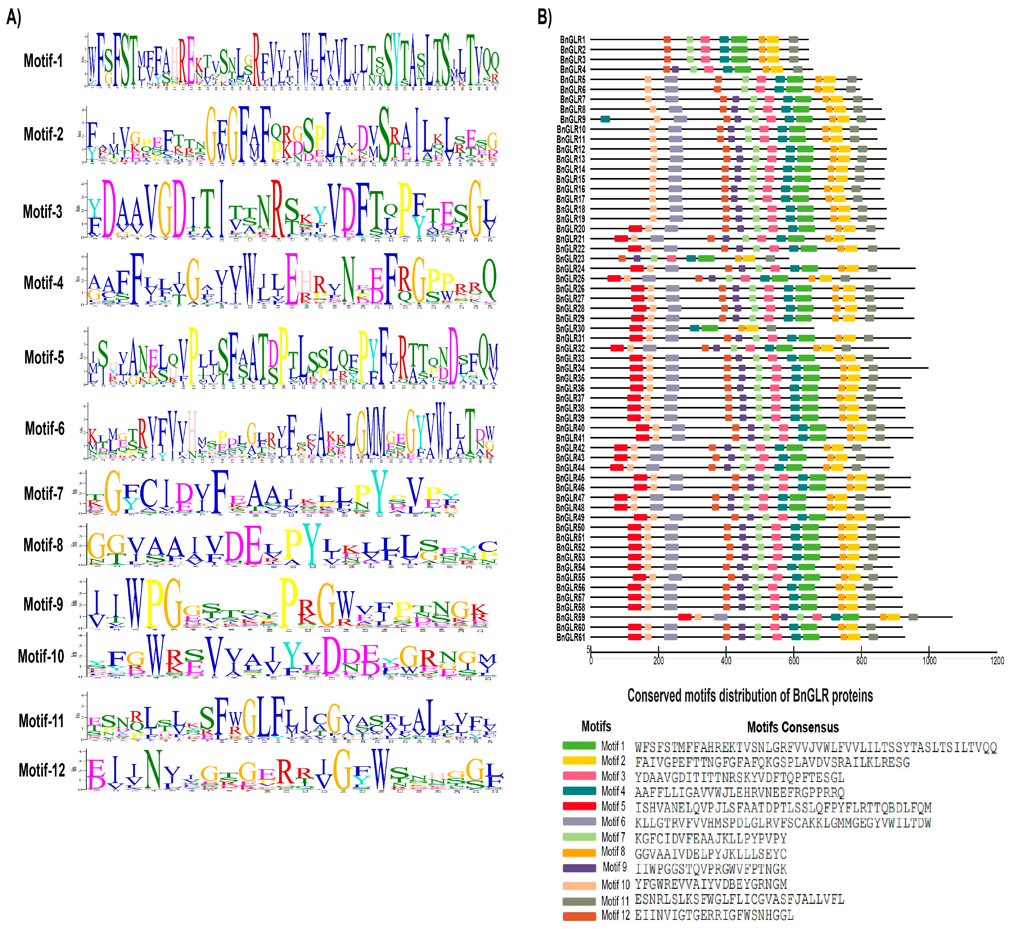
To further obtain clues for the functions of BnGLRs, the conserved motifs of BnGLR proteins were predicted using MEME. Consequently, 12 motifs with 20–50 aa that were conserved in the BnGLR protein family were generated (Figure 2A). The structure of BnGLR proteins was group-dependent and dominated by a motif organization of 5-10-6-12-9-7-3-4-1-8-2-11 or lacking the N-terminal motif 5 (Figure 2B). Most of the proteins within the same group had common features in terms of motif composition and distribution. All BnGLR proteins of Groups II and III exhibited the motif pattern 5-10-6-12-9-7-3-4-1-8-2-11, except one protein (BnGLR23), which was shorter with a lack of the N-terminal triple motifs 5-10-6. BnGLR proteins of Group I dominated with the motif pattern 10-6-12-9-7-3-4-1-8-2-11, which lacked the N-terminal motif 5 and was otherwise identical to that of Groups II and III BnGLRs. The exceptions were BnGLR9, with additional N-terminal motif 4; BnGLR5 and BnGLR6, which lacked motif 9; BnGLR4, which lacked motifs 10 and 6; and BnGLR1-3, which lacked motifs 10, 6, and 9 (Figure 2B).
Intriguingly, multiple alignment of the ion channel domain by ClustalW revealed the existence of the conserved motif “SYTANLTS” in the C-terminus of motif 1 among BnGLRs, which was similar but not identical to the known functional motif “SYTANLAA” reported in two rat iGluRs and Rosaceae and Arabidopsis GLRs to be involved in ion exchange or transportation through the plasma membrane (Figure S1). Additionally, 12 amino acids, F (9), E (20), F (27, 42, 44), S (45), R (61), W (67), F (69), S (77), Y (78), and A (80), were identical in this domain of all BnGLRs (Figure S1).

2.4. Domains and Gene Structures of BnGLRs

Domain composition analyses revealed that all BnGLRs contained a ligand-binding domain and an ion channel domain, a bacterial periplasmic substrate-binding protein domain (PBPe) (accession: SM000079), or a low complexity region (LCR) and 2–4 short transmembrane TMDs in “SMART” database, while a periplasmic_binding_protein_type_1 (PBP1) and a PBP2, or, more specifically, a PBP1_GABAb_receptor_plant (accession: cd06366) and a Glur_Plant (accession: cd13686) in the NCBI-CDD database. Remarkably, BnGLR21 carried a B3 domain (Table 1; Figures S2 and S3). Whether it is functional in regulating the transcription of target genes remains to be experimentally confirmed.
To study the GLR gene structure, the distribution of the exons and introns in BnGLR genes was identified. The number of introns ranged from 3 to 7. On average the introns were predicted to be 4.05, 4.47, and 5.52 in Group I, II, and III, respectively, indicating that the BnGLR genes of Group III contained more introns than those of the other groups (Table 1; Figure 3). Additionally, the size of introns also differed significantly in the BnGLR genes of different groups (Figure 3). Together, these results indicate that the gene structure of BnGLR genes is group-specific.

2.5. Cis-Acting Elements in Promoters of BnGLR Genes

In order to obtain preliminary clues for BnGLR gene function, cis-acting elements in the upstream (1.5 kb) sequence of BnGLR genes were analyzed using the search scan program in the PLACE database. Generally, very few (3.15%) CAMTA-binding cis-acting elements (CGCG-box) were predicted in BnGLR promoters (Figure 4). Only nine BnGLRs, one in Group I (BnGLR12), two in Group II (BnGLR34 and BnGLR35), and six in Group III (BnGLR37, -38, -39, -53, -60, and -61), contained 1~2 CGCG-boxes, indicating that these genes might be regulated by CAMTA3. BnGLRs contained several biotic stress responsive elements such as BOXLCOREDCPAL, MYB1LEPR, and SEBFCONSSTPR10A (Figure 4). Two BnGLRsBnGLR21 and BnGLR39—from Group II contained the maximum (10) biotic-stress responsive elements in their promoter, whereas two BnGLRsBnGLR22 and BnGLR36—from the same group and four BnGLRs from Group III (BnGLR46, -47, -48, and -49) did not have any single biotic-stress related cis-elements. Moreover, BnGLRs carried a set of abiotic-stress responsive cis-elements such as ELRECOREPCRP1, GT1GMSCAM4, MYCATERD1, MYCONSENSUSAT, and MYB2AT (Figure 4). Two BnGLRs from Group III (BnGLR42 and BnGLR55) contained the maximum number (16) of these cis-acting elements, while BnGLR10 (Group I), BnGLR22, -29, and -36 (Group II) and eight members from Group III (BnGLR37, -38, -43, -44, -46, -47, -48, and -49) did not have a single element. These results indicated that the BnGLR family might be widely involved in biotic- and abiotic-stress responses but in a member-dependent manner (Figure 4).

2.6. Tertiary Structure of BnGLR Proteins

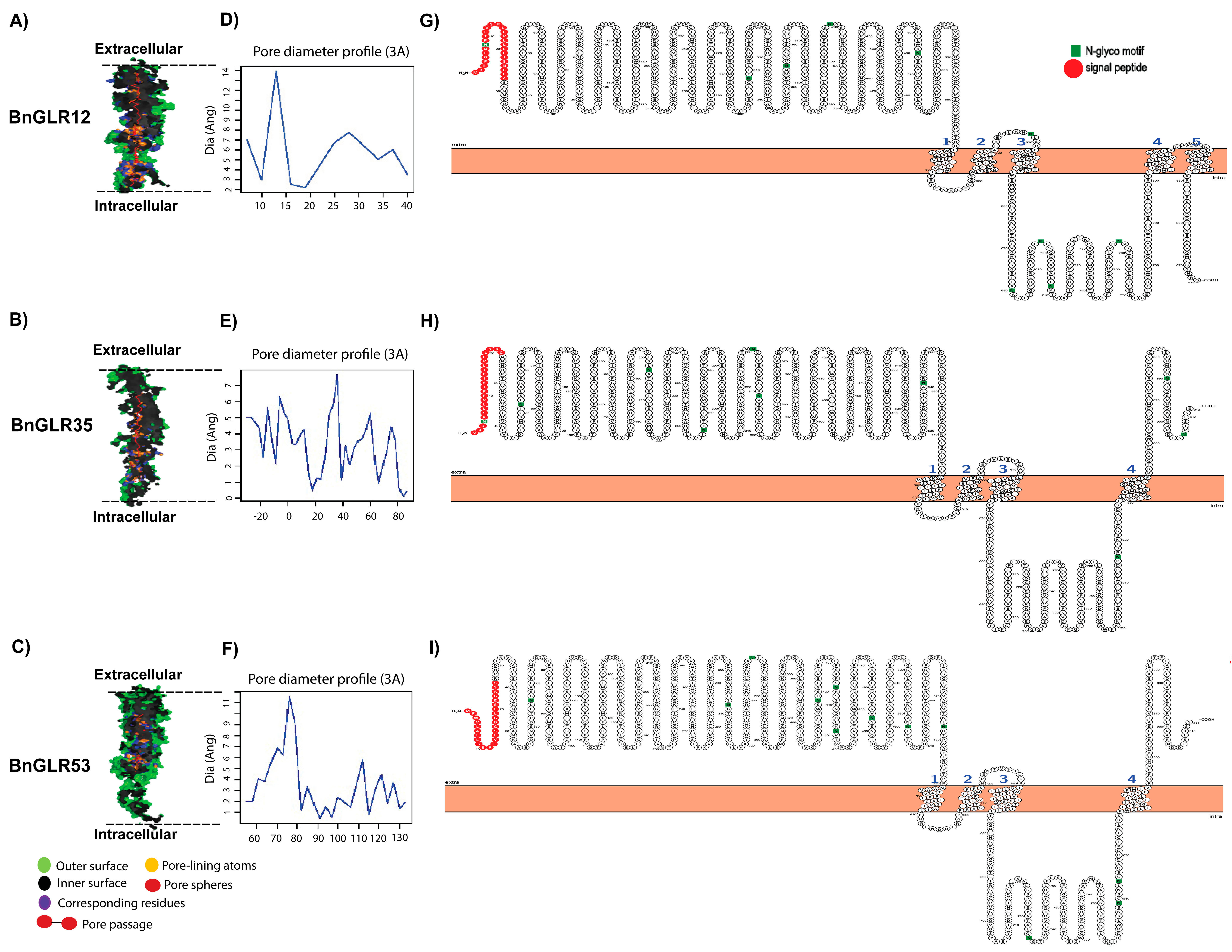
To examine whether BnGLRs were possible transmembrane protein channels, the PoreWalker webserver was used to predict the tertiary structures of three BnGLR proteins representing each group, BnGLR12, BnGLR35, and BnGLR53. The pore structure and 3D geometry of these BnGLR proteins were shown to fit with a pore morphology that longitudinally passed through the extracellular to intracellular opening of the proteins (Figure 5A–F). The pore size and constraints, which are considered as selective barriers, were predicted as shown in Figure 5A–F. Moreover, the Protter online tool was used for the structural visualization of these BnGLR proteins. The results showed that these BnGLR proteins were predicted to be membrane proteins with four transmembrane domains (Figure 5G–I). Together, these data support the BnGLR proteins to be transmembrane channels.

2.7. Amino Acid Ligands for BnGLRs

The interaction between 61 BnGLR proteins (as receptors) and 7 important amino acid ligands (Ala, Asn, Cys, Gln, Gly, Met, Ser) were observed based on ligand positions on receptor-binding pockets. Most BnGLR proteins had different binding sites for each of the above mentioned amino acid ligands but at distinct positions (Table S2). BnGLRs interacted with different amino acids via hydrogen bonding. BnGLR37 did not bind to any of the examined amino acids; BnGLR7 interacted uniquely with Ala; BnGLRs -3, -17, -30, -35, -36, and -39 interacted with two amino acids; and BnGLRs -2, -5, -8, -21, -33, and -51 bound to all seven amino acid ligands but at distinct sites. Distinguished from hydrogen bond interactions, all BnGLRs had hydrophobic interactions with all the amino acid ligands at different binding pockets consisting of 5–10 amino acids, most frequently Met, Phe, Thr, Arg, Leu, Ser, Glu, Tyr, Gly, and Ala. The most abundant binding residues in BnGLR receptor proteins through hydrogen bonds were Glu (867) in receptor BnGLR53 and Ala (76, 946, and 346) in BnGLR15, -25, and -60, respectively. Additionally, Arg (765) in BnGLR47, Tyr (432) in BnGLR52, and Asn (335 and 352) in BnGLR22 and BnGLR24 were also observed, respectively. On the contrary, binding residues through hydrophobic bonds were widely predicted in BnGLR proteins. They were Phe (335), Ala (273 and 344), Met (229), and Asp (175) in BnGLRs in Group I; Phe (333), Asp (260), and Arg (27) in BnGLRs in Group II; and Phe (339, 362), Asp (905), Ala (346), Leu (368), and Val (55, 79) in BnGLRs in Group III. Only five amino acid residues in BnGLRs bound to some of the above mentioned ligands through external bonds. These included Asp (175) and Ser (149) in BnGLR5, Gln (588) in BnGLR6, Glu (418) in BnGLR7, Pro (846) in BnGLR17, and Glu (732) in BnGLR33 (Table S2). These potential bindings need to be proved with further biochemical assays.

2.8. Post-Translational Modification Sites in BnGLRs

2.8.1. Phosphorylation

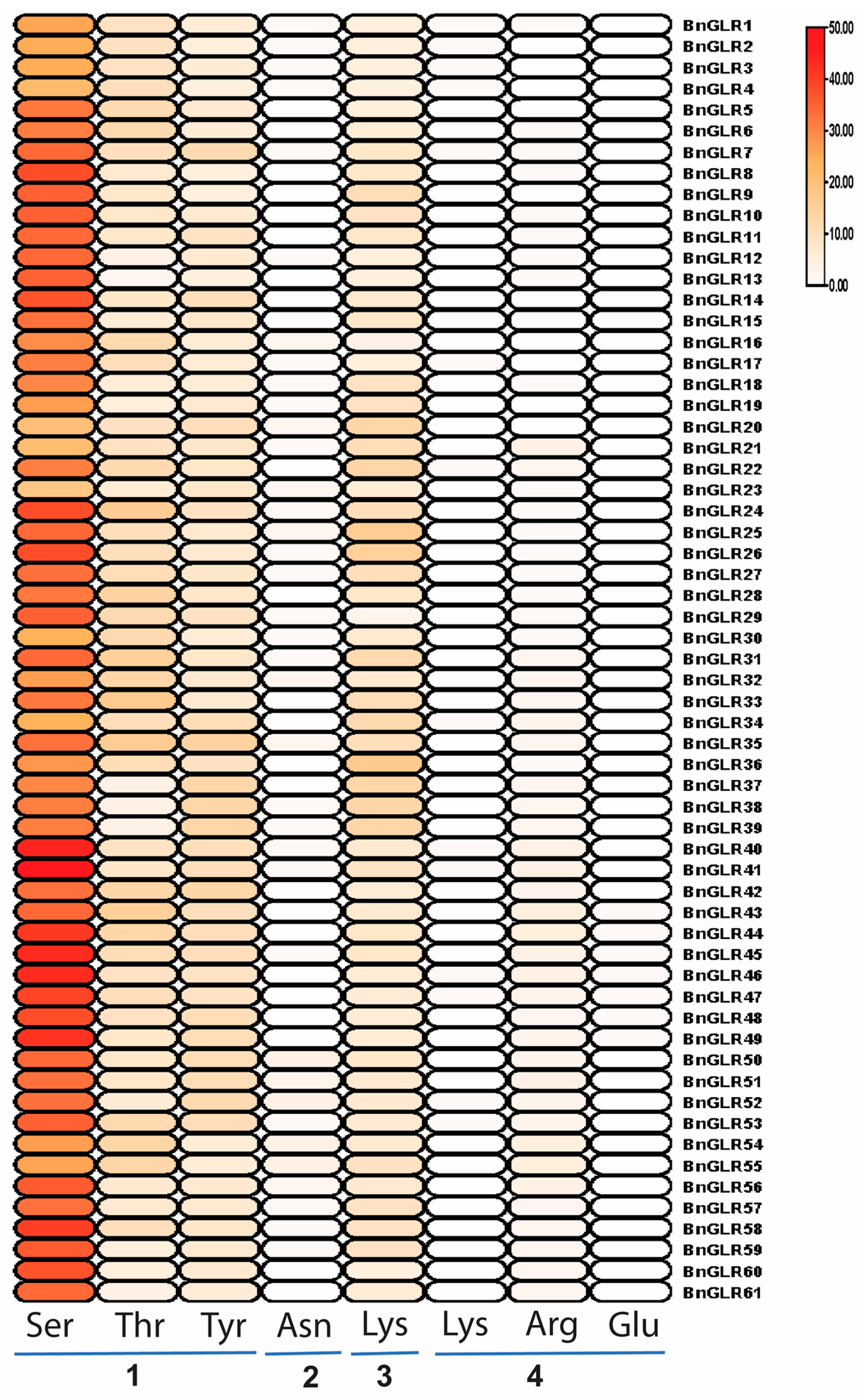
The attachment of the phosphate group to the serine, threonine, and tyrosine amino acids of BnGLR proteins was predicted. Generally, all BnGLR proteins contained all these three phosphorylation sites. The maximum putative phosphorylation serine (48) and threonine (17) were found in BnGLR41 and BnGLR33, respectively. Whereas BnGLR37, -38, -39, and -42 had the maximum number (13) of putative phosphorylation tyrosine in their protein sequences (Figure 6, Table S3). This result indicated that BnGLRs are likely phosphorylation-inducible proteins.

2.8.2. Glycosylation

There are four main categories of glycosylation based on the linkage between amino acids and sugars: N-linked glycans, O-linked glycans, GPI anchors, and C-mannosylation. Glycosylation analyses revealed the O-linked glycans in BnGLRs and AtGLRs, which were characterized by the interaction of a sugar with the hydroxyl group of serine or threonine. BnGLR50, BnGLR52, BnGLR54, and BnGLR55 carried the maximal four “Asn” residues, while 24 BnGLRs out of 61 did not contain any “Asn” residue (Figure 6, Table S3). This result implied that only some members of BnGLRs can be glycosylated.

2.8.3. Sumoylation

Sumoylation sites were characterized by a covalent bond of small ubiquitin-like modifiers to a specific lysine residue through an enzymatic action. All BnGLRs had sumo sites. A maximum of 17 sumoylation sites were predicted in BnGLR36. The positions and scores of these sumoylation sites are clearly marked in Figure 6 and Table S3. This result suggests that all BnGLRs can likely be sumoylated.

2.8.4. Methylation

The methylation sites in BnGLR proteins were predicted by using the online webserver, MASA. Methylation sites in lysine (Lys), arginine (Arg), and glutamate (Glu), but not in asparagine (N), were found in BnGLRs. The methylation site frequency was highest in Arg, followed by Lys, and lowest in Glu, which appeared only in six BnGLRs in Group III. Overall, the methylation sites in BnGLRs appeared to be site- and group-dependent. Group III BnGLRs contained more methylation sites, especially in Arg and Glu (Figure 6, Table S3).
Collectively, all BnGLRs carried phosphorylation and sumoylation sites, while glycosylation and methylation sites seemed to be BnGLR-dependent. Some Group I BnGLRs did not contain any methylation sites, while the absence of glycosylation site in BnGLRs was not group-dependent.

2.9. Response of BnGLRs to the Pathogen Sclerotinia sclerotiorum

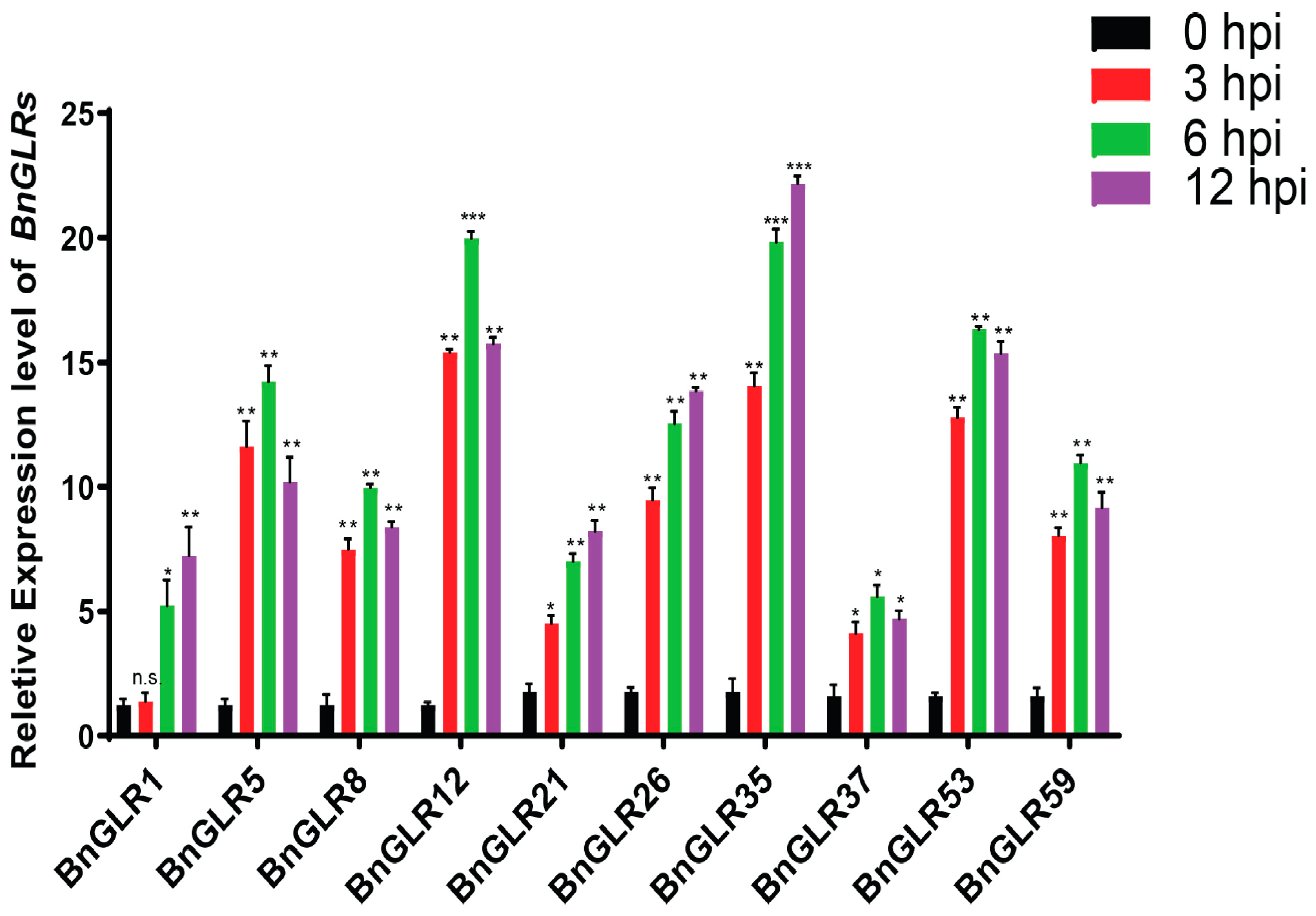
To analyze the response of BnGLR genes to the necrotrophic pathogen S. sclerotiorum (Ss), expression analysis was performed using quantitative real-time PCR (qRT-PCR). The primers that were used for expression analysis are listed in Table S1. The Ss was inoculated on oilseed rape leaves and sampled at 3, 6, and 12 h post-inoculation (hpi) and mock-inoculated samples were collected as a control. A total of 10 BnGLR genes, including BnGLR1, -5, -8, and -12 (Group I), BnGLR21, -26, and -35 (Group II), and BnGLR37, -53, and -59 (Group III) were selected, representing each group for expression analysis. Results showed that all genes were upregulated significantly in response to Ss inoculation. However, the extent of upregulation of BnGLR expression differed obviously. The expression was highest with a 23-fold peak enhancement at 12 hpi for BnGLR35, 20-fold at 6 hpi for BnGLR12, and 17-fold at 6hpi for BnGLR53, while it was lowest with a 5-fold peak enhancement at 6 hpi for BnGLR37 and 7~8-fold at 12 hpi for BnGLR1 and BnGLR21 (Figure 7). This result indicated that the upregulation of BnGLR expression was not group-dependent. Generally, BnGLRs were induced remarkably in response to Ss inoculation, indicating that these BnGLRs might be involved in resistance to Ss.

2.10. Silencing of BnGLR12, BnGLR35, and BnGLR53 Distinctly Altered Oilseed Rape Resistance against S. sclerotiorum

The three BnGLR genes (BnGLR12, BnGLR35, and BnGLR53) that were expressed most highly in response to Ss were selected for further functional analysis in oilseed rape by reverse genetics technique Cabbage leaf curl virus (CaLCuV)-based virus-induced gene silencing (VIGS). The plants treated with empty vectors (EV) showed normal growth, suggesting that the CaLCuV-based vector infection did not obviously affect the vegetative growth of seedlings. BnGLR expression in the plants agro-infiltrated with PCVA-BnGLR12, PCVA-BnGLR35, and PCVA-BnGLR53 was significantly reduced (Figure 8A), indicating that these BnGLR genes were efficiently silenced. Compared with the plants treated with EV, the leaves of BnGLR35-silenced oilseed rape plants exhibited more severe necrosis (Figure 8B) with larger lesion areas (Figure 8C) and increased relative fungal biomass of S. sclerotiorum (Figure 8D), indicating that the silencing of BnGLR35 reduced oilseed rape resistance to Ss. In contrast, the BnGLR12- and BnGLR53-silenced plants displayed milder disease symptoms (Figure 8B) with smaller lesion areas (Figure 8C) and lower S. sclerotiorum biomass (Figure 8D), indicating that the silencing of BnGLR12 and BnGLR53 enhanced oilseed rape resistance to Ss. These results demonstrated that BnGLR12, BnGLR35, and BnGLR53 play distinct roles in oilseed rape resistance to Ss; BnGLR35 positively, while BnGLR12 and BnGLR53 negatively, regulate this resistance.

2.11. Arabidopsis Mutants of BnGLR Orthologs Exhibited Altered Plant Resistance against S. sclerotiorum

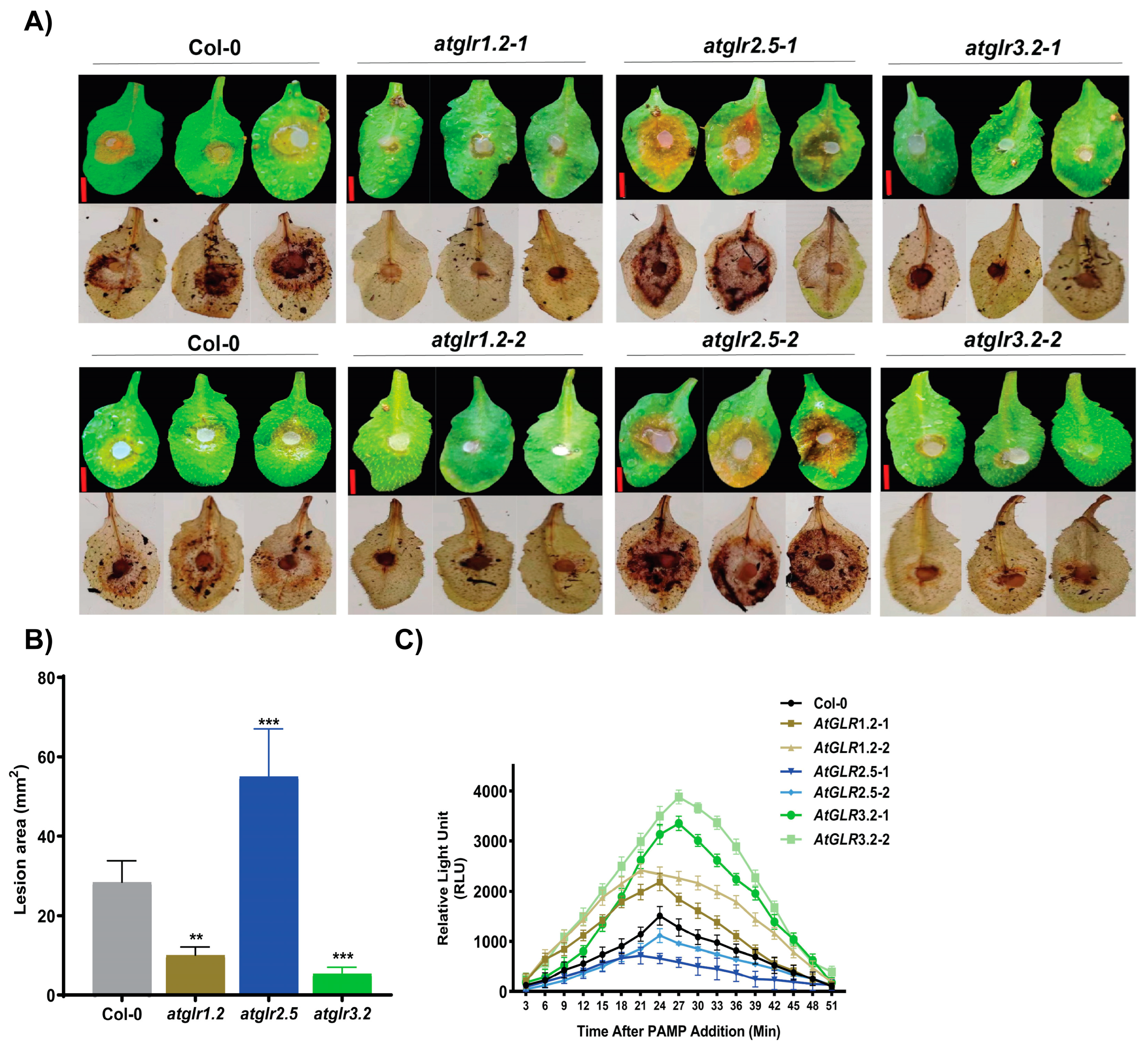
To further explore the role of BnGLR genes in plant resistance against S. sclerotiorum, inoculation analyses were performed in leaves of 4-week-old plants of six A. thaliana T-DNA insertion mutants of BnGLR orthologs, atglr1.2 (mutant of the ortholog of BnGLR12), atglr2.5 (mutant of the ortholog of BnGLR35), and atglr3.2 (mutant of the ortholog of BnGLR53). Prior to inoculation analysis, these Arabidopsis mutants were confirmed by PCR analysis and only homozygous mutant plants were selected for functional analysis. Compared with the wild type Col-0 (28 mm2), the leaves of both lines of atglr2.5 plants showed more severe necrosis (53 mm2) with more DAB staining (Figure 9A) and larger lesion areas (Figure 9B), while the leaves of two lines each of atglr1.2 (6 mm2) and atglr3.2 (8 mm2) plants displayed less severe necrosis with less DAB staining (Figure 9A) and smaller lesion areas (Figure 9B). It revealed that atglr2.5 mutants exhibited increased susceptibility to S. sclerotiorum, while atglr1.2 and atglr3.2 mutants displayed reduced susceptibility compared with the wild type Col-0. In addition, compared with the Col-0 plants, atglr3.2 and atglr1.2 mutants manifested the enhanced generation of SsNLP1-stimulated H2O2. Conversely, the atglr2.5 mutants exhibited reduced SsNLP1-triggered H2O2 production (Figure 9C). These findings suggest that the GLR genes play a crucial role in plant resistance against Ss, probably via modulating ROS accumulation.

3. Discussion

In this study, we identified 61 BnGLRs in the genome of oilseed rape cv. ZS11. A set of evidence supports them to be potentially functional GLRs. They all contained a ligand-binding domain and an ion channel domain (Table 1; Figure S2). Their ion channel domain bore the conserved motif “SYTANLTS” (Figure S1), which was reported to be involved in ion exchange or transportation through the plasma membrane in rat, Rosaceae, and Arabidopsis [9]. Furthermore, the BnGLRs were predicted to be localized to plasma membrane and their tertiary structure fitted with membrane channel morphology, as exemplified by BnGLR12, BnGLR35, and BnGLR53 (Figure 5) as reported for known other gene family (DTX) [30]. Additionally, binding pockets of seven amino acids existed in all BnGLR proteins (Table S2) as reported typical GLRs [16]. Nevertheless, whether these BnGLRs function as calcium channels awaits further experimental confirmation.
One of our findings in this study is the group-dependent characteristics of BnGLRs. Firstly, group-wide chromosomal distribution of BnGLRs is obviously uneven. Regardless of the uncertainty of the localization of BnGLR4 (Group I) and BnGLR34 (Group II), Group I BnGLRs are absent in chromosomes A03–A05, A07–A08, A10, C03–C06, and C08, Group II BnGLRs are lacking in chromosomes A01–A02, A06–A10, C01, and C05–C08, whereas Group III BnGLRs are missing in chromosomes A01-A02, A04-A06, C02, and C06 (Table 1). Secondly, the motif composition of BnGLRs differs group-wide. For example, motif 5 is absent from Group I BnGLRs while present in those of the other two groups (Figure 2). Thirdly, the gene structure of BnGLRs distinguishes group-wide. BnGLR genes of Group III carry more introns than those of the other groups. BnGLR genes of Group II generally contain a long first-intron, while those of the other groups do not (Figure 3). Finally, the methylation sites discriminate between the groups of BnGLRs. The methylation sites in Glu only exist in six BnGLRs of Group III. Group III BnGLRs also contain more methylation sites in Arg and Glu than other group BnGLRs (Figure 6, Table S3). Collectively, our findings clarify the differentiation of BnGLRs of different groups, which implies that the functions and mechanisms of BnGLRs of different groups may be distinct. In this context, it is intriguing that we indeed found the distinct function of BnGLRs from various groups—BnGLR12 from Group I, BnGLR35 from Group II, and BnGLR53 from Group III—in oilseed rape resistance to the necrotrophic fungal pathogen S. sclerotiorum (Figure 8). Additionally, our results on chromosomal distribution, protein motif, and gene structure of BnGLRs differ in some aspects from a previous report, which documented that Group I BnGLRs distributed in chromosomes A03, A05, A07, A08, C03–C06, and C08; Group II BnGLRs existed in A02, C06, and C07; and Group III BnGLRs presented in A01, A02, and C02. Motif 5 existed in Group I BnGLRs. Group II BnGLRs contained more introns (2–12 introns) than other groups BnGLRs [14]. These differences might be due to the variety in the oilseed rape cultivars used in the two studies [14].
Interestingly, 9 out of the total 61 BnGLRs carried one to several CGCG-boxes (the CAMTA-binding sites in their promoters (Figure 4)). These included BnGLR12 from Group I, BnGLR34 and BnGLR35 from Group II, and BnGLR37, -38, -39, -53, -60, and -61 from Group III. This demonstrates that these BnGLRs might be directly regulated by CAMTA3, an essential transcription factor in the calcium signaling pathways. Notably, we found previously that CAMTA3 plays a role in plant disease resistance against Ss [31]. Therefore, it is likely that CAMTA3 and these GLRs coordinate in plant resistance to Ss.
PTM is one of the most important ways to regulate protein functions. We systematically analyzed four types of PTMs, including phosphorylation, sumoylation, glycosylation, and methylation, in BnGLRs. Consequently, we found that all BnGLRs carry plenty of phosphorylation and sumoylation sites, while glycosylation and methylation sites only exist in some BnGLRs (Figure 6, Table S3). Interestingly, AtGLR3.6 and AtGLR3.7 were recently reported to be phosphorylated by calcium-dependent protein kinases at Serine-856 and Serine-860, respectively, thereby leading to salt tolerance [32,33], indicating that oilseed rape GLRs are also highly likely phosphorylated at Serine. It would be interesting to examine whether GLRs are also phosphorylated at Thr and Tyr in accordance with our prediction results. It also deserves to be confirmed whether GLRs are also sumoylated, glycosylated, and/or methylated following the clues from our study.
The function of GLRs in plant resistance to pathogens has only been studied to a limited extent mainly in the model plant Arabidopsis. For example, Group III AtGLRs were reported to play a role in plant resistance against the oomycete H. arabidopsidis [24], fungus B. cinerea [25], and bacterium P. syringae [26]. In this study, we explored the role of BnGLRs in resistance to S. sclerotiorum, one of the most destructive pathogens in oilseed rape, which is one of the most important oil crop species. We found that three BnGLRsBnGLR12 from Group I, BnGLR35 from Group II, and BnGLR53 from Group III—are highly responsive to S. sclerotiorum infection with an increase in expression by over 15-fold at 6 hpi (Figure 7). More importantly, VIGS analyses demonstrated that BnGLR12, BnGLR35, and BnGLR53 play distinct roles in oilseed rape resistance to Ss; BnGLR35 positively, while BnGLR12 and BnGLR53 negatively, regulate this resistance (Figure 8). Furthermore, inoculation analysis and ROS detection employing Arabidopsis mutants of the BnGLR gene homologs (BnGLR12/AtGLR1.2, BnGLR35/AtGLR2.5, and BnGLR53/AtGLR3.2) confirmed this conclusion and extended their roles in PAMP (SsNLP1)-triggered immunity (Figure 9). The response of Group III GLR, AtGLR3.2, and its ortholog in B. napus (BnGLR53) are distinguished from previous studies of AtGLRs from Group III, in which radish GLR homologs of AtGLR3.3 and AtGLR3.4 play a positive role in resistance against B. cinerea and Alternaria brassicae [25,34], possibly due to the difference in the pathogens used for inoculation analyses. Together with these reports, our findings reveal the complex member-dependent functions of the GLR family in plant resistance to pathogens. The mechanisms underlying BnGLR-mediated plant immunity await further dissection.

4. Materials and Methods

4.1. Identification of BnGLR Proteins

Using AtGLR proteins as queries, BLASTP searches were carried out against sequenced genomes of green plants in the Phytozome (https://phytozome-next.jgi.doe.gov, accessed on 26 August 2020) and NCBI (http://www.ncbi.nlm.nih.gov/, accessed on 26 August 2020) databases. Using the ClustalW2 program (http://www.ebi.ac.uk/Tools/msa/clustalw2/, accessed on 15 September 2020) with default settings, all retrieved sequences were compared with AtGLR proteins and analyzed using the GeneDoc program [8].
For the measurement of physicochemical properties such as isoelectric point (pI) and amino acid (aa) composition, an online tool ProtParam server was used (http://web.expasy.org/protparam/, accessed on 12 November 2020) [14].

4.2. Construction of BnGLR Phylogenic Tree

MegaX software was used to align the AtGLRs and BnGLRs by applying the clustal W function. For the construction of a phylogenetic tree in Mega X (v10.2.2) [35], the maximum likelihood (ML) algorithm with 1000 bootstrap replicates and partial deletion (95% site coverage as cut-off) were used for gaps and missing data. Based on their sequence similarity with AtGLRs in the phylogenetic tree, the members of the BnGLR family were named.

4.3. Identification of Motifs, Domains, and Gene Structure

Conserved motifs in the BnGLR proteins were identified using the web tool Multiple Expectation Maximization for Motif Elicitation (MEME) (http://meme-suite.org/tools/meme, accessed on 29 November 2020) [28]. The parameters were as follows: the maximum number of motifs was set to 12, the motif width ranged from 6 to 50 (inclusive), and the site distribution was limited to 0 or 1 occurrence (of a contributing motif site) per sequence. Logo motif analysis was also performed to demonstrate the conservativeness of each group of BnGLR proteins using the weblogo3 (v3.7) platform [36].
The conserved domains of BnGLR proteins were predicted using the SMART [37] and CDD databases [38].
By matching the cDNA to the corresponding gene sequences, the Gene Structure Display Server (GSDS) was used [39] to analyze the CDS and exon/intron structures of the GLR genes.

4.4. Prediction of Subcellular Localization of BnGLRs

The subcellular localization of the BnGLR proteins was predicted using the CELLO [14] web tool (http://cello.life.nctu.edu.tw/, accessed on 8 December 2023).

4.5. Promoter Profiling of BnGLRs

To identify the potential cis-acting elements in BnGLRs, upstream (1.5 kb) sequence of BnGLRs were obtained from NCBI and were analyzed using the search scan program in the PLACE database [8].

4.6. Prediction of Tertiary Protein Structure of BnGLRs

A protein modeling server, Phyre2, was used to predict the tertiary protein structures of BnGLR proteins (www.sbg.bio.ic.ac.uk/phyre2, accessed on 8 January 2021). To identify the transmembrane protein channels from their 3D structures, the PoreWalker webserver was used with pdb files of BnGLR proteins (http://www.ebi.ac.uk/thornton-srv/software/PoreWalker, accessed on 12 January 2021). To validate their secondary structures, the Protter online tool was used (http://wlab.ethz.ch/protter, accessed on 15 January 2021) [40].

4.7. Prediction of Amino Acid Ligands Interacting with BnGLR Proteins

To visualize the predicted physical interactions between amino acid ligands and receptor BnGLR proteins, PDB images of receptor proteins and amino acid ligands (Ala, Asn, Cys, Glu, Gly, Met, and Ser) were obtained from Phyre2 (Protein Homology/Analogy Recognition Engine V 2.0) and Pubchem (https://pubchem.ncbi.nlm.nih.gov/, accessed on 8 January 2021), respectively. Interactions were predicted by using the online tool Patchdock [41] and the complex (receptor-ligand complex) was obtained in PDB format, and the complex PDB image was visualized using LIGPLOT (v2.2) software for identification of binding pocket positions [42].

4.8. Post-Translational Modifications in BnGLR Proteins

The sites of PTMs, including methylation at lysine, arginine, and glutamate; N-glycosylation at asparagine; phosphorylation at serine, threonine, and tyrosine; and sumoylation were predicted by using online bioinformatics tools given below as described [43].
http://masa.mbc.nctu.edu.tw/, accessed on 20 January 2021 (Methylation);
http://www.cbs.dtu.dk/services/NetOGlyc/, accessed on 25 January 2021 (Glycosylation);
http://www.cbs.dtu.dk/services/NetPhos/, accessed on 29 January 2021 (Phosphorylation);
http://sumosp.biocuckoo.org/online.php, accessed on 2 February 2021 (Sumoylation).

4.9. Plant Materials and Pathogen Inoculation Analysis

Brassica napus plants were grown in growth cabinets at 23 °C under a 14 h/10 h light/dark photoperiod [36]. All Arabidopsis thaliana plants used in this study were of Col-0 ecotype background and were grown at 16 h photoperiod and 70% humidity at 21 ± 1 °C in the growth chamber [44]. The following mutants were used in this study and were obtained through the AraShare (Fuzhou, China): atglr1.2 (SALK_136614C, SALK_053535C), atglr2.5 (SALK_078407C, SALK_050593C), and atglr3.2 (SALK_063873C, SALK_133700C). Homozygous plants were screened based on growth on selectable medium and PCR using a combination of gene-specific and transposon- or T-DNA-specific primers (Table S1).
Plant leaves were inoculated using Sclerotinia sclerotiorum (Ss) mycelial plugs. Fresh sclerotia of Ss strain UF1 were cultured at 23 °C on potato dextrose agar medium (PDA) to produce mycelia, which were transferred to new PDA plates and grown for 2 days. The PDA plugs containing young Ss mycelia were punched to inoculate the leaves of 30 d old plants [36]. Area of disease lesions was measured using the ImageJ (v1.54e) software [36]. For disease resistance evaluation, at least five plants for each genotype were examined and the experiments were conducted three times independently.

4.10. Gene Expression Analysis

A total of 10 BnGLR genes including BnGLR1, -5, -8, and -12 (Group I), BnGLR21, -26, and -35 (Group II), and BnGLR37, -53, and -59 (Group III) were selected representing each group for expression analysis. One-month old oilseed rape leaves were sampled at 0, 3, 6, and 12 h post-inoculation with S. sclerotiorum. Total RNA was extracted using Trizol reagent (Vazyme, Nanjing, China) following the previous study [45]. Quantitative real-time PCR (qRT-PCR) was performed using the StepOne Real-Time PCR system (Applied Biosystems, Waltham, MA, USA) with SYBR Green PCR Master Mix (TaKaRa, Dalian, China). The relative fold changes were calculated using the 2−∆∆Ct method as previously described [45], with two technical replicates for each of the three biological replicates. The housekeeping gene BnActin7 was used as an internal control. Primers used for qRT-PCR are listed in Table S1.

4.11. Cabbage Leaf Curl Virus (CaLCuV)-Induced Gene Silencing Manipulation Procedure

The VIGS experiment with cabbage leaf curl virus was carried out as previously reported [46], with a few adjustments. (i) The gene-specific sequences of BnGLR12, BnGLR35, and BnGLR53 were selected to ensure gene-specific silencing. Primers containing the restriction site (Xba I) and homologous arms were designed (Table S1). (ii) Using first-strand cDNA from the ZS11 cultivar, the target sequences of BnGLRs were amplified. The digested material was kept at 37 °C overnight to obtain the digested PCVA vector (digested with Xba I) (NEB, Ipswich, MA, USA). Using the ClonExpress Entry One Step Cloning Kit (Vazyme), the target sequence and the digested PCVA were ligated for 15 min at 50 °C. (iii) After transforming the ligation mix into competent DH5α cells (Invitrogen, Darmstadt, Germany), the cells were plated on LB plates containing antibiotics (kanamycin at 50 mg/mL) at 37 °C for overnight. A single colony was chosen, shaken well in liquid LB (containing 50 mg/mL kanamycin), and then sequenced using gene-specific primers. The plasmids from empty PCVA, PCVB, PCVA-BnGLR12, PCVA-35, and PCVA-53 were purified using the FastPure EndoFree Plasmid Maxi Kit (Vazyme). (iv) Using the freeze–thaw transformation technique, plasmids were transformed into Agrobacterium tumefaciens (GV3101). Following that, single colonies were chosen and shaken for 48–72 h at 28 °C in liquid LB (containing 50 mg/mL of kanamycin and 50 mg/mL of rifampicin). The BnGLRs were sequenced using gene-specific primers and primers A and B for the PCVA and PCVB empty vectors, respectively, to confirm that the constructed vector was effectively transformed into Agrobacterium. A mixture of Agrobacterium culture and glycerol (1:3) was placed in a 2 mL cryotube to preserve bacteria for long-term preservation at −80 °C. For inoculation with PCVA/PCVB and PCVA-BnGLRs, ZS11 seedlings (2–3 leaf-stage) were used for VIGS experiments. Two-week-old oilseed rape plants were infiltrated with a 1 mL syringe and the infiltrated plants were placed in the dark for 24 h. The seedlings were then placed in a chamber for the following 15–21 days for successful silencing. To confirm whether plants were truly silent, half of the leaves were checked for BnGLR gene silencing by qRT-PCR. The remaining half of the same leaves were used for Ss inoculation analyses using mycelial plugs.

4.12. ROS Detection

For quantitative ROS measurement, 3 mm diameter leaf disks were immersed in 50 μL of distilled water in a 96-well plate and left in the dark overnight. A 100 μL solution comprising 100 μM luminol (Sigma-Aldrich, St. Louis, MO, USA) and 1 μg of horseradish peroxidase was used as a substitute for water. A microplate luminometer was used to measure the luminescence of H2O2 for 51 min after the addition of H2O2 elicitor, the fungal PAMP, SsNLP1 (1 μM) (TITERTEK BERTHOLD, Pforzheim, Germany), as previously described [36].

4.13. Statistical Analysis

All experiments were performed with three biological replicates. Data were statistically analyzed to determine the significant differences by Student’s t-test and shown as the mean ± SE using Graphpad Prism 8.0 [36].

5. Conclusions

A genome-wide analysis of Brassica napus cultivar ZS11 identified 61 GLR genes, grouped similarly to Arabidopsis thaliana. The BnGLR family exhibited some group-dependent characteristics such as chromosomal distribution, amino acid sequence motifs, gene intron number, and some PTM sites. Comprehensive bioinformatics analyses supported these BnGLRs as functional ion channels. BnGLRs were potential amino acid receptors carrying multiple PTM sites, including phosphorylation and sumoylation sites and probably also glycosylation and methylation sites. Functional assays demonstrated the positive role of BnGLR35 and the negative role of BnGLR12 and BnGLR53 in oilseed rape resistance against the destructive necrotrophic pathogen Sclerotinia sclerotiorum. These findings advance our understanding of GLR family functions in plant immunity; nevertheless, functions of more BnGLR genes in plant immunity need to be clarified. Mechanisms underlying BnGLR-dependent plant immunity await further dissection. How plants elaborate the BnGLR genes with distinct roles in resistance to counter the pathogens represents one of the intriguing questions to be addressed in the near future.

Supplementary Materials

The following supporting information can be downloaded at: https://www.mdpi.com/article/10.3390/ijms25115670/s1.

Author Contributions

Conceptualization, X.-Z.C.; experiments, R.M.A.G.; data analyses, R.M.A.G., C.-X.R., X.F., Y.-P.X. and M.A.S., writing, R.M.A.G. and X.-Z.C.; supervision, X.-Z.C.; funding acquisition, X.-Z.C. All authors have read and agreed to the published version of the manuscript.

Funding

This research was funded by the Zhejiang Science and Technology Major Program on Agricultural New Variety Breeding (No. 2021C02064), the Hainan Provincial Natural Science Foundation of China (No. 324CXTD430), and the National Natural Science Foundation of China (No. 31871947).

Institutional Review Board Statement

Not applicable.

Informed Consent Statement

Not applicable.

Data Availability Statement

Data is contained within the article and Supplementary Materials.

Acknowledgments

We are grateful to Honghao Lv, Chinese Academy of Agricultural Sciences, for providing the CaLCuV-based VIGS vector. We appreciate Yanhua Zhang, Jilin University, for providing the Sclerotinia sclerotiorum strain UF1. We also thank the laboratory members for their technical help and critical discussions.

Conflicts of Interest

The authors declare no conflicts of interest.

References

  1. Naz, R.; Khan, A. An insight into animal glutamate receptors homolog of Arabidopsis thaliana and their potential applications-a review. Plants 2022, 19, 2580. [Google Scholar] [CrossRef] [PubMed]
  2. Iwano, M.; Ito, K.; Fujii, S.; Kakita, M.; Asano-Shimosato, H.; Igarashi, M.; Kaothien-Nakayama, P.; Entani, T.; Kanatani, A.; Takehisa, M.; et al. Calcium signalling mediates self-incompatibility response in the Brassicaceae. Nat. Plants 2015, 1, 15128. [Google Scholar] [CrossRef] [PubMed]
  3. Shoshan-Barmatz, V.; De, S. Mitochondrial VDAC, the Na+/Ca2+ exchanger, and the Ca2+ uniporter in Ca2+ dynamics and signaling. Adv. Exp. Med. Biol. 2017, 981, 323–347. [Google Scholar] [PubMed]
  4. Forde, B.G.; Roberts, M.R. Glutamate receptor-like channels in plants: A role as amino acid sensors in plant defence? Prime Rep. 2014, 6, 37. [Google Scholar] [CrossRef] [PubMed]
  5. Shi, Y.; Zeng, W.; Xu, M.; Li, H.; Zhang, F.; Chen, Z.; Anicet, G.; Huang, S.; Huang, Y.; Wang, X. Comprehensive analysis of glutamate receptor-like genes in rice (Oryza sativa L.): Genome-wide identification, characteristics, evolution, chromatin accessibility, gcHap diversity, population variation and expression analysis. Curr. Issues Mol. Biol. 2022, 44, 6404–6427. [Google Scholar] [CrossRef] [PubMed]
  6. Zhang, J.; Cui, T.; Su, Y.; Zang, S.; Zhao, Z.; Zhang, C.; Zou, W.; Chen, Y.; Cao, Y.; Chen, Y. Genome-wide identification, characterization, and expression analysis of glutamate receptor-like gene (GLR) family in sugarcane. Plants 2022, 11, 2440. [Google Scholar] [CrossRef] [PubMed]
  7. Yin, L.; Liu, S.; Sun, W.; Ke, X.; Zuo, Y. Genome-wide identification of glutamate receptor genes in adzuki bean and the roles of these genes in light and rust fungal response. Gene 2023, 879, 147593. [Google Scholar] [CrossRef] [PubMed]
  8. Saand, M.A.; Xu, Y.P.; Li, W.; Wang, J.P.; Cai, X.Z. Cyclic nucleotide gated channel gene family in tomato: Genome-wide identification and functional analyses in disease resistance. Front. Plant Sci. 2015, 6, 303. [Google Scholar] [CrossRef]
  9. Chen, J.; Jing, Y.; Zhang, X.; Li, L.; Wang, P.; Zhang, S.; Zhou, H.; Wu, J. Evolutionary and expression analysis provides evidence for the plant glutamate-like receptors family is involved in woody growth-related function. Sci. Rep. 2016, 6, 32013. [Google Scholar] [CrossRef]
  10. Luo, B.; Zhang, H.; Li, D.; Wu, Q.; Ge, W.; Zhu, T.; Chen, Y.; Huang, Y.; Lin, Y.; Lai, Z. Genome-wide identification of the banana GLR gene family and its expression analysis in response to low temperature and abscisic acid/methyl jasmonate. Sheng Wu Gong Cheng Xue Bao 2023, 39, 2874–2896. [Google Scholar]
  11. Yang, L.; Zhao, Y.; Wu, X.; Zhang, Y.; Fu, Y.; Duan, Q.; Ma, W.; Huang, J. Genome-wide identification and expression analysis of BraGLRs reveal their potential roles in abiotic stress tolerance and sexual reproduction. Cells 2022, 11, 3729. [Google Scholar] [CrossRef] [PubMed]
  12. Kang, S.; Kim, H.B.; Lee, H.; Choi, J.Y.; Heu, S.; Oh, C.J.; Kwon, S.I.; An, C.S. Overexpression in Arabidopsis of a plasma membrane-targeting glutamate receptor from small radish increases glutamate-mediated Ca2+ Influx and delays fungal infection. Mol. Cells 2006, 21, 418–427. [Google Scholar] [CrossRef]
  13. Li, X.; Zhu, T.; Wang, X.; Zhu, M. Genome-wide identification of glutamate receptor-like gene family in soybean. Heliyon 2023, 9, e21655. [Google Scholar] [CrossRef]
  14. Roy, B.C.; Shukla, N.; Gachhui, R.; Mukherjee, A. Genome-wide analysis of glutamate receptor gene family in allopolyploid Brassica napus and its diploid progenitors. Genetica 2023, 151, 293–310. [Google Scholar] [CrossRef] [PubMed]
  15. Grenzi, M.; Bonza, M.C. Structural insights into long-distance signal transduction pathways mediated by plant glutamate receptor-like channels. New Phytol. 2021, 229, 1261–1267. [Google Scholar] [CrossRef] [PubMed]
  16. Alfieri, A.; Doccula, F.G. The structural bases for agonist diversity in an Arabidopsis thaliana glutamate receptor-like channel. Proc. Natl. Acad. Sci. USA 2020, 117, 752–760. [Google Scholar] [CrossRef] [PubMed]
  17. Grenzi, M.; Bonza, M.C.; Costa, A. Signaling by plant glutamate receptor-like channels: What else! Curr. Opin. Plant Biol. 2022, 68, 102253. [Google Scholar] [CrossRef] [PubMed]
  18. Reddy, A.S.; Ali, G.S.; Celesnik, H.; Day, I.S. Coping with stresses: Roles of calcium-and calcium/calmodulin-regulated gene expression. Plant Cell 2011, 23, 2010–2032. [Google Scholar] [CrossRef] [PubMed]
  19. Kong, D.; Hu, H.C.; Okuma, E.; Lee, Y.; Lee, H.S.; Munemasa, S.; Cho, D.; Ju, C.; Pedoeim, L.; Rodriguez, B. L-Met activates AtGLR Ca2+ channels upstream of ROS production and regulates stomatal movement. Cell Rep. 2016, 17, 2553–2561. [Google Scholar] [CrossRef]
  20. Wang, Y.; Yu, D.; Zhao, H.; Jiang, L.; Gao, L.; Song, Y.; Liu, Z.; Bao, F.; Hou, C.; He, Y. A glutamate receptor-like gene is involved in ABA-mediated growth control in Physcomitrium patens. Plant Signal Behav. 2022, 17, 2145057. [Google Scholar] [CrossRef]
  21. Yu, B.; Wu, Q.; Li, X.; Zeng, R.; Min, Q.; Huang, J. Glutamate receptor-like gene OsGLR3.4 is required for plant growth and systemic wound signaling in rice (Oryza sativa). New Phytol. 2022, 233, 1238–1256. [Google Scholar] [CrossRef] [PubMed]
  22. Meyerhoff, O.; Müller, K.; Roelfsema, M.R.; Latz, A.; Lacombe, B.; Hedrich, R.; Dietrich, P.; Becker, D. AtGLR3.4, a glutamate receptor channel-like gene is sensitive to touch and cold. Planta 2005, 222, 418–427. [Google Scholar] [CrossRef] [PubMed]
  23. Cheng, Y.; Zhang, X.; Sun, T.; Tian, Q.; Zhang, W.-H. Glutamate receptor homolog AtGLR3.4 is involved in regulation of seed germination under salt stress in Arabidopsis. Plant Cell Physiol. 2018, 59, 978–988. [Google Scholar] [CrossRef] [PubMed]
  24. Manzoor, H.; Kelloniemi, J.; Chiltz, A.; Wendehenne, D.; Pugin, A.; Poinssot, B.; Garcia-Brugger, A. Involvement of the glutamate receptor AtGLR3.3 in plant defense signaling and resistance to Hyaloperonospora arabidopsidis. Plant J. 2013, 76, 466–480. [Google Scholar] [CrossRef] [PubMed]
  25. Feng, S.; Pan, C.; Ding, S.; Ma, Q.; Hu, C.; Wang, P.; Shi, K. The glutamate receptor plays a role in defense against Botrytis cinerea through electrical signaling in tomato. Appl. Sci. 2021, 11, 11217. [Google Scholar] [CrossRef]
  26. Li, F.; Wang, J.; Ma, C.; Zhao, Y.; Wang, Y.; Hasi, A.; Qi, Z. Glutamate receptor-like channel AtGLR3.3 is involved in mediating glutathione-triggered cytosolic calcium transients, transcriptional changes, and innate immunity responses in Arabidopsis. Plant Physiol. 2013, 162, 1497–1509. [Google Scholar] [CrossRef] [PubMed]
  27. Roy, B.C.; Mukherjee, A. Computational analysis of the glutamate receptor gene family of Arabidopsis thaliana. J. Biomol. Struct. Dyn. 2017, 35, 2454–2474. [Google Scholar] [CrossRef] [PubMed]
  28. Bailey, T.L.; Johnson, J.; Grant, C.E.; Noble, W.S. The MEME suite. Nucleic Acids Res. 2015, 43, W39–W49. [Google Scholar] [CrossRef]
  29. Chen, C.; Wu, Y.; Li, J.; Wang, X.; Zeng, Z.; Xu, J.; Liu, Y.; Feng, J.; Chen, H.; He, Y.; et al. TBtools-II: A "one for all, all for one" bioinformatics platform for biological big-data mining. Mol. Plant 2023, 16, 1733–1742. [Google Scholar] [CrossRef]
  30. Ali, E.; Saand, M.A.; Khan, A.R.; Shah, J.M.; Feng, S.; Ming, C.; Sun, P. Genome-wide identification and expression analysis of detoxification efflux carriers (DTX) genes family under abiotic stresses in flax. Plant Physiol. 2021, 171, 483–501. [Google Scholar] [CrossRef]
  31. Rahman, H.; Xu, Y.P.; Zhang, X.R.; Cai, X.Z. Brassica napus genome possesses extraordinary high number of CAMTA genes and CAMTA3 contributes to PAMP triggered immunity and resistance to Sclerotinia sclerotiorum. Front. Plant Sci. 2016, 7, 581. [Google Scholar] [CrossRef] [PubMed]
  32. Silamparasan, D.; Chang, I.F.; Jinn, T.L. Calcium-dependent protein kinase CDPK16 phosphorylates serine-856 of glutamate receptor-like AtGLR3.6 protein leading to salt-responsive root growth in Arabidopsis. Front. Plant Sci. 2023, 14, 1093472. [Google Scholar] [CrossRef] [PubMed]
  33. Wang, P.H.; Lee, C.E.; Lin, Y.S.; Lee, M.H.; Chen, P.Y.; Chang, H.C.; Chang, I.F. The glutamate receptor-like protein GLR3.7 interacts with 14-3-3ω and participates in salt stress response in Arabidopsis thaliana. Front. Plant Sci. 2019, 10, 1169. [Google Scholar] [CrossRef] [PubMed]
  34. Johnson, J.M.; Ludwig, A.; Furch, A.C.U.; Mithöfer, A.; Scholz, S. The beneficial root-colonizing fungus Mortierella hyalina promotes the aerial growth of Arabidopsis and activates calcium-dependent responses that restrict Alternaria brassicae-induced disease development in roots. Mol. Plant Microbe Interact. 2019, 32, 351–363. [Google Scholar] [CrossRef]
  35. Khan, M.S.I.; Gao, X.; Liang, K.; Mei, S.; Zhan, J. Virulent drexlervirial bacteriophage MSK, morphological and genome resemblance with Rtp bacteriophage inhibits the multidrug-resistant bacteria. Front. Microbiol. 2021, 12, 706700. [Google Scholar] [CrossRef] [PubMed]
  36. He, Y.H.; Zhang, Z.R.; Xu, Y.P.; Chen, S.Y.; Cai, X.Z. Genome-wide identification of rapid alkalinization factor family in Brassica napus and functional analysis of BnRALF10 in immunity to Sclerotinia sclerotiorum. Front. Plant Sci. 2022, 13, 877404. [Google Scholar] [CrossRef] [PubMed]
  37. Letunic, I.; Bork, P. 20 years of the SMART protein domain annotation resource. Nucleic Acids Res. 2017, 46, D493–D496. [Google Scholar] [CrossRef] [PubMed]
  38. Wang, J.; Chitsaz, F.; Derbyshire, M.K.; Gonzales, N.R.; Gwadz, M.; Lu, S.; Marchler, G.H.; Song, J.S.; Thanki, N.; Yamashita, R.A. The conserved domain database in 2023. Nucleic Acids Res 2023, 51, D384–D388. [Google Scholar] [CrossRef] [PubMed]
  39. Hu, B.; Jin, J.; Guo, A.Y.; Zhang, H.; Luo, J.; Gao, G. GSDS 2.0: An upgraded gene feature visualization server. Bioinformatics 2015, 31, 1296–1297. [Google Scholar] [CrossRef]
  40. Kakar, K.U.; Nawaz, Z.; Kakar, K.; Ali, E.; Almoneafy, A.A.; Ullah, R.; Ren, X.L.; Shu, Q.Y. Comprehensive genomic analysis of the CNGC gene family in Brassica oleracea: Novel insights into synteny, structures, and transcript profiles. BMC Genom. 2017, 18, 869. [Google Scholar] [CrossRef]
  41. Sympli, H.D. Estimation of drug-likeness properties of GC–MS separated bioactive compounds in rare medicinal Pleione maculata using molecular docking technique and Swiss ADME in silico tools. Netw. Model. Anal. Health Inform. Bioinform. 2021, 10, 1–36. [Google Scholar] [CrossRef] [PubMed]
  42. Laskowski, R.A.; Swindells, M.B. LigPlot+: Multiple ligand–protein interaction diagrams for drug discovery. J. Chem. Inf. Model. 2011, 51, 2778–2786. [Google Scholar] [CrossRef] [PubMed]
  43. Baloch, A.A.; Raza, A.M.; Rana, S.S.A.; Ullah, S.; Khan, S.; Zahid, H.; Malghani, G.K.; Kakar, K.U. BrCNGC gene family in field mustard: Genome-wide identification, characterization, comparative synteny, evolution and expression profiling. Sci. Rep. 2021, 11, 24203. [Google Scholar] [CrossRef] [PubMed]
  44. He, Y.H.; Chen, S.Y.; Chen, X.Y.; Xu, Y.P.; Liang, Y.; Cai, X.Z. RALF22 promotes plant immunity and amplifies the Pep3 immune signal. J. Integr. Plant Biol. 2023, 65, 2519–2534. [Google Scholar] [CrossRef]
  45. Cao, J.Y.; Xu, Y.P.; Cai, X.Z. TMT-based quantitative proteomics analyses reveal novel defense mechanisms of Brassica napus against the devastating necrotrophic pathogen Sclerotinia sclerotiorum. J. Proteom. 2016, 143, 265–277. [Google Scholar] [CrossRef]
  46. Xiao, Z.; Xing, M.; Liu, X.; Fang, Z.; Yang, L.; Zhang, Y.; Wang, Y.; Zhuang, M.; Lv, H. An efficient virus-induced gene silencing (VIGS) system for functional genomics in Brassica using a cabbage leaf curl virus (CaLCuV)-based vector. Planta 2020, 252, 42. [Google Scholar] [CrossRef]
Figure 1. Phylogenic tree for AtGLR and BnGLR proteins. The maximum-likelihood (ML) phylogenetic tree of the AtGLR and BnGLR proteins was constructed by MEGA X software (v10.2.2) with 1000 bootstrap replicates. Dotted lines represent the AtGLRs, whereas plain lines stand for BnGLRs. Distinct colors indicate different groups of GLRs, Group I in brown, Group II in green, and Group III in yellow.
Ijms 25 05670 g001
Figure 2. Conserved motifs among BnGLR proteins. (A) Logos of the predicted motifs. The BnGLR-specific motif logos were generated by using weblogo3 (v3.7). The bit scores for each position in the sequence are indicated to the left. (B) Motif profile of BnGLRs. The conserved motifs identified by MEME [28] are indicated by different colors. The consensus of each motif is provided.
Ijms 25 05670 g002
Figure 3. The exon–intron structures of 61 BnGLR genes. The constituents of BnGLR genes are indicated in differentially colored shapes. CDS: green box; upstream/downstream: red box; introns: black lines.
Ijms 25 05670 g003
Figure 4. Cis-acting elements in BnGLR gene promoters. Major stress responsive cis-elements predicted in BnGLRs are shown. The types of cis-elements are provided. The bubble heat map showing the number of elements was prepared using the TB-tool (v2.084) [29].
Ijms 25 05670 g004
Figure 5. Pore morphology, dimensions, and topology of BnGLR proteins. (AC) Protein tertiary structures of BnGLRs. Pore structure of three BnGLRs are shown; (DF) pore diameter profile of BnGLRs. Pore dimensions were obtained using the PoreWalker software (v1.0); (GI) topology of BnGLRs. Secondary structure of three BnGLRs was visualized by the online tool Protter (v1.0). The numbers count the transmembrane domains.
Ijms 25 05670 g005
Figure 6. Predicted post-translational modification sites in 61 BnGLR proteins. TBtool was used to draw heatmap [29]. Bubble colors (white to red) indicate the number of PTM sites. 1. Phosphorylation sites (Ser—serine; Thr—threonine; Tyr—tyrosine). 2. N-Glycosylation site (Asn—asparagine). 3. Sumoylation site (Lys—lysine). 4. Methylation sites (Lys—lysine; Arg—arginine; Glu—glutamate).
Ijms 25 05670 g006
Figure 7. Transcriptional regulation of BnGLRs in response to S. sclerotiorum infection in oilseed rape plants. Gene expression was detected by qRT-PCR, in which BnActin7 gene was used as reference. This experiment was performed with three biological replicates, each yielding similar results. The asterisks indicate significant differences (* p ≤ 0.05, ** p ≤ 0.01, *** p ≤ 0.001, n.s.—not significant) of BnGLR gene expression in four time points (0, 3, 6, 12 h post-inoculation (hpi)) when statistically analyzed by Student’s t-test.
Ijms 25 05670 g007
Figure 8. Virus-induced gene silencing analyses for BnGLR genes in oilseed rape. (A) Efficient silencing of BnGLRs in leaves as manifested by the dramatically reduced level of BnGLR transcript, which was detected by qRT-PCR analysis using B. napus actin (BnActin7) as reference gene; (B) representative disease symptoms on BnGLR-silent (PCVA-BnGLRs) and BnGLR-non silent (EV) leaves of oilseed rape cv. ZS11; (C) lesion areas of BnGLR-silenced leaves at 24 h post-inoculation with S. sclerotiorum; (D) relative fungal biomass. Data were statistically analyzed by Student’s t-test (n = 10) and shown as the mean ± SE. The asterisks indicate significant differences (* p ≤ 0.05, ** p ≤ 0.01, *** p ≤ 0.001, **** p ≤ 0.0001) between leaves infiltrated with PCVA-EV and PCVA-BnGLRs. Scale bar: 1 cm. This experiment was performed with three biological replicates, each yielding similar results.
Ijms 25 05670 g008
Figure 9. Arabidopsis mutants of BnGLR orthologs exhibited altered plant resistance against S. sclerotiorum. (A) Disease symptoms at 24 h post-inoculation (hpi). Leaves before DAB staining (upper panels) and after staining (lower panels) are shown. (B) Lesion areas at 24 hpi. (C) SsNLP1-triggered ROS in atglr plants. ROS was measured in Col-0 and atglr leaf disks after addition of 1 µM SsNLP1. The dynamics of ROS production within 50 min after SsNLP1 addition is shown. ROS production at each time point was calculated as mean values of total photon counts. Data were statistically analyzed by Student’s t-test (n = 8) and shown as the mean ± SE. The asterisks indicate significant differences (** p ≤ 0.01, *** p ≤ 0.001) of lesion areas between Col-0 and atglr mutants. Scale bar: 1 cm. This experiment was performed with three biological replicates, each yielding similar results.
Ijms 25 05670 g009
Table 1. BnGLRs identified in this study.
ProteinAccessionProtein Size (aa)LocalizationExon/IntronMol. Weight (Da)Chromosomal PositionIsoelectric Point (pI)Domains
SMART DatabaseCDD Database
BnGLR1XP_013666141.1643Plasma membrane5/472,014ChrC099.263TMD, 3LCRPBP1, PBP2
BnGLR2XP_013666200.1644Plasma membrane5/472,135ChrA109.443TMD, 2LCRPBP1, PBP2
BnGLR3XP_013720491.1644Plasma membrane5/472,155ChrA109.443TMD, 2LCRPBP1, PBP2
BnGLR4XP_013666142.1656Plasma membrane5/473,622ChrCnn_random9.593TMD, 2LCRPBP1, PBP2
BnGLR5XP_013660587.1802Plasma membrane5/489,788ChrA017.694TMD, 2LCRPBP1, PBP2
BnGLR6XP_013745308.1795Plasma membrane6/589,191ChrC017.044TMDPBP1, PBP2
BnGLR7XP_013700213.1833Plasma membrane6/593,803ChrC016.344TMD, 3LCRPBP1_GABAb_receptor_plant, PBP2
BnGLR8XP_013724384.1859Plasma membrane5/496,549ChrC096.215TMD, 1LCRPBP1_GABAb_receptor_plant, Glur_Plant
BnGLR9XP_013660762.1869Plasma membrane5/498,155ChrA097.644TMD, 2LCRPBPI, Glur_Plant
BnGLR10XP_013724386.1845Plasma membrane6/594,723ChrC096.345TMD, 1LCRPBP1_GABAb_receptor_plant, PBP2
BnGLR11XP_013660763.1846Plasma membrane5/494,693ChrC096.574TMD, 1LCRPBP1_GABAb_receptor_plant, PBP2
BnGLR12XP_013663332.1874Plasma membrane4/397,865ChrA068.413TMD, 3LCRPBP1_GABAb_receptor_plant, PBP2
BnGLR13XP_013644299.1873Plasma membrane4/397,979ChrC078.685TMD, 1LCRPBP1_GABAb_receptor_plant, PBP2
BnGLR14XP_013674287.1867Plasma membrane5/497,316ChrC027.655TMD, 2LCRPBP1_GABAb_receptor_plant, PBP2
BnGLR15XP_022564985.1867Plasma membrane5/497,481ChrC026.796TMD, 1LCRPBP1_GABAb_receptor_plant, PBP2
BnGLR16XP_013721387.1855Plasma membrane5/496,045ChrA026.996TMD, 1LCRPBP1_GABAb_receptor_plant, PBP2
BnGLR17XP_013675190.1866Plasma membrane5/497,047ChrA028.066TMD, 2LCRPBP1_GABAb_receptor_plant, PBP2
BnGLR18XP_013721377.1873Plasma membrane5/498,377ChrC026.213TMD, 2LCRPBP1_GABAb_receptor_plant, PBP2
BnGLR19XP_013677704.1873Plasma membrane5/497,852ChrC026.133TMD, 1LCRPBP1_GABAb_receptor_plant, PBP2
BnGLR20XP_013716662.2867Plasma membrane5/497,436ChrA036.932TMD, 2LCR, 1PBPePBP1_GABAb_receptor_plant, Glur_Plant, PBP2
BnGLR21XP_013739323.1840Plasma membrane7/694,418ChrC038.371TMD, 1B3PBP1_GABAb_receptor_plant, Glur_Plant, B3, PBP2
BnGLR22XP_022574381.1912Plasma membrane7/6102,694ChrA047.131TMD, 2LCR, 1PBPePBP1_GABAb_receptor_plant, Glur_Plant
BnGLR23XP_013745928.1585Plasma membrane4/366,027ChrA048.762TMD, 1PBPeGlur_Plant, PBP1, LGC
BnGLR24XP_022545808.1959Plasma membrane5/4107,726ChrA058.692TMD, 1PBPePBP1_GABAb_receptor_plant, Glur_Plant
BnGLR25XP_013687288.1956Plasma membrane5/4108,094ChrA048.532TMD, 2LCR, 1PBPePBP1_GABAb_receptor_plant, Glur_Plant
BnGLR26XP_013744174.1957Plasma membrane5/4108,035ChrA047.902TMD, 1LCR, 1PBPePBP1_GABAb_receptor_plant, Glur_Plant
BnGLR27XP_013744172.1925Plasma membrane5/4104,404ChrA046.812TMD, 2LCR, 1PBPePBP1_GABAb_receptor_plant, Glur_Plant
BnGLR28XP_013744173.2922Plasma membrane5/4102,918ChrA048.152TMD, 1PBPePBP1_GABAb_receptor_plant, Glur_Plant
BnGLR30XP_013746034.1659Plasma membrane4/374,709ChrC045.894TMDPBP1, LGC, Glur_Plant
BnGLR31XP_022557020.1946Plasma membrane5/4106,999ChrC046.192TMD, 1PBPePBP1_GABAb_receptor_plant, Glur_Plant
BnGLR32XP_013746035.1880Plasma membrane6/599,879ChrC046.241TMD, 1PBPePBP1_GABAb_receptor_plant, Glur_Plant
BnGLR33XP_013721832.1946Plasma membrane5/4106,780ChrC046.122TMD, 1PBPePBP1_GABAb_receptor_plant, Glur_Plant
BnGLR34XP_013699507.1997Plasma membrane6/5112,457ChrAnn_randm8.942TMD, 2LCR, 1PBPePBP1_GABAb_receptor_plant, Glur_Plant
BnGLR35XP_013717843.1947Plasma membrane6/7106,852ChrC099.062TMD, 1PBPePBP1_GABAb_receptor_plant, Glur_Plant
BnGLR36XP_013678599.1914Plasma membrane5/4103,158ChrC026.093TMD, 3LCRPBP1_GABAb_receptor_plant, Glur_Plant, LGC
BnGLR37XP_013701564.1921Plasma membrane6/5103,095ChrA088.571TMD, 2LCR, 1PBPePBP1_GABAb_receptor_plant, Glur_Plant
BnGLR38XP_022545804.1929Plasma membrane7/6103,972ChrA088.571TMD, 2LCR, 1PBPePBP1_GABAb_receptor_plant, Glur_Plant
BnGLR39XP_013656439.1929Plasma membrane8/7103,924ChrA088.571TMD, 2LCR, 1PBPePBP1_GABAb_receptor_plant, Glur_Plant
BnGLR40XP_013666559.1952Plasma membrane6/5106,246ChrA108.341TDM, 2LCR, 1PBPePBP1_GABAb_receptor_plant, PBP2, LGC
BnGLR41XP_013696828.1952Plasma membrane5/4106,246ChrC057.942TMD, 2LCR, 1PBPePBP1_GABAb_receptor_plant, PBP2, LGC
BnGLR42XP_013664803.1884Plasma membrane6/599,044ChrA096.361TDM, 1LCR, 1PBPePBP1_GABAb_receptor_plant, PBP2, LGC
BnGLR43XP_013737627.1894Plasma membrane6/5100,672ChrC046.441TDM, 2LCR, 1PBPePBP1_GABAb_receptor_plant, PBP2
BnGLR44XP_013737625.1952Plasma membrane6/5106,516ChrC047.161TMD, 2LCR, 1PBPePBP1_GABAb_receptor_plant, PBP2
BnGLR45XP_013748685.1945Plasma membrane6/5105,775ChrA107.562TMD, 3LCR, 1PBPePBP1_GABAb_receptor_plant, Glur_Plant
BnGLR46XP_013683371.1944Plasma membrane6/5105,706ChrC056.331TMD, 3LCR, 1PBPePBP1_GABAb_receptor_plant, Glur_Plant
BnGLR47XP_013683372.1886Plasma membrane6/599,953ChrC036.101TDM, 2LCR, 1PBPePBP1_GABAb_receptor_plant, Glur_Plant
BnGLR48XP_013734086.1885Plasma membrane6/599,802ChrA076.481TDM, 2LCR, 1PBPePBP1_GABAb_receptor_plant, Glur_Plant
BnGLR49XP_013734084.1943Plasma membrane6/5105,585ChrC056.891TMD, 3LCR, 1PBPePBP1_GABAb_receptor_plant, Glur_Plant
BnGLR50XP_013727017.1912Plasma membrane5/4101,668ChrA077.971TDM, 1LCR, 1PBPePBP1_GABAb_receptor_plant, Glur_Plant, LGC
BnGLR51XP_013692909.1912Plasma membrane5/4101,543ChrC077.261TMD, 2LCR, 1PBPePBP1_GABAb_receptor_plant, Glur_Plant, LGC
BnGLR52XP_013751706.1913Plasma membrane7/6101,458ChrC018.541 TMD, 1PBPePBP1, GABAb_receptor_plant, Glur_Plant
BnGLR53XP_013670580.1912Plasma membrane7/6101,572ChrC018.551 TMD, 1PBPePBP1_GABAb_receptor_plant, Glur_Plant, LGC
BnGLR54XP_013663029.1891Plasma membrane7/699,380ChrC088.771TMD, 1LCR, 1PBPePBP1_GABAb_receptor_plant, Glur_Plant
BnGLR55XP_013663027.1906Plasma membrane7/6100,844ChrC088.531TMD, 1LCR, 1PBPePBP1_GABAb_receptor_plant, Glur_Plant
BnGLR56XP_013662917.1891Plasma membrane7/699,338ChrA098.651TDM, 1LCR, 1PBPePBP1_GABAb_receptor_plant, Glur_Plant
BnGLR57XP_013734089.1921Plasma membrane7/6103,167ChrA038.761TMD, 1LCR, 1PBPePBP1_GABAb_receptor_plant, Glur_Plant
BnGLR58XP_013683373.1921Plasma membrane7/6102,816ChrC038.601TMD, 1LCR, 1PBPePBP1_GABAb_receptor_plant, Glur_Plant
BnGLR59XP_022557765.11068Plasma membrane8/7120,615ChrC098.901TMD, 1PBPePBP1, Glur_Plant
BnGLR60XP_013737629.1921Plasma membrane8/7102,807ChrC047.951TMD, 2LCR, 1PBPePBP1_GABAb_receptor_plant, Glur_Plant
BnGLR61XP_013737628.1928Plasma membrane8/7103,572ChrC048.181TMD, f2LCR, 1PBPePBP1_GABAb_receptor_plant, Glur_Plant
Abbreviations: BnGLR—B. napus glutamate receptor like proteins; Chr—chromosome; TMD—transmembrane domain; LCR—low complexity region; PBPe—bacterial periplasmic substrate binding proteins domain 1; PBP1—periplasmic binding protein type 1; PBP2—periplasmic binding protein type 2; GABAb_receptor—gamma-aminobutyric acid type B receptor; Glur_Plant—glutamate receptor in plants; LGC—ligand-gated ion channel; B3—B3 DNA binding domain.
Disclaimer/Publisher’s Note: The statements, opinions and data contained in all publications are solely those of the individual author(s) and contributor(s) and not of MDPI and/or the editor(s). MDPI and/or the editor(s) disclaim responsibility for any injury to people or property resulting from any ideas, methods, instructions or products referred to in the content.