Article

Arabidopsis Transcriptomics Reveals the Role of Lipoxygenase2 (AtLOX2) in Wound-Induced Responses

1
Department of Plant Science, McGill University, 21,111 rue Lakeshore, Ste-Anne-de-Bellevue, QC H9X 3V9, Canada
2
Institut de Recherche en Biologie Végétale, Université de Montréal, 4101 rue Sherbrooke E., Montréal, QC H1X 2B2, Canada
3
German Centre for Integrative Biodiversity Research (iDiv) Halle-Jena-Leipzig, Deutscher Platz 52, 04103 Leipzig, Germany
4
Institute of Biodiversity, Friedrich Schiller University Jena, 07743 Jena, Germany
5
German Biomass Research Centre (DBFZ), Torgauer Straße 116, 04347 Leipzig, Germany
6
Department of Animal Science, McGill University, 21,111 rue Lakeshore, Ste-Anne-de-Bellevue, QC H9X 3V9, Canada
7
Leibniz Institute for Vegetable and Ornamental Crops (IGZ), Theodor-Echtermeyerweg-1, 14979 Großbeeren, Germany
*
Author to whom correspondence should be addressed.
Int. J. Mol. Sci. 2024, 25(11), 5898; https://doi.org/10.3390/ijms25115898
Submission received: 25 April 2024 / Revised: 22 May 2024 / Accepted: 24 May 2024 / Published: 28 May 2024
(This article belongs to the Special Issue Plant Response to Insects and Microbes 2.0)

Abstract

:
In wounded Arabidopsis thaliana leaves, four 13S-lipoxygenases (AtLOX2, AtLOX3, AtLOX4, AtLOX6) act in a hierarchical manner to contribute to the jasmonate burst. This leads to defense responses with LOX2 playing an important role in plant resistance against caterpillar herb-ivory. In this study, we sought to characterize the impact of AtLOX2 on wound-induced phytohormonal and transcriptional responses to foliar mechanical damage using wildtype (WT) and lox2 mutant plants. Compared with WT, the lox2 mutant had higher constitutive levels of the phytohormone salicylic acid (SA) and enhanced expression of SA-responsive genes. This suggests that AtLOX2 may be involved in the biosynthesis of jasmonates that are involved in the antagonism of SA biosynthesis. As expected, the jasmonate burst in response to wounding was dampened in lox2 plants. Generally, 1 h after wounding, genes linked to jasmonate biosynthesis, jasmonate signaling attenuation and abscisic acid-responsive genes, which are primarily involved in wound sealing and healing, were differentially regulated between WT and lox2 mutants. Twelve h after wounding, WT plants showed stronger expression of genes associated with plant protection against insect herbivory. This study highlights the dynamic nature of jasmonate-responsive gene expression and the contribution of AtLOX2 to this pathway and plant resistance against insects.

1. Introduction

In addition to their roles in plant development, such as root elongation and pollen development [1,2], jasmonates are most recognized for their critical role in induced plant resistance to necrotrophic pathogens and chewing insect herbivores [3]. In Arabidopsis thaliana (arabidopsis), jasmonate biosynthesis is initiated from chloroplast membrane-derived galactolipids generating α-linolenic acid (18:3) upon wounding or recognition of pathogen or insect attack [1,4,5]. A stromal 13S-lipoxygenase (LOX) then catalyzes the oxygenation of α-linolenic acid at the 13C position, producing (13S)-hydroperoxyoctadecatrienoic acid (HPOA). Through sequential reactions catalyzed by allene oxide synthase (AOS) and allene oxide cyclase (AOC), HPOA is converted to the active phytohormone (9S,13S)-12-oxo-phytodienoic acid (OPDA), which is transported from the chloroplast by JASSY transporters [6,7,8]. Through the CTS/PXA1 transporters [9], OPDA is transported from the chloroplast to the peroxisome and converted to jasmonic acid (JA) through reduction followed by β-oxidation. JA is exported to the cytosol, possibly through the transporter AtJAT2 [10], where it is conjugated to isoleucine, a step catalyzed by jasmonate-resistant 1 (JAR1) [11], to form the bioactive (+)-7-iso-jasmonoyl-isoleucine (JA-Ile) [12]. The resultant JA-Ile enters the nucleus via AtJAT1 to bridge jasmonate-ZIM domain (JAZ) transcriptional repressors to the SCFCOI1 complex that ubiquitinates the JAZ protein, targeting them for degradation by the 26S-proteosome [13,14,15]. Removal of JAZ proteins releases MYC transcription factors from the JAZ-NINJA-TOPLESS repressor complex, allowing MED25 to interact with the MYC transcription factors and jasmonate-responsive transcript expression to occur, leading to gene expression involved in plant defenses [16,17].
In arabidopsis, jasmonate signaling leads increased foliar levels of glucosinolates (GSLs), S- and N-rich specialized metabolites that contribute protection against insects and pathogens [18,19,20]. In the leaves, GSLs are spatially separated from their activating enzyme, myrosinase [21]. Herbivore damage disrupting foliar tissue allows contact between the myrosinases and GSLs, resulting in the hydrolysis of the thioester bond and releasing an unstable compound that undergoes rearrangement to produce toxic compounds such as isothiocyanates, thiocyanates and nitriles [18]. Though both aliphatic and indolic GSLs may negatively affect insect herbivores, in general, aliphatic GSLs have a stronger negative impact on generalist caterpillar herbivores while indolic GSLs negatively affect aphids [22,23,24,25,26].
Given the increase in jasmonates within seconds after wounding [27,28], jasmonate biosynthetic enzymes are thought to be constitutively present and regulated both transcriptionally and post-translationally in response to stress [29,30]. Indeed, an enzyme that catalyzes an early step in jasmonate biosynthesis, AtLOX2, is constitutively phosphorylated at Ser600 and dephosphorylated in wounded plants [31]. In vitro studies using phosphovariants confirmed that phosphorylation of AtLOX2 at Ser600 may affect enzyme activity with lower activity observed in the phosphomimics (Ser600-to-Asp, Ser600-to-Met) compared with the unphosphorylated WT (Ser600) or phosphonull variants (Ser600-to-Ala) [32]. This is likely due to the fact that Ser600 phosphorylation interferes with the positioning of the substrate on the enzyme active site.
13S-LOXs, such as AtLOX2, catalyze the addition of oxygen to the 13-position of a chloroplast membrane-derived polyunsaturated fatty acid, typically α-linolenic acid. In arabidopsis, there are four functional 13S-LOXs, AtLOX2, AtLOX3, AtLOX4 and AtLOX6, that contribute to the jasmonate burst. A hierarchy is noted where LOX2 and LOX6 activity results in jasmonate production that leads to the expression of AtLOX2, AtLOX3 and AtLOX4 [33]. Thus, AtLOX2 contributes to the early foliar jasmonate burst, as does AtLOX6, and is primarily associated with mesophyll and bundle sheath cells and important in local signaling [27,28,33]. In contrast, AtLOX6 is localized near the vasculature and likely plays an important role in long-range jasmonate signals. AtLOX3 and AtLOX4 are associated with the xylem and phloem, respectively, and are responsible for later (4 h after wounding) jasmonate biosynthesis, along with AtLOX2.
Even though these four functional AtLOXs contribute to jasmonate biosynthesis in response to plant stress, AtLOX2 is thought to be the most important for plant resistance against caterpillar herbivory [28,33]. Generalist caterpillars of the Egyptian cotton leafworm, Spodoptera littoralis, gained nearly twice the biomass when reared on the lox2 mutant compared with WT or lox3/4/6 plants [33]. As well, LOX2 activity results in singlet oxygen (1O2) production in wounded leaf tissue, which may affect transcriptional responses [34,35].
In this study, we seek to dissect the role of AtLOX2 in plant defense further by comparing wound-induced responses after foliar wounding of Col-0 and the loss-of-function lox2 mutant. We combined bioinformatic analyses of transcriptomics data with phytohormone and GSL analyses focusing on phytohormone signaling and 1O2-responsive gene expression as well as GSL metabolism. Our results show that constitutive AtLOX2-associated jasmonate biosynthesis may be involved in antagonizing salicylic acid (SA) levels. In wounded arabidopsis, AtLOX2 exerts a high degree of control on the plants’ response to damage. In particular, the mechanically damage-associated jasmonate burst was lower in the lox2 mutant, which is reflected in the reduced expression of late wound-induced genes.

2. Results

Arabidopsis has four functional 13S-LOXs involved in the biosynthesis of jasmonates in response to wounding or biotic stresses, such as insect herbivory or pathogens, making it complex to tease out the role of individual enzymes in plant resistance [36]. In wounded arabidopsis leaves, these proteins work in a hierarchical arrangement with AtLOX2, primarily contributing to plant resistance against caterpillar herbivory [33]. Using a lox2 mutant [28], we conducted a time course transcriptomic experiment to understand the role of AtLOX2 in the plant response to wounding, particularly in connection to plant protection.

2.1. Transcriptome Profiling and Mapping onto the Arabidopsis Genome

After processing to remove the adaptor and poor-quality reads, RNA-Seq produced an average of 39.7 million 100 bp reads per sample, all with a Pfred score at or above 20. The high-quality reads were aligned with the arabidopsis TAIR10 genome with an average mapping efficiency of 97.8% uniquely mapped reads (Supplemental Table S4).

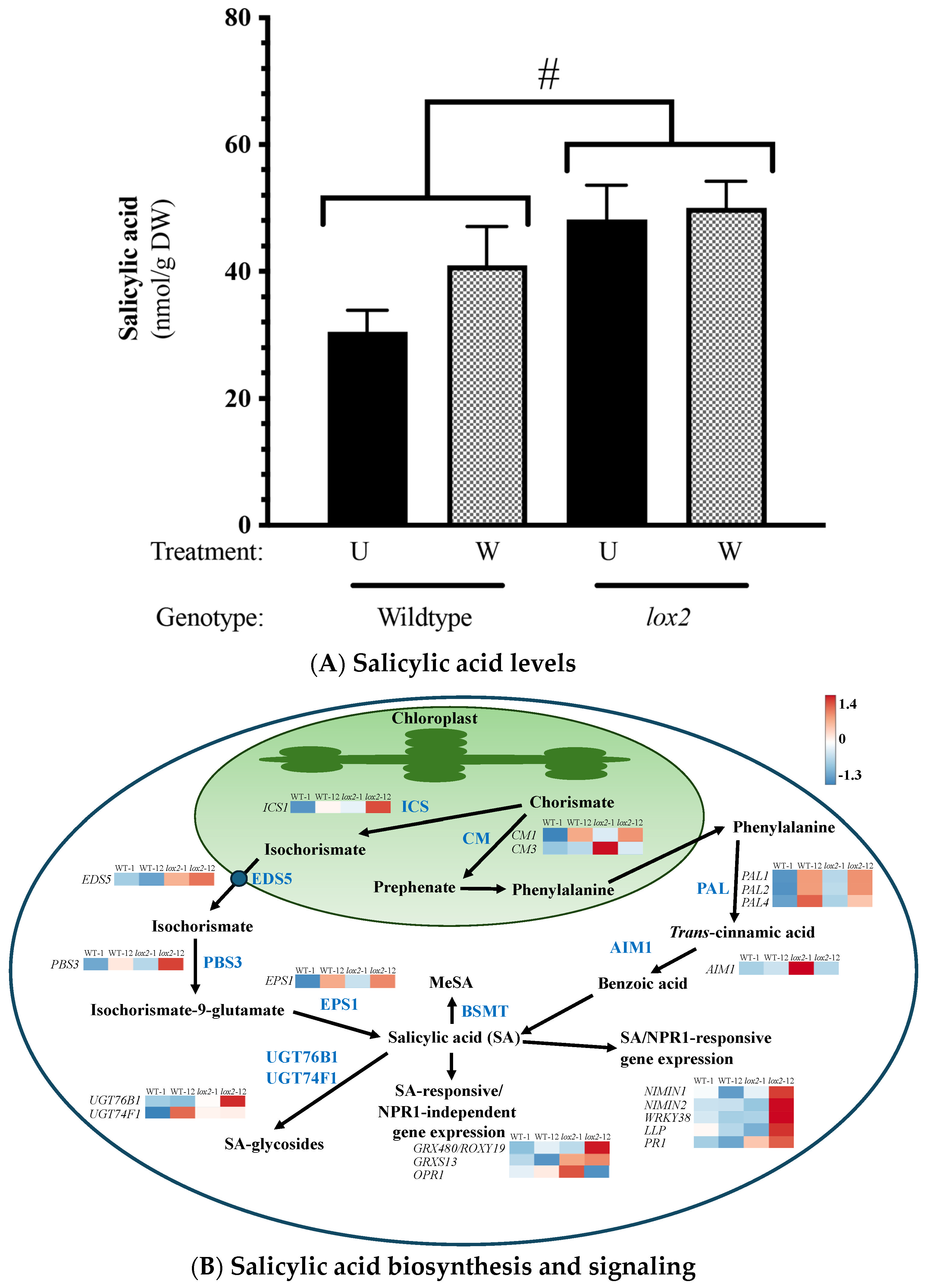
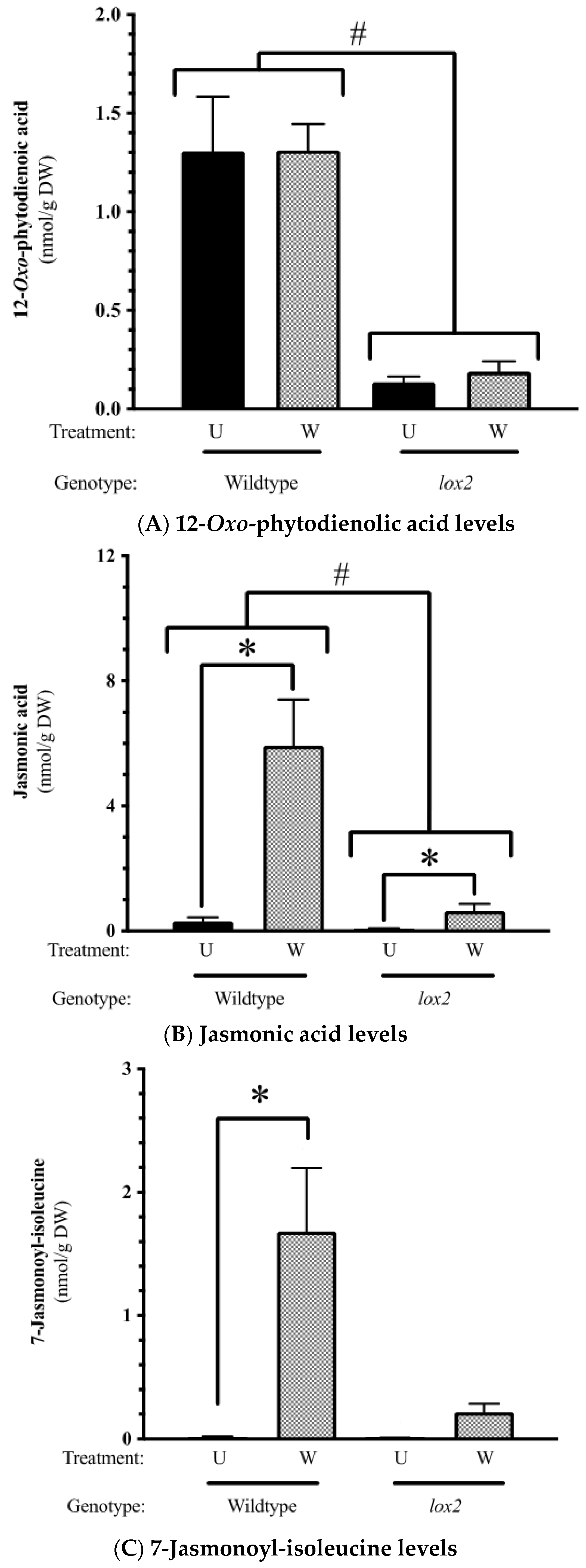
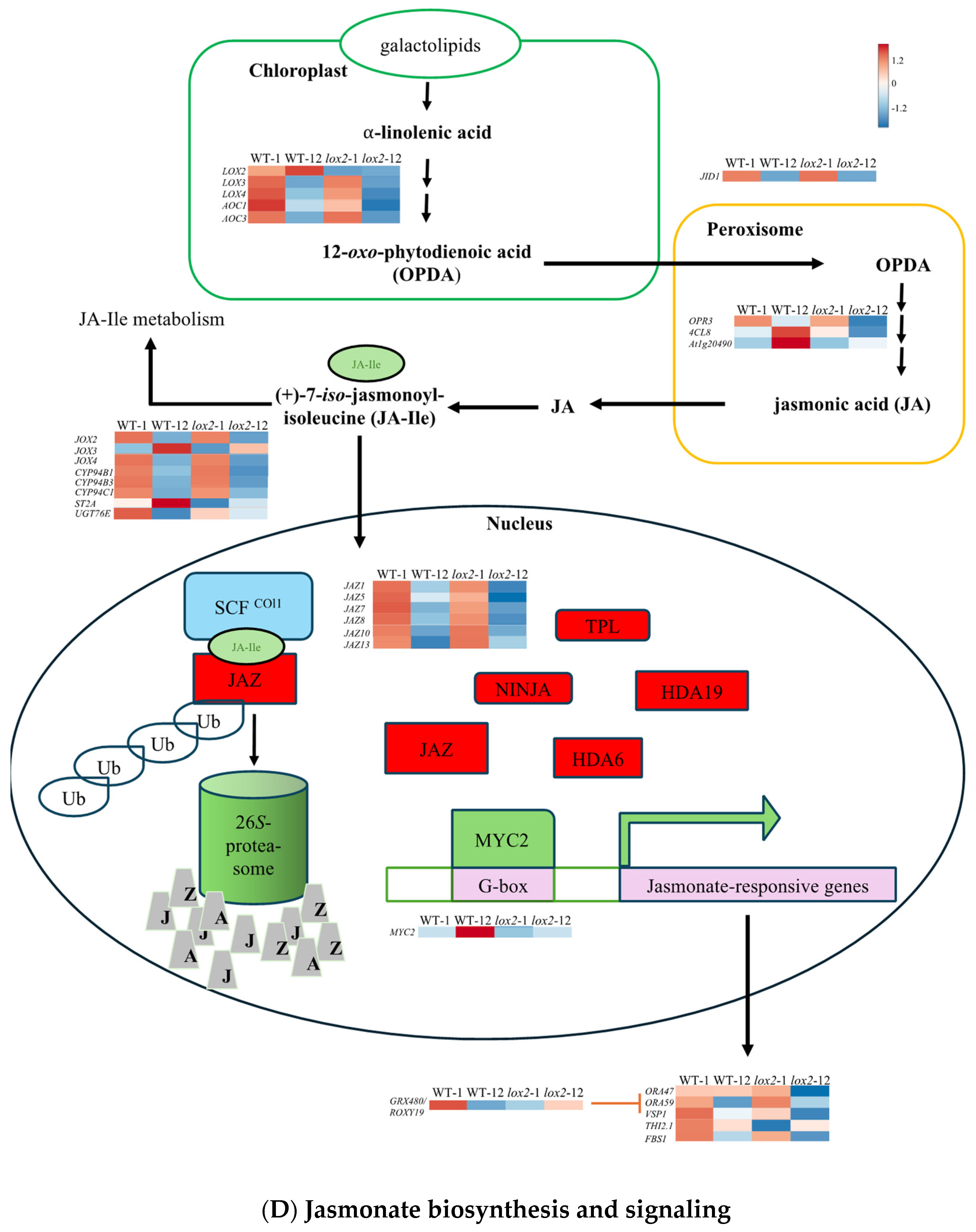
2.2. Constitutive Phytohormone and Gene Expression Levels

Constitutively, gene expression was highly conserved between the two genotypes (~20,469 genes) (Figure 1A); less than 1% were differentially expressed between WT and lox2 at any time point. In WT plants compared with lox2 mutants, there was an increase in specialized metabolism, glutathione metabolism and phenylpropanoid biosynthesis (Figure 1B,C). However, some pathogen resistance-related genes had higher expression levels in lox2 plants compared with WT plants, which may reflect the increased SA levels in these plants (Figure 2). These transcripts included genes that encode enzymes in SA biosynthesis (i.e., AtICS1, AtCM3, AtAIM1 [37]) and SA-responsive genes (i.e., AtPR1, AtNIMIN1, AtNIMIN2, AtGRXS13 [38]). In contrast, constitutive levels of the jasmonate phytohormone OPDA and JA were higher in WT plants compared with the lox2 mutant (Figure 3A,B).

2.3. Arabidopsis Responses to Foliar Damage

After wounding, the levels of JA and JA-Ile rose in damaged rosette leaves in both or only WT genotypes, respectively (Figure 3B,C). As expected, levels of these phytohormones were ~7.5 times higher in WT than the lox2 mutant. Jasmonate signaling is dynamic with the early expression of genes encoding biosynthetic enzymes and positive regulators (Figure 3D) [39,40,41]. In addition, the jasmonate burst is tempered either by catabolizing jasmonates to a less active form or by the activity of JAZ proteins that bind to and repress MYC2/3/4 transcription factors [42,43].
In WT plants, 324 genes were differentially expressed in response to wounding, with 252, 58 and 14 expressed early, late or at both time points, respectively. At 1 h after wounding, a strong increase in the expression of transcripts that encode proteins involved in α-linolenic and linoleic acid biosynthesis, phytohormone and MAPK signaling, terpenoid biosynthesis and wound healing (cutin, suberin and wax biosynthesis) as well as redox metabolism (glutathione metabolism) was observed (Figure 4A,B,E,F). Though most of the processes observed 1 h post-damage were also observed later, there was also an increase in primary metabolic pathways 12 h after wounding. In contrast, though the same general pathways were upregulated in the lox2 mutant (Figure 4C,D), fewer genes were strongly expressed (Figure 4G,H).
In general, WT and lox2 plants showed similar wound-induced responses (Figure 5A,D). One hour post-damage, out of the 249 wound-induced genes observed in WT plants, 81% were also induced in damaged lox2 (Figure 5A,B). A stronger difference in wound-induced gene expression between WT and lox2 plants was observed 12 h after wounding (Figure 5D,E). At this time point, only 76 wound-induced genes were identified in WT and shared 31% gene expression with damaged lox2.
Based on this, wound-induced gene expression patterns were divided into six distinct groups as follows: Pattern 1—general early (1 h) wound-induced gene expression (Figure 5A), Pattern 2—early (1 h) wound-induced gene expression with higher expression in WT plants (Figure 5B), Pattern 3—early (1 h) wound-induced gene expression with higher expression in lox2 plants (Figure 5C), Pattern 4—general late (12 h) wound-induced gene expression (Figure 5D), Pattern 5—late (12 h) wound-induced gene expression with higher expression in WT plants (Figure 5E) and Pattern 6—late (12 h) wound-induced gene expression with higher expression in lox2 plants (Figure 5F).

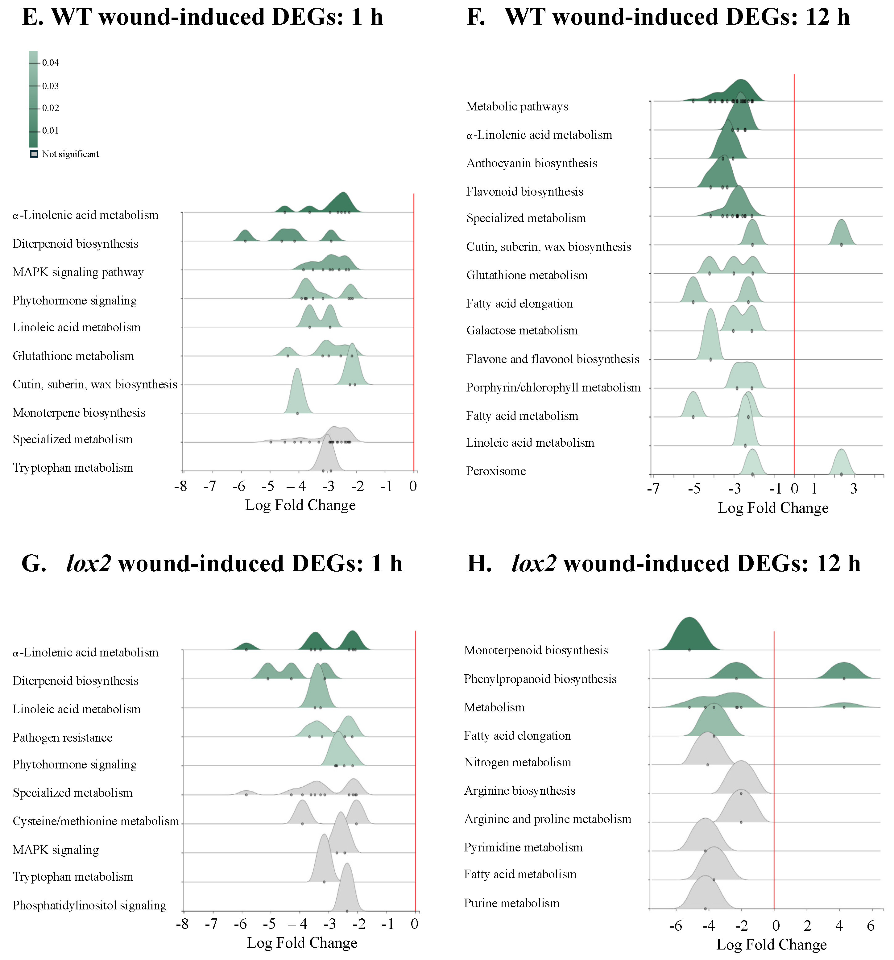
2.4. Pattern 1: General Early Wound-Induced Gene Expression

In this group, wound-induced gene expression is higher at 1 h and typically returned to near-basal levels at 12 h in both genotypes (Figure 5A, Supplemental Table S5A). One hour post-damage, the expression of genes encoding proteins involved in jasmonate biosynthesis and signaling were strongly upregulated in both genotypes (i.e., AtLOX3, AtLOX4, AtLOX6, AtAOC1, AtAOC3, AtOPR3, AtOPCL1 and AtORA47 [39,40,41]) (Figure 3D). In addition, genes encoding proteins involved in jasmonate anabolism (i.e., AtJOX2, AtJOX4, AtCYP94B1, AtCYP94B3, AtILL6 and AtJID1 [44,45,46,47,48]) and JAZ proteins (AtJAZ1, AtJAZ2, AtJAZ5, AtJAZ7, AtJAZ8, AtJAZ10, AtJAZ13 [42,43]) were also induced.
In line with this, jasmonate-responsive genes were among the early responding genes including those that encode enzymes in volatile biosynthesis (i.e., AtTPS04/GES, AtCYP82G1 [49,50]), enzymes involved in sealing damaged tissue (i.e., AtPP2-A5 [51]), transcription factors (i.e., AtRRTF1, AtWRKY40, AtRAP2.6 [52,53,54,55,56]) and proteins involved in plant defense against pathogens or insects (i.e., AtTI6, AtMAPKKK21 [57,58]).
Gene expression associated with other phytohormones and their signaling pathways that may be involved in crosstalk were also observed. Wound-induced expression of AtGA2OX6 and AtGA2OX8, encoding enzymes that oxidize gibberellins lowering their availability [59], as well as the negative growth regulator DELLA protein AtRGAL3 [60,61], may reflect the shift from growth to defense [62]. Even though abscisic acid (ABA) levels were not affected by wounding (Supplemental Figure S1), a number of ABA-related genes were induced 1 h after wounding. The expression of the ABA receptor AtPYL6 and ABA-responsive genes (i.e., AtRAS1, AtERD7, AtOSCA1.4, AtERD10, AtCOR78 [63,64,65,66,67]) were upregulated early after foliar damage. ABA-related responses help minimize water loss from damaged leaves as well as enhance plant defense responses [68,69,70]. In response to wounding, synergistic defensive responses between jasmonates and ethylene are often observed [71]; genes encoding ethylene biosynthetic enzymes (i.e., AtCSP2 and AtACS8) and ethylene responses (i.e., AtERF2 and AtRAP2.6) were noted in early wound-induced responses.
LOX2 has been implicated in wound-associated, chloroplastic generation of 1O2 [34,35]. Comparing transcript expression of 66 1O2-responsive genes in our damaged foliar tissues 1 h post-wounding [72,73], a genotype-difference in wound-induced 1O2-responsive transcript expression was not observed (Supplemental Figure S2).

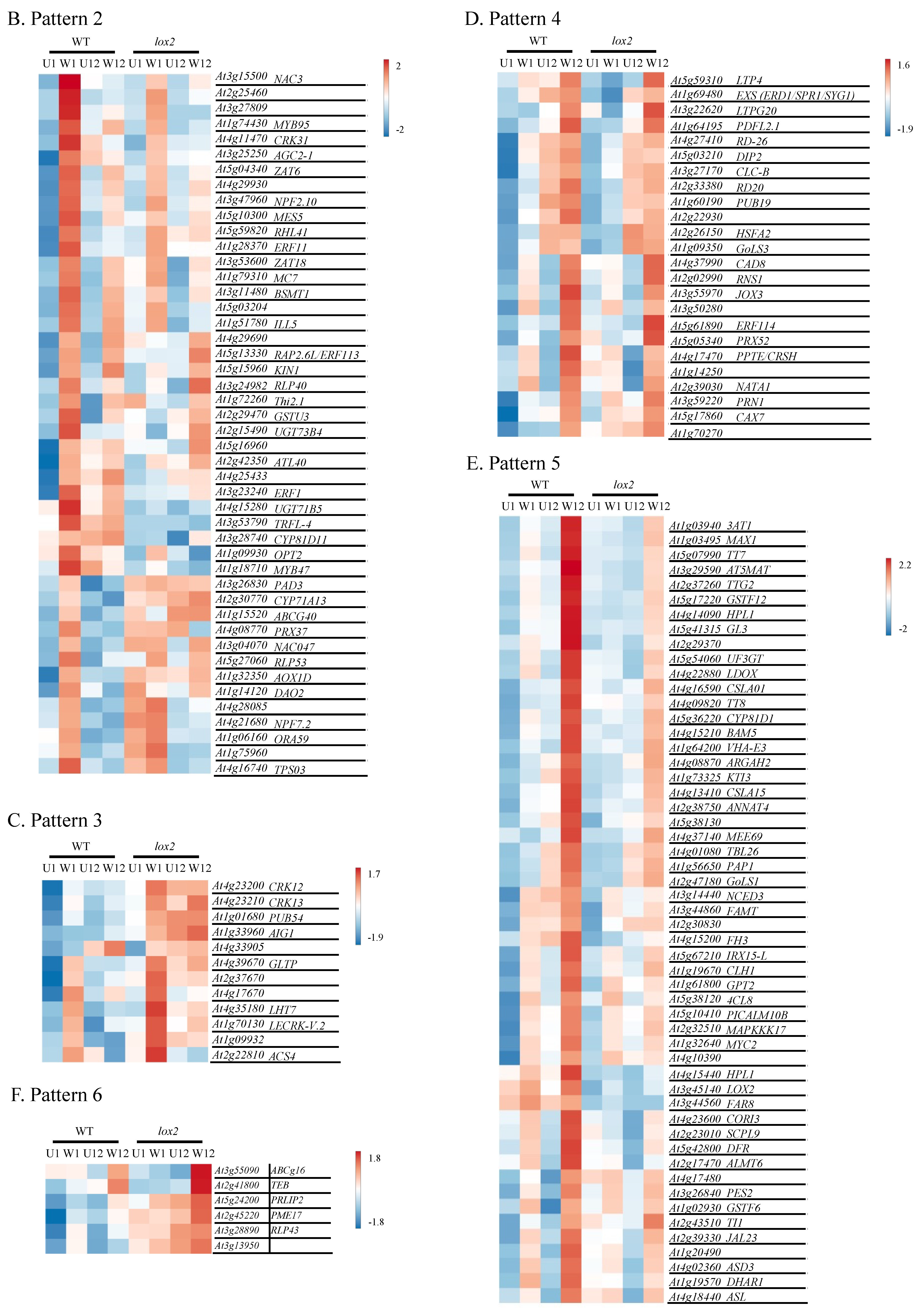
2.5. Pattern 2: Early Wound-Induced Gene Expression in WT Plants

In general, genes in this group were often wound-induced in both WT and lox2 but showed earlier, higher expression levels in WT plants (Figure 5B, Supplemental Table S5B). These early damage-induced genes include transcriptional regulators of jasmonate responses (i.e., AtORA59 [74]) as well as jasmonate- and wound/insect-responsive genes (i.e., AtTHI2.1, AtTPS03, AtCYP81D11 [75,76,77,78]).

2.6. Pattern 3: Early Wound-Induced Gene Expression in lox2 Plants

Likely reflecting elevated SA levels (Figure 2, Supplemental Table S5C), SA-responsive genes (AtAIG1, AtCRK13 [79,80]) were expressed at higher levels in wounded lox2 mutants compared with WT plants (Figure 5C).

2.7. Pattern 4: General Late Wound-Induced Gene Expression

Pattern 4 shows wound-induced genes in both genotypes that were more highly expressed 12 h after mechanical damage (Figure 5D, Supplemental Table S5D). Late wound-induced genes in both plant genotypes include AtPDFL2.1, AtNATA1, AtRD20, AtPRN1, AtPPTE/AtCRSH and AtRNS1. Genes encoding enzymes in lignin biosynthesis (i.e., AtCAD8, AtPRX52) may also contribute to wound-induced lignin deposition (Figure 6) [81,82].

2.8. Pattern 5: Late Wound-Induced Gene Expression in WT Plants

Most late-induced genes showed higher expression in WT plants compared with lox2 plants 12 h post-damage (Figure 5E, Supplemental Table S5E). This likely reflects the expression of genes encoding enzymes in jasmonate biosynthesis (AtLOX2, At4CL8) and signaling (AtMYC2) that were more highly expressed in wounded WT compared with lox2 plants [4]. Jasmonate-responsive genes involved in plant resistance to insect herbivory, such as AtHPL/AtCYP74B2, which encodes an enzyme involved in volatile biosynthesis [83], AtCLH1, AtKTI3, AtTI1, AtARGAH2 and AtMAPKKK17, reflect this pattern [58,84,85,86]. Genes involved in antioxidant pathways are upregulated in wounded WT plants, including those that encode proteins involved in the ascorbate/glutathione cycle (i.e., AtDHAR1 [87]) and anthocyanin biosynthesis (i.e., AtPAP1, AtTT8, AtGL3, AtTTG2, At4CL, AtTT7, AtDFR, AtLDOX, AtUF3GT, At1g14090, At3AT1, At3AT2, AtGSTF12, At5MAT [88,89,90]) (Figure 6). Of note, AtTTG1 and AtGL3 also regulate trichome development [91].

2.9. Pattern 6: Late Wound-Induced Gene Expression in lox2 Plants

Only a few genes showed lox2-specific late wound-induced expression (Figure 5F, Supplemental Table S5F). Expression of the SA-responsive gene AtPRLIP2 was observed in wounded lox2 mutants [92,93].

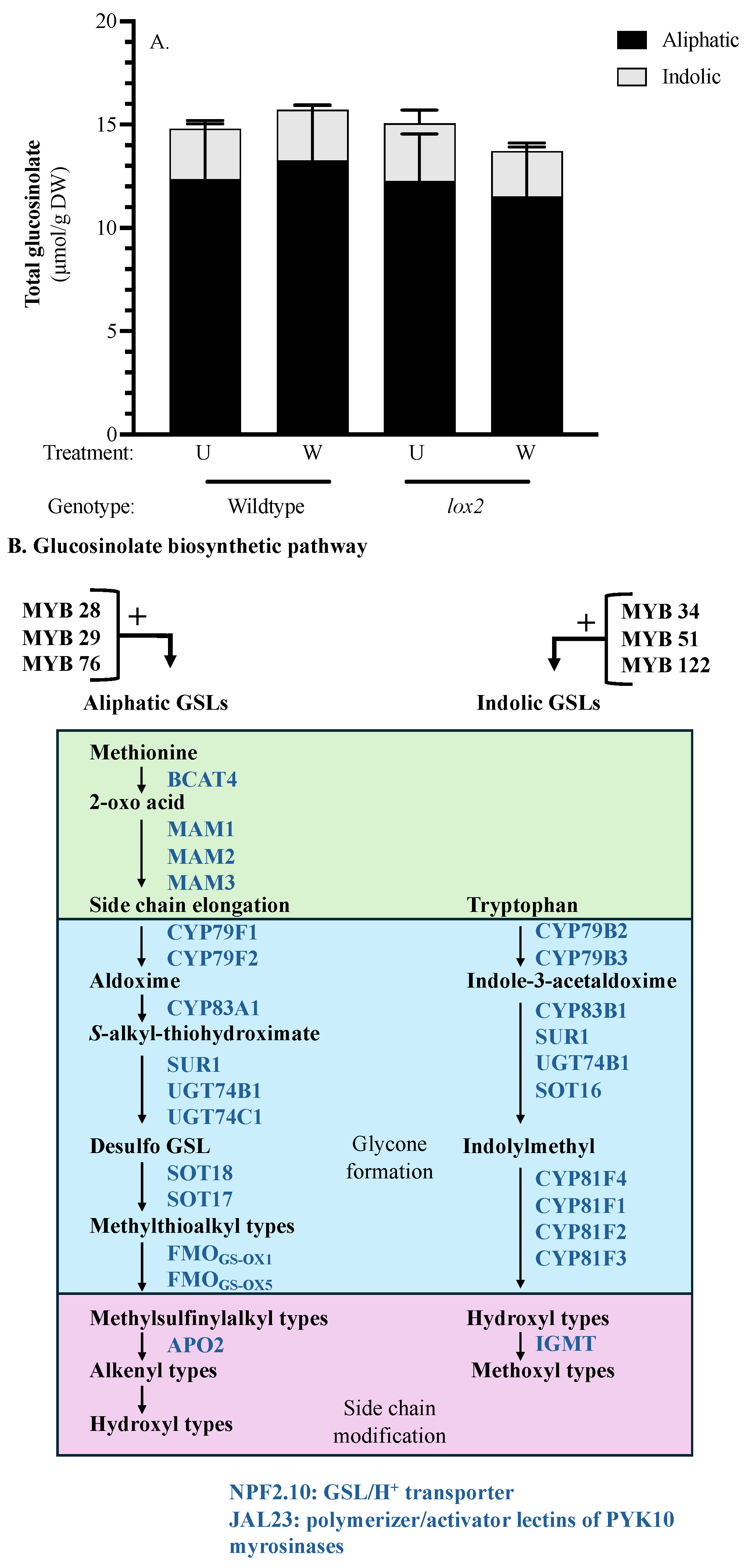
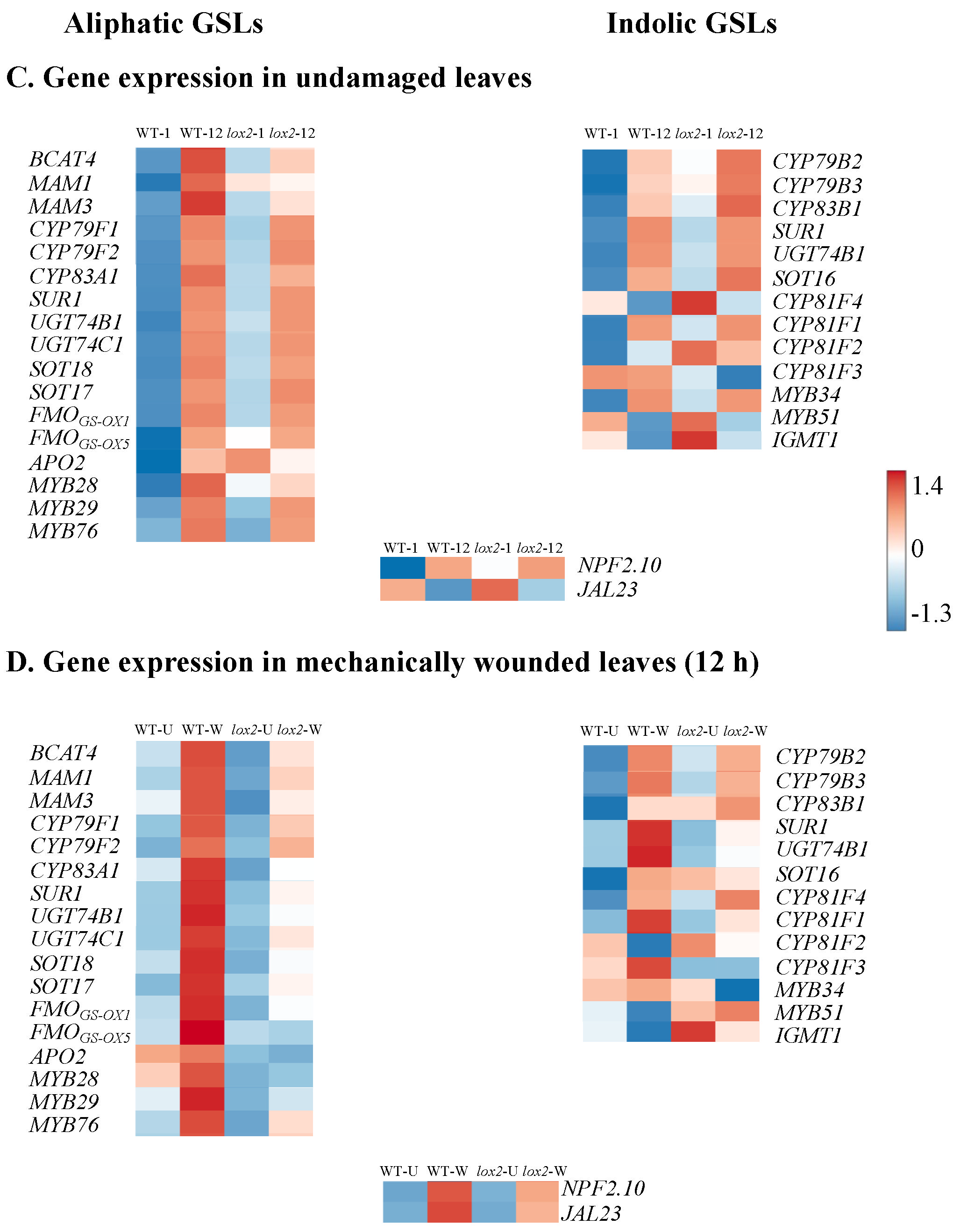
2.10. Glucosinolates

Constitutively or 12 h post-wounding, the foliar GSL profile did not differ between the two genotypes, reflecting the gene expression profile of GSL biosynthetic enzymes (Figure 7A). However, genes encoding GSL transcriptional regulators and biosynthetic enzymes show a strong diurnal cycle with stronger expression in the light phase, particularly for genes involved in aliphatic GSL biosynthesis (Figure 7B). Genes encoding indole GSL methyltransferase1 (AtIGMT1), the GSL transporter AtNPF2.10 and lectin JAL23 (a polymerization factor and putative activator of the myrosinase PYK10 [94]) showed strong wound-induction in WT plants.

3. Discussion

Our results highlight the transcriptional dynamics of wound-induced jasmonate biosynthesis and the importance of AtLOX2 in sustained jasmonate signaling leading to plant resistance against insect herbivores.

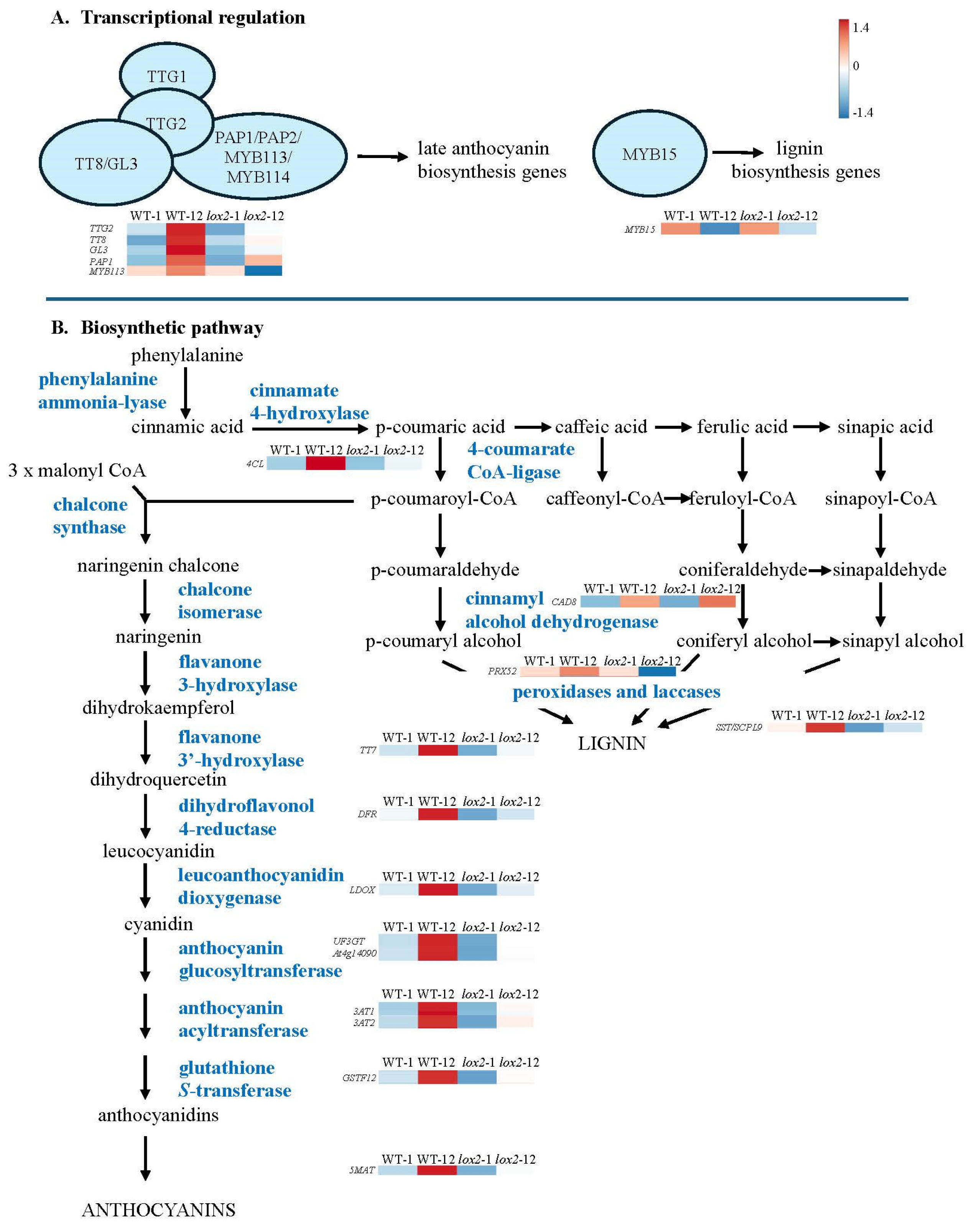
3.1. The Dynamic Jasmonate Burst

Early wound-induced genes in both genotypes include enzymes in the jasmonate biosynthetic pathway (i.e., AtLOX3, AtLOX4, AtAOC1, AtAOC3, AtOPR3, AtOPCL1 [4]) and transcriptional regulators (i.e AtORA47) (Pattern 1, Figure 5A). ORA47 coordinates the expression of genes that encode jasmonate and ABA biosynthetic enzymes as well as in the general jasmonate-responsive stress network [41,95,96]. The expression of later genes that encode jasmonate biosynthetic enzymes (i.e., AtLOX2, At4CL8, At1g20490 (putative) [4,97]) were generally higher in WT compared with lox2 plants (Pattern 5, Figure 5E). This was reflected in the expression of jasmonate-responsive gene expression, particularly in late wound-induced genes (Figure 5).
The wound-induced jasmonate burst is dynamic, and the initial strong jasmonate wave is dampened over time. Indeed, early wound-induced transcriptional expression included genes encoding proteins that attenuate the jasmonate-mediated signaling response, in particular, enzymes that metabolize jasmonates to their inactive form (i.e., AtST2A, AtJOX2, AtJOX4, AtCYP94B1, AtCYP94B3, AtILL6, AtJID1) and JAZ proteins (i.e., AtJAZ1, AtJAZ5, AtJAZ7, AtJAZ8, AtJAZ10, AtJAZ13) (Pattern 1, Figure 5A). JAZ proteins are negative regulators that bind to jasmonate signaling MYC2, MYC3 and MYC4 transcription factors [42,43]. Early wound-induced JAZ transcripts encode AtJAZ1, AtAtJAZ5, AtJAZ8, AtJAZ10 and AtJAZ13, which interact with AtMYC2 or AtMYC3, as well as AtJAZ7, which interacts with AtMYC2 [98,99,100].
In addition, early wound-induced genes include a number of enzymes that convert jasmonates into an potentially inactive forms (Pattern 1, Figure 5A). 2-oxo-glutarate-dependent dioxygenase genes jasmonate-induced oxidase2 (AtJOX2) and AtJOX4 as well as the aminohydrolase AtILL6 are expressed early after wounding, while AtJOX3 is expressed later (Figure 3); these genes encode enzymes that hydroxylate JA to a biologically inactive form [45,46,47]. Subsequently, the hydroxylated JA can be further metabolized by sulfotransferase 2A (ST2A) to the sulfated form [101]. Furthermore, the early wound-induced cytochrome P450 genes encoding AtCYP94B1, AtCYP94C1 and AtCYP94B3 potentially work in sequence to catalyze the formation of 12-OH-JA-Ile and 12-COOH-JA-Ile, respectively [44,102,103,104]. JID1 is a cytosolic enzyme thought to be involved in OPDA anabolism [48].
Often working synergistically, ethylene and jasmonate signaling leads to increased plant resistance [105]. Early genes encoding enzymes in ethylene biosynthesis, AtACS2, AtACS8 and AtACS4, as well as the transcription factors AtRAP2.6, AtERF2 and AtERF114, were wound-induced in WT or both genotypes 1 h post-damage (Figure 5A,C).
In comparison, SA and jasmonates often have an antagonistic relationship that acts to shape the plant defense response [106]. In general, the lox2 mutant expressed higher constitutive expression of genes involved in SA biosynthesis (i.e., AtICS, AtEDS5, AtPBS3, AtCM3, AtAIMI [37,107]) that translated into higher SA levels and SA-responsive gene expression (i.e., AtGRX480/AtROXY19, AtCRXS13, AtOPR1, AtNIMIN1, AtNIMIN2, AtWRKY38, AtLLP, AtPR1) (Figure 2). Of these, AtNIMIN1, AtNIMIN2 and AtWRKY38, which encode negative regulators of basal immunity [108,109,110], were expressed in the light (9 am), which may reflect the diurnal rhythm of SA, which is lower in the morning [111]. In wounded plants, SA-responsive AtCRK13, AtAIG1 and AtPRLIP2 were more highly expressed in lox2 compared with WT plants (Pattern 3, Figure 5C; Pattern 6, Figure 5F) [79,80,92,93].

3.2. Plant Resistance against Insect Herbivory

3.2.1. Response to Egg Deposition

In some species, plants respond to insect eggs by eliciting an SA-dependent “hypersensitive-like response” that results in necrotrophic tissue forming under the eggs, often leading to egg desiccation and plant resistance [112]. Treatment of leaves by spider mite egg extract resulted in the induction of genes encoding enzymes in the jasmonate biosynthesis and signaling pathway (i.e., AtAOC1, AtAOC3, AtLOX2, AtMYC2, AtJAZ9, AtST2A, AtJOX2, AtJOX3, AtJOX4, AtILL6, AtCYP82G1, AtMDHAR, AtFAMT, AtNATA1, AtZAT10/STZ, AtBSMT1, RNS1 At3g23350 and AtCORI3), which were also seen in our study (Figure 3D and Figure 5) [113].

3.2.2. Signal Transduction

In this and other studies, AtMAPKKK17, induced late in response to wounding in WT plants (Pattern 5, Figure 5E), activates the MKK3-MPK1/2/7 module [114,115]. In plants challenged with the two-spotted spider mite, Tetranchus urtica, plants with higher constitutive AtMAPKKK17 expression had higher resistance against mite herbivory, exhibiting less leaf damage with mites having lower fecundity [58]. AtMAPKKK21 is expressed early after wounding in both genotypes (Pattern 1, Figure 5A). In contrast to MAPKKK17, this kinase was found to be a negative regulator of plant resistance to mites [58].

3.2.3. Physical Defense

Foliar trichomes create a mechanical defense against insect herbivores and, as well, may be a site for the production of chemical defenses [116]. Even though arabidopsis trichomes are non-glandular, volatile organic compounds as well as aliphatic and indolic GSLs may be biosynthesized in these cells [21,50,117]. In addition, arabidopsis trichomes may serve as mechanosensors, possibly leading to specialized metabolite production when triggered [118,119]. AtGL3, a late wound-induced gene that shows higher stress-associated expression in WT compared with lox2 plants (Pattern 5, Figure 5E), encodes a bHLH transcription factor that regulates trichome initiation as well as anthocyanin biosynthesis [120,121]. Constitutively, this transcription factor interacts with JAZ proteins, specifically, JAZ1, JAZ2, JAZ8 and JAZ11, leading to its repression [122]. In response to wounding, the resultant degradation of JAZ proteins coupled to increased AtGL3 expression supports observations of a wound-associated increase in arabidopsis foliar trichomes [123].

3.2.4. Chemical Defense

GSLs and flavonoids are important defensive compounds in the Brassicaceae [18,124,125]. A wound-induced difference in foliar GSL levels was not observed (Figure 7). However, in both genotypes, a strong expression of GSL biosynthetic genes was observed late after wounding, which corresponds to the light phase in our experiment. This supports previous observations of a possible role of circadian rhythms in foliar GSL biosynthesis [21].
In contrast, a genotype difference in wound-induced genes associated with flavonoid biosynthesis was identified in this study, with genes expressed more highly in WT plants (Pattern 5, Figure 6). Flavonoids can act as feeding or oviposition deterrents or toxins against insect herbivores [124,125]. Even though AtRNS1, a jasmonate-independent, wound-induced gene that encodes a ribonuclease that is a negative regulator of anthocyanin biosynthesis [126], is expressed in both genotypes (Pattern 4, Figure 5D), the late wound-induced expression of genes that encode transcriptional regulators or flavonoid biosynthetic enzymes was observed in WT plants (Pattern 5, Figure 5E). Thus, particularly in WT plants, there is a strong upregulation of genes involved in flavonoid biosynthesis, particularly anthocyanins, that may act as antioxidants or be involved in plant resistance against insect herbivory [125,127].

3.2.5. Indirect Chemical Defense

Wounding of arabidopsis leaves results in the biosynthesis and release of volatile organic compounds (VOCs) that are involved in numerous ecological roles, including intra-plant stress signaling and tritrophic interactions, such as attracting parasitoids or predators of the herbivore [128,129]. From the diterpenoid precursor geranylgeranyl diphosphate, AtTPS04, which is expressed in arabidopsis non-glandular trichomes [50], catalyzes the production of (Ε,Ε)-geranyllinalool, which is converted by AtCYP82G1 into volatile 4,8,12-trimethyltrideca-1,3,7,11-tetraene (TMTT) [49], an important attractant in tritrophic interactions for beneficial parasitoid and predatory arthropods [130]. AtTPS04 is induced in response to herbivory by caterpillars of the specialist diamond backmoth, Plutella xylostella, or the generalist African cotton leafworm, Spodoptera littoralis [131,132,133,134]. In our study, both AtTPS04 and AtCYP82G1 show early wound-induced gene expression (Pattern 1, Figure 5A). AtCYP82G1 is also induced in response to the treatment of arabidopsis leaves with spider mite egg extract [113].
The expression of AtTPS03 and AtCYP81D11 is predominant in wounded WT plants (Pattern 2, Figure 5B). AtTPS03 encodes a terpene synthase that produces (E,E)-α-farnesene, an important component in highly attractive volatile blends attractive to parasitic wasps such as Microplitis croceipes, Apanteles taragamae, Anaphes iole and Gonatocerus ashmeadi, as well as acting as a deterrent to soybean cyst nematodes [135,136,137,138,139]. AtCYP81D11 also contributes to the volatile profile responsible for the attraction of parasitoid wasps, such as Cotesia plutellae, to wounded plants [140].
AtHPL had higher late wound-induced expression in WT compared with lox2 plants (Pattern 5, Figure 5E). This jasmonate-responsive gene is also induced in response to herbivory by P. rapae or S. littoralis caterpillars [141]. Cytosolic AtHPL competes with the jasmonate biosynthetic pathway for 13-hydroxyperoxide precursors to generate alkenals, which are converted into C6 volatiles [83]. The AtHPL in the arabidopsis Col-0 ecotype (which was also used in this study) has a deletion resulting in a truncated protein that is unable to use 13-hydroperoxide linolenic acid as a precursor but uses 13-hydroperoxide linoleic acid to produce hexenals [142].
AtBSMT1 was also more highly wound-induced in WT plants (Pattern 2, Figure 5B). This methyltransferase, involved in volatile methyl salicylate biosynthesis, is induced in response to P. rapae and Pieris brassicae herbivory [143,144]. The resultant methyl salicylate is an attractant for female parasitic Diadegma semiclausum wasps and also deters oviposition by female P. brassicae butterflies, thus decreasing herbivore damage [144,145].
In addition to the role that these VOCs play in attracting natural enemies of the herbivore [146], these volatiles can also act on the plant itself, leading to systemic upregulation of plant defense responses [147,148]. Recently, the importance of these volatiles as conspecific signals has been recognized [149]. Thus, volatiles may serve as systemic signals resulting in the induction of plant defense responses.

3.2.6. Interference with Insect Nutrition or Physiology

Obtaining sufficient nitrogen to maintain development and fitness is a key challenge for phytophagous insects [150]. Thus, plants have numerous strategies to interfere with a herbivore’s ability to obtain sufficient nitrogen to limit herbivore success.
In herbivorous insects, serine proteinases, such as trypsin and chymotrypsin, initiate protein digestion [151]; However, plants produce inhibitors of these enzymes, known as proteinase inhibitors [152]. Overexpression of a soybean trypsin inhibitor in arabidopsis negatively affected the larval biomass of corn earworm, Helicoverpa zea, caterpillars [153]. In our study, we identified the expression of three wound-induced trypsin inhibitors. AtTI1 is induced early in both genotypes and has previously been found to be induced by aphid attack (Pattern 1, Figure 5A) [154]. AtKTI3 and AtTI6 are more highly expressed in WT plants late after wounding (Pattern 5, Figure 5E).
Another late wound-induced gene identified in our transcriptomic study that interferes with nitrogen resources for the insect encodes arginase (AtARGAH2) (Pattern 5, Figure 5E). This enzyme catabolizes nitrogen-rich arginine to produce urea and ornithine. Direct feeding of Manduca sexta caterpillars on tomato plants overexpressing arginase resulted in smaller insects, presumably because of the decreased availability of the essential amino acid arginine [86]. However, this may also reflect the potential toxicity of arginase products, where ornithine may be converted by AtNATA1, a late pattern 4 gene (Figure 5D), to Nd-acetylornithine [155]. This derivative negatively affects the fecundity of the green peach aphid, Myzus persicae. It is of interest that the pH optimum of plant arginase is alkaline [86], which likely allows it to be most active in its native cellular location, the mitochondrial matrix [156], and also in the alkaline midgut of caterpillars [157]. In addition to these activities, ARGAH2 activity may affect nitric oxide (NO) production [158]. Though this has been shown in the marine green algae Ostreococcus tauri [159], in higher plants, NOS-like activity is controversial; however, NO has been proposed to be released from arginine upon its conversion to citrulline [160,161]. Consistent with this, AtARGAH2 knockout plants had increased NO accumulation [158]. Differences in NO, or its more biologically stable form S-nitrosoglutathione (GSNO), can impact protein S-nitrosation and S-glutathionylation status of regulatory proteins involved in plant defense [162,163,164,165]. Manduca sexta caterpillars grew larger on plants silenced in their ability to produce GSNO. Methyl jasmonate-induced levels of proteinase inhibitors and some defensive specialized metabolites (caffeoylputrescine, diterpene glycosides) were lower in these plants [162]. Therefore, AtARGAH2 may affect caterpillar nutrition or plant defense.
The phloem-associated protein AtPP2-A5 has two domains, a PP2 (lectin activity) domain at the C-terminus and a Toll/Interleukin-1 receptor domain at the N-terminus [166]. Though this protein plays a major role in sealing wounded sieve elements (Pattern 1, Figure 5A), AtPP2-A5 overexpression or knockout lines show modified transcriptional patterns to spider mite herbivory, suggesting that the receptor portion of this protein recognizes an herbivore-specific effector to remodel gene expression [166]. The knockout line was more susceptible to spider mite damage, and the arthropods had higher mortality on the overexpression lines. This likely reflects direct interactions with the insect, potentially by binding to the arthropod gut epithelial [167]. Feeding aphids a diet spiked with recombinant AtPP2-A5 did not affect mortality but did negatively affect the weight gain of two different aphid species, the pea aphid Acythosiphon pisum (~30% smaller) and the green peach aphid Myzus persicae (10–20% smaller) [168]. Aphids reared on AtPP2-A5 overexpression lines had lower colonization and spent less time feeding on the phloem compared with WT plants [169].
Chlorophyllase, AtCHL1, catalyzes the hydrolysis of chlorophyll to chlorophyllide. Under stress conditions, this enzyme may be involved in chlorophyll degradation to minimize chlorophyll-associated ROS generation [170,171]. However, as this protein is associated with the endoplasmic reticulum or tonoplast, only upon cell disruption, such as that incurred by herbivory, does the enzyme come into contact with its substrate, chlorophyll. The product of this reaction, chlorophyllide, is toxic to S. littoralis caterpillars and binds to the midgut of Bombyx mori caterpillars, potentially impairing digestion [172]. However, other studies suggest that chlorophyllide may have roles that benefit the insect herbivore [173]. In our study, AtChl1 was induced late after wounding and showed higher expression in WT plants (Pattern 5, Figure 5E).

4. Materials and Methods

4.1. Plant Maintenance

Arabidopsis thaliana wildtype (WT) Columbia (Col-0) seeds were obtained from the Arabidopsis Biological Resource Center. Seeds of the lox2-1 mutant line, which has a mutation in the tryptophan amino acid at position 630 to produce a stop codon resulting in a truncated non-functional protein, were generously provided by Dr. E. E. Farmer [28]. The seeds were surface-sterilized in 70% (v/v) ethanol for one min, then 0.6% (v/v) bleach (NaOCl) for 3 min, followed by five successive washes in sterile ddH2O. Between these treatments, the seeds were recovered by centrifugation, and the liquid was removed. The seeds were then placed in Petri dishes containing Murashige and Skoog salts, pH 5.8, in 0.8% agar, followed by stratification in the dark at 4 °C to promote synchronized germination. After 2 days, the Petri dishes were transferred to a growth cabinet with 14 h of light with an intensity of 250 μmol m−2 s−1 followed by 10 h darkness. The temperature of the light–dark cycle was 23:20 °C. After one week, the germinated seedlings were transplanted into pots (12 cm diameter × 11 cm height) containing Fafard Agromix G6 potting medium and grown under the same conditions. Plants were bottom-watered 3 times per week with 20:20:20 NPK fertilizer (0.14 g/L distilled water) and used for experimentation at the vegetative 3.9 stage [174].

4.2. Experimental Design

Two days prior to the wounding experiment, a plexiglass sheet was placed between randomly chosen plants that were to remain unwounded or mechanically damaged to separate the treatments and avoid volatile signaling between these groups. At 9 PM (time 0; dark phase), arabidopsis lines were either mechanically damaged, whereby each leaf of the rosette was wounded once by a hole punch, without harming the mid-vein, or left unwounded. Whole rosettes were collected for phytohormones (1 h; dark phase), GSLs (12 h; light phase) or transcriptomics (1 h and 12 h, dark and light phase, respectively) post-damage, flash-frozen in liquid nitrogen and stored at −80 °C until analysis. The experiment was repeated temporally three times to collect samples for transcriptomics and five times for phytohormone and GSL analyses. At each temporal replicate, one sample was taken for the different analyses (RNA-Seq or metabolite analysis) of transcriptomics (n = 3), phytohormones (n = 5) and GSLs (n = 5).

4.3. Phytohormone Analysis

Following the protocol described in Martinez Henao et al. [175], plant samples were finely ground and extracted in ethyl acetate containing isotopically labeled standards (D6-JA, D6-JA-Ile and D4-SA (OlChemim, s.r.o)). The samples were vigorously vortexed and centrifuged (19,000× g, 10 min, 4 °C), and the supernatant was transferred to a new tube. The extraction was repeated, and the supernatants were pooled. Following evaporation using a vacuum concentrator at room temperature, the resulting pellet was resuspended in 70% (v/v) methanol (HPLC-MS grade). A final centrifugation step was performed as above to ensure the removal of all non-soluble debris. Metabolites were separated by ultrahigh performance liquid chromatography (UHPLC) followed by detection on a triple quadrupole mass spectrometer (EVOQ-TQ-MS, Bruker, Hamburg, Germany). Reverse phase UHPLC was performed using a Zorbax Extend-C18 column (4.6 × 50 mm, 1.8 μm, Agilent Technologies, Santa Clara, CA, USA). The mobile phase began with 5% (v/v) acetonitrile (ACN), 0.05% (v/v) formic acid for 30 s and then increased to 50% (v/v) ACN, 0.05% (v/v) formic acid over 2 min. After separation, the compounds were nebulized by electron spray ionization and detected using the EVOQ-TQ-MS. Phytohormones were identified based on their retention time, in comparison with known standards, as well as their m/z.

4.4. Glucosinolate Analysis

GSLs were extracted from lyophilized leaf tissue and analyzed by high-performance liquid chromatography (HPLC)-pulsed amperometric detection following Grosser and van Dam [176]. Briefly, 70% MeOH was added to the finely ground tissues and incubated at 90 °C for 6 min to inactivate myrosinases followed by sonication for 15 min. After centrifugation (2975× g for 10 min), the supernatant was transferred to a clean tube, and the pellet was re-extracted. Pooled supernatants were passed through a diethylaminoethyl Sephadex A-25 ion exchange column preconditioned with ddH2O. After washing (2 × 1 mL 70% MeOH, 2 × 1 mL ddH2O, 1 × 1 mL 20 mM sodium acetate buffer, pH 5.5), the column was treated with 10 U of arylsulfatase and incubated for 12 h at RT. Desulfated GSLs were eluted in sterile MilliQ H2O (2 × 0.75 mL) and lyophilized.
GSLs were separated by reverse-phase chromatography on a C18 column (Alltima C18, 150 × 4.6 mm, 3 μm, Alltech, Lexington, USA) using a mobile gradient from 2% acetonitrile (ACN) to 35% ACN in 30 min at a flow rate of 0.75 mL min−1. GSLs were identified based on retention time to known standards (glucoiberin (3-methylsulfenylpropyl GSL), glucoerucin (4-methylthiobutyl GSL), progoitrin (2-hydroxy-3-butenyl GSL), sinigrin (2-propenyl GSL), gluconapin (3-butenyl GSL), glucobrassicanapin (4-pentenyl GSL), glucobrassicin (indol-3-ylmethyl GSL), sinalbin (4-hydoxybenzyl GSL), glucotropaeolin (benzyl GSL) and gluconasturtiin (2-phenylethyl GSL); Phytoplan, Heidelberg, Germany)) and UV spectra. GSL concentrations were calculated from a sinigrin standard curve according to Grosser and van Dam [176].

4.5. RNA Extraction, Library Preparation and Transcriptomics

Total RNA was extracted from flash-frozen pulverized plant rosettes (~100 mg) using the RNeasy Plant Mini Kit (Qiagen, Venlo, The Netherlands) following the manufacturer’s protocol with an additional on-column DNase digestion step to avoid genomic DNA contamination. The 2100 Bioanalyzer instrument (Agilent Technologies) was used to determine RNA quality using an RNA integrity number (RIN) of 8.7 as a minimum threshold. Total RNA samples were processed by Genome Québec Innovation Centre for library preparation and next-generation RNA sequencing (RNA-Seq). Libraries were generated from 250 ng of total RNA as follows: mRNA enrichment and ribosomal RNA removal were performed using the Next Poly(A) Magnetic Isolation following the manufacturer’s instructions (New England BioLabs, Ipswich, USA). cDNA synthesis was performed with Next RNA First Strand Synthesis and Next Ultra Directional RNA Second Strand Synthesis kits (New England BioLabs). The remaining steps of library preparation were performed using the Next Ultra II DNA Library Prep Kit for Illumina (New England BioLabs). Adapters and PCR primers were purchased from New England BioLabs.
The libraries were normalized, pooled and denatured in 0.02 N NaOH, followed by neutralization using HT1 hybridization buffer. Twenty-four strand-specific mRNA libraries were generated and sequenced by loading 200 pM on the NovaSeq 6000 S4 (Illumina) using the Xp protocol as per the manufacturer’s recommendations, with paired-end (PE) mode for 2 × 100 base pairs (bp) resulting in >25 million reads in each direction. The Illumina phiX control v3 library was used as a control and mixed with libraries at a 1% level. Base calling was performed using Real-Time Analysis (RTA) v3 software. The program bcl2fastq2 v2.20 was used to demultiplex samples and generate fastq reads.

4.6. Quality Check, Clipping and Mapping

Raw read quality was examined by performing FastQC 0.11.9 on “.fastq.gz” file [177]. FastQC produces a control statistic and evaluates every metric using a classification system of pass, warn or fail. The adapter clipping and bad end quality trimming were performed by subjecting the raw reads to fastp tool version 0.20.1 to remove adapters, low-quality bases with a Phred score less than 20 and reads shorter than 25 bp from the tail (3’ end) from further analysis [178]. This preprocessing was followed by another quality check with FastQC. Read mapping was conducted using a splice-aware alignment tool STAR v2.7.9a to align the trimmed reads against the arabidopsis reference genome downloaded from The Arabidopsis Information Resource (TAIR, assembly ID: TAIR10, https://www.arabidopsis.org/download_files/Genes/TAIR10_genome_release/TAIR10_gff3/TAIR10_GFF3_genes.gff (accessed on 9 December 2022)) [179].

4.7. Differential Gene Expression

Strand-specific transcript abundance was calculated using featureCounts (a tool included in the subread package 2.0.3) on mapped and sorted BAM files. The resulting read counts per transcript values were imported into the web-based tool NetworkAnalyst 3.0 for data filtering, Log2 normalization and differential analysis [180]. The statistical method DESeq2 with a negative binomial (Gamma–Poisson) distribution was used to identify differentially expressed genes using the log2-fold change (logFC) calculation [181,182]. Genes with a False Discovery Rate (FDR)-corrected p-value (padj) ≤ 0.05 and log2 fold change ≥ 2 or ≤−2 were deemed significant. The transcriptomic data generated by this study are available in the Supplementary Materials of this article (Supplemental Table S1) and the raw read data (FASTQ) were deposited in the NCBI Sequence Read Archive (Bioproject ID #PRJNA1077722).
Transcriptomic visualization (ridgeplots, heatmaps, principal component analysis (PCA)) was performed using MetaboAnalyst 5.0 and ExpressAnalyst [183,184]. For visualization, the data were normalized by log10 transformation.

4.8. Statistics

For phytohormone and GSL analyses, a two-factor analysis of variance (ANOVA) (factors: genotype and treatment) was conducted using the statistical program SPSS vers. 29 (Supplemental Tables S2 and S3).

5. Conclusions

By comparing early (1 h) and late (12 h) constitutive and wound responses between arabidopsis WT and lox2 mutants, we identified gene expression differences related to the regulation of jasmonate biosynthesis and, in mechanically damaged plants, resistance to insect herbivores. Both constitutively and in wounded plants, SA levels are higher in lox2, which is reflected in the expression of SA-responsive genes. Since jasmonate and SA pathways are mutually antagonistic [106], this suggests that AtLOX2 may be involved in the constitutive biosynthesis of jasmonates that modulate SA levels.
As expected, the wound-associated jasmonate burst is dampened in lox2 compared with WT plants. In the early response to wounding, genes are similarly expressed in both genotypes with few genotype-specific differences. In contrast, in the later transcriptional responses, higher expression of numerous genes involved in insect resistance is observed in WT plants compared to lox2, highlighting the role of AtLOX2 in arabidopsis resistance to insects (Figure 8).

Supplementary Materials

The following supporting information can be downloaded at: https://www.mdpi.com/article/10.3390/ijms25115898/s1.

Author Contributions

The experimental design was conceived by D.K. and J.C.B. and conducted by D.K. (wounding experiments, transcriptomic analyses), J.M.H. (transcriptomic analyses) and C.L. (molecular training) under the supervision of J.R. and J.C.B. The phytohormone analysis was conducted by A.S. under the supervision of N.M.v.D. The data were analyzed and this manuscript was written by D.K. and J.C.B., with valuable input from J.M.H., C.L., A.S., N.M.v.D. and J.R. All authors have read and agreed to the published version of the manuscript.

Funding

We gratefully acknowledge our funding sources for supporting this research including the Deutsche Forshungsgemeinshaft (DFG-FZT 118, 202548816) (NMvD, AS), the Natural Science and Engineering Research Council (NSERC) Discovery grants (NSERC-2019-04516 to JCB and NSERC-2019-05955 to JR), and the Fonds de recherche du Québec—nature et technologies (FRQNT) Centre SÈVE (FRQNT-RQT00335) to JCB and JR.

Institutional Review Board Statement

Not applicable.

Informed Consent Statement

Not applicable.

Data Availability Statement

The data that support the findings of this study are available within this article. The transcriptomic data (FASTQ) generated by this study are available through the NCBI Sequence Read Archive (Bioproject ID #PRJNA1077722).

Acknowledgments

We are grateful to Edward Farmer for providing the background lox2-1 mutant lines. We thank Genome Québec for the preparation of the RNA libraries and RNA-Seq analyses as well as Compute Canada and the Digital Alliance of Canada (alliancecan.ca) for computational support.

Conflicts of Interest

The authors declare that this research was conducted in the absence of any commercial or financial relationships that could be construed as a potential conflict of interest.

Abbreviations

ABA: abscisic acid, ACN: acetonitrile, ANOVA: analysis of variance, AOC: allene oxide cyclase, AOS: allene oxide synthase, BPs: base pairs, Col-0: Columbia, DEGs: differentially expressed genes, EDS1: enhanced disease susceptibility 1, GSH: reduced glutathione, GSSG: oxidized glutathione, JA-Ile: (+)-7-iso-jasmonoyl-isoleucine, JAR1: jasmonate resistant1, JAZ: jasmonate zim domain, KOBAS: KEGG orthology-based annotation system, LOX: lipoxygenase, MACP: membrane attack complex/perforin, OPDA: (9S,13S)-12-oxo-phytodienoic acid, PCA: principal component analysis, PE: paired-end, PR1: pathogenesis-related 1, RIN: RNA integrity number, RNA-Seq: RNA sequencing, ROS: reactive oxygen species, TIR-NBS-LRR: toll interleukin receptor–nucleotide binding site–leucine-rich repeat, UHPLC-EVOQ-TQ-MS: ultrahigh performance liquid chromatography–Evoq–triple quadrupole–mass spectrometry, VOCs: volatile organic compounds, VSP: vegetative storage protein.

References

  1. Wasternack, C.; Hause, B. Jasmonates: Biosynthesis, perception, signal transduction and action in plant stress response, growth and development. An update to the 2007 review in Annals of Botany. Ann. Bot. 2013, 111, 1021–1058. [Google Scholar] [CrossRef] [PubMed]
  2. Jang, G.; Yoon, Y.; Do Choi, Y. Crosstalk with jasmonic acid integrates multiple responses in plant development. Int. J. Mol. Sci. 2020, 21, 305. [Google Scholar] [CrossRef] [PubMed]
  3. Zhang, L.; Zhang, F.; Melotto, M.; Yao, J.; He, S.Y. Jasmonate signaling and manipulation by pathogens and insects. J. Exp. Bot. 2017, 68, 1371–1385. [Google Scholar] [CrossRef] [PubMed]
  4. Wasternack, C.; Song, S. Jasmonates: Biosynthesis, metabolism, and signaling by proteins activating and repressing transcription. J. Exp. Bot. 2017, 68, 1308–1321. [Google Scholar] [CrossRef] [PubMed]
  5. Li, H.-M.; Yu, C.-W. Chloroplast galactolipids: The link between photosynthesis, chloroplast shape, jasmonates, phosphate starvation and freezing tolerance. Plant Cell Physiol. 2018, 59, 1128–1134. [Google Scholar] [CrossRef] [PubMed]
  6. Stenzel, I.; Hause, B.; Miersch, O.; Kurz, T.; Maucher, H.; Weichert, H.; Ziegler, J.; Feussner, I.; Wasternack, C. Jasmonate biosynthesis and the allene oxide cyclase family of Arabidopsis thaliana. Plant Mol. Biol. 2003, 51, 895–911. [Google Scholar] [CrossRef]
  7. Farmer, E.E.; Goossens, A. Jasmonates: What ALLENE OXIDE SYNTHASE does for plants. J. Exp. Bot. 2019, 70, 3373–3378. [Google Scholar] [CrossRef] [PubMed]
  8. Guan, L.; Denkert, N.; Eisa, A.; Lehmann, M.; Sjuts, I.; Weiberg, A.; Soll, J.; Meinecke, M.; Schwenkert, S. Jassy, a chloroplast outer membrane protein required for jasmonate biosynthesis. Proc. Natl. Acad. Sci. USA 2019, 116, 10568–10575. [Google Scholar] [CrossRef] [PubMed]
  9. Theodoulou, F.L.; Job, K.; Slocombe, S.P.; Footitt, S.; Holdsworth, M.; Baker, A.; Larson, T.R.; Graham, I.A. Jasmonic acid levels are reduced in COMATOSE ATP-binding cassette transporter mutants. Implications for transport of jasmonate precursors into peroxisomes. Plant Physiol. 2005, 137, 835–840. [Google Scholar] [CrossRef]
  10. Wang, F.; Yu, G.; Liu, P. Transporter-mediated subcellular distribution of the metabolism and signaling of jasmonates. Front. Plant Sci. 2019, 10, 390. [Google Scholar] [CrossRef]
  11. Staswick, P.E.; Tiryaki, I. The oxylipin signal jasmonic acid is activated by an enzyme that conjugates it to isoleucine in Arabidopsis. Plant Cell 2004, 16, 2117–2127. [Google Scholar] [CrossRef] [PubMed]
  12. Fonseca, S.; Chini, A.; Hamberg, M.; Adie, B.; Porzel, A.; Kramell, R.; Miersch, O.; Wasternack, C.; Solano, R. (+)-7-iso-jasmonoyl-L-isoleucine is the endogenous bioactive jasmonate. Nat. Chem. Biol. 2009, 5, 344–350. [Google Scholar] [CrossRef]
  13. Thines, B.; Katsir, L.; Melotto, M.; Niu, Y.; Mandaokar, A.; Liu, G.; Nomura, K.; He, S.Y.; Howe, G.A.; Browse, J. JAZ repressor proteins are targets of the SCFCOI1 complex during jasmonate signalling. Nature 2007, 448, 661–665. [Google Scholar] [CrossRef] [PubMed]
  14. Sheard, L.B.; Tan, X.; Mao, H.; Withers, J.; Ben-Nissan, G.; Hinds, T.R.; Kobayashi, Y.; Hsu, F.F.; Sharon, M.; He, S.Y.; et al. Jasmonate perception by inositol-phosphate-potentiated COI1-JAZ co-receptor. Nature 2010, 468, 400–405. [Google Scholar] [CrossRef] [PubMed]
  15. Li, Q.; Zheng, J.; Li, S.; Huang, G.; Skilling, S.J.; Wang, L.; Li, L.; Li, M.; Yuan, L.; Liu, P. Transporter-mediated nuclear entry of jasmonoyl-isoleucine is essential for jasmonate signaling. Mol. Plant 2017, 10, 695–708. [Google Scholar] [CrossRef] [PubMed]
  16. Zhai, Q.; Deng, L.; Li, C. Mediator subunit MED25: At the nexus of jasmonate signaling. Curr. Opin. Plant Biol. 2020, 57, 78–86. [Google Scholar] [CrossRef] [PubMed]
  17. Takaoka, Y.; Suzuki, K.; Nozawa, A.; Takahashi, H.; Sawasaki, T.; Ueda, M. Protein–protein interactions between jasmonate-related master regulator MYC and transcriptional mediator MED25 depend on a short binding domain. J. Biol. Chem. 2022, 298, 101504. [Google Scholar] [CrossRef] [PubMed]
  18. Hopkins, R.J.; van Dam, N.M.; van Loop, J.J.A. Role of glucosinolates in insect-plant relationships and multitrophic interactions. Annu. Rev. Entomol. 2009, 54, 57–83. [Google Scholar] [CrossRef] [PubMed]
  19. Mitreiter, S.; Gigolashvilli, T. Regulation of glucosinolate biosynthesis. J. Exp. Bot. 2021, 72, 70–91. [Google Scholar] [CrossRef]
  20. Liu, B.; Seong, K.; Pang, S.; Song, J.; Gao, H.; Wang, C.; Zhai, J.; Zhang, Y.; Gao, S.; Li, X.; et al. Functional specificity, diversity, and redundancy of Arabidopsis JAZ family repressors in jasmonate and COI1-regulated growth, development, and defense. New Phytol. 2021, 231, 1525–1545. [Google Scholar] [CrossRef]
  21. Burow, M.; Hilkier, B.A. How does a plant orchestrate defense in time and space? Using glucosinolates in Arabidopsis as a case study. Curr. Opin. Plant Biol. 2017, 38, 142–147. [Google Scholar] [CrossRef] [PubMed]
  22. Beekwilder, J.; van Leeuwen, W.; van Dam, N.M.; Bertossi, M.; Grandi, V.; Mizzi, L.; Soloviev, M.; Szabados, L.; Molthoff, J.W.; Schipper, B.; et al. The impact of the absence of aliphatic glucosinolates on insect herbivory in arabidopsis. PLoS ONE 2008, 3, e2068. [Google Scholar] [CrossRef] [PubMed]
  23. Kim, J.H.; Lee, B.W.; Schroeder, F.C.; Jander, G. Identification of indole glucosinolate breakdown products with antifeedent effects on Myzus persicae (green peach aphid). Plant J. 2008, 54, 1015–1026. [Google Scholar] [CrossRef] [PubMed]
  24. Müller, R.; de Vos, M.; Sun, J.Y.; Sønderby, I.E.; Halkier, B.A.; Wittstock, U.; Jander, G. Differential effects of indole and aliphatic glucosinolates on lepidopteran herbivores. J. Chem. Ecol. 2010, 36, 905–913. [Google Scholar] [CrossRef]
  25. Jeschke, V.; Kearney, E.E.; Schramm, K.; Kunert, G.; Shekhov, A.; Gershenzon, J.; Vassão, D.G. How glucosinolates affect generalist lepidopteran larvae: Growth, development and glucosinolate metabolism. Front. Plant Sci. 2017, 8, 1995. [Google Scholar] [CrossRef] [PubMed]
  26. Jeschke, V.; Zalucki, J.M.; Raguschke, B.; Gershenzon, J.; Heckel, D.G.; Zalucki, M.P.; Vassão, D.G. So much for glucosinolates: A generalist does survive and develop on Brassicas, but at what cost. Plants 2021, 10, 962. [Google Scholar] [CrossRef] [PubMed]
  27. Glauser, G.; Grata, E.; Dubugnon, L.; Rudaz, S.; Farmer, E.E.; Wolfender, J.L. Spatial and temporal dynamics of jasmonate synthesis and accumulation in Arabidopsis in response to wounding. J. Biol. Chem. 2008, 283, 16400–16407. [Google Scholar] [CrossRef] [PubMed]
  28. Glauser, G.; Dubugnon, L.; Mousavi, S.A.; Rudaz, S.; Wolfender, J.L.; Farmer, E.E. Velocity estimates for signal propagation leading to systemic jasmonic acid accumulation in wounded Arabidopsis. J. Biol. Chem. 2009, 284, 34506–34513. [Google Scholar] [CrossRef] [PubMed]
  29. Scholz, S.S.; Reichelt, M.; Boland, W.; Mithöfer, A. Additional evidence against jasmonate-induced jasmonate induction hypothesis. Plant Sci. 2015, 239, 9–14. [Google Scholar] [CrossRef]
  30. Schaller, A.; Stintzi, A. Jasmonate biosynthesis and signaling for induced plant defense against herbivory. In Induced Plant Resistance to Herbivores; Schaller, A., Ed.; Springer: Dordrecht, The Netherlands, 2008; pp. 349–366. [Google Scholar] [CrossRef]
  31. Thivierge, K.; Prado, A.; Driscoll, B.T.; Bonneil, É.; Thibault, P.; Bede, J.C. Caterpillar- and salivary-specific modification of plant proteins. J. Proteome Res. 2010, 9, 5887–5895. [Google Scholar] [CrossRef]
  32. Kaur, D.; Dorion, S.; Jmii, S.; Cappadocia, L.; Bede, J.C.; Rivoal, J. Pseudophosphorylation of Arabidopsis jasmonate biosynthesis enzyme lipoxygenase 2 via mutation of Ser600 inhibits enzyme activity. J. Biol. Chem. 2023, 299, 102898. [Google Scholar] [CrossRef]
  33. Chauvin, A.; Lenglet, A.; Wolfender, J.L.; Farmer, E.E. Paired hierarchical organization of 13-lipoxygenases in Arabidopsis. Plants 2016, 5, 16. [Google Scholar] [CrossRef]
  34. Prasad, A.; Sedlářová, M.; Kale, R.S.; Pospišil, P. Lipoxygenase in singlet oxygen generation as a response to wounding: In vivo imaging in Arabidopsis thaliana. Sci. Rep. 2017, 7, 9831. [Google Scholar] [CrossRef] [PubMed]
  35. Prasad, A.; Sedlářová, M.; Balukova, A.; Rác, M.; Pospišil, P. Reactive oxygen species as a response to wounding: In vivo imaging in Arabidopsis thaliana. Front. Plant Sci. 2020, 10, 1660. [Google Scholar] [CrossRef]
  36. Bannenberg, G.; Martínez, M.; Hamberg, M.; Castresana, C. Diversity of the enzymatic activity in the lipoxygenase gene family of Arabidopsis thaliana. Lipids 2009, 44, 85–95. [Google Scholar] [CrossRef] [PubMed]
  37. Lefevere, H.; Bauters, L.; Gheysen, G. Salicylic acid biosynthesis in plants. Front. Plant Sci. 2020, 11, 338. [Google Scholar] [CrossRef]
  38. Blanco, F.; Salinas, P.; Cecchini, N.M.; Jordana, X.; Van Hummelen, P.; Alvarez, M.E.; Holuigue, L. Early genomic responses to salicylic acid in Arabidopsis. Plant Mol. Biol. 2009, 70, 79–102. [Google Scholar] [CrossRef] [PubMed]
  39. Schaller, A.; Stintzi, A. Enzymes in jasmonate biosynthesis—Structure, function, regulation. Phytochemistry 2009, 70, 1532–1538. [Google Scholar] [CrossRef] [PubMed]
  40. Fernández-Calvo, P.; Chini, A.; Fernández-Barbero, G.; Chico, J.M.; Gimenez-Ibanez, S.; Geerinck, J.; Eckhout, D.; Schweizer, F.; Godoy, M.; Franco-Zorrilla, J.M.; et al. The Arabidopsis bHLH transcription factors MYC3 and MYC4 are targets of JAZ repressors and act additively with MYC2 in the activation of jasmonate responses. Plant Cell 2011, 23, 701–715. [Google Scholar] [CrossRef]
  41. Zeng, L.; Chen, H.; Wang, Y.; Hicks, D.; Ke, H.; Pruneda-Paz, J.; Dehesh, K. ORA47 is a transcriptional regulator of a general stress response hub. Plant J. 2022, 110, 562–571. [Google Scholar] [CrossRef]
  42. Pauwels, L.; Goossens, A. The JAZ proteins: A crucial interface in the jasmonate signaling cascade. Plant Cell 2011, 23, 3089–3100. [Google Scholar] [CrossRef] [PubMed]
  43. Liu, Z.; Wang, H.; Xie, J.; Lv, J.; Zhang, G.; Hu, L.; Luo, S.; Li, L.; Yu, J. The roles of cruciferae glucosinolates in disease and pest resistance. Plants 2021, 10, 1097. [Google Scholar] [CrossRef] [PubMed]
  44. Heitz, T.; Widemann, E.; Lugan, R.; Miesch, L.; Ullmann, P.; Désaubry, L.; Holder, E.; Grausem, B.; Kandel, S.; Miesch, M.; et al. Cytochromes P450 CYP94C1 and CYP94B3 catalyze two successive oxidation steps of plant hormone jasmonoyl-isoleucine for catabolic turnover. J. Biol. Chem. 2012, 287, 6296–6306. [Google Scholar] [CrossRef] [PubMed]
  45. Widemann, E.; Miesch, L.; Lugan, R.; Holder, E.; Heinrich, C.; Aubert, Y.; Miesch, M.; Pinot, F.; Heitz, T. The amidohydrolases IAR3 and ILL6 contribute to jasmonoyl-isoleucine hormone turnover and generate 12-hydroxyjasmonic acid upon wounding in Arabidopsis leaves. J. Biol. Chem. 2013, 288, 31701–31714. [Google Scholar] [CrossRef] [PubMed]
  46. Caarls, L.; Elberse, J.; Awwanah, M.; Ludwig, N.R.; De Vries, M.; Zeilmaker, T.; Van Wees, S.C.; Schuurink, R.C.; Van den Ackerveken, G. Arabidopsis JASMONATE-INDUCED OXYGENASES down-regulate plant immunity by hydroxylation and inactivation of the hormone jasmonic acid. Proc. Natl. Acad. Sci. USA 2017, 114, 6388–6393. [Google Scholar] [CrossRef] [PubMed]
  47. Smirnova, E.; Marquis, V.; Poirier, L.; Aubert, Y.; Zumsteg, J.; Ménard, R.; Miesch, L.; Heitz, T. Jasmonic acid oxidase 2 hydroxylates jasmonic acid and represses basal defense and resistance responses against Botrytis cinerea infection. Mol. Plant 2017, 10, 1159–1173. [Google Scholar] [CrossRef] [PubMed]
  48. Yi, R.; Du, R.; Wang, J.; Yan, J.; Chu, J.; Yan, J.; Shan, X.; Xie, D. Dioxygenase JID1 mediates the modification of OPDA to regulate jasmonate homeostasis. Cell Discov. 2023, 9, 29. [Google Scholar] [CrossRef] [PubMed]
  49. Lee, S.; Badieyan, S.; Bevan, D.R.; Herde, M.; Gatz, C.; Tholl, D. Herbivore-induced and floral homoterpene volatiles are biosynthesized by a single P450 enzyme (CYP82G1) in Arabidopsis. Proc. Natl. Acad. Sci. USA 2010, 107, 21205–21210. [Google Scholar] [CrossRef] [PubMed]
  50. Parker, M.T.; Zhong, Y.; Dai, X.; Wang, S.; Zhao, P. Comparative genomic and transcriptomic analysis of terpene synthases in Arabidopsis and Medicago. IET Syst. Biol. 2013, 8, 146–153. [Google Scholar] [CrossRef]
  51. Woolfson, K.N.; Esfandiari, M.; Bernards, M.A. Suberin biosynthesis, assembly, and regulation. Plants 2022, 11, 555. [Google Scholar] [CrossRef]
  52. Krishnamurthy, S.; Verma, S.; Rahman, M.H.; Kav, N.N.V. Functional characterization of four APETALA2-family genes (RAP2.6, RAP2.6L, DREB19 and DREB26) in Arabidopsis. Plant Mol. Biol. 2011, 75, 107–127. [Google Scholar] [CrossRef] [PubMed]
  53. Matsuo, M.; Johnson, J.M.; Hieno, A.; Tokizawa, M.; Nomoto, M.; Tada, Y.; Godfrey, R.; Obokata, J.; Sherameti, I.; Yamamoto, Y.Y.; et al. High REDOX RESPONSIVE TRANSCRIPTION FACTOR1 levels result in accumulation of reactive oxygen species in Arabidopsis thaliana shoots and roots. Mol. Plant 2015, 8, 1253–1273. [Google Scholar] [CrossRef] [PubMed]
  54. Matsuo, M.; Oelmüller, R. REDOX RESPONSIVE TRANSCRIPTION FACTOR1 is involved in age-dependent and systemic stress signaling. Plant Signal. Behav. 2015, 10, e1051279. [Google Scholar] [CrossRef] [PubMed]
  55. Dong, X.; Zhu, R.; Kang, E.; Shang, Z. RRFT1 (Redox Responsive Transcription Factor 1) is involved in extracellular ATP-regulated gene expression in Arabidopsis thaliana seedlings. Plant Signal. Behav. 2020, 15, 1748282. [Google Scholar] [CrossRef] [PubMed]
  56. Pandey, S.P.; Roccaro, M.; Schön, M.; Logemann, E.; Somssich, I.E. Transcriptional reprogramming regulated by WRKY18 and WRKY40 facilitates powdery mildew infection of Arabidopsis. Plant J. 2020, 64, 912–923. [Google Scholar] [CrossRef] [PubMed]
  57. Jongsma, M.A.; Beekwilder, J. Plant Protease Inhibitors: Functional Evolution for Defense. In Induced Plant Resistance to Herbivores; Schaller, A., Ed.; Springer: Dordrecht, The Netherlands, 2008; pp. 235–251. [Google Scholar] [CrossRef]
  58. Romero-Hernandez, G.; Martinez, M. Opposite roles of MAPKKK17 and MAPKKK21 against Tetranychus urticae in Arabidopsis. Front. Plant Sci. 2022, 13, 5057. [Google Scholar] [CrossRef] [PubMed]
  59. Rieu, I.; Ruiz-Rivero, O.; Fernandez-Garcia, N.; Griffiths, J.; Powers, S.J.; Gong, F.; Linhartova, T.; Eriksson, S.; Nilsson, O.; Thomas, S.G.; et al. The gibberllin biosynthetic genes AtGA20ox1 and AtGA20ox2 act, partially redundantly, to promote growth and development throughout the Arabidopsis life cycle. Plant Cell 2008, 53, 488–504. [Google Scholar] [CrossRef]
  60. Wild, M.; Davière, J.-M.; Cheminant, S.; Regnault, T.; Baumbergerm, N.; Heintz, D.; Baltz, R.; Genschik, P.; Achard, P. The Arabidopsis DELLA RGA-LIKE3 is a direct target of MYC2 and modulates jasmonate signaling responses. Plant Cell 2012, 24, 3307–3319. [Google Scholar] [CrossRef] [PubMed]
  61. Thomas, S.G.; Blázquez, M.A.; Alabadí, D. DELLA proteins: Master regulators of gibberellin-responsive growth and development. Annu. Plant Rev. 2016, 49, 189–228. [Google Scholar] [CrossRef]
  62. Wasternack, C. A plant’s balance of growth and defense—Revisited. New Phytol. 2017, 215, 1291–1294. [Google Scholar] [CrossRef]
  63. Kiyosue, T.; Yamaguchi-Shinozaki, Y.; Shinozaki, K. Characterization of two cDNAs (ERD10 and ERD14) corresponding to genes that respond rapidly to dehydration stress in Arabidopsis thaliana. Plant Cell Physiol. 1994, 35, 225–231. [Google Scholar] [CrossRef]
  64. Ren, Z.; Zheng, Z.; Chinnusamy, V.; Zhu, J.; Cui, X.; Iida, K.; Zhu, J.-K. RAS1, a quantitative trait locus for salt tolerance and ABA sensitivity in Arabidopsis. Proc. Natl. Acad. Sci. USA 2010, 107, 5669–5674. [Google Scholar] [CrossRef]
  65. Aleman, F.; Yazaki, J.; Lee, M.; Takahashi, Y.; Kim, A.Y.; Li, Z.; Kinoshita, T.; Ecker, J.R.; Schroeder, J.I. An ABA-increased interaction of the PYL6 ABA receptor with MYC2 transcription factor: A putative link of ABA and JA signaling. Sci. Rep. 2016, 6, 28941. [Google Scholar] [CrossRef] [PubMed]
  66. de Dios Barajas-Lopez, J.; Tiwari, A.; Zarza, X.; Shaw, M.W.; Pascual, J.; Punkkinen, M.; Bakowska, J.C.; Munnik, T.; Fujii, H. EARLY RESPONSE TO DEHYDRATION 7 remodels cell membrane lipid composition during cold stress in Arabidopsis. Plant Cell Physiol. 2021, 62, 80–91. [Google Scholar] [CrossRef] [PubMed]
  67. Zhang, X.; Cheng, X.; Zhang, C.; Ma, X.; Zhang, Y.; Song, J.; Xie, M. Genome-wide analysis of hyperosmolality-gated calcium-permeable channel (OSCA) family members and their involvement in various osmotic stresses in Brassica napus. Gene 2023, 856, 147137. [Google Scholar] [CrossRef]
  68. Ton, J.; Flors, V.; Mauch-Mani, B. The multifaceted role of ABA in disease resistance. Trends Plant Sci. 2009, 14, 310–317. [Google Scholar] [CrossRef]
  69. Nguyen, D.; Rieu, I.; Mariani, C.; van Dam, N.M. How plants handle multiple stresses: Hormonal interactions underlying responses to abiotic stress and insect herbivory. Plant Mol. Biol. 2016, 91, 27–740. [Google Scholar] [CrossRef] [PubMed]
  70. Chapman, K.M.; Marchi-Werle, L.; Hunt, T.E.; Heng-Moss, T.M.; Louis, J. Abscisic and jasmonic acids contribute to soybean tolerance to the soybean aphid (Aphis glycines Matsumura). Sci. Rep. 2018, 8, 15148. [Google Scholar] [CrossRef]
  71. Zhu, Z. Molecular basis for jasmonate and ethylene signal interactions in Arabidopsis. J. Exp. Bot. 2014, 65, 5743–5748. [Google Scholar] [CrossRef]
  72. Meskauskiene, R.; Nater, M.; Goslings, D.; Kessler, F.; op den Camp, R.; Apel, K. Flu: A negative regulator of chlorophyll biosynthesis in Arabidopsis thaliana. Proc. Natl. Acad. Sci. USA 2001, 98, 12826–12831. [Google Scholar] [CrossRef]
  73. op den Camp, R.G.; Przybyla, D.; Ochsenbein, C.; Laloi, C.; Kim, C.; Danon, A.; Wagner, D.; Hideg, É.; Göbel, C.; Feussner, I.; et al. Rapid induction of distinct stress responses after the release of singlet oxygen in arabidopsis. Plant Cell 2003, 15, 2320–2332. [Google Scholar] [CrossRef] [PubMed]
  74. Memelink, J. Regulation of gene expression by jasmonate hormones. Phytochemistry 2009, 70, 156–1570. [Google Scholar] [CrossRef]
  75. Bohlmann, H.; Vignutelli, A.; Hilpert, B.; Miersch, O.; Wasternack, C.; Apel, K. Wounding and chemicals induce expression of the Arabidopsis thaliana gene Thi2.1, encoding a fungal defense thionin, via the octadecanoid pathway. FEBS Lett. 1998, 437, 281–286. [Google Scholar] [CrossRef]
  76. Huang, M.; Abel, C.; Sohrabi, R.; Petri, J.; Haupt, I.; Cosimano, J.; Gershenzon, J.; Tholl, D. Variation of herbivore-induced volatile terpenes among arabidopsis ecotypes depends on allelic differences and subcellular targeting of two terpene synthases, TPS02 and TPS03. Plant Physiol. 2010, 153, 1293–1310. [Google Scholar] [CrossRef]
  77. Matthes, M.; Bruce, T.; Chamberlain, K.; Pickett, J.; Napier, J. Emerging roles in plant defense for cis-jasmone-induced cytochrome P450 CYP81D11. Plant Signal. Behav. 2011, 6, 563–565. [Google Scholar] [CrossRef]
  78. Köster, J.; Thurow, C.; Kruse, K.; Meier, A.; Ivan, T.; Feussner, I.; Gatz, C. Xenobiotic- and jasmonic acid-inducible signal transduction pathway have become interdependent at the Arabidopsis CYP81D11 promoter. Plant Physiol. 2012, 159, 391–402. [Google Scholar] [CrossRef] [PubMed]
  79. Rate, D.N.; Cuenca, J.V.; Bowman, G.R.; Guttman, D.S.; Greenberg, J.T. The gain-of-function arabidopsis acd6 mutant reveals novel regulation and function of salicylic acid signaling pathway in controlling cell death, defenses, and cell growth. Plant Cell 1999, 11, 1695–1708. [Google Scholar] [CrossRef] [PubMed]
  80. Acharya, B.R.; Raina, S.; Maqbool, S.B.; Jagadeeswaran, G.; Mosher, S.L.; Appel, H.M.; Schultz, J.C.; Klessig, D.F.; Raina, R. Overexpression of CRK13, an Arabidopsis cysteine-rich receptor-like kinase, results in enhanced resistance to Pseudomonas syringae. Plant J. 2007, 50, 488–499. [Google Scholar] [CrossRef]
  81. Raes, J.; Rohde, A.; Christensen, J.H.; Van de Peer, Y.; Boerjan, W. Genome-wide characterization of the lignification toolbox in Arabidopsis. Plant Physiol. 2003, 133, 1051–1071. [Google Scholar] [CrossRef]
  82. Hoffmann, N.; Benske, A.; Betz, H.; Schuetz, M.; Samuels, A.L. Laccases and peroxidases co-localize in lignified secondary cell walls throughout stem development. Plant Physiol. 2020, 184, 806–822. [Google Scholar] [CrossRef]
  83. Ameye, M.; Allmann, S.; Verwaeren, J.; Smagghe, G.; Haesaert, G.; Schuurink, R.C.; Audenaert, K. Green leaf volatile production by plants: A meta-analysis. New Phytol. 2018, 220, 666–683. [Google Scholar] [CrossRef]
  84. Tsuchiya, T.; Ohta, H.; Okawa, K.; Iwamatsu, A.; Shimada, H.; Masuda, T.; Takamiya, K.-I. Cloning of chlorophyllase, the key enzyme in chlorophyll degradation: Finding of a lipase motife and the induction by methyl jasmonate. Proc. Natl. Acad. Sci. USA 1999, 96, 15362–15367. [Google Scholar] [CrossRef]
  85. Brownfield, D.L.; Todd, C.D.; Deyholos, M.K. Analysis of Arabidopsis arginase gene transcription patterns indicates specific biological functions for recently diverged paralogs. Plant Mol. Biol. 2008, 67, 429–440. [Google Scholar] [CrossRef] [PubMed]
  86. Chen, H.; Wilkerson, C.G.; Kuchar, J.A.; Phinney, B.S.; Howe, G.A. Jasmonate-inducible plant enzymes degrade essential amino acids in the herbivore midgut. Proc. Natl. Acad. Sci. USA 2005, 102, 19237–19242. [Google Scholar] [CrossRef] [PubMed]
  87. Ding, H.; Wang, B.; Han, Y.; Li, S. The pivotal function of dehydroascorbate reductase in glutathione homeostasis in plants. J. Exp. Bot. 2020, 71, 3405–3416. [Google Scholar] [CrossRef]
  88. Gonsalez, A.; Zhao, M.; Leavitt, J.M.; Lloyd, A.M. Regulation of the anthocyanin biosynthetic pathway by the TTG1/bHLH/Myb transcription complex in Arabidopsis seedlings. Plant J. 2008, 53, 814–827. [Google Scholar] [CrossRef]
  89. Shan, X.; Zhang, Y.; Peng, W.; Wang, Z.; Xie, D. Molecular mechanism for jasmonate-induction of anthocyanin accumulation in Arabidopsis. J. Exp. Bot. 2009, 60, 3849. [Google Scholar] [CrossRef] [PubMed]
  90. Cappellini, F.; Marinelli, A.; Toccaceli, M.; Tonelli, C.; Petroni, K. Anthocyanins: From mechanisms of regulation in plants to health benefits in foods. Front. Plant Sci. 2021, 12, 748049. [Google Scholar] [CrossRef] [PubMed]
  91. Payne, T.C.; Zhang, F.; Lloyd, A.M. GL3 encodes a bHLH protein that regulates trichome development in arabidopsis through interaction with GL1 and TTG1. Genetics 2000, 156, 1349–1362. [Google Scholar] [CrossRef]
  92. Jakab, G.; Manrique, A.; Zimmerli, L.; Métraux, J.-P.; Mauch-Mani, B. Molecular characterization of a novel lipase-like pathogen-inducible gene family of Arabidopsis. Plant Physiol. 2003, 132, 2230–2239. [Google Scholar] [CrossRef]
  93. Szalontai, B.; Jakab, G. Differential expression of PRLIPs, a pathogenesis-related gene family encoding class 3 lipase-like proteins in Arabidopsis. Acta Biol. Hung. 2010, 61 (Suppl. 1), 156–171. [Google Scholar] [CrossRef] [PubMed]
  94. Nakano, R.T.; Piślewska-Bednarek, M.; Yamada, K.; Edger, P.P.; Miyahara, M.; Kondo, M.; Böttcher, C.; Mori, M.; Nishimura, M.; Schulze-Lefert, P.; et al. PYK10 myrosinase reveals a functional coordination between endoplasmic reticulum bodies and glucosinolates in Arabidopsis thaliana. Plant J. 2017, 89, 204–220. [Google Scholar] [CrossRef] [PubMed]
  95. Chen, H.-Y.; Hsieh, E.-J.; Cheng, M.-C.; Chen, C.-Y.; Hwang, S.-Y.; Lin, T.-P. ORA47 (octadecanoid-responsive AP2/ERF-domain transcription factor 47) regulates jasmonic acid and abscisic acid biosynthesis and signaling through binding to a novel cis-element. New Phytol. 2016, 211, 599–613. [Google Scholar] [CrossRef]
  96. Hickman, R.; Van Verk, M.C.; Van Dijken, A.J.H.; Pereira Mendes, M.; Vroegop-Vos, I.A.; Caarls, L.; Steenbergen, M.; Van der Nagel, I.; Wesselink, G.J.; Jironkin, A.; et al. Architecture and dynamics of the jasmonic acid gene regulatory network. Plant Cell 2017, 29, 2086–2105. [Google Scholar] [CrossRef]
  97. Koo, A.J.K.; Howe, G.A. Role of peroxisomal β-oxidation in the production of plant signaling compounds. Plant Signal. Behav. 2007, 2, 20–22. [Google Scholar] [CrossRef]
  98. Cheng, Z.; Sun, L.; Qi, T.; Zhang, B.; Peng, W.; Liu, Y.; Xie, D. The bHLH transcription factor MYC3 interacts with the jasmonate ZIM-domain proteins to mediate jasmonate response in Arabidopsis. Mol. Plant 2011, 4, 279–288. [Google Scholar] [CrossRef] [PubMed]
  99. Niu, Y.; Figueroa, P.; Browse, J. Characterization of JAZ-interacting bHLH transcription factors that regulate jasmonate responses in Arabidopsis. J. Exp. Bot. 2011, 62, 2143–2154. [Google Scholar] [CrossRef]
  100. Thireault, C.; Shyu, C.; Yoshida, Y.; St. Aubin, B.; Campos, M.L.; Howe, G.A. Repression of jasmonate signaling by a non-TIFY JAZ protein in Arabidopsis. Plant J. 2015, 82, 669–679. [Google Scholar] [CrossRef]
  101. Fernández-Milmanda, G.L.; Crocco, C.D.; Reichelt, M.; Mazza, C.A.; Köllner, T.G.; Zhang, T.; Cargnel, M.D.; Lichy, M.Z.; Fiorucci, A.-S.; Fankhauser, C.; et al. A light-dependent molecular link between competition cues and defence responses in plants. Nat. Plants 2020, 6, 223–230. [Google Scholar] [CrossRef]
  102. Koo, A.J.; Cooke, T.F.; Howe, G.A. Cytochrome P450 CYP94B3 mediates catabolism and inactivation of the plant hormone jasmonoyl-L-isoleucine. Proc. Natl. Acad. Sci. USA 2011, 108, 9298–9303. [Google Scholar] [CrossRef]
  103. Bruckhoff, V.; Haroth, S.; Feussner, K.; König, S.; Brodhun, F.; Feussner, I. Functional characterization of CYP94-genes and identification of a novel jasmonate catabolite in flowers. PLoS ONE 2016, 11, e0159875. [Google Scholar] [CrossRef]
  104. Poudel, A.N.; Holtsclaw, R.E.; Kimberlin, A.; Sen, S.; Zeng, S.; Joshi, T.; Lei, Z.; Sumner, L.W.; Singh, K.; Matsuura, H.; et al. 12-Hydroxy-jasmonoyl-L-isoleucine is an active jasmonate that signals through CORONATINE INSENSITIVE 1 and contributes to the wound response in Arabidopsis. Plant Cell Physiol. 2019, 60, 2152–2166. [Google Scholar] [CrossRef] [PubMed]
  105. Zhu, Z.; Lee, B. Friends or foes: New insights in jasmonate and ethylene co-actions. Plant Cell Physiol. 2015, 56, 414–420. [Google Scholar] [CrossRef]
  106. Caarls, L.; Pieterse, C.M.J.; Van Wees, S.C.M. How salicylic acid takes transcriptional control over jasmonic acid signaling. Front. Plant Sci. 2015, 6, 170. [Google Scholar] [CrossRef]
  107. Torrens-Spence, M.P.; Bobokalonova, A.; Carballo, V.; Glinkerman, C.M.; Pluskal, T.; Shen, A.; Weng, J.-K. PBS3 and EPS1 complete salicylic acid biosynthesis from isochorismate in Arabidopsis. Mol. Plant 2019, 12, 1577–1586. [Google Scholar] [CrossRef]
  108. Zwicker, S.; Mast, S.; Stos, V.; Pfitznerm, A.J.P.; Pfitzner, U.M. Tobacco NIMIN2 proteins control PR gene induction through transient repression early in systemic acquired resistance. Mol. Plant Pathol. 2007, 8, 385–400. [Google Scholar] [CrossRef]
  109. Kim, K.-C.; Lai, Z.; Fan, B.; Chen, Z. Arabidopsis WRKY38 and WRKY62 transcription factors interact with histone deacetylase 19 in basal defense. Plant Cell 2008, 20, 2357–2371. [Google Scholar] [CrossRef]
  110. Hermann, M.; Maier, F.; Masroor, A.; Hirth, S.; Pfitzner, A.J.P.; Pfitzner, U.M. The Arabidopsis NIMIN proteins affect NPR1 differentially. Front. Plant Sci. 2013, 4, 88. [Google Scholar] [CrossRef] [PubMed]
  111. Spoel, S.H.; van Ooijen, G. Circadian redox signaling in plant immunity and abiotic stress. Antioxid. Redox Signal. 2014, 20, 3024–3039. [Google Scholar] [CrossRef]
  112. Hilker, M.; Fatouros, N.E. Plant responses to insect egg deposition. Annu. Rev. Entomol. 2015, 60, 493–515. [Google Scholar] [CrossRef]
  113. Ojeda-Martinez, D.; Martinez, M.; Diaz, I.; Estrella Santamaria, M. Spider mite egg extract modifies Arabidopsis response to future infestations. Sci. Rep. 2021, 11, 17692. [Google Scholar] [CrossRef] [PubMed]
  114. Danquah, A.; de Zélicourt, A.; Boudsocq, M.; Neubauer, J.; Frei dit Frey, N.; Leonhardt, N.; Pateyron, S.; Gwinner, F.; Tamby, J.P.; Ortiz-Masia, D.; et al. Identification and characterization of an ABA-activated MAP kinase cascade in Arabidopsis thaliana. Plant J. 2015, 82, 232–244. [Google Scholar] [CrossRef]
  115. Sözen, C.; Schenk, S.T.; Boudsocq, M.; Chardin, C.; Almeida-Trapp, M.; Krapp, A.; Hirt, H.; Mithöfer, A.; Colcombet, J. Wounding and insect feeding trigger two independent MAPK pathways with distinct regulation and kinetics. Plant Cell 2020, 32, 1988–2003. [Google Scholar] [CrossRef]
  116. Bar, M.; Shtein, I. Plant trichomes and the biomechanics of defense in various systems, with Solanaceae as a model. Botany 2019, 97, 651–660. [Google Scholar] [CrossRef]
  117. Frerigmann, H.; Böttcher, C.; Baatout, D.; Gigolashvili, T. Glucosinolates are produced in trichomes of Arabidopsis thaliana. Front. Plant Sci. 2012, 3, 242. [Google Scholar] [CrossRef]
  118. Zhou, L.H.; Liu, S.B.; Wang, P.F.; Lu, T.J.; Xu, F.; Genin, G.M.; Pickard, B.G. The Arabidopsis trichome is an active mechanosensory switch. Plant Cell Environ. 2017, 40, 611–621. [Google Scholar] [CrossRef] [PubMed]
  119. Matsumura, M.; Nomoto, M.; Itaya, T.; Aratani, Y.; Iwamoto, M.; Matsuura, T.; Hayashi, Y.; Mori, T.; Skelly, M.J.; Yamamoto, Y.Y.; et al. Mechanosensory trichome cells evoke a mechanical stimuli–induced immune response in Arabidopsis thaliana. Nat. Commun. 2022, 13, 1216. [Google Scholar] [CrossRef]
  120. Morohashi, K.; Grotewold, E. A systems approach reveals regulatory circuitry for Arabidopsis trichome initiation by the GL3 and GL1 selectors. PLoS Genet. 2009, 5, e1000396. [Google Scholar] [CrossRef]
  121. Zhou, L.-L.; Shi, M.-Z.; Xie, D.-Y. Regulation of anthocyanin biosynthesis by nitrogen in TTG1-GL3/TT8-PAP1-programmed red cells of Arabidopsis thaliana. Planta 2012, 236, 825–837. [Google Scholar] [CrossRef]
  122. Wen, J.; Li, Y.; Qi, T.; Gao, H.; Liu, B.; Zhang, M.; Huang, H.; Song, S. The C-terminal domains of Arabidopsis GL3/EGL3/TT8 interact with JAZ proteins and mediate dimeric interactions. Plant Signal. Behav. 2018, 13, 1795–1814. [Google Scholar] [CrossRef]
  123. Yoshida, Y.; Sano, R.; Wada, T.; Takabayashi, J.; Okada, K. Jasmonic acid control of GLABRA3 links inducible defense and trichome patterning in Arabidopsis. Development 2009, 136, 1039–1048. [Google Scholar] [CrossRef] [PubMed]
  124. Simmonds, M.S.J. Importance of flavonoids in insect-plant interactions: Feeding and oviposition. Phytochemistry 2001, 56, 245–252. [Google Scholar] [CrossRef] [PubMed]
  125. Ramaroson, M.-L.; Koutouan, C.; Helesbeux, J.-J.; Le Clerc, V.; Hamama, L.; Geoffriau, E.; Briard, M. Role of phenylpropanoids and flavonoids in plant resistance to pests and diseases. Molecules 2022, 27, 8371. [Google Scholar] [CrossRef] [PubMed]
  126. Bariola, P.A.; MacIntosh, G.C.; Green, P.J. Regulation of S-like ribonuclease levels in Arabidopsis. Antisense inhibition of RNS1 or RNS2 elevates anthocyanin accumulation. Plant Physiol. 1999, 119, 331–342. [Google Scholar] [CrossRef] [PubMed]
  127. Agati, G.; Brunetti, C.; Fini, A.; Gori, A.; Guidi, L.; Landi, M.; Sebastiani, F.; Tattini, M. Are flavonoids effective antioxidants in plants? Twenty years of our investigation. Antioxidants 2020, 9, 1098. [Google Scholar] [CrossRef] [PubMed]
  128. Xiao, Y.; Wang, Q.; Erb, M.; Turlings, T.C.J.; Ge, L.; Hu, L.; Li, J.; Han, X.; Zhang, T.; Lu, J.; et al. Specific herbivore-induced volatiles defend plants and determine insect community composition in the field. Ecol. Lett. 2012, 15, 1130–1139. [Google Scholar] [CrossRef] [PubMed]
  129. Turlings, T.C.J.; Erb, M. Tritrophic interactions mediated by herbivore-induced plant volatiles: Mechanisms, ecological relevance, and application potential. Annu. Rev. Entomol. 2018, 63, 433–452. [Google Scholar] [CrossRef] [PubMed]
  130. Gurr, G.M.; Liu, J.; Pickett, J.A.; Stevenson, P.C. Review of the chemical ecology of homoterpenes in arthropod-plant interactions. Austral Entomol. 2023, 62, 2–14. [Google Scholar] [CrossRef]
  131. Ehlting, J.; Chowrira, S.G.; Mattheus, N.; Aeschliman, D.S.; Arimura, G.I.; Bohlmann, J. Comparative transcriptome analysis of Arabidopsis thaliana infested by diamond back moth (Plutella xylostella) larvae reveals signatures of stress response, secondary metabolism, and signalling. BMC Genom. 2008, 9, 154. [Google Scholar] [CrossRef]
  132. Herde, M.; Gartner, K.; Kollner, T.G.; Fode, B.; Boland, W.; Gershenzon, J.; Gatz, C.; Tholl, D. Identification and regulation of TPS04/GES, an Arabidopsis geranyllinalool synthase catalyzing the first step in the formation of the insect-induced volatile C16-homoterpene TMTT. Plant Cell 2008, 20, 1152–1168. [Google Scholar] [CrossRef]
  133. Schweizer, F.; Bodenhausen, N.; Lassueur, S.; Masclaux, F.G.; Reymond, P. Differential contribution of transcription factors to Arabidopsis thaliana defense against Spodoptera littoralis. Front. Plant Sci. 2013, 4, 13. [Google Scholar] [CrossRef] [PubMed]
  134. Kroes, A.; Broekgaarden, C.; Castellanos Uribe, M.; May, S.; van Loon, J.J.; Dicke, M. Brevicoryne brassicae aphids interfere with transcriptome responses of Arabidopsis thaliana to feeding by Plutella xylostella caterpillars in a density-dependent manner. Oecologia 2017, 183, 107–120. [Google Scholar] [CrossRef] [PubMed]
  135. Williams III, L.; Rodriquez-Saona, C.; Castle, S.C.; Zhu, S. EAG-active herbivore-induced plant volatiles modify behavioral responses and host attack by an egg parasitoid. J. Chem. Ecol. 2008, 34, 1190–1201. [Google Scholar] [CrossRef] [PubMed]
  136. Krugner, R.; Wallis, C.M.; Walse, S.S. Attraction of the egg parasitoid, Gonatocerus ashmeidi Giralt (Hymenoptera:Mymaridae) to synthetic formulation of a (E)- β-ocimene and (E,E)-α-farnesene mixture. Biol. Control 2014, 77, 23–28. [Google Scholar] [CrossRef]
  137. Morawo, T.; Fadamiro, H. Identification of key plant-associated volatiles emitted by Heliothis virescens larvae that attract the parasitoid Microplitis croceipes: Implications for parasitoid perception of odor blends. J. Chem. Ecol. 2016, 42, 1112–1121. [Google Scholar] [CrossRef] [PubMed]
  138. Lin, J.; Wang, D.; Chen, X.; Köllner, T.G.; Mazareim, M.; Guo, H.; Pantalone, V.R.; Arelli, P.; Stewart, C.N., Jr.; Wang, N.; et al. An (E,E)-α-farnesene synthase genes of soybean has a role in defence against nematodes and is involved in synthesizing insect-induced volatiles. Plant Biotechnol. J. 2017, 15, 510–519. [Google Scholar] [CrossRef] [PubMed]
  139. Nurkomar, I.; Pudjianto; Manuwoto, S.; Buchori, D.; Matsuyama, S.; Taylor, D.; Kainoh, Y. (E,E)-α-farnesene as a host-induced plant volatile that attracts Apanteles taragamae (Hymenoptera: Braconidae) to host-infested cucumber plants. Biocontrol Sci. Technol. 2018, 28, 34–48. [Google Scholar] [CrossRef]
  140. Matthes, M.C.; Bruce, T.J.A.; Ton, J.; Verrier, P.J.; Pickett, J.A.; Napier, J.A. The transcriptome of cis-jasmone-induced resistance in Arabidopsis thaliana and its role in indirect defence. Planta 2010, 232, 1163–1180. [Google Scholar] [CrossRef]
  141. Bodenhausen, N.; Reymond, P. Signaling pathways controlling induced resistance to insect herbivores in Arabidopsis. Mol. Plant-Microbe Interact. 2007, 20, 1406–1420. [Google Scholar] [CrossRef]
  142. Duan, H.; Huang, M.Y.; Palacio, K.; Schuler, M.A. Variations in CYP74B2 (Hydroperoxide lyase) gene expression differentially affect hexenal signaling in the Columbia and Landsberg erecta ecotypes of Arabidopsis. Plant Physiol. 2005, 139, 1529–1544. [Google Scholar] [CrossRef]
  143. Snoeren, T.A.L.; Mumm, R.; Poelman, E.H.; Yang, Y.; Pichersky, E.; Dicke, M. The herbivore-induced plant volatiles methyl salicylate negatively affects attraction of the parasitoid Diadegma semiclausum. J. Chem. Ecol. 2010, 36, 479–489. [Google Scholar] [CrossRef] [PubMed]
  144. Groux, R.; Hilfiker, O.; Gouhier-Darimont, C.; Gomes Villalba Penaflor, M.F.; Erb, M.; Reymond, P. Role of methyl salicylate on oviposition deterrence in Arabidopsis thaliana. J. Chem. Ecol. 2014, 40, 754–759. [Google Scholar] [CrossRef] [PubMed]
  145. Kroes, A.; Weldegergis, B.T.; Cappai, F.; Dicke, M.; van Loon, J.J.A. Terpenoid biosynthesis in Arabidopsis attacked by caterpillars and aphids: Effects of aphid density on the attraction of a caterpillar parasitoid. Oecologia 2017, 185, 699–712. [Google Scholar] [CrossRef] [PubMed]
  146. Wei, J.; Kang, L. Roles of (Z)-3-hexenol in plant-insect interactions. Plant Signal. Behav. 2011, 6, 369–371. [Google Scholar] [CrossRef] [PubMed]
  147. Kishimoto, K.; Matsui, K.; Ozawa, R.; Takabayashi, J. Volatile C6-aldehydes and allo-ocimene activate defense genes and induce resistance against Botrytis cinerea in Arabidopsis thaliana. Plant Cell Physiol. 2005, 46, 1093–1102. [Google Scholar] [CrossRef] [PubMed]
  148. Hirao, T.; Okazawa, A.; Harada, K.; Kobayashi, A.; Muranaka, T.; Hirata, K. Green leaf volatiles enhance methyl jasmonate response in Arabidopsis. J. Biosci. Bioeng. 2012, 114, 540–545. [Google Scholar] [CrossRef] [PubMed]
  149. Rosenkranz, M.; Chen, Y.; Zhu, P.; Vlot, A.C. Volatile terpenes—Mediators of plant-to-plant communication. Plant J. 2021, 108, 617–631. [Google Scholar] [CrossRef] [PubMed]
  150. Fagan, W.F.; Siemann, E.; Mitter, C.; Denno, R.F.; Huberty, A.F.; Woods, H.A.; Elser, J.J. Nitrogen in insects: Implications for trophic complexity and species diversification. Am. Nat. 2002, 160, 784–802. [Google Scholar] [CrossRef] [PubMed]
  151. Holtof, M.; Lenaerts, C.; Cullen, D.; Vanden Broeck, J. Extracellular nutrient digestion and absorption in the insect gut. Cell Tissue Res. 2019, 377, 397–414. [Google Scholar] [CrossRef] [PubMed]
  152. Jongsma, M.A.; Beekwilder, J. Co-evolution of insect proteases and plant protease inhibitors. Curr. Protein Pept. Sci. 2011, 12, 437–447. [Google Scholar] [CrossRef]
  153. Sultana, M.S.; Mazarei, M.; Jurat-Fuentes, J.L.; Hewezi, T.; Millwood, R.J.; Stewart, C.N. Overexpression of soybean trypsin inhibitor genes decreases defoliation by corn earworm (Helicoverpa zea) in soybean (Glycine max) and Arabidopsis thaliana. Front. Plant Sci. 2023, 14, 370. [Google Scholar] [CrossRef]
  154. Kuśnierczyk, A.; Winge, P.; Midelfart, H.; Armbruster, W.S.; Rossiter, J.T.; Bones, A.M. Transcriptional responses of Arabidopsis thaliana ecotypes with different glucosinolate profiles after attack by polyphagous Myzus persicae and oligophagous Brevicoryne brassicae. J. Exp. Bot. 2007, 58, 2537–2552. [Google Scholar] [CrossRef]
  155. Adio, A.M.; Casteel, C.L.; De Vos, M.; Kim, J.H.; Joshi, V.; Li, B.; Juery, C.; Daron, J.; Kliebenstein, D.J.; Jander, G. Biosynthesis and defensive function of N δ-acetylornithine, a jasmonate-induced Arabidopsis metabolite. Plant Cell 2011, 23, 3303–3318. [Google Scholar] [CrossRef]
  156. Taylor, N.L.; Howell, K.A.; Heazlewood, J.L.; Tan, T.Y.W.; Narsai, R.; Huang, S.; Whelan, J.; Millar, A.H. Analysis of the rice mitochondrial carrier family reveals anaerobic accumulation of a basic amino acid carrier involved in arginine metabolism during seed germination. Plant Physiol. 2010, 154, 691–704. [Google Scholar] [CrossRef]
  157. Dow, J.A. pH gradients in lepidopteran midgut. J. Exp. Biol. 1992, 172, 355–375. [Google Scholar] [CrossRef]
  158. Flores, T.; Todd, C.D.; Tovar-Mendez, A.; Dhanoa, P.K.; Correa-Aragunde, N.; Hoyos, M.E.; Brownfield, D.M.; Mullen, R.T.; Lamattina, L.; Polacco, J.C. Arginase-negative mutants of Arabidopsis exhibit increased nitric oxide signaling in root development. Plant Physiol. 2008, 147, 1936–1946. [Google Scholar] [CrossRef] [PubMed]
  159. Foresci, N.; Correa-Aragunde, N.; Parisi, G.; Caló, G.; Salerno, G.; Lamattina, L. Characterization of a nitric oxide synthase from the plant kingdom: NO generation from the green alga Ostreococcus tauri is light irradiance and growth phase dependent. Plant Cell 2010, 22, 381603830. [Google Scholar] [CrossRef] [PubMed]
  160. Winter, G.; Todd, C.D.; Trovato, M.; Forlani, G.; Funck, D. Physiological implications of arginine metabolism in plants. Front. Plant Sci. 2015, 6, 534. [Google Scholar] [CrossRef] [PubMed]
  161. Kolbert, Z.; Barroso, J.B.; Brouquisse, R.; Corpas, F.J.; Gupta, K.J.; Lindermayr, C.; Loake, G.J.; Palma, J.M.; Petřivalský, M.; Wendehennem, D.; et al. A forty year journey: The generation and roles of NO in plants. Nitric Oxide 2019, 93, 53–70. [Google Scholar] [CrossRef]
  162. Wünsche, H.; Baldwin, I.T.; Wu, J. S-Nitrosoglutathione reductase (GSNOR) mediates the biosynthesis of jasmonic acid and ethylene induced by feeding of the insect herbivore Manduca sexta and is important for jasmonate-elicited responses in Nicotiana attenuata. J. Exp. Bot. 2011, 62, 4605–4616. [Google Scholar] [CrossRef]
  163. Espunya, M.C.; De Michele, R.; Gomez-Cadenas, A.; Martínez, M.C. S-Nitrosoglutathione is a component of wound- and salicylic acid-induced systemic responses in Arabidopsis thaliana. J. Exp. Bot. 2012, 63, 3219–3227. [Google Scholar] [CrossRef]
  164. Hu, J.; Huang, X.; Chen, L.; Sun, X.; Lu, C.; Zhang, L.; Wang, Y.; Zuo, J. Site-specific nitrosoproteomic identification of endogenously S-nitrosylated proteins in arabidopsis. Plant Physiol. 2015, 167, 1731–1746. [Google Scholar] [CrossRef]
  165. Feng, J.; Chen, L.; Zuo, J. Protein S-nitrosylation in plants: Current progresses and challenges. J. Integr. Plant Biol. 2019, 61, 1206–1223. [Google Scholar] [CrossRef] [PubMed]
  166. Santamaría, M.E.; Martínez, M.; Arnaiz, A.; Rioja, C.; Burow, M.; Grbic, V.; Díaz, I. An Arabidopsis TIR-lectin two-domain protein confers defense properties against Tetranychus urticae. Plant Physiol. 2019, 179, 1298–1314. [Google Scholar] [CrossRef]
  167. Michiels, K.; Van Damme, E.J.; Smagghe, G. Plant-insect interactions: What can we learn from plant lectins? Arch. Insect Biochem. Physiol. 2010, 73, 193–212. [Google Scholar] [CrossRef] [PubMed]
  168. Beneteau, J.; Renard, D.; Marché, L.; Douville, E.; Lavenant, L.; Rahbé, Y.; Dupont, D.; Vilaine, F.; Dinant, S. Binding properties of the N-acetylglucosamine and high-mannose N-glycan PP2-A1 phloem lectin in Arabidopsis. Plant Physiol. 2010, 153, 1345–1361. [Google Scholar] [CrossRef]
  169. Zhang, C.; Shi, H.; Chen, L.; Wang, X.; Lü, B.; Zhang, S.; Liang, Y.; Liu, R.; Qian, J.; Sun, W.; et al. Harpin-induced expression and transgenic overexpression of the phloem protein gene AtPP2-A1 in Arabidopsis repress phloem feeding of the green peach aphid Myzus persicae. BMC Plant Biol. 2011, 11, 11. [Google Scholar] [CrossRef] [PubMed]
  170. Kariola, T.; Brader, G.; Li, J.; Palva, E.T. Chlorophyllase 1, a damage control enzyme, affects the balance between defense pathways in plants. Plant Cell 2005, 17, 282–294. [Google Scholar] [CrossRef]
  171. Pospíšil, P. Molecular mechanisms of production and scavenging of reactive oxygen species by photosystem II. Biochim. Biophys. Acta 2012, 1817, 218–231. [Google Scholar] [CrossRef]
  172. Hu, X.; Makita, S.; Schelbert, S.; Sano, S.; Ochiai, M.; Tsuchiya, T.; Hasegawa, S.F.; Hörtensteiner, S.; Tanaka, A.; Tanaka, R. Reexamination of chlorophyllase function implies its involvement in defense against chewing herbivores. Plant Physiol. 2015, 167, 660–670. [Google Scholar] [CrossRef]
  173. Pandian, G.N.; Ishikawa, T.; Togashi, M.; Shitomi, Y.; Haginoya, K.; Yamamoto, S.; Nishiumi, T.; Hori, H. Bombyx mori midgut membrane protein P252, which binds to Bacillus thuringiensis Cry1A, is a chlorophyllide-binding protein, and the resulting complex has antimicrobial activity. Appl. Environ. Microbiol. 2008, 74, 1324–1331. [Google Scholar] [CrossRef] [PubMed]
  174. Boyes, D.C.; Zayed, A.M.; Ascenzi, R.; McCaskill, A.J.; Hoffman, N.E.; Davis, K.R.; Gorlach, J. Growth stage–based phenotypic analysis of Arabidopsis: A model for high throughput functional genomics in plants. Plant Cell 2001, 13, 1499–1510. [Google Scholar] [CrossRef] [PubMed]
  175. Martinez Henao, J.; Demers, L.E.; Grosser, K.; Schedl, A.; Van Dam, N.M.; Bede, J.C. Fertilizer rate-associated increase in foliar jasmonate burst observed in wounded Arabidopsis thaliana leaves is attenuated at eCO2. Front. Plant Sci. 2020, 10, 1636. [Google Scholar] [CrossRef] [PubMed]
  176. Grosser, K.; van Dam, N.M. A straightforward method for glucosinolate extraction and analysis with high-pressure liquid chromatography (HPLC). J. Vis. Exp. 2017, 121, e55425. [Google Scholar] [CrossRef]
  177. Andrews, S.; Krueger, F.; Segonds-Pichon, A.; Biggins, L.; Krueger, C.; Wingett, S. FastQC: A Quality Control Tool for High Throughput Sequence Data. 2010. Available online: http://www.bioinformatics.babraham.ac.uk/projects/fastqc/ (accessed on 1 August 2022).
  178. Chen, S.; Zhou, Y.; Chen, Y.; Gu, J. Fastp: An ultra-fast all-in-one FASTQ preprocessor. Bioinformatics 2018, 34, i884–i890. [Google Scholar] [CrossRef] [PubMed]
  179. Dobin, A.; Davis, C.A.; Schlesinger, F.; Drenkow, J.; Zaleski, C.; Jha, S.; Batut, P.; Chaisson, M.; Gingeras, T.R. STAR: Ultrafast universal RNA-seq aligner. Bioinformatics 2013, 29, 15–21. [Google Scholar] [CrossRef] [PubMed]
  180. Zhou, G.; Soufan, O.; Ewald, J.; Hancock, R.E.W.; Basu, N.; Xia, J. NetworkAnalyst 3.0: A visual analytics platform for comprehensive gene expression profiling and meta-analysis. Nucleic Acids Res. 2019, 47, W234–W241. [Google Scholar] [CrossRef]
  181. Love, M.I.; Huber, W.; Anders, S. Moderated estimation of fold change and dispersion for RNA-Seq data with DESeq2. Genome Biol. 2014, 15, 550. [Google Scholar] [CrossRef] [PubMed]
  182. Zhu, A.; Ibrahim, J.G.; Love, M.I. Heavy-tailed prior distributions for sequence count data: Removing the noise and preserving large differences. Bioinformatics 2019, 35, 2084–2092. [Google Scholar] [CrossRef]
  183. Pang, Z.; Chong, J.; Zhou, G.; de Lima Morais, D.A.; Chang, L.; Barrette, M.; Gauthier, C.; Jacques, P.-É.; Li, S.; Xia, J. MetaboAnalyst 5.0: Narrowing the gap between raw spectra and functional insights. Nucleic Acids Res. 2021, 49, W389. [Google Scholar] [CrossRef]
  184. Ewald, J.; Zhou, G.; Lu, Y.; Xia, J. Using ExpressAnalyst for comprehensive gene expression analysis in model and non-model organisms. Curr. Protoc. 2023, 3, e922. [Google Scholar] [CrossRef] [PubMed]
Figure 1. Foliar constitutive gene expression. (A): Venn diagram of constitutive genes differentially expressed between wildtype (WT) and lox2 Arabidopsis thaliana plants at two time points (1 and 12 h). Differentially expressed genes between the different treatments and times were determined by DESeq2 (p-value (padj) ≤ 0.05 and log2 fold change ≥2 or ≤−2). (B): Gene set enrichment analysis (GSEA) and ridgeline plot highlighting constitutive metabolic pathways differentially expressed between WT and lox2 plants at 1 h. (C): GSEA and ridgeline plot highlighting constitutive metabolic pathways differentially expressed between WT and lox2 plants at 12 h. The ridgeline plot visualizes the distribution of differential enrichment categories identified by GSEA. Significantly differentially expressed pathways in ridgeline plots are indicated in green.
Ijms 25 05898 g001
Figure 2. Foliar salicylic acid (SA) levels and SA-related gene expression. Four-week-old Arabidopsis thaliana wildtype (WT) or lox2 plants were left undamaged (U) or wounded (W) with a hole punch on each fully expanded rosette leaf and harvested 1 or 12 h post-damage. (A): Foliar salicylic acid (SA) levels. (B): Constitutive expression of genes involved in SA biosynthesis and SA-responsive gene expression. Phytohormone levels are represented by the mean ± SE. Differences in phytohormone levels were determined by two-factor analysis of variance (2-factor ANOVA) (factors: genotype, treatment) followed by Tukey HSD (Supplemental Table S2). A hashtag (#) indicates genotype differences. Heatmaps visualize constitutive gene expression (wildtype—1 h (WT-1), wildtype—12 h (WT-12), lox2—1 h (lox2-1), lox2—12 h (lox2-12)). Genes: ICS1/SID2/EDS16 (At1g74710), EDS5 (At4g39030), PBS3 (At5g13320), EPS1 (At5g67160), CM1 (At3g29200), CM3 (At1g69370), PAL1 (At2g37040), PAL2 (At3g53260), PAL4 (At3g10340), AIM1 (At4g29010), NIMIN1 (At1g02450), NIMIN2 (At3g25882), WRKY38 (At5g22570), LLP (At5g03350), PR1 (At2g14610), GRX480/ROXY19 (At1g28480), GRXS13 (At1g03850), OPR1 (At1g76680), UGT74F1 (At2g43840), UGT76B1 (At3g11340), BSMT1 (At3g11480).
Ijms 25 05898 g002
Figure 3. Foliar jasmonate levels and jasmonate-related gene expression. Four-week-old Arabidopsis thaliana wildtype (WT) or lox2 plants were left undamaged (U) or wounded (W) with a hole punch on each fully expanded rosette leaf and harvested 1 or 12 h post-damage. Jasmonate levels 1 h after wounding: (A): 12-oxo-phytodienoic acid (OPDA), (B): jasmonic acid and (C): 7-jasmonyl-isoleucine (JA-Ile). (D): Foliar wound-induced jasmonate-related gene expression (1 and 12 h). Phytohormone levels are represented by the mean ± SE. Differences in phytohormone levels were determined by two-factor analysis of variance (2-factor ANOVA) (Factors: genotype, treatment) followed by Tukey HSD (Supplemental Table S2). An asterisk (*) indicates wound-induced phytohormone levels and a hashtag (#) represents genotype differences. Wound-induced genes were determined by DESeq2 (p-value (padj) ≤ 0.05 and log2 fold change ≥2 or ≤−2). Heatmaps visualize wound-induced gene expression (wildtype—1 h (WT-1), wildtype—12 h (WT-12), lox2—1 h (lox2-1), lox2—12 h (lox2-12)). Genes: LOX2 (AT3g45140), LOX3 (At1g17420), LOX4 (At1g72520), AOC1 (At3g25760), AOC3 (At3g25780), OPR3 (At2g06050), OPCL1 (At1g20510), At1g20490, FBS1 (At1g61340), MYC2 (At1g32640), JAZ1 (At1g19180), JAZ5 (At1g17380), JAZ7 (At2g34600), JAZ8 (At1g30135), JAZ10 (At5g13220), JAZ13 (At3g22275), JOX2 (At5g05600), JOX3 (At3g55970), JOX4 (At2638240), CYP94B1 (At5g63450), CYP94B3 (At3g48520), CYP94C1 (At2g27690), ST2A (At5g07010), JID1 (At1g06620), ORA47 (At1g74930), ORA59 (At1g6160), VSP1 (At5g24780), THI2.1 (At1g72260), GRX480/ROXY19 (At1g28480).
Ijms 25 05898 g003aIjms 25 05898 g003b
Figure 4. Wound-induced foliar gene expression. Four-week-old Arabidopsis thaliana wildtype (WT) or lox2 plants were wounded with a hole punch on each fully expanded rosette leaf and harvested at 1 and 12 h. Gene set enrichment analyses (GSEA) and ridgeline plots of WT (1 h) (A,E), WT (12 h) (B,F), lox2 (1 h) (C,G) and lox2 (12 h) (D,H). Wound-induced differentially expressed genes (DEGs) were determined by DESeq2 (p-value (padj) ≤ 0.05 and log2 fold change ≥2 or ≤−2). The ridgeline plot visualizes the distribution of differential enrichment categories identified by GSEA. Significantly differentially expressed pathways in ridgeline plots are indicated in green.
Ijms 25 05898 g004aIjms 25 05898 g004b
Figure 5. Wound-induced foliar gene expression: Patterns. Four-week-old Arabidopsis thaliana wildtype (WT) or lox2 plants were wounded with a hole punch on each fully expanded rosette leaf and harvested at 1 and 12 h. Wound-induced genes fell into six general expression patterns visualized by heatmaps. Early gene expression (peak at 1 h): (A) general-both genotypes, (B) WT and (C) lox2. Later gene expression (peak 12 h): (D) general-both genotypes, (E) WT and (F) lox2. Wound-induced genes were determined by DESeq2 (p-value (padj) ≤ 0.05 and log2 fold change ≥2 or ≤−2) (Supplemental Table S5). Heatmaps visualize gene expression (undamaged-U, wound-W, 1 h-1, 12 h-12).
Ijms 25 05898 g005aIjms 25 05898 g005b
Figure 6. Wound-induced genes involved in polyphenol biosynthesis. Four-week-old Arabidopsis thaliana wildtype (WT) or lox2 plants were wounded with a hole punch on each fully expanded rosette leaf and harvested at 1 or 12 h. (A) Transcriptional regulators and (B) biosynthetic genes. Wound-induced genes were determined by DESeq2 (p-value (padj) ≤ 0.05 and log2 fold change ≥2 or ≤−2). Heatmaps visualize wound-induced gene expression (wildtype—1 h (WT-1), wildtype—12 h (WT-12), lox2—1 h (lox2-1), lox2—12 h (lox2-12)). Genes: TTG2 (At2g37260), TT8 (At4g09820), GL3 (At5g41315), PAP1 (At1g56650), MYB113 (At1g66379), 4CL (At1g20490), TT7 (At5g07990), DFR (At5g42800), LDOX (At4g22880), UF3GT (At5g54060), At4g14090, 3AT1 (At1g03840), 3AT2 (At1g03495), GSTF12 (At5g17220), 5MAT (At3g29590), MYB15 (At3g23250), CAD8 (At4g37990), PRX52 (At5g05340), SST/SCPL9 (At2g23010).
Ijms 25 05898 g006
Figure 7. Foliar glucosinolate (GSL) levels and expression of GSL biosynthesis genes. Four-week-old Arabidopsis thaliana wildtype (WT) or lox2 plants were wounded with a hole punch on each fully expanded rosette leaf and harvested at 1 and 12 h. (A) Foliar aliphatic and indolic GSL levels in undamaged (U) and wounded (W) WT and lox2 plants taken 12 h after mechanical damage. (B) GSL pathway illustrating transcriptional activators, biosynthetic enzymes and other GSL-related proteins. Gene expression in (C) undamaged and (D) mechanically damaged arabidopsis rosettes. Glucosinolate levels are represented by the mean ± SE. Differences in GSL levels were determined by a two-factor analysis of variance (2-factor ANOVA) (factors: genotype (WT or lox2), treatment) followed by Tukey HSD (Supplemental Table S3). Heatmaps visualize gene expression (for C: wildtype—1 h (WT-1), wildtype—12 h (WT-12), lox2—1 h (lox2-1), lox2—12 h (lox2-12); for D: wildtype undamaged—WT-U, wildtype wounded—WT-W, lox2 undamaged—lox2-U, lox2 wounded—lox2-W)). Genes: BCAT4 (At3g19710), MAM1 (At5g23010), MAM3 (At5g23020), CYP79F1 (At1g16410), CYP79F2 (At1g16400), CYP83A1 (At4g13770), SUR1 (At2g20610), UGT74B1 (At1g24100), UGT14C1 (At2g31790), SOT18 (At1g74090), SOT17 (At1g18590), FMOGS-OX1 (At1g65860), FMOGS-OX5 (At1g12140), APO2 (At5g57930), MYB28 (At5g61420), MYB29 (At5g07690), MYB76 (At5g07700), NPF2.10 (At3g47960), JAL23 (At2g39330), CYP79B2 (At4g39950), CYP79B3 (At2g22330), CYP83B1 (At4g31500), UGT74B1 (At1g24100), SOT16 (At1g74100), CYP81F4 (At4g37410), CYP81F1 (At4g37430), CYP81F2 (At5g57220), CYP81F3 (At4g37400), IGMT1 (At1g21100), MYB34 (At5g60890), MYB51 (At1g18570).
Ijms 25 05898 g007aIjms 25 05898 g007b
Figure 8. Wound-induced genes involved in plant–insect interactions in arabidopsis WT and lox2 mutants. Foliar gene expression in wounded lox2 mutants, which has a truncated non-functional enzyme, was compared to WT plants. Proteins encoded by early genes (1 h) are shown in blue, whereas later transcript expression (12 h) is depicted in brown. (A). General wound-induced responses found in both genotypes. Jasmonate biosynthesis begins in the chloroplast from 18C membrane lipid-derived precursors, typically α-linolenic acid, to finally form one of the biologically active jasmonates, jasmonoyl-isoleucine (JA-Ile). Numerous genes encoding enzymes in the jasmonate biosynthetic pathway are upregulated in wounded plants. JA-Ile enters the nucleus and forms a bridge between the SCFCOI1 and jasmonate ZIM domain (JAZ) repressors, leading to their degradation through the proteasome. The degradation of JAZ proteins releases MYC2/3/4 transcription factors, leading to jasmonate-responsive gene expression. The abscisic acid (ABA)-PYL6 receptor complex positively interacts with MYC2. Genes encoding JAZ-negative regulators as well as those further metabolizing jasmonic acid (JA) (i.e., JOX, ST2A) to inactive derivatives are also wound-induced. Gibberellin (GA) bound to its receptor GID1 activates a pathway that leads to the proteasome-mediated degradation of negative DELLA growth regulators, such as RGL3. In wounded leaves, the increase in RGL3 expression and genes that encode GA2OX6 and GA2OX8, involved in gibberellin metabolism to inactive products, results in the suppression of plant growth. Numerous genes involved in oxidative stress are wound-induced. For example, the oxidative stress-associated transcription factor RRTF1 and MDAR3, which are part of the Foyer–Halliwell–Asada cycle, a series of interconnected enzymatic reactions to detoxify the reactive oxygen species hydrogen peroxide (H2O2). Wound-induced MYB15 leads to the expression of ELI3/CAD8 and PRX52, which contribute to lignin biosynthesis. TPS04 and CYP82G1 are involved in the biosynthesis of volatiles, such as 4,8,12-trimethyltrideca-1,3,7,11-tetraene (TMTT), that are attractive to natural enemies of the herbivorous insect. TI1 and NATA1 contribute to plant resistance against arthropods, whereas MAPKKK21 is a negative regulator of arthropod resistance. (B). LOX2-specific responses. Wound-induced genes expressed at higher levels in WT compared with lox2 plants. Wound-induced expression of genes that encode LOX2 and MYC2 involved in jasmonate biosynthesis and signaling, respectively, is noted. CORI3 is a cysteine lyase involved in cysteine biosynthesis that produces precursors for ethylene, aliphatic glucosinolates (GSLs) and reduced glutathione (GSH) involved in the Foyer–Halliwell–Asada cycle. Also, the expression of the gene encoding DHAR1 in this pathway is wound-induced. Other cellular antioxidants whose biosynthetic pathway is positively regulated in these plants are anthocyanins. Indirect defenses involved in volatile biosynthesis are wound-induced. An increase the genes that encode proteins involved in antinutritive defenses, such as CLH1, ARGAH2 and TIs, as well as MAPKKK17, an important signaling kinase involved in arthropod resistance, is also seen. Abbreviations: ABA: abscisic acid, AOC: allene oxide cyclase, AOS: allene oxide synthase, DHAR1: dehydroascorbate reductase1, GA: gibberellin, GSH: reduced glutathione, GSL: glucosinolate, GSSG: oxidized glutathione, JA: jasmonic acid, JA-Ile: jasmonoyl-isoleucine, JAZ: jasmonate-Zim domain, JOX: jasmonate oxidase, LOX: lipoxygenase, OPDA: 12-oxo-phytodienoic acid, OPR: oxo-phytodienoate reductase, OPCL: OPC-8-CoA ligase, PUFA: polyunsaturated fatty acid, TF: transcription factor, TMTT: 4,8,12-trimethyltrideca-1,3,7,11-tetraene.
Ijms 25 05898 g008
Disclaimer/Publisher’s Note: The statements, opinions and data contained in all publications are solely those of the individual author(s) and contributor(s) and not of MDPI and/or the editor(s). MDPI and/or the editor(s) disclaim responsibility for any injury to people or property resulting from any ideas, methods, instructions or products referred to in the content.

