Article

Molecular Basis of CO2 Sensing in Hyphantria cunea

1
School of Life Sciences, Changchun Normal University, Changchun 130033, China
2
School of Life Sciences, Northeast Normal University, Changchun 130024, China
*
Authors to whom correspondence should be addressed.
These authors contributed equally to this work.
Int. J. Mol. Sci. 2024, 25(11), 5987; https://doi.org/10.3390/ijms25115987
Submission received: 23 January 2024 / Revised: 25 May 2024 / Accepted: 26 May 2024 / Published: 30 May 2024
(This article belongs to the Special Issue Plant Response to Insects and Microbes 2.0)

Abstract

:
Carbon dioxide (CO2) released by plants can serve as a cue for regulating insect behaviors. Hyphantria cunea is a widely distributed forestry pest that may use CO2 as a cue for foraging and oviposition. However, the molecular mechanism underlying its ability to sense CO2 has not been elucidated. Our initial study showed that CO2 is significantly attractive to H. cunea adults. Subsequently, 44 H. cunea gustatory receptors (GRs) were identified using transcriptome data, and 3 candidate CO2 receptors that are specifically expressed in the labial palps were identified. In vivo electrophysiological assays revealed that the labial palp is the primary organ for CO2 perception in H. cunea, which is similar to findings in other lepidopteran species. By using the Xenopus oocyte expression system, we showed that the HcunGR1 and HcunGR3 co-expressions produced a robust response to CO2, but HcunGR2 had an inhibitory effect on CO2 perception. Finally, immunohistochemical staining revealed sexual dimorphism in the CO2-sensitive labial pit organ glomerulus (LPOG). Taken together, our results clarified the mechanism by which H. cunea sense CO2, laying the foundation for further investigations into the role of CO2 in the rapid spread of H. cunea.

1. Introduction

CO2, as a ubiquitous gas in the natural atmosphere, is produced by almost all organisms when they obtain energy via respiration. Insects possess the ability to detect subtle changes in the concentration of carbon dioxide (CO2) in their environment [1]. CO2 not only serves as a danger signal for insects, but in social insects such as honeybees, CO2 could also cause an imminent increase in the nest temperature; thus, they expel CO2 by flapping their wings to lower the temperature of their nest [2]. CO2 is also important in host-seeking and oviposition in insects; e.g., blood-sucking mosquitoes use CO2 as a cue to locate their vertebrate hosts [3,4]; in phytophagous insects, evidence has shown that CO2 can guide Elasmopalpus lignosellus larvae to locate the freshest parts of the plant [5], while Manduca sexta and Cactoblastis cactorum use CO2 to locate suitable places to lay eggs [6,7]. However, the CO2-sensing mechanism of most other lepidopteran pests, such as the major forestry pest Hyphantria cunea, remains unclear. H. cunea is a highly polyphagous and fertile pest; one female can lay up to 500 eggs with a hatch rate of over 95% [8], and can forage on more than 400 plant species, resulting in great damage to forest ecosystems, which is known as “smokeless fires” locally [9]. One of the predominant reasons for their rapid spread is their ability to choose suitable foraging and oviposition sites [10], suggesting that CO2 cues might contribute to their adaptation to the environment.
The CO2 sensing pathway in insects varies among different taxa. Two CO2 receptor genes were found in Drosophila melanogaster, and only DmelGr21a and DmelGr63a were co-expressed in response to CO2 [11,12], suggesting that a heterodimer is needed in D. melanogaster to carry out CO2 sensing. However, CO2 receptor homologs have not been identified in honeybees, ants, or blacklegged ticks [13], suggesting that these species utilize distinct mechanisms for CO2 sensing [14,15]. Three CO2 receptors have been found in lepidopterans, such as Danaus Plexippus, Bombyx mori, Heliconius Melpomene, and Helicoverpa armigera [16,17]. A functional study of H. armigera showed that although HarmGr1, HarmGr3, and HarmGr2 were expressed in the same neuron of the labial palps, only the co-expression of HarmGr1+HarmGr3 or HarmGr1+HarmGr2+HarmGr3 resulted in a robust response to Sodium bicarbonate (NaHCO3) [18], indicating that HarmGr1 and HarmGr3 were necessary for CO2 perception in H. armigera, whereas the role of HarmGr2 is still unclear. To date, the molecular CO2 sensing pathway in H. cunea is unclear, and whether the CO2 sensing mechanism in H. cunea is conserved among lepidopteran species remains unknown.
At the central nervous system (CNS) level, the location and projection modes of CO2-sensing glomeruli also vary among different insects. In D. melanogaster, CO2-sensitive neurons in the antennae project to a ventrally distributed glomerulus called the V glomerulus [19,20]; in Aedes aegypti and in several other mosquitos, CO2-sensitive neurons in the maxillary palps are linked to the dorsomedial glomerulus in the antennal lobe (AL) [21,22,23]. These two species both have an ipsilateral projection; i.e., CO2 neurons project to only one side of the AL, which is also called the “single side projection mode”. In Lepidoptera, the well-accepted hypothesis is that labial palp pit organs (LPO) are used for CO2 sensing [24], and LPO neurons project to a specific glomerulus located on the most ventral side of the antennal lobe; this type of glomerulus is referred to as the “labial pit organ glomerulus” (LPOG) [24,25]. The LPO neurons in lepidopterans project to both the ipsilateral and contralateral antennal lobes via bilateral projection. LPOG is considered a specific CO2 glomerulus, and no other olfactory neurons project to this glomerulus; therefore, an assessment of its projection mechanisms and olfactory glomerulus volume could provide further motivation to assess the importance of CO2 sensing in H. cunea through higher-level mechanisms. In conclusion, although the rapid spread of H. cunea may be related to herbivore-induced plant volatiles (HIPVs) such as pinene [26], its ability to sense CO2 also plays a major role. But to our knowledge, no studies have investigated the role of CO2 in the spread of H. cunea using molecular-level approaches. Our study will contribute to a better understanding of the important role that CO2 plays in the dispersal of H. cunea, which will provide a theoretical basis for pest management.

2. Results

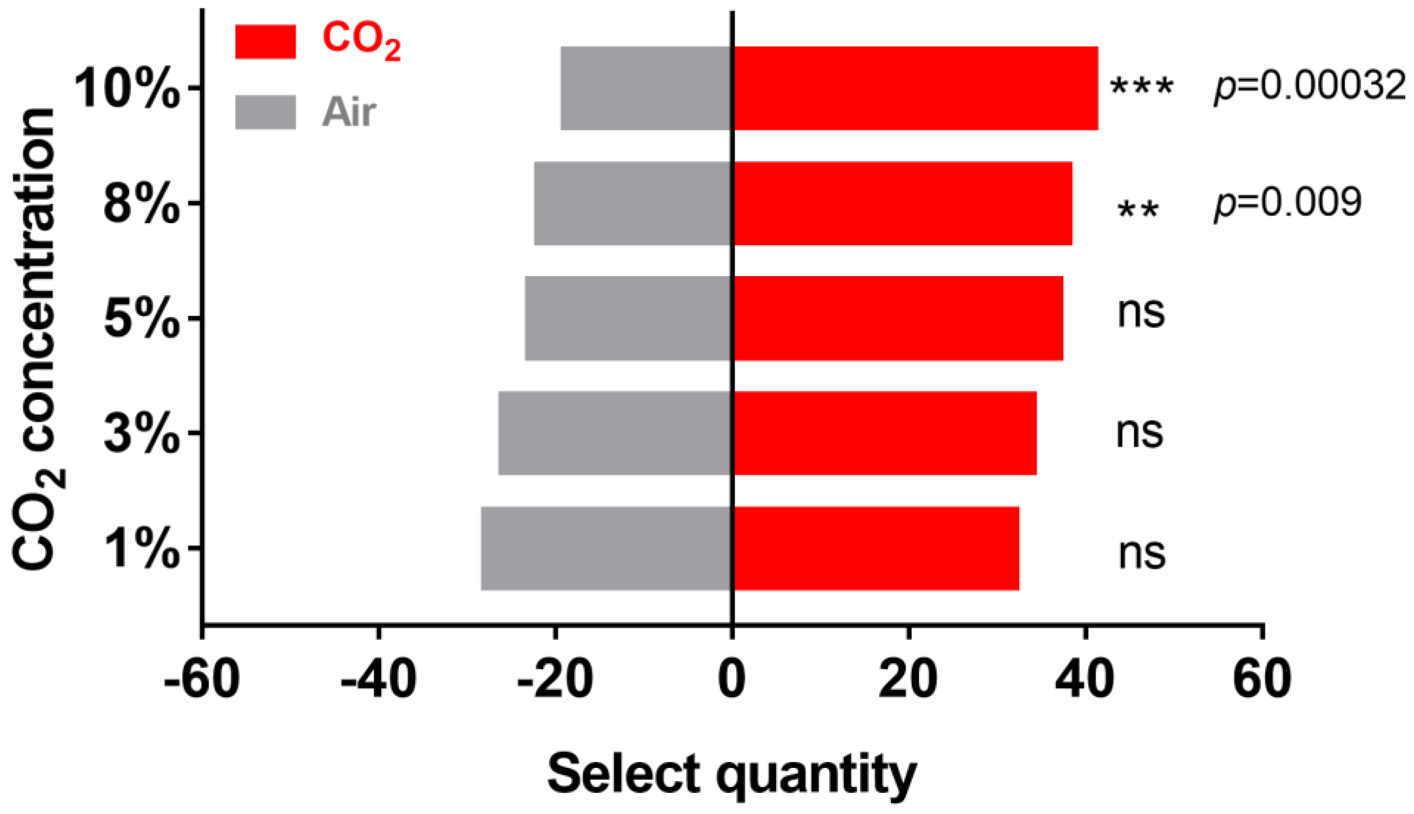
2.1. Effect of CO2 on H. cunea Behavior

To verify the effect of CO2 on the behavior of female H. cunea, Y-tube olfactometer tests were conducted with 60 H. cunea at different CO2 concentrations. Compared with those of the negative control (pure air without CO2, Figure 1), 32 moths preferred 1% CO2. As the CO2 concentration increased, 34 moths were attracted to the side with CO2 at a 3% concentration, 37 moths were attracted to 5% CO2, 38 moths were attracted to 8% CO2, and 41 moths were attracted to 10% CO2. These results showed that CO2 concentrations ranging from 8%-10% had an attractive effect on H. cunea adults; within this range, the attractive effect of CO2 increased with increasing CO2 concentration.

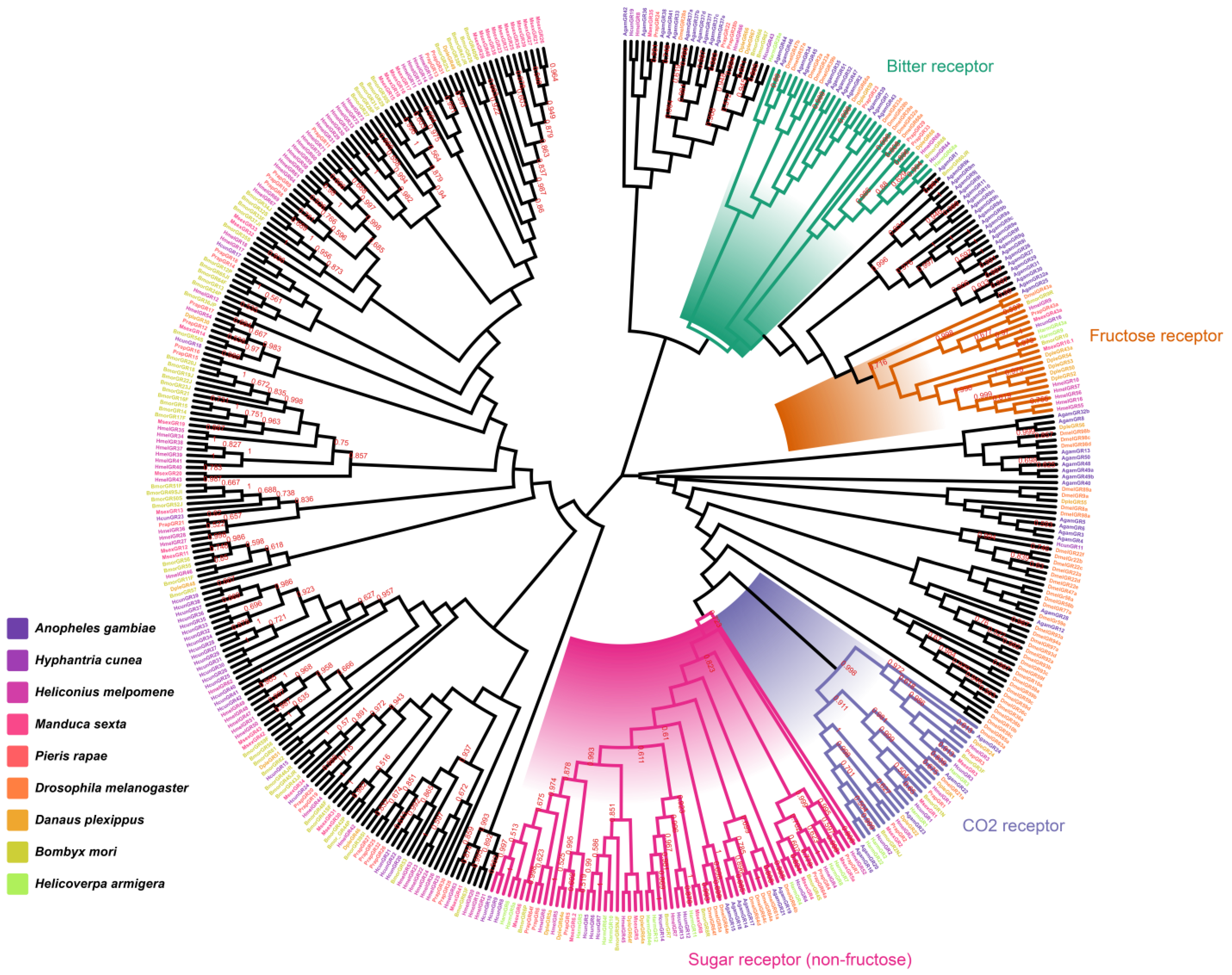
2.2. Identification and Homology Analysis of HcunGRs

To identify CO2 receptors in H. cunea, 44 candidate gustatory receptors (GRs) were identified from H. cunea transcriptome data; 24 GRs were full-length sequences, and 20 were truncated sequences. Blast results revealed that the similarity between HcunGRs and those of other species ranged from 35% to 91% (Table S1). HcunGR16 was the homolog of the fructose receptors of D. melanogaster, B. mori, H. armigera, and H. melpomene and was also hypothesized to be a candidate fructose receptor in H. cunea (Figure 2) [16]. HcunGR4-10 and HcunGR12-14 clustered together with the conserved sugar receptor DmelGR64 [27], suggesting that these GRs have a sugar receptor (non-fructose) sensing role. HcunGR44 is a candidate for bitter receptors in H. cunea. Most importantly, we found that HcunGR1, DmelGR21a, and HarmGR1; HcunGR2 and HarmGR2; and HcunGR3, DmelGR63a, and HarmGR3 were grouped together, indicating that these three HcunGRs might play a role in CO2 sensing in H. cunea.
Notably, the expression levels of the GRs showed that HcunGR1, HcunGR2, and HcunGR3 were highly abundant in the labial palps (Figure 3a). The expression patterns of HcunGR1, HcunGR2, and HcunGR3 were further verified by q-PCR (Figure 3b), and the qPCR results were consistent with the heatmap. These results indicate that the main organ responsible for CO2 sensing in H. cunea is the labial palp, which is similar to that in other lepidopteran species.

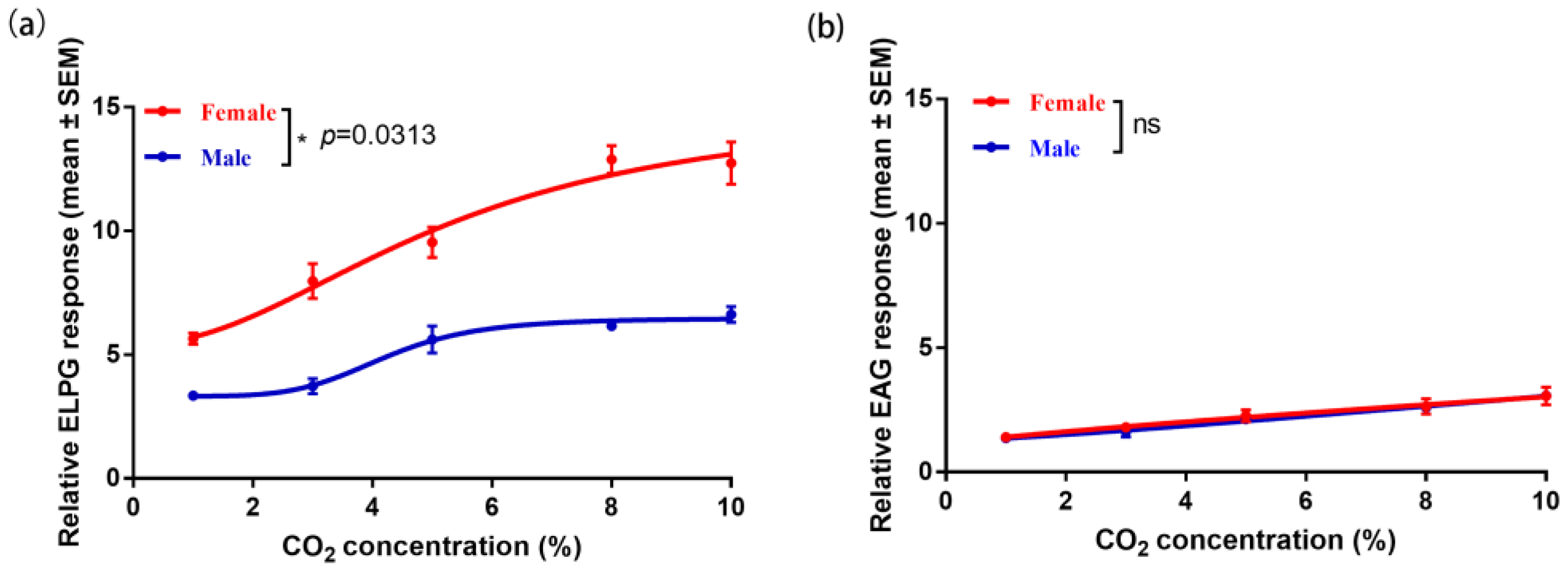
2.3. Electrophysiological Response of the Antennae and Labial Palp to CO2

The results of electrolabialpalpography (ELPG) and electroantennogram (EAG) showed that the response to CO2 was significantly higher in the labial palp of female H. cunea than in males (Figure 4a) (p = 0.0313), but there was no significant difference in the antennae response to CO2 (Figure 4b). In addition, the response of the labial palp to CO2 was consistently higher than that of the antennae at 1%-10% concentrations (Figure 4). These findings suggest that the labial palp is the main organ involved in CO2 sensing in H. cunea and that female moths have stronger CO2-sensing abilities than males.

2.4. Two-Electrode Voltage Clamp (TEVC) Response of HcunGr1, HcunGr2, HcunGr3, and Their Combinations

By analyzing the dissolved CO2 concentration in NaHCO3 solution, a direct correlation between the concentration of NaHCO3 and the dissolved CO2 concentration was identified (Figure S1). Moreover, we found that Na+ in NaCl solution also elicits a channel current (Figure 5a–g and Figure S2). Therefore, eliminating the Na+ effect from the NaCl solution is necessary when calculating the “real response”; we used full response minus NaCl response to look at the real response of CO2 [28]. Oocytes expressing HcunGR1, HcunGR2, or HcunGR3 alone or with co-expressions of HcunGR1+HcunGR2 or HcunGR2+HcunGR3 did not respond to CO2 after excluding the effect of Na+ (Figure 5c–g). However, the HcunGR1+HcunGR3 and HcunGR1+HcunGR2+HcunGR3 expression sets significantly responded to CO2 beginning at a concentration of 100 mM (equivalent to 51 ± 3 ppm CO2) (Figure S2), and the response increased gradually with increasing CO2 concentration (Figure 5a,b). In the range of 100–300 mM, the response of HcunGR1+HcunGR3 ranged from 257 ± 87 nA to 1387.67 ± 162.7 nA, and the response of HcunGR1+HcunGR2+HcunGR3 ranged from 245.67 ± 114.33 nA to 575.3 ± 89.3 nA (Figure 5h). The response of the HcunGR1+HcunGR2+HcunGR3 set was significantly lower than that of the HcunGR1+HcunGR3 set from a concentration of 200 mM or greater (Figure 5i).

2.5. Anterograde Dye Filling of Labial Palps in H. cunea

After we concluded that the labial palp is the main organ for sensing CO2, an anterograde dye-filling experiment on the labial palps was conducted to further explore the transmission of CO2 signals to the central nervous system. We observed the dye’s entry from the labial palp nerves, passing through the gnathal ganglion (GNG), then dividing into two bundles, and finally arriving at the LPOG (Figure 6a–f). The LPOG is located in the ventral region of the ALs, where it is similar to the DP region in D. melanogaster; we named this region DP1. The projections displayed a clear boundary, and the neurons exhibited a bilateral projection pattern.
To precisely locate and calculate the volume of the LPOG in the AL, we, based on the nine individuals (five females and four males), performed two-dimensional and three-dimensional reconstructions of all the glomeruli (Figure 6g–j) (Figure S3). It was found that female H. cunea possess 81 glomeruli, whereas male moths have only 74; most of the glomeruli were roughly spherical, while a few were irregularly shaped, forming a central fiber nucleus. After computing the surface area and volume of DP1 in both male and female H. cunea, we found that the total surface area of DP1 in females (5741.25 μm2) was nearly the same as that in males (5728.94 μm2); however, the average volume of DP1 in females (34,388.3 μm3) was significantly (p = 0.0115) greater than that in males (28,852.4 μm3) (Figure 6k), suggesting that sexual dimorphism existed in the DP1 glomerulus.

3. Discussion

Initially, we discovered that H. cunea adults exhibit a preference for CO2 concentrations ranging from 1% to 10%; this preference might be linked to the foraging and oviposition behavior of H. cunea. Studies have shown that many plants, such as Nepenthes, release in excess of 5% concentration of CO2 [29], which is consistent with the CO2 concentration in our behavioral test. In addition, researchers have found that adults H. cunea prefer to forage and oviposition at night [30], which is the peak time of CO2 release, which partially explains why the high concentration of CO2 could also attract H. cunea females. Previous studies have shown that the level of respiratory metabolism in plants is a main indicator reflecting plant quality. A high respiration rate indicates the presence of more nutrients, such as carbohydrates [7]; therefore, H. cunea can choose strong nectar as food by sensing the CO2 released by plants [31]. On the other hand, it has been shown that insect spawning increases with increasing CO2 concentration [6], suggesting that H. cunea can also choose oviposition sites by CO2 cues to maximize the survival of their offspring. Field observations have also shown that H. cunea prefer to lay eggs on the dorsal sides of leaves [32], which may be because the undersides of terrestrial plant leaves have more stomata and can generate higher CO2 concentrations [33]. H. cunea can use this CO2 cue to lay eggs on the dorsal side of the leaf to prevent damage from direct sunlight. In summary, our behavioral results showed that CO2 can attract adult H. cunea, which may be related to their oviposition behavior and its adaptation. And the possibility of further study about CO2 baited traps such as mosquitos will be an exciting topic in the future [34].
Homology analysis revealed that three CO2 receptor homologs exist in H. cunea, which is consistent with findings in other lepidopteran insects [16,17], indicating that the CO2 sensing pathway might be relatively conserved in Lepidoptera. The expression levels of the three candidate CO2 receptors demonstrated that they were all specifically expressed in labial palps; however, their expression was low in the antennae. And the response of the labial palp is significantly greater than that of the antennae, suggesting that the labial palp is the primary organ for sensing CO2 in H. cunea, as is the response of other lepidopteran species, such as H. armigera and M. sexta [18,25]. The fact that females produce stronger action potentials than males in the labial palp also suggests that CO2 may play an important role in female-specific behaviors such as spawn selection in H. cunea. In D. melanogaster, the molecular mechanisms for CO2 sensing in taste and olfaction are mutually independent [35], and ionotropic receptors (IRs) are involved in the detection of CO2 [36,37,38]. In addition, the response of D. melanogaster to CO2 is regulated by two different neural pathways, one for CO2 attraction and the other for avoidance [39]. Therefore, it is necessary to further investigate whether there are other CO2-sensing pathways in H. cunea.
In vitro expression of HcunGR1, HcunGR2, and HcunGR3 revealed that CO2 responses occurred only when HcunGR1 was combined with HcunGR3 or when HcunGR1 was combined with HcunGR2+HcunGR3. These results support the crucial role of the HcunGR1+HcunGR3 co-expression in CO2 sensing in H. cunea, which is consistent with the findings in H. armigera [18]. Notably, the response of the HcunGR1+HcunGR2+HcunGR3 ternary complex was significantly lower than that of the co-expression at concentrations greater than 200 mm. The role of HcunGR2 in CO2 sensing remains unknown. Our results could be due to two possible explanations. One possibility is that the limited number of ion channels on the oocyte surface results in a reduction in the expression ratio of the HcunGR1+HcunGR3 co-expression when HcunGR2 is expressed [40], therefore suppressing the TEVC response of HcunGR1+HcunGR3; another possibility is that HcunGR2 may serve as a modulator, playing an inhibitory role in the CO2 sensing process in H. cunea. In conclusion, we have shown that the co-expression formed by HcunGR1+HcunGR3 plays a primary role in CO2 sensing, but the function of HcunGR2 requires further exploration.
At the CNS level, a bilateral projection pattern of labial palps was observed in H. cunea; this result is consistent with what has been observed in H. armigera and Anopheles gambiae (A. gambiae) but differs from the unilateral projection pattern observed in D. melanogaster and A. aegypti. Our heatmap result showed that GR1, GR2, and GR3 were the top three highest expressing receptors in labial palps, which were responsible for CO2 sensing; thus, the projection from the labial palp into the brain was mainly for CO2 sensing. But we also found that there are other GRs also expressed in the labial palp, which may be related to wider sensing of different cues [41]. And previous research has suggested that bilateral projections enable olfactory receptor neurons (ORNs) to release an asymmetric amount of neurotransmitters on both sides of ALs [42], which leads to a stronger signal in one projection neuron (PN) than in the other, enhancing the contrast of odor concentration gradients between the two brain hemispheres [43]. This asymmetrical projection pattern may help H. cunea detect the differences in CO2 concentrations between the two labial palps and rapidly locate the CO2 source. Moreover, the average volume of DP1 in females was significantly higher than that of males; however, the total surface area of DP1 was nearly the same in both genders. This indicates that the shape of DP1 is different between males and females, which is consistent with our observations that the male’s DP1 had an irregularly shaped (Figure S3). And the sexual dimorphism was observed in the morphology of LPOGs; the average volume of LPOGs was significantly greater in females than in males. It is well accepted that a larger glomerulus indicates greater odor sensitivity due to the greater number of synaptic connections [44,45]. An enlarged LPOG may reflect the crucial role of CO2 sensing in H. cunea females; in contrast, no significant sexual dimorphism was observed on the LPOG in M. sexta [46]. In summary, the identified sexual dimorphism of labial palp projections in H. cunea may somewhat explain the differences in CO2 function between males and females, which might be linked to female-specific behavior such as oviposition.

4. Materials and Methods

4.1. Insect

A single fall webworm egg mass was collected from a Manchurian ash (Fraxinus mandschurica) tree in Animal and Plant Park, Jinlin Province, China (43°86′96.71″ N, 125°33′29.82″ E), and was reared in an artificial incubator (BIC-300 artificial incubator, Boxun, Shanghai, China) at 26 °C, 80% RH and a 19:5 light: dark cycle beginning in 2019. After each 12 generations, the wild population was collected again from the same place and crossed with the laboratory colony for rejuvenation for more than 30 generations. The larvae were fed on the leaves of mulberry trees (Morus alba), and the adults were given a 10% sucrose solution for energy supplementation.

4.2. Binary Choice Assay

The airflow of CO2 (Juyang Company, Changchun, China) was controlled by a flow meter and mixed with Zero Air (21% O2 and 79% N2, CO2 free, Juyang Company, China) to ensure the delivery of 1%, 3%, 5%, 8%, and 10% CO2. A Y-tube olfactometer (1.6 cm in diameter, 7.2 cm in base and arm length) was used for the binary choice assay, and pure air without any CO2 was used as a negative control. At 7 p.m. (peak spawning), day 3 after emergence, females were selected. A moth was first placed at the entrance of the main arm, and a “choice” was recorded when it entered an arm and stayed for more than 30 s. If no choice was made within 5 min, the data were recorded as “no choice”. A total of 60 H. cunea were tested. The Y-tube was cleaned with hexane, and the position of the stimulus was exchanged before and after each test. All the assays were performed in a dark room with red light (Intelligent LED solutions, Berkshire, UK) to avoid light interference.

4.3. Homology Analysis of Gustatory Receptors (GRs)

The GR sequence of H. cunea used in this study was obtained from our previous transcriptome studies (Table S1) [47], and the genome sequences of six lepidopteran species, B. mori (GCA_026075555.1), M. sexta (GCA_014839805.1), H. melpomene (GCA_900068175.1), Pieris rapae (GCA_905147795.1), H. armigera (GCA_026262555.1), and D. plexippus (GCA_009731565.1) were used for homology analysis, and D. melanogaster (GCA_000001215.4) and A. gambiae (GCA_000005575.1) were used as outgroups. The amino acid sequences of the GRs were first aligned by MUSCLE, after which Molecular Evolutionary Genetics Analysis (MEGA; State College, PA, USA) version 6 was used to construct a maximum likelihood (ML) tree with the Jones–Taylor–Thornton (JTT) model [48]. Bootstrap support values were based on 1000 replicates. The resulting homological tree was visualized with FigTree 1.42 (http://tree.bio.ed.ac.uk/software/figtree/, accessed 29 May 2023). And the fragments per kilobase of transcript per million fragments mapped (FPKM) were used to measure gene expression [49]. R (R Foundation for Statistical Computer, Vienna, Austria) version 4.1.3 was used to construct the heatmaps.

4.4. Electrophysiological Recording

EAG and ELPG were used to detect the electrophysiological responses of the antennae and labial palp to CO2 in H. cunea. Three days after emergence, females and males were selected at night (oviposition peak time). Glass electrodes were pulled by using a micropipette PC-10 (Narishige, Tokyo, Japan) and then filled with 1 M potassium chloride containing 1% polyvinylpyrrolidone. The reference electrode was subsequently inserted into one eye of the insects, while the recording electrode was placed in contact with the tips of the antennae and labial palp using the micromanipulator MP-12 (Syntech, Kirchzarten, Germany). The obtained signals were amplified by a high-impedance ac/dc preamplifier (Syntech, Kirchzarten, Germany). CO2 stimuli ranging from 1% to 10% were injected into a carbon-filtered and humidified airflow for 0.2 s to deliver the stimulus to the antenna and labial palp at 500 mL/min generated by an air stimulus controller CS-55 (Syntech, Kirchzarten, Germany). A minimum of 3 individuals were tested, and 3 puffs were performed for each antenna or labial palps. The EAG and ELPG data were acquired with EAG Pro version 2.0 software (Syntech, Kirchzarten, Germany) and normalized by the response of negative control (21% O2 and 79% N2, CO2 free) (Juyang Company, China) by the equation “relative EAG response = EAG response of CO2 / EAG response of negative control”. The data were subsequently analyzed with GraphPad Prism 6.0 (GraphPad Software, San Diego, CA, USA).

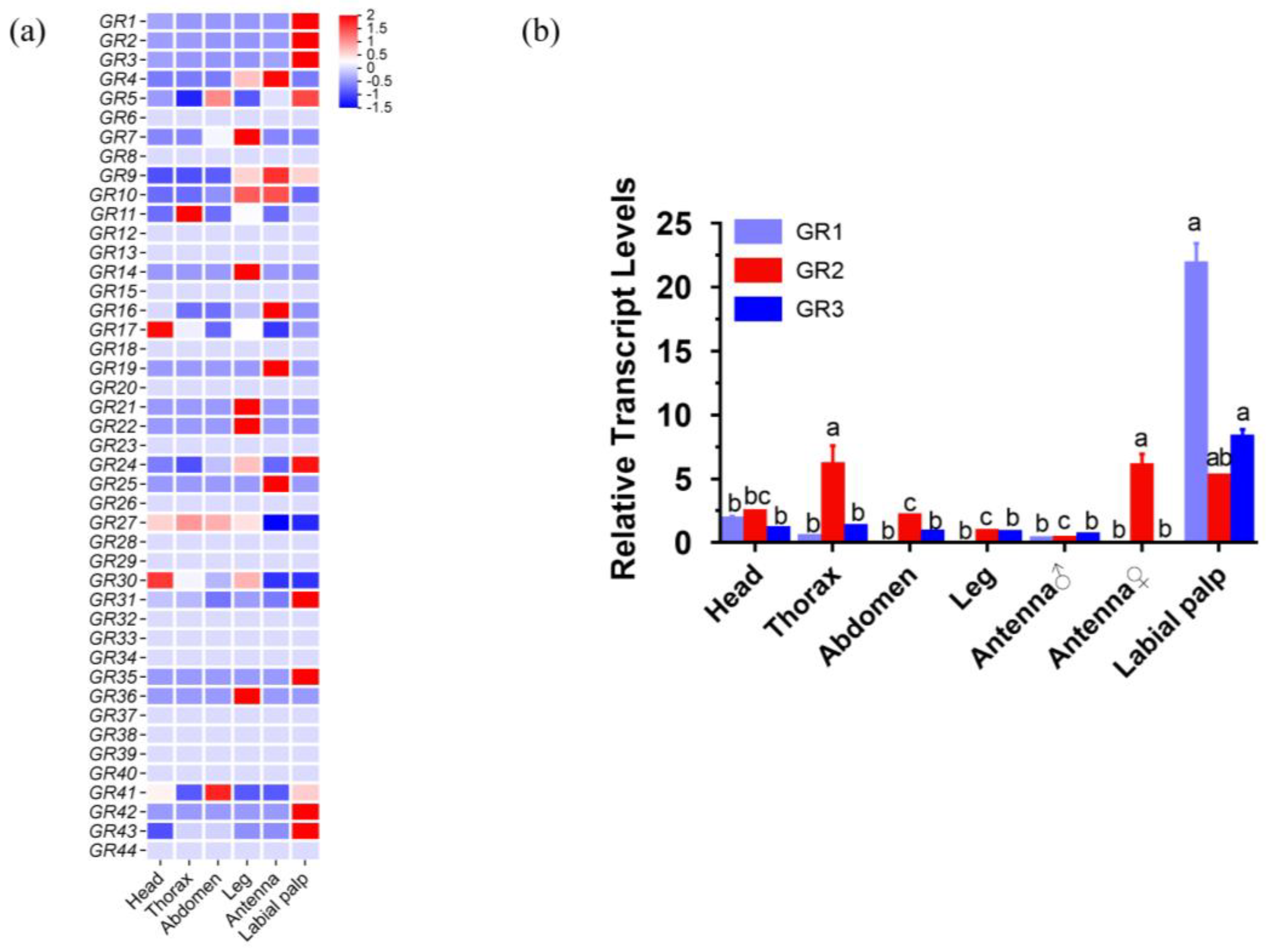
4.5. RNA Extraction, Expression Pattern Analysis, Quantitative PCR, and Cloning

Previous studies have shown that some GRs are differently expressed in the olfactory sensing organ (antennae) between males and females, but in non-olfactory organs (such as the head, chest, abdomen, and legs), no sex-based different expressions were detected; thus, we use mix samples of these body parts as a negative control [50]. The head, thorax, abdomen, leg, and labial palp carefully dissected from 15 individuals (female:male = 1:1) with DEPC-treated forceps under a stereomicroscope (Motic, Hong Kong, China). Then, female and male antennae were dissected from 15 individuals in the same way. Total RNA was isolated from homogenized body parts with TRIzol reagent (Invitrogen, Carlsbad, CA, USA) following the manufacturer’s protocol. After extraction, the total RNA concentration was assessed with a NanoDrop 2000 spectrophotometer (Thermo Fisher Scientific, Waltham, MA, USA) and 1% agarose gel (Sangon Biotech, Shanghai, China) electrophoresis.
For the qPCR assay, 1 μg of total RNA was transcribed into cDNA by using TransScript One-Step gDNA Removal and cDNA Synthesis SuperMix (TransGen Biotech, Beijing, China). qPCR was performed with a LightCycler 480 II Detection System (Roche, Shanghai, China) and TransStar Tip Top Green qPCR Supermix (TransGen Biotech, Beijing, China) under the following conditions: 94 °C for 30 s; 45 cycles of 94 °C for 5 s, 55 °C for 15 s, and 72 °C for 10 s; the β-actin gene was used as an internal control. The primers used in this study are listed in Table S2. The qPCR results were analyzed via the 2−ΔΔCT method [51]. The data were subsequently analyzed with GraphPad Prism 6.0 (GraphPad Software, CA, USA).
The primer was designed by Primer 3 (https://bioinfo.ut.ee/primer3-0.4.0, accessed on 29 June 2023) (Table S3), the full ORF of GRs containing 5′UTR and 3′UTR were obtained by PCR and ligated to pUCm-T Vector (Sangon Biotech, Shanghai, China) for sequencing. Subsequently, the ORF of GRs were amplified by a specific primer and subcloned to pGEMHE using pEASY-Uni Seamless Cloning and Assembly Kit (TransGen Biotech, Beijing, China) with BamHI and HindIII restriction sites (New England Biolabs, Ipswich, MA, USA). The recombinant plasmids were transformed in DH5α (TransGen Biotech, Beijing, China) competent cell, and then plasmids were extracted with a SanPrep Column Plasmid Mini-Preps Kit (Sangon Bio, Shanghai, China). After transformation, the inserts were verified via DNA sequencing (Sangon Biotech, Shanghai, China).

4.6. cRNA Synthesis and Oocyte Microinjection

The full-length open reading frames (ORFs) of HcunGr1, HcunGr2, and HcunGr3 were expressed in Xenopus laevis oocytes individually or in combination; thus, seven sets of oocytes were obtained expressing the following: HcunGr1, HcunGr2, HcunGr3, HcunGr1+HcunGr2, HcunGr1+HcunGr3, HcunGr2+HcunGr3, and HcunGr1+HcunGr2+HarmGr3. cRNAs of HcunGr1, HcunGr2, and HcunGr3 containing the 3′ (126bp) and 5′ (43bp) Xenopus globin UTR from the pGEMHE vector were synthesized using the mMACHINE T7 Transcription Kit (Ambion, Austin, USA) according to the manufacturer’s instructions. RNA concentration and purity were analyzed by a NanoDrop 2000 spectrophotometer (Thermo Fisher Scientific, Waltham, USA) and 1% agarose gel (Sangon Biotech, Shanghai, China) electrophoresis. A total of 46 nL (36.8 ng) of relevant cRNA was microinjected into each oocyte at a 1:1 or 1:1:1 ratio by a NanoLiter 2000 injector (World Precision Instruments, Sarasota, FL, USA). Afterward, the injected oocytes were incubated at 18 °C for 2 to 8 days in Barth’s solution (96 mM NaCl, 2 mM KCL, 5 mM MgCl2, 0.8 mM CaCl2 and 5 mM HEPES; pH adjusted to 7.6 by NaOH) supplemented with 10 μg/ mL gentamycin, 50 μg/ mL tetracycline, 100 μg/ mL streptomycin and 500 μg/ mL sodium pyruvate.

4.7. CO2 Quantification and TEVC

Since a TEVC requires a liquid environment, it is not possible to directly measure the response of GRs to CO2 in air; we chose to quantify the function of GRs by their response to dissolved CO2 in NaHCO3 solution, which was previously described by Xu et al. [28]. In brief, the concentration of dissolved CO2 in NaHCO3 solution can be calculated by the following equation; thus, we can obtain different concentrations of dissolved CO2 by controlling the pH and the concentration of bicarbonate.
C O 2 a q = 10 pKoverall PH × [ H C O 3 ] n o m i n a l 1 + 10 pKoverall PH
Koverall, which is sometimes referred to as Ka, is a constant that incorporates the CO2 hydrolysis constant (Kh) and the first dissociation constant of carbonic acid (Ka1), i.e., Koverall = Kh × Ka1. The pKa value is 6.3 (https://pubchem.ncbi.nlm.nih.gov/compound/sodium-bicarbonate#section=pKa, accessed on 6 August 2023).
The TEVC technique was used to record the channel currents in Xenopus oocytes at a holding potential of −80 Mv [52]. Signals were amplified with an Axonclamp 900A amplifier (Molecular Devices, San Jose, CA, USA) and 50-Hz low-pass filters and digitized at 1 kHz. Data acquisition and analysis were performed using Axon Digi 1550B and pCLAMP10 software (Molecular Devices, San Jose, CA, USA). We used the same concentration of Na+ in NaCl solution as negative control and then used full response minus NaCl response to look at the real response of CO2. The data were subsequently analyzed with GraphPad Prism 6.0 (GraphPad Software, San Diego, CA, USA).

4.8. Anterograde Dye Filling and Immunohistochemical Staining of the Labial Palps

In order to furthermore explore labial palps projection on AL, the anterograde dye filling was performed on labial palp with both genders. According to previous studies [53], the adult insects were fixed in a plastic tube with dental wax so that their heads were exposed. The base of the labial palp was then cut off, and the fluorescent dye, tetramethylrhodamine dextran (MicroRuby, Molecular Probes; Invitrogen, Eugene, OR, USA), was applied at the cutting surface by using a needle. After staining, the insects were placed in a refrigerator with moist filter paper overnight, allowing transportation of the dye to the sensory axons. The next day, the brain and ventral nerve cord were dissected in Ringer’s saline, fixed in 4% paraformaldehyde (PFA) for 1 h, dehydrated via an EtOH gradient, cleared with methyl salicylate, and mounted in Permount on perforated aluminum slides with coverslips.
For immunohistochemical staining, the brains of H. cunea were carefully removed with forceps, fixed with 4% PFA at 4 °C overnight, and then rinsed with PBST three times for 45 min. The fixed brain body part was then transferred to 5% normal goat serum (NGS; Thermo Fisher Scientific, Waltham, MA, USA) and preincubated at 4 °C for 15 h. After preincubation, the primary antibody 3C11 (anti-SYNORF1, 1:100 dilution with 5% NGS and PBST) (DSHB, University of Iowa, Johnson County, IA, USA) was applied and incubated at 4 °C for 5 days. Afterward, the brain was rinsed with PBST again and treated with the secondary antibody Cy2 coupled to an Alexa FluorTM 488 (1:300 dilution with 1% NGS and PBST) (Invitrogen, Eugene, OR, USA) at 4 °C for 3 days. Finally, the brain was dehydrated using an alcohol gradient, cleared with methyl salicylate, stored at 4 °C, and mounted with 1 mm aluminum slides for confocal laser microscopy imaging.
The slides were observed under a laser scanning confocal microscope (LSM880, Carl Zeiss, Jena, Germany) with an excitation wavelength of 488 nm and collected between 490 nm and 560 nm. A clear image was captured with ZEN v2.6. The identified neuropils within the brain and ventral nerve cords were reconstructed by using the 3D reconstruction software Amira 5.4.3 (FEI, Hillsboro, OR, USA) [54].

4.9. Statistical Analysis

One-way ANOVA followed by Tukey’s test or Dunnett’s test was used for multigroup comparisons (Figure 3b). Wilcoxon signed-rank test was used as a comparison between two curves (Figure 4). When two sets of data were compared, the data were first evaluated by the Shapiro–Wilk normality test. If p ≤ 0.05, the data sets will be applied for the Mann–Whitney test. If p > 0.05, the data will subsequently apply to the F-test to check the equal variances. If the data pass the F-test, it will apply to unpaired t-tests (Figure 6k and Figure S2). If they do not pass the F-test, the data will apply to the unpaired t-tests with Welch’s correction. The data were analyzed with SPSS 27.0 (IBM Corp., Armonk, NY, USA) and GraphPad Prism 6.0 (GraphPad Software, San Diego, CA, USA). And for electrophysiological and TEVC experiments, the value of each biological replicate is the average of its three technical replicates, and three technical replicates mean that when conducting an antenna and labial palp or oocyte preparation, the same stimulus was applied three times.

5. Conclusions

Although the detailed biological functions of CO2 in foraging and oviposition in H. cunea could not be fully clarified, some conclusions were reached at this stage. First, CO2 (ranging from 1% to 10%) strongly affects H. cunea adults; second, the main organ involved in CO2 sensing in H. cunea is the labial palp, and female moths have a more sensitive ability to perceive CO2 than male moths, whereas HcunGR1 and HcunGR3 are indispensable elements in the CO2 sensing process; and third, sexual dimorphism is observed in the volume of the LPOG, the main CO2 projection region in the antennal lobe. In summary, these results showed that CO2 has an attractive effect on H. cunea, which may be related to their oviposition behaviors; by identifying the CO2 receptors in H. cunea, the olfactory sensing pathway was further clarified. These results would benefit the development of a CO2-based trap for the monitoring and control of H. cunea, meanwhile providing more evidence on the biological function of CO2 among varied insects, especially those pests that take serious damage to the agriculture and forest.

Supplementary Materials

The following supporting information can be downloaded at https://www.mdpi.com/article/10.3390/ijms25115987/s1.

Author Contributions

Conceptualization, Y.W. and D.L.; methodology, Y.W. and S.D.; software, J.Z.; validation, J.Z. and S.D. formal analysis, J.Z., S.D. and W.W.; writing—original draft preparation, S.D. and Y.W.; writing—review and editing, Y.W., D.L., J.Z. and S.D.; funding acquisition, Y.W. and D.L. All authors have read and agreed to the published version of the manuscript.

Funding

This research was supported by the National Key R&D Program of China (2023FYE0113600) and the Natural Science Foundation of Jilin Province (grant 20230101252JC).

Institutional Review Board Statement

Xenopus laevis approved by the Ethics Committee of Northeast Normal University and used for this study were collected, cared for, and treated under strict compliance with all ethical practices and laws.

Informed Consent Statement

Not applicable.

Data Availability Statement

The data sets analyzed in the current study are available from the corresponding author upon reasonable request.

Acknowledgments

We thank our colleagues for their assistance in the specimen collection of the insects. The authors would like to thank all the reviewers who participated in the review.

Conflicts of Interest

The authors declare no conflicts of interest.

References

  1. Guerenstein, P.G.; Hildebrand, J.G. Roles and Effects of Environmental Carbon Dioxide in Insect Life. Annu. Rev. Entomol. 2008, 53, 161–178. [Google Scholar] [CrossRef]
  2. Seeley, T.D. Atmospheric carbon dioxide regulation in honey-bee (Apis mellifera) colonies. J. Insect Physiol. 1974, 20, 2301–2305. [Google Scholar] [CrossRef] [PubMed]
  3. Bowen, M.F. The Sensory Physiology of Host-Seeking Behavior in Mosquitoes. Annu. Rev. Entomol. 1991, 36, 139–158. [Google Scholar] [CrossRef] [PubMed]
  4. Gillies, M.T. The role of carbon dioxide in host-finding by mosquitoes (Diptera: Culicidae): A review. Bull. Entomol. Res. 1980, 70, 525–532. [Google Scholar] [CrossRef]
  5. Huang, X.; Mack, T.P. Artificial Carbon Dioxide Source to Attract Lesser Cornstalk Borer (Lepidoptera: Pyralidae) Larvae. J. Econ. Entomol. 2001, 94, 860–867. [Google Scholar] [CrossRef] [PubMed]
  6. Abrell, L.; Guerenstein, P.G.; Mechaber, W.L.; Stange, G.; Christensen, T.A.; Nakanishi, K.; Hildebrand, J.G. Effect of elevated atmospheric CO2 on oviposition behavior in Manduca sexta moths. Glob. Chang. Biol. 2005, 11, 1272–1282. [Google Scholar] [CrossRef]
  7. Stange, G.; Monro, J.; Stowe, S.; Osmond, C.B. The CO2 sense of the moth Cactoblastis cactorum and its probable role in the biological control of the CAM plant Opuntia stricta. Oecologia 1995, 102, 341–352. [Google Scholar] [CrossRef] [PubMed]
  8. Loewy, K.J.; Flansburg, A.L.; Grenis, K.; Kjeldgaard, M.K.; Mccarty, J.; Montesano, L.; Vernick, J.; Murphy, S.M. Life History Traits and Rearing Techniques for Fall Webworms (Hyphantria cunea Drury) in Colorado. J. Lepid. Soc. 2013, 67, 196–205. [Google Scholar] [CrossRef]
  9. Schowalter, T.D.; Ring, D.R. Biology and Management of the Fall Webworm, Hyphantria cunea (Lepidoptera: Erebidae). J. Integr. Pest Manag. 2017, 8. [Google Scholar] [CrossRef]
  10. Wu, N.; Zhang, S.; Li, X.; Cao, Y.; Liu, X.; Wang, Q.; Liu, Q.; Liu, H.; Hu, X.; Zhou, X.J.; et al. Fall webworm genomes yield insights into rapid adaptation of invasive species. Nat. Ecol. Evol. 2019, 3, 105–115. [Google Scholar] [CrossRef]
  11. Jones, W.D.; Cayirlioglu, P.; Kadow, I.G.; Vosshall, L.B. Two chemosensory receptors together mediate carbon dioxide detection in Drosophila. Nature 2006, 445, 86–90. [Google Scholar] [CrossRef] [PubMed]
  12. Kwon, J.Y.; Dahanukar, A.; Weiss, L.A.; Carlson, J.R. The molecular basis of CO2 reception in Drosophila. Proc. Natl. Acad. Sci. USA 2007, 104, 3574–3578. [Google Scholar] [CrossRef] [PubMed]
  13. Robertson, H.M.; Kent, L.B. Evolution of the Gene Lineage Encoding the Carbon Dioxide Receptor in Insects. J. Insect Sci. 2009, 9, 1–14. [Google Scholar] [CrossRef] [PubMed]
  14. Hu, J.; Zhong, C.; Ding, C.; Chi, Q.; Walz, A.; Mombaerts, P.; Matsunami, H.; Luo, M. Detection of Near-Atmospheric Concentrations of CO2 by an Olfactory Subsystem in the Mouse. Science 2007, 317, 953–957. [Google Scholar] [CrossRef] [PubMed]
  15. Fischler, W.; Kong, P.; Marella, S.; Scott, K. The detection of carbonation by the Drosophila gustatory system. Nature 2007, 448, 1054–1057. [Google Scholar] [CrossRef] [PubMed]
  16. Engsontia, P.; Sangket, U.; Chotigeat, W.; Satasook, C. Molecular Evolution of the Odorant and Gustatory Receptor Genes in Lepidopteran Insects: Implications for Their Adaptation and Speciation. J. Mol. Evol. 2014, 79, 21–39. [Google Scholar] [CrossRef] [PubMed]
  17. Xu, W.; Anderson, A. Carbon dioxide receptor genes in cotton bollworm Helicoverpa armigera. Sci. Nat. 2015, 102, 1–9. [Google Scholar] [CrossRef] [PubMed]
  18. Ning, C.; Yang, K.; Xu, M.; Huang, L.-Q.; Wang, C.-Z. Functional validation of the carbon dioxide receptor in labial palps of Helicoverpa armigera moths. Insect Biochem. Mol. Biol. 2016, 73, 12–19. [Google Scholar] [CrossRef] [PubMed]
  19. Stocker, R.F.; Singh, R.N.; Schorderet, M.; Siddiqi, O. Projection patterns of different types of antennal sensilla in the antennal glomeruli of Drosophila melanogaster. Cell Tissue Res. 1983, 232, 237–248. [Google Scholar] [CrossRef]
  20. Suh, G.S.B.; Wong, A.M.; Hergarden, A.C.; Wang, J.W.; Simon, A.F.; Benzer, S.; Axel, R.; Anderson, D.J. A single population of olfactory sensory neurons mediates an innate avoidance behaviour in Drosophila. Nature 2004, 431, 854–859. [Google Scholar] [CrossRef]
  21. Distler, P.G.; Boeckh, J. Central Projections of the Maxillary and Antennal Nerves in the Mosquito Aedes Aegypti. J. Exp. Biol. 1997, 200, 1873–1879. [Google Scholar] [CrossRef]
  22. Anton, S.; van Loon, J.J.; Meijerink, J.; Smid, H.M.; Takken, W.; Rospars, J.-P. Central projections of olfactory receptor neurons from single antennal and palpal sensilla in mosquitoes. Arthropod Struct. Dev. 2003, 32, 319–327. [Google Scholar] [CrossRef]
  23. Anton, S.; Rospars, J. Quantitative analysis of olfactory receptor neuron projections in the antennal lobe of the malaria mosquito, Anopheles gambiae. J. Comp. Neurol. 2004, 475, 315–326. [Google Scholar] [CrossRef]
  24. Zhao, X.-C.; Tang, Q.-B.; Berg, B.G.; Liu, Y.; Wang, Y.-R.; Yan, F.-M.; Wang, G.-R. Fine structure and primary sensory projections of sensilla located in the labial-palp pit organ of Helicoverpa armigera (Insecta). Cell Tissue Res. 2013, 353, 399–408. [Google Scholar] [CrossRef]
  25. Guerenstein, P.G.; Christensen, T.A.; Hildebrand, J.G. Sensory processing of ambient CO2 information in the brain of the moth Manduca sexta. J. Comp. Physiol. A 2004, 190, 707–725. [Google Scholar] [CrossRef]
  26. Yu, H.; Khashaveh, A.; Li, Y.; Li, X.; Zhang, Y. Field Trapping of Predaceous Insects With Synthetic Herbivore-Induced Plant Volatiles in Cotton Fields. Environ. Entomol. 2017, 47, 114–120. [Google Scholar] [CrossRef]
  27. Slone, J.; Daniels, J.; Amrein, H. Sugar Receptors in Drosophila. Curr. Biol. 2007, 17, 1809–1816. [Google Scholar] [CrossRef]
  28. Xu, P.; Wen, X.; Leal, W.S. CO2 per se activates carbon dioxide receptors. Insect Biochem. Mol. Biol. 2019, 117, 103284. [Google Scholar] [CrossRef]
  29. Baby, S.; Johnson, A.J.; Zachariah, E.J.; Hussain, A.A. Nepenthes pitchers are CO2-enriched cavities, emit CO2 to attract preys. Sci. Rep. 2017, 7, 11281. [Google Scholar] [CrossRef]
  30. Edosa, T.T.; Jo, Y.H.; Keshavarz, M.; Anh, Y.S.; Noh, M.Y.; Han, Y.S. Current status of the management of fall webworm, Hyphantria cunea: Towards the integrated pest management development. J. Appl. Entomol. 2019, 143, 1–10. [Google Scholar] [CrossRef]
  31. Krenn, H.W. Feeding Mechanisms of Adult Lepidoptera: Structure, Function, and Evolution of the Mouthparts. Annu. Rev. Entomol. 2010, 55, 307–327. [Google Scholar] [CrossRef]
  32. Morris, R.F. Fecundity and colony size in natural populations of hyphantria cunea. Can. Entomol. 1972, 104, 399–409. [Google Scholar] [CrossRef]
  33. Clark, D. Images of Leaf Stomata: Little Things that Matter. Microsc. Today 2019, 27, 12–17. [Google Scholar] [CrossRef]
  34. Eshun, O.; Gerry, A.; Hayes, W.K. Mosquito Capture Rate Using CO2-Baited Traps in Relation to Distance From Water and Height: Implications for Avian Disease Transmission. J. Med Entomol. 2016, 53, 1378–1384. [Google Scholar] [CrossRef]
  35. Chen, Y.-C.D.; Dahanukar, A. Recent advances in the genetic basis of taste detection in Drosophila. Cell. Mol. Life Sci. 2019, 77, 1087–1101. [Google Scholar] [CrossRef]
  36. Benton, R.; Vannice, K.S.; Gomez-Diaz, C.; Vosshall, L.B. Variant Ionotropic Glutamate Receptors as Chemosensory Receptors in Drosophila. Cell 2009, 136, 149–162. [Google Scholar] [CrossRef]
  37. Sánchez-Alcañiz, J.A.; Silbering, A.F.; Croset, V.; Zappia, G.; Sivasubramaniam, A.K.; Abuin, L.; Sahai, S.Y.; Münch, D.; Steck, K.; Auer, T.O.; et al. An expression atlas of variant ionotropic glutamate receptors identifies a molecular basis of carbonation sensing. Nat. Commun. 2018, 9, 4252, Correction in Nat. Commun. 2020, 11, 1131. [Google Scholar] [CrossRef]
  38. Ai, M.; Min, S.; Grosjean, Y.; Leblanc, C.; Bell, R.; Benton, R.; Suh, G.S.B. Acid sensing by the Drosophila olfactory system. Nature 2010, 468, 691–695. [Google Scholar] [CrossRef]
  39. van Breugel, F.; Huda, A.; Dickinson, M.H. Distinct activity-gated pathways mediate attraction and aversion to CO2 in Drosophila. Nature 2018, 564, 420–424. [Google Scholar] [CrossRef]
  40. Terhag, J.; Cavara, N.A.; Hollmann, M. Cave Canalem: How endogenous ion channels may interfere with heterologous expression in Xenopus oocytes. Methods 2010, 51, 66–74. [Google Scholar] [CrossRef]
  41. Amat, C.; Marion-Poll, F.; Navarro-Roldán, M.A.; Gemeno, C. Gustatory function of sensilla chaetica on the labial palps and antennae of three tortricid moths (Lepidoptera: Tortricidae). Sci. Rep. 2022, 12, 1–11. [Google Scholar] [CrossRef]
  42. Gaudry, Q.; Hong, E.J.; Kain, J.; de Bivort, B.L.; Wilson, R.I. Asymmetric neurotransmitter release enables rapid odour lateralization in Drosophila. Nature 2012, 493, 424–428. [Google Scholar] [CrossRef]
  43. Mohamed, A.A.M.; Hansson, B.S.; Sachse, S. Third-Order Neurons in the Lateral Horn Enhance Bilateral Contrast of Odor Inputs Through Contralateral Inhibition in Drosophila. Front. Physiol. 2019, 10, 851. [Google Scholar] [CrossRef]
  44. Mysore, K.; Subramanian, K.; Sarasij, R.; Suresh, A.; Shyamala, B.V.; VijayRaghavan, K.; Rodrigues, V. Caste and sex specific olfactory glomerular organization and brain architecture in two sympatric ant species Camponotus sericeus and Camponotus compressus (Fabricius, 1798). Arthropod Struct. Dev. 2009, 38, 485–497. [Google Scholar] [CrossRef]
  45. Varela, N.; Avilla, J.; Gemeno, C.; Anton, S. Ordinary glomeruli in the antennal lobe of male and female tortricid moth Grapholita molesta (Busck) (Lepidoptera: Tortricidae) process sex pheromone and host-plant volatiles. J. Exp. Biol. 2011, 214, 637–645. [Google Scholar] [CrossRef]
  46. Kent, K.S.; Harrow, I.D.; Quartararo, P.; Hildebrand, J.G. An accessory olfactory pathway in Lepidoptera: The labial pit organ and its central projections in Manduca sexta and certain other sphinx moths and silk moths. Cell Tissue Res. 1986, 245, 237–245. [Google Scholar] [CrossRef]
  47. Chen, Q.; Zhao, H.; Wen, M.; Li, J.; Zhou, H.; Wang, J.; Zhou, Y.; Liu, Y.; Du, L.; Kang, H.; et al. Genome of the webworm Hyphantria cunea unveils genetic adaptations supporting its rapid invasion and spread. BMC Genom. 2020, 21, 1–22. [Google Scholar] [CrossRef]
  48. Kumar, S.; Stecher, G.; Tamura, K. MEGA7: Molecular Evolutionary Genetics Analysis Version 7.0 for Bigger Datasets. Mol. Biol. Evol. 2016, 33, 1870–1874. [Google Scholar] [CrossRef]
  49. Trapnell, C.; Williams, B.A.; Pertea, G.; Mortazavi, A.; Kwan, G.; Van Baren, M.J.; Salzberg, S.L.; Wold, B.J.; Pachter, L. Transcript assembly and quantification by RNA-Seq reveals unannotated transcripts and isoform switching during cell differentiation. Nat. Biotechnol. 2010, 28, 511–515. [Google Scholar] [CrossRef]
  50. Zhao, Y.; Wang, F.; Zhang, X.; Zhang, S.; Guo, S.; Zhu, G.; Liu, Q.; Li, M. Transcriptome and Expression Patterns of Chemosensory Genes in Antennae of the Parasitoid Wasp Chouioia cunea. PLoS ONE 2016, 11, e0148159. [Google Scholar] [CrossRef]
  51. Livak, K.J.; Schmittgen, T.D. Analysis of relative gene expression data using real-time quantitative PCR and the 2−ΔΔCT Method. Methods 2001, 25, 402–408. [Google Scholar] [CrossRef]
  52. Zhou, Y.; Han, Q.; Feng, K.; Wang, J.; Zhou, H.; Wen, M.; Duan, H.; Wang, Y.; Ren, B. New binding sites of nicotinic acetylcholine receptors from Myzus persicae. Entomol. Gen. 2023, 43, 461–470. [Google Scholar] [CrossRef]
  53. Ignell, R.; Anton, S.; Hansson, B.S. The maxillary palp sensory pathway of Orthoptera. Arthropod Struct. Dev. 2000, 29, 295–305. [Google Scholar] [CrossRef]
  54. Ye, Z.; Liu, F.; Liu, N. Three-dimensional structure of the antennal lobe in the Southern house mosquito Culex quinquefasciatus. Insect Sci. 2021, 28, 93–102. [Google Scholar] [CrossRef]
Figure 1. The number of female Hyphantria cunea attracted to different concentrations of CO2. Statistical differences were evaluated via the Chi−square test. ** p < 0.01, *** p < 0.001, ns: no significance.
Ijms 25 05987 g001
Figure 2. The maximum likelihood tree of candidate gustatory receptors (GRs). Bootstrap replications up to 1000.
Ijms 25 05987 g002
Figure 3. Expression profiles of HcunGRs in different body parts of adult H. cunea. (a) Characteristic expression patterns of 44 HcunGRs in different body parts based on FPKM (normalization by row). FPKM: Fragments per kilobase of transcript per million fragments mapped. (b) Expression patterns of three candidate CO2 GRs in different body parts of adult H. cunea. A different lowercase indicates a significant difference based on one-way ANOVA followed by Tukey’s multiple comparison test (p < 0.05). The data are presented as the means ± standard errors of the means (SEMs), N = 3.
Ijms 25 05987 g003
Figure 4. Electrolabialpalpography (ELPG) and electroantennogram (EAG) nonlinear regression curve for CO2. (a) Response of the labial palps of the H. cunea to CO2; (b) response of the antennae of the H. cunea to CO2. Statistical differences were evaluated by the Wilcoxon signed-rank test. * p < 0.05, ns: no significance. The data are presented as the means ± SEMs, N = 3 biological replicates (Tables S4 and S5).
Ijms 25 05987 g004
Figure 5. Two-electrode voltage clamp recording (TEVC) responses of HcunGR1, HcunGR2, and HcunGR3 alone and in combination with different concentrations of NaCl and NaHCO3. (a) Response of HcunGR1+HcunGR3; (b) response of HcunGR1+HcunGR2+HcunGR3; (c) response of HcunGR1; (d) response of HcunGR2; (e) response of HcunGR3; (f) response of HcunGR1+HcunGR2; (g) response of HcunGR2+HcunGR3; green traces represent the response in NaCl solution; red traces represent the response in NaHCO3 solution; the number involved in (ag) indicates concentration NaCl and NaHCO3; (h) nonlinear regression curve of HcunGR1, HcunGR2, and HcunGR3 alone and in combination after excluding the influence of Na+. N = 3 biological replicates (Table S6); The data are presented as the mean ± SEM.
Ijms 25 05987 g005
Figure 6. Anterograde dye-filling of labial palps and two-dimensional reconstructions of the antennal lobe (AL) in the DP1 region of H. cunea. (ac) The central projections of female labial pit organ sensory neurons passing through the gnathal ganglion (GNG) to DP1; (df) the central projections of male labial pit organ sensory neurons passing through the gnathal ganglion (GNG) to DP1; (g,h) confocal images of the male H. cunea AL glomeruli seen from the ventral view. The sections are from anterior to posterior at a depth of 156 μm. Scale bar = 50 μm; (i,j) confocal images of female H. cunea AL glomeruli taken from the ventral view. The sections are from anterior to posterior at a depth of 142 μm. Scale bar = 50 μm; (k) the volume compares male and female DP1s. The data are presented as the mean ± SEM, N ≥ 4. Statistical differences were evaluated by unpaired t tests. * p < 0.05.
Ijms 25 05987 g006
Disclaimer/Publisher’s Note: The statements, opinions and data contained in all publications are solely those of the individual author(s) and contributor(s) and not of MDPI and/or the editor(s). MDPI and/or the editor(s) disclaim responsibility for any injury to people or property resulting from any ideas, methods, instructions or products referred to in the content.

