Review

Ferroptosis-Regulated Natural Products and miRNAs and Their Potential Targeting to Ferroptosis and Exosome Biogenesis

1
Department of Biomedical Science and Environmental Biology, PhD Program in Life Sciences, College of Life Science, Kaohsiung Medical University, Kaohsiung 80708, Taiwan
2
School of Dentistry, Taipei Medical University, Taipei 11031, Taiwan
3
Department of Oral and Maxillofacial Surgery, Chi-Mei Medical Center, Tainan 71004, Taiwan
4
Department of Urology, Kaohsiung Medical University Hospital, Kaohsiung 80708, Taiwan
5
School of Post-Baccalaureate Medicine, Kaohsiung Medical University, Kaohsiung 80708, Taiwan
6
Department of Urology, Kaohsiung Gangshan Hospital, Kaohsiung Medical University, Kaohsiung 820111, Taiwan
7
Graduate Institute of Natural Products, Kaohsiung Medical University, Kaohsiung 80708, Taiwan
8
Department of Pharmacy and Master Program, College of Pharmacy and Health Care, Tajen University, Pingtung 907101, Taiwan
9
Department of Computer Science and Information Engineering, National Pingtung University, Pingtung 900391, Taiwan
10
Department of Radiation Oncology, Kaohsiung Medical University Hospital, Kaohsiung Medical University, Kaohsiung 80708, Taiwan
11
Department of Medical Research, Kaohsiung Medical University Hospital, Kaohsiung 80708, Taiwan
12
Center for Cancer Research, Kaohsiung Medical University, Kaohsiung 80708, Taiwan
*
Authors to whom correspondence should be addressed.
These authors contributed equally to this work.
Int. J. Mol. Sci. 2024, 25(11), 6083; https://doi.org/10.3390/ijms25116083
Submission received: 28 April 2024 / Revised: 22 May 2024 / Accepted: 29 May 2024 / Published: 31 May 2024
(This article belongs to the Special Issue Anticancer Activity of Natural Products and Related Compounds)

Abstract

:
Ferroptosis, which comprises iron-dependent cell death, is crucial in cancer and non-cancer treatments. Exosomes, the extracellular vesicles, may deliver biomolecules to regulate disease progression. The interplay between ferroptosis and exosomes may modulate cancer development but is rarely investigated in natural product treatments and their modulating miRNAs. This review focuses on the ferroptosis-modulating effects of natural products and miRNAs concerning their participation in ferroptosis and exosome biogenesis (secretion and assembly)-related targets in cancer and non-cancer cells. Natural products and miRNAs with ferroptosis-modulating effects were retrieved and organized. Next, a literature search established the connection of a panel of ferroptosis-modulating genes to these ferroptosis-associated natural products. Moreover, ferroptosis-associated miRNAs were inputted into the miRNA database (miRDB) to bioinformatically search the potential targets for the modulation of ferroptosis and exosome biogenesis. Finally, the literature search provided a connection between ferroptosis-modulating miRNAs and natural products. Consequently, the connections from ferroptosis–miRNA–exosome biogenesis to natural product-based anticancer treatments are well-organized. This review sheds light on the research directions for integrating miRNAs and exosome biogenesis into the ferroptosis-modulating therapeutic effects of natural products on cancer and non-cancer diseases.

1. Introduction

1.1. Relationship between Exosomes and Ferroptosis

Exosomes are extracellular vesicles with a nanometer-scale size (30–150 nm) [1]. Exosome biogenesis requires the processes of assembly and secretion. Exosomes may contain diverse cargos, such as proteins, lipids, and nucleic acids (DNA, mRNAs, and noncoding RNAs) [2,3,4]. Exosomal noncoding RNAs play a crucial role in regulating carcinogenesis [5]. Exosomal noncoding RNAs contain different types of nucleic acids, such as circular RNAs, long noncoding RNAs, and microRNAs (miRNAs). Literature reports indicate that cancer patients generally exhibit higher miRNAs in exosomes than normal controls [6]. Consequently, this review focuses on exosomal miRNAs.
Ferroptosis comprises iron-dependent non-apoptotic cell death characterized by the overexpression of membrane lipid peroxidation, an increase in cellular iron uptake, and the triggering of ferroptosis signaling [7]. Modulating ferroptosis is an anticancer strategy [8,9]. Ferroptosis-inducing compounds may improve pharmaceutical effects with respect to inhibiting metastasis, drug resistance, and tumor regression [9].
Notably, exosome and ferroptosis may interplay with each other [10,11,12,13,14,15]. Exosomes may enhance or suppress ferroptosis in several diseases and cancers [16]. The inhibition of ferroptosis may be triggered via exosome-mediated regulation that acts on ferroptosis signaling [10]. Exosomes may deliver biomolecules to recipient cells and, in turn, regulate ferroptosis-modulating signaling and response [11,12]. For example, breast cancerous exosomes suppress migration mediated by ferroptosis [13]. Bladder-cancer-isolated exosomes demonstrate ferroptosis-inducing effects by transporting miR-217 [14]. Exosomes isolated from the adipose tissue macrophages of an obesity-induced cardiac injury trigger ferroptosis by targeting ferroptosis-modulating signaling [17]. Consequently, drugs that regulate ferroptosis may modulate cancer and other medical therapies. Although exosome and ferroptosis exhibit an interplay relationship, this review only focuses on exploring the impact of ferroptosis on exosome biogenesis regarding the ferroptosis-modulating natural products and miRNAs.

1.2. Exosome-Biogenesis-Modulating Genes

The search methodology for exosome biogenesis genes was performed bioinformatically via the Gene Oncology database [18]. Exosome biogenesis genes such as secretion and assembly were summarized from the Gene Oncology (GO:1990182) in Mouse Genome Database [18] (accessed on 1 March 2023), including ATPase class II, type 9A (ATP9A) and ATP13A2; CD34 antigen (CD34); charged multivesicular body protein 2A (CHMP2A); COP9 signalosome subunit 5 (COPS5); hepatocyte growth-factor-regulated tyrosine kinase substrate (HGS); myosin VB (MYO5B); parkin RBR E3 ubiquitin protein ligase (PRKN); programmed cell death 6 interacting protein (PDCD6IP); RAB11A, a member of the RAS oncogene family (RAB11A), RAB7A, RAB7B, and RAB27A; syndecan 1 (SCD1) and SDC4; syndecan-binding protein (SDCBP); sphingomyelin phosphodiesterase 3; neutral (SMPD3); SNF8 subunit of the endosomal sorting complexes required for transport (ESCRT)-II complex (SNF8); SH3 domain and ITAM motif (STAM); STEAP family member 3 (STEAP3); tumor susceptibility gene 101 (TSG101); vacuolar protein sorting 4A (VPS4A); and VPS4B. These exosomal biogenesis-modulating genes (Figure 1) are used as candidates for determining the potential regulation of exosome biogenesis by carrying out a literature search (PubMed and Google Scholar) and bioinformatic data-mining (miRDB) [19]. These exosome-biogenesis-modulating genes are the potential target candidates for natural-product-regulated miRNAs, but they still need further assessment.

1.3. Ferroptosis-Modulating Genes

Several ferroptosis-modulating genes, such as 25 ferroptosis-inducing genes and 24 ferroptosis-inhibiting genes, are collected in this review. These ferroptosis-modulating genes (Figure 1) are used as candidates for determining the potential regulation of ferroptosis by carrying out a literature search (PubMed and Google Scholar) and bioinformatic data-mining (miRDB) [19].

1.3.1. Ferroptosis-Inducing Genes

As stated above, twenty-five ferroptosis-inducing genes (Figure 1) were reported [20,21,22]. These include the following: Acyl-CoA synthetase long-chain family member 4 (ACSL4); activating transcription factor 3 (ATF3) and ATF4; autophagy-related 5 (ATG5) and ATG7; arachidonate 12-lipoxygenase (ALOX12), ALOX15, ALOX5, and ALOXE3; dipeptidyl-peptidase 4 (DPP4); endothelial PAS domain protein 1 (EPAS1; HIF2A); heme oxygenase-1 (HMOX1; HO-1); iron-responsive element-binding protein 2 (IREB2); lysophosphatidylcholine acyltransferase 3 (LPCAT3); microtubule-associated protein 1 light chain 3 alpha (MAP1LC3A); microtubule-associated protein 1 light chain 3 beta (MAP1LC3B); nuclear receptor coactivator 4 (NCOA4); cytochrome p450 oxidoreductase (POR); spermidine N1-acetyltransferase 1 (SAT1); transferrin (TF); transferrin receptor (TFRC); voltage-dependent anion channel 2 (VDAC2) and VDAC3; Yes 1-associated transcriptional regulator (YAP1); and WW domain-containing transcription regulator 1 (WWTR1).

1.3.2. Ferroptosis-Inhibiting Genes

Twenty-four ferroptosis-inhibiting genes (Figure 1) were reported [20,23,24,25,26,27,28,29]. These include apoptosis-inducing factor mitochondria-associated 1 (AIFM1) and AIFM2; biglycan (BGN); ceruloplasmin (CP); ferritin heavy chain 1 (FTH1); ferritin light chain (FTL); ferritin mitochondrial (FTMT); glutamate–cysteine ligase catalytic subunit (GCLC); glutathione synthetase (GSS); glutathione peroxidase 4 (GPX4); hypoxia-inducible factor 1 subunit alpha (HIF1A); NEDD4 E3 ubiquitin protein ligase (NEDD4); nuclear factor erythroid 2 like 2 (NFE2L2; NRF2); prominin 2 (PROM2); solute carrier family 3 member 2 (SLC3A2); solute carrier family 7 member 11 (SLC7A11; xCT); solute carrier family 40 member 1 (SLC40A1; FPN); solute carrier family 11 member 2 (SLC11A2; DMT1); SP1 transcription factor (SP1); transcription factor AP-2 gamma (TFAP2C); tumor protein p53 (TP53); thioredoxin (TXN); transducin beta-like 1X-linked (TBLR1); and thioredoxin reductase 1 (TXNRD1). These ferroptosis-modulating genes are the potential target candidates for natural-product-regulated miRNAs, but still need further evaluation.

1.4. The Knowledge Gaps of miRNA-Modulating Natural Products for the Induction and Inhibition of Ferroptosis and Exosome Biogenesis

Several natural products were evaluated, and they demonstrated the modulation function for exosomal miRNAs and exosome biogenesis [30]. However, this exosomal miRNA review did not consider the impact of ferroptosis in natural product studies.
Several literature reports have recently organized comprehensive ferroptosis-modulating natural products [31,32,33,34]. For example, the effects of various natural products exhibiting inducing and inhibiting effects on ferroptosis in several diseases and cancers have been summarized [31,32]. Their functional targets and experimental models for these ferroptosis-modulating natural products are demonstrated. In comparison, the chemical structures of ferroptosis-modulating natural products are presented but do not discuss the target genes of ferroptosis [32,34]. Another review provides information on the compound source, cell line names, concentration, and treatment time of natural products with ferroptosis, necroptosis, and pyroptosis-inducing ability [33]. However, these reviews summarize ferroptosis-modulating natural products without considering the impact of miRNAs [31,32,33,34].
Some studies focused on exploring the role of miRNAs in regulating ferroptosis [35,36,37]. Several miRNAs that modulate cardiomyopathy, neuronal injury, and cancer-associated ferroptosis are summarized [35]. Ferroptosis-regulating miRNAs [36,37] and circular RNAs [36] occurring in several cancer cells are summarized [36]. The cell line models and targets for these ferroptosis-modulating miRNAs are provided. However, these reported miRNA functions focus on regulating ferroptosis without considering the contribution of natural products [35,36,37].
As mentioned, those studies of ferroptosis-modulating natural products and miRNAs were individually investigated without exploring their relationships. A knowledge gap was discovered in the connection between the modulating effects of miRNAs and natural products acting on ferroptosis. Moreover, there is another knowledge gap: the ferroptosis- and exosome-biogenesis-modulating targets of natural products-regulated miRNAs were limitedly reported. Hence, comprehensive assessments of natural product studies targeting the modulation of ferroptosis and exosome biogenesis by miRNAs are warranted.

1.5. The Novelty, Rationale, and Outline of This Review

Several reviews have specific scopes for natural products, miRNAs, exosome biosynthesis, or ferroptosis, but the connection between them is weakly emphasized and organized. The novelty of the current review is to fix these disconnections by introducing bioinformatics retrieval and deep literature searches.
Many types of cell death play differential roles in anticancer responses [38,39]. Interestingly, ferroptosis comprises iron-dependent non-apoptotic cell death. Switching from apoptosis to ferroptosis may improve the anticancer effects against cancer stem cells [40]. Moreover, many natural products exhibit modulating effects on ferroptosis. Therefore, this review focuses on ferroptosis-associated responses involving natural products. The rationale of this review is to propose a regulatory axis where ferroptosis-modulating natural products affect ferroptosis-modulating miRNAs, which in turn control ferroptosis- and exosome-biogenesis-modulating targets (Figure 1). Those two gaps are innovatively filled in this review as described below.
In this review, several ferroptosis-modulating natural products and their potential role of modulating ferroptosis signaling were overviewed by a literature search (PubMed and Google Scholar) (Section 2). There is a knowledge gap with respect to the connections between the modulating effects of the miRNA of natural products and ferroptosis and exosome biogenesis (Figure 1). To fill this gap, we performed a literature search (PubMed and Google Scholar) and bioinformatic database mining (miRDB) that allowed ferroptosis-modulating miRNAs and their potential ferroptosis-targeting genes to be retrieved (Section 3). Similarly, ferroptosis-modulating miRNAs and their potential exosome biogenesis-targeting genes were retrieved (Section 4).
Another gap is the connection between ferroptosis-modulating natural products and miRNAs because they were individually reported (Figure 1). After the literature search, the relationship between ferroptosis-modulating miRNAs and some natural products was explored to fill this gap (Section 5). Consequently, this review provides the connecting information between the potential modulation of miRNAs and exosome biogenesis in ferroptosis-modulating natural products.

2. Ferroptosis-Modulating Natural Products

Many natural products showing ferroptosis-modulating effects are well-reviewed [31,32,33,34]. Generally, these studies provide one or two ferroptosis-modulating targets from literature reports. However, these reports did not assess most of the 49 ferroptosis-modulating genes mentioned (Figure 1). Three main concerns are described as follows: (1) Ferroptosis-inducing (Section 2.1) and (2) ferroptosis-inhibiting (Section 2.2) natural products and their ferroptosis-modulating targets are illustrated. Finally, (3) the regulation of exosome biogenesis by ferroptosis-modulating natural products is discussed (Section 2.3).
The Google Scholar search methodology for ferroptosis-modulating natural products was performed as follows: Based on the search term “natural products ferroptosis”, only pure compounds are included for natural products with ferroptosis-modulating effects (inducing and inhibiting) (Table 1), whereas the crude extracts are excluded. Moreover, the ferroptosis-inducing and inhibiting genes (as described in Figure 1 or Section 1.3.1 and Section 1.3.2) regulated by these natural products are also retrieved by Google Scholar.
Table 1. Ferroptosis-modulating natural products and their responses to ferroptosis-inducing and ferroptosis-inhibiting genes.
Natural ProductsFerroptosis Modulation by Natural Products *Ferroptosis-Inducing GenesFerroptosis-Inhibiting Genes
Ferroptosis inducersArtesunate [41]Lymphoma ATG5 [42], ATG7 [43], NCOA4 [44], TFRC [45] SLC11A2 [46], FTH1 [47], GPX4 [48], SP1 [49]
Albiziabioside A [50]Breast Ca GPX4 [51]
Alloimperatorin [24]Breast Ca SLC7A11, GPX4, p-AIFM1 [24]
Amentoflavone [52]Gastric Ca↓ FTH1 [31], SP1 [53]
Ardisiacrispin B [54]Leukemia
Aridanin [55]Liver Ca
Artemisinin [56]Osteosarcoma ATF3 [57] GPX4 [58]
Artenimol [59]Leukemia
Auriculasin [25]Colon Ca pAIFM1 [25]
Bromelain [60]Colon Ca ATG5, ATG7 [61], ACSL4 [60]
Curcumin [62]Colon Ca ATF3 [63], ACSL4 [64], IREB2 [63], HMOX1 [65] SLC7A11, GPX4 [62,64], HIF1A [66], NEDD4 [67,68]
Dihydroartemisinin [69]Glioma ATF4 [70,71], NCOA4 [72], HMOX1 [69] GPX4, SLC7A11, SLC3A2 [70], FTH1 [73]
Dihydroisotanshinone I [74]Glioma ACSL4 [74] GPX4 [74]
Diplacone [75]Lung Ca
DMOCPTL [76]Breast Ca GPX4 [76]
Epigallocatechin gallate [77]Pancreatic Ca ATF4 [78], ATG5, ATG7 [79], YAP1 [80] SP1, TP53 [81]
Epunctanone [82]Leukemia
Erianin [83]Renal Ca stem cells ALOX12 [83] GPX4, FTH1, SLC7A11 [83], TP53 [84]
Ferroptocide [23]Ovarian Ca TXN [23]
Gallic acid [85]Breast Ca ATF4 [86] GPX4, SLC7A11 [86]
Heteronemin [87]Liver Ca ATG5, ATG7 [88] GPX4 [87], TP53 [89,90]
Matrine [91]Colon Ca ATF4 [91] GPX4, SLC7A11 [91]
Nitidine chloride [92]Myeloma NEDD4 [93]
Piperlongumine [94]Pancreas Ca ATF4 [95], HMOX1 [96] FTH1, SLC7A11, GPX4 [97], SP1 [98]
Pseudolaric acid B [99]GBM TFRC [31] SLC7A11 [99]
Punicic acid [100]Colon Ca
Quercetin [101]Breast Ca ATF3 [102], HMOX1 [103] SLC40A1 [104], FTL [105], SP1 [106]
Ruscogenin [31]Pancreas Ca TF [31,107] SLC40A1 [107]
Salinomycin [108]Head/neck Ca ATF3 [109], ATF4 [110], ATG5, ATG7 [111], DPP4 [112], IREB2, TFRC [108,113] FTH1, FTL [113], NFE2L2, GCLC [114], HIF1A [115], TP53 [116], FTH1 [108]
Sanguinarine [117,118]Cervical Ca SLC7A11 [118]
Solasonine [31]Liver Ca GPX4, GSS [119], SLC7A11 [120]
Sulforaphane [121]Leukemia ATF3 [122], ATG7 [123], ALOX12 [124], HMOX1 [125], YAP1 [126] GPX4 [124], SLC7A11 [127]
Tagitinin C [128]Colon Ca HMOX1 [128]
Talaroconvolutin A [22]Colon Ca ALOXE3 [22] SLC7A11 [22]
Trigonelline [31]Liver Ca DPP4 [129] NFE2L2 [129]
Typhaneoside [130]Leukemia
Ungeremine [131]Leukemia
Withaferin A [31]Neuroblastoma ATF3, ATF4, HMOX1 [132], ATG5, ATG7 [133] GPX4, NFE2L2 [31]
β-Elemene [134]Colon Ca HIF1A [135], SP1 [136]
β-Phenethyl isothiocyanate (PEITC) [137]Osteosarcoma ATG4 [138], HMOX1 [139] SLC11A2, SLC40A1, FTH1 [140], GPX4, SLC11A2 [137], HIF1A [141]
Ferroptosis inhibitorsNodosin [142]Bladder Ca AIFM2, GPX4 [142]
Nordihydroguaiareticacid [143]Leukemia ALOX12, ALOX15 [143]
Cryptotanshinone [144]Pancreas Ca
Artepillin C [145]Neuron
Bakuchiol [146]Neuron
Berberine [147]Cardiomyocytes ATG5 [148], ACSL4 [149] Cruloplasmin [150], NFE2L2 [151], SLC7A11 [149]
Glycyrrhizin [152]Acute liver failure GPX4 [153], SLC40A1 [154]
Psoralidin [146]Neuron ALOX5 [146]
Butein [155]BMSCs SP1 [156]
Baicalein [157]Pancreas Ca ATF3 [158], ACSL4 [157], ALOX12 [159], ALOX15 [160] GPX4 [157,161], SLC7A11 [157], GCLC [162]
7-O-cinnamoyl-taxifolin [163]Neuron NFE2L2 [163]
3-Hydroxybakuchiol [146]Neuron
Morachalcone D [164]Cardiomyocytes SLC7A11, NFE2L2, GPX4 [164]
Sterubin [165]Neuron NFE2L2 [165]
Proanthocyanidin [166]Spinal cord injury mice ATG5, ATG7 [167], ACSL4 [168], DDP4 [169] GPX4, SLC7A11 [168], NFE2L2 [151]
Puerarin [170]Cardiomyocyte injury FTH1 [31], GCLC [171], NFE2L2, GPX4 [172]
* All ferroptosis-inducing natural products show antiproliferative effects on cancer cells, while ferroptosis-inhibiting natural products show protective effects on ferroptosis-induced cell death of cancer cells and non-cancer cell injury. Ca, cancer; GBM, glioblastoma multiforme. Ferroptosis-inducing (top) and ferroptosis-inhibiting (bottom) genes have been mentioned (Section 1.3). The blank column indicates data are not available by Google Scholar retrieval. ↓ and ↑ indicate the inhibiting and inducing effects by natural products.

2.1. Ferroptosis-Inducing Natural Products

Several natural products are potential ferroptosis inducers (Table 1). Although the review focuses on ferroptosis, some ferroptosis-inducing natural products with ferroptotic and non-ferroptotic effects are mentioned. Some reviews on ferroptosis-inducing natural products did not examine ferroptosis but showed the impact of ferroptosis by the modulation of ferroptosis-inducing or ferroptosis-inhibiting genes.
Generally, natural products upregulating the ferroptosis-inducing genes or downregulating the ferroptosis-inhibiting genes are potential ferroptosis inducers (Figure 2). The ferroptosis-inducing natural products and miRNAs were retrieved by a literature search (PubMed and Google Scholar), while potential targets of ferroptosis-inducing RNAs were retrieved from the miRDB database [19]. The ferroptosis-inducing functions of the reported natural products and their modulations (inducing or inhibiting) on ferroptosis-inducing or ferroptosis-inhibiting targets are exemplified as follows (Table 1). Natural products that modulate specific proteins in the molecular pathway of inducing ferroptosis are shown (Figure 3).

2.1.1. Artesunate

Several artesunate studies demonstrate the upregulation of ferroptosis-inducing genes (Table 1). Artesunate preferentially inhibits the proliferation of head and neck cancer cells with low cytotoxicity relative to normal cells by promoting ferroptosis [190]. Artesunate causes ferritinophagy, i.e., ferritin (FTH1 and FTL)-degradation-dependent ferroptosis, by upregulating the ferroptosis-inducing NCOA4 gene and downregulating ferroptosis-inhibiting genes (FTH1 and FTL) [44]. In addition to ferroptosis, artesunate triggers endoplasmic reticulum (ER) stress in Burkitt’s lymphoma cells, accompanied by upregulating the ferroptosis-inducing ATF4 gene [41].
Without assessing ferroptosis, artesunate induces apoptosis [42], autophagy [43], and anti-angiogenesis [45] effects by upregulating ferroptosis-inducing genes (Table 1). Artesunate induces the antiproliferation and apoptosis of endometrial cancer cells, accompanied by upregulating the ferroptosis-inducing ATG5 gene, which enhances the cytotoxicity of natural killer cells. In contrast, the knockdown of ATG5 reduces the cytotoxicity of natural killer cells [42]. ATG7 knockdown inhibits the artesunate-induced autophagy of cervical cancer cells [43], suggesting that artesunate may upregulate the ferroptosis-inducing ATG7 gene. Moreover, artesunate inhibits renal cancer cell proliferation, migration, and angiogenesis by upregulating the ferroptosis-inducing TFRC gene [45].
In contrast, several artesunate studies demonstrate the downregulation of ferroptosis-inhibiting genes associated with ferroptosis and/or other non-ferroptosis responses (Table 1). Artesunate alleviates ocular fibrosis by promoting ferroptosis associated with downregulating the ferroptosis-inhibiting GPX4 gene [48]. Artesunate triggers the apoptosis of leukemia cells by downregulating the ferroptosis-inhibiting SLC11A2 gene [46]. Artesunate improves the antiproliferation and apoptosis of lymphoblasts [46] by downregulating the ferroptosis-inhibiting SLC40A1 gene [46]. Artesunate enhances anti-human cytomegalovirus (HCMV) effects by downregulating the ferroptosis-inhibiting SP1 gene [49].
Artesunate may demonstrate both the upregulation of ferroptosis-inducing genes and the downregulation of ferroptosis-inhibiting genes associated with ferroptosis and/or other non-ferroptosis responses (Table 1). Artesunate induces ferritinophagy-mediated ferroptosis and the anti-fibrosis effects of activated hepatic stellate cells by upregulating the ferroptosis-inducing ATG5 gene and downregulating ferroptosis-inhibiting FTH1 gene [47]. Notably, the participation of ferroptosis in these natural products still warrants further assessment.

2.1.2. Albiziabioside A and Alloimperatorin

The responses of the upregulation of ferroptosis-inducing genes and/or the downregulation of ferroptosis-inhibiting genes were also reported in several ferroptosis-inducing natural products, such as albiziabioside A and alloimperatorin (Table 1). Albiziabioside A triggers the ferroptosis of colon cancer cells, which is associated with apoptosis [50]. Similarly, albiziabioside A induces the apoptosis and ferroptosis of breast cancer cells, accompanied by the downregulation of the ferroptosis-inhibiting GPX4 gene [51]. For alloimperatorin, it induces the antiproliferation, apoptosis, and ferroptosis of breast cancer cells by downregulating the ferroptosis-inhibiting genes (SLC7A11, GPX4, and phosphorylated AIFM1) [24]. The detailed impacts of these natural products on ferroptosis still warrant further assessment.

2.1.3. Amentoflavone, Artemisinin, Auriculasin, and Bromelain

Similar ferroptosis modulation is also shown for amentoflavone, artemisinin, auriculasin, and bromelain (Table 1). Amentoflavone inhibits proliferation and triggers the autophagy-mediated ferroptosis of human gastric cancer cells, which is accompanied by downregulating the ferropto-sis-inhibiting FTH1 gene [31]. Amentoflavone causes apoptosis in glioma cells by downregulating the ferroptosis-inhibiting SP1 gene [53]. Artemisinin triggers the ferroptosis of osteosarcoma cells [56]. Artemisinin inhibits breast and lung cancer cell migration by upregulating the ferroptosis-inducing ATF3 gene [57]. In comparison, artemisinin enhances the ferroptosis of cancer cells by downregulating the ferroptosis-inhibiting GPX4 gene [58]. Auriculasin, a Flemingia philippinensis-derived flavonoid, triggers the ferroptosis and apoptosis of colon cancer cells by downregulating the ferroptosis-inhibiting protein, such as phosphorylated AIFM1 [25]. For bromelain, it suppresses hepatic lipid accumulation by upregulating ferroptosis-inducing proteins, such as phosphorylated ATG5 and ATG7, in the liver of high-fat-diet-fed mice [61]. In comparison, bromelain inhibits proliferation and induces the ferroptosis of colon cancer cells by upregulating the ferroptosis-inducing ACSL4 gene [60]. The impacts on the ferroptosis of these natural products still warrant further assessment.

2.1.4. Curcumin

In the case of the upregulation of ferroptosis-inducing genes, several curcumin studies demonstrate the induction of ferroptosis (Table 1). Curcumin enhances the ferroptosis of lung cancer cells by upregulating the ferroptosis-inducing ACSL4 gene [64]. Curcumin promotes ferroptosis, thereby inhibiting lung cancer tumor growth, by upregulating ACSL4 and downregulating ferroptosis-inhibiting genes (SLC7A11 and GPX4) [64]. In addition to ferroptosis, curcumin promotes the autophagy of lung cancer cells, and this is alleviated by downregulating the ferroptosis-inhibiting IREB2 gene [63]. This suggests that curcumin-induced ferroptosis and autophagy are associated with the upregulation of IREB2.
Without assessing ferroptosis, curcumin induces apoptosis [63,65] by upregulating ferroptosis-inducing genes (Table 1). Curcumin causes the antiproliferation and apoptosis of leiomyoma cells by inducing the expression of the ferroptosis-inducing ATF3 gene [63]. Similarly, curcumin triggers the apoptosis of breast cancer cells by promoting the expression of the ferroptosis-inducing HMOX1 gene [65].
In the case of the downregulation of ferroptosis-inhibiting genes, several curcumin studies demonstrate the induction of ferroptosis (Table 1). Curcumin promotes the antiproliferation and ferroptosis of the colon [62] and lung [64] cancer cells by downregulating ferroptosis-inhibiting genes (SLC7A11 and GPX4). Without assessing ferroptosis, curcumin modulates non-ferroptosis effects such as tumor neovascularization [66] and migration [67,68] by downregulating ferroptosis-inhibiting genes (Table 1). Curcumin inhibits the gene expression of tumor neovascularization, such as that of the ferroptosis-inhibiting HIF1A gene, in pituitary adenomas [66]. Curcumin suppresses the proliferation and migration of prostate [68] and glioma [67] cancer cells by inhibiting the expression of the ferroptosis-inhibiting NEDD4 gene.

2.1.5. Dihydroartemisinin, Dihydroisotanshinone I, and DMOCPTL

The upregulation of ferroptosis-inducing genes and/or the downregulation of ferroptosis-inhibiting genes were demonstrated in several ferroptosis-inducing natural products, such as dihydroartemisinin, dihydroisotanshinone I, and DMOCPTL (Table 1).
Dihydroartemisinin, a common artemisinin derivative, triggers the ferroptosis of several cancer and non-cancer cells (Table 1). In cancer cell studies, dihydroartemisinin inhibits proliferation and migration and triggers the ferroptosis of glioma cells by upregulating the ferroptosis-inducing HMOX1 gene and downregulating the ferroptosis-inhibiting GPX4 gene [69]. Dihydroartemisinin promotes the antiproliferation and ferroptosis of liver cancer cells by upregulating the ferroptosis-inducing ATF4 gene and downregulating ferroptosis-inhibiting genes (GPX4, SLC7A11, and SLC3A2) [70]. In non-cancer cell studies, dihydroartemisinin triggers the ferroptosis of hepatic stellate cells by upregulating the ferroptosis-inducing NCOA4 gene [72]. Moreover, dihydroartemisinin also induces a non-ferroptosis response, such as ER stress. Dihydroartemisinin also induces the ER stress of porcine ovarian granulosa cells by upregulating the ferroptosis-inducing ATF4 gene [71]. However, its impact on the regulation of ferroptosis warrants a detailed assessment.
Dihydroisotanshinone I suppresses proliferation and induces the ferroptosis of glioma cells by upregulating the ferroptosis-inducing ACSL4 gene and downregulating the ferroptosis-inhibiting GPX4 gene [74] (Table 1). Moreover, DMOCPTL, a derivative of the natural product parthenolide, induces the apoptosis and ferroptosis of breast cancer cells by directly binding to GPX4 and causing degradation of GPX4 [76]. The impacts on ferroptosis of these natural products still warrant further investigation.

2.1.6. Epigallocatechin Gallate (EGCG)

Similar ferroptosis modulation is attributed to EGCG (Table 1). EGCG may upregulate and downregulate ferroptosis-inducing and inhibiting genes, respectively. For the modulation of ferroptosis-inducing genes, EGCG enhances the ferroptosis of pancreatic cancer cells by promoting the degradation of the ferroptosis-inhibiting GPX4 gene [77]. For non-ferroptosis responses, EGCG may induce ER stress [78], autophagy [79,80], and apoptosis [79]. EGCG triggers the ER stress of colon cancer cells by upregulating the ferroptosis-inducing ATF4 gene [78]. EGCG induces the autophagy-mediated death of breast cancer cells by retaining the ferroptosis-inducing YAP1 gene in the cytoplasm [80]. EGCG induces the antiproliferation, apoptosis, and autophagy of umbilical vein endothelial cells grown on 316L stainless steel by upregulating ferroptosis-inducing genes (ATG5 and ATG7) [79].
In contrast, EGCG may downregulate ferroptosis-inhibiting genes (Table 1). Without assessing ferroptosis, EGCG modulates non-ferroptosis effects such as apoptosis. EGCG suppresses proliferation and causes the apoptosis of oral cancer cells by downregulating the ferroptosis-inhibiting TP53 gene [81]. Moreover, EGCG also downregulates the ferroptosis-inhibiting SP1 gene in terms of molecular docking experiments [81]. The ferroptosis response of these non-ferroptosis studies of EGCG warrants a detailed investigation.

2.1.7. Erianin and Ferroptocide

Similar ferroptosis modulation is attributed to erianin and ferroptocide (Table 1). Erianin induces the ferroptosis of renal cancer stem cells by upregulating the ferroptosis-inducing ALOX12 gene [83] and downregulating ferroptosis-inhibiting genes (GPX4, FTH1, and SLC7A11) [83]. Erianin causes the antiproliferation of cervical cancer cells by downregulating the ferroptosis-inhibiting TP53 gene [84], but the impact of ferroptosis warrants a detailed investigation.
Ferroptocide, a novel compound derived from pleuromutilin, promotes the ferroptosis of ovarian cancer cells [23] (Table 1). Ferroptocide is also a TXN inhibitor [23] with the potential to inhibit antioxidant systems and cause oxidative stress, but its ferroptotic effects warrant a detailed assessment.

2.1.8. Gallic Acid, Heteronemin, Matrine, Nitidine Chloride, and Sanguinarine

By regulating ferroptosis-modulating genes, several studies using gallic acid, heteronemin, matrine, nitidine chloride, and sanguinarine are exemplified (Table 1). Gallic acid induces the ferroptosis of breast cancer cells [85]. In addition to ferroptosis, gallic acid also triggers the apoptosis of breast cancer cells by downregulating the ferroptosis-inhibiting GPX4 gene [85]. Similarly, gallic acid suppresses the proliferation of colon cancer cells, which is reversed by a ferroptosis inhibitor [86], indicating that gallic acid is a ferroptosis inducer. Mechanistically, gallic acid regulates the ferroptosis of colon cancer cells by upregulating the ferroptosis-inducing ATF4 gene and downregulating ferroptosis-inhibiting genes (GPX4 and SLC7A11) [86].
Heteronemin, a marine terpenoid, triggers the apoptosis and ferroptosis of liver cancer cells by downregulating GPX4 [87] (Table 1). Heteronemin also regulates several ferroptosis modulators in cancers. In the case of pancreatic cancer cells, heteronemin causes ferroptosis-based cell death by upregulating ferroptosis-inducing genes (ATG5 and ATG7) [88]. Without assessing ferroptosis, heteronemin inhibits lung [89] and oral [90] cancer cell growth and downregulates the ferroptosis-inhibiting TP53 gene. However, more of its ferroptotic effects warrant a detailed examination.
Literature reports support both matrine and nitidine chloride as potential ferroptosis inducers. Matrine shows antiproliferation and promotes the ferroptosis of colon cancer cells by upregulating the ferroptosis-inducing ATF4 gene and downregulating the ferroptosis-inhibiting genes (GPX4 and SLC7A11) [91]. Similarly, nitidine chloride, a natural product derived from the traditional Chinese medicine Zanthoxylum nitidum, induces ferroptosis in multiple myeloma [92]. A study mentioned that nitidine chloride inhibited the proliferation of lung cancer cells by downregulating the ferroptosis-inhibiting NEDD4 gene [93]. Moreover, other natural benzophenanthridine alkaloids, such as sanguinarine [117], also induce ferroptosis in cervical cancer cells by downregulating SLC7A11 [118].

2.1.9. Piperlongumine and Pseudolaric Acid B

Similar ferroptosis modulation is attributed to piperlongumine and pseudolaric acid B (Table 1). Piperlongumine triggers the ferroptosis and cell death of pancreatic cancer cells [94]. Piperlongumine has been reported to show anticancer effects by modulating ferroptosis inducers and inhibitors, but the impact of ferroptosis was not assessed. For example, piperlongumine induces the apoptosis of liver cancer cells by upregulating the ferroptosis-inducing ATF4 gene [95]. Piperlongumine promotes the apoptosis of pancreatic cancer cells by upregulating the ferroptosis-inducing HMOX1 gene [96]. Piperlongumine shows the antiproliferation of oral cancer cells by downregulating ferroptosis-inhibiting genes (FTH1, SLC7A11, and GPX4) [97]. Piperlongumine downregulates the ferroptosis-inhibiting SP1 gene in kidney cancer cells [98]. Since the expressions of these ferroptosis inducers and inhibitors have changed, a detailed evaluation of ferroptosis induction in these cancer studies treated with piperlongumine is warranted. Moreover, pseudolaric acid B induces the antiproliferation and ferroptosis of glioma cells by downregulating the ferroptosis-inhibiting SLC7A11 gene [99].

2.1.10. Quercetin

Several quercetin studies demonstrate the upregulation of ferroptosis-inducing genes (Table 1). Quercetin promotes the ferroptosis of breast cancer cells by improving the lysosomal degradation of ferritin (FTH1 and FTL), which are ferroptosis-inhibiting proteins [101]. Quercetin was reported to upregulate several ferroptosis-inducing genes, but the impact of ferroptosis was not assessed. For example, quercetin enhances macrophage M2 polarization by inducing the expression of the ferroptosis-inducing ATF3 gene [102]. Quercetin promotes lipopolysaccharide (LPS)-influenced NO generation to alleviate the inflammatory responses of microglial cells by upregulating the ferroptosis-inducing HMOX1 gene [103]. This warrants a detailed investigation of the ferroptosis responses of the above quercetin studies.
In contrast, quercetin was reported to downregulate ferroptosis-inhibiting genes (SP1 [104], SLC40A1 [104], and FTL [105]), but the impact of ferroptosis was not assessed. In a cancer study, quercetin causes the antiproliferation and apoptosis of malignant pleural mesothelioma by inhibiting the expression of the ferroptosis-inhibiting SP1 gene [106]. In non-cancer studies, quercetin downregulates the ferroptosis-inhibiting SLC40A1 gene of colon cancer cells and reduces intestinal iron absorption in rats [104]. Similarly, quercetin reduces alcohol-fed mice’s liver damage and iron levels by downregulating FTL [105]. In contrast, other ferroptosis events are rarely investigated in these studies [104,105]. An assessment of the impact of quercetin treatments on ferroptosis is warranted in these studies.

2.1.11. Ruscogenin, Sulforaphane, and Solasonine

Similar ferroptosis modulation is attributed to ruscogenin, solasonine, and sulforaphane (Table 1). Ruscogenin inhibits proliferation and triggers the ferroptosis of pancreatic cancer cells by upregulating the ferroptosis-inducing TF gene and downregulating the ferroptosis-inhibiting SLC40A1 gene [107]. Moreover, sulforaphane is a ferroptosis inducer for leukemia cell death by downregulating the ferroptosis-inhibiting GPX4 gene [121].
Solasonine, a Solanum melongena-derived natural product, inhibits proliferation and promotes the ferroptosis of liver cancer cells by downregulating ferroptosis-inhibiting genes (GPX4 and GSS) [119]. Without assessing ferroptosis, solasonine induces cancer cell proliferation by downregulating ferroptosis-inhibiting genes. Solasonine inhibits the proliferation of pancreatic cancer cells by downregulating the ferroptosis-inhibiting SLC7A11 gene in a ubiquitination–degradation manner [120]. The impact of ferroptosis on this solasonine study needs further assessment.

2.1.12. Salinomycin

Salinomycin studies showing similar ferroptosis modulation (upregulation and downregulation) are exemplified (Table 1). Salinomycin induces ferroptosis in head and neck cancer cells by upregulating the ferroptosis-inducing TFRC gene and downregulating FTH1 [108]. Without assessing ferroptosis, salinomycin induces non-ferroptotic effects, such as migration [109], ER stress [110], autophagy [111], and apoptosis [112], by upregulating ferroptosis-inducing genes (Table 1). Salinomycin inhibits prostate cancer growth and migration by upregulating the ferroptosis-inducing ATF3 gene [109]. Salinomycin induces ER stress in prostate cancer cells associated with the upregulation of the ferroptosis-inducing ATF4 gene [110]. Salinomycin induces the autophagy and cell death of breast cancer cells by upregulating ferroptosis-inducing genes (ATG5 and ATG7) [111]. Salinomycin inhibits proliferation, induces apoptosis, and upregulates the expression of the ferroptosis-inducing DPP4 gene in colon cancer cells [112].
For comparison, without assessing ferroptosis, salinomycin induces non-ferroptotic effects, such as ER stress [114] and apoptosis [114,115,116], by downregulating ferroptosis-inhibiting genes (Table 1). Salinomycin triggers ER stress and the apoptosis of prostate cancer cells, and this is associated with the downregulation of ferroptosis-inhibiting genes (NFE2L2 and GCLC) [114]. Salinomycin decreases the proliferation and promotes the apoptosis of endometrial cancer cells by downregulating the ferroptosis-inhibiting HIF1A gene [115]. Salinomycin induces the apoptosis of liver cancer cells by downregulating the ferroptosis-inhibiting TP53 gene [116]. Additionally, salinomycin is reported to be a SLC11A2 inhibitor in cancer stem cells by upregulating iron homeostasis [191].
Moreover, salinomycin may upregulate and downregulate ferroptosis-modulating genes. Salinomycin causes the antiproliferation of cancer stem cells by upregulating the ferroptosis-inducing genes (IREB2 and TFRC) and downregulating ferritin consisting of FTH1 and FTL [113].

2.1.13. Sulforaphane

Sulforaphane studies showing the ferroptosis modulation (upregulation and downregulation) are exemplified (Table 1). Sulforaphane inhibits the proliferation of colon cancer cells by upregulating the ferroptosis-inducing ATF3 gene [122]. Sulforaphane demonstrates ferroptosis-mediated antiproliferation of lung cancer cells by downregulating the ferroptosis-inhibiting SLC7A11 gene [127].
Without assessing ferroptosis, sulforaphane induces non-ferroptotic effects, such as autophagy [123] and apoptosis [123,124], by modulating ferroptosis-inducing or inhibiting genes (Table 1). Sulforaphane induces the autophagy and apoptosis of hepatoblastoma cells, associated with the upregulation of the ferroptosis-inducing ATG7 gene [123]. Sulforaphane induces the apoptosis of cervical cancer cells by enhancing the ferroptosis-inducing ALOX12 gene and inhibiting the expression of the ferroptosis-inhibiting GPX4 gene [124].
Some sulforaphane reports also regulate ferroptosis-modulating genes without assessing ferroptosis responses. Sulforaphane inhibits the proliferation of head and neck cancer cells by upregulating the ferroptosis-inducing HMOX1 gene [125]. Sulforaphane inhibits the proliferation of cancer stem cells by upregulating the ferroptosis-inducing YAP1 gene [126]. The impact of ferroptosis on these solasonine studies needs further assessment.

2.1.14. Tagitinin C, Talaroconvolutin A, Trigonelline, and Withaferin A

Similar ferroptosis modulation is applied to tagitinin C, talaroconvolutin A, trigonelline, and withaferin A (Table 1). Tagitinin C, a Tithonia diversifolia-derived sesquiterpene lactone, triggers the antiproliferation and ferroptosis of colon cancer cells by upregulating the HMOX1 ferroptosis-inducing gene [128]. Talaroconvolutin A, a Talaromyces purpureogenus-derived natural product, inhibits proliferation and promotes the ferroptosis of colon cancer cells by upregulating the ferroptosis-inducing ALOXE3 gene and downregulating SLC7A11 [22].
Trigonelline inhibits the ferroptosis-inducing NFE2L2 gene [192]. The upregulation of NFE2L2 inhibits the ferroptosis of liver cancer cells [193]. It can be concluded, therefore, that trigonelline is a ferroptosis inducer [31]. Furthermore, DPP4 inhibition triggers the senescence of endothelial cells by upregulating NFE2L2 [129]. Trigonelline is a potential activator for the ferroptosis-inducing DPP4 gene, but its ferroptotic effects warrant a detailed investigation.
Withaferin A is a ferroptosis inducer and tumor-suppressing agent of neuroblastoma [194]. Without assessing ferroptosis, withaferin A induces non-ferroptotic effects, such as apoptosis [132] and autophagy [133], by modulating ferroptosis-inducing genes (Table 1). Withaferin A induces the intrinsic apoptosis of glioblastoma cells by upregulating ferroptosis-inducing genes (ATF3, ATF4, and HMOX1) [132]. Withaferin A induces autophagy in liver-cancer-xenografted mice by upregulating ferroptosis-inducing genes (ATG5 and ATG7) [133]. Ferroptosis responses within these withaferin A studies need further evaluation.

2.1.15. β-Elemene and β-Phenethyl Isothiocyanate (PEITC)

Similar ferroptosis modulation is attributed to β-elemene and PEITC (Table 1). β-elemene demonstrating the downregulation of ferroptosis-inhibiting genes is exemplified. β-elemene induces the ferroptosis-mediated cell death of colon cancer cells [134]. Non-ferroptosis responses are also modulated by β-elemene. For example, β-elemene inhibits the proliferation of pancreatic cancer cells by reducing the expression of the ferroptosis-inhibiting HIF1A gene [135]. β-elemene inhibits the proliferation of lung cancer cells by downregulating the ferroptosis-inhibiting SP1 gene [136].
In comparison, PEITC studies showing the downregulation of ferroptosis-inhibiting genes are exemplified. PEITC suppresses proliferation and promotes ferroptosis, apoptosis, and autophagy in osteosarcoma cells [137]. PEITC treatment triggers autophagy and the apoptosis of prostate cancer cells by upregulating the ferroptosis-inducing ATG5 gene [138]. PEITC induces the expression of the ferroptosis-inducing HMOX1 gene in primary mouse hepatocytes [139]. Moreover, PEITC induces ferroptosis, apoptosis, and autophagy by downregulating ferroptosis-inhibiting genes (SLC11A2, SLC40A1, and FTH1) in osteosarcoma [140]. Similarly, PEITC causes the ferroptosis, autophagy, and apoptosis of murine osteosarcoma cells by downregulating ferroptosis-inhibiting genes (GPX4 and SLC11A2) [137]. PEITC downregulates the expression of the ferroptosis-inhibiting gene HIF1A in breast cancer cells [141]. The impact of ferroptosis on these PEITC studies needs further investigation.

2.1.16. Other Ferroptosis-Inducing Natural Products

Some natural products (Table 1) also demonstrate ferroptosis-inducing effects, but their potential ferroptosis-modulating targets could not be retrieved by a literature search. For example, ardisiacrispin B induces the apoptosis and ferroptosis of breast cancer cells [54]. Aridanin [55], artenimol [59], and epunctanone [82] induce the antiproliferation and ferroptosis of leukemia cells, and this is reversed by ferroptosis-inhibiting genes. Diplacone, a Paulownia tomentosa fruit-derived natural product, causes the antiproliferation and ferroptosis of lung cancer cells [75]. Punicic acid, a main bioactive component in pomegranate seed oil, exhibits antiproliferation and triggers ferroptosis in colon cancer cells [100]. Typhaneoside inhibits the proliferation of acute myeloid leukemia (AML) by upregulating ferroptosis and autophagy [130]. Ungeremine inhibits the proliferation of leukemia cells by inducing apoptosis, ferroptosis, necroptosis, and autophagy [131]. This warrants advanced experiments to explore potential ferroptosis-modulating targets in the future.

2.2. Ferroptosis-Inhibiting Natural Products

Many natural products are potential ferroptosis-inhibiting genes (Table 1). Although this review focuses on ferroptosis, some ferroptosis-inhibiting natural products exhibiting ferroptotic and non-ferroptotic effects are mentioned. Some studies on ferroptosis-inhibiting natural products did not examine ferroptosis but showed the impact of ferroptosis due to the modulation of ferroptosis-inhibiting genes.
In general, natural products upregulate ferroptosis-inhibiting genes or downregulate ferroptosis-inducing genes (Figure 2). The ferroptosis-inhibiting natural products and miRNAs were retrieved by a literature search (PubMed and Google Scholar), while the potential targets of ferroptosis-inhibiting RNAs were retrieved from the miRDB database [19]. The ferroptosis-inhibiting functions of reported natural products and their modulation (inducing or inhibiting) on ferroptosis-inducing or ferroptosis-inhibiting targets are exemplified as follows (Table 1). Natural products that modulate specific proteins in the molecular pathway of inhibiting ferroptosis are shown (Figure 3).

2.2.1. Cancer Studies for Ferroptosis-Inhibiting Natural Products

Nodosin, Nordihydroguaiaretic Acid, and Cryptotanshinone

The modulating effects of ferroptosis-inhibiting natural products, such as nodosin, nordihydroguaiaretic acid, and cryptotanshinone, are reported in cancer studies (Table 1). Nodosin and nordihydroguaiaretic acid inhibit ferroptosis by regulating ferroptosis-modulating genes. Nodosin inhibits the migration of bladder cancer by inhibiting ferroptosis [142]. Nodosin promotes the expression of the ferroptosis-inhibiting AIFM2 gene, and in turn, AIFM2 interacts with the ferroptosis-inhibiting GPX4 protein to inhibit lipid peroxidation and ferroptosis [142]. Nordihydroguaiaretic acid, an inhibitor of the ferroptosis-inducing proteins ALOX12/15, alleviates the GPX4 inhibitor (RLS3)-triggered ferroptosis of acute lymphoblastic leukemia cells [143], suggesting nordihydroguaiaretic acid as a ferroptosis inhibitor. Cryptotanshinone, a Salvia miltiorrhiza-derived diterpenoid anthraquinone, suppresses erastin-induced ferroptosis and the cell death of pancreatic cancer cells [144]. However, its regulation on ferroptosis-modulating genes is rarely investigated.

2.2.2. Cancer and Non-Cancer Studies for Ferroptosis-Inhibiting Natural Products

The modulating effects of ferroptosis-inhibiting natural products, such as artepillin C, bakuchiol, glycyrrhizin, psoralidin, and baicalein, are reported in both cancer and non-cancer studies (Table 1).

Artepillin C and Bakuchiol

Several natural products demonstrate neuroprotective and neuron-related tumor regression effects by downregulating ferroptosis (Table 1). Artepillin C, a natural product derived from Brazilian green propolis, was shown to have anticancer (neurofibromatosis-associated tumors) impacts [195]. For the non-cancer study, artepillin C exhibits neuroprotective effects on mouse hippocampal HT22 cells by downregulating ferroptosis [145]. Bakuchiol, a Cullen corylifolium-derived natural product, shows antiproliferative effects on skin cancer cells [196], but the impact of ferroptosis is not assessed. In comparison, the potential ferroptosis of bakuchiol was reported in non-cancer cells. For example, bakuchiol suppresses the erastin-triggered ferroptosis of mouse hippocampal cells [146]. The impact of ferroptosis on these artepillin C and bakuchiol studies needs to be further assessed.

Berberine

Berberine studies showing similar ferroptosis modulation (upregulation and downregulation) are exemplified (Table 1). Berberine alleviates erastin and RSL3 (GPX4 inhibitor)-triggered cell death and the ferroptosis of cardiac cells [147]. Berberine exhibits anticancer effects, such as breast cancer cells [197], but the participation of ferroptosis is not assessed.
For non-cancer studies, berberine was reported to exhibit ferroptosis-modulating effects. Berberine suppresses cerebral ischemia–reperfusion-injury-induced ferroptosis by downregulating ACSL4 and upregulating SLC7A11 expression [149]. Without assessing ferroptosis, berberine induces non-ferroptosis effects by modulating ferroptosis-inducing and ferroptosis-inhibiting genes (Table 1). Berberine upregulates the ferroptosis-inhibiting gene ceruloplasmin in diabetic rats [150]. Alternatively, berberine may downregulate the ferroptosis-inducing gene. For example, berberine suppresses liver fibrosis by downregulating the ferroptosis-inducing ATG5 gene [148]. Berberine suppresses apoptosis by upregulating the ferroptosis-inhibiting NFE2L2 gene [151]. The impact of ferroptosis on these studies needs to be further assessed.

Glycyrrhizin, Psoralidin, and Butein

Similar ferroptosis modulation is applied to glycyrrhizin, psoralidin, and butein (Table 1). Glycyrrhizin inhibits ferroptosis in acute liver failure [152]. Glycyrrhizin demonstrates the antiproliferation of liver cancer cells [198], but the impact of ferroptosis was not investigated. Glycyrrhizin upregulates the ferroptosis-inhibiting GPX4 gene for hypoxic–ischemic brain damage [153]. Glycyrrhizin stimulates the expression of the ferroptosis-inhibiting SLC40A1 gene in human lung epithelial cells [154].
Psoralidin suppresses the ferroptosis of hippocampal cells based on an erastin-induced ferroptosis-mediated cell viability assay [146]. Psoralidin exerts tumor-growth-suppressing effects based on a breast cancer cell xenografted mouse model [199]. However, the impact of ferroptosis was not assessed.
Butein alleviates the erastin-induced ferroptosis of bone-marrow-derived mesenchymal stem (BMSCs) cells via its antioxidant properties [155]. Notably, butein may regulate non-ferroptosis responses. Butein triggers apoptosis and suppresses the migration of liver cancer cells by upregulating the ferroptosis-inhibiting SP1 gene [156]. The role of ferroptosis in these non-ferroptosis studies needs to be further validated.

Baicalein

Similar ferroptosis modulation is applied to baicalein (Table 1). Baicalein is a ferroptosis inhibitor [200]. For example, baicalein suppresses ferroptosis to decrease cerebral ischemia–reperfusion injury by downregulating the ferroptosis-inducing ACSL4 gene and upregulating ferroptosis-inhibiting genes (GPX4 and SLC7A11) in brain tissues [157]. Baicalein alleviates carbon-tetrachloride-triggered acute liver injury and ferroptosis in mice by downregulating the ferroptosis-inducing ALOX12 gene [159]. Moreover, baicalein inhibits the ferroptosis-inducing ALOX15 gene [160]. Baicalein suppresses ferroptosis and restores the phagocytosis of monocytes by upregulating the ferroptosis-inducing and ferroptosis-inhibiting GPX4 gene [161].
Without assessing ferroptosis, baicalein induces non-ferroptosis effects by modulating ferroptosis-inducing and ferroptosis-inhibiting genes (Table 1). Baicalein downregulates the palmitate-induced ferroptosis-inducing ATF3 gene in rat insulinoma cells, avoiding lipotoxicity [158]. Baicalein reduces acetaminophen-induced hepatotoxicity in mice by upregulating the hepatic ferroptosis-inhibiting GCLC gene [162]. A detailed evaluation of their ferroptosis response is warranted.

2.2.3. Non-Cancer Studies for Ferroptosis-Inhibiting Natural Products

The modulating effects of ferroptosis-inhibiting natural products are reported in non-cancer studies (Table 1).
Several natural products, such as 7-O-cinnamoyl-taxifolin, 3-hydroxybakuchiol, and Morachalcone D, demonstrate the inhibition of ferroptosis of mouse hippocampal neuronal cells (Table 1). The natural product 7-O-cinnamoyl-taxifolin suppresses the RSL3 (GPX4 inhibitor)-induced ferroptosis of hippocampal neuronal cells by upregulating the ferroptosis-inhibiting GPX4 gene [163]. The natural product 3-hydroxybakuchiol, derived from Cullen corylifolium, is a ferroptosis inhibitor for hippocampal cells based on an erastin-induced ferroptosis cell viability assay [146]. Morachalcone D, a mulberry-leaf-derived prenylated chalcone, inhibits the erastin-induced ferroptosis-based cell death of hippocampal cells by upregulating ferroptosis-inhibiting genes (SLC7A11, NFE2L2, and GPX4) [164]. Moreover, sterubin exerts neuroprotective and anti-inflammatory effects by upregulating the ferroptosis-inhibiting NFE2L2 gene [165], but its ferroptosis effects need further validation.
Ferroptosis-inhibiting natural products such as puerarin and proanthocyanidin show non-neuroprotection effects (Table 1). Puerarin, a Pueraria lobata-derived natural product, attenuates ferroptosis-dependent cardiomyocyte injury [170]. Puerarin inhibits subarachnoid-hemorrhage-induced ferroptosis in rats by upregulating NFE2L2 and GPX4 [172]. Moreover, puerarin activates the ferroptosis-inhibiting GCLC gene [171]. Proanthocyanidin enhances the locomotion of spinal cord injury mice by downregulating ferroptosis [166]. Proanthocyanidin inhibits ferroptosis and influenza-virus-induced acute lung injury in mice by downregulating ACSL4 and upregulating GPX4 and SLC7A11 [168]. Proanthocyanidin modulates the ferroptosis-inducing and ferroptosis-inhibiting genes without assessing ferroptosis in non-cancer studies. Proanthocyanidins inhibit influenza-virus-induced autophagy by downregulating ATG7 and ATG5 [167]. Proanthocyanidins inhibit gluconeogenesis in type 2 DM mice, achieving an anti-diabetes effect, by downregulating DPP4 activity [169]. Consequently, a detailed assessment of the puerarin and proanthocyanidin treatments is warranted in cancer studies.

2.3. Exosome Regulation by Ferroptosis-Modulating Natural Products

Among these 55 ferroptosis-modulating natural products, only 9 natural products are also reported in exosome modulation studies. For example, artesunate upregulates exosomal small nucleolar RNA host gene 7 (SNHG7) to enhance osteoblast activity and suppress osteogenesis in mice [201]. Curcumin induces anti-lung-cancer effects by upregulating exosomal transcription factor 21 (TCF21) expression and downregulating DNA (cytosine-5)-methyltransferase 1 (DNMT1), i.e., a TCF21 suppressor [202]. In EGCG treatment, breast cancer exosomes exhibit tumor-suppressive effects by suppressing macrophage M2 polarization [203]. Gallic acid exerts anti-breast-cancer effects by suppressing exosomal secretion [204]. Matrine induces the antiproliferation of colon cancer cells by suppressing exosomal circSLC7A6 secretion from cancer-associated fibroblasts (CAFs) [205]. Quercetin exhibits the antiproliferation of colon cancer cells, accompanied by enriching exosome miRNA amounts [206]. Sulforaphane blocks proliferation and autophagy by inducing exosome-dependent paracrine senescence [207]. Milk-derived exosomes of withaferin-A-treated breast cancer cells have been effectively transferred to target cells for anticancer purposes [208]. Exosomes derived from β-elemene-treated breast cancer cells trigger apoptosis and inhibit chemoresistance, accompanied by downregulating multiple drug-resistant proteins, such as p-glycoprotein [209]. Colon-cancer-cell-isolated exosomes exhibit tumor-promoting effects, which are suppressed by berberine [210].
Detailed investigation of the regulation of exosome biogenesis by the remaining ferroptosis-modulating natural products is warranted. The potential impact on exosome biogenesis by ferroptosis-modulating natural products is discussed later.

3. Ferroptosis-Modulating miRNAs and Their Ferroptosis-Targeting Genes

Many miRNAs showing ferroptosis-modulating effects have been well-reviewed [35,36,37]; however, these studies provide one or two ferroptosis-modulating targets for ferroptosis-modulating miRNAs from literature reports. Utilizing miRDB database [19] mining (Figure 2), more ferroptosis-modulating (inducing and inhibiting) genes were retrieved in ferroptosis-modulating miRNA studies, although they did not assess ferroptosis changes (Table 2). Ferroptosis-inducing (Section 3.1) and ferroptosis-inhibiting (Section 3.2) miRNAs and their ferroptosis-modulating targets are illustrated later.
The Google Scholar search methodology for ferroptosis-modulating miRNAs and their target genes is described as follows: Based on the search term “miRNA ferroptosis”, only those miRNAs with identified complete names showing 3p or 5p information are included if available (Table 2). Then, this complete name information for miRNAs is suitable for an miRDB-targeted search for the modulation of ferroptosis bioinformatically.
Table 2. Ferroptosis-modulating miRNAs and their ferroptosis-targeting genes.
Ferroptosis-
Modulating miRNA
Cancer CellsTargetsmiRDB-Targeting Ferroptosis-Inducing/
Inhibiting Genes (Targets)
Ferroptosis-inducing miRNAsmiR-1261 [211]Liver CaSLC7A11
miR-143-3p [212]Renal CaSLC7A11
miR-34c-3p [213]Oral Ca
miR-382-5p [214]Ovarian Ca
miR-489-3p [215]Gastric Ca
miR-25-3p [216]Prostate CaSLC7A11, AIFM1, SLC11A2
miR-409-3p [217]Cervical CaSLC7A11
miR-515-5p [217]Cervical Ca
miR-545-3p [218]Thyroid CaGCLC, SLC11A2
miR-27a-3p [219]Bladder CaSLC7A11, NEDD4, NFE2L2
miR-375-3p [220]Oral CaSLC7A11
miR-205-5p [221]Airway epithelial *TXNRD1
miR-302a-3p [222]Lung CaSLC40A1SLC40A1, AIFM1
miR-4735-3p [223]Renal CaHIF1A, NEDD4, GCLC, SLC40A1
miR-142-3p [224]Liver CaSLC3A2SLC7A11
miR-1231 [225]Thyroid CaGPX4BGN
miR-1287-5p [226]Lung Ca
miR-15a-5p [227]Prostate CaSLC11A2
miR-15a-3p [228]Colon CaHIF1A
miR-539-5p [229]Colon CaSP1, TXNRD1, SLC11A2, SLC7A11, SLC40A1
miR-541-3p [230]Liver Ca
miR-324-3p [231]Lung CaSLC7A11
miR-450b-5p [232]Liver CaNFE2L2, CP, SLC7A11, AIFM1
miR-125b-5p [233]Oral CaNFE2L2AIFM1, TXNRD1
miR-144-3p [234]LeukemiaNFE2L2, SLC7A11, GCLC
miR-28-5p [235]Breast CaNFE2L2
miR-507 [236]Esophageal CaNFE2L2
miR-29b-1-5p [237]Breast CaPROM2
miR-365a-3p [238]Liver Ca
miR-214-3p [239]Liver CaATF4TFAP2C, GPX4
miR-3200-5p [240]Liver Ca
miR-1228-3p [241]Breast CaAIFM2
miR-429 [29]Gastric CaBGN
miR-19b-3p [242]Lung CaFTH1SLC11A2
miR-129-5p [243]Bladder CaPROM2NFE2L2
miR-101-3p [27]Lung CaTBLR1NFE2L2, SLC7A11, GCLC
Ferroptosis-inhibiting miRNAsmiR-23a-3p [244]Liver CaACSL4EPAS1
miR-424-5p [245]Ovarian CaACSL4, YAP1
miR-4291 [246]Cervical CaDPP4, NCOA4, YAP1
miR-670-3p [247]GBMACSL4
miR-18a-5p [248]GBMALOXE3WWTR1
miR-522-3p [249]Gastric CaALOX15WWTR1, ACSL4
miR-19a-3p [21]Colon CaIREB2IREB2, ACSL4, NCOA4, ATG5
Ca: cancer cells. * Non-cancer cells. Ferroptosis-inducing genes (ACSL4, ALOXE3, ALOX15, and IREB2) and ferroptosis-inhibiting genes (SLC7A11, SLC40A1, SLC3A2, GPX4, NFE2L2, ATF4, AIFM2, BGN, FTH1, PROM2, and TBLR1) have been mentioned (Section 1.3). The blank column indicates data are not available by miRDB retrieval.

3.1. Ferroptosis-Inducing miRNAs and Their Ferroptosis-Targeting Genes

miRNAs can bind to their target and downregulate target expression. The rationale is that ferroptosis-inducing miRNAs are expected to target ferroptosis-inhibiting genes (Figure 2). Different ferroptosis miRNAs may have the same targets. Many ferroptosis-inducing miRNAs and targets from literature reports and miRDB mining were exemplified in the order of target genes (Table 2).

3.1.1. SLCA11 (Ferroptosis-Inhibiting Gene)

In general, miRNAs that show the downregulation of ferroptosis-inhibiting SLC7A11 genes are potential ferroptosis-inducing miRNAs (Table 2). Several miRNAs, such as miR-1261, miR-143-3p, miR-34c-3p, miR-382-5p, and miR-489-3p, were reported to induce ferroptosis in cancer cells by targeting and downregulating the ferroptosis-inhibiting SLC7A11 gene. This has been shown in the ferroptosis induction of liver cancer cells by miR-1261 [211], renal cancer cells by miR-143-3p [212], oral cancer cells by miR-34c-3p [213], ovarian cancer cells by miR-382-5p [214], and gastric cancer cells by miR-489-3p [215].
Some miRNAs show SLC7A11-modulating effects, but the evidence of ferroptosis induction is indirect (Table 2). TFAP2C mediated the inhibition of ferroptosis on docetaxel-resistant prostate cancer cells by downregulating miR-25-3p and upregulating miR-25-3p to target SLC7A11 [216], suggesting that miR-25-3p is a ferroptosis-inducing miRNA. circEPSTI1, which is highly expressed in cervical cancer, inhibits ferroptosis, which downregulates miR-409-3p and miR-515-5p and upregulates SLC7A11 [217], suggesting miR-409-3p and miR-515-5p are ferroptosis-inducing miRNAs. Similarly, circ_0067934 suppresses ferroptosis and improves the cell viability of thyroid cancer cells by sponging miR-545-3p and upregulating SLC7A11 [218], indicating that miR-545-3p functions as ferroptosis-inducing miRNA of thyroid cancer cells.
Some miRNAs show SLC7A11-modulating effects but lack evidence with respect to ferroptosis induction (Table 2). miR-27a-3p downregulates SLC7A11 in cisplatin-resistant bladder cancer cells [219]. miR-375-3p inhibits the proliferation of oral cancer cells by downregulating SLC7A11, which is reversed by SLC7A11 overexpression [220]. Moreover, miR-205-5p targets SLC7A11 based on the dataset for COPD small-airway epithelial cells [221]. This warrants a thoughtful characterization of more ferroptosis responses, such as lipid peroxidation and iron uptake.
For ferroptosis-inducing miRNAs, some reported targets have been retrieved by the miRDB database [19] (Table 2). For example, SLCA11 is searchable for miR-143-3p, miR-25-3p, miR-27-3p, miR-375-3p, and miR-409-3p by performing an miRDB search. Moreover, some potential ferroptosis-inhibiting genes have not been reported but are retrieved by miRDB.
In addition to SLCA11, other potential targets for miR-25-3p are AIFM1 and SCL11A2; GCLC and SLC11A2 are potential targets for miR-27-3p; NEDD4 and NFE2L2 are potential targets for miR-545-3p; and TXNRD1 is a potential target for miR-205-5p (Table 2). All these potential miRDB-targets are ferroptosis-inhibiting genes that are possibly downregulated by these ferroptosis-inducing miRNAs. This warrants a detailed examination of the participation of these ferroptosis-inhibiting genes in studies for ferroptosis-inducing miRNAs.

3.1.2. SLC40A1 and SLC3A2 (Ferroptosis-Inhibiting Genes)

In general, miRNAs showing the downregulation of ferroptosis-inhibiting genes, such as SLC40A1 and SLC3A2, are potential ferroptosis-inducing miRNAs (Table 2). miR-302a-3p and miR-4735-3p trigger the ferroptosis of the lung [222] and renal [223] cancer cells by downregulating SLC40A1, respectively. Liver-cancer-cell-derived exosomal miR-142-3p induces ferroptosis in M1-type macrophages by downregulating SLC3A2 [224].
In addition to SLC40A1, another potential miRDB target for miR-302a-3p is AIFM1, while those for miR-4735-3p include HIF1A, NEDD4, and GCLC (Table 2). SLC7A11 is retrieved by performing an miRDB search. These potential miRDB targets are ferroptosis-inhibiting genes that are possibly downregulated by ferroptosis-inducing miRNAs as described. This warrants a detailed assessment of the participation of these ferroptosis-inhibiting genes in these ferroptosis-inducing miRNA experiments.

3.1.3. GPX4 (Ferroptosis-Inhibiting Gene)

In general, miRNAs demonstrating the downregulation of the ferroptosis-inhibiting GPX4 gene exhibit the potential for being ferroptosis-inducing miRNAs (Table 2). This has been demonstrated in the ferroptosis induction and antiproliferation of papillary thyroid cancer cells by miR-1231 [225], osteosarcoma by miR-1287-5p [226], prostate cancer cells by miR-15a-5p [227], colon cancer cells by miR-15a-3p [228], and colon cancer cells by miR-539-5p [229]. Furthermore, inhibiting miR-541-3p promotes the proliferation and inhibits the ferroptosis of liver cancer cells by upregulating GPX4 [230], suggesting miR-541-3p is a ferroptosis-inducing miRNA.
Moreover, ferroptosis may attenuate drug resistance, involving miRNAs with GPX4-downregulating abilities (Table 2). By downregulating GPX4 for ferroptosis induction, miR-324-3p attenuates cisplatin resistance in lung cancer cells [231], and miR-450b-5p suppresses sorafenib resistance in liver cancer cells [232].
In addition to GPX4, other non-GPX4 miRDB targets (ferroptosis-inhibiting genes) are retrieved for miR-1231 (BGN), miR-15a-5p (SLC11A2), miR-15a-3p (HIF1A), miR-539-5p (SP1, TXNRD1, SLC11A2, SLC7A11, and SLC40A1), miR-324-3p (SLC7A11), and miR-450b-5p (NFE2L2, CP, SLC7A11, and AIFM1) (Table 2). A detailed assessment is warranted to validate the contribution of these miRDB targets to ferroptosis induction.

3.1.4. NFE2L2 (Ferroptosis-Inhibiting Gene)

In general, miRNAs demonstrating the downregulation of the ferroptosis-inhibiting gene NFE2L2 are potential ferroptosis-inducing miRNAs (Table 2). This has been demonstrated in the cisplatin sensitization of oral cancer cells by miR-125b-5p [233], acute myeloid leukemia by miR-144-3p [234], breast cancer cells by miR-28-5p [235], and esophageal cancer cells by miR-507 [236].
Moreover, natural products and toxins may downregulate NFE2L2 (Table 2). Parthenolide upregulates miR-29b-1-5p by downregulating NFE2L2 in breast cancer cells [237]. Cadmium stress induces liver cancer cell death by upregulating miR-365a-3p and downregulating its target NFE2L2 [238]. Although these miRNAs show NFE2L2-modulating ability, this warrants a thoughtful characterization for more ferroptosis responses in these studies in the future.
The reported NFE2L2 is also searchable for miR-144-3p, miR-28-5p, and miR-507 by miRDB (Table 2). In addition to NFE2L2, other potential miRDB targets are retrieved for miR-125b-5p (AIFM1 and TXNRD1), miR-144-3p (SLC7A11 and GCLC), and miR-29b-1-5p (PROM2). An advanced assessment to validate their ferroptosis-modulating functions is warranted.

3.1.5. ATF4 (Ferroptosis-Inhibiting Gene)

miRNAs demonstrating the downregulation of the ferroptosis-inhibiting ATF4 gene are potential ferroptosis-inducing miRNAs (Table 2). With the downregulation of ATF4, miR-214-3p promotes erastin-triggered ferroptosis and the cell death of liver cancer cells [239], and miR-3200-5p improves ferroptosis and inhibits the proliferation and metastasis of liver cancer cells [240].
In addition to NFE2L2, other potential miRDB targets are retrieved for miR-214-3p (TFAP2C and GPX4) (Table 2).

3.1.6. AIFM2, BGN, FTH1, PROM2, and TBLR1 (Ferroptosis-Inhibiting Genes)

miRNAs showing the downregulation of ferroptosis-inhibiting genes, such as AIFM2, BGN, FTH1, PROM2, and TBLR1, are potential ferroptosis-inducing genes (Table 2). miR-1228-3p promotes ferroptosis by targeting the ferroptosis-inhibiting AIFM2 gene in breast cancer cells [241]. miR-429 causes the ferroptosis of gastric cancer cells by downregulating BGN [29]. Curcumenol exerts ferroptosis-promoting effects on lung cancer cells by upregulating miR-19b-3p and downregulating FTH1 [242]. Downregulating miR-129-5p improves proliferation and suppresses the ferroptosis of bladder cancer cells by upregulating PROM2 [243]. miR-101-3p is underexpressed in lung cancer tissues and cells used for promoting proliferation, which is reversed by miR-101-3p overexpression restoring the ferroptosis of lung cancer cells by targeting TBLR1 [27] to downregulate GPX4 [179]. These results suggest that miR-19b-3p, miR-129-5p, and miR-101-3p are ferroptosis-inducing genes.
In addition to FTH1, PROM2, and TBLR1, other potential miRDB targets are retrieved for miR-19b-3p (SLC11A2), miR-129-3p (NFE2L2), and miR-101-3p (NFE2L2, SLC7A11, and GCLC) (Table 2).

3.2. Ferroptosis-Inhibiting miRNAs and Their Ferroptosis-Targeting Genes

miRNAs can bind to their target and downregulate target expression. The rationale is that ferroptosis-inhibiting miRNAs are expected to target ferroptosis-inducing genes (Figure 2). Several ferroptosis-inhibiting miRNAs and targets from literature reports and miRDB mining (Table 2) are exemplified in the order of target genes.

3.2.1. ACSL4 (Ferroptosis-Inducing Gene)

miRNAs showing the downregulation of the ferroptosis-inhibiting ACSL4 gene have the potential to induce ferroptosis (Table 2). miR-23a-3p, highly expressed in liver cancer cells, inhibits ferroptosis by targeting ACSL4 [244]. miR-424-5p inhibits ferroptosis and the cell death of ovarian cancer cells by targeting ACSL4, which is reversed by ACSL4 overexpression or miR-424-5p underexpression [245]. CircLMO1 enhances ferroptosis and the antiproliferation of cervical cancer cells by upregulating ACSL4, which is reversed by miR-4291 overexpression or ACSL4 underexpression [246]. This suggests that miR-4291 is a ferroptosis-inhibiting miRNA. Furthermore, miR-670-3p inhibits the ferroptosis of glioblastoma cells by downregulating ACSL4 [247].
The reported ACSL4 is also searchable for miR-424-5p and miR-670-3p by miRDB (Table 2). In addition to ACSL4, other potential miRDB targets are retrieved for miR-23a-3p (EPAS1), miR-424-5p (YAP1), and miR-4291 (DPP4, NCOA4, and YAP1).

3.2.2. ALOXE3, IREB2, and ALOX15 (Ferroptosis-Inducing Gene)

miRNAs showing the downregulation of ferroptosis-inhibiting genes, such as ALOXE3, IREB2, and ALOX15, have the potential to induce ferroptosis (Table 2). miR-18a-5p suppresses ferroptosis to improve glioblastoma development by downregulating ALOXE3 [248]. miR-19a-3p inhibits ferroptosis and promotes the proliferation and migration of colorectal cancer cells by targeting IREB2 [21]. Cisplatin and paclitaxel enhance miR-522-3p secretion from cancer-associated fibroblasts (CAFs), leading to the suppression of ferroptosis and enhancing chemoresistance on gastric cancer cells by downregulating ALOX15 [249].
The reported IREB2 is also searchable for miR-19a-3p using miRDB (Table 2). In addition to ALOXE3, ALOS15, and IREB2, other potential miRDB targets are retrieved for miR-18a-5p (WWTR1), miR-522-3p (WWTR1 and ACSL4), and miR-19a-3p (ACSL4, NCOA4, and ATG5).

4. Ferroptosis-Modulating miRNAs and Their Exosome-Biogenesis-Targeting Genes

The ferroptosis-inducing exosomal miRNAs are summarized in Table 2. However, the potential regulations of the exosome biogenesis function of these miRNAs are not investigated in the literature reports listed in Table 2. Using the literature search (PubMed and Google Scholar), the participation of these ferroptosis-inducing (Section 4.1 and Section 4.2) and ferroptosis-inhibiting (Section 4.3 and Section 4.4) exosomal miRNAs in exosome studies were retrieved (Table 3). Similarly, the potential functions of these ferroptosis-inducing and ferroptosis-inhibiting exosomal miRNAs in modulating exosome biogenesis are investigated by performing miRDB, which is a robust database for providing miRNA targets (Table 3). Moreover, several anti-cancer and non-cancer studies have reported the impacts of several ferroptosis-inducing (Section 4.1 and Section 4.2) and ferroptosis-inhibiting (Section 4.3 and Section 4.4) exosomal miRNAs on exosome biogenesis, although they did not assess the ferroptosis-modulating effects. The detailed information on these concerns is mentioned as follows.
The Google Scholar search methodology for ferroptosis-modulating miRNAs and their exosome biogenesis target genes is described as follows: Ferroptosis-modulating miRNAs listed in Table 2 were combined with “exosome” for the search to obtain the results for “exosomal miRNA studies” (Table 3). Then, this complete name information for miRNAs was suitable for an miRDB-targeted search for the modulation of exosome biogenesis bioinformatically.
Table 3. Ferroptosis-modulating miRNAs and their exosome-biogenesis-targeting genes.
Ferroptosis-
Modulating miRNA
Exosomal miRNA StudiesExosome Biogenesis Genes (miRDB)
Ferroptosis-inducing miRNAsmiR-101-3p [27]Medulloblastoma [250]RAB27A
miR-1231 [225]Pancreatic ca [251]
miR-1287-5p [226]Inflammatory injury [252]RAB7A
miR-129-5p [243]Colon ca [253]VPS4B, ATP9A, PDCD6IP
miR-142-3p [224]Retinoblastoma [254]STAM, HGS
miR-143-3p [212]Lung ca [255], pancreatic ca [256]RAB7A
miR-144-3p [234]Endothelial cells [257]VPS4B, PDCD6IP, SMPD3
miR-15a-5p [227]Endometrial ca [258], lung ca [259]MYO5B, VPS4A
miR-15a-3p [228]Wound repair [260]
miR-19b-3p [242]Lung ca [261]SDC1, VPS4B, MYO5B
miR-28-5p [235]Lung injury [262]SDC1
miR-29b-1-5p [237] COPS5
miR-302a-3p [222]Preeclampsia [263]SDC1, RAB11A
miR-3200-5p [240] SMPD3
miR-324-3p [231] RAB7B
miR-34c-3p [213]Lung ca [264]CD34
miR-365a-3p [238] MYO5B
miR-409-3p [217]Mast cells [265]STAM
miR-450b-5p [232]Rat [266]ATP9A, RAB11A, PDCD6IP
miR-507 [236] RAB11A, STEAP3, PDCD6IP
miR-515-5p [217] RAB11A
miR-539-5p [229]Stem cells [267]STAM
miR-545-3p [218] RAB11A
Ferroptosis-
inhibiting miRNAs
miR-18a-5p [248]Osteoblast cells [268]
miR-19a-3p [21]Ischemic myocardium [269]SDC1, VPS4B, MYO5B
miR-23a-3p [244]Cholangiocarcinoma [270]
miR-424-5p [245]Endothelial cells [271]VPS4A, MYO5B
miR-4291 [246] ATP9A, SMPD3, TSG101, MYO5B
miR-522-3p [249] PDCD6IP
miR-670-3p [247] CD34, RAB27A
Exosome-biogenesis-modulating (inducing and inhibiting) genes have been mentioned (Section 1.2). ca = cancer cells. The blank column indicates data are not available by Google Scholar and miRDB retrieval.

4.1. The Potential Role of the Exosome Biogenesis Modulation of Ferroptosis-Inducing miRNA in Cancer Studies

4.1.1. Anticancer Effects of Ferroptosis-Inducing miRNAs

Several ferroptosis-inducing miRNAs (Table 2) were reported to regulate exosome biogenesis and contribute to anti-cancer effects (Table 3). Based on cell and animal models, exosomal miR-101-3p suppresses medulloblastoma growth [250]. Exosomal miRNA-1231 derived from bone marrow mesenchymal stem cells inhibits the proliferation and migration of pancreatic cancer cells [251]. Exosomal miR-129-5p inhibits the proliferation and migration of colon cancer cells [253]. Monocyte-derived exosomal miR-142-3p induces the antiproliferation of retinoblastoma cells [254]. Exosomal miR-143-3p is more highly expressed in human mesenchymal stem cells (hMSCs) than in pancreatic cancer cells. Exosomal miR-143-3p inhibits proliferation and triggers the apoptosis of pancreatic cancer cells [256]. Apelin (APLN), a tumor promoter in tumors, inhibits lung cancer cell proliferation by suppressing exosomal miR-15a-5p expression [259], suggesting that exosomal miR-15a-5p has a tumor suppressor function. Moreover, low exosomal miR-34c-3p enhances the migration of lung cancer cells [264], indicating that exosomal miR-34c-3p exhibits a tumor suppressor function.

4.1.2. miRDB Targets of Ferroptosis-Inducing miRNAs

For cancer studies, the miDRB-targeted exosome biogenesis genes for ferroptosis-inducing miRNAs are retrieved as follows (Table 3): miR-101-3p (RAB27A), miR-129-5p (VPS4B, ATP9A, and PDCD6IP), miR-142-3p (STAM and HGS), miR-143-3p (RAB7A), miR-144-3p (VPS4B, PDCD6IP, and SMPD3), miR-15a-5p (MYO5B and VPS4A), miR-19b-3p (SDC1, VPS4B, and MYO5B), miR-29b-1-5p (COPS5), miR-3200-5p (SMPD3), miR-324-3p (RAB7B), miR-34c-3p (CD34), miR-365a-3p (MYO5B), miR-507 (RAB11A, STEAP3, and PDCD6IP), miR-515-5p (RAB11A), and miR-545-3p (RAB11A). These predicted exosome biogenesis targets support the potential exosome-modulating effects for cancer cells, as reported (Table 3).
Some miDRB-targeted exosome biogenesis genes are shared with several ferroptosis-inducing miRNAs. RAB11A is targeted by miR-507, miR-515-5p, and miR-545-5p. VPS4B is targeted by miR-129-5p, miR-144-3p, and miR-19b-3p. MYO5B is targeted by miR-19b-3p and miR-365a-3p. PDCD6IP is targeted by miR-129-5p, miR-144-3p, and miR-507.

4.2. The Potential Role of the Exosome Biogenesis Modulation of Ferroptosis-Inducing miRNA in Non-Cancer Studies

4.2.1. Non-Cancer Functions of Ferroptosis-Inducing miRNAs

Several ferroptosis-inducing miRNAs (Table 2) were reported to regulate exosome biogenesis and modulate non-cancer functions, such as inflammatory, diabetes, organ injury, and angiogenesis responses, as follows (Table 3).
In inflammatory responses, several exosomal miRNAs, such as miR-1287-5p, miR-409-3p, and miR-539-5p, have been reported. Exosomal miR-1287-5p isolated from microscopic polyangiitis plasma can be taken up by human umbilical vein endothelial cells (HUVEC). Subsequently, exosomal miR-1287-5p upregulates inflammatory and adhesion factors and promotes neutrophil adhesion, which is reversed by downregulating miR-1287-5p [252]. Exosomal miR-409-3p released from lipopolysaccharide-stimulated mast cells activates microglial mobility and neuroinflammation [265]. Furthermore, the exosomal miR-539-5p of bone marrow mesenchymal stem cells suppresses pyroptosis. It decreases proinflammatory cytokines, thereby attenuating inflammatory bowel disease by targeting and downregulating the pyroptotic protein, NLR family pyrin domain containing 3 (NLRP3) [267].
In diabetes response, several exosomal miRNAs, such as miR-144-3p and miR-15a-3p, have been reported. Circulating exosomal miR-144-3p, derived from mice with streptozotocin-induced diabetes, inhibits the migration of endothelial progenitor cells and ischemia-triggered neovascularization [257]. Plasma exosomal miR-15a-3p from diabetic patients shows inhibitory effects on diabetic wound repair, which are reversed by miR-15a-3p knockdown [260].
In organ injury, several exosomal miRNAs, such as miR-28-5p and miR-450b-5p, have been reported. Mesenchymal stem cells can protect against acute lung injury (ALI). ALI exosomal miR-28-5p from a phosgene-stimulated lung enhances the proliferation and immunomodulation of mesenchymal stem cells [262]. Exosomal miR-450b-5p in plasma shows similar levels of miR-450b-5p as cerebrospinal fluid. Plasma exosomal miR-450b-5p was validated to be a promising biomarker for transient ischemia in rats [266].
Some exosomal miRNAs are reported to modulate angiogenesis. For example, the migration and proliferation of trophoblasts are improved, and the angiogenesis of HUVEC is inhibited, by exosomal miR-302a [263].

4.2.2. miRDB Targets of Ferroptosis-Inducing miRNAs

For non-cancer studies, the miDRB-targeted exosome biogenesis genes for ferroptosis-inducing miRNAs are retrieved as follows (Table 3): miR-1287-5p (RAB7A), miR-28-5p (SDC1), miR-302a-3p (SDC1 and RAB11A), miR-409-3p (STAM), miR-450b-5p (ATP9A, RAB11A, and PDCD6IP), miR-450b-5p (ATP9A, RAB11A, and PDCD6IP), and miR-539-5p (STAM). These predicted exosome biogenesis targets of ferroptosis-inducing miRNAs support the potential exosome-modulating effects for non-cancer cells, as reported (Table 3).
Some ferroptosis-inducing miRNAs, such as miR-29b-1-5p, miR-3200-5p, miR-324-3p, miR-365a-3p, miR-507, miR-515-5p, and miR-545-3p, showing the modulation of exosome biogenesis, are rarely investigated based on the PubMed and Google Scholar search. This review fills the gap by performing the miRDB search. The miRDB-targeted exosome biogenesis genes for these ferroptosis-inducing miRNAs are retrieved as follows (Table 3): miR-29b-1-5p (COPS5), miR-3200-5p (SMPD3), miR-324-3p (RAB7B), miR-365a-3p (MYO5B), miR-507 (RAB11A, STEAP3, and PDCD6IP), miR-515-5p (RAB11A), and miR-545-3p (RAB11A).

4.3. The Potential Role of the Exosome Biogenesis Modulation of Ferroptosis-Inhibiting miRNA in Cancer Studies

Several ferroptosis-inhibiting miRNAs, such as miR-18a-5p and miR-23a-3p, can exhibit exosome-associated regulation, as shown in cancer studies (Table 3). miR-18a-5p is overexpressed in the bone metastases of prostate cancer patients. The inhibition of prostate-cancer-cell-derived exosomal miR-18a-5p suppresses sclerotic lesions from bone metastases in mice [268]. miR-23a-3p is highly expressed in cholangiocarcinoma and promotes proliferation, which is reversed by miR-23a-3p knockdown. The exosomal miR-23a-3p of cholangiocarcinoma improves proliferation and metastasis [270].

4.4. The Potential Role of the Exosome Biogenesis Modulation of Ferroptosis-Inhibiting miRNA in Non-Cancer Studies

Several ferroptosis-inhibiting miRNAs, such as miR-19a-3p and miR-424-5p, can regulate exosome-associated non-cancer diseases. For example, shock wave therapy can improve the functions of myocardial ischemia, such as angiogenesis and anti-myocardial fibrosis, by releasing angiogenic exosomal miR-19a-3p [269]. Exosomal miR-424-5p from oxygen–glucose-deprivation-activated microglia enhances the injury of brain microvascular endothelial cells and inhibits their vascular formation, which is reversed by miR-424-5p knockdown [271].
For non-cancer studies, the miDRB-targeted exosome biogenesis genes for ferroptosis-inhibiting miRNAs are retrieved as follows (Table 3): miR-19a-3p (SDC1, VPS4B, and MYO5B), miR-424-5p (VPS4A and MYO5B), miR-4291 (ATP9A, SMPD3, TSG101, and MYO5B), miR-522-3p (PDCD6IP), and miR-670-3p (CD34 and RAB27A). As reported, these predicted exosome biogenesis targets of ferroptosis-inhibiting miRNAs support the potential exosome-modulating effects of non-cancer cells (Table 3).
Some ferroptosis-inhibiting miRNAs, such as miR-4291, miR-522-3p, and miR-670-3p, modulating exosome biogenesis are rarely investigated based on PubMed and Google Scholar searches. This gap is filled by performing an miRDB search. The miRDB-targeted exosome biogenesis genes for these ferroptosis-inhibiting miRNAs are retrieved as follows (Table 3): miR-4291 (ATP9A, SMPD3, TSG101, and MYO5B), miR-522-3p (PDCD6IP), and miR-670-3p (CD34 and RAB27A).

5. Ferroptosis-Modulating miRNAs Are Associated with Some Natural Products

Several natural products (Table 1) and miRNAs (Table 2) exhibiting ferroptosis-modulating effects have been individually reported as described above. However, the relationship between these ferroptosis-modulating natural products and miRNAs is rarely investigated. This gap is filled by the literature search using Google Scholar. The results are exemplified in the order of these ferroptosis-inducing (Section 5.1, Section 5.2, Section 5.3, Section 5.4 and Section 5.5) and ferroptosis-inhibiting (Section 5.6) miRNAs as follows (Table 4). Finally, the natural-product-centric overview connecting to miRNAs is explored (Section 5.7).
Notably, the literature results (Table 4) demonstrate that these natural products (Table 1) are modulated by various miRNAs (Table 2). However, their impacts on the modulation of ferroptosis have not yet been reported. A detailed investigation of ferroptosis responses for these natural products and miRNAs is warranted.
Table 4. The connection between ferroptosis-modulating miRNAs and natural products.
miRNAsFerroptosis-Modulating Natural Products
Ferroptosis-inducing miRNAsmiR-101-3pCurcumin [272]
miR-125b-5pPEITC [273], quercetin [274], EGCG [275], berberine [276]
miR-1287-5pCurcumin [277]
miR-129-5pMatrine [278]
miR-142-3pArtesunate [279], quercetin [280], curcumin [281]
miR-143-3pCurcumin [282], EGCG [283,284], sulforaphane [285], quercetin [286]
miR-144-3pCurcumin [287]
miR-15a-5pCurcumin [288], baicalin [289], Withaferin A [290]
miR-15a-3pEGCG [284]
miR-205-5pCryptotanshinone ↓ [291] proanthocyanidins ↓ [292], curcumin [293]
miR-214-3pEGCG [284], sulforaphane [285]
miR-25-3pWithaferin A [294]
miR-27a-3pβ-elemene [295], quercetin [296]
miR-28-5pCurcumin [297]
miR-302a-3pCurcumin [298]
miR-324-3pSalinomycin ↓ [299]
miR-365a-3pSulforaphane [300]
miR-375-3pSolasonine [301]
miR-409-3pCurcumin [302]
miR-429Curcumin [303], berberine ↓ [304,305]
miR-489-3pCurcumin [306]
Ferroptosis-
inhibiting
miRNAs
miR-18a-5pCurcumin ↓ [307]
miR-19a-3pBerberine [308], proanthocyanidins [292], matrine ↓ [309], Sulforaphane ↓ [310]
miR-23a-3pBerberine [311]
miR-424-5pCurcumin [312]
miR-522-3pEGCG ↓ [284]
Listed natural products upregulate most miRNAs except for the symbol ↓, which indicates miRNA downregulation. Ferroptosis-modulating natural products and miRNAs have been mentioned in Table 1 and Table 2 and were independently reported in different studies. The references shown in Table 3 provide findings that these natural products can modulate these miRNAs. However, these studies did not report on the changes in ferroptosis.

5.1. Ferroptosis-Inducing miRNAs: miR-101-3p, miR-125b-5p, miR-1287-5p, and miR-129-5p

Several ferroptosis-inducing natural products (Table 1) are connected to ferroptosis-inducing miRNAs (Table 3) by carrying out a literature search using PubMed and Google Scholar. The connecting results are summarized (Table 4).
Several ferroptosis-inducing natural products, such as curcumin and matrine, are correlated with the upregulation of the ferroptosis-inducing miRNAs, such as miR-101-3p, miR-1287-5p, and miR-129-5p (Table 4). Curcumin, a ferroptosis inducer, inhibits the cell proliferation of colon cancer cells by upregulating the ferroptosis-inducing miR-101-3p [272]. Similarly, ischemic stroke patients contain low levels of miR-1287-5p. Curcumin may alleviate ischemic stroke by suppressing the oxygen–glucose deprivation/reperfusion (OGD/R)-downregulated miR-1287-5p in neuroblastoma cells [277]. Consequently, curcumin upregulates miR-1287-5p to protect against ischemic stroke. Matrine suppresses lipopolysaccharide (LPS)-triggered epithelial–mesenchymal transition (EMT) in human peritoneal mesothelial cells (HPMCs) by upregulating miR-129-5p [278].
Several ferroptosis-inducing natural products, such as PEITC, quercetin, and EGCG, are correlated with the upregulation of the ferroptosis-inducing miR-125b-5p (Table 4). PEITC induces miR-125b-5p in environmental-cigarette-smoke-exposed mice [273]. Quercetin-enriched high-fat diets promote miR-125b-5p expression in mice [274]. EGCG improves cognitive function in mice models with early-onset Alzheimer’s disease by upregulating circulating exosomal miR-125b-5p [275]. In contrast, ferroptosis-inhibiting natural products, such as berberine, are correlated with the downregulation of the ferroptosis-inducing miR-125b-5p. The ferroptosis inhibitor berberine inhibits the expression of the miR-99a-125b cluster (miR-99a, let-7c, and miR-125b-5p), inducing the apoptosis of multiple myeloma [276].

5.2. Ferroptosis-Inducing miRNAs: miR-142-3p, miR-143-3p, miR-144-3p, and miR-15a-5p

Several ferroptosis-inducing natural products, such as artesunate, quercetin, and curcumin, are correlated with the upregulation of ferroptosis-inducing miR-142-3p (Table 4). Artesunate induces the apoptosis of ovarian cancer by promoting Th1 differentiation and upregulating miR-142-3p [279]. Quercetin inhibits pancreatic cancer cell proliferation by upregulating miR-142-3p [280]. Curcumin suppresses the proteasome and proliferation activity of breast cancer cells by upregulating miR-142-3p to target PSMB5, reversed by downregulating miR-142-3p [281].
Similarly, ferroptosis-inducing natural products, such as curcumin and quercetin, are associated with upregulating ferroptosis-inducing miR-143-3p (Table 4). Curcumin inhibits autophagy and enhances the radiosensitivity of prostate cancer cells by upregulating miR-143-3p [282]. EGCG inhibits the proliferation of preadipocytes [283] and breast cancer cells [284] by upregulating miR-143-3p. Additionally, gastric cancer cells express low levels of miR-143-3p. miR-143-3p inhibits autophagy to improve the quercetin-induced antiproliferation of gastric cancer cells [286]. Consequently, miR-143-3p provides a positive regulation of the antiproliferative effects of quercetin.
Other ferroptosis-inducing natural products, such as curcumin and withaferin A, are associated with upregulating ferroptosis-inducing miRNAs (Table 4). Curcumin suppresses the H2O2-induced apoptosis of cardiomyocytes by upregulating miR-144-3p [287]. Wilms’ tumor 1 (WT1) is highly expressed in leukemia. Curcumin downregulates Wilms’ tumor 1 (WT1) of leukemic cells by upregulating miR-15a-5p, which is reversed by miR-15a-5p inhibition [288]. For withaferin A, it upregulates miR-15a-5p in breast cancer cells [290].

5.3. Ferroptosis-Inducing miRNAs: miR-15a-3p, miR-205-5p, miR-214-3p, and miR-27a-3p

Several ferroptosis-inducing natural products, such as salinomycin, sulforaphane, EGCG, and β-elemene, are correlated with the upregulation of the ferroptosis-inducing miRNAs (Table 4). Salinomycin induces the antiproliferation, apoptosis, and EMT expression of head and neck cancer cells by upregulating miR-15a-3p [299]. Sulforaphane reduces cancer stemness and cisplatin resistance of lung cancer cells by upregulating miR-214-3p [285]. EGCG inhibits the proliferation of breast cancer cells by upregulating miR-214-3p [284]. β-elemene suppresses the oxygen-stimulated retinal neovascularization of mice by upregulating miR-27a-3p [295].
Ferroptosis-inducing natural products such as curcumin, cryptotanshinone, and proanthocyanidins may be associated with upregulating ferroptosis-inducing miRNAs (Table 4). Curcumin intake upregulates miR-205-5p in murine melanoma [293]. In comparison, some natural products, such as cryptotanshinone and proanthocyanidins, may be associated with downregulating ferroptosis-inhibiting miRNAs. Cryptotanshinone suppresses the invasion of lung cancer cells by downregulating miR-205-5p [291]. Grape seed proanthocyanidins downregulate the miR-205-5p of colon cancer in mice [292].

5.4. Ferroptosis-Inducing miRNAs: miR-28-5p, miR-302a-3p, miR-365a-3p, miR-375-3p, miR-429, and miR-489

Ferroptosis-inducing natural products, such as curcumin, sulforaphane, and solasonine, are associated with upregulating ferroptosis-inducing miRNAs (Table 4). Curcumin induces the antiproliferation and apoptosis of large B-cell lymphoma cells by upregulating miR-28-5p, which is reversed by miR-28-5p inhibition [297]. Similarly, curcumin inhibits the proliferation and EMT gene expression of colon cancer cells by inducing miR-302a-3p expression [298]. Sulforaphane induces apoptosis and inhibits pancreatic tumor growth by upregulating miR-365a-3p [300]. Solasonine inhibits the proliferation of liver cancer cells by upregulating miR-375-3p [301]. Curcumin inhibits the cell proliferation of colon cancer cells by upregulating miR-429 [272]. Curcumin alleviates glucose-fluctuation-promoted renal injury and inhibits Warburg effects by upregulating miR-489-3p [306].

5.5. Ferroptosis-Inducing miRNAs: miR-382-5p and miR-409-3p

Ferroptosis-inducing natural product curcumin may enhance drug sensitivity by upregulating ferroptosis-inducing miRNAs (Table 4). Compared to parent cells, the oxaliplatin resistance of colorectal cancer cells is enhanced by downregulating miR-409-3p. Accordingly, curcumin reduces oxaliplatin resistance by targeting excision repair cross-complementing 1 (ERCC1) expression and upregulating miR-409-3p [302].
In comparison, the ferroptosis-inhibiting natural product berberine is associated with downregulating ferroptosis-inducing miRNAs (Table 4). miR-429 is highly expressed in colon cancer. Compared to normal tissues, berberine downregulates miR-429 in colon tumors [305]. Berberine suppresses the proliferation and migration of endometrial stromal cells by decreasing miR-429 expression, which is reversed by miR-429 overexpression [304].

5.6. Ferroptosis-Inhibiting miRNAs: miR-18a-5p, miR-19a-3p, miR-23a-3p, and miR-552-3p

Ferroptosis-inducing natural products, such as curcumin, EGCG, and matrine, are associated with downregulating ferroptosis-inhibiting miRNAs (Table 4). Curcumin triggers the apoptosis of RT4 schwannoma cells by downregulating miR-18a-5p [307]. EGCG downregulates miR-552-3p in breast cancer cells [284]. Moreover, matrine alleviates preeclampsia by enhancing trophoblast invasion and suppressing inflammation, accompanied by the downregulation of miR-19a-3p [309].
Ferroptosis-inhibiting natural products, such as berberine and proanthocyanidins, are associated with upregulating ferroptosis-inhibiting miRNAs (Table 4). Berberine raises the level of miR-19a-3p and lowers the level of TF, thus activating MAPK signaling and leading to the apoptosis of cancer cells [308]. The ferroptosis inhibitor berberine induces the G2/M arrest and tumor regression of liver cancer cells by upregulating miR-23a-3p, which is reversed by miR-23a-3p inhibition [311]. Proanthocyanidins are generally isolated from grape seeds. Grape seed proanthocyanidins suppress azoxymethane-promoted colon tumorigenesis in mice by upregulating miR-19a-3p [292].

5.7. Natural-Product-Centric Overview Connecting to Ferroptosis-Modulating miRNAs

Ferroptosis-modulating natural products (Table 1) and miRNAs (Table 2) were independently reported by different studies. Their relationship was summarized in a miRNA-centric manner (Table 4). Alternatively, the natural-product-centric overview to connect with ferroptosis-inducing and ferroptosis-inhibiting miRNAs is summarized (Figure 4).
For ferroptosis-inducing natural products, several natural products, such as curcumin, EGCG, and matrine, may participate in the modulation of multiple miRNAs in different studies. For example, ferroptosis-inducing curcumin upregulates 12 ferroptosis-inducing miRNAs: miR-101-3p [272], miR-125b-5p [277], miR-142-3p [281], miR-143-3p [282], miR-144-3p [287], miR-15a-5p [288], miR-205-5p [293], miR-28-5p [297], miR-302a-3p [298], miR-409-3p [302], miR-429 [303], and miR-489-3p [306]. It downregulates one ferroptosis-inhibiting miRNA (miR-18a-5p [307]). Similarly, EGCG upregulates four ferroptosis-inducing miRNAs (miR-125b-5p [275], miR-143-3p [283,284], miR-15a-3p [284], and miR-214-3p [284]) and downregulates one ferroptosis-inhibiting miRNA (miR-522-3p [284]). Matrine upregulates ferroptosis-inducing miRNA (miR-129-5p) [278] and downregulates ferroptosis-inhibiting miRNA (miR-19a-3p) [309].
Furthermore, one or several ferroptosis-inducing miRNAs are upregulated by the remaining natural products, including artesunate, cryptotanshinone, quercetin, salinomycin, solasonine, sulforaphane, withaferin A, β-elemene, and PEITC (Figure 4).
As for ferroptosis-inhibiting natural products, several natural products, such as cryptotanshinone, berberine, and proanthocyanidin, may contribute to the modulation of multiple miRNAs in different studies. For example, ferroptosis-inhibiting cryptotanshinone upregulates the ferroptosis-inhibiting miRNA (miR-205-5p [291]). Ferroptosis-inhibiting berberine upregulates ferroptosis-inhibiting miRNAs (miR-19a-3p [308] and miR-23a-3p [311]) and downregulates ferroptosis-inducing miRNAs (miR-125b-5p [276] and miR-429 [304,305]). Proanthocyanidin upregulates the ferroptosis-inhibiting miRNA (miR-19a-3p [292]) and downregulates the ferroptosis-inducing miRNA (miR-205-5p) [292].
Consequently, these results indicate that ferroptosis-inducing natural products may upregulate ferroptosis-inducing miRNAs and/or downregulate ferroptosis-inhibiting miRNAs (Figure 2). In contrast, ferroptosis-inhibiting natural products may upregulate ferroptosis-inhibiting miRNAs and/or downregulate ferroptosis-inducing miRNAs.

6. Conclusions

Ferroptosis and exosome biogenesis can regulate cell physiological responses in cancer and non-cancer cells. Ferroptosis has the potential to avoid the drug-induced apoptosis resistance of cancer cells [313]. Modulating ferroptosis and exosome biogenesis is a novel strategy for cancer and non-cancer therapies. Natural products are rich resources when applied to cancer and other disease therapies involving the modulation of ferroptosis. Moreover, miRNAs are important in regulating ferroptosis and exosome biogenesis. Therefore, this review proposes a rationale that ferroptosis-modulating natural products may regulate ferroptosis-modulating miRNAs, which in turn control ferroptosis- and exosome-biogenesis-modulating targets.
For the literature search, ferroptosis-modulating natural products (Table 1) and miRNAs (Table 2) were individually reported. However, there is a knowledge gap with respect to the connection to the modulating effects of the miRNA of natural products acting on ferroptosis. There is another knowledge gap with respect to the limited reports on ferroptosis- and exosome-biogenesis-modulating targets of natural-product-regulated miRNAs. Those two gaps are innovatively connected in this review, providing a systemic integration for natural-product-modulated miRNAs and their potential targeting for ferroptosis and exosome biogenesis. To fill these gaps, we used literature-search-derived ferroptosis-modulating miRNAs that were added to the miRDB database for identifying the potential targets of ferroptosis- (Table 2) and exosome-biogenesis-modulating genes (Table 3). Moreover, the connection between natural products and miRNAs regarding their ferroptosis induction was organized (Table 4 and Figure 4).
The central concept of this review clearly and innovatively illustrates that ferroptosis modulation may regulate ferroptosis-modulating miRNAs and target their potential genes for regulating ferroptosis and exosome biogenesis in an integrated scope. In other words, ferroptosis-inducing natural products may upregulate ferroptosis-inducing miRNAs and/or downregulate ferroptosis-inhibiting miRNAs. In turn, they target ferroptosis-inhibiting genes and/or ferroptosis-inducing genes. The ferroptosis-inhibiting natural products show the opposite responses.
Notably, this review provides a straightforward literature survey with insufficient evidence of critical assessments for ferroptosis and exosome biogenesis targeting by natural products. Although miRDB is an authoritative and evidence-based miRNA target prediction database, this information may be based on specific cell lines and treatments. It may be differentially expressed or target different cases. When applied to other therapies, a careful assessment of this miRNA targeting information is warranted. Moreover, the connection between natural products and miRNAs is organized by a literature search without validating ferroptosis responses in these reported studies. A detailed assessment of ferroptosis responses, including iron uptake, membrane lipid peroxidation, and ferroptosis signaling, for these natural products and miRNAs is warranted.
Moreover, the ferroptosis-modulating effects of the natural products mentioned in the review are not the sole reason for regulating cancer- and non-cancer-cell responses. Other non-ferroptosis effects are also reported in some of those literature reports. Additionally, this review only focuses on exploring the impact of ferroptosis on exosome biogenesis regarding ferroptosis-modulating natural products and miRNAs, whereas the molecular mechanisms by which exosomes may induce ferroptosis need to be investigated in the future.
Consequently, the connections in the natural product–miRNA–ferroptosis–exosome biogenesis axis are well organized. This review sheds light on the potential directions for integrating miRNAs, exosome biogenesis, and ferroptosis-modulated effects with therapies for cancer and other diseases via natural products.

Author Contributions

Conceptualization, Y.-T.C., C.-Y.Y., J.-Y.T. and H.-W.C.; methodology, Y.-T.C., C.-Y.Y., T.-M.C., F.-R.C., Y.-H.T. and K.-C.W.; supervision, J.-Y.T. and H.-W.C.; writing—original draft, Y.-T.C., T.-M.C. and H.-W.C.; writing—review and editing, J.-Y.T. and H.-W.C. All authors have read and agreed to the published version of the manuscript.

Funding

This study was partly supported by funds from the Ministry of Science and Technology (MOST 111-2320-B-037-015-MY3 and MOST 110-2314-B-037-074-MY3), the Kaohsiung Medical University (KMU-DK(A)113003), and the Kaohsiung Medical University Research Center (KMU-TC113A04).

Institutional Review Board Statement

Not applicable.

Informed Consent Statement

Not applicable.

Data Availability Statement

No new data were created.

Conflicts of Interest

The authors declare no conflicts of interest.

References

  1. Crescitelli, R.; Lasser, C.; Lotvall, J. Isolation and characterization of extracellular vesicle subpopulations from tissues. Nat. Protoc. 2021, 16, 1548–1580. [Google Scholar] [CrossRef]
  2. Zhang, C.; Ji, Q.; Yang, Y.; Li, Q.; Wang, Z. Exosome: Function and role in cancer metastasis and drug resistance. Technol. Cancer Res. Treat. 2018, 17, 1533033818763450. [Google Scholar] [CrossRef]
  3. Thery, C.; Zitvogel, L.; Amigorena, S. Exosomes: Composition, biogenesis and function. Nat. Rev. Immunol. 2002, 2, 569–579. [Google Scholar] [CrossRef] [PubMed]
  4. Dilsiz, N. Role of exosomes and exosomal microRNAs in cancer. Future Sci. OA 2020, 6, FSO465. [Google Scholar] [CrossRef] [PubMed]
  5. Anastasiadou, E.; Jacob, L.S.; Slack, F.J. Non-coding RNA networks in cancer. Nat. Rev. Cancer 2018, 18, 5–18. [Google Scholar] [CrossRef]
  6. Hsu, C.C.; Yang, Y.; Kannisto, E.; Zeng, X.; Yu, G.; Patnaik, S.K.; Dy, G.K.; Reid, M.E.; Gan, Q.; Wu, Y. Simultaneous detection of tumor derived exosomal protein-microRNA pairs with an Exo-PROS biosensor for cancer diagnosis. ACS Nano 2023, 17, 8108–8122. [Google Scholar] [CrossRef]
  7. Lei, G.; Zhuang, L.; Gan, B. Targeting ferroptosis as a vulnerability in cancer. Nat. Rev. Cancer 2022, 22, 381–396. [Google Scholar] [CrossRef] [PubMed]
  8. Nie, Q.; Hu, Y.; Yu, X.; Li, X.; Fang, X. Induction and application of ferroptosis in cancer therapy. Cancer Cell Int. 2022, 22, 12. [Google Scholar] [CrossRef]
  9. Koeberle, S.C.; Kipp, A.P.; Stuppner, H.; Koeberle, A. Ferroptosis-modulating small molecules for targeting drug-resistant cancer: Challenges and opportunities in manipulating redox signaling. Med. Res. Rev. 2023, 43, 614–682. [Google Scholar] [CrossRef]
  10. Shi, Y.; Qiu, B.; Huang, L.; Lin, J.; Li, Y.; Ze, Y.; Huang, C.; Yao, Y. Exosomes and ferroptosis: Roles in tumour regulation and new cancer therapies. PeerJ 2022, 10, e13238. [Google Scholar] [CrossRef]
  11. Zhou, Z.; You, B.; Ji, C.; Zhang, L.; Wu, F.; Qian, H. Implications of crosstalk between exosome-mediated ferroptosis and diseases for pathogenesis and treatment. Cells 2023, 12, 311. [Google Scholar] [CrossRef] [PubMed]
  12. Liu, J. Research advances in the understanding of how exosomes regulate ferroptosis in cancer. Clin. Transl. Oncol. 2023, 25, 1906–1915. [Google Scholar] [CrossRef] [PubMed]
  13. Yi, C.; Wu, S.; Duan, Q.; Liu, L.; Li, L.; Luo, Y.; Wang, A. Ferroptosis-dependent breast cancer cell-derived exosomes inhibit migration and invasion of breast cancer cells by suppressing M2 macrophage polarization. PeerJ 2023, 11, e15060. [Google Scholar] [CrossRef] [PubMed]
  14. Huang, Z.M.; Wang, H.; Ji, Z.G. Bladder cancer tissue-derived exosomes suppress ferroptosis of T24 bladder cancer cells by transporting miR-217. Environ. Mol. Mutagen. 2023, 64, 39–49. [Google Scholar] [CrossRef] [PubMed]
  15. Farooqi, A.A.; Kapanova, G.; Kalmakhanov, S.; Kussainov, A.Z.; Datkhayeva, Z. Regulation of ferroptosis by non-coding RNAs: Mechanistic insights. J. Pharmacol. Exp. Ther. 2023, 384, 20–27. [Google Scholar] [CrossRef] [PubMed]
  16. Liu, L.; Ye, Y.; Lin, R.; Liu, T.; Wang, S.; Feng, Z.; Wang, X.; Cao, H.; Chen, X.; Miao, J.; et al. Ferroptosis: A promising candidate for exosome-mediated regulation in different diseases. Cell Commun. Signal 2024, 22, 6. [Google Scholar] [CrossRef] [PubMed]
  17. Zhao, X.; Si, L.; Bian, J.; Pan, C.; Guo, W.; Qin, P.; Zhu, W.; Xia, Y.; Zhang, Q.; Wei, K. Adipose tissue macrophage-derived exosomes induce ferroptosis via glutathione synthesis inhibition by targeting SLC7A11 in obesity-induced cardiac injury. Free Radic. Biol. Med. 2022, 182, 232–245. [Google Scholar] [CrossRef] [PubMed]
  18. Bult, C.J.; Blake, J.A.; Smith, C.L.; Kadin, J.A.; Richardson, J.E.; Mouse Genome Database, G. Mouse Genome Database (MGD) 2019. Nucleic Acids Res 2019, 47, D801–D806. [Google Scholar] [CrossRef]
  19. Chen, Y.; Wang, X. miRDB: An online database for prediction of functional microRNA targets. Nucleic Acids Res. 2020, 48, D127–D131. [Google Scholar] [CrossRef]
  20. Kim, S.W.; Kim, Y.; Kim, S.E.; An, J.Y. Ferroptosis-related genes in neurodevelopment and central nervous system. Biology 2021, 10, 35. [Google Scholar] [CrossRef]
  21. Fan, H.; Ai, R.; Mu, S.; Niu, X.; Guo, Z.; Liu, L. MiR-19a suppresses ferroptosis of colorectal cancer cells by targeting IREB2. Bioengineered 2022, 13, 12021–12029. [Google Scholar] [CrossRef]
  22. Xia, Y.; Liu, S.; Li, C.; Ai, Z.; Shen, W.; Ren, W.; Yang, X. Discovery of a novel ferroptosis inducer-talaroconvolutin A-killing colorectal cancer cells in vitro and in vivo. Cell Death Dis. 2020, 11, 988. [Google Scholar] [CrossRef]
  23. Llabani, E.; Hicklin, R.W.; Lee, H.Y.; Motika, S.E.; Crawford, L.A.; Weerapana, E.; Hergenrother, P.J. Diverse compounds from pleuromutilin lead to a thioredoxin inhibitor and inducer of ferroptosis. Nat. Chem. 2019, 11, 521–532. [Google Scholar] [CrossRef]
  24. Zhang, J.; Gao, R.F.; Li, J.; Yu, K.D.; Bi, K.X. Alloimperatorin activates apoptosis, ferroptosis, and oxeiptosis to inhibit the growth and invasion of breast cancer cells in vitro. Biochem. Cell Biol. 2022, 100, 213–222. [Google Scholar] [CrossRef]
  25. Wang, C.X.; Chen, L.H.; Zhuang, H.B.; Shi, Z.S.; Chen, Z.C.; Pan, J.P.; Hong, Z.S. Auriculasin enhances ROS generation to regulate colorectal cancer cell apoptosis, ferroptosis, oxeiptosis, invasion and colony formation. Biochem. Biophys. Res. Commun. 2022, 587, 99–106. [Google Scholar] [CrossRef]
  26. Bao, W.D.; Pang, P.; Zhou, X.T.; Hu, F.; Xiong, W.; Chen, K.; Wang, J.; Wang, F.; Xie, D.; Hu, Y.Z.; et al. Loss of ferroportin induces memory impairment by promoting ferroptosis in Alzheimer’s disease. Cell Death Differ. 2021, 28, 1548–1562. [Google Scholar] [CrossRef]
  27. Luo, Y.; Niu, G.; Yi, H.; Li, Q.; Wu, Z.; Wang, J.; Yang, J.; Li, B.; Peng, Y.; Liang, Y.; et al. Nanomedicine promotes ferroptosis to inhibit tumour proliferation in vivo. Redox Biol. 2021, 42, 101908. [Google Scholar] [CrossRef]
  28. Guo, W.; Wu, Z.; Chen, J.; Guo, S.; You, W.; Wang, S.; Ma, J.; Wang, H.; Wang, X.; Wang, H.; et al. Nanoparticle delivery of miR-21-3p sensitizes melanoma to anti-PD-1 immunotherapy by promoting ferroptosis. J. Immunother. Cancer 2022, 10, e004381. [Google Scholar] [CrossRef]
  29. Wu, C.; Hou, X.; Li, S.; Luo, S. Long noncoding RNA ZEB1-AS1 attenuates ferroptosis of gastric cancer cells through modulating miR-429/BGN axis. J. Biochem. Mol. Toxicol. 2023, 37, e23381. [Google Scholar] [CrossRef]
  30. Chuang, Y.T.; Tang, J.Y.; Shiau, J.P.; Yen, C.Y.; Chang, F.R.; Yang, K.H.; Hou, M.F.; Farooqi, A.A.; Chang, H.W. Modulating effects of cancer-derived exosomal miRNAs and exosomal processing by natural products. Cancers 2023, 15, 318. [Google Scholar] [CrossRef]
  31. Ge, C.; Zhang, S.; Mu, H.; Zheng, S.; Tan, Z.; Huang, X.; Xu, C.; Zou, J.; Zhu, Y.; Feng, D.; et al. Emerging mechanisms and disease implications of ferroptosis: Potential applications of natural products. Front. Cell Dev. Biol. 2021, 9, 774957. [Google Scholar] [CrossRef]
  32. Zheng, K.; Dong, Y.; Yang, R.; Liang, Y.; Wu, H.; He, Z. Regulation of ferroptosis by bioactive phytochemicals: Implications for medical nutritional therapy. Pharmacol. Res. 2021, 168, 105580. [Google Scholar] [CrossRef]
  33. Greco, G.; Catanzaro, E.; Fimognari, C. Natural products as inducers of non-canonical cell death: A weapon against cancer. Cancers 2021, 13, 304. [Google Scholar] [CrossRef]
  34. Wu, Z.; Zhong, M.; Liu, Y.; Xiong, Y.; Gao, Z.; Ma, J.; Zhuang, G.; Hong, X. Application of natural products for inducing ferroptosis in tumor cells. Biotechnol. Appl. Biochem. 2022, 69, 190–197. [Google Scholar] [CrossRef]
  35. Guo, L.; Zhang, Q.; Liu, Y. The role of microRNAs in ferroptosis. Front. Mol. Biosci. 2022, 9, 1003045. [Google Scholar] [CrossRef]
  36. Zhi, Y.; Gao, L.; Wang, B.; Ren, W.; Liang, K.X.; Zhi, K. Ferroptosis holds novel promise in treatment of cancer mediated by non-coding RNAs. Front. Cell Dev. Biol. 2021, 9, 686906. [Google Scholar] [CrossRef]
  37. Dai, S.M.; Li, F.J.; Long, H.Z.; Zhou, Z.W.; Luo, H.Y.; Xu, S.G.; Gao, L.C. Relationship between miRNA and ferroptosis in tumors. Front. Pharmacol. 2022, 13, 977062. [Google Scholar] [CrossRef]
  38. Peng, F.; Liao, M.; Qin, R.; Zhu, S.; Peng, C.; Fu, L.; Chen, Y.; Han, B. Regulated cell death (RCD) in cancer: Key pathways and targeted therapies. Signal Transduct. Target. Ther. 2022, 7, 286. [Google Scholar] [CrossRef]
  39. Tang, R.; Xu, J.; Zhang, B.; Liu, J.; Liang, C.; Hua, J.; Meng, Q.; Yu, X.; Shi, S. Ferroptosis, necroptosis, and pyroptosis in anticancer immunity. J. Hematol. Oncol. 2020, 13, 110. [Google Scholar] [CrossRef]
  40. Pandrangi, S.L.; Chittineedi, P.; Chalumuri, S.S.; Meena, A.S.; Neira Mosquera, J.A.; Sanchez Llaguno, S.N.; Pamuru, R.R.; Mohiddin, G.J.; Mohammad, A. Role of intracellular iron in switching apoptosis to ferroptosis to target therapy-resistant cancer stem cells. Molecules 2022, 27, 3011. [Google Scholar] [CrossRef]
  41. Wang, N.; Zeng, G.Z.; Yin, J.L.; Bian, Z.X. Artesunate activates the ATF4-CHOP-CHAC1 pathway and affects ferroptosis in Burkitt’s Lymphoma. Biochem. Biophys. Res. Commun. 2019, 519, 533–539. [Google Scholar] [CrossRef]
  42. Zhang, J.; Zhou, L.; Xiang, J.D.; Jin, C.S.; Li, M.Q.; He, Y.Y. Artesunate-induced ATG5-related autophagy enhances the cytotoxicity of NK92 cells on endometrial cancer cells via interactions between CD155 and CD226/TIGIT. Int. Immunopharmacol. 2021, 97, 107705. [Google Scholar] [CrossRef]
  43. Zhang, J.; Sun, X.; Wang, L.; Wong, Y.K.; Lee, Y.M.; Zhou, C.; Wu, G.; Zhao, T.; Yang, L.; Lu, L.; et al. Artesunate-induced mitophagy alters cellular redox status. Redox Biol. 2018, 19, 263–273. [Google Scholar] [CrossRef]
  44. Su, Y.; Zhao, B.; Zhou, L.; Zhang, Z.; Shen, Y.; Lv, H.; AlQudsy, L.H.H.; Shang, P. Ferroptosis, a novel pharmacological mechanism of anti-cancer drugs. Cancer Lett. 2020, 483, 127–136. [Google Scholar] [CrossRef]
  45. Jeong, D.E.; Song, H.J.; Lim, S.; Lee, S.J.; Lim, J.E.; Nam, D.H.; Joo, K.M.; Jeong, B.C.; Jeon, S.S.; Choi, H.Y.; et al. Repurposing the anti-malarial drug artesunate as a novel therapeutic agent for metastatic renal cell carcinoma due to its attenuation of tumor growth, metastasis, and angiogenesis. Oncotarget 2015, 6, 33046–33064. [Google Scholar] [CrossRef]
  46. Liu, X.; Gong, J.; Du, Q.; Li, R.; Zhang, S.; Zhu, H.; Liu, Z.; Ouyang, Z.; Ouyang, L. Study on the change of iron transporter expression in k562 cells apoptosis induced by artesunate. Blood 2008, 112, 5019. [Google Scholar]
  47. Kong, Z.; Liu, R.; Cheng, Y. Artesunate alleviates liver fibrosis by regulating ferroptosis signaling pathway. Biomed. Pharmacother. 2019, 109, 2043–2053. [Google Scholar] [CrossRef]
  48. Liu, J.; Pan, Z.; Tong, B.; Wang, C.; Yang, J.; Zou, J.; Jiang, J.; Zhang, L.; Jiang, B. Artesunate protects against ocular fibrosis by suppressing fibroblast activation and inducing mitochondria-dependent ferroptosis. FASEB J. 2023, 37, e22954. [Google Scholar] [CrossRef]
  49. Drouot, E.; Piret, J.; Boivin, G. Artesunate demonstrates in vitro synergism with several antiviral agents against human cytomegalovirus. Antivir. Ther. 2016, 21, 535–539. [Google Scholar] [CrossRef]
  50. Wei, G.; Sun, J.; Hou, Z.; Luan, W.; Wang, S.; Cui, S.; Cheng, M.; Liu, Y. Novel antitumor compound optimized from natural saponin Albiziabioside A induced caspase-dependent apoptosis and ferroptosis as a p53 activator through the mitochondrial pathway. Eur. J. Med. Chem. 2018, 157, 759–772. [Google Scholar] [CrossRef]
  51. Wei, G.; Sun, J.; Luan, W.; Hou, Z.; Wang, S.; Cui, S.; Cheng, M.; Liu, Y. Natural product albiziabioside A conjugated with pyruvate dehydrogenase kinase inhibitor dichloroacetate to induce apoptosis-ferroptosis-M2-TAMs polarization for combined cancer therapy. J. Med. Chem. 2019, 62, 8760–8772. [Google Scholar] [CrossRef]
  52. Tang, F.; Xu, Y.; Gao, E.; Zhang, W.; Zhang, F.; Xiang, Y.; Xu, L.; Dong, F. Amentoflavone attenuates cell proliferation and induces ferroptosis in human gastric cancer by miR-496/ATF2 axis. Chem. Biol. Drug Des. 2023, 102, 782–792. [Google Scholar]
  53. Zhaohui, W.; Yingli, N.; Hongli, L.; Haijing, W.; Xiaohua, Z.; Chao, F.; Liugeng, W.; Hui, Z.; Feng, T.; Linfeng, Y. Amentoflavone induces apoptosis and suppresses glycolysis in glioma cells by targeting miR-124-3p. Neurosci. Lett. 2018, 686, 1–9. [Google Scholar] [CrossRef]
  54. Mbaveng, A.T.; Ndontsa, B.L.; Kuete, V.; Nguekeu, Y.M.M.; Celik, I.; Mbouangouere, R.; Tane, P.; Efferth, T. A naturally occuring triterpene saponin ardisiacrispin B displayed cytotoxic effects in multi-factorial drug resistant cancer cells via ferroptotic and apoptotic cell death. Phytomedicine 2018, 43, 78–85. [Google Scholar] [CrossRef]
  55. Mbaveng, A.T.; Chi, G.F.; Bonsou, I.N.; Abdelfatah, S.; Tamfu, A.N.; Yeboah, E.M.O.; Kuete, V.; Efferth, T. N-acetylglycoside of oleanolic acid (aridanin) displays promising cytotoxicity towards human and animal cancer cells, inducing apoptotic, ferroptotic and necroptotic cell death. Phytomedicine 2020, 76, 153261. [Google Scholar] [CrossRef]
  56. Isani, G.; Bertocchi, M.; Andreani, G.; Farruggia, G.; Cappadone, C.; Salaroli, R.; Forni, M.; Bernardini, C. Cytotoxic effects of Artemisia annua L. and pure artemisinin on the D-17 canine osteosarcoma cell line. Oxid. Med. Cell. Longev. 2019, 2019, 1615758. [Google Scholar] [CrossRef]
  57. Ma, G.T.; Lee, S.K.; Park, K.K.; Park, J.; Son, S.H.; Jung, M.; Chung, W.Y. Artemisinin-daumone hybrid inhibits cancer cell-mediated osteolysis by targeting cancer cells and osteoclasts. Cell Physiol. Biochem. 2018, 49, 1460–1475. [Google Scholar] [CrossRef]
  58. Zhu, S.; Yu, Q.; Huo, C.; Li, Y.; He, L.; Ran, B.; Chen, J.; Li, Y.; Liu, W. Ferroptosis: A novel mechanism of artemisinin and its derivatives in cancer therapy. Curr. Med. Chem. 2021, 28, 329–345. [Google Scholar] [CrossRef]
  59. Ooko, E.; Saeed, M.E.; Kadioglu, O.; Sarvi, S.; Colak, M.; Elmasaoudi, K.; Janah, R.; Greten, H.J.; Efferth, T. Artemisinin derivatives induce iron-dependent cell death (ferroptosis) in tumor cells. Phytomedicine 2015, 22, 1045–1054. [Google Scholar] [CrossRef]
  60. Park, S.; Oh, J.; Kim, M.; Jin, E.J. Bromelain effectively suppresses Kras-mutant colorectal cancer by stimulating ferroptosis. Anim. Cells Syst. 2018, 22, 334–340. [Google Scholar] [CrossRef]
  61. Hu, P.A.; Hsu, M.C.; Chen, S.H.; Chen, C.H.; Kou, Y.R.; Huang, J.W.; Lee, T.S. Bromelain activates the AMP-activated protein kinase-autophagy pathway to alleviate hepatic lipid accumulation. J. Food Drug Anal. 2014, 30, 357. [Google Scholar] [CrossRef]
  62. Chen, M.; Tan, A.H.; Li, J. Curcumin represses colorectal cancer cell proliferation by triggering ferroptosis via PI3K/Akt/mTOR signaling. Nutr. Cancer 2023, 75, 726–733. [Google Scholar] [CrossRef]
  63. Malik, M.; Mendoza, M.; Payson, M.; Catherino, W.H. Curcumin, a nutritional supplement with antineoplastic activity, enhances leiomyoma cell apoptosis and decreases fibronectin expression. Fertil. Steril. 2009, 91, 2177–2184. [Google Scholar] [CrossRef] [PubMed]
  64. Tang, X.; Ding, H.; Liang, M.; Chen, X.; Yan, Y.; Wan, N.; Chen, Q.; Zhang, J.; Cao, J. Curcumin induces ferroptosis in non-small-cell lung cancer via activating autophagy. Thorac. Cancer 2021, 12, 1219–1230. [Google Scholar] [CrossRef]
  65. Kreutz, D.; Sinthuvanich, C.; Bileck, A.; Janker, L.; Muqaku, B.; Slany, A.; Gerner, C. Curcumin exerts its antitumor effects in a context dependent fashion. J. Proteom. 2018, 182, 65–72. [Google Scholar] [CrossRef]
  66. Shan, B.; Schaaf, C.; Schmidt, A.; Lucia, K.; Buchfelder, M.; Losa, M.; Kuhlen, D.; Kreutzer, J.; Perone, M.J.; Arzt, E.; et al. Curcumin suppresses HIF1A synthesis and VEGFA release in pituitary adenomas. J. Endocrinol. 2012, 214, 389–398. [Google Scholar] [CrossRef] [PubMed]
  67. Wang, X.; Deng, J.; Yuan, J.; Tang, X.; Wang, Y.; Chen, H.; Liu, Y.; Zhou, L. Curcumin exerts its tumor suppressive function via inhibition of NEDD4 oncoprotein in glioma cancer cells. Int. J. Oncol. 2017, 51, 467–477. [Google Scholar] [CrossRef] [PubMed]
  68. Su, J.; Zhou, X.; Yin, X.; Wang, L.; Zhao, Z.; Hou, Y.; Zheng, N.; Xia, J.; Wang, Z. The effects of curcumin on proliferation, apoptosis, invasion, and NEDD4 expression in pancreatic cancer. Biochem. Pharmacol. 2017, 140, 28–40. [Google Scholar] [CrossRef]
  69. Gong, H.; Gao, M.; Lin, Y.; Liu, J.; Hu, Z.; Liu, J. TUG1/MAZ/FTH1 axis attenuates the antiglioma effect of dihydroartemisinin by inhibiting ferroptosis. Oxid. Med. Cell. Longev. 2022, 2022, 7843863. [Google Scholar] [CrossRef]
  70. Wang, Z.; Li, M.; Liu, Y.; Qiao, Z.; Bai, T.; Yang, L.; Liu, B. Dihydroartemisinin triggers ferroptosis in primary liver cancer cells by promoting and unfolded protein response-induced upregulation of CHAC1 expression. Oncol. Rep. 2021, 46, 240. [Google Scholar] [CrossRef]
  71. Luo, Y.; Guo, Q.; Zhang, L.; Zhuan, Q.; Meng, L.; Fu, X.; Hou, Y. Dihydroartemisinin exposure impairs porcine ovarian granulosa cells by activating PERK-eIF2alpha-ATF4 through endoplasmic reticulum stress. Toxicol. Appl. Pharmacol. 2020, 403, 115159. [Google Scholar] [CrossRef] [PubMed]
  72. Zhang, Z.; Wang, X.; Wang, Z.; Zhang, Z.; Cao, Y.; Wei, Z.; Shao, J.; Chen, A.; Zhang, F.; Zheng, S. Dihydroartemisinin alleviates hepatic fibrosis through inducing ferroptosis in hepatic stellate cells. Biofactors 2021, 47, 801–818. [Google Scholar] [CrossRef] [PubMed]
  73. Grignano, E.; Cantero-Aguilar, L.; Tuerdi, Z.; Chabane, T.; Vazquez, R.; Johnson, N.; Zerbit, J.; Decroocq, J.; Birsen, R.; Fontenay, M.; et al. Dihydroartemisinin-induced ferroptosis in acute myeloid leukemia: Links to iron metabolism and metallothionein. Cell Death Discov. 2023, 9, 97. [Google Scholar] [CrossRef] [PubMed]
  74. Tan, S.; Hou, X.; Mei, L. Dihydrotanshinone I inhibits human glioma cell proliferation via the activation of ferroptosis. Oncol. Lett. 2020, 20, 122. [Google Scholar] [CrossRef] [PubMed]
  75. Oh, E.S.; Ryu, H.W.; Song, Y.N.; Kang, M.J.; Huh, Y.H.; Park, J.Y.; Oh, S.M.; Lee, S.Y.; Park, Y.J.; Kim, D.Y.; et al. Diplacone isolated from Paulownia tomentosa mature fruit induces ferroptosis-mediated cell death through mitochondrial Ca2+ influx and mitochondrial permeability transition. Int. J. Mol. Sci. 2023, 24, 7057. [Google Scholar] [CrossRef] [PubMed]
  76. Ding, Y.; Chen, X.; Liu, C.; Ge, W.; Wang, Q.; Hao, X.; Wang, M.; Chen, Y.; Zhang, Q. Identification of a small molecule as inducer of ferroptosis and apoptosis through ubiquitination of GPX4 in triple negative breast cancer cells. J. Hematol. Oncol. 2021, 14, 19. [Google Scholar] [CrossRef] [PubMed]
  77. Zhu, S.; Zhang, Q.; Sun, X.; Zeh, H.J., 3rd; Lotze, M.T.; Kang, R.; Tang, D. HSPA5 regulates ferroptotic cell death in cancer cells. Cancer Res. 2017, 77, 2064–2077. [Google Scholar] [CrossRef] [PubMed]
  78. Md Nesran, Z.N.; Shafie, N.H.; Ishak, A.H.; Mohd Esa, N.; Ismail, A.; Md Tohid, S.F. Induction of endoplasmic reticulum stress pathway by green tea epigallocatechin-3-gallate (EGCG) in colorectal cancer cells: Activation of PERK/p-eIF2alpha/ATF4 and IRE1alpha. Biomed. Res. Int. 2019, 2019, 3480569. [Google Scholar] [CrossRef] [PubMed]
  79. Wang, J.; Wang, Y.; Zhao, Y.; Zhao, J.; Zhang, B.; Xu, K. EGCG regulates cell apoptosis of human umbilical vein endothelial cells grown on 316L stainless steel for stent implantation. Drug Des. Dev. Ther. 2021, 15, 493–499. [Google Scholar] [CrossRef]
  80. Guo, Y.; Cui, Y.; Li, Y.; Jin, X.; Wang, D.; Lei, M.; Chen, F.; Liu, Y.; Xu, J.; Yao, G.; et al. Cytoplasmic YAP1-mediated ESCRT-III assembly promotes autophagic cell death and is ubiquitinated by NEDD4L in breast cancer. Cancer Commun. 2023, 43, 582–612. [Google Scholar] [CrossRef]
  81. Sivakumar, A.; Emerson, I.A.; Jayaraman, G. Docking studies on transcription factor sp1: The transcriptional down-regulation of TACO gene by EGCG and the Importance of TACO in M. tuberculosis survival. Int. J. Drug Des. Discov. 2010, 1, 265–272. [Google Scholar]
  82. Mbaveng, A.T.; Fotso, G.W.; Ngnintedo, D.; Kuete, V.; Ngadjui, B.T.; Keumedjio, F.; Andrae-Marobela, K.; Efferth, T. Cytotoxicity of epunctanone and four other phytochemicals isolated from the medicinal plants Garcinia epunctata and Ptycholobium contortum towards multi-factorial drug resistant cancer cells. Phytomedicine 2018, 48, 112–119. [Google Scholar] [CrossRef] [PubMed]
  83. Shen, H.; Geng, Z.; Nie, X.; Liu, T. Erianin induces ferroptosis of renal cancer stem cells via promoting ALOX12/P53 mRNA N6-methyladenosine modification. J. Cancer 2023, 14, 367–378. [Google Scholar] [CrossRef] [PubMed]
  84. Li, M.; He, Y.; Peng, C.; Xie, X.; Hu, G. Erianin inhibits human cervical cancer cell through regulation of tumor protein p53 via the extracellular signal-regulated kinase signaling pathway. Oncol. Lett. 2018, 16, 5006–5012. [Google Scholar] [CrossRef] [PubMed]
  85. Khorsandi, K.; Kianmehr, Z.; Hosseinmardi, Z.; Hosseinzadeh, R. Anti-cancer effect of gallic acid in presence of low level laser irradiation: ROS production and induction of apoptosis and ferroptosis. Cancer Cell Int. 2020, 20, 18. [Google Scholar] [CrossRef] [PubMed]
  86. Hong, Z.; Tang, P.; Liu, B.; Ran, C.; Yuan, C.; Zhang, Y.; Lu, Y.; Duan, X.; Yang, Y.; Wu, H. Ferroptosis-related genes for overall survival prediction in patients with colorectal cancer can be inhibited by gallic acid. Int. J. Biol. Sci. 2021, 17, 942–956. [Google Scholar] [CrossRef] [PubMed]
  87. Chang, W.T.; Bow, Y.D.; Fu, P.J.; Li, C.Y.; Wu, C.Y.; Chang, Y.H.; Teng, Y.N.; Li, R.N.; Lu, M.C.; Liu, Y.C.; et al. A marine terpenoid, heteronemin, induces both the apoptosis and ferroptosis of hepatocellular carcinoma cells and involves the ROS and MAPK pathways. Oxid. Med. Cell. Longev. 2021, 2021, 7689045. [Google Scholar] [CrossRef] [PubMed]
  88. Kaftan, G.; Erdogan, M.A.; El-Shazly, M.; Lu, M.C.; Shih, S.P.; Lin, H.Y.; Saso, L.; Armagan, G. Heteronemin promotes iron-dependent cell death in pancreatic cancer. Naunyn Schmiedebergs Arch. Pharmacol. 2024, 397, 1865–1874. [Google Scholar] [CrossRef] [PubMed]
  89. Chung, C.C.; Huang, T.Y.; Chu, H.R.; De Luca, R.; Candelotti, E.; Huang, C.H.; Yang, Y.S.H.; Incerpi, S.; Pedersen, J.Z.; Lin, C.Y.; et al. Heteronemin and tetrac derivatives suppress non-small cell lung cancer growth via ERK1/2 inhibition. Food Chem. Toxicol. 2022, 161, 112850. [Google Scholar] [CrossRef]
  90. Huang, C.H.; Huang, T.Y.; Chang, W.J.; Pan, Y.S.; Chu, H.R.; Li, Z.L.; Unson, S.; Chin, Y.T.; Lin, C.Y.; Huang, H.M.; et al. Combined treatment of heteronemin and tetrac induces antiproliferation in oral cancer cells. Mar. Drugs 2020, 18, 348. [Google Scholar] [CrossRef]
  91. Wang, J.; Tang, P.; Cai, Q.; Xie, S.; Duan, X.; Pan, Y. Matrine can inhibit the growth of colorectal cancer cells by inducing ferroptosis. Nat. Prod. Commun. 2020, 15, 1934578X20982779. [Google Scholar] [CrossRef]
  92. Yin, Z.; Lv, Y.; Deng, L.; Li, G.; Ou, R.; Chen, L.; Zhu, Y.; Zhong, Q.; Liu, Z.; Huang, J.; et al. Targeting ABCB6 with nitidine chloride inhibits PI3K/AKT signaling pathway to promote ferroptosis in multiple myeloma. Free Radic. Biol. Med. 2023, 203, 86–101. [Google Scholar] [CrossRef] [PubMed]
  93. Zhang, J.; Cao, R.; Lian, C.; Cao, T.; Shi, Y.; Ma, J.; Wang, P.; Xia, J. Nitidine chloride suppresses NEDD4 expression in lung cancer cells. Aging 2021, 13, 782. [Google Scholar] [CrossRef] [PubMed]
  94. Yamaguchi, Y.; Kasukabe, T.; Kumakura, S. Piperlongumine rapidly induces the death of human pancreatic cancer cells mainly through the induction of ferroptosis. Int. J. Oncol. 2018, 52, 1011–1022. [Google Scholar] [CrossRef] [PubMed]
  95. Zhang, Q.; Chen, W.; Lv, X.; Weng, Q.; Chen, M.; Cui, R.; Liang, G.; Ji, J. Piperlongumine, a novel TrxR1 inhibitor, induces apoptosis in hepatocellular carcinoma cells by ROS-mediated ER stress. Front. Pharmacol. 2019, 10, 1180. [Google Scholar] [CrossRef] [PubMed]
  96. Mohammad, J.; Singh, R.R.; Riggle, C.; Haugrud, B.; Abdalla, M.Y.; Reindl, K.M. JNK inhibition blocks piperlongumine-induced cell death and transcriptional activation of heme oxygenase-1 in pancreatic cancer cells. Apoptosis 2019, 24, 730–744. [Google Scholar] [CrossRef] [PubMed]
  97. Wang, Z.Q.; Li, Y.Q.; Wang, D.Y.; Shen, Y.Q. Natural product piperlongumine inhibits proliferation of oral squamous carcinoma cells by inducing ferroptosis and inhibiting intracellular antioxidant capacity. Transl. Cancer Res. 2023, 12, 2911–2922. [Google Scholar] [CrossRef] [PubMed]
  98. Karki, K.; Hedrick, E.; Kasiappan, R.; Jin, U.H.; Safe, S. Piperlongumine induces reactive oxygen species (ROS)-dependent downregulation of specificity protein transcription factors. Cancer Prev. Res. 2017, 10, 467–477. [Google Scholar] [CrossRef]
  99. Wang, Z.; Ding, Y.; Wang, X.; Lu, S.; Wang, C.; He, C.; Wang, L.; Piao, M.; Chi, G.; Luo, Y.; et al. Pseudolaric acid B triggers ferroptosis in glioma cells via activation of Nox4 and inhibition of xCT. Cancer Lett. 2018, 428, 21–33. [Google Scholar] [CrossRef]
  100. Vermonden, P.; Vancoppenolle, M.; Dierge, E.; Mignolet, E.; Cuvelier, G.; Knoops, B.; Page, M.; Debier, C.; Feron, O.; Larondelle, Y. Punicic acid triggers ferroptotic cell death in carcinoma cells. Nutrients 2021, 13, 2751. [Google Scholar] [CrossRef]
  101. An, S.; Hu, M. Quercetin promotes TFEB nuclear translocation and activates lysosomal degradation of ferritin to induce ferroptosis in breast cancer cells. Comput. Intell. Neurosci. 2022, 2022, 5299218. [Google Scholar] [CrossRef] [PubMed]
  102. Zhu, Y.; Fan, S.; Lu, Y.; Wei, Y.; Tang, J.; Yang, Y.; Li, F.; Chen, Q.; Zheng, J.; Liu, X. Quercetin confers protection of murine sepsis by inducing macrophage M2 polarization via the TRPM2 dependent calcium influx and AMPK/ATF3 activation. J. Funct. Foods 2019, 56, 1–13. [Google Scholar] [CrossRef]
  103. Sun, G.Y.; Chen, Z.; Jasmer, K.J.; Chuang, D.Y.; Gu, Z.; Hannink, M.; Simonyi, A. Quercetin attenuates inflammatory responses in BV-2 microglial cells: Role of MAPKs on the Nrf2 pathway and induction of heme oxygenase-1. PLoS ONE 2015, 10, e0141509. [Google Scholar] [CrossRef] [PubMed]
  104. Lesjak, M.; Hoque, R.; Balesaria, S.; Skinner, V.; Debnam, E.S.; Srai, S.K.; Sharp, P.A. Quercetin inhibits intestinal iron absorption and ferroportin transporter expression in vivo and in vitro. PLoS ONE 2014, 9, e102900. [Google Scholar] [CrossRef] [PubMed]
  105. Liu, J.; Chen, H.; Lin, H.; Peng, S.; Chen, L.; Cheng, X.; Yao, P.; Tang, Y. Iron-frataxin involved in the protective effect of quercetin against alcohol-induced liver mitochondrial dysfunction. J. Nutr. Biochem. 2023, 114, 109258. [Google Scholar] [CrossRef] [PubMed]
  106. Chae, J.I.; Cho, J.H.; Lee, K.A.; Choi, N.J.; Seo, K.S.; Kim, S.B.; Lee, S.H.; Shim, J.H. Role of transcription factor Sp1 in the quercetin-mediated inhibitory effect on human malignant pleural mesothelioma. Int. J. Mol. Med. 2012, 30, 835–841. [Google Scholar] [CrossRef] [PubMed]
  107. Song, Z.; Xiang, X.; Li, J.; Deng, J.; Fang, Z.; Zhang, L.; Xiong, J. Ruscogenin induces ferroptosis in pancreatic cancer cells. Oncol. Rep. 2020, 43, 516–524. [Google Scholar] [CrossRef]
  108. Lee, J.; Roh, J.L. Promotion of ferroptosis in head and neck cancer with divalent metal transporter 1 inhibition or salinomycin. Hum. Cell 2023, 36, 1090–1098. [Google Scholar] [CrossRef]
  109. Ketola, K.; Hilvo, M.; Hyotylainen, T.; Vuoristo, A.; Ruskeepaa, A.L.; Oresic, M.; Kallioniemi, O.; Iljin, K. Salinomycin inhibits prostate cancer growth and migration via induction of oxidative stress. Br. J. Cancer 2012, 106, 99–106. [Google Scholar] [CrossRef]
  110. Zhang, Y.; Li, F.; Liu, L.; Jiang, H.; Hu, H.; Du, X.; Ge, X.; Cao, J.; Wang, Y. Salinomycin triggers endoplasmic reticulum stress through ATP2A3 upregulation in PC-3 cells. BMC Cancer 2019, 19, 381. [Google Scholar] [CrossRef]
  111. Verdoodt, B.; Vogt, M.; Schmitz, I.; Liffers, S.T.; Tannapfel, A.; Mirmohammadsadegh, A. Salinomycin induces autophagy in colon and breast cancer cells with concomitant generation of reactive oxygen species. PLoS ONE 2012, 7, e44132. [Google Scholar] [CrossRef] [PubMed]
  112. Skeberdyte, A.; Sarapiniene, I.; Aleksander-Krasko, J.; Stankevicius, V.; Suziedelis, K.; Jarmalaite, S. Dichloroacetate and salinomycin exert a synergistic cytotoxic effect in colorectal cancer cell lines. Sci. Rep. 2018, 8, 17744. [Google Scholar] [CrossRef]
  113. Mai, T.T.; Hamai, A.; Hienzsch, A.; Caneque, T.; Muller, S.; Wicinski, J.; Cabaud, O.; Leroy, C.; David, A.; Acevedo, V.; et al. Salinomycin kills cancer stem cells by sequestering iron in lysosomes. Nat. Chem. 2017, 9, 1025–1033. [Google Scholar] [CrossRef] [PubMed]
  114. Yu, J.; Yang, Y.; Li, S.; Meng, P. Salinomycin triggers prostate cancer cell apoptosis by inducing oxidative and endoplasmic reticulum stress via suppressing Nrf2 signaling. Exp. Ther. Med. 2021, 22, 946. [Google Scholar] [CrossRef]
  115. Januszyk, S.; Mieszczanski, P.; Lurka, H.; Sagan, D.; Boron, D.; Grabarek, B.O. Expression profile of mRNAs and miRNAs related to the oxidative-stress phenomenon in the ishikawa cell line treated either cisplatin or salinomycin. Biomedicines 2022, 10, 1190. [Google Scholar] [CrossRef] [PubMed]
  116. Niwa, A.M.; Semprebon, S.C.; D’Epiro, G.F.R.; Marques, L.A.; Zanetti, T.A.; Mantovani, M.S. Salinomycin induces cell cycle arrest and apoptosis and modulates hepatic cytochrome P450 mRNA expression in HepG2/C3a cells. Toxicol. Mech. Methods 2022, 32, 341–351. [Google Scholar] [CrossRef]
  117. Peng, R.; Xu, M.; Xie, B.; Min, Q.; Hui, S.; Du, Z.; Liu, Y.; Yu, W.; Wang, S.; Chen, X.; et al. Insights on antitumor activity and mechanism of natural benzophenanthridine alkaloids. Molecules 2023, 28, 6588. [Google Scholar] [CrossRef]
  118. Alakkal, A.; Thayyullathil, F.; Pallichankandy, S.; Subburayan, K.; Cheratta, A.R.; Galadari, S. Sanguinarine induces H2O2-dependent apoptosis and ferroptosis in human cervical cancer. Biomedicines 2022, 10, 1795. [Google Scholar] [CrossRef]
  119. Jin, M.; Shi, C.; Li, T.; Wu, Y.; Hu, C.; Huang, G. Solasonine promotes ferroptosis of hepatoma carcinoma cells via glutathione peroxidase 4-induced destruction of the glutathione redox system. Biomed. Pharmacother. 2020, 129, 110282. [Google Scholar] [CrossRef]
  120. Liang, X.; Hu, C.; Han, M.; Liu, C.; Sun, X.; Yu, K.; Gu, H.; Zhang, J. Solasonine inhibits pancreatic cancer progression with involvement of ferroptosis induction. Front. Oncol. 2022, 12, 834729. [Google Scholar] [CrossRef]
  121. Greco, G.; Schnekenburger, M.; Catanzaro, E.; Turrini, E.; Ferrini, F.; Sestili, P.; Diederich, M.; Fimognari, C. Discovery of sulforaphane as an inducer of ferroptosis in U-937 leukemia cells: Expanding its anticancer potential. Cancers 2021, 14, 76. [Google Scholar] [CrossRef] [PubMed]
  122. Jeong, B.G.; Kim, S.Y.; Lee, K.J.; Kim, J.S. Up-regulation of NAG-1 and p21 genes by sulforaphane. J. Life Sci. 2012, 22, 360–365. [Google Scholar] [CrossRef]
  123. Lin, J.; Xu, Y.; Zhao, X.; Qiu, Z. Anticancer activity of sulforaphane against human hepatoblastoma cells involves apoptosis, autophagy and inhibition of β-catenin signaling pathway. Arch. Med. Sci. 2020, 16, 1–9. [Google Scholar] [CrossRef]
  124. Sundaram, M.K.; Khan, M.A.; Alalami, U.; Somvanshi, P.; Bhardwaj, T.; Pramodh, S.; Raina, R.; Shekfeh, Z.; Haque, S.; Hussain, A. Phytochemicals induce apoptosis by modulation of nitric oxide signaling pathway in cervical cancer cells. Eur. Rev. Med. Pharmacol. Sci. 2020, 24, 11827–11844. [Google Scholar] [PubMed]
  125. Hu, L.; Li, H.; Lee, E.D.; Grandis, J.R.; Bauman, J.E.; Johnson, D.E. Gene targets of sulforaphane in head and neck squamous cell carcinoma. Mol. Med. Rep. 2019, 20, 5335–5344. [Google Scholar] [CrossRef] [PubMed]
  126. Fisher, M.L.; Ciavattone, N.; Grun, D.; Adhikary, G.; Eckert, R.L. Sulforaphane reduces YAP/∆Np63alpha signaling to reduce cancer stem cell survival and tumor formation. Oncotarget 2017, 8, 73407–73418. [Google Scholar] [CrossRef] [PubMed]
  127. Iida, Y.; Okamoto-Katsuyama, M.; Maruoka, S.; Mizumura, K.; Shimizu, T.; Shikano, S.; Hikichi, M.; Takahashi, M.; Tsuya, K.; Okamoto, S.; et al. Effective ferroptotic small-cell lung cancer cell death from SLC7A11 inhibition by sulforaphane. Oncol. Lett. 2021, 21, 71. [Google Scholar] [CrossRef] [PubMed]
  128. Wei, R.; Zhao, Y.; Wang, J.; Yang, X.; Li, S.; Wang, Y.; Yang, X.; Fei, J.; Hao, X.; Zhao, Y.; et al. Tagitinin C induces ferroptosis through PERK-Nrf2-HO-1 signaling pathway in colorectal cancer cells. Int. J. Biol. Sci. 2021, 17, 2703–2717. [Google Scholar] [CrossRef]
  129. Chen, Z.; Yu, J.; Fu, M.; Dong, R.; Yang, Y.; Luo, J.; Hu, S.; Li, W.; Xu, X.; Tu, L. Dipeptidyl peptidase-4 inhibition improves endothelial senescence by activating AMPK/SIRT1/Nrf2 signaling pathway. Biochem. Pharmacol. 2020, 177, 113951. [Google Scholar] [CrossRef]
  130. Zhu, H.Y.; Huang, Z.X.; Chen, G.Q.; Sheng, F.; Zheng, Y.S. Typhaneoside prevents acute myeloid leukemia (AML) through suppressing proliferation and inducing ferroptosis associated with autophagy. Biochem. Biophys. Res. Commun. 2019, 516, 1265–1271. [Google Scholar] [CrossRef]
  131. Mbaveng, A.T.; Bitchagno, G.T.M.; Kuete, V.; Tane, P.; Efferth, T. Cytotoxicity of ungeremine towards multi-factorial drug resistant cancer cells and induction of apoptosis, ferroptosis, necroptosis and autophagy. Phytomedicine 2019, 60, 152832. [Google Scholar] [CrossRef] [PubMed]
  132. Tang, Q.; Ren, L.; Liu, J.; Li, W.; Zheng, X.; Wang, J.; Du, G. Withaferin A triggers G2/M arrest and intrinsic apoptosis in glioblastoma cells via ATF4-ATF3-CHOP axis. Cell Prolif. 2020, 53, e12706. [Google Scholar] [CrossRef] [PubMed]
  133. Siddharth, S.; Muniraj, N.; Saxena, N.K.; Sharma, D. Concomitant inhibition of cytoprotective autophagy augments the efficacy of withaferin A in hepatocellular carcinoma. Cancers 2019, 11, 453. [Google Scholar] [CrossRef] [PubMed]
  134. Chen, P.; Li, X.; Zhang, R.; Liu, S.; Xiang, Y.; Zhang, M.; Chen, X.; Pan, T.; Yan, L.; Feng, J.; et al. Combinative treatment of beta-elemene and cetuximab is sensitive to KRAS mutant colorectal cancer cells by inducing ferroptosis and inhibiting epithelial-mesenchymal transformation. Theranostics 2020, 10, 5107–5119. [Google Scholar] [CrossRef] [PubMed]
  135. Zhu, J.; Li, B.; Ji, Y.; Zhu, L.; Zhu, Y.; Zhao, H. beta-elemene inhibits the generation of peritoneum effusion in pancreatic cancer via suppression of the HIF1A-VEGFA pathway based on network pharmacology. Oncol. Rep. 2019, 42, 2561–2571. [Google Scholar] [PubMed]
  136. Zhao, S.; Wu, J.; Zheng, F.; Tang, Q.; Yang, L.; Li, L.; Wu, W.; Hann, S.S. β-elemene inhibited expression of DNA methyltransferase 1 through activation of ERK1/2 and AMPKalpha signalling pathways in human lung cancer cells: The role of Sp1. J. Cell. Mol. Med. 2015, 19, 630–641. [Google Scholar] [CrossRef] [PubMed]
  137. Lv, H.H.; Zhen, C.X.; Liu, J.Y.; Shang, P. PEITC triggers multiple forms of cell death by GSH-iron-ROS regulation in K7M2 murine osteosarcoma cells. Acta Pharmacol. Sin. 2020, 41, 1119–1132. [Google Scholar] [CrossRef] [PubMed]
  138. Bommareddy, A.; Hahm, E.R.; Xiao, D.; Powolny, A.A.; Fisher, A.L.; Jiang, Y.; Singh, S.V. Atg5 regulates phenethyl isothiocyanate-induced autophagic and apoptotic cell death in human prostate cancer cells. Cancer Res. 2009, 69, 3704–3712. [Google Scholar] [CrossRef] [PubMed]
  139. Lamsa, V.; Levonen, A.L.; Leinonen, H.; Yla-Herttuala, S.; Yamamoto, M.; Hakkola, J. Cytochrome P450 2A5 constitutive expression and induction by heavy metals is dependent on redox-sensitive transcription factor Nrf2 in liver. Chem. Res. Toxicol. 2010, 23, 977–985. [Google Scholar] [CrossRef]
  140. Lv, H.; Zhen, C.; Liu, J.; Shang, P. beta-Phenethyl isothiocyanate induces cell death in human osteosarcoma through altering iron metabolism, disturbing the redox balance, and activating the MAPK signaling pathway. Oxid. Med. Cell. Longev. 2020, 2020, 5021983. [Google Scholar] [CrossRef]
  141. Wang, X.H.; Cavell, B.E.; Syed Alwi, S.S.; Packham, G. Inhibition of hypoxia inducible factor by phenethyl isothiocyanate. Biochem. Pharmacol. 2009, 78, 261–272. [Google Scholar] [CrossRef] [PubMed]
  142. Hao, X.; Fan, H.; Yang, J.; Tang, J.; Zhou, J.; Zhao, Y.; Huang, L.; Xia, Y. Network pharmacology research and dual-omic analyses reveal the molecular mechanism of natural product nodosin inhibiting muscle-invasive bladder cancer in vitro and in vivo. J. Nat. Prod. 2022, 85, 2006–2017. [Google Scholar] [CrossRef] [PubMed]
  143. Probst, L.; Dachert, J.; Schenk, B.; Fulda, S. Lipoxygenase inhibitors protect acute lymphoblastic leukemia cells from ferroptotic cell death. Biochem. Pharmacol. 2017, 140, 41–52. [Google Scholar] [CrossRef] [PubMed]
  144. Gao, H.; Bai, Y.; Jia, Y.; Zhao, Y.; Kang, R.; Tang, D.; Dai, E. Ferroptosis is a lysosomal cell death process. Biochem. Biophys. Res. Commun. 2018, 503, 1550–1556. [Google Scholar] [CrossRef] [PubMed]
  145. Takashima, M.; Ichihara, K.; Hirata, Y. Neuroprotective effects of Brazilian green propolis on oxytosis/ferroptosis in mouse hippocampal HT22 cells. Food Chem. Toxicol. 2019, 132, 110669. [Google Scholar] [CrossRef] [PubMed]
  146. Yaseen, A.; Yang, F.; Zhang, X.; Li, F.; Chen, B.; Faraag, A.H.I.; Wang, M.K.; Shen, X.F.; Wang, L. Ferroptosis inhibitory constituents from the fruits of Cullen corylifolium. Nat. Prod. Res. 2021, 35, 5364–5368. [Google Scholar] [CrossRef]
  147. Yang, K.T.; Chao, T.H.; Wang, I.C.; Luo, Y.P.; Ting, P.C.; Lin, J.H.; Chang, J.C. Berberine protects cardiac cells against ferroptosis. Tzu Chi Med. J. 2022, 34, 310–317. [Google Scholar] [PubMed]
  148. Tan, Y.; Li, C.; Zhou, J.; Deng, F.; Liu, Y. Berberine attenuates liver fibrosis by autophagy inhibition triggering apoptosis via the miR-30a-5p/ATG5 axis. Exp. Cell Res. 2023, 427, 113600. [Google Scholar] [CrossRef]
  149. Wang, X.; Zhang, J.; Wang, S.; Song, Z.; Sun, H.; Wu, F.; Lin, X.; Jin, K.; Jin, X.; Wang, W.; et al. Berberine modulates gut microbiota to attenuate cerebral ferroptosis induced by ischemia-reperfusion in mice. Eur. J. Pharmacol. 2023, 953, 175782. [Google Scholar] [CrossRef]
  150. Punitha, I.S.R.; Shirwaikar, A.; Shirwaikar, A. Antidiabetic activity of benzyl tetra isoquinoline alkaloid berberine in streptozotocin-nicotinamide induced type 2 diabetic rats. Diabetol. Croat. 2005, 34, 117–128. [Google Scholar]
  151. Zhang, X.; Liang, D.; Lian, X.; Jiang, Y.; He, H.; Liang, W.; Zhao, Y.; Chi, Z.H. Berberine activates Nrf2 nuclear translocation and inhibits apoptosis induced by high glucose in renal tubular epithelial cells through a phosphatidylinositol 3-kinase/Akt-dependent mechanism. Apoptosis 2016, 21, 721–736. [Google Scholar] [CrossRef]
  152. Wang, Y.; Chen, Q.; Shi, C.; Jiao, F.; Gong, Z. Mechanism of glycyrrhizin on ferroptosis during acute liver failure by inhibiting oxidative stress. Mol. Med. Rep. 2019, 20, 4081–4090. [Google Scholar] [CrossRef]
  153. Zhu, K.; Zhu, X.; Liu, S.; Yu, J.; Wu, S.; Hei, M. Glycyrrhizin attenuates hypoxic-ischemic brain damage by inhibiting ferroptosis and neuroinflammation in neonatal rats via the HMGB1/GPX4 pathway. Oxid. Med. Cell. Longev. 2022, 2022, 8438528. [Google Scholar] [CrossRef]
  154. Gowda, P.; Patrick, S.; Joshi, S.D.; Kumawat, R.K.; Sen, E. Glycyrrhizin prevents SARS-CoV-2 S1 and Orf3a induced high mobility group box 1 (HMGB1) release and inhibits viral replication. Cytokine 2021, 142, 155496. [Google Scholar] [CrossRef] [PubMed]
  155. Liu, J.; Li, X.; Cai, R.; Ren, Z.; Zhang, A.; Deng, F.; Chen, D. Simultaneous study of anti-ferroptosis and antioxidant mechanisms of butein and (S)-butin. Molecules 2020, 25, 674. [Google Scholar] [CrossRef]
  156. Jayasooriya, R.; Molagoda, I.M.N.; Park, C.; Jeong, J.W.; Choi, Y.H.; Moon, D.O.; Kim, M.O.; Kim, G.Y. Molecular chemotherapeutic potential of butein: A concise review. Food Chem. Toxicol. 2018, 112, 1–10. [Google Scholar] [CrossRef]
  157. Li, M.; Meng, Z.; Yu, S.; Li, J.; Wang, Y.; Yang, W.; Wu, H. Baicalein ameliorates cerebral ischemia-reperfusion injury by inhibiting ferroptosis via regulating GPX4/ACSL4/ACSL3 axis. Chem. Biol. Interact. 2022, 366, 110137. [Google Scholar] [CrossRef] [PubMed]
  158. Kwak, H.J.; Yang, D.; Hwang, Y.; Jun, H.S.; Cheon, H.G. Baicalein protects rat insulinoma INS-1 cells from palmitate-induced lipotoxicity by inducing HO-1. PLoS ONE 2017, 12, e0176432. [Google Scholar] [CrossRef] [PubMed]
  159. Dai, C.; Li, H.; Wang, Y.; Tang, S.; Velkov, T.; Shen, J. Inhibition of oxidative stress and ALOX12 and NF-kappaB pathways contribute to the protective effect of baicalein on carbon tetrachloride-induced acute liver injury. Antioxidants 2021, 10, 976. [Google Scholar] [CrossRef]
  160. Jiang, Y.N.; Guo, Y.Z.; Lu, D.H.; Pan, M.H.; Liu, H.Z.; Jiao, G.L.; Bi, W.; Kurihara, H.; Li, Y.F.; Duan, W.J.; et al. Tianma Gouteng granules decreases the susceptibility of Parkinson’s disease by inhibiting ALOX15-mediated lipid peroxidation. J. Ethnopharmacol. 2020, 256, 112824. [Google Scholar] [CrossRef]
  161. Yi, Z.H.; Li, S.Q.; Ke, J.Y.; Wang, Y.; Zhao, M.Z.; Li, J.; Li, M.Q.; Zhu, Z.L. Baicalein relieves ferroptosis-mediated phagocytosis inhibition of macrophages in ovarian endometriosis. Curr. Issues Mol. Biol. 2022, 44, 6189–6204. [Google Scholar] [CrossRef] [PubMed]
  162. Shi, L.; Hao, Z.; Zhang, S.; Wei, M.; Lu, B.; Wang, Z.; Ji, L. Baicalein and baicalin alleviate acetaminophen-induced liver injury by activating Nrf2 antioxidative pathway: The involvement of ERK1/2 and PKC. Biochem. Pharmacol. 2018, 150, 9–23. [Google Scholar] [CrossRef] [PubMed]
  163. Gunesch, S.; Hoffmann, M.; Kiermeier, C.; Fischer, W.; Pinto, A.F.M.; Maurice, T.; Maher, P.; Decker, M. 7-O-Esters of taxifolin with pronounced and overadditive effects in neuroprotection, anti-neuroinflammation, and amelioration of short-term memory impairment in vivo. Redox Biol. 2020, 29, 101378. [Google Scholar] [CrossRef] [PubMed]
  164. Wen, L.; Shi, D.; Zhou, T.; Tu, J.; He, M.; Jiang, Y.; Yang, B. Identification of two novel prenylated flavonoids in mulberry leaf and their bioactivities. Food Chem. 2020, 315, 126236. [Google Scholar] [CrossRef] [PubMed]
  165. Liang, Z.; Maher, P. Structural requirements for the neuroprotective and anti-inflammatory activities of the flavanone sterubin. Antioxidants 2022, 11, 2197. [Google Scholar] [CrossRef] [PubMed]
  166. Zhou, H.; Yin, C.; Zhang, Z.; Tang, H.; Shen, W.; Zha, X.; Gao, M.; Sun, J.; Xu, X.; Chen, Q. Proanthocyanidin promotes functional recovery of spinal cord injury via inhibiting ferroptosis. J. Chem. Neuroanat. 2020, 107, 101807. [Google Scholar] [CrossRef] [PubMed]
  167. Gramza-Michałowska, A.; Sidor, A.; Kulczyński, B. Berries as a potential anti-influenza factor—A review. J. Funct. Foods 2017, 37, 116–137. [Google Scholar] [CrossRef]
  168. Lv, Y.W.; Du, Y.; Ma, S.S.; Shi, Y.C.; Xu, H.C.; Deng, L.; Chen, X.Y. Proanthocyanidins attenuates ferroptosis against influenza-induced acute lung injury in mice by reducing IFN-gamma. Life Sci. 2023, 314, 121279. [Google Scholar] [CrossRef] [PubMed]
  169. Kashiwada, M.; Nakaishi, S.; Usuda, A.; Miyahara, Y.; Katsumoto, K.; Katsura, K.; Terakado, I.; Jindo, M.; Nakajima, S.; Ogawa, S.; et al. Analysis of anti-obesity and anti-diabetic effects of acacia bark-derived proanthocyanidins in type 2 diabetes model KKAy mice. J. Nat. Med. 2021, 75, 893–906. [Google Scholar] [CrossRef]
  170. Liu, B.; Zhao, C.; Li, H.; Chen, X.; Ding, Y.; Xu, S. Puerarin protects against heart failure induced by pressure overload through mitigation of ferroptosis. Biochem. Biophys. Res. Commun. 2018, 497, 233–240. [Google Scholar] [CrossRef]
  171. Li, C.; Pan, Z.; Xu, T.; Zhang, C.; Wu, Q.; Niu, Y. Puerarin induces the upregulation of glutathione levels and nuclear translocation of Nrf2 through PI3K/Akt/GSK-3beta signaling events in PC12 cells exposed to lead. Neurotoxicol. Teratol. 2014, 46, 1–9. [Google Scholar] [CrossRef] [PubMed]
  172. Huang, Y.; Wu, H.; Hu, Y.; Zhou, C.; Wu, J.; Wu, Y.; Wang, H.; Lenahan, C.; Huang, L.; Nie, S.; et al. Puerarin attenuates oxidative stress and ferroptosis via AMPK/PGC1alpha/Nrf2 pathway after subarachnoid hemorrhage in rats. Antioxidants 2022, 11, 1259. [Google Scholar] [CrossRef] [PubMed]
  173. Chen, X.; Li, J.; Kang, R.; Klionsky, D.J.; Tang, D. Ferroptosis: Machinery and regulation. Autophagy 2021, 17, 2054–2081. [Google Scholar] [CrossRef] [PubMed]
  174. Kang, L.; Wang, D.; Shen, T.; Liu, X.; Dai, B.; Zhou, D.; Shen, H.; Gong, J.; Li, G.; Hu, Y.; et al. PDIA4 confers resistance to ferroptosis via induction of ATF4/SLC7A11 in renal cell carcinoma. Cell Death Dis. 2023, 14, 193. [Google Scholar] [CrossRef] [PubMed]
  175. Yang, Y.; Sun, S.; Xu, W.; Zhang, Y.; Yang, R.; Ma, K.; Zhang, J.; Xu, J. Piperlongumine inhibits thioredoxin reductase 1 by targeting selenocysteine residues and sensitizes cancer cells to erastin. Antioxidants 2022, 11, 710. [Google Scholar] [CrossRef] [PubMed]
  176. Jiang, X.; Stockwell, B.R.; Conrad, M. Ferroptosis: Mechanisms, biology and role in disease. Nat. Rev. Mol. Cell Biol. 2021, 22, 266–282. [Google Scholar] [CrossRef] [PubMed]
  177. Tang, D.; Chen, X.; Kang, R.; Kroemer, G. Ferroptosis: Molecular mechanisms and health implications. Cell Res. 2021, 31, 107–125. [Google Scholar] [CrossRef]
  178. Li, J.; Cao, F.; Yin, H.L.; Huang, Z.J.; Lin, Z.T.; Mao, N.; Sun, B.; Wang, G. Ferroptosis: Past, present and future. Cell Death Dis. 2020, 11, 88. [Google Scholar] [CrossRef] [PubMed]
  179. Kong, J.; Lyu, H.; Ouyang, Q.; Shi, H.; Zhang, R.; Xiao, S.; Guo, D.; Zhang, Q.; Chen, X.Z.; Zhou, C.; et al. Insights into the roles of epigenetic modifications in ferroptosis. Biology 2024, 13, 122. [Google Scholar] [CrossRef]
  180. Chen, X.; Yu, C.; Kang, R.; Tang, D. Iron metabolism in ferroptosis. Front. Cell Dev. Biol. 2020, 8, 590226. [Google Scholar] [CrossRef]
  181. Sonnenberg, G.F.; Nair, M.G.; Kirn, T.J.; Zaph, C.; Fouser, L.A.; Artis, D. Pathological versus protective functions of IL-22 in airway inflammation are regulated by IL-17A. J. Exp. Med. 2010, 207, 1293–1305. [Google Scholar] [CrossRef]
  182. Wang, Y.Q.; Chang, S.Y.; Wu, Q.; Gou, Y.J.; Jia, L.P.; Cui, Y.M.; Yu, P.; Shi, Z.H.; Wu, W.S.; Gao, G.F.; et al. The protective role of mitochondrial ferritin on erastin-induced ferroptosis. Front. Aging Neurosci. 2016, 8, 308. [Google Scholar] [CrossRef] [PubMed]
  183. Wang, X.; Chen, Y.; Yang, X.; Cheng, L.; He, Z.; Xin, Y.; Huang, S.; Meng, F.; Zhang, P.; Luo, L. Activation of ALOX12 by a multi-organelle-orienting photosensitizer drives ACSL4-independent cell ferroptosis. Cell Death Dis. 2022, 13, 1040. [Google Scholar] [CrossRef]
  184. Yang, W.H.; Huang, Z.; Wu, J.; Ding, C.C.; Murphy, S.K.; Chi, J.T. A TAZ-ANGPTL4-NOX2 axis regulates ferroptotic cell death and chemoresistance in epithelial ovarian cancer. Mol. Cancer Res. 2020, 18, 79–90. [Google Scholar] [CrossRef] [PubMed]
  185. Dong, H.; Zhang, C.; Shi, D.; Xiao, X.; Chen, X.; Zeng, Y.; Li, X.; Xie, R. Ferroptosis related genes participate in the pathogenesis of spinal cord injury via HIF-1 signaling pathway. Brain Res. Bull. 2023, 192, 192–202. [Google Scholar] [CrossRef]
  186. Balihodzic, A.; Prinz, F.; Dengler, M.A.; Calin, G.A.; Jost, P.J.; Pichler, M. Non-coding RNAs and ferroptosis: Potential implications for cancer therapy. Cell Death Differ. 2022, 29, 1094–1106. [Google Scholar] [CrossRef] [PubMed]
  187. Santana-Codina, N.; Mancias, J.D. The role of NCOA4-mediated ferritinophagy in health and disease. Pharmaceuticals 2018, 11, 114. [Google Scholar] [CrossRef]
  188. Li, H.; Zhang, X.; Yi, C.; He, Y.; Chen, X.; Zhao, W.; Yu, D. Ferroptosis-related gene signature predicts the prognosis in oral squamous cell carcinoma patients. BMC Cancer 2021, 21, 835. [Google Scholar] [CrossRef]
  189. Vabulas, R.M. Ferroptosis-related flavoproteins: Their function and stability. Int. J. Mol. Sci. 2021, 22, 430. [Google Scholar] [CrossRef]
  190. Roh, J.L.; Kim, E.H.; Jang, H.; Shin, D. Nrf2 inhibition reverses the resistance of cisplatin-resistant head and neck cancer cells to artesunate-induced ferroptosis. Redox Biol. 2017, 11, 254–262. [Google Scholar] [CrossRef]
  191. Turcu, A.L.; Versini, A.; Khene, N.; Gaillet, C.; Caneque, T.; Muller, S.; Rodriguez, R. DMT1 inhibitors kill cancer stem cells by blocking lysosomal iron translocation. Chemistry 2020, 26, 7369–7373. [Google Scholar] [CrossRef] [PubMed]
  192. Arlt, A.; Sebens, S.; Krebs, S.; Geismann, C.; Grossmann, M.; Kruse, M.L.; Schreiber, S.; Schafer, H. Inhibition of the Nrf2 transcription factor by the alkaloid trigonelline renders pancreatic cancer cells more susceptible to apoptosis through decreased proteasomal gene expression and proteasome activity. Oncogene 2013, 32, 4825–4835. [Google Scholar] [CrossRef] [PubMed]
  193. Sun, X.; Ou, Z.; Chen, R.; Niu, X.; Chen, D.; Kang, R.; Tang, D. Activation of the p62-Keap1-NRF2 pathway protects against ferroptosis in hepatocellular carcinoma cells. Hepatology 2016, 63, 173–184. [Google Scholar] [CrossRef] [PubMed]
  194. Hassannia, B.; Wiernicki, B.; Ingold, I.; Qu, F.; Van Herck, S.; Tyurina, Y.Y.; Bayir, H.; Abhari, B.A.; Angeli, J.P.F.; Choi, S.M.; et al. Nano-targeted induction of dual ferroptotic mechanisms eradicates high-risk neuroblastoma. J. Clin. Investig. 2018, 128, 3341–3355. [Google Scholar] [CrossRef] [PubMed]
  195. Messerli, S.M.; Ahn, M.R.; Kunimasa, K.; Yanagihara, M.; Tatefuji, T.; Hashimoto, K.; Mautner, V.; Uto, Y.; Hori, H.; Kumazawa, S.; et al. Artepillin C (ARC) in Brazilian green propolis selectively blocks oncogenic PAK1 signaling and suppresses the growth of NF tumors in mice. Phytother. Res. 2009, 23, 423–427. [Google Scholar] [CrossRef] [PubMed]
  196. Kim, J.E.; Kim, J.H.; Lee, Y.; Yang, H.; Heo, Y.S.; Bode, A.M.; Lee, K.W.; Dong, Z. Bakuchiol suppresses proliferation of skin cancer cells by directly targeting Hck, Blk, and p38 MAP kinase. Oncotarget 2016, 7, 14616–14627. [Google Scholar] [CrossRef]
  197. Guo, X.H.; Jiang, S.S.; Zhang, L.L.; Hu, J.; Edelbek, D.; Feng, Y.Q.; Yang, Z.X.; Hu, P.C.; Zhong, H.; Yang, G.H.; et al. Berberine exerts its antineoplastic effects by reversing the Warburg effect via downregulation of the Akt/mTOR/GLUT1 signaling pathway. Oncol. Rep. 2021, 46, 253. [Google Scholar] [CrossRef] [PubMed]
  198. El-Senduny, F.F.; Zidane, M.M.; Youssef, M.M.; Badria, F.A. An approach to treatment of liver cancer by novel glycyrrhizin derivative. Anticancer. Agents Med. Chem. 2019, 19, 1863–1873. [Google Scholar] [CrossRef]
  199. Amani, D.; Shakiba, E.; Motaghi, E.; Alipanah, H.; Jalalpourroodsari, M.; Rashidi, M. Psoralidin exerts anti-tumor, anti-angiogenic, and immunostimulatory activities in 4T1 tumor-bearing balb/c mice. Horm. Mol. Biol. Clin. Investig. 2021, 43, 71–79. [Google Scholar] [CrossRef]
  200. Xie, Y.; Song, X.; Sun, X.; Huang, J.; Zhong, M.; Lotze, M.T.; Zeh, H.J.R.; Kang, R.; Tang, D. Identification of baicalein as a ferroptosis inhibitor by natural product library screening. Biochem. Biophys. Res. Commun. 2016, 473, 775–780. [Google Scholar] [CrossRef]
  201. Huang, M.Z.; Chen, H.Y.; Peng, G.X.; Sun, H.; Peng, H.C.; Li, H.Y.; Liu, X.H.; Li, Q. Exosomes from artesunate-treated bone marrow-derived mesenchymal stem cells transferring SNHG7 to promote osteogenesis via TAF15-RUNX2 pathway. Regen. Med. 2022, 17, 819–833. [Google Scholar] [CrossRef] [PubMed]
  202. Wu, H.; Zhou, J.C.; Zeng, C.; Wu, D.; Mu, Z.M.; Chen, B.K.; Xie, Y.C.; Ye, Y.W.; Liu, J.X. Curcumin increases exosomal TCF21 thus suppressing exosome-induced lung cancer. Oncotarget 2016, 7, 87081–87090. [Google Scholar] [CrossRef] [PubMed]
  203. Jang, J.Y.; Lee, J.K.; Jeon, Y.K.; Kim, C.W. Exosome derived from epigallocatechin gallate treated breast cancer cells suppresses tumor growth by inhibiting tumor-associated macrophage infiltration and M2 polarization. BMC Cancer 2013, 13, 421. [Google Scholar] [CrossRef]
  204. Jabbari, N.; Feghhi, M.; Esnaashari, O.; Soraya, H.; Rezaie, J. Inhibitory effects of gallic acid on the activity of exosomal secretory pathway in breast cancer cell lines: A possible anticancer impact. Bioimpacts 2022, 12, 549–559. [Google Scholar] [CrossRef] [PubMed]
  205. Gu, C.; Lu, H.; Qian, Z. Matrine reduces the secretion of exosomal circSLC7A6 from cancer-associated fibroblast to inhibit tumorigenesis of colorectal cancer by regulating CXCR5. Biochem. Biophys. Res. Commun. 2020, 527, 638–645. [Google Scholar] [CrossRef] [PubMed]
  206. Becer, E.; Özsoy, S.; Kabadayı, H.; Vatansever, H.S. Effect of quercetin on tumor-derived exosomal miRNA circulation in primary (Colo 320) and metastatic (Colo 741) colon cancer cell lines. Res. Sq. 2022. [Google Scholar] [CrossRef]
  207. Zheng, K.; Ma, J.; Wang, Y.; He, Z.; Deng, K. Sulforaphane inhibits autophagy and induces exosome-mediated paracrine senescence via regulating mTOR/TFE3. Mol. Nutr. Food Res. 2020, 64, e1901231. [Google Scholar] [CrossRef] [PubMed]
  208. Kumar, D.N.; Chaudhuri, A.; Aqil, F.; Dehari, D.; Munagala, R.; Singh, S.; Gupta, R.C.; Agrawal, A.K. Exosomes as emerging drug delivery and diagnostic modality for breast cancer: Recent advances in isolation and application. Cancers 2022, 14, 1435. [Google Scholar] [CrossRef] [PubMed]
  209. Zhang, J.; Zhang, H.D.; Yao, Y.F.; Zhong, S.L.; Zhao, J.H.; Tang, J.H. beta-Elemene reverses chemoresistance of breast cancer cells by reducing resistance transmission via Exosomes. Cell Physiol. Biochem. 2015, 36, 2274–2286. [Google Scholar] [CrossRef]
  210. Sun, Q.; Shan, R.; Qi, T.; Yang, P. Berberine reverses the tumorigenic function of colon cancer cell-derived exosomes. Tohoku J. Exp. Med. 2023, 260, 75–85. [Google Scholar] [CrossRef]
  211. Lyu, N.; Zeng, Y.; Kong, Y.; Chen, Q.; Deng, H.; Ou, S.; Bai, Y.; Tang, H.; Wang, X.; Zhao, M. Ferroptosis is involved in the progression of hepatocellular carcinoma through the circ0097009/miR-1261/SLC7A11 axis. Ann. Transl. Med. 2021, 9, 675. [Google Scholar] [CrossRef] [PubMed]
  212. Li, Y.Z.; Zhu, H.C.; Du, Y.; Zhao, H.C.; Wang, L. Silencing lncRNA SLC16A1-AS1 induced ferroptosis in renal cell carcinoma through miR-143-3p/SLC7A11 signaling. Technol. Cancer Res. Treat. 2022, 21, 15330338221077803. [Google Scholar] [CrossRef] [PubMed]
  213. Sun, K.; Ren, W.; Li, S.; Zheng, J.; Huang, Y.; Zhi, K.; Gao, L. MiR-34c-3p upregulates erastin-induced ferroptosis to inhibit proliferation in oral squamous cell carcinomas by targeting SLC7A11. Pathol. Res. Pract. 2022, 231, 153778. [Google Scholar] [CrossRef] [PubMed]
  214. Sun, D.; Li, Y.C.; Zhang, X.Y. Lidocaine promoted ferroptosis by targeting miR-382-5p/SLC7A11 axis in ovarian and breast cancer. Front. Pharmacol. 2021, 12, 681223. [Google Scholar] [CrossRef] [PubMed]
  215. Mao, S.H.; Zhu, C.H.; Nie, Y.; Yu, J.; Wang, L. Levobupivacaine induces ferroptosis by miR-489-3p/SLC7A11 signaling in gastric cancer. Front. Pharmacol. 2021, 12, 681338. [Google Scholar] [CrossRef] [PubMed]
  216. Jiang, X.; Guo, S.; Xu, M.; Ma, B.; Liu, R.; Xu, Y.; Zhang, Y. TFAP2C-mediated lncRNA PCAT1 inhibits ferroptosis in docetaxel-resistant prostate cancer through c-Myc/miR-25-3p/SLC7A11 signaling. Front. Oncol. 2022, 12, 862015. [Google Scholar] [CrossRef] [PubMed]
  217. Wu, P.; Li, C.; Ye, D.M.; Yu, K.; Li, Y.; Tang, H.; Xu, G.; Yi, S.; Zhang, Z. Circular RNA circEPSTI1 accelerates cervical cancer progression via miR-375/409-3P/515-5p-SLC7A11 axis. Aging 2021, 13, 4663–4673. [Google Scholar] [CrossRef] [PubMed]
  218. Wang, H.H.; Ma, J.N.; Zhan, X.R. Circular RNA Circ_0067934 attenuates ferroptosis of thyroid cancer cells by miR-545-3p/SLC7A11 signaling. Front. Endocrinol. 2021, 12, 670031. [Google Scholar] [CrossRef] [PubMed]
  219. Drayton, R.M.; Dudziec, E.; Peter, S.; Bertz, S.; Hartmann, A.; Bryant, H.E.; Catto, J.W. Reduced expression of miRNA-27a modulates cisplatin resistance in bladder cancer by targeting the cystine/glutamate exchanger SLC7A11. Clin. Cancer Res. 2014, 20, 1990–2000. [Google Scholar] [CrossRef]
  220. Wu, Y.; Sun, X.; Song, B.; Qiu, X.; Zhao, J. MiR-375/SLC7A11 axis regulates oral squamous cell carcinoma proliferation and invasion. Cancer Med. 2017, 6, 1686–1697. [Google Scholar] [CrossRef]
  221. Lin, Z.; Xu, Y.; Guan, L.; Qin, L.; Ding, J.; Zhang, Q.; Zhou, L. Seven ferroptosis-specific expressed genes are considered as potential biomarkers for the diagnosis and treatment of cigarette smoke-induced chronic obstructive pulmonary disease. Ann. Transl. Med. 2022, 10, 331. [Google Scholar] [CrossRef]
  222. Wei, D.; Ke, Y.Q.; Duan, P.; Zhou, L.; Wang, C.Y.; Cao, P. MicroRNA-302a-3p induces ferroptosis of non-small cell lung cancer cells via targeting ferroportin. Free Radic. Res. 2021, 55, 821–830. [Google Scholar] [CrossRef] [PubMed]
  223. Zhu, C.; Song, Z.; Chen, Z.; Lin, T.; Lin, H.; Xu, Z.; Ai, F.; Zheng, S. MicroRNA-4735-3p facilitates ferroptosis in clear cell renal cell carcinoma by targeting SLC40A1. Anal. Cell. Pathol. 2022, 2022, 4213401. [Google Scholar] [CrossRef] [PubMed]
  224. Hu, Z.; Yin, Y.; Jiang, J.; Yan, C.; Wang, Y.; Wang, D.; Li, L. Exosomal miR-142-3p secreted by hepatitis B virus (HBV)-hepatocellular carcinoma (HCC) cells promotes ferroptosis of M1-type macrophages through SLC3A2 and the mechanism of HCC progression. J. Gastrointest. Oncol. 2022, 13, 754–767. [Google Scholar] [CrossRef]
  225. Chen, W.; Fu, J.; Chen, Y.; Li, Y.; Ning, L.; Huang, D.; Yan, S.; Zhang, Q. Circular RNA circKIF4A facilitates the malignant progression and suppresses ferroptosis by sponging miR-1231 and upregulating GPX4 in papillary thyroid cancer. Aging 2021, 13, 16500–16512. [Google Scholar] [CrossRef] [PubMed]
  226. Xu, Z.; Chen, L.; Wang, C.; Zhang, L.; Xu, W. MicroRNA-1287-5p promotes ferroptosis of osteosarcoma cells through inhibiting GPX4. Free Radic. Res. 2021, 55, 1119–1129. [Google Scholar] [CrossRef]
  227. Xu, P.; Wang, Y.; Deng, Z.; Tan, Z.; Pei, X. MicroRNA-15a promotes prostate cancer cell ferroptosis by inhibiting GPX4 expression. Oncol. Lett. 2022, 23, 67. [Google Scholar] [CrossRef]
  228. Liu, L.; Yao, H.; Zhou, X.; Chen, J.; Chen, G.; Shi, X.; Wu, G.; Zhou, G.; He, S. MiR-15a-3p regulates ferroptosis via targeting glutathione peroxidase GPX4 in colorectal cancer. Mol. Carcinog. 2022, 61, 301–310. [Google Scholar] [CrossRef]
  229. Yang, Y.; Lin, Z.; Han, Z.; Wu, Z.; Hua, J.; Zhong, R.; Zhao, R.; Ran, H.; Qu, K.; Huang, H.; et al. miR-539 activates the SAPK/JNK signaling pathway to promote ferropotosis in colorectal cancer by directly targeting TIPE. Cell Death Discov. 2021, 7, 272. [Google Scholar] [CrossRef]
  230. Xu, Q.; Zhou, L.; Yang, G.; Meng, F.; Wan, Y.; Wang, L.; Zhang, L. CircIL4R facilitates the tumorigenesis and inhibits ferroptosis in hepatocellular carcinoma by regulating the miR-541-3p/GPX4 axis. Cell Biol. Int. 2020, 44, 2344–2356. [Google Scholar] [CrossRef]
  231. Deng, S.H.; Wu, D.M.; Li, L.; Liu, T.; Zhang, T.; Li, J.; Yu, Y.; He, M.; Zhao, Y.Y.; Han, R.; et al. miR-324-3p reverses cisplatin resistance by inducing GPX4-mediated ferroptosis in lung adenocarcinoma cell line A549. Biochem. Biophys. Res. Commun. 2021, 549, 54–60. [Google Scholar] [CrossRef] [PubMed]
  232. Li, X.; Li, Y.; Lian, P.; Lv, Q.; Liu, F. Silencing lncRNA HCG18 regulates GPX4-inhibited ferroptosis by adsorbing miR-450b-5p to avert sorafenib resistance in hepatocellular carcinoma. Hum. Exp. Toxicol. 2023, 42, 9603271221142818. [Google Scholar] [CrossRef] [PubMed]
  233. Chen, Y.F.; Wei, Y.Y.; Yang, C.C.; Liu, C.J.; Yeh, L.Y.; Chou, C.H.; Chang, K.W.; Lin, S.C. miR-125b suppresses oral oncogenicity by targeting the anti-oxidative gene PRXL2A. Redox Biol. 2019, 22, 101140. [Google Scholar] [CrossRef]
  234. Sun, X.; Liu, D.; Xue, Y.; Hu, X. Enforced miR-144-3p expression as a non-invasive biomarker for the acute myeloid leukemia patients mainly by targeting NRF2. Clin. Lab. 2017, 63, 679–687. [Google Scholar] [CrossRef]
  235. Yang, M.; Yao, Y.; Eades, G.; Zhang, Y.; Zhou, Q. MiR-28 regulates Nrf2 expression through a Keap1-independent mechanism. Breast Cancer Res. Treat. 2011, 129, 983–991. [Google Scholar] [CrossRef]
  236. Yamamoto, S.; Inoue, J.; Kawano, T.; Kozaki, K.; Omura, K.; Inazawa, J. The impact of miRNA-based molecular diagnostics and treatment of NRF2-stabilized tumors. Mol. Cancer Res. 2014, 12, 58–68. [Google Scholar] [CrossRef] [PubMed]
  237. De Blasio, A.; Di Fiore, R.; Pratelli, G.; Drago-Ferrante, R.; Saliba, C.; Baldacchino, S.; Grech, G.; Scerri, C.; Vento, R.; Tesoriere, G. A loop involving NRF2, miR-29b-1-5p and AKT, regulates cell fate of MDA-MB-231 triple-negative breast cancer cells. J. Cell. Physiol. 2020, 235, 629–637. [Google Scholar] [CrossRef]
  238. Gao, M.; Li, C.; Xu, M.; Liu, Y.; Cong, M.; Liu, S. LncRNA MT1DP aggravates cadmium-induced oxidative stress by repressing the function of Nrf2 and is dependent on interaction with miR-365. Adv. Sci. 2018, 5, 1800087. [Google Scholar] [CrossRef]
  239. Bai, T.; Liang, R.; Zhu, R.; Wang, W.; Zhou, L.; Sun, Y. MicroRNA-214-3p enhances erastin-induced ferroptosis by targeting ATF4 in hepatoma cells. J. Cell. Physiol. 2020, 235, 5637–5648. [Google Scholar] [CrossRef]
  240. Guan, L.; Wang, F.; Wang, M.; Han, S.; Cui, Z.; Xi, S.; Xu, H.; Li, S. Downregulation of HULC induces ferroptosis in hepatocellular carcinoma via targeting of the miR-3200-5p/ATF4 axis. Oxid. Med. Cell. Longev. 2022, 2022, 9613095. [Google Scholar] [CrossRef]
  241. Bazhabayi, M.; Qiu, X.; Li, X.; Yang, A.; Wen, W.; Zhang, X.; Xiao, X.; He, R.; Liu, P. CircGFRA1 facilitates the malignant progression of HER-2-positive breast cancer via acting as a sponge of miR-1228 and enhancing AIFM2 expression. J. Cell. Mol. Med. 2021, 25, 10248–10256. [Google Scholar] [CrossRef] [PubMed]
  242. Zhang, R.; Pan, T.; Xiang, Y.; Zhang, M.; Xie, H.; Liang, Z.; Chen, B.; Xu, C.; Wang, J.; Huang, X.; et al. Curcumenol triggered ferroptosis in lung cancer cells via lncRNA H19/miR-19b-3p/FTH1 axis. Bioact. Mater. 2022, 13, 23–36. [Google Scholar] [CrossRef] [PubMed]
  243. Luo, W.; Wang, J.; Xu, W.; Ma, C.; Wan, F.; Huang, Y.; Yao, M.; Zhang, H.; Qu, Y.; Ye, D.; et al. LncRNA RP11-89 facilitates tumorigenesis and ferroptosis resistance through PROM2-activated iron export by sponging miR-129-5p in bladder cancer. Cell Death Dis. 2021, 12, 1043. [Google Scholar] [CrossRef] [PubMed]
  244. Lu, Y.; Chan, Y.T.; Tan, H.Y.; Zhang, C.; Guo, W.; Xu, Y.; Sharma, R.; Chen, Z.S.; Zheng, Y.C.; Wang, N.; et al. Epigenetic regulation of ferroptosis via ETS1/miR-23a-3p/ACSL4 axis mediates sorafenib resistance in human hepatocellular carcinoma. J. Exp. Clin. Cancer Res. 2022, 41, 3. [Google Scholar] [CrossRef] [PubMed]
  245. Ma, L.L.; Liang, L.; Zhou, D.; Wang, S.W. Tumor suppressor miR-424-5p abrogates ferroptosis in ovarian cancer through targeting ACSL4. Neoplasma 2021, 68, 165–173. [Google Scholar] [CrossRef] [PubMed]
  246. Ou, R.; Lu, S.; Wang, L.; Wang, Y.; Lv, M.; Li, T.; Xu, Y.; Lu, J.; Ge, R.S. Circular RNA circLMO1 suppresses cervical cancer growth and metastasis by triggering miR-4291/ACSL4-mediated ferroptosis. Front. Oncol. 2022, 12, 858598. [Google Scholar] [CrossRef] [PubMed]
  247. Bao, C.; Zhang, J.; Xian, S.Y.; Chen, F. MicroRNA-670-3p suppresses ferroptosis of human glioblastoma cells through targeting ACSL4. Free Radic. Res. 2021, 55, 853–864. [Google Scholar] [CrossRef]
  248. Yang, X.; Liu, J.; Wang, C.; Cheng, K.K.; Xu, H.; Li, Q.; Hua, T.; Jiang, X.; Sheng, L.; Mao, J.; et al. miR-18a promotes glioblastoma development by down-regulating ALOXE3-mediated ferroptotic and anti-migration activities. Oncogenesis 2021, 10, 15. [Google Scholar] [CrossRef]
  249. Zhang, H.; Deng, T.; Liu, R.; Ning, T.; Yang, H.; Liu, D.; Zhang, Q.; Lin, D.; Ge, S.; Bai, M.; et al. CAF secreted miR-522 suppresses ferroptosis and promotes acquired chemo-resistance in gastric cancer. Mol. Cancer 2020, 19, 43. [Google Scholar] [CrossRef]
  250. Xue, P.; Huang, S.; Han, X.; Zhang, C.; Yang, L.; Xiao, W.; Fu, J.; Li, H.; Zhou, Y. Exosomal miR-101-3p and miR-423-5p inhibit medulloblastoma tumorigenesis through targeting FOXP4 and EZH2. Cell Death Differ. 2022, 29, 82–95. [Google Scholar] [CrossRef]
  251. Shang, S.; Wang, J.; Chen, S.; Tian, R.; Zeng, H.; Wang, L.; Xia, M.; Zhu, H.; Zuo, C. Exosomal miRNA-1231 derived from bone marrow mesenchymal stem cells inhibits the activity of pancreatic cancer. Cancer Med. 2019, 8, 7728–7740. [Google Scholar] [CrossRef] [PubMed]
  252. Zhu, Y.; Liu, L.; Chu, L.; Lan, J.; Wei, J.; Li, W.; Xue, C. Microscopic polyangiitis plasma-derived exosomal miR-1287-5p induces endothelial inflammatory injury and neutrophil adhesion by targeting CBL. PeerJ 2023, 11, e14579. [Google Scholar] [CrossRef] [PubMed]
  253. Ji, Y.; Ji, J.; Yin, H.; Chen, X.; Zhao, P.; Lu, H.; Wang, T. Exosomes derived from microRNA-129-5p-modified tumor cells selectively enhanced suppressive effect in malignant behaviors of homologous colon cancer cells. Bioengineered 2021, 12, 12148–12156. [Google Scholar] [CrossRef] [PubMed]
  254. Plousiou, M.; De Vita, A.; Miserocchi, G.; Bandini, E.; Vannini, I.; Melloni, M.; Masalu, N.; Fabbri, F.; Serra, P. Growth inhibition of retinoblastoma cell line by exosome-mediated transfer of miR-142-3p. Cancer Manag. Res. 2022, 14, 2119–2131. [Google Scholar] [CrossRef] [PubMed]
  255. Zhou, J.H.; Yao, Z.X.; Zheng, Z.; Yang, J.; Wang, R.; Fu, S.J.; Pan, X.F.; Liu, Z.H.; Wu, K. G-MDSCs-derived exosomal miRNA-143-3p promotes proliferation via targeting of ITM2B in lung cancer. Onco Targets Ther. 2020, 13, 9701–9719. [Google Scholar] [CrossRef] [PubMed]
  256. Wang, B.; Xu, Y.; Wei, Y.; Lv, L.; Liu, N.; Lin, R.; Wang, X.; Shi, B. Human mesenchymal stem cell-derived exosomal microRNA-143 promotes apoptosis and suppresses cell growth in pancreatic cancer via target gene regulation. Front. Genet. 2021, 12, 581694. [Google Scholar] [CrossRef] [PubMed]
  257. Liu, Y.; Xu, J.; Gu, R.; Li, Z.; Wang, K.; Qi, Y.; Sun, X.; Xie, J.; Wang, L.; Xu, B.; et al. Circulating exosomal miR-144-3p inhibits the mobilization of endothelial progenitor cells post myocardial infarction via regulating the MMP9 pathway. Aging 2020, 12, 16294–16303. [Google Scholar] [CrossRef] [PubMed]
  258. Zhou, L.Y.; Wang, W.; Wang, F.F.; Yang, S.Q.; Hu, J.Q.; Lu, B.J.; Pan, Z.M.; Ma, Y.; Zheng, M.Y.; Zhou, L.Y.; et al. Plasma-derived exosomal miR-15a-5p as a promising diagnostic biomarker for early detection of endometrial carcinoma. Mol. Cancer 2021, 20, 1–6. [Google Scholar] [CrossRef] [PubMed]
  259. Ran, J.; Li, Y.; Liu, L.; Zhu, Y.; Ni, Y.; Huang, H.; Liu, Z.; Miao, Z.; Zhang, L. Apelin enhances biological functions in lung cancer A549 cells by downregulating exosomal miR-15a-5p. Carcinogenesis 2021, 42, 243–253. [Google Scholar] [CrossRef]
  260. Xiong, Y.; Chen, L.; Yu, T.; Yan, C.; Zhou, W.; Cao, F.; You, X.; Zhang, Y.; Sun, Y.; Liu, J.; et al. Inhibition of circulating exosomal microRNA-15a-3p accelerates diabetic wound repair. Aging 2020, 12, 8968–8986. [Google Scholar] [CrossRef]
  261. Chen, J.; Zhang, K.; Zhi, Y.; Wu, Y.; Chen, B.; Bai, J.; Wang, X. Tumor-derived exosomal miR-19b-3p facilitates M2 macrophage polarization and exosomal LINC00273 secretion to promote lung adenocarcinoma metastasis via Hippo pathway. Clin. Transl. Med. 2021, 11, e478. [Google Scholar] [CrossRef] [PubMed]
  262. Xu, N.; He, D.; Shao, Y.; Qu, Y.; Ye, K.; Memet, O.; Zhang, L.; Shen, J. Lung-derived exosomes in phosgene-induced acute lung injury regulate the functions of mesenchymal stem cells partially via miR-28-5p. Biomed. Pharmacother. 2020, 121, 109603. [Google Scholar] [CrossRef]
  263. Wu, M.; Zhao, Y.; Li, L.; Wang, G.; Xing, L. Exosomal microRNA-302a promotes trophoblast migration and proliferation, and represses angiogenesis by regulating the expression levels of VEGFA in preeclampsia. Mol. Med. Rep. 2021, 24, 1–10. [Google Scholar] [CrossRef]
  264. Huang, W.; Yan, Y.; Liu, Y.; Lin, M.; Ma, J.; Zhang, W.; Dai, J.; Li, J.; Guo, Q.; Chen, H.; et al. Exosomes with low miR-34c-3p expression promote invasion and migration of non-small cell lung cancer by upregulating integrin alpha2beta1. Signal Transduct. Target. Ther. 2020, 5, 39. [Google Scholar] [CrossRef] [PubMed]
  265. Hu, L.; Si, L.; Dai, X.; Dong, H.; Ma, Z.; Sun, Z.; Li, N.; Sha, H.; Chen, Y.; Qian, Y.; et al. Exosomal miR-409-3p secreted from activated mast cells promotes microglial migration, activation and neuroinflammation by targeting Nr4a2 to activate the NF-kappaB pathway. J. Neuroinflammation 2021, 18, 68. [Google Scholar] [CrossRef]
  266. Luo, X.; Wang, W.; Li, D.; Xu, C.; Liao, B.; Li, F.; Zhou, X.; Qin, W.; Liu, J. Plasma exosomal miR-450b-5p as a possible biomarker and therapeutic target for transient ischaemic attacks in rats. J. Mol. Neurosci. 2019, 69, 516–526. [Google Scholar] [CrossRef]
  267. Wang, D.D.; Xue, H.; Tan, J.F.; Liu, P.L.; Qiao, C.X.; Pang, C.J.; Zhang, L.Z. Bone marrow mesenchymal stem cells-derived exosomes containing miR-539-5p inhibit pyroptosis through NLRP3/caspase-1 signalling to alleviate inflammatory bowel disease. Inflamm. Res. 2022, 71, 833–846. [Google Scholar] [CrossRef] [PubMed]
  268. Zeng, F.; Zhao, C.; Wang, R.; Ren, L.; Qiu, H.; Zou, Z.; Ding, H.; Sun, Z.; Li, J.; Dong, S. Antagonizing exosomal miR-18a-5p derived from prostate cancer cells ameliorates metastasis-induced osteoblastic lesions by targeting Hist1h2bc and activating Wnt/beta-catenin pathway. Genes Dis. 2023, 10, 1626–1640. [Google Scholar] [CrossRef] [PubMed]
  269. Gollmann-Tepekoylu, C.; Polzl, L.; Graber, M.; Hirsch, J.; Nagele, F.; Lobenwein, D.; Hess, M.W.; Blumer, M.J.; Kirchmair, E.; Zipperle, J.; et al. miR-19a-3p containing exosomes improve function of ischaemic myocardium upon shock wave therapy. Cardiovasc. Res. 2020, 116, 1226–1236. [Google Scholar] [CrossRef]
  270. Ni, Q.; Zhang, H.; Shi, X.; Li, X. Exosomal microRNA-23a-3p contributes to the progression of cholangiocarcinoma by interaction with Dynamin3. Bioengineered 2022, 13, 6208–6221. [Google Scholar] [CrossRef]
  271. Xie, L.; Zhao, H.; Wang, Y.; Chen, Z. Exosomal shuttled miR-424-5p from ischemic preconditioned microglia mediates cerebral endothelial cell injury through negatively regulation of FGF2/STAT3 pathway. Exp. Neurol. 2020, 333, 113411. [Google Scholar] [CrossRef] [PubMed]
  272. Toden, S.; Okugawa, Y.; Jascur, T.; Wodarz, D.; Komarova, N.L.; Buhrmann, C.; Shakibaei, M.; Boland, C.R.; Goel, A. Curcumin mediates chemosensitization to 5-fluorouracil through miRNA-induced suppression of epithelial-to-mesenchymal transition in chemoresistant colorectal cancer. Carcinogenesis 2015, 36, 355–367. [Google Scholar] [CrossRef] [PubMed]
  273. Izzotti, A.; Calin, G.A.; Steele, V.E.; Cartiglia, C.; Longobardi, M.; Croce, C.M.; De Flora, S. Chemoprevention of cigarette smoke-induced alterations of MicroRNA expression in rat lungs. Cancer Prev. Res. 2010, 3, 62–72. [Google Scholar] [CrossRef] [PubMed]
  274. Boesch-Saadatmandi, C.; Wagner, A.E.; Wolffram, S.; Rimbach, G. Effect of quercetin on inflammatory gene expression in mice liver in vivo—Role of redox factor 1, miRNA-122 and miRNA-125b. Pharmacol. Res. 2012, 65, 523–530. [Google Scholar] [CrossRef] [PubMed]
  275. Bahlakeh, G.; Gorji, A.; Soltani, H.; Ghadiri, T. MicroRNA alterations in neuropathologic cognitive disorders with an emphasis on dementia: Lessons from animal models. J. Cell. Physiol. 2021, 236, 806–823. [Google Scholar] [CrossRef] [PubMed]
  276. Feng, M.; Luo, X.; Gu, C.; Li, Y.; Zhu, X.; Fei, J. Systematic analysis of berberine-induced signaling pathway between miRNA clusters and mRNAs and identification of mir-99a approximately 125b cluster function by seed-targeting inhibitors in multiple myeloma cells. RNA Biol. 2015, 12, 82–91. [Google Scholar] [CrossRef] [PubMed]
  277. Zhang, T.; Chen, X.; Qu, Y.; Ding, Y. Curcumin alleviates oxygen-glucose-deprivation/reperfusion-induced oxidative damage by regulating miR-1287-5p/LONP2 axis in SH-SY5Y cells. Anal. Cell. Pathol. 2021, 2021, 5548706. [Google Scholar] [CrossRef] [PubMed]
  278. Li, Y.Z.; Peng, X.; Ma, Y.H.; Li, F.J.; Liao, Y.H. Matrine suppresses lipopolysaccharide-induced fibrosis in human peritoneal mesothelial cells by inhibiting the epithelial-mesenchymal transition. Chin. Med. J. 2019, 132, 664–670. [Google Scholar] [CrossRef] [PubMed]
  279. Chen, X.; Zhang, X.L.; Zhang, G.H.; Gao, Y.F. Artesunate promotes Th1 differentiation from CD4+ T cells to enhance cell apoptosis in ovarian cancer via miR-142. Braz. J. Med. Biol. Res. 2019, 52, e7992. [Google Scholar] [CrossRef]
  280. MacKenzie, T.N.; Mujumdar, N.; Banerjee, S.; Sangwan, V.; Sarver, A.; Vickers, S.; Subramanian, S.; Saluja, A.K. Triptolide induces the expression of miR-142-3p: A negative regulator of heat shock protein 70 and pancreatic cancer cell proliferation. Mol. Cancer Ther. 2013, 12, 1266–1275. [Google Scholar] [CrossRef]
  281. Liu, L.; Fu, Y.; Zheng, Y.; Ma, M.; Wang, C. Curcumin inhibits proteasome activity in triple-negative breast cancer cells through regulating p300/miR-142-3p/PSMB5 axis. Phytomedicine 2020, 78, 153312. [Google Scholar] [CrossRef] [PubMed]
  282. Liu, J.; Li, M.; Wang, Y.; Luo, J. Curcumin sensitizes prostate cancer cells to radiation partly via epigenetic activation of miR-143 and miR-143 mediated autophagy inhibition. J. Drug Target. 2017, 25, 645–652. [Google Scholar] [CrossRef] [PubMed]
  283. Chen, C.P.; Su, T.C.; Yang, M.J.; Chen, W.T.; Siao, A.C.; Huang, L.R.; Lin, Y.Y.; Kuo, Y.C.; Chung, J.F.; Cheng, C.F.; et al. Green tea epigallocatechin gallate suppresses 3T3-L1 cell growth via microRNA-143/MAPK7 pathways. Exp. Biol. Med. 2022, 247, 1670–1679. [Google Scholar] [CrossRef] [PubMed]
  284. Banerjee, S.; Mandal, A.K.A. Role of epigallocatechin-3- gallate in the regulation of known and novel microRNAs in breast carcinoma cells. Front. Genet. 2022, 13, 995046. [Google Scholar] [CrossRef] [PubMed]
  285. Li, Q.Q.; Xie, Y.K.; Wu, Y.; Li, L.L.; Liu, Y.; Miao, X.B.; Liu, Q.Z.; Yao, K.T.; Xiao, G.H. Sulforaphane inhibits cancer stem-like cell properties and cisplatin resistance through miR-214-mediated downregulation of c-MYC in non-small cell lung cancer. Oncotarget 2017, 8, 12067–12080. [Google Scholar] [CrossRef] [PubMed]
  286. Du, F.; Feng, Y.; Fang, J.; Yang, M. MicroRNA-143 enhances chemosensitivity of Quercetin through autophagy inhibition via target GABARAPL1 in gastric cancer cells. Biomed. Pharmacother. 2015, 74, 169–177. [Google Scholar] [CrossRef] [PubMed]
  287. Kang, J.Y.; Kim, H.E.; Mun, D.S.; Yun, N.R.; Joung, B.Y. Curcumin-loaded extracellular vesicles endowed with heart targeting properties facilitate treatment of myocardial infarction. Eur. Heart J. 2020, 41, 3609. [Google Scholar] [CrossRef]
  288. Gao, S.M.; Yang, J.J.; Chen, C.Q.; Chen, J.J.; Ye, L.P.; Wang, L.Y.; Wu, J.B.; Xing, C.Y.; Yu, K. Pure curcumin decreases the expression of WT1 by upregulation of miR-15a and miR-16-1 in leukemic cells. J. Exp. Clin. Cancer Res. 2012, 31, 27. [Google Scholar] [CrossRef] [PubMed]
  289. Zhen, J.; Chen, W.; Liu, Y.; Zang, X. Baicalin protects against acute pancreatitis involving JNK signaling pathway via regulating miR-15a. Am. J. Chin. Med. 2021, 49, 147–161. [Google Scholar] [CrossRef]
  290. Shuaib, M.; Prajapati, K.S.; Gupta, S.; Kumar, S. Natural steroidal lactone induces G1/S phase cell cycle arrest and intrinsic apoptotic pathway by up-regulating tumor suppressive miRNA in triple-negative breast cancer cells. Metabolites 2023, 13, 29. [Google Scholar] [CrossRef]
  291. Wang, H.; Zhang, Y.; Zhang, Y.; Liu, W.; Wang, J. Cryptotanshinone inhibits lung cancer invasion via microRNA-133a/matrix metalloproteinase 14 regulation. Oncol. Lett. 2019, 18, 2554–2559. [Google Scholar] [CrossRef] [PubMed]
  292. Derry, M.M.; Raina, K.; Balaiya, V.; Jain, A.K.; Shrotriya, S.; Huber, K.M.; Serkova, N.J.; Agarwal, R.; Agarwal, C. Grape seed extract efficacy against azoxymethane-induced colon tumorigenesis in A/J mice: Interlinking miRNA with cytokine signaling and inflammation. Cancer Prev. Res. 2013, 6, 625–633. [Google Scholar] [CrossRef] [PubMed]
  293. Dahmke, I.N.; Backes, C.; Rudzitis-Auth, J.; Laschke, M.W.; Leidinger, P.; Menger, M.D.; Meese, E.; Mahlknecht, U. Curcumin intake affects miRNA signature in murine melanoma with mmu-miR-205-5p most significantly altered. PLoS ONE 2013, 8, e81122. [Google Scholar] [CrossRef] [PubMed]
  294. Kim, J.H.; Kim, S.J. Overexpression of microRNA-25 by withaferin A induces cyclooxygenase-2 expression in rabbit articular chondrocytes. J. Pharmacol. Sci. 2014, 125, 83–90. [Google Scholar] [CrossRef] [PubMed]
  295. Zhang, W.; Chen, L.; Geng, J.; Liu, L.; Xu, L. beta-Elemene inhibits oxygen-induced retinal neovascularization via promoting miR-27a and reducing VEGF expression. Mol. Med. Rep. 2019, 19, 2307–2316. [Google Scholar] [PubMed]
  296. Zhang, X.; Guo, Q.; Chen, J.; Chen, Z. Quercetin enhances cisplatin sensitivity of human osteosarcoma cells by modulating microRNA-217-KRAS axis. Mol. Cells 2015, 38, 638–642. [Google Scholar] [CrossRef] [PubMed]
  297. Kang, T.; Sun, W.L.; Lu, X.F.; Wang, X.L.; Jiang, L. MiR-28-5p mediates the anti-proliferative and pro-apoptotic effects of curcumin on human diffuse large B-cell lymphoma cells. J. Int. Med. Res. 2020, 48, 300060520943792. [Google Scholar] [CrossRef]
  298. Wu, Z.; Zhao, X.; Sun, Y.; Yu, H. Curcumin suppresses colorectal cancer development with epithelial-mesenchymal transition via modulating circular RNA HN1/miR-302a-3p/PIK3R3 axis. J. Physiol. Pharmacol. 2022, 73, 219–231. [Google Scholar]
  299. Kuo, S.Z.; Blair, K.J.; Rahimy, E.; Kiang, A.; Abhold, E.; Fan, J.B.; Wang-Rodriguez, J.; Altuna, X.; Ongkeko, W.M. Salinomycin induces cell death and differentiation in head and neck squamous cell carcinoma stem cells despite activation of epithelial-mesenchymal transition and Akt. BMC Cancer 2012, 12, 556. [Google Scholar] [CrossRef]
  300. Yin, L.; Xiao, X.; Georgikou, C.; Yin, Y.; Liu, L.; Karakhanova, S.; Luo, Y.; Gladkich, J.; Fellenberg, J.; Sticht, C.; et al. MicroRNA-365a-3p inhibits c-Rel-mediated NF-kappaB signaling and the progression of pancreatic cancer. Cancer Lett. 2019, 452, 203–212. [Google Scholar] [CrossRef]
  301. Liu, Z.; Ma, C.; Tang, X.; Tang, Q.; Lou, L.; Yu, Y.; Zheng, F.; Wu, J.; Yang, X.B.; Wang, W.; et al. The reciprocal interaction between lncRNA CCAT1 and miR-375-3p contribute to the downregulation of IRF5 gene expression by solasonine in HepG2 human hepatocellular carcinoma cells. Front. Oncol. 2019, 9, 1081. [Google Scholar] [CrossRef] [PubMed]
  302. Han, W.; Yin, H.; Ma, H.; Wang, Y.; Kong, D.; Fan, Z. Curcumin regulates ERCC1 expression and enhances oxaliplatin sensitivity in resistant colorectal cancer cells through its effects on miR-409-3p. Evid. Based Complement. Altern. Med. 2020, 2020, 8394574. [Google Scholar] [CrossRef]
  303. Tsunekuni, K.; Konno, M.; Haraguchi, N.; Koseki, J.; Asai, A.; Matsuoka, K.; Kobunai, T.; Takechi, T.; Doki, Y.; Mori, M.; et al. CD44/CD133-positive colorectal cancer stem cells are sensitive to trifluridine exposure. Sci. Rep. 2019, 9, 14861. [Google Scholar] [CrossRef]
  304. Gu, Y.; Zhou, Z. Berberine inhibits the proliferation, invasion and migration of endometrial stromal cells by downregulating miR-429. Mol. Med. Rep. 2021, 23, 416. [Google Scholar] [CrossRef]
  305. Liu, H.; Huang, C.; Wu, L.; Wen, B. Effect of evodiamine and berberine on miR-429 as an oncogene in human colorectal cancer. Onco Targets Ther. 2016, 9, 4121–4127. [Google Scholar] [PubMed]
  306. Fu, X.; Zhang, J.; Huang, X.; Mo, Z.; Sang, Z.; Duan, W.; Huang, W. Curcumin antagonizes glucose fluctuation-induced renal injury by inhibiting aerobic glycolysis via the miR-489/LDHA pathway. Mediat. Inflamm. 2021, 2021, 6104529. [Google Scholar] [CrossRef] [PubMed]
  307. Sohn, E.J.; Bak, K.M.; Nam, Y.K.; Park, H.T. Upregulation of microRNA 344a-3p is involved in curcumin induced apoptosis in RT4 schwannoma cells. Cancer Cell Int. 2018, 18, 199. [Google Scholar] [CrossRef]
  308. Chen, Q.Q.; Shi, J.M.; Ding, Z.; Xia, Q.; Zheng, T.S.; Ren, Y.B.; Li, M.; Fan, L.H. Berberine induces apoptosis in non-small-cell lung cancer cells by upregulating miR-19a targeting tissue factor. Cancer Manag. Res. 2019, 11, 9005–9015. [Google Scholar] [CrossRef]
  309. Yang, W.N.; Kang, Q.; Li, C.L.; Bo, S.C.; Wang, Y. Matrine promotes trophoblast invasion and reduces inflammation via miR-19a-3p to prevent preeclampsia. Mol. Cell. Toxicol. 2023, 19, 591–599. [Google Scholar] [CrossRef]
  310. Zhu, J.; Wang, S.; Chen, Y.; Li, X.; Jiang, Y.; Yang, X.; Li, Y.; Wang, X.; Meng, Y.; Zhu, M.; et al. miR-19 targeting of GSK3beta mediates sulforaphane suppression of lung cancer stem cells. J. Nutr. Biochem. 2017, 44, 80–91. [Google Scholar] [CrossRef]
  311. Wang, N.; Zhu, M.; Wang, X.; Tan, H.Y.; Tsao, S.W.; Feng, Y. Berberine-induced tumor suppressor p53 up-regulation gets involved in the regulatory network of MIR-23a in hepatocellular carcinoma. Biochim. Biophys. Acta 2014, 1839, 849–857. [Google Scholar] [CrossRef] [PubMed]
  312. Howell, J.C.; Chun, E.; Farrell, A.N.; Hur, E.Y.; Caroti, C.M.; Iuvone, P.M.; Haque, R. Global microRNA expression profiling: Curcumin (diferuloylmethane) alters oxidative stress-responsive microRNAs in human ARPE-19 cells. Mol. Vis. 2013, 19, 544–560. [Google Scholar] [PubMed]
  313. Wang, X.; Hua, P.; He, C.; Chen, M. Non-apoptotic cell death-based cancer therapy: Molecular mechanism, pharmacological modulators, and nanomedicine. Acta Pharm. Sin. B 2022, 12, 3567–3593. [Google Scholar] [CrossRef] [PubMed]
Figure 1. The rationale of ferroptosis-modulating miRNAs of ferroptosis-modulating natural products and their potential targeting of ferroptosis and exosome biogenesis modulation. There are two knowledge gaps for the natural product–miRNA–ferroptosis/exosome biogenesis target axis. The first knowledge gap is the disconnection between the modulating effects of miRNAs and natural products acting on ferroptosis. The second knowledge gap is disconnection between the ferroptosis- and exosome-biogenesis-modulating targets and natural-product-regulated miRNAs. This review focuses on retrieving natural products with ferroptosis-modulating effects. The involvement of miRNAs in ferroptosis-modulating natural products is explored by Google Scholar to fill the first gap. To fill the second gap, these ferroptosis-modulating miRNAs are used to retrieve the potential targets for ferroptosis and exosome biogenesis by utilizing Google Scholar and the miRDB database. Notably, the modulations of ferroptosis and exosome biogenesis are based on miRDB retrieval to identify the possible targets for ferroptosis and exosome biogenesis by these ferroptosis-modulated miRNAs. Ferroptosis-inducing genes, ferroptosis-inhibiting genes, and exosome-biogenesis-modulating genes are mentioned (Section 1.2, Section 1.3.1 and Section 1.3.2). Consequently, the rationale for the natural product–miRNA–ferroptosis/exosome biogenesis target axis is established.
Ijms 25 06083 g001
Figure 2. Connecting ferroptosis-modulating natural products to their regulating miRNAs and targets. Potential ferroptosis inducers may upregulate ferroptosis-inducing miRNAs and/or downregulate ferroptosis-inhibiting miRNAs and, in turn, suppress the functions of ferroptosis-inhibiting and/or ferroptosis-inducing genes (Section 2.1). Similarly, the potential ferroptosis inhibitors may upregulate ferroptosis-inhibiting miRNAs and/or downregulate ferroptosis-inducing miRNAs, which, in turn, suppress the functions of ferroptosis-inducing and/or ferroptosis-inhibiting genes (Section 2.2).
Ijms 25 06083 g002
Figure 3. Natural products that modulate specific proteins in the molecular pathway of inducing and inhibiting ferroptosis. This pathway is drawn by considering the information from many literature reports [173,174,175,176,177,178,179,180,181,182,183,184,185,186,187,188,189]. Natural products that target these specific proteins have been described in Table 1. The ferroptosis-inducing and inhibiting natural products are indicated in black and red, respectively. Although these natural products potentially target the ferroptosis signaling pathway, their potential impact on ferroptosis still warrants detailed investigation. The ferroptosis-inducing and inhibiting targets (as described in Section 1.3.1 and Section 1.3.2) are indicated in blue and red, respectively. Natural products that may induce the targets are indicated by an arrow line, while natural products that may inhibit the targets are indicated by a “T” line. Abbreviations (natural products): Albiziabioside A (ALBA), Alloimperatorin (ALLO), Amentoflavone (AMEN), Artemisinin (ARTM), Artesunate (ARTS), Auriculasin (AURI), Baicalein (BAI), Berberine (BERB), Bromelain (BROM), Butein (BUT), Curcumin (CUR), Dihydroartemisinin (DHART), Dihydroisotanshinone I (DIHI), DMOCPTL (DMO), Epigallocatechin Gallate (EGCG), β-Elemene (βELE), Erianin (ERI), Gallic acid (GALA), Glycyrrhizin (GLYC), Heteronemin (HET), Matrine (MAT), Morachalcone D (MORD), Nitidine chloride (NICH), Nodosin (NOD), Nordihydroguaiaretic acid (NORA), 7-O-cinnamoyl-taxifolin (7OCI), β-Phenethyl isothiocyanate (PEITC), Piperlongumine (PIPE), Proanthocyanidin (PRCY), Pseudolaric acid B (PSEB), Psoralidin (PSO), Puerarin (PUE), Quercetin (QUE), Ruscogenin (RUS), Salinomycin (SALI), Sanguinarine (SANG), Sulforaphane (SULF), Solasonine (SOLA), Sterubin (STE), Talaroconvolutin A (TALA), Trigonelline (TRIG), Tagitinin C (TAGC), Withaferin A (WFA).
Ijms 25 06083 g003
Figure 4. Natural-product-centric overview connecting with ferroptosis-modulating miRNAs. Ferroptosis-modulating natural products and miRNAs are mentioned in Table 1 and Table 2.
Ijms 25 06083 g004
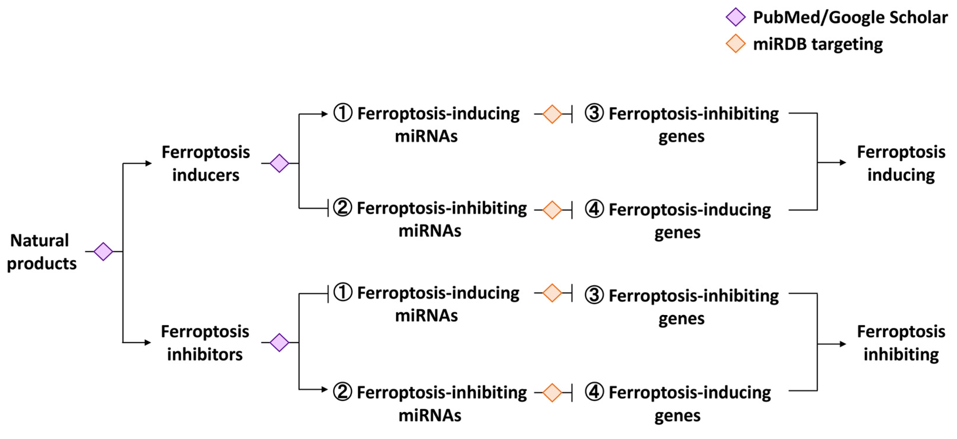
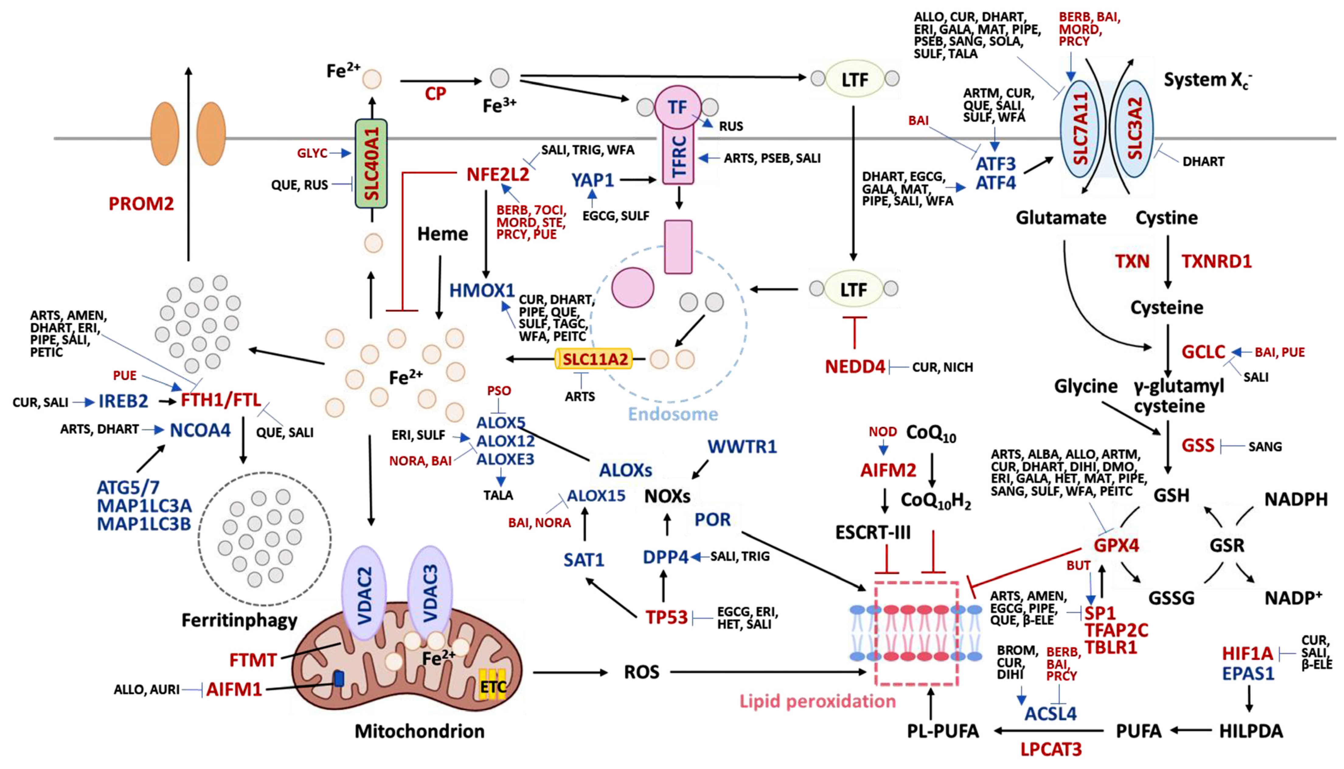
Disclaimer/Publisher’s Note: The statements, opinions and data contained in all publications are solely those of the individual author(s) and contributor(s) and not of MDPI and/or the editor(s). MDPI and/or the editor(s) disclaim responsibility for any injury to people or property resulting from any ideas, methods, instructions or products referred to in the content.

