
The Gap Junction Inhibitor Octanol Decreases Proliferation and Increases Glial Differentiation of Postnatal Neural Progenitor Cells

 ,
,  , and
, and 
Abstract
:1. Introduction
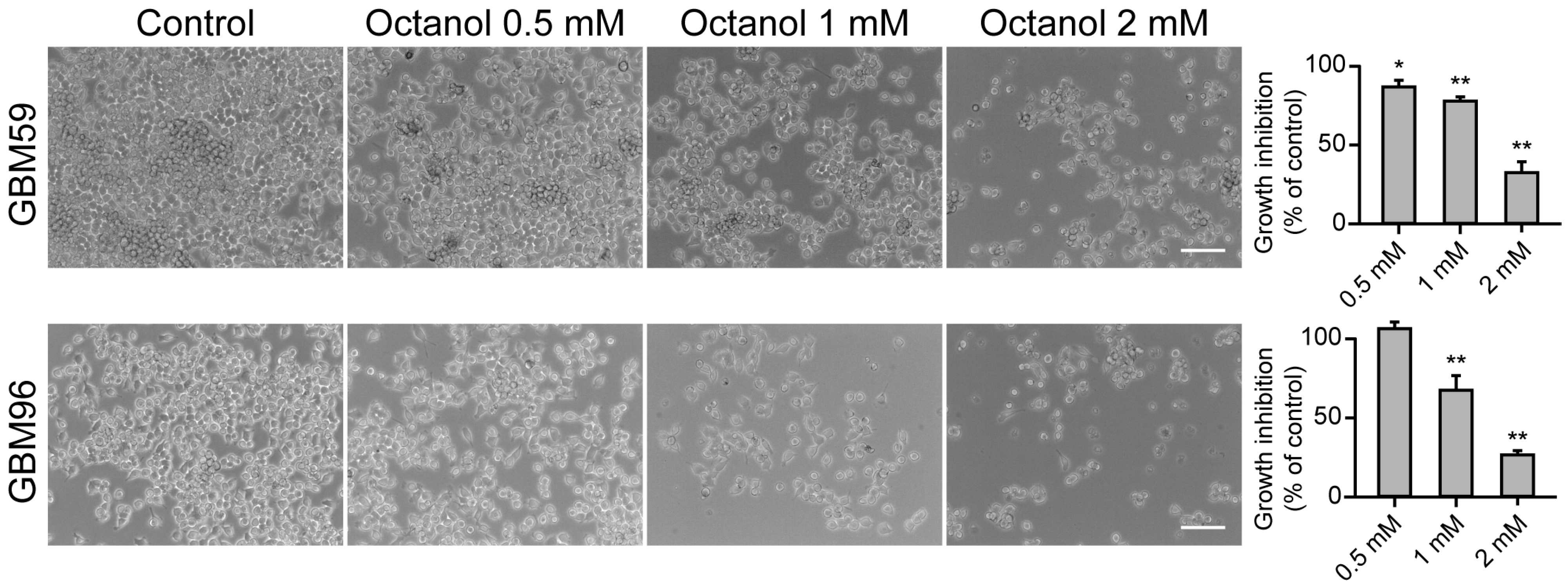
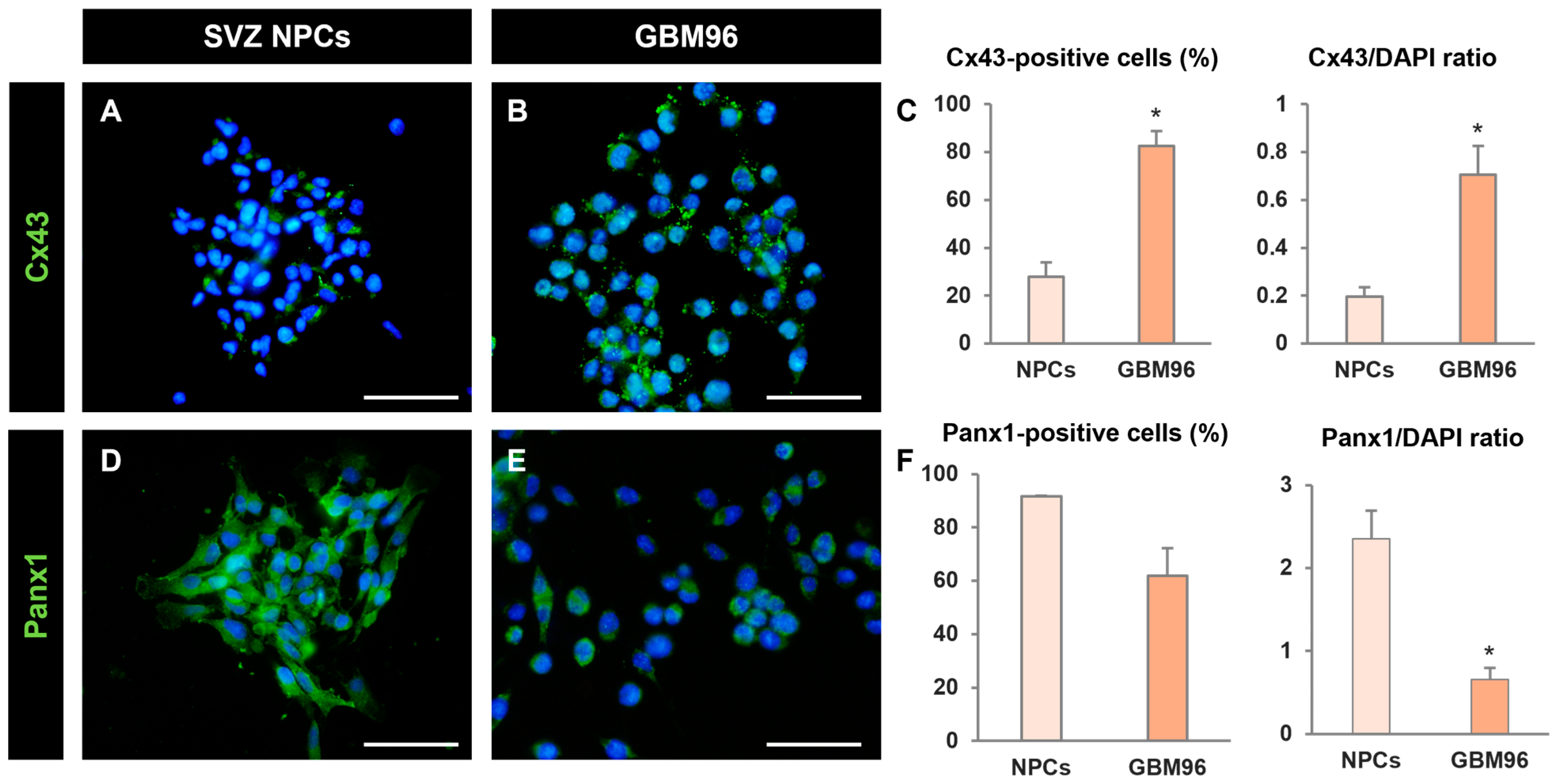
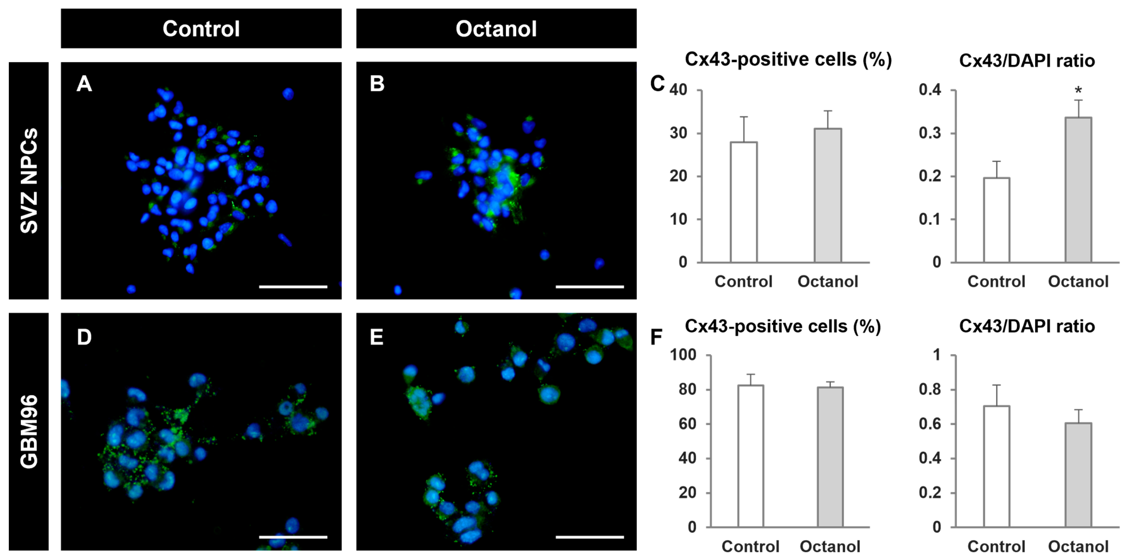
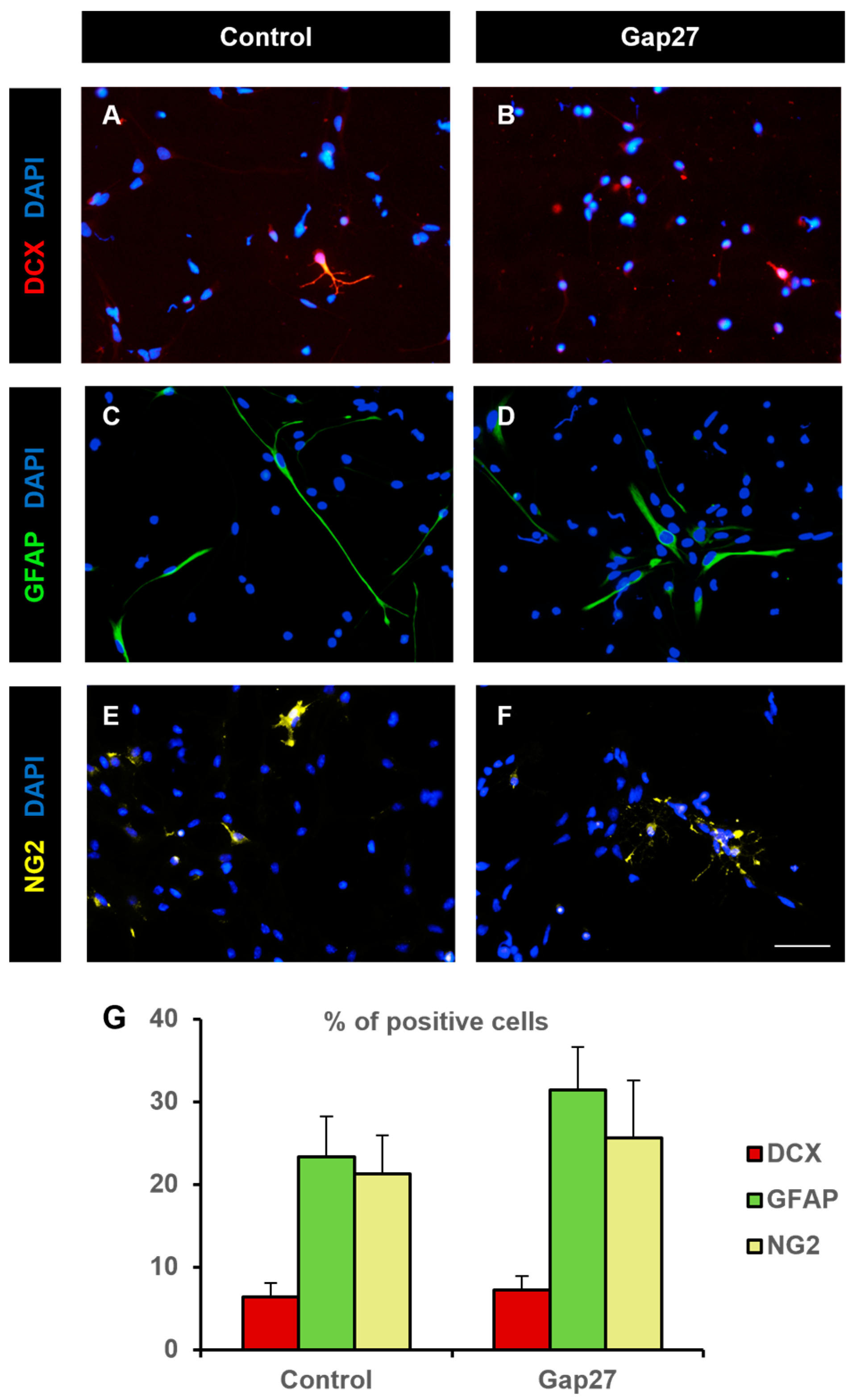
2. Results
3. Discussion
4. Materials and Methods
4.1. Neural Precursor Cell Culture
4.2. Fixation and Neurosphere Preparation for Electron Microscopy
4.3. Glioblastoma Cell Culture
4.4. Analysis of Proliferation in Neurospheres
4.5. BrdU Immunohistochemistry
4.6. Evaluation of Apoptosis
4.7. MTT Assay
4.8. Identification of Hemichannel Proteins via Immunocytochemistry
4.9. Analysis of Differentiation of Neurosphere-Derived Cells
4.10. Epifluorescence Microscopy
4.11. Statistics
Supplementary Materials
Author Contributions
Funding
Institutional Review Board Statement
Informed Consent Statement
Data Availability Statement
Acknowledgments
Conflicts of Interest
References
- Kar, R.; Batra, N.; Riquelme, M.A.; Jiang, J.X. Biological Role of Connexin Intercellular Channels and Hemichannels. Arch. Biochem. Biophys. 2012, 524, 2–15. [Google Scholar] [CrossRef] [PubMed]
- Willecke, K.; Eiberger, J.; Degen, J.; Eckardt, D.; Romualdi, A.; Guldenagel, M.; Deutsch, U.; Sohl, G.; Güldenagel, M.; Deutsch, U.; et al. Structural and Functional Diversity of Connexin Genes in the Mouse and Human Genome. Biol. Chem. 2002, 383, 725–737. [Google Scholar] [CrossRef]
- Goodenough, D.A.; Paul, D.L. Beyond the Gap: Functions of Unpaired Connexon Channels. Nat. Rev. Mol. Cell Biol. 2003, 4, 285–294. [Google Scholar] [CrossRef] [PubMed]
- Kumar, N.M.; Gilula, N.B. The Gap Junction Communication Channel. Cell 1996, 84, 381–388. [Google Scholar] [CrossRef]
- Saez, J.C.; Berthoud, V.M.; Branes, M.C.; Martinez, A.D.; Beyer, E.C. Plasma Membrane Channels Formed by Connexins: Their Regulation and Functions. Physiol. Rev. 2003, 83, 1359–1400. [Google Scholar] [CrossRef] [PubMed]
- Zhou, J.Z.; Jiang, J.X. Gap Junction and Hemichannel-Independent Actions of Connexins on Cell and Tissue Functions—An Update. FEBS Lett. 2014, 588, 1186–1192. [Google Scholar] [CrossRef] [PubMed]
- Alvarez-Buylla, A.; García-Verdugo, J.M. Neurogenesis in Adult Subventricular Zone. J. Neurosci. 2002, 22, 629–634. [Google Scholar] [CrossRef] [PubMed]
- Gage, F.H. Mammalian Neural Stem Cells. Science 2000, 287, 1433–1438. [Google Scholar] [CrossRef] [PubMed]
- Carpenter, M.K.; Cui, X.; Hu, Z.; Jackson, J.; Sherman, S.; Seiger, Å.; Wahlberg, L.U. In Vitro Expansion of a Multipotent Population of Human Neural Progenitor Cells. Exp. Neurol. 1999, 158, 265–278. [Google Scholar] [CrossRef]
- Reynolds, B.A.; Weiss, S. Generation of Neurons and Astrocytes from Isolated Cells of the Adult Mammalian Central Nervous System. Science 1992, 255, 1707–1710. [Google Scholar] [CrossRef]
- Vescovi, A.L.; Parati, E.A.; Gritti, A.; Poulin, P.; Ferrario, M.; Wanke, E.; Frölichsthal-Schoeller, P.; Cova, L.; Arcellana-Panlilio, M.; Colombo, A.; et al. Isolation and Cloning of Multipotential Stem Cells from the Embryonic Human CNS and Establishment of Transplantable Human Neural Stem Cell Lines by Epigenetic Stimulation. Exp. Neurol. 1999, 156, 71–83. [Google Scholar] [CrossRef] [PubMed]
- Talaverón, R.; Fernández, P.; Escamilla, R.; Pastor, A.M.; Matarredona, E.R.; Sáez, J.C. Neural Progenitor Cells Isolated from the Subventricular Zone Present Hemichannel Activity and Form Functional Gap Junctions with Glial Cells. Front. Cell. Neurosci. 2015, 9, 411. [Google Scholar] [CrossRef] [PubMed]
- Jiménez-Madrona, E.; Morado-Díaz, C.J.; Talaverón, R.; Tabernero, A.; Pastor, A.M.; Sáez, J.C.; Matarredona, E.R. Antiproliferative Effect of Boldine on Neural Progenitor Cells and on Glioblastoma Cells. Front. Neurosci. 2023, 17, 1211467. [Google Scholar] [CrossRef] [PubMed]
- Lacar, B.; Young, S.Z.; Platel, J.C.; Bordey, A. Gap Junction-Mediated Calcium Waves Define Communication Networks among Murine Postnatal Neural Progenitor Cells. Eur. J. Neurosci. 2011, 34, 1895–1905. [Google Scholar] [CrossRef] [PubMed]
- Ravella, A.; Ringstedt, T.; Brion, J.; Pandolfo, M.; Herlenius, E. Adult Neural Precursor Cells Form Connexin-Dependent Networks That Improve Their Survival. Neuroreport 2015, 26, 928–936. [Google Scholar] [CrossRef] [PubMed]
- Wiencken-Barger, A.E.; Djukic, B.; Casper, K.B.; McCarthy, K.D. A Role for Connexin43 during Neurodevelopment. Glia 2007, 55, 675–686. [Google Scholar] [CrossRef] [PubMed]
- Cheng, A.; Tang, H.; Cai, J.; Zhu, M.; Zhang, X.; Rao, M.; Mattson, M.P. Gap Junctional Communication Is Required to Maintain Mouse Cortical Neural Progenitor Cells in a Proliferative State. Dev. Biol. 2004, 272, 203–216. [Google Scholar] [CrossRef] [PubMed]
- Duval, N.; Gomès, D.; Calaora, V.; Calabrese, A.; Meda, P.; Bruzzone, R. Cell Coupling and Cx43 Expression in Embryonic Mouse Neural Progenitor Cells. J. Cell Sci. 2002, 115, 3241–3251. [Google Scholar] [CrossRef] [PubMed]
- Lemcke, H.; Nittel, M.L.; Weiss, D.G.; Kuznetsov, S.A. Neuronal Differentiation Requires a Biphasic Modulation of Gap Junctional Intercellular Communication Caused by Dynamic Changes of Connexin43 Expression. Eur. J. Neurosci. 2013, 38, 2218–2228. [Google Scholar] [CrossRef]
- Cai, J.; Cheng, A.; Luo, Y.; Lu, C.; Mattson, M.P.; Rao, M.S.; Furukawa, K. Membrane Properties of Rat Embryonic Multipotent Neural Stem Cells. J. Neurochem. 2003, 88, 212–226. [Google Scholar] [CrossRef]
- Wong, R.C.B.; Dottori, M.; Koh, K.L.L.; Nguyen, L.T.V.; Pera, M.F.; Pébay, A. Gap Junctions Modulate Apoptosis and Colony Growth of Human Embryonic Stem Cells Maintained in a Serum-Free System. Biochem. Biophys. Res. Commun. 2006, 344, 181–188. [Google Scholar] [CrossRef]
- Todorova, M.G.; Soria, B.; Quesada, I. Gap Junctional Intercellular Communication Is Required to Maintain Embryonic Stem Cells in a Non-Differentiated and Proliferative State. J. Cell. Physiol. 2008, 214, 354–362. [Google Scholar] [CrossRef] [PubMed]
- Lee, J.H.; Lee, J.E.; Kahng, J.Y.; Kim, S.H.; Park, J.S.; Yoon, S.J.; Um, J.Y.; Kim, W.K.; Lee, J.K.; Park, J.; et al. Human Glioblastoma Arises from Subventricular Zone Cells with Low-Level Driver Mutations. Nature 2018, 560, 243–247. [Google Scholar] [CrossRef] [PubMed]
- Matarredona, E.R.; Pastor, A.M. Neural Stem Cells of the Subventricular Zone as the Origin of Human Glioblastoma Stem Cells. Therapeutic Implications. Front. Oncol. 2019, 9, 779. [Google Scholar] [CrossRef] [PubMed]
- Louis, D.N.; Ohgaki, H.; Wiestler, O.D.; Cavenee, W.K.; Burger, P.C.; Jouvet, A.; Scheithauer, B.W.; Kleihues, P. The 2007 WHO Classification of Tumours of the Central Nervous System. Acta Neuropathol. 2007, 114, 97–109. [Google Scholar] [CrossRef] [PubMed]
- Bani-Yaghoub, M.; Bechberger, J.F.; Underhill, T.M.; Naus, C.C. The Effects of Gap Junction Blockage on Neuronal Differentiation of Human NTera2/Clone D1 Cells. Exp. Neurol. 1999, 156, 16–32. [Google Scholar] [CrossRef] [PubMed]
- Hartfield, E.M.; Rinaldi, F.; Glover, C.P.; Wong, L.F.; Caldwell, M.A.; Uney, J.B. Connexin 36 Expression Regulates Neuronal Differentiation from Neural Progenitor Cells. PLoS ONE 2011, 6, e14746. [Google Scholar] [CrossRef]
- Imbeault, S.; Gauvin, L.G.; Toeg, H.D.; Pettit, A.; Sorbara, C.D.; Migahed, L.; DesRoches, R.; Menzies, A.S.; Nishii, K.; Paul, D.L.; et al. The Extracellular Matrix Controls Gap Junction Protein Expression and Function in Postnatal Hippocampal Neural Progenitor Cells. BMC Neurosci. 2009, 10, 13. [Google Scholar] [CrossRef]
- Leung, D.S.Y.; Unsicker, K.; Reuss, B. Expression and Developmental Regulation of Gap Junction Connexins Cx26, Cx32, Cx43 and Cx45 in the Rat Midbrain-Floor. Int. J. Dev. Neurosci. 2002, 20, 63–75. [Google Scholar] [CrossRef]
- Rozental, R.; Morales, M.; Mehler, M.F.; Urban, M.; Kremer, M.; Dermietzel, R.; Kessler, J.A.; Spray, D.C. Changes in the Properties of Gap Junctions during Neuronal Differentiation of Hippocampal Progenitor Cells. J. Neurosci. 1998, 18, 1753–1762. [Google Scholar] [CrossRef]
- Kunze, A.; Rubenecia, M.; Hartmann, C.; Wallraff-beck, A.; Hu, K.; Bedner, P.; Requardt, R.; Seifert, G.; Redecker, C.; Willecke, K.; et al. Connexin Expression by Radial Glia-like Cells Is Required for Neurogenesis in the Adult Dentate Gyrus. Proc. Natl. Acad. Sci. USA 2009, 106, 3–8. [Google Scholar] [CrossRef] [PubMed]
- Zhang, J.; Griemsmann, S.; Wu, Z.; Dobrowolski, R.; Willecke, K.; Theis, M.; Steinhäuser, C.; Bedner, P. Connexin43, but Not Connexin30, Contributes to Adult Neurogenesis in the Dentate Gyrus. Brain Res. Bull. 2018, 136, 91–100. [Google Scholar] [CrossRef] [PubMed]
- Talaverón, R.; Matarredona, E.R.; de la Cruz, R.R.; Macías, D.; Gálvez, V.; Pastor, A.M. Implanted Neural Progenitor Cells Regulate Glial Reaction to Brain Injury and Establish Gap Junctions with Host Glial Cells. Glia 2014, 62, 623–638. [Google Scholar] [CrossRef] [PubMed]
- Spray, D.C.; Rozental, R.; Srinivas, M. Prospects for Rational Development of Pharmacological Gap Junction Channel Blockers. Curr. Drug Targets 2002, 3, 455–464. [Google Scholar] [CrossRef]
- Willebrords, J.; Maes, M.; Crespo Yanguas, S.; Vinken, M. Inhibitors of connexin and pannexin channels as potential therapeutics. Pharmacol. Ther. 2017, 180, 144–160. [Google Scholar] [CrossRef]
- Chaytor, A.T.; Evans, W.H.; Griffith, T.M. Peptides Homologous to Extracellular Loop Motifs of Connexin 43 Reversibly Abolish Rhythmic Contractile Activity in Rabbit Arteries. J. Physiol. 1997, 503, 99–110. [Google Scholar] [CrossRef] [PubMed]
- Howard Evans, W.; Leybaert, L. Mimetic Peptides as Blockers of Connexin Channel-Facilitated Intercellular Communication. Cell Commun. Adhes. 2007, 14, 265–273. [Google Scholar] [CrossRef] [PubMed]
- Evans, W.H.; Boitano, S. Connexin mimetic peptides: Specific inhibitors of gap-junctional intercellular communication. Biochem. Soc. Trans. 2001, 29, 606–612. [Google Scholar] [CrossRef]
- Krysko, D.V.; Leybaert, L.; Vandenabeele, P.; D’Herde, K. Gap Junctions and the Propagation of Cell Survival and Cell Death Signals. Apoptosis 2005, 10, 459–469. [Google Scholar] [CrossRef]
- Weissman, T.A.; Riquelme, P.A.; Ivic, L.; Flint, A.C.; Kriegstein, A.R. Calcium Waves Propagate through Radial Glial Cells and Modulate Proliferation in the Developing Neocortex. Neuron 2004, 43, 647–661. [Google Scholar] [CrossRef]
- Talukdar, S.; Emdad, L.; Das, S.K.; Fisher, P.B. GAP Junctions: Multifaceted Regulators of Neuronal Differentiation. Tissue Barriers 2022, 10, 1982349. [Google Scholar] [CrossRef] [PubMed]
- Wang, J.; Shen, J.; Kirschen, G.W.; Gu, Y.; Jessberger, S.; Ge, S. Lateral Dispersion Is Required for Circuit Integration of Newly Generated Dentate Granule Cells. Nat. Commun. 2019, 10, 3324. [Google Scholar] [CrossRef] [PubMed]
- Lacar, B.; Herman, P.; Platel, J.-C.; Kubera, C.; Hyder, F.; Bordey, A. Neural Progenitor Cells Regulate Capillary Blood Flow in the Postnatal Subventricular Zone. J. Neurosci. 2012, 32, 16435–16448. [Google Scholar] [CrossRef] [PubMed]
- Manjarrez-Marmolejo, J.; Franco-Pérez, J. Gap Junction Blockers: An Overview of Their Effects on Induced Seizures in Animal Models. Curr. Neuropharmacol. 2016, 14, 759–771. [Google Scholar] [CrossRef]
- Talaverón, R.; Matarredona, E.R.; Herrera, A.; Medina, J.M.; Tabernero, A. Connexin43 Region 266–283, via Src Inhibition, Reduces Neural Progenitor Cell Proliferation Promoted by Egf and Fgf-2 and Increases Astrocytic Differentiation. Int. J. Mol. Sci. 2020, 21, 8852. [Google Scholar] [CrossRef] [PubMed]
- Joksovic, P.M.; Choe, W.J.; Nelson, M.T.; Orestes, P.; Brimelow, B.C.; Todorovic, S.M. Mechanisms of Inhibition of T-Type Calcium Current in the Reticular Thalamic Neurons by 1-Octanol: Implication of the Protein Kinase C Pathway. Mol. Pharmacol. 2010, 77, 87–94. [Google Scholar] [CrossRef] [PubMed]
- Sanai, N.; Alvarez-Buylla, A.; Berger, M.S. Mechanisms of Disease: Neural Stem Cells and the Origin of Gliomas. N. Engl. J. Med. 2005, 353, 811–822. [Google Scholar] [CrossRef]
- Mettang, M.; Meyer-Pannwitt, V.; Karpel-Massler, G.; Zhou, S.; Carragher, N.O.; Föhr, K.J.; Baumann, B.; Nonnenmacher, L.; Enzenmüller, S.; Dahlhaus, M.; et al. Blocking Distinct Interactions between Glioblastoma Cells and Their Tissue Microenvironment: A Novel Multi-Targeted Therapeutic Approach. Sci. Rep. 2018, 8, 5527. [Google Scholar] [CrossRef] [PubMed]
- Holdhoff, M.; Ye, X.; Supko, J.G.; Nabors, L.B.; Desai, A.S.; Walbert, T.; Lesser, G.J.; Read, W.L.; Lieberman, F.S.; Lodge, M.A.; et al. Timed Sequential Therapy of the Selective T-Type Calcium Channel Blocker Mibefradil and Temozolomide in Patients with Recurrent High-Grade Gliomas. Neuro Oncol. 2017, 19, 845–852. [Google Scholar] [CrossRef]
- Talaverón, R.; Matarredona, E.R.; de la Cruz, R.R.; Pastor, A.M. Neural Progenitor Cell Implants Modulate Vascular Endothelial Growth Factor and Brain-Derived Neurotrophic Factor Expression in Rat Axotomized Neurons. PLoS ONE 2013, 8, e54519. [Google Scholar] [CrossRef]
- Torroglosa, A.; Murillo-Carretero, M.; Romero-Grimaldi, C.; Matarredona, E.R.; Campos-Caro, A.; Estrada, C. Nitric Oxide Decreases Subventricular Zone Stem Cell Proliferation by Inhibition of Epidermal Growth Factor Receptor and Phosphoinositide-3-Kinase/Akt Pathway. Stem Cells 2007, 25, 88–97. [Google Scholar] [CrossRef]
- Wang, Q.; Wang, Z.; Tian, Y.; Zhang, H.; Fang, Y.; Yu, Z.; Wang, W.; Xie, M.; Ding, F. Inhibition of Astrocyte Connexin 43 Channels Facilitates the Differentiation of Oligodendrocyte Precursor Cells Under Hypoxic Conditions In Vitro. J. Mol. Neurosci. 2018, 64, 591–600. [Google Scholar] [CrossRef]
- Stafford, M.R.; Bartlett, P.F.; Adams, D.J. Purinergic Receptor Activation Inhibits Mitogen-Stimulated Proliferation in Primary Neurospheres from the Adult Mouse Subventricular Zone. Mol. Cell. Neurosci. 2007, 35, 535–548. [Google Scholar] [CrossRef]
- Scemes, E.; Duval, N.; Meda, P. Reduced Expression of P2Y1 Receptors in Connexin43-Null Mice Alters Calcium Signaling and Migration of Neural Progenitor Cells. J. Neurosci. 2003, 23, 11444–11452. [Google Scholar] [CrossRef]
- Santiago, M.F.; Alcami, P.; Striedinger, K.M.; Spray, D.C.; Scemes, E. The Carboxyl-Terminal Domain of Connexin43 Is a Negative Modulator of Neuronal Differentiation. J. Biol. Chem. 2010, 285, 11836–11845. [Google Scholar] [CrossRef]
- Rinaldi, F.; Hartfield, E.M.; Crompton, L.A.; Badger, J.L.; Glover, C.P.; Kelly, C.M.; Rosser, A.E.; Uney, J.B.; Caldwell, M.A. Cross-Regulation of Connexin43 and β-Catenin Influences Differentiation of Human Neural Progenitor Cells. Cell Death Dis. 2014, 5, e1017. [Google Scholar] [CrossRef]
- Sawada, N.; Kotani, T.; Konno, T.; Setiawan, J.; Nishigaito, Y.; Saito, Y.; Murata, Y.; Nibu, K.I.; Matozaki, T. Regulation by Commensal Bacteria of Neurogenesis in the Subventricular Zone of Adult Mouse Brain. Biochem. Biophys. Res. Commun. 2018, 498, 824–829. [Google Scholar] [CrossRef]
- Wang, X.; Mao, X.; Xie, L.; Greenberg, D.A.; Jin, K. Involvement of Notch1 Signaling in Neurogenesis in the Subventricular Zone of Normal and Ischemic Rat Brain in Vivo. J. Cereb. Blood Flow Metab. 2009, 29, 1644–1654. [Google Scholar] [CrossRef]


| Antibody Name | Immunogen | Antibody Information | Working Concentration |
|---|---|---|---|
| Bromodeoxyuridine (BrdU) | The epitope is inside the DNA helix. DNA must be denatured before antibody efficiently binds to DNA-BrdU | Roche Diagnostics GmBH Mannheim, Germany Cat#11 170 376 001 | 1:100 |
| Doublecortin (DCX) | Amino acids 123–402 mapping at the C-terminus of doublecortin of human origin | Santa Cruz Biotechnology Santa Cruz, CA, USA Cat# sc-271390 Mouse monoclonal | 1:100 |
| Glial Fibrillary Acidic Protein (GFAP) | Purified GFAP from pig spinal cord | Sigma-Aldrich Cat# G3893 Mouse monoclonal | 1: 1400 |
| Chondroitin sulphate proteoglycan (NG2) | Purified NG2 chondroitin sulphate proteoglycan from rat | Millipore Cat#AB5320 Rabbit polyclonal | 1:400 |
| Connexin43 (Cx43) | Peptide corresponding to a segment of the 3rd cytoplasmic domain (C-terminal portion) of rat connexin43 | Thermo Scientific Cat #71-0700 Rabbit polyclonal | 1:50 |
| Pannexin1 (Panx1) | Recombinant Protein Epitope Signature Tag (PrEST) antigen sequence available in datasheet | Sigma Aldrich Cat #HPA016930 Rabbit polyclonal | 1:500 |
Disclaimer/Publisher’s Note: The statements, opinions and data contained in all publications are solely those of the individual author(s) and contributor(s) and not of MDPI and/or the editor(s). MDPI and/or the editor(s) disclaim responsibility for any injury to people or property resulting from any ideas, methods, instructions or products referred to in the content. |
© 2024 by the authors. Licensee MDPI, Basel, Switzerland. This article is an open access article distributed under the terms and conditions of the Creative Commons Attribution (CC BY) license (https://creativecommons.org/licenses/by/4.0/).
Share and Cite
Talaverón, R.; Morado-Díaz, C.J.; Herrera, A.; Gálvez, V.; Pastor, A.M.; Matarredona, E.R. The Gap Junction Inhibitor Octanol Decreases Proliferation and Increases Glial Differentiation of Postnatal Neural Progenitor Cells. Int. J. Mol. Sci. 2024, 25, 6288. https://doi.org/10.3390/ijms25126288
Talaverón R, Morado-Díaz CJ, Herrera A, Gálvez V, Pastor AM, Matarredona ER. The Gap Junction Inhibitor Octanol Decreases Proliferation and Increases Glial Differentiation of Postnatal Neural Progenitor Cells. International Journal of Molecular Sciences. 2024; 25(12):6288. https://doi.org/10.3390/ijms25126288
Chicago/Turabian StyleTalaverón, Rocío, Camilo J. Morado-Díaz, Alejandro Herrera, Victoria Gálvez, Angel M. Pastor, and Esperanza R. Matarredona. 2024. "The Gap Junction Inhibitor Octanol Decreases Proliferation and Increases Glial Differentiation of Postnatal Neural Progenitor Cells" International Journal of Molecular Sciences 25, no. 12: 6288. https://doi.org/10.3390/ijms25126288