Article

Amelioration of Fibrosis via S1P Inhibition Is Regulated by Inactivation of TGF-β and SPL Pathways in the Human Cornea

by
Sarah E. Nicholas
1,2,
Sandip K. Basu
3,
Nawajes Mandal
3,4 and
Dimitrios Karamichos
1,2,5,*
1
North Texas Eye Research Institute, University of North Texas Health Science Center, Fort Worth, TX 76107, USA
2
Department of Pharmaceutical Sciences, University of North Texas Health Science Center, Fort Worth, TX 76107, USA
3
Department of Ophthalmology, University of Tennessee Health Science Center, Memphis, TN 38163, USA
4
Department of Anatomy and Neurobiology, University of Tennessee Health Science Center, Memphis, TN 38163, USA
5
Department of Pharmacology and Neuroscience, University of North Texas Health Science Center, Fort Worth, TX 76107, USA
*
Author to whom correspondence should be addressed.
Int. J. Mol. Sci. 2024, 25(12), 6560; https://doi.org/10.3390/ijms25126560
Submission received: 24 May 2024 / Revised: 10 June 2024 / Accepted: 11 June 2024 / Published: 14 June 2024
(This article belongs to the Special Issue Sphingolipid Metabolism and Signaling: Role in Health and Diseases)

Abstract

:
Human corneal fibrosis can lead to opacity and ultimately partial or complete vision loss. Currently, corneal transplantation is the only treatment for severe corneal fibrosis and comes with the risk of rejection and donor shortages. Sphingolipids (SPLs) are known to modulate fibrosis in various tissues and organs, including the cornea. We previously reported that SPLs are tightly related to both, transforming growth factor beta (TGF-β) signaling and corneal fibrogenesis. The aim of this study was to investigate the effects of sphingosine-1-phosphate (S1P) and S1P inhibition on specific TGF-β and SPL family members in corneal fibrosis. Healthy human corneal fibroblasts (HCFs) were isolated and cultured in EMEM + FBS + VitC (construct medium) on 3D transwells for 4 weeks. The following treatments were prepared in a construct medium: 0.1 ng/mL TGF-β1 (β1), 1 μM sphingosine-1-phosphate (S1P), and 5 μM Sphingosine kinase inhibitor 2 (I2). Five groups were tested: (1) control (no treatment); rescue groups; (2) β1/S1P; (3) β1/I2; prevention groups; (4) S1P/β1; and (5) I2/β1. Each treatment was administered for 2 weeks with one treatment and switched to another for 2 weeks. Using Western blot analysis, the 3D constructs were examined for the expression of fibrotic markers, SPL, and TGF-β signaling pathway members. Scratch assays from 2D cultures were also utilized to evaluate cell migration We observed reduced fibrotic expression and inactivation of latent TGF-β binding proteins (LTBPs), TGF-β receptors, Suppressor of Mothers Against Decapentaplegic homologs (SMADs), and SPL signaling following treatment with I2 prevention and rescue compared to S1P prevention and rescue, respectively. Furthermore, we observed increased cell migration following stimulation with I2 prevention and rescue groups, with decreased cell migration following stimulation with S1P prevention and rescue groups after 12 h and 18 h post-scratch. We have demonstrated that I2 treatment reduced fibrosis and modulated the inactivation of LTBPs, TGF-β receptors, SPLs, and the canonical downstream SMAD pathway. Further investigations are warranted in order to fully uncover the potential of utilizing SphK I2 as a novel therapy for corneal fibrosis.

1. Introduction

Corneal fibrosis is a leading cause of blindness worldwide, affecting over 10 million people [1]. Injury or trauma to the cornea can initiate resident keratocyte differentiation into myofibroblasts, causing high expression levels of α smooth muscle actin (αSMA) and Collagen III, which can cause irregular deposition of extracellular matrix (ECM) components [2,3]. This disruption of the homeostatic corneal environment can cause cornea scarring, which can result in vision loss. While the corneal clarity is maintained by orchestrated signaling cascades, the specific mechanisms driving corneal fibrosis are very complex and still not well understood [1,4,5,6]. Unfortunately, for severe corneal scars, corneal transplantation is often the only viable option for those suffering from this condition.
Previous studies have investigated the role of transforming growth factor beta (TGF-β) isoforms in cells and fibrotic tissues [7,8,9,10,11,12,13]. The three isoforms found in humans share approximately 80% homology but their actions on cells and tissues vary greatly. Our group and others have found that the TGF-β1 isoform induces corneal fibrosis, whereas the TGF-β3 isoform is known for its anti-fibrotic abilities [4,14,15,16,17,18,19,20,21,22,23,24]. Various signaling cascades are initiated when TGF-β isoforms bind to TGF-β receptors [7,25,26,27]. The activation of latent TGF-β is regulated largely by latent TGF-β binding proteins (LTBPs). LTBPs 1–4 are secreted by various cells and tissues [28,29,30,31], including the cornea; however, their role in the cornea is not currently well understood.
Sphingosine 1-phosphate (S1P) is a pleiotropic bioactive lipid mediator that is formed from the catalysis of sphingosine kinases (SphK1 and SphK2). Sphingolipids (SPLs), such as S1P, bind to S1P receptors (S1PR1-5), inducing various cellular responses, and have been implicated in many studies as key regulators of fibrosis in various tissues and organs [32,33,34,35,36,37,38,39,40,41,42,43,44,45,46,47,48], including the cornea [26,33,49,50,51,52,53,54,55,56].
In the cornea, the cross-talks between TGF-β and S1P signaling remain elusive; however, studies in other tissues suggest that TGF-β induces the activity of sphingosine kinase [45,57]. Several recent studies have demonstrated the fibrotic protective effects of TGF-β and S1P inhibition [58,59,60,61,62]. Figure 1 represents the “inside out” signaling of S1P and TGF-β, both autocrine and paracrine, as observed by our team and other researchers [34,35,63,64,65]. Our group recently investigated the interplay of S1P and SPHK I2 (I2; a selective inhibitor of SphK1) interactions with TGF-β signaling and downstream signaling in human corneal fibrosis [26]. We demonstrated differential regulation of TGF-βRII following S1P stimulation vs. I2 treatment. Additionally, S1P inhibition downregulated pSMAD2 and SMAD4 and showed similar signaling patterns as TGF-β3 treatment.
The present study sought to understand the role of SPLs and TGF-β signaling pathway members, their cross-talks, and downstream targets in corneal fibrosis. Future studies will reveal whether S1P inhibition can be tailored as a novel therapy for the management of corneal fibrosis.

2. Results

2.1. Latent Transforming Growth Factor Beta Binding Proteins (LTBPs)

Latent TGF-β binding proteins-1 through -4 (LTBPs 1–4) are known activators of TGF-β and were investigated here for their protein expressions following treatment with all groups tested. LTBP1 expression was significantly upregulated by β1-S1P stimulation compared to the controls (p = 0.0005), S1P-β1 (p = 0.0048), and β1-I2 (p < 0.0001) treatments. In Figure 2A, I2-β1 stimulation led to a significant downregulation of LTBP1 compared to S1P-β1 treatment (p < 0.0001) and the controls (p = 0.006). LTBP2 expression was significantly upregulated by β1-S1P stimulation compared to the controls (p < 0.0001) and β1-I2 treatment (p < 0.0001) (Figure 2B). Figure 2B showed that treatment with I2-β1 caused a significant downregulation of LTBP2 compared to S1P-β1 treatment (p < 0.0001) and the controls (p = 0.0258). The expression of LTBP3 was significantly upregulated by β1-S1P stimulation compared to the controls (p < 0.0001), S1P-β1 (p < 0.0001), and β1-I2 (p < 0.0001) treatments (Figure 2C). The I2-β1 stimulation caused a significant downregulation of LTBP3 compared to S1P-β1 treatment (p = 0.0298) (Figure 2C). LTBP4 was significantly upregulated by β1-S1P stimulation compared to the controls (p < 0.0001) and β1-I2 treatment (p < 0.0001) (Figure 2D). Treatment with I2-β1 led to a significant downregulation of LTBP4 compared to S1P-β1 (p < 0.0001) treatment and the controls (Figure 2D).

2.2. Transforming Growth Factor Beta Receptors (TGF-βRs)

TGF-β receptors I and II (TGF-βRI and TGF-βRII), which are activated via binding with active TGF-β, were investigated for their protein expressions for all groups tested. TGF-βRI expression was significantly upregulated by stimulation with β1-S1P compared to the controls (p = 0.0106), β1-I2 (p = 0.0004), and S1P-β1 compared to the controls only (p = 0.0041) (Figure 3A). β1-I2 treatment caused a significant downregulation of TGF-βRI compared to I2-β1 (p = 0.0023; Figure 3A). TGF-βRII was significantly upregulated by the S1P-β1 treatment compared to the controls (p < 0.0001), β1-S1P (p = 0.0076), and I2-β1 (p < 0.0001) treatments (Figure 3B). β1-S1P stimulation significantly upregulated TGF-βRII compared to the controls (p < 0.0001) and β1-I2 treatment (p < 0.0001) (Figure 3B).

2.3. Canonical Downstream SMAD Pathway

SMADs 2–4, which are the main signal transducers for TGF-β receptors, were investigated for their protein expressions for all groups tested. Figure 4A shows that pSMAD2 expression was significantly upregulated by S1P-β1 stimulation compared to the controls (p = 0.0008), β1-S1P (p < 0.0001), and I2-β1 (p < 0.0001) treatments. Stimulation with β1-I2 led to a significant downregulation of pSMAD2 compared to the controls (p = 0.0035) and β1-S1P treatment (p = 0.0446) (Figure 4A). pSMAD3 expression was significantly upregulated by S1P-β1 compared to the controls (p < 0.0001), β1-S1P (p < 0.0001), and I2-β1 (p < 0.0001) treatments (Figure 4B). SMAD4 expression was significantly upregulated by S1P-β1 treatment compared to the controls (p < 0.0001), β1-S1P (p = 0.0002), and I2-β1 (p < 0.0001) treatments (Figure 4C). Stimulation with β1-S1P significantly upregulated SMAD4 compared to the controls (p < 0.0001) and β1-I2 treatment (p < 0.0001) (Figure 4C).

2.4. Sphingolipid Members Pathway

Sphingolipid members (SphK1, SphK2, and S1PR3), which are known to transactivate the TGF-β pathway, were examined for their protein expressions for all groups tested. SphK1 expression was significantly upregulated by β1-S1P treatment compared to the controls (p = 0.0006), S1P-β1 (p = 0.0181), and β1-I2 stimulation (p = 0.001) (Figure 5A). Tge expression of SphK2 was significantly upregulated by β1-S1P compared to the controls (p < 0.0001), S1P-β1 (p < 0.0001), and β1-I2 (p < 0.0001) treatments (Figure 5B). The I2-β1 treatment led to a significant downregulation of SphK2 compared to S1P-β1 stimulation (p = 0.0259; Figure 5B). S1PR3 was significantly upregulated by the β1-S1P treatment compared to the controls (p < 0.0001), S1P-β1 (p = 0.0002), and β1-I2 (p < 0.0001) treatments (Figure 5C). Stimulation with I2-β1 caused a significant downregulation of S1PR3 compared to the S1P-β1 treatment (p = 0.0009; Figure 5C).

2.5. Fibrosis Markers

Corneal fibrosis markers, α-smooth muscle actin (α-SMA), and Collagen III were examined for their protein expressions for all groups tested. α-SMA expression was significantly downregulated by the I2-β1 treatment compared to the controls (p = 0.0001) and the S1P-β1 (p = 0.0062) treatment (Figure 6A). β1-I2 stimulation caused a significant downregulation of α-SMA compared to the controls (p < 0.0001) and β1-S1P (p < 0.0001) stimulation (Figure 6A). The expression of Collagen III was significantly downregulated with the β1-I2 treatment compared to the controls (p = 0.0018) and β1-S1P (p = 0.0014) stimulation (Figure 6B). S1P-β1 caused a significant upregulation of Collagen III compared to the control (p < 0.0001), β1-S1P (p < 0.0001), and I2-β1 (p < 0.0001) treatments (Figure 6B).

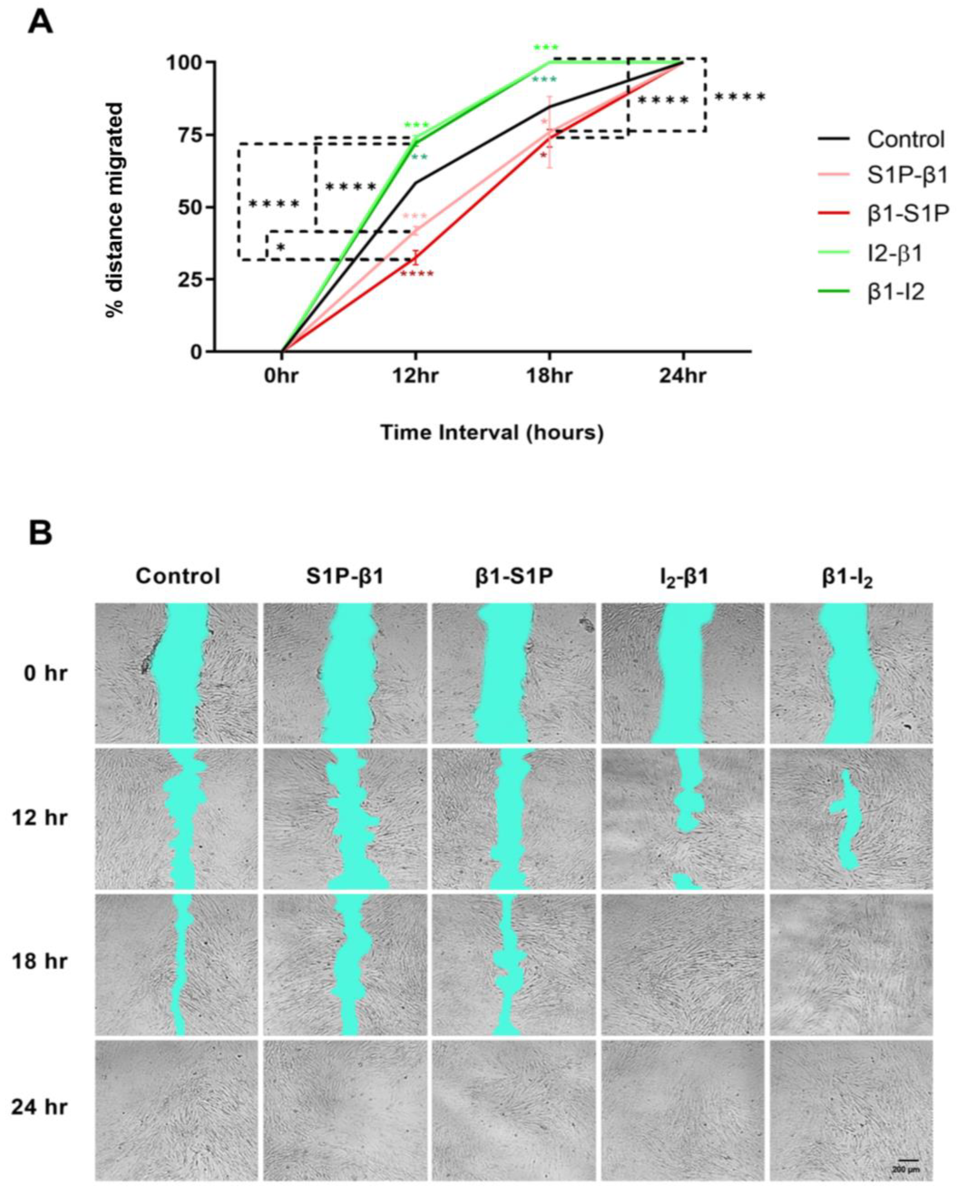
2.6. Effects of S1P and TGF-β Treatment Groups on HCF Cellular Migration

Cellular migration of HCFs was examined in response to stimulation with all groups tested over a period of 24 h. Following 12 h of stimulation with the I2-β1 group, we observed significantly increased wound closure compared with the controls (p = 0.0006) and S1P-β1 (p < 0.0001; Figure 7A). Similarly, the β1-I2 group caused significantly increased wound closure after 12 h compared to the controls (p = 0.0024) and β1-S1P (p < 0.0001) stimulation (Figure 7A). After 18 h, we observed significantly increased wound closure with I2-β1 compared to the controls (p = 0.0008) and β1-S1P (p < 0.0001) and in β1I2 compared to the controls (p = 0.0008) and β1-S1P (p < 0.0001) stimulation (Figure 7A). After 24 h, all treatment groups reached 100% wound closure (Figure 7A). Representative cell migration images with all groups tested over the course of 24 h are shown in Figure 7B.

3. Discussion

The mechanisms involving TGF-β and S1P in the cornea have largely remained a mystery due to the complexity of their signaling effects the lack of studies. Our group previously reported on the signaling expressions of SPLs, TGF-β members, canonical downstream SMAD, non-canonical downstream, and fibrotic markers in HCF 3D constructs treated with S1P, I2, TGF-β1, and TGF-3 [26]. Our current study demonstrated the impact of I2-induced fibrotic prevention and rescue modulated by SPLs and TGF-β family member signaling in HCF 3D constructs.
Inactive TGF-β isoforms are secreted from cells and are activated in covalent association with LTBP molecules [30,66]. Acosta et al., 2023, reported the presence of increased LTBP1 expression in murine corneal fibroblasts [16]. Another recent study found an increased expression of LTBP1 in the keratotomy wounds of mouse corneas [67]. LTBP2 dysregulation has been linked to eye diseases in numerous studies, including glaucoma [68,69,70,71,72,73,74,75,76,77]. Two studies reported LTBP1 and LTBP2 upregulation in the anterior segment of human tissues with pseudoexfoliation syndrome [78,79]. Similarly, De Maria et al., 2021, found LTBP2 and LTBP3 upregulation in the lens capsule and aqueous humor of patients with exfoliation syndrome [80]. A review by Su et al., 2021, reported on the link of LTBP3 dysregulation with various physical developmental disorders in mice and humans [81]. One group demonstrated the compensation of LTBP4 for the loss of LTBP2 in the microfibril formation of mouse embryonic fibroblasts [82]. Furthermore, LTBP4 genetic mutations have been linked to various disorders, including cutis laxa [81,83,84,85], scleroderma [81,86], pulmonary [81,87,88,89], cardiac [81,90,91,92], and cancer [81,93,94]. Our group reported that both TGF-β1 and TGF-β3 modulated LTBP1 expression, but only TGF-β3 modulated LTBP2 expression in 3D HCF constructs. Interestingly, LTBPs were not modulated by exogenous S1P or S1P inhibition (I2) [26]. Our current study revealed that fibrotic rescue via stimulation with β1-I2 caused a significant downregulation in the expression of LTBPs 1–4; contrastingly, the β1-S1P group caused an upregulation of LTBPs 1–4. Fibrotic prevention via stimulation with I2-β1 induced the downregulation of LTBPs 1 and 2, whereas the S1P-β1 group led to the significant upregulation of LTBPs 2 and 4 only. TGF-β activation is dependent on LTBP regulation and release from the large latent complex (LLC), and our findings demonstrated that LTBPs were heavily regulated by S1P prevention and rescue treatments, whereas I2 prevention and rescue treatments inhibited their expressions.
TGF-β and S1P overlapping convergence and cell signaling effects have been well documented [62,95], and recent studies involving endometriosis [57], pulmonary fibrosis [58,61], EMT/asthma [59], and renal interstitial fibrosis [60] have reported their involvement in the development of the aforementioned disorders. Although TGF-β and S1P cross-talks have been rigorously investigated in various cells and tissues, their role in the cornea has been understudied and is not yet well understood. Herein, we observed the upregulation of TGF-βRI and II expressions following S1P-β1 and β1-S1P treatment groups but significant downregulation following I2-β1 and β1-I2 group treatments, indicating that S1P prevention and rescue activated TGF-β receptors, whereas I2 prevention and rescue treatments did not.
SMADs are major downstream signaling transducers for TGF-β receptors and have been previously documented for their role in corneal fibrosis [65,66]. Recent studies have reported the impact of fibrosis in response to SMAD inhibition in the cornea. A reduction in corneal fibrosis was observed via the inhibition of SMAD2/3 in human [96,97] and mouse corneas [98]. One study previously demonstrated that murine corneal fibrosis was regulated in part by TGF-β1/SMAD2 activation [99]. Nuwormegbe et al., 2021 [100], revealed that TGF-β1-induced fibrosis was suppressed via SMAD3 signal inhibition in the human cornea. This finding was substantiated by another group, which reported that SMAD3 overexpression enhanced TGF-β1-induced fibroblasts to myofibroblast differentiation in HCFs [101]. Our current study demonstrated pSMAD2/3 and SMAD4 downregulation in response to I2-β1 treatment and upregulation following S1P-β1 treatment. Additionally, β1-I2 treatment caused the downregulation of pSMAD2 and SMAD4, but β1-S1P led to the upregulation of SMAD4 only.
In the sphingosine rheostat, sphingosine kinase is known as the “fulcrum” due to its critical role in controlling the balance between S1P and ceramide levels [44,46]. Many previous studies have implicated S1P as a fibrotic inducer in various cells and tissues [62]. S1P is generated from ceramide, which is phosphorylated by sphingosine kinases, SphK1 and Sphk2, which can demonstrate oppositional effects. S1P produced by SphK1 in the cytosol can act as a second messenger or can be secreted to bind to S1P receptors and TGF-β receptors, whereas SphK2 resides in the cell nucleus where S1P is generated and regulates gene expression [55]. Recently, SphK1 was found to influence S1P upregulation more than SphK2, and S1PR3 was linked to fibrotic manifestations in the lung [44]. Wang et al., 2023, reported that abnormal S1P content in the circulation affected cardiovascular disorder pathogenesis and S1PR3 mediation of cell proliferation and vascular permeability [45]. Furthermore, the S1PR3 antagonist was observed to improve graft viability in rat heart transplants [102]. Another recent study revealed that S1P-induced epithelial endometriotic cell fibrosis was reliant on S1PR3 activation [103]. Although investigations on S1P in the cornea have been under documented in the past, two recent studies have demonstrated the effects of SphK1/S1P in mouse corneas. Yasuda et al., 2021, revealed that TGF-β1-induced injury increased S1P via SphK1 upregulation modulated by S1PR3 and VEGF-A and angiogenesis [104]. Wilkerson et al., 2022, reported that SphK1 knockout mice had reduced corneal neovascularization following injury [55]. Previously, our group reported that TGF-β1 induced the upregulation of S1PR3 in HCF 3D in vitro constructs [26]. In the current study, we observed the upregulation of S1PR3 following S1P-β1 and β1-S1P treatment groups but a significant downregulation following I2-β1 and β1-I2 group treatments.
S1P is known to elicit cell and tissue-specific effects but is known largely as an inducer of fibrosis. A recent study observed that S1P stimulation enhanced retinal pigment epithelial cell migration, activated S1PR3, and stimulated αSMA transcription [105]. Meanwhile, another study found that exogenous S1P treatment in human Müller glial cells led to the significant upregulation of α-SMA expression [106]. Yang et al., 2024, found that an S1P agonist reduced cell migration compared to high glucose treatment in rat retinal Müller cells [107]. Our current study found increased cell migration following stimulation with I2-β1 and β1-I2 treatment groups, with decreased cell migration following stimulation with S1P-β1 and β1-S1P groups after 12 h and 18 h post-scratch. Moreover, we observed reduced fibrosis via αSMA and Collagen III expression regulated by I2-β1 and β1-I2 treatments.
Our observations demonstrated that S1P complementation to TGF-β1-induced fibrosis led to the activation of SPL and TGF-β pathways, whereas I2 treatment inhibited the pathways and resulted in reduced corneal fibrosis. Future investigations would explore the potential involvement of SMAD pathway inhibitors and S1P inhibition as a novel therapy for corneal fibrosis management.

4. Materials and Methods

4.1. Ethical Approval

Primary human corneal stromal fibroblasts (HCFs) were isolated from human cadaver corneas with no history of ocular or systemic disease and were de-identified prior to analysis. All cadaver corneas were obtained from the National Disease Research Interchange (NDRI, Philadelphia, PA, USA). All studies herein were approved by the North Texas Regional Institutional Review Board (IRB # 2020-030) and adhered to the Declaration of Helsinki.

4.2. Human Corneal Fibroblast Cell Isolation and 3D In Vitro Model Cultures

HCFs were isolated from healthy donors by scraping away the epithelium and endothelium, cutting the stromal tissue into 2 × 2 mm pieces, and allowing them to adhere in T25 flasks. The corneal explants were cultured in complete media consisting of Eagle’s Minimum Essential Medium (EMEM: ATCC; Manassas, VA, USA) with 10% fetal bovine serum (FBS: R&D Systems, Minneapolis, MN, USA) and 1% antibiotic-antimycotic (A.A.; Gibco, Life Technologies; Grand Island, NY, USA). HCFs were seeded onto polycarbonate transwell membranes in 6-well plates with 1 × 10⁶ cells/well. The cells were incubated for 24 h hours to allow adherence to the membranes and thereafter were stimulated with 0.5 mM stable vitamin C (0.5 mM 2-O-α-D-glucopyranosyl-L-ascorbic acid [108], Sigma-Aldrich, St. Louis, MO, USA) in a complete medium containing the following treatments: 0.1 ng/mL TGF-β1 (β1), 1 μM sphingosine-1-phosphate (S1P), or 5 μM SPHK I2 (I2). The TGF-β1 treatment was administered for the first two weeks and was then switched to the S1P or I2 treatment for the last two weeks. Additionally, the S1P or I2 treatment was administered for the first two weeks and was then switched to the TGF-β1 treatment for the last two weeks. The treatment groups are abbreviated as follows: S1P-β1 (S1P prevention), β1-S1P (S1P rescue), I2-β1 (I2 prevention), and β1-I2 (I2 rescue). Constructs with complete media and vitamin C only served as the controls. Fresh treatments were supplied every other day for a total of four weeks. A TGF-β1 stock solution was made at a concentration of 20 μg/mL by dissolving TGF-β1 powder (#240-B; R&D Systems; Minneapolis, MN, USA) in 1 mg/mL bovine serum albumin (BSA) and 4mM HCl. An S1P stock solution was prepared at a concentration of 125 μm by dissolving S1P powder (#860492P; Avanti Polar Lipids; Alabaster, AL, USA) in 4 mg/mL BSA in water at 37 °C inside a glass vessel. A stock solution of SPHK I2 (#10009222; Cayman Chemicals; Ann Arbor, MI, USA) was made at a concentration of 5 mM by dissolving the powder in DMSO. Protein was extracted from the 3D constructs for Western blot analysis.

4.3. Western Blot Analysis

Protein was extracted from the 3D constructs as previously described [51], and their concentrations and purities were examined using a Pierce™ BCA Protein Assay (ThermoFisher Scientific; Rockford, IL, USA) by measuring absorbance at 562 nm with Gen5 version 3.10 software (BioTek EPOCH2 microplate reader; BioTek; Winooski, VT, USA). The proteins were denatured, added into Novex 4–20% Tris-Glycine Mini Gels (Life Technologies; Carlsbad, CA, USA) at equal concentrations, electrophoresed, and then transferred onto PVDF membranes (Invitrogen, ThermoFisher Scientific; Waltham, MA, USA). The membranes were incubated at room temperature on a shaker for 1 h in a 1X blocking solution (#37565; ThermoFisher Scientific; Rockford, IL, USA). Next, the membranes were incubated overnight at 4 °C in the following primary antibodies: anti-TGF-βRI (ab121024; Abcam; Cambridge, MA, USA), anti-TGF-βRII (ab61213; Abcam; Cambridge, MA, USA), anti-SphK1 (ab302714; Abcam; Cambridge, MA, USA), anti-SphK2 (ab215750; Abcam; Cambridge, MA, USA), anti-S1PR3 (ab126622; Abcam; Cambridge, MA, USA), anti-pSMAD2 (ab53100; Abcam; Cambridge, MA, USA), anti-pSMAD3 (ab52903; Abcam; Cambridge, MA, USA), anti-SMAD4 (ab40759; Abcam; Cambridge, MA, USA), anti-LTBP1 (MBS9603049; MyBioSource; San Diego, CA, USA), anti-LTBP2 (PA551930; Invitrogen; ThermoFisher Scientific; Waltham, MA, USA), anti-LTBP3 (BS72733; Bioworld Technology; St. Louis Park, MN, USA), anti-LTBP4 (MBS9402509; MyBioSource; San Diego, CA, USA), and anti-βactin (ab184092; Abcam; Cambridge, MA, USA). Lastly, the membranes were incubated for 1 h at room temperature in Alexa Flour® 488 Goat anti-Rabbit secondary antibody (A-11008; Life Technologies; Carlsbad, CA, USA). Primary and secondary antibody dilutions were used as recommended by the manufacturers. The iBright 1500 FL imaging system (ThermoFisher Scientific; Rockford, IL, USA) was used for fluorescent signal detection. The membranes were stripped and reprobed with primary antibodies as needed. Pre-conjugated anti-βactin housekeeping antibody was used to adjust all target values, and their fold expressions were plotted. All experimental conditions were repeated 3 times.

4.4. 2D Scratch Assay—Cell Migration

HCFs were seeded at a density of 5 × 10⁵ cells/well in 12-well plates in a control medium. Following 24 incubations, a scratch was administered through the confluent cell layer using a 10 μL pipette tip, and cell migration was observed at 0, 12, 18, and 24 h post-scratch. The TGF-β1 treatment was administered for the first 6 h and the S1P or I2 treatment was administered for the last 6 h. Additionally, the S1P or I2 treatment was administered for the first 6 h and the TGF-β1 treatment was administered for the last 6 h. An EXI-310 inverted microscope (Accu-Scope Inc.; Commack, NY, USA) was used to capture images of the wound closure progression. Cell migration was measured and quantified using ImageJ 1.53e software. All experimental conditions were repeated 5 times.

4.5. Statistical Analysis

All data were reported as mean ± SEM. GraphPad Prism 9.4.1 (GraphPad Prism; San Diego, CA, USA) software was used to calculate statistically significant differences using one-way ANOVA, where p < 0.05 was considered statistically significant: * p < 0.05, ** p < 0.01, *** p < 0.001, and **** p < 0.0001.

Author Contributions

Conceptualization, S.E.N., N.M. and D.K.; methodology, S.E.N., N.M. and D.K.; software, S.E.N.; validation, S.E.N., S.K.B., N.M. and D.K.; formal analysis, S.E.N.; investigation, S.E.N.; resources, N.M. and D.K.; data curation, S.E.N.; writing—original draft preparation, S.E.N., N.M. and D.K.; writing—review and editing, S.E.N., S.K.B., N.M. and D.K.; visualization, S.E.N.; supervision, N.M. and D.K.; funding acquisition, N.M. and D.K. All authors have read and agreed to the published version of the manuscript.

Funding

The authors would like to acknowledge the National Eye Institute for their funding support (EY031316).

Institutional Review Board Statement

This study was conducted according to the guidelines of the Declaration of Helsinki and was performed by the North Texas Internal Review Board approval (protocol #2020-030; approval date: 20 December 2019).

Informed Consent Statement

Written informed consent was obtained from all patient(s) involved in the study. All samples included in this study are de-identified.

Data Availability Statement

The data in this manuscript study are available upon request from the corresponding author.

Conflicts of Interest

The authors declare no conflicts of interest.

References

  1. Barrientez, B.; Nicholas, S.E.; Whelchel, A.; Sharif, R.; Hjortdal, J.; Karamichos, D. Corneal injury: Clinical and molecular aspects. Exp. Eye Res. 2019, 186, 107709. [Google Scholar] [CrossRef]
  2. Yang, C.J.; Nguyen, D.D.; Lai, J.Y. Poly(l-Histidine)-Mediated On-Demand Therapeutic Delivery of Roughened Ceria Nanocages for Treatment of Chemical Eye Injury. Adv. Sci. 2023, 10, e2302174. [Google Scholar] [CrossRef]
  3. Funderburgh, J.L.; Funderburgh, M.L.; Mann, M.M.; Corpuz, L.; Roth, M.R. Proteoglycan expression during transforming growth factor beta -induced keratocyte-myofibroblast transdifferentiation. J. Biol. Chem. 2001, 276, 44173–44178. [Google Scholar] [CrossRef]
  4. Mohan, R.R.; Kempuraj, D.; D’Souza, S.; Ghosh, A. Corneal stromal repair and regeneration. Prog. Retin. Eye Res. 2022, 91, 101090. [Google Scholar] [CrossRef]
  5. Torricelli, A.A.; Wilson, S.E. Cellular and extracellular matrix modulation of corneal stromal opacity. Exp. Eye Res. 2014, 129, 151–160. [Google Scholar] [CrossRef]
  6. Pouw, A.E.; Greiner, M.A.; Coussa, R.G.; Jiao, C.; Han, I.C.; Skeie, J.M.; Fingert, J.H.; Mullins, R.F.; Sohn, E.H. Cell-Matrix Interactions in the Eye: From Cornea to Choroid. Cells 2021, 10, 687. [Google Scholar] [CrossRef]
  7. Noom, A.; Sawitzki, B.; Knaus, P.; Duda, G.N. A two-way street–cellular metabolism and myofibroblast contraction. NPJ Regen. Med. 2024, 9, 15. [Google Scholar] [CrossRef]
  8. Li, R.; Frangogiannis, N.G. Integrins in cardiac fibrosis. J. Mol. Cell Cardiol. 2022, 172, 1–13. [Google Scholar] [CrossRef]
  9. Li, Y.; Fan, W.; Link, F.; Wang, S.; Dooley, S. Transforming growth factor beta latency: A mechanism of cytokine storage and signalling regulation in liver homeostasis and disease. JHEP Rep. 2022, 4, 100397. [Google Scholar] [CrossRef]
  10. Ortiz-Zapater, E.; Signes-Costa, J.; Montero, P.; Roger, I. Lung Fibrosis and Fibrosis in the Lungs: Is It All about Myofibroblasts? Biomedicines 2022, 10, 1423. [Google Scholar] [CrossRef]
  11. Stolfi, C.; Troncone, E.; Marafini, I.; Monteleone, G. Role of TGF-Beta and Smad7 in Gut Inflammation, Fibrosis and Cancer. Biomolecules 2020, 11, 17. [Google Scholar] [CrossRef]
  12. Moretti, L.; Stalfort, J.; Barker, T.H.; Abebayehu, D. The interplay of fibroblasts, the extracellular matrix, and inflammation in scar formation. J. Biol. Chem. 2022, 298, 101530. [Google Scholar] [CrossRef]
  13. Gimeno, L.I.; Benito-Jardon, M.; Guerrero-Barbera, G.; Burday, N.; Costell, M. The Role of the Fibronectin Synergy Site for Skin Wound Healing. Cells 2022, 11, 2100. [Google Scholar] [CrossRef]
  14. Karamichos, D.; Hutcheon, A.E.; Zieske, J.D. Transforming growth factor-beta3 regulates assembly of a non-fibrotic matrix in a 3D corneal model. J. Tissue Eng. Regen. Med. 2011, 5, e228–e238. [Google Scholar] [CrossRef]
  15. Pot, S.A.; Lin, Z.; Shiu, J.; Benn, M.C.; Vogel, V. Growth factors and mechano-regulated reciprocal crosstalk with extracellular matrix tune the keratocyte-fibroblast/myofibroblast transition. Sci. Rep. 2023, 13, 11350. [Google Scholar] [CrossRef]
  16. Acosta, A.C.; Joud, H.; Sun, M.; Avila, M.Y.; Margo, C.E.; Espana, E.M. Keratocyte-Derived Myofibroblasts: Functional Differences With Their Fibroblast Precursors. Investig. Ophthalmol. Vis. Sci. 2023, 64, 9. [Google Scholar] [CrossRef]
  17. Guo, X.; Hutcheon, A.E.K.; Zieske, J.D. Molecular insights on the effect of TGF-beta1/-beta3 in human corneal fibroblasts. Exp. Eye Res. 2016, 146, 233–241. [Google Scholar] [CrossRef]
  18. Kamil, S.; Mohan, R.R. Corneal stromal wound healing: Major regulators and therapeutic targets. Ocul. Surf. 2021, 19, 290–306. [Google Scholar] [CrossRef]
  19. Karamichos, D.; Hutcheon, A.E.; Zieske, J.D. Reversal of fibrosis by TGF-beta3 in a 3D in vitro model. Exp. Eye Res. 2014, 124, 31–36. [Google Scholar] [CrossRef]
  20. Meng, N.; Wu, J.; Chen, J.; Luo, Y.; Xu, L.; Li, X. Basement membrane regeneration and TGF-beta1 expression in rabbits with corneal perforating injury. Mol. Vis. 2023, 29, 58–67. [Google Scholar]
  21. Shah, M.; Foreman, D.M.; Ferguson, M.W. Neutralisation of TGF-beta 1 and TGF-beta 2 or exogenous addition of TGF-beta 3 to cutaneous rat wounds reduces scarring. J. Cell Sci. 1995, 108 Pt 3, 985–1002. [Google Scholar] [CrossRef]
  22. Tsai, C.H.; Liu, E.; Phan, A.; Lu, K.L.; Mei, H. NBL1 Reduces Corneal Fibrosis and Scar Formation after Wounding. Biomolecules 2023, 13, 1570. [Google Scholar] [CrossRef]
  23. Wilson, S.E. Corneal myofibroblasts and fibrosis. Exp. Eye Res. 2020, 201, 108272. [Google Scholar] [CrossRef]
  24. Yeung, V.; Sriram, S.; Tran, J.A.; Guo, X.; Hutcheon, A.E.K.; Zieske, J.D.; Karamichos, D.; Ciolino, J.B. FAK Inhibition Attenuates Corneal Fibroblast Differentiation In Vitro. Biomolecules 2021, 11, 1682. [Google Scholar] [CrossRef]
  25. Vander Ark, A.; Cao, J.; Li, X. TGF-beta receptors: In and beyond TGF-beta signaling. Cell Signal 2018, 52, 112–120. [Google Scholar] [CrossRef]
  26. Nicholas, S.E.; Choi, A.J.; Lam, T.N.; Basu, S.K.; Mandal, N.; Karamichos, D. Potentiation of Sphingolipids and TGF-beta in the human corneal stroma reveals intricate signaling pathway crosstalks. Exp. Eye Res. 2023, 231, 109487. [Google Scholar] [CrossRef]
  27. Priyadarsini, S.; McKay, T.B.; Sarker-Nag, A.; Karamichos, D. Keratoconus in vitro and the key players of the TGF-beta pathway. Mol. Vis. 2015, 21, 577–588. [Google Scholar]
  28. Klingberg, F.; Hinz, B.; White, E.S. The myofibroblast matrix: Implications for tissue repair and fibrosis. J. Pathol. 2013, 229, 298–309. [Google Scholar] [CrossRef]
  29. Hinz, B. Myofibroblasts. Exp. Eye Res. 2016, 142, 56–70. [Google Scholar] [CrossRef]
  30. Rifkin, D.; Sachan, N.; Singh, K.; Sauber, E.; Tellides, G.; Ramirez, F. The role of LTBPs in TGF beta signaling. Dev. Dyn. 2022, 251, 95–104. [Google Scholar] [CrossRef]
  31. Robertson, I.B.; Horiguchi, M.; Zilberberg, L.; Dabovic, B.; Hadjiolova, K.; Rifkin, D.B. Latent TGF-beta-binding proteins. Matrix Biol. 2015, 47, 44–53. [Google Scholar] [CrossRef]
  32. Shea, B.S.; Tager, A.M. Sphingolipid regulation of tissue fibrosis. Open Rheumatol. J. 2012, 6, 123–129. [Google Scholar] [CrossRef]
  33. Swaney, J.S.; Moreno, K.M.; Gentile, A.M.; Sabbadini, R.A.; Stoller, G.L. Sphingosine-1-phosphate (S1P) is a novel fibrotic mediator in the eye. Exp. Eye Res. 2008, 87, 367–375. [Google Scholar] [CrossRef]
  34. Takuwa, Y.; Ikeda, H.; Okamoto, Y.; Takuwa, N.; Yoshioka, K. Sphingosine-1-phosphate as a mediator involved in development of fibrotic diseases. Biochim. Biophys. Acta 2013, 1831, 185–192. [Google Scholar] [CrossRef]
  35. Watterson, K.R.; Lanning, D.A.; Diegelmann, R.F.; Spiegel, S. Regulation of fibroblast functions by lysophospholipid mediators: Potential roles in wound healing. Wound Repair. Regen. 2007, 15, 607–616. [Google Scholar] [CrossRef]
  36. Borodzicz, S.; Rudnicka, L.; Mirowska-Guzel, D.; Cudnoch-Jedrzejewska, A. The role of epidermal sphingolipids in dermatologic diseases. Lipids Health Dis. 2016, 15, 13. [Google Scholar] [CrossRef]
  37. Masuda-Kuroki, K.; Di Nardo, A. Sphingosine 1-Phosphate Signaling at the Skin Barrier Interface. Biology 2022, 11, 809. [Google Scholar] [CrossRef]
  38. Park, W.J.; Song, J.H.; Kim, G.T.; Park, T.S. Ceramide and Sphingosine 1-Phosphate in Liver Diseases. Mol. Cells 2020, 43, 419–430. [Google Scholar] [CrossRef]
  39. Ishay, Y.; Nachman, D.; Khoury, T.; Ilan, Y. The role of the sphingolipid pathway in liver fibrosis: An emerging new potential target for novel therapies. Am. J. Physiol. Cell Physiol. 2020, 318, C1055–C1064. [Google Scholar] [CrossRef]
  40. Choi, R.H.; Tatum, S.M.; Symons, J.D.; Summers, S.A.; Holland, W.L. Ceramides and other sphingolipids as drivers of cardiovascular disease. Nat. Rev. Cardiol. 2021, 18, 701–711. [Google Scholar] [CrossRef]
  41. Cirillo, F.; Piccoli, M.; Ghiroldi, A.; Monasky, M.M.; Rota, P.; La Rocca, P.; Tarantino, A.; D’Imperio, S.; Signorelli, P.; Pappone, C.; et al. The antithetic role of ceramide and sphingosine-1-phosphate in cardiac dysfunction. J. Cell Physiol. 2021, 236, 4857–4873. [Google Scholar] [CrossRef]
  42. Sharma, L.; Prakash, H. Sphingolipids Are Dual Specific Drug Targets for the Management of Pulmonary Infections: Perspective. Front. Immunol. 2017, 8, 378. [Google Scholar] [CrossRef]
  43. Tibboel, J.; Reiss, I.; de Jongste, J.C.; Post, M. Sphingolipids in lung growth and repair. Chest 2014, 145, 120–128. [Google Scholar] [CrossRef]
  44. Jayant, G.; Kuperberg, S.; Somnay, K.; Wadgaonkar, R. The Role of Sphingolipids in Regulating Vascular Permeability in Idiopathic Pulmonary Fibrosis. Biomedicines 2023, 11, 1728. [Google Scholar] [CrossRef]
  45. Wang, N.; Li, J.Y.; Zeng, B.; Chen, G.L. Sphingosine-1-Phosphate Signaling in Cardiovascular Diseases. Biomolecules 2023, 13, 818. [Google Scholar] [CrossRef]
  46. Nojima, H.; Shimizu, H.; Murakami, T.; Shuto, K.; Koda, K. Critical Roles of the Sphingolipid Metabolic Pathway in Liver Regeneration, Hepatocellular Carcinoma Progression and Therapy. Cancers 2024, 16, 850. [Google Scholar] [CrossRef] [PubMed]
  47. Ueda, N. A Rheostat of Ceramide and Sphingosine-1-Phosphate as a Determinant of Oxidative Stress-Mediated Kidney Injury. Int. J. Mol. Sci. 2022, 23, 4010. [Google Scholar] [CrossRef]
  48. Kleuser, B.; Baumer, W. Sphingosine 1-Phosphate as Essential Signaling Molecule in Inflammatory Skin Diseases. Int. J. Mol. Sci. 2023, 24, 1456. [Google Scholar] [CrossRef]
  49. Priyadarsini, S.; Sarker-Nag, A.; Allegood, J.; Chalfant, C.; Karamichos, D. Description of the sphingolipid content and subspecies in the diabetic cornea. Curr. Eye Res. 2015, 40, 1204–1210. [Google Scholar] [CrossRef]
  50. Qi, H.; Priyadarsini, S.; Nicholas, S.E.; Sarker-Nag, A.; Allegood, J.; Chalfant, C.E.; Mandal, N.A.; Karamichos, D. Analysis of sphingolipids in human corneal fibroblasts from normal and keratoconus patients. J. Lipid Res. 2017, 58, 636–648. [Google Scholar] [CrossRef]
  51. Nicholas, S.E.; Rowsey, T.G.; Priyadarsini, S.; Mandal, N.A.; Karamichos, D. Unravelling the interplay of sphingolipids and TGF-beta signaling in the human corneal stroma. PLoS ONE 2017, 12, e0182390. [Google Scholar] [CrossRef] [PubMed]
  52. Yin, F.; Watsky, M.A. LPA and S1P increase corneal epithelial and endothelial cell transcellular resistance. Investig. Ophthalmol. Vis. Sci. 2005, 46, 1927–1933. [Google Scholar] [CrossRef]
  53. Wang, D.A.; Du, H.; Jaggar, J.H.; Brindley, D.N.; Tigyi, G.J.; Watsky, M.A. Injury-elicited differential transcriptional regulation of phospholipid growth factor receptors in the cornea. Am. J. Physiol. Cell Physiol. 2002, 283, C1646–C1654. [Google Scholar] [CrossRef]
  54. Wang, J.; Carbone, L.D.; Watsky, M.A. Receptor-mediated activation of a Cl(-) current by LPA and S1P in cultured corneal keratocytes. Investig. Ophthalmol. Vis. Sci. 2002, 43, 3202–3208. [Google Scholar]
  55. Wilkerson, J.L.; Basu, S.K.; Stiles, M.A.; Prislovsky, A.; Grambergs, R.C.; Nicholas, S.E.; Karamichos, D.; Allegood, J.C.; Proia, R.L.; Mandal, N. Ablation of Sphingosine Kinase 1 Protects Cornea from Neovascularization in a Mouse Corneal Injury Model. Cells 2022, 11, 2914. [Google Scholar] [CrossRef]
  56. Basu, S.K.; Mandal, N. Image-Based Longitudinal Characterization of Corneal Wound to Understand the Role of Sphingosine-1-Phosphate. Methods Mol. Biol. 2023, 2625, 337–345. [Google Scholar] [CrossRef]
  57. Garcia Garcia, J.M.; Vannuzzi, V.; Donati, C.; Bernacchioni, C.; Bruni, P.; Petraglia, F. Endometriosis: Cellular and Molecular Mechanisms Leading to Fibrosis. Reprod. Sci. 2023, 30, 1453–1461. [Google Scholar] [CrossRef]
  58. Li, J.; Xiao, Y.; Zhang, Y.; Li, S.; Zhao, M.; Xia, T.; Meng, H. Pulmonary Delivery of Specialized Pro-Resolving Mediators-Based Nanotherapeutics Attenuates Pulmonary Fibrosis in Preclinical Animal Models. ACS Nano 2023, 17, 15354–15370. [Google Scholar] [CrossRef]
  59. Riemma, M.A.; Cerqua, I.; Romano, B.; Irollo, E.; Bertolino, A.; Camerlingo, R.; Granato, E.; Rea, G.; Scala, S.; Terlizzi, M.; et al. Sphingosine-1-phosphate/TGF-beta axis drives epithelial mesenchymal transition in asthma-like disease. Br. J. Pharmacol. 2022, 179, 1753–1768. [Google Scholar] [CrossRef]
  60. Lin, Q.; Long, C.; Wang, Z.; Wang, R.; Shi, W.; Qiu, J.; Mo, J.; Xie, Y. Hirudin, a thrombin inhibitor, attenuates TGF-beta-induced fibrosis in renal proximal tubular epithelial cells by inhibition of protease-activated receptor 1 expression via S1P/S1PR2/S1PR3 signaling. Exp. Ther. Med. 2022, 23, 3. [Google Scholar] [CrossRef]
  61. Cui, L.; Wang, X.; Zhao, X.; Sun, B.; Xia, T.; Hu, S. CeO(2) nanoparticles induce pulmonary fibrosis via activating S1P pathway as revealed by metabolomics. Nano Today 2022, 45, 101559. [Google Scholar] [CrossRef] [PubMed]
  62. Jimenez-Uribe, A.P.; Gomez-Sierra, T.; Aparicio-Trejo, O.E.; Orozco-Ibarra, M.; Pedraza-Chaverri, J. Backstage players of fibrosis: NOX4, mTOR, HDAC, and S1P; companions of TGF-beta. Cell Signal 2021, 87, 110123. [Google Scholar] [CrossRef] [PubMed]
  63. Gellings Lowe, N.; Swaney, J.S.; Moreno, K.M.; Sabbadini, R.A. Sphingosine-1-phosphate and sphingosine kinase are critical for transforming growth factor-beta-stimulated collagen production by cardiac fibroblasts. Cardiovasc. Res. 2009, 82, 303–312. [Google Scholar] [CrossRef] [PubMed]
  64. Lebman, D.A.; Spiegel, S. Cross-talk at the crossroads of sphingosine-1-phosphate, growth factors, and cytokine signaling. J. Lipid Res. 2008, 49, 1388–1394. [Google Scholar] [CrossRef] [PubMed]
  65. Donati, C.; Cencetti, F.; Bernacchioni, C.; Vannuzzi, V.; Bruni, P. Role of sphingosine 1-phosphate signalling in tissue fibrosis. Cell Signal 2021, 78, 109861. [Google Scholar] [CrossRef]
  66. Wilson, S.E. TGF beta-1, -2 and -3 in the modulation of fibrosis in the cornea and other organs. Exp. Eye Res. 2021, 207, 108594. [Google Scholar] [CrossRef] [PubMed]
  67. Donovan, C.; Cogswell, D.; Sun, M.; Adams, S.; Avila, M.Y.; Margo, C.E.; Koch, M.; Espana, E.M. Collagen XII regulates stromal wound closure. Exp. Eye Res. 2023, 230, 109456. [Google Scholar] [CrossRef]
  68. Guo, D.; Li, S.; Xiao, X.; Jiang, Y.; Wang, Y.; Jin, G.; Wang, J.; Ouyang, J.; Jia, X.; Sun, W.; et al. Clinical and Genetic Landscape of Ectopia Lentis Based on a Cohort of Patients From 156 Families. Investig. Ophthalmol. Vis. Sci. 2024, 65, 20. [Google Scholar] [CrossRef] [PubMed]
  69. Huang, L.; Xu, T.; Gan, J.; Mao, Y.; Zhao, L.; Jiao, X.; Fan, M.; Wang, T.; Zhang, D.; Xu, M.; et al. Zonule-Associated Gene Variants in Isolated Ectopia Lentis and Glaucoma. J. Glaucoma 2023, 32, e80–e89. [Google Scholar] [CrossRef]
  70. Lewis, C.J.; Hedberg-Buenz, A.; DeLuca, A.P.; Stone, E.M.; Alward, W.L.M.; Fingert, J.H. Primary congenital and developmental glaucomas. Hum. Mol. Genet. 2017, 26, R28–R36. [Google Scholar] [CrossRef]
  71. Gupta, V.; Birla, S.; Varshney, T.; Somarajan, B.I.; Gupta, S.; Gupta, M.; Panigrahi, A.; Singh, A.; Gupta, D. In vivo identification of angle dysgenesis and its relation to genetic markers associated with glaucoma using artificial intelligence. Indian J. Ophthalmol. 2024, 72, 339–346. [Google Scholar] [CrossRef] [PubMed]
  72. Bergman, Z.; Anderson, K.; Kuchtey, R.W. Compound Heterozygous LTBP2 Mutations Associated With Juvenile-Onset Open-Angle Glaucoma and Marfan-Like Phenotype. JAMA Ophthalmol. 2023, 141, 607–609. [Google Scholar] [CrossRef] [PubMed]
  73. Kumar, A.; Duvvari, M.R.; Prabhakaran, V.C.; Shetty, J.S.; Murthy, G.J.; Blanton, S.H. A homozygous mutation in LTBP2 causes isolated microspherophakia. Hum. Genet. 2010, 128, 365–371. [Google Scholar] [CrossRef] [PubMed]
  74. Ali, M.; McKibbin, M.; Booth, A.; Parry, D.A.; Jain, P.; Riazuddin, S.A.; Hejtmancik, J.F.; Khan, S.N.; Firasat, S.; Shires, M.; et al. Null mutations in LTBP2 cause primary congenital glaucoma. Am. J. Hum. Genet. 2009, 84, 664–671. [Google Scholar] [CrossRef] [PubMed]
  75. Narooie-Nejad, M.; Paylakhi, S.H.; Shojaee, S.; Fazlali, Z.; Rezaei Kanavi, M.; Nilforushan, N.; Yazdani, S.; Babrzadeh, F.; Suri, F.; Ronaghi, M.; et al. Loss of function mutations in the gene encoding latent transforming growth factor beta binding protein 2, LTBP2, cause primary congenital glaucoma. Hum. Mol. Genet. 2009, 18, 3969–3977. [Google Scholar] [CrossRef] [PubMed]
  76. Azmanov, D.N.; Dimitrova, S.; Florez, L.; Cherninkova, S.; Draganov, D.; Morar, B.; Saat, R.; Juan, M.; Arostegui, J.I.; Ganguly, S.; et al. LTBP2 and CYP1B1 mutations and associated ocular phenotypes in the Roma/Gypsy founder population. Eur. J. Hum. Genet. 2011, 19, 326–333. [Google Scholar] [CrossRef] [PubMed]
  77. Inoue, T.; Ohbayashi, T.; Fujikawa, Y.; Yoshida, H.; Akama, T.O.; Noda, K.; Horiguchi, M.; Kameyama, K.; Hata, Y.; Takahashi, K.; et al. Latent TGF-beta binding protein-2 is essential for the development of ciliary zonule microfibrils. Hum. Mol. Genet. 2014, 23, 5672–5682. [Google Scholar] [CrossRef] [PubMed]
  78. Schlotzer-Schrehardt, U.; Zenkel, M.; Kuchle, M.; Sakai, L.Y.; Naumann, G.O. Role of transforming growth factor-beta1 and its latent form binding protein in pseudoexfoliation syndrome. Exp. Eye Res. 2001, 73, 765–780. [Google Scholar] [CrossRef] [PubMed]
  79. Zenkel, M.; Poschl, E.; von der Mark, K.; Hofmann-Rummelt, C.; Naumann, G.O.; Kruse, F.E.; Schlotzer-Schrehardt, U. Differential gene expression in pseudoexfoliation syndrome. Investig. Ophthalmol. Vis. Sci. 2005, 46, 3742–3752. [Google Scholar] [CrossRef]
  80. De Maria, A.; Zientek, K.D.; David, L.L.; Wilmarth, P.A.; Bhorade, A.M.; Harocopos, G.J.; Huang, A.J.W.; Hong, A.R.; Siegfried, C.J.; Tsai, L.M.; et al. Compositional Analysis of Extracellular Aggregates in the Eyes of Patients With Exfoliation Syndrome and Exfoliation Glaucoma. Investig. Ophthalmol. Vis. Sci. 2021, 62, 27. [Google Scholar] [CrossRef]
  81. Su, C.T.; Urban, Z. LTBP4 in Health and Disease. Genes 2021, 12, 795. [Google Scholar] [CrossRef] [PubMed]
  82. Fujikawa, Y.; Yoshida, H.; Inoue, T.; Ohbayashi, T.; Noda, K.; von Melchner, H.; Iwasaka, T.; Shiojima, I.; Akama, T.O.; Nakamura, T. Latent TGF-beta binding protein 2 and 4 have essential overlapping functions in microfibril development. Sci. Rep. 2017, 7, 43714. [Google Scholar] [CrossRef]
  83. Ravel, J.M.; Comel, M.; Wandzel, M.; Bronner, M.; Tatopoulos, A.; Renaud, M.; Lambert, L.; Bursztejn, A.C.; Bonnet, C. First report of a short in-frame biallelic deletion removing part of the EGF-like domain calcium-binding motif in LTBP4 and causing autosomal recessive cutis laxa type 1C. Am. J. Med. Genet. A 2022, 188, 3343–3349. [Google Scholar] [CrossRef]
  84. Callewaert, B.L.; Urban, Z. LTBP4-Related Cutis Laxa. In GeneReviews®; Adam, M.P., Feldman, J., Mirzaa, G.M., Pagon, R.A., Wallace, S.E., Bean, L.J.H., Gripp, K.W., Amemiya, A., Eds.; University of Washington: Seattle, WA, USA, 1993. [Google Scholar]
  85. Mazaheri, M.; Jahantigh, H.R.; Yavari, M.; Mirjalili, S.R.; Vahidnezhad, H. Autosomal recessive cutis laxa type 1C with a homozygous LTBP4 splicing variant: A case report and update of literature. Mol. Biol. Rep. 2022, 49, 4135–4140. [Google Scholar] [CrossRef]
  86. Lu, J.; Liu, Q.; Wang, L.; Tu, W.; Chu, H.; Ding, W.; Jiang, S.; Ma, Y.; Shi, X.; Pu, W.; et al. Increased expression of latent TGF-beta-binding protein 4 affects the fibrotic process in scleroderma by TGF-beta/SMAD signaling. Lab. Investig. 2017, 97, 1121. [Google Scholar] [CrossRef]
  87. Park, H.J.; Choi, Y.J.; Park, C.H.; Kim, T.H.; Lee, S.S.; Moon, D.H.; Lee, K.A.; Lee, S.E.; Park, M.S.; Kim, S.Y.; et al. Outstanding Characteristics of Birt-Hogg-Dube Syndrome in Korea. Diagnostics 2023, 13, 2047. [Google Scholar] [CrossRef]
  88. Koloko Ngassie, M.L.; De Vries, M.; Borghuis, T.; Timens, W.; Sin, D.D.; Nickle, D.; Joubert, P.; Horvatovich, P.; Marko-Varga, G.; Teske, J.J.; et al. Age-associated differences in the human lung extracellular matrix. Am. J. Physiol. Lung Cell Mol. Physiol. 2023, 324, L799–L814. [Google Scholar] [CrossRef] [PubMed]
  89. Sun, B.; Tomita, B.; Salinger, A.; Tilvawala, R.R.; Li, L.; Hakami, H.; Liu, T.; Tsoyi, K.; Rosas, I.O.; Reinhardt, D.P.; et al. PAD2-mediated citrullination of Fibulin-5 promotes elastogenesis. Matrix Biol. 2021, 102, 70–84. [Google Scholar] [CrossRef]
  90. Liu, Y.N.; Lv, X.; Chen, X.; Yan, M.; Guo, L.C.; Liu, G.; Yao, L.; Jiang, H.F. Specific Overexpression of YAP in Vascular Smooth Muscle Attenuated Abdominal Aortic Aneurysm Formation by Activating Elastic Fiber Assembly via LTBP4. J. Cardiovasc. Transl. Res. 2023, 16, 65–76. [Google Scholar] [CrossRef] [PubMed]
  91. Chang, Y.; Wang, X.; Tian, X.; Cao, Z.; Zhen, X.; Zhao, W.; Luo, B.; Gao, Y. Novel indel variation of LTBP4 gene associates with risk of sudden cardiac death in Chinese populations with coronary artery disease. Leg. Med. 2024, 69, 102437. [Google Scholar] [CrossRef] [PubMed]
  92. Bello, L.; Sabbatini, D.; Fusto, A.; Gorgoglione, D.; Borin, G.U.; Penzo, M.; Riguzzi, P.; Villa, M.; Vianello, S.; Calore, C.; et al. The IAAM LTBP4 Haplotype is Protective Against Dystrophin-Deficient Cardiomyopathy. J. Neuromuscul. Dis. 2024, 11, 285–297. [Google Scholar] [CrossRef] [PubMed]
  93. Wang, L.; Tang, D.; Wu, T.; Sun, F. Disruption of LTBP4 Inhibition-Induced TGFbeta1 Activation Promoted Cell Proliferation and Metastasis in Skin Melanoma by Inhibiting the Activation of the Hippo-YAP1 Signaling Pathway. Front. Cell Dev. Biol. 2021, 9, 673904. [Google Scholar] [CrossRef] [PubMed]
  94. Jang, H.N.; Moon, S.J.; Jung, K.C.; Kim, S.W.; Kim, H.; Han, D.; Kim, J.H. Mass Spectrometry-Based Proteomic Discovery of Prognostic Biomarkers in Adrenal Cortical Carcinoma. Cancers 2021, 13, 3890. [Google Scholar] [CrossRef] [PubMed]
  95. Wang, E.; He, X.; Zeng, M. The Role of S1P and the Related Signaling Pathway in the Development of Tissue Fibrosis. Front. Pharmacol. 2018, 9, 1504. [Google Scholar] [CrossRef] [PubMed]
  96. Chang, Y.M.; Cian, A.A.; Weng, T.H.; Liang, C.M.; Pao, S.I.; Chen, Y.J. Beneficial Effects of Hypercapnic Acidosis on the Inhibition of Transforming Growth Factor beta-1-induced Corneal Fibrosis in Vitro. Curr. Eye Res. 2021, 46, 648–656. [Google Scholar] [CrossRef] [PubMed]
  97. Sinha, N.R.; Tripathi, R.; Balne, P.K.; Suleiman, L.; Simkins, K.; Chaurasia, S.S.; Mohan, R.R. Mustard Gas Exposure Actuates SMAD2/3 Signaling to Promote Myofibroblast Generation in the Cornea. Cells 2023, 12, 1533. [Google Scholar] [CrossRef] [PubMed]
  98. Li, Z.; Liu, R.; Zhang, X.; Guo, Z.; Geng, X.; Chu, D.; Cui, H.; Zhang, A.; Li, W.; Zhu, L.; et al. An injectable thermoresponsive-hydrogel for lamellar keratoplasty: In-situ releases celastrol and hampers corneal scars. J. Control Release 2024, 369, 604–616. [Google Scholar] [CrossRef] [PubMed]
  99. Zhang, J.; Su, L.; Liu, Z.; Tang, J.; Zhang, L.; Li, Z.; Zhou, D.; Sun, Z.; Xi, K.; Lu, P.; et al. A responsive hydrogel modulates innate immune cascade fibrosis to promote ocular surface reconstruction after chemical injury. J. Control Release 2024, 365, 1124–1138. [Google Scholar] [CrossRef]
  100. Nuwormegbe, S.; Park, N.Y.; Kim, S.W. Lobeglitazone attenuates fibrosis in corneal fibroblasts by interrupting TGF-beta-mediated Smad signaling. Graefes Arch. Clin. Exp. Ophthalmol. 2022, 260, 149–162. [Google Scholar] [CrossRef]
  101. Tang, Y.; Du, E.; Wang, G.; Qin, F.; Meng, Z.; Dai, L.; Wang, Y.; Ren, S. A negative feedback loop centered on SMAD3 expression in transforming growth factor beta1-induced corneal myofibroblast differentiation. Exp. Eye Res. 2023, 236, 109654. [Google Scholar] [CrossRef]
  102. Kanemitsu, E.; Zhao, X.; Iwaisako, K.; Inoue, A.; Takeuchi, A.; Yagi, S.; Masumoto, H.; Ohara, H.; Hosokawa, M.; Awaya, T.; et al. Antagonist of sphingosine 1-phosphate receptor 3 reduces cold injury of rat donor hearts for transplantation. Transl. Res. 2023, 255, 26–36. [Google Scholar] [CrossRef] [PubMed]
  103. Bernacchioni, C.; Rossi, M.; Vannuzzi, V.; Prisinzano, M.; Seidita, I.; Raeispour, M.; Muccilli, A.; Castiglione, F.; Bruni, P.; Petraglia, F.; et al. Sphingosine-1-phosphate receptor 3 is a non-hormonal target to counteract endometriosis-associated fibrosis. Fertil. Steril. 2024, 121, 631–641. [Google Scholar] [CrossRef] [PubMed]
  104. Yasuda, S.; Sumioka, T.; Iwanishi, H.; Okada, Y.; Miyajima, M.; Ichikawa, K.; Reinach, P.S.; Saika, S. Loss of sphingosine 1-phosphate receptor 3 gene function impairs injury-induced stromal angiogenesis in mouse cornea. Lab. Investig. 2021, 101, 245–257. [Google Scholar] [CrossRef] [PubMed]
  105. Simon, M.V.; Vera, M.S.; Tenconi, P.E.; Soto, T.; Prado Spalm, F.H.; Torlaschi, C.; Mateos, M.V.; Rotstein, N.P. Sphingosine-1-phosphate and ceramide-1-phosphate promote migration, pro-inflammatory and pro-fibrotic responses in retinal pigment epithelium cells. Exp. Eye Res. 2022, 224, 109222. [Google Scholar] [CrossRef] [PubMed]
  106. Kim, M.; Kwon, S.; Jeon, S.; Jung, B.J.; Kim, K.S. Sphingosine-1-phosphate expression in human epiretinal membranes. PLoS ONE 2022, 17, e0273674. [Google Scholar] [CrossRef]
  107. Yang, Y.; Liu, Y.; Tang, H.; Zhou, Q.; Li, H.; Song, E. FTY720 Suppresses Pathogenic Retinal Muller Cell Activation and Chronic Progression by Inhibiting the mTOR/NF-kappaB Signaling Pathway and Regulating Autophagy. Curr. Eye Res. 2024, 1–10. [Google Scholar] [CrossRef]
  108. Karamichos, D.; Guo, X.Q.; Hutcheon, A.E.; Zieske, J.D. Human corneal fibrosis: An in vitro model. Investig. Ophthalmol. Vis. Sci. 2010, 51, 1382–1388. [Google Scholar] [CrossRef]
Figure 1. S1P and TGF-β signaling cross-talk: TGF-β receptor activation induces SMAD signaling cascades, resulting in the regulation of gene expression, including SphK regulation. Stimulation of SphK1 leads to the formation of S1P, which activates S1P receptors, inducing fibrotic cell responses. Created with Biorender.com.
Ijms 25 06560 g001
Figure 2. LTBPs 1–4 protein expressions in HCF 3D constructs in response to stimulation with S1P-β1 (S1P prevention), β1-S1P (S1P rescue), I2-β1 (I2 prevention), and β1-I2 (I2 rescue). (A) LTBP1 expression in HCFs (n = 3). (B) LTBP2 expression in HCFs (n = 3). (C) LTBP3 expression in HCFs (n = 3). (D) LTBP4 expression in HCFs (n = 3). One-way ANOVA; * p < 0.05, ** p < 0.01, *** p < 0.001, and **** p < 0.0001.
Ijms 25 06560 g002
Figure 3. TGF-β receptors protein expressions in HCF 3D constructs in response to stimulation with S1P-β1 (S1P prevention), β1-S1P (S1P rescue), I2-β1 (I2 prevention), and β1-I2 (I2 rescue). (A) TGF-βRI expression in HCFs (n = 3). (B) TGF-βRII expression in HCFs (n = 3). One-way ANOVA; * p < 0.05, ** p < 0.01, *** p < 0.001, and **** p < 0.0001.
Ijms 25 06560 g003
Figure 4. SMAD pathway protein expressions in HCF 3D constructs in response to stimulation with S1P-β1 (S1P prevention), β1-S1P (S1P rescue), I2-β1 (I2 prevention), and β1-I2 (I2 rescue). (A) pSMAD2 expression in HCFs (n = 3). (B) pSMAD3 expression in HCFs (n = 3). (C) SMAD4 expression in HCFs (n = 3). One-way ANOVA; * p < 0.05, ** p < 0.01, *** p < 0.001, and **** p < 0.0001.
Ijms 25 06560 g004
Figure 5. Sphingolipid pathway protein expressions in HCF 3D constructs in response to stimulation with S1P-β1 (S1P prevention), β1-S1P (S1P rescue), I2-β1 (I2 prevention), and β1-I2 (I2 rescue). (A) SphK1 expression in HCFs (n = 3). (B) SphK2 expression in HCFs (n = 3). (C) S1PR3 expression in HCFs (n = 3). One-way ANOVA; * p < 0.05, *** p < 0.001, and **** p < 0.0001.
Ijms 25 06560 g005
Figure 6. Fibrosis marker protein expressions in HCF 3D constructs in response to stimulation with S1P-β1 (S1P prevention), β1-S1P (S1P rescue), I2-β1 (I2 prevention), and β1-I2 (I2 rescue). (A) α-SMA expression in HCFs (n = 3). (B) Collagen III expression in HCFs (n = 3). One-way ANOVA; * p < 0.05, ** p < 0.01, *** p < 0.001, and **** p < 0.0001.
Ijms 25 06560 g006
Figure 7. Effects of HCF cellular migration in response to stimulation with S1P-β1 (S1P prevention), β1-S1P (S1P rescue), I2-β1 (I2 prevention), and β1-I2 (I2 rescue) via the scratch assay. (A) Cell migration % quantification (n = 5). (B) Representative scratch assay images at 0, 12, 18, and 24 h post-scratch. Two-way ANOVA; * p < 0.05, ** p < 0.01, *** p < 0.001, and **** p < 0.0001.
Ijms 25 06560 g007
Disclaimer/Publisher’s Note: The statements, opinions and data contained in all publications are solely those of the individual author(s) and contributor(s) and not of MDPI and/or the editor(s). MDPI and/or the editor(s) disclaim responsibility for any injury to people or property resulting from any ideas, methods, instructions or products referred to in the content.

