Article

Functional Characterization of the Effects of CsDGAT1 and CsDGAT2 on Fatty Acid Composition in Camelina sativa

1
Department of Agricultural Biotechnology, National Institute of Agricultural Sciences, Rural Development Administration, Jeonju 54875, Republic of Korea
2
Department of Life Science, Sogang University, Seoul 04107, Republic of Korea
3
Department of Bioindustry and Bioresource Engineering, Plant Engineering Research Institute, Sejong University, Seoul 05006, Republic of Korea
*
Author to whom correspondence should be addressed.
Int. J. Mol. Sci. 2024, 25(13), 6944; https://doi.org/10.3390/ijms25136944
Submission received: 2 May 2024 / Revised: 21 June 2024 / Accepted: 21 June 2024 / Published: 25 June 2024
(This article belongs to the Special Issue Molecular Breeding and Genetic Regulation of Crops)

Abstract

:
Triacylglycerols (TAGs) are the storage oils of plant seeds, and these lipids provide energy for seed germination and valuable oils for human consumption. Three diacylglycerol acyltransferases (DGAT1, DGAT2, and DGAT3) and phospholipid:diacylglycerol acyltransferases participate in the biosynthesis of TAGs. DGAT1 and DGAT2 participate in the biosynthesis of TAGs through the endoplasmic reticulum (ER) pathway. In this study, we functionally characterized CsDGAT1 and CsDGAT2 from camelina (Camelina sativa). Green fluorescent protein-fused CsDGAT1 and CsDGAT2 localized to the ER when transiently expressed in Nicotiana benthamiana leaves. To generate Csdgat1 and Csdgat2 mutants using the CRISPR/Cas9 system, camelina was transformed with a binary vector carrying Cas9 and the respective guide RNAs targeting CsDGAT1s and CsDGAT2s via the Agrobacterium-mediated floral dip method. The EDD1 lines had missense and nonsense mutations in the CsDGAT1 homoeologs, suggesting that they retained some CsDGAT1 function, and their seeds showed decreased eicosaenoic acid (C20:1) contents and increased C18:3 contents compared to the wild type (WT). The EDD2 lines had a complete knockout of all CsDGAT2 homoeologs and a slightly decreased C18:3 content compared to the WT. In conclusion, CsDGAT1 and CsDGAT2 have a small influence on the seed oil content and have an acyl preference for C20:1 and C18:3, respectively. This finding can be applied to develop oilseed plants containing high omega-3 fatty acids or high oleic acid.

1. Introduction

Seed oils with high energy density serve as a critical energy source for seed germination and seedling development. Seed oils are also extensively used by humans as food and for industrial purposes, including the production of biofuels and chemical feedstocks, the renewable and eco-friendly alternatives to fossil fuels, and feedstocks. Triacylglycerol (TAG), the storage lipid in seeds, is biosynthesized through sequential acylation with an acyl-CoA from glycerol-3-phosphate (G3P) by three acyltransferases, namely G3P acyltransferase (GPAT), lysophosphatidate acyltransferase (LPAT), and diacylglycerol acyltransferase (DGAT), in the Kennedy pathway [1]. Additionally, phosphatidylcholine:diacylglycerol acyltransferase (PDAT) biosynthesizes TAG in an acyl-CoA–independent manner [2,3]. Acylation of diacylglycerol (DAG) by DGAT is a rate-limiting step of seed oil accumulation in plant seeds [4].
Plants have three types of DGAT, all sharing the same function in TAG biosynthesis but differing in gene and protein structures. DGAT1, belonging to the membrane-bound O-acyltransferase (MBOAT) protein family, was initially identified in mice (Mus musculus) and later in various plants, including Arabidopsis (Arabidopsis thaliana) [5,6,7,8,9,10,11,12]. DGAT1 plays a critical role in seed oil accumulation in Arabidopsis [7,13]. In contrast, DGAT2, a member of the monoacylglycerol acyltransferase (MGAT) family, was initially discovered in the oleaginous fungus Umbelopsis ramanniana (formerly Mortierella ramanniana) [14]. Arabidopsis thaliana DGAT2 (AtDGAT2), however, was found to have little impact on TAG biosynthesis, as the oilseed content was not changed in an Arabidopsis dgat2 T-DNA insertional mutant [15]. Nevertheless, DGAT2s are involved in the accumulation of unusual fatty acids in plants that accumulate epoxy or hydroxy fatty acids in seed oil, such as the castor plant (Ricinus communis), ironweed (Vernonia galamensis), wild spurge (Euphorbia lagascae), and Stokes’ aster (Stokesia laevis) [16,17,18,19]. DGAT3, a soluble form of DGAT, was first found in peanut (Arachis hypogaea) cotyledons [20]. AtDGAT3 is involved in TAG accumulation in Arabidopsis and has a preference for α-linolenic acid (C18:3), as demonstrated using agroinfiltration in Nicotiana benthamiana leaves [21]. It was also reported that DGAT3 is involved in TAG biosynthesis in the immature seeds, flowers, and leaves of the tung tree (Vernicia fordii) [22].
Camelina (Camelina sativa), also called false flax or gold-of-pleasure, is an emerging oilseed crop in the Brassicaceae family [23,24,25]. Its seeds have a high oil content (35–45%) and a high C18:3 content (about 35%) compared to other common vegetable oils, and camelina seed oil is used to produce soaps, polishes, cosmetics, and biokerosene for use as aircraft fuel [23,24,26]. Its genome is allohexaploid (2n = 6x = 40), with three almost-identical homoeologous sets of chromosomes [27,28]. Camelina can be cultivated in poor soils due to its low nutrient requirements [23] and shows good tolerance of abiotic stresses such as drought and low temperatures [23,24]. Camelina also has a short life cycle (15–17 weeks), making it potentially amenable to double cropping and more economical to grow than other oilseed crops [26]. Furthermore, camelina can be easily transformed using the Agrobacterium (Agrobacterium tumefaciens)-mediated floral dip method [29,30], with a comparable transformation efficiency to Arabidopsis [31]. Therefore, camelina is considered an ideal host plant for vegetable oil biotechnology and industry [32].
Despite camelina’s long history of cultivation, unlike model plants, such as Arabidopsis, and major oilseed crops, such as rapeseed, few studies have explored the genes related to fatty acid metabolism, such as DGATs, in camelina. The functional analysis of three camelina DGAT1 genes (CsDGAT1) revealed that overexpressing CsDGAT1B in the Arabidopsis dgat1 mutant (AS11) background increased the seed mass and storage oil content [11]. In in vitro assays of CsDGAT1 in microsomal fractions from developing seeds, artificial microRNA-mediated silencing of CsDGAT1 increased the C18:3 content [33]. Lager et al. [34] showed that CsDGAT1 prefers acyl donors with saturated and monounsaturated fatty acids, while CsDGAT2 exhibits a high specificity for acyl acceptors containing only polyunsaturated fatty acids (PUFAs). Though DGAT3 has not been reported to have a major role in seed oil accumulation in plants, recent studies confirmed that CsDGAT3 is an important contributor to TAG accumulation in camelina seeds [35,36].
In this study, we investigated the roles of CsDGAT1 and CsDGAT2 in seed oil accumulation, as well as their fatty acid preferences, by generating Csdgat1 and Csdgat2 knockout (KO) camelina plants using the clustered regularly interspaced short palindromic repeats (CRISPR)/CRISPR-associated nuclease 9 (Cas9) system. CsDGAT1 and CsDGAT2 have a smaller impact on seed oil content than CsDGAT3 does, but their effects on TAG biosynthesis were sufficient to alter the fatty acid composition. The C18:3 content and the C20:1 content of the seed oil increased and decreased, respectively, in Csdgat1 KO camelina plants, indicating that CsDGAT1 has a weak preference for C18:3 and a strong preference for C20:1, whereas the C18:3 content of seed oil decreased in Csdgat2 KO camelina plants, indicating that CsDGAT2 has a strong preference for C18:3. These findings could potentially be applied for developing tailored oilseed plants.

2. Results

2.1. Phylogenetic Relationships of CsDGAT1s and CsDGAT2s

CsDGAT1A, CsDGAT1B, and CsDGAT1C were named as designated by Kim et al. [11] and are located on the chromosomes 19, 1, and 15, respectively, as revealed by a BLAST search in EnsemblPlants (Supplementary Table S1). CsDGAT1A and CsDGAT1B each comprise 1563 bp and encode the proteins of 520 amino acids (aa), similar to Arabidopsis thaliana DGAT1 (AtDGAT1), whereas CsDGAT1C is 1566 bp long and encodes a protein of 521 aa. The similarities between AtDGAT1 and CsDGAT1s are very high (93.6–93.8%).
The BLAST search results with AtDGAT2 as a query in EnsemblPlants showed 90.1–90.4% similarities to CsDGAT2s. The three CsDGAT2 homoeologs, each comprising 948 bp and encoding proteins of 315 aa, located on chromosomes 4 (Csa04g037310), 6 (Csa06g025650), and 9 (Csa09g058550), were named CsDGAT2A, CsDGAT2B, and CsDGAT2C, respectively. We analyzed the phylogenetic relationships among CsDGAT1s, CsDGAT2s, and 46 DGATs from various plant species and found that CsDGAT1, CsDGAT2, and CsDGAT3 clustered into different groups (Supplementary Figure S1). The three homoeologs of CsDGAT1 and CsDGAT2 were closely clustered with the DGAT1 and DGAT2 of Arabidopsis (and other Brassica spp.), respectively, but not with the DGATs from plant species belonging to different families, such as the peanut, soybean (Glycine max), and sesame (Sesamum indicum). These results suggest that the DGAT1, DGAT2, and DGAT3 genes were independently diverged, but the three homoeologs of CsDGAT1, CsDGAT2, and CsDGAT3 have been diverged from the common ancestor of camelina species [27].
Although all plant DGATs function in TAG biosynthesis, they share a low identity with each other. Plant DGAT1s and DGAT2s have 14–16 and 8–9 exons, respectively [20,21,37]. In addition, DGAT1 and DGAT2 have 6–9 and 1–2 transmembrane domains (TMDs), respectively [37]. The UniProt analysis revealed that CsDGAT1s have 16 exons and 9 TMDs, while CsDGAT2s have 9 exons and 2 TMDs (Supplementary Table S1, Figure S2). RARESPLSSDAIFKQSHAG, just upstream of TMD1 in CsDGAT1, was assumed to be a putative acyl-CoA-binding site [38]. Another proposed acyl-CoA-binding site, FYXDWWN, which is largely conserved among DGAT1s, was found adjacent to TMD6 [39,40]. The putative DAG/phorbol ester binding site, HKWXXRHXYXP, was also located between TMD6 and TMD7 [7]. We found similar putative active motifs of acyltransferases, comprising a conserved histidine and aspartic acid residue (HXXXXD and HXXXD), adjacent to TMD4 and TMD5 in CsDGAT1 [37]. DGAT2 has not been previously reported to possess a putative acyl-CoA-binding site or a putative DAG-binding site, nor did we identify one; however, we found a putative acyltransferase active motif, HXXXD, from aa 185 to 189, in the CsDGAT2 homoeologs.

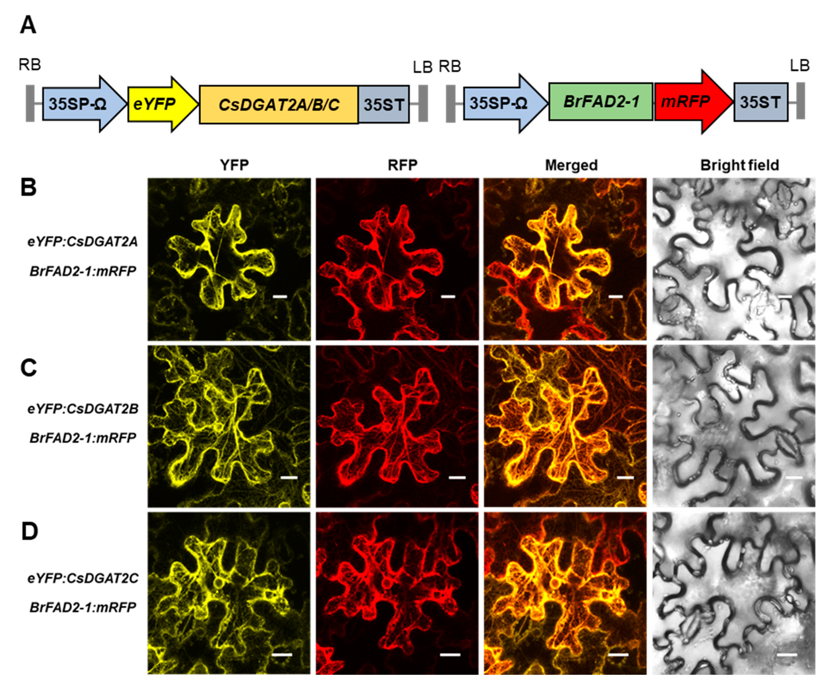
2.2. Both DGAT1 and DGAT2 Are Membrane-Bound Proteins Localized to the Endoplasmic Reticulum (ER)

DGAT1 and DGAT2 are endoplasmic reticulum (ER) membrane-bound proteins [17]. The ER retrieval motif of CsDGAT1 is YYHDL (Supplementary Figure S2A), similar to those in Arabidopsis and tung tree VfDGAT1s [17]. The ER retrieval motif of CsDGAT2 is LQLNI (Supplementary Figure S2B), which differs from the LELKI motif of AtDGAT2 and the LKLEI motif of VfDGAT2 [17,41]. Previously, agroinfiltration in Nicotiana benthamiana leaf epidermal cells using constructs expressing enhanced yellow fluorescent protein (eYFP)-fused CsDGAT1s revealed that the CsDGAT1s were localized to the ER [11]. To analyze the subcellular localization of CsDGAT2s, we co-infiltrated Agrobacterium carrying eYFP:CsDGAT2, Brassica rapa BrFAD2-1:mRFP (an ER marker), and tobacco bushy stunt virus p19 as a gene-silencing suppressor into 5-week-old N. benthamiana leaves. At 48 h after infiltration, we observed eYFP and mRFP fluorescence signals in the ER, indicating that all CsDGAT2s localize to the ER (Figure 1). Quantification of the co-localized signal intensity in the fluorescence images using Pearson’s correlation coefficient supported the localization of all three CsDGAT2s to the ER (Supplementary Figure S3).

2.3. CsDGAT1 and CsDGAT2 Have Seed-Specific Expression, but the Expression Level of CsDGAT1 Was Higher

To determine the expression pattern and the transcript level for each homoeolog of CsDGAT1 and CsDGAT2 in camelina, we cloned all homoeologs and designed homoeolog-specific primers. However, despite using homoeolog-specific primers, we were unable to obtain homoeolog-specific amplification. Since camelina has more than 97% identity among homoeologs, designing efficient homoeolog-specific primers is difficult. According to the method of Hayashi et al. [42], we attempted to confirm homoeolog-specific amplification using primers in which one or more mismatch sequences were replaced at the 3′ end region, but the expression level was too low. Even after several trials using different 3′-end mismatch sequences, the homoeolog-specific reverse-transcription quantitative PCR (RT-qPCR) was unsuccessful. Therefore, we measured the total transcript level for each set of three homoeologs, but not that of the individual homoeologs. The transcript levels of the CsDGAT1, CsDGAT2, CsDGAT3, and CsPDAT1 genes in flowers, leaves, stems, roots, cotyledons, and developing seeds at stage 1 (DS1), DS2, DS3, DS4, and DS5 are shown in Supplementary Figure S4. The CsDGAT1s, CsDGAT2s, and CsDGAT3s showed seed-specific expression, and their expression pattern in developing seeds showed a bell-shaped pattern similar to that of the genes related to seed oil biosynthesis and accumulation (Supplementary Figure S4A–C). In developing seeds, the highest expression levels of CsDGAT1 was 6.5-fold higher than that of CsDGAT2 but was only 32% that of CsDGAT3. CsDGAT3 expression was much higher than that of the other genes in all tissues. CsDGAT2 expression peaked at DS3, which differed from the expression of CsDGAT1 and CsDGAT3, which peaked at DS4 (Supplementary Figure S4A–C). These results were similar to those reported previously [36]. In contrast to the seed-specific expression of CsDGATs, the expression level of CsPDAT1s, which are also involved in seed oil biosynthesis, was highest in leaves and cotyledons, and the expression of CsPDAT1s in developing seeds did not show a bell-shaped pattern similar to those of other genes related to seed oil biosynthesis and accumulation (Supplementary Figure S4D). Taken together, these results indicate that CsDGAT1s, CsDGAT2s, and CsDGAT3s are expressed mainly in developing seeds, and the expression level of CsDGAT1s is higher than that of CsDGAT2s, but lower than that of CsDGAT3s.

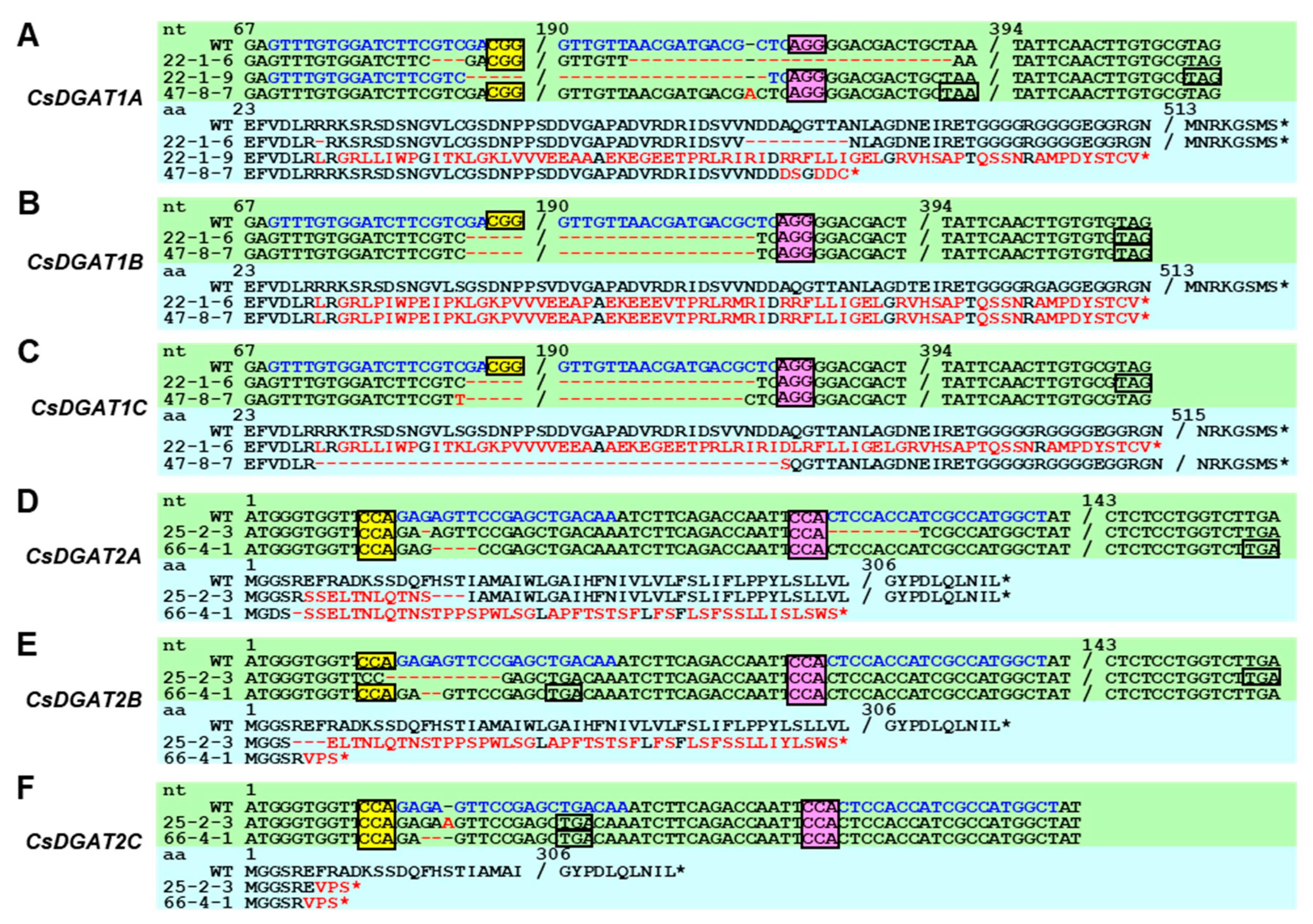
2.4. Csdgat1 KO Lines and Csdgat2 KO Lines Were Obtained Using CRISPR/Cas

To generate CsDGAT1 and CsDGAT2 KO camelina plants, we used the pHEE401E-Red vector, which can accommodate the insertion of two guide RNAs [36]. We selected two optimal gRNA sequences to knock out all CsDGAT1 and CsDGAT2 homoeologs, respectively, using Cas Designer [43] according to the criteria described in Section 4 (Supplementary Table S2). The dual gRNAs for CsDGAT1s or CsDGAT2s were inserted into pHEE401E-Red, and the complete vectors were named pRedEDD1 (EC1.2P:zCas9 + Dual gRNA for DGAT1) and pRedEDD2, respectively (Supplementary Figure S5). Camelina plants were transformed using the floral dip method with the Agrobacterium strain GV3101, carrying pRedEDD1 or pRedEDD2. We selected 67 and 172 T1 camelina seeds for EDD1 and EDD2, respectively, emitting orange–red fluorescence (Supplementary Figure S6A,B). The transformation efficiencies of EDD1 and EDD2 camelina plants were approximately 0.25% and 0.37%, respectively (Supplementary Table S3).
To determine the insertion–deletion mutant (indel) frequency of transformed camelina, we conducted homoeolog-specific PCR followed by Sanger sequencing. First, we designed CsDGAT1 and CsDGAT2 homoeolog-specific primers to determine the indel frequency, and the sequencing results of the PCR products using WT genomic DNA demonstrated that they amplified the individual homoeologs. Next, we performed homoeolog-specific PCR using the EDD1 and EDD2 plants (Supplementary Figure S6C,D), followed by Sanger sequencing of the PCR products. Finally, we analyzed the homoeolog-specific PCR-based Sanger sequencing results using two web-based tools, TIDE and DsDecodeM [44,45]. Our results showed that in the 67 T1 EDD1 lines, only five individuals had over 15% indel frequency in a single homoeolog (Supplementary Table S4). Only EDD1#22 had over 15% indel frequency in all three homoeologs of CsDGAT1, whereas EDD1#10, #45, #47, and #49 had over 15% indel frequency in one or two homoeologs of CsDGAT1. To obtain a complete Csdgat1 KO camelina line, we selected five EDD1 T1 lines for T2 generation by self-fertilization. In the 172 T1 EDD2 lines, 34 individuals had over 15% indel frequency in a single homoeolog (Supplementary Table S5). Compared to the EDD1 lines, there were many more gene-edited EDD2 lines (8.5% vs. 21.1%), and the proportion of lines with over 15% indel frequency in all three homoeologs was much higher (20.0% vs. 58.8%). In particular, EDD2#25 and EDD2#66 were predicted to be complete CsDGAT2 KO lines. Therefore, we chose the 12 EDD2 T1 lines (EDD2#9, #24, #25, #26, #42, #44, #59, #66, #83, #106, #137, and #147) with high indel frequencies in all CsDGAT2 homoeologs as the T2 generation.
The indel patterns and zygosities of EDD1 and EDD2 T2 lines, except for the lines that were confirmed to be unedited at all, are listed in Table 1 and Table 2. Deletions of 120–121 nt and 36–41 nt, spanning the sgRNA1 and sgRNA2 sites, were found in EDD1 and EDD2 lines, respectively. Interestingly, cases of co-occurrence of indels at two sgRNA sites on the same haploid gene were identified in the T2 descendants of EDD1#10 and #22 and those of EDD2#25 and #137 (Table 1 and Table 2). However, all of these cases were expected to result in missense mutations. Overall, there were a large number of indel patterns that were expected to cause missense mutations. In the EDD1 T2 generation, we did not find a complete Csdgat1 KO line. EDD1#22-1 and #22-4, which had a missense mutation in only the haploid of CsDGAT1A and nonsense mutations in the other CsDGATs (aa′bbcc; lowercase letters and ′ indicate a nonsense mutation and a missense mutation, respectively), were expected to have the lowest CsDGAT1 activity among the EDD1 T2 descendants (Table 2). In the EDD2 T2 generation, complete Csdgat2 KO lines were found in the T2 descendants of EDD2 #42, #59, and #66 (Table 2).

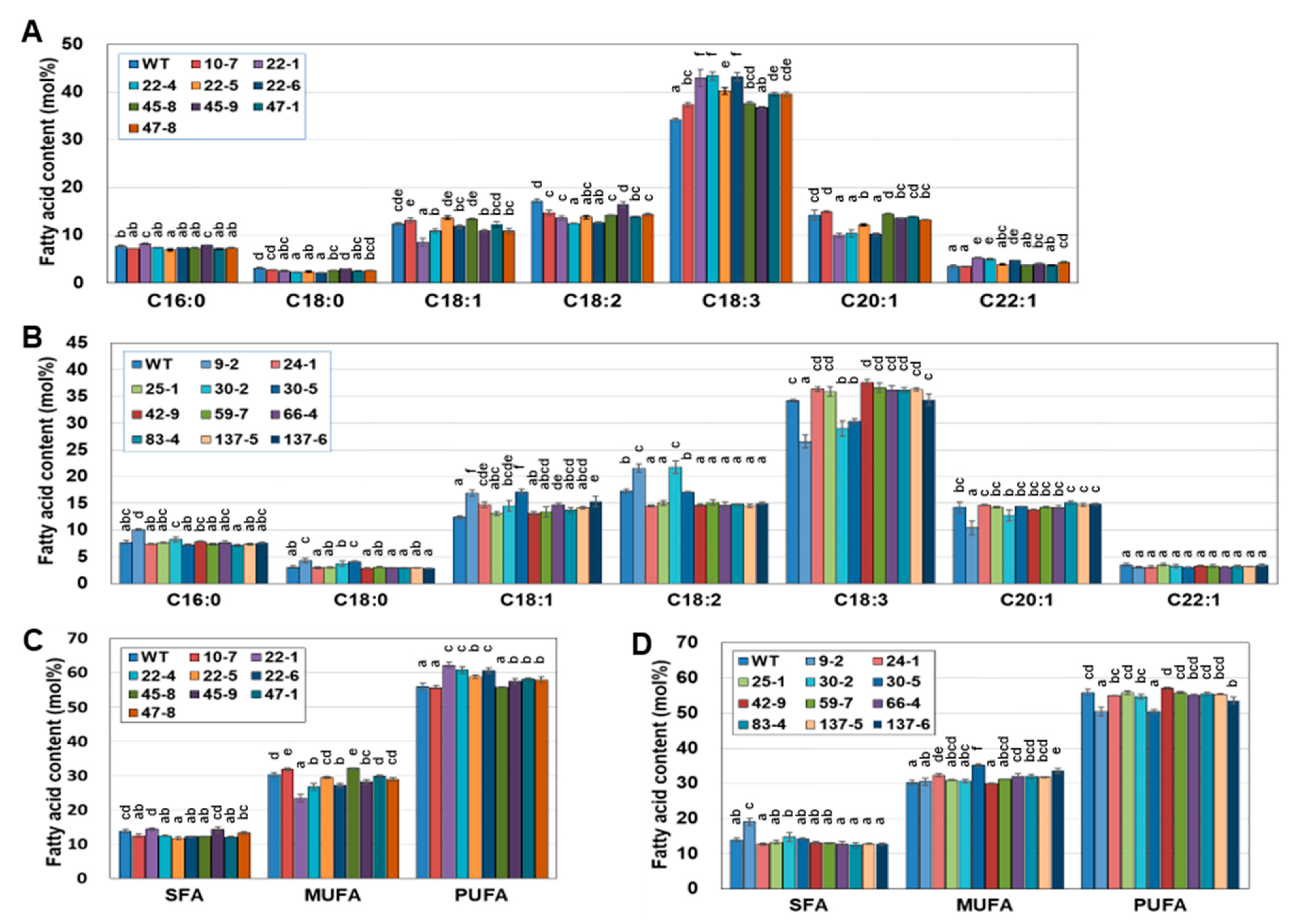
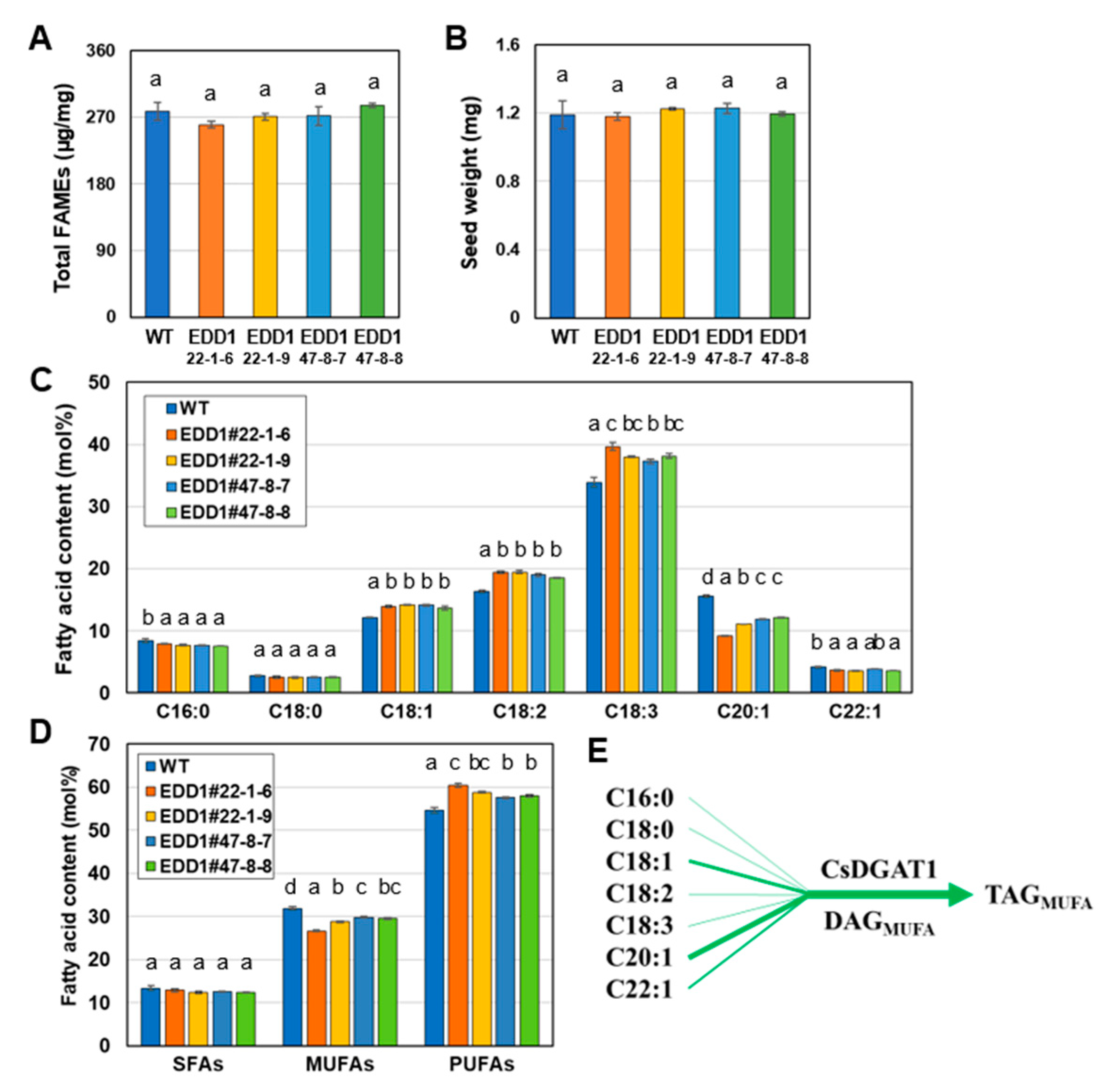
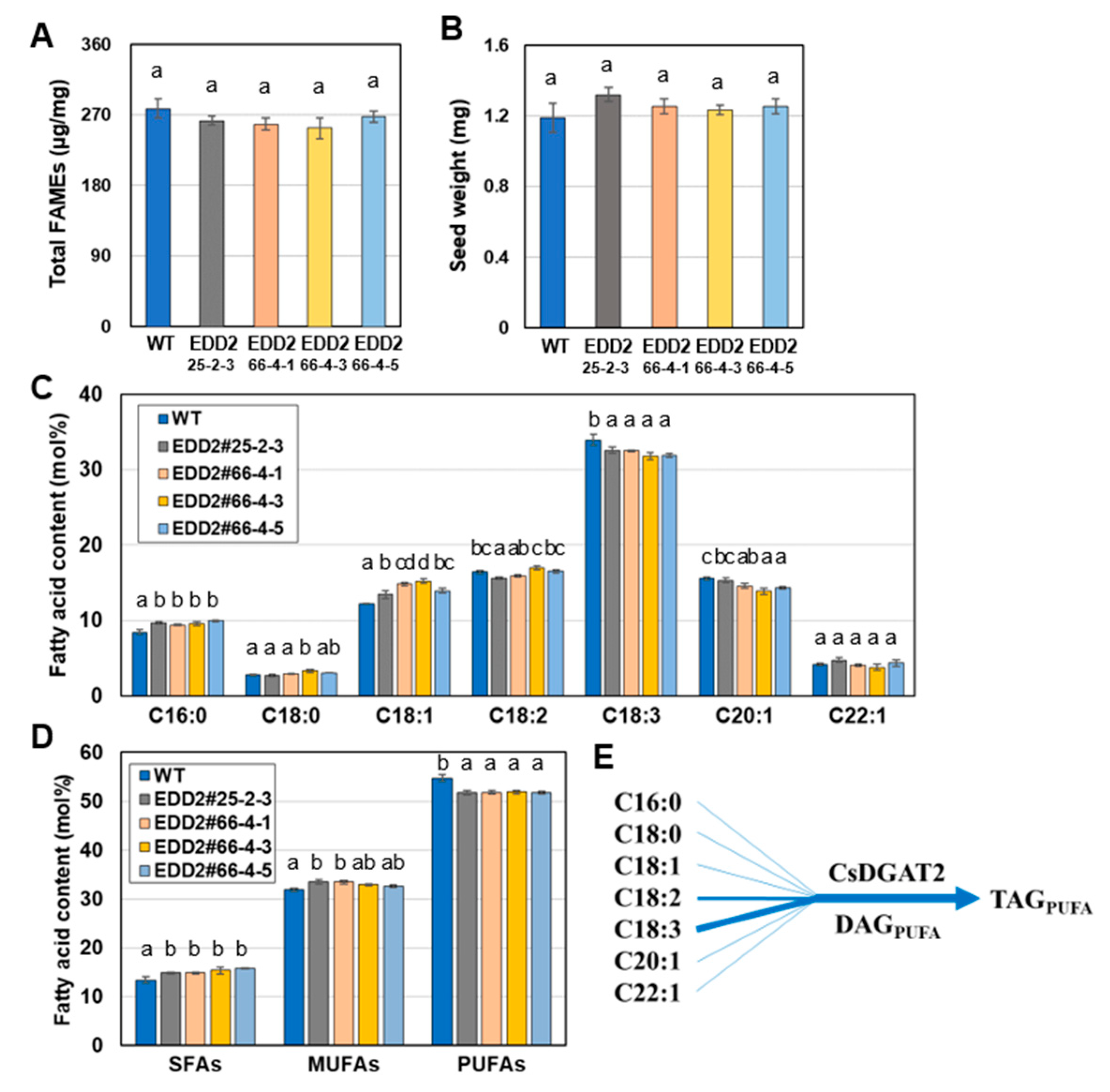
2.5. EDD1 and EDD2 Seeds Have Changed Fatty Acid Content Compared to WT Seeds

To observe how the mutations in CsDGAT1 and CsDGAT2 caused changes in the fatty acid composition of seeds, we analyzed the seed fatty acids from EDD1 and EDD2 T2 lines. Nine EDD1 T2 lines and 11 EDD2 T2 lines were used for seed fatty acid analysis. EDD1#10-7, #22-1, #22-4, #22-5, #22-6, #45-8, #45-9, #47-1, and #47-8; EDD2#9-2, #24-1, #25-1, #30-2, #30-5, #42-9, #59-7, #66-4, #83-4, #137-5, and #137-6. The changes in seed fatty acid composition of the EDD1 T2 plants are shown in Figure 2A. The C16:0 contents of EDD1 T2 plants were generally similar to those of the WT. The C18:2 contents of the EDD1 T2 lines were generally lower than that of the WT, while the C18:1 and C20:1 contents of some EDD1 T2 lines were lower than those of the WT. By contrast, the C18:3 contents of most EDD1 T2 lines were increased compared to that of the WT. EDD1#45-9 was an exception, in that the C18:2 and C18:3 contents were not significantly changed. Among the EDD1 T2 lines, #22-4 had a C18:2 content of 12.4 mol%, which was a decrease of 4.8 mol% compared to WT, and a C18:3 content of 43.4 mol%, an increase of 9.2 mol% compared to WT. The C20:1 and erucic acid (C22:1) contents of EDD1 T2 lines were similar to those of the WT, except for the T2 descendants of #22 (#22-1, -4, -5, and -6), which generally showed a decrease in the C20:1 content and an increase in the C22:1 content.
Figure 2C showed the comparison results by grouping saturated fatty acids (SFAs), monounsaturated fatty acids (MUFAs), and PUFAs. In the seed oil from EDD1 T2 plants, the SFA contents were significantly decreased compared to the WT, except for #10-7, #45-9, and #47-8. Compared to the MUFA content of the WT, that of #47-1 and #47-8 was similar, whereas that of #10-7 and #45-8 was significantly increased, and that of other lines was significantly decreased. Compared to the PUFA content of the WT, those of all EDD1 lines were increased, except for #10-7 and #45-8. However, most of the individuals with a significantly increased C18:3 content had a decreased C18:2 content (Figure 2A); thus, the PUFA content of all EDD1 lines slightly increased compared to that of the WT. EDD1 #22-1 had a PUFA content increased by 6.3 mol% and a MUFA content decreased by 6.8 mol% compared to the WT, showing the greatest fatty acid changes.
The changes in the seed fatty acid composition of the EDD2 T2 lines are shown in Figure 2B. Compared to the palmitic acid (C16:0) and stearic acid (C18:0) contents of the WT, only #9-2 increased. The C18:1 content was similar or increased compared to the WT, and the increase in #9-2 and #30-5 was remarkable among the EDD2 T2 lines. Compared to the WT, the C18:2 content was increased in #9-2 and #30-2, similar in #30-5, and decreased in the other lines. The C18:3 content was decreased in #9-2, #30-2, and #30-5 and significantly increased only in #42-9; the rest were similar to the WT. The C20:1 content decreased only in #9-2, and the rest were similar to the WT. The C22:1 content of EDD2 T2 lines was almost the same as that of the WT. Figure 2D shows the SFA, MUFA, and PUFA contents of seed oil from EDD2 T2 plants. The SFA content of seed oil from EDD2 T2 plants increased only in #9-2 compared to that of the WT. The MUFA content was increased in #24-1, #30-3, and #137-6, while the rest were similar to that of the WT. The PUFA contents were all similar, except in #9-2 and #137-6, in which they were decreased compared to the WT. Overall, the seed fatty acid composition of EDD1 T2 lines differed significantly among individuals, whereas that of EDD2 T2 lines was relatively similar in all individuals, except for #9-2, #30-2, and #30-5, which changed dramatically compared to the WT. Taken together, the seed fatty acid composition of the EDD1 T2 lines was different from that of the WT and slightly differed among each other. In contrast, the seed fatty acid composition of the EDD2 T2 lines, except for the #9-2, #30-2, and #30-5 lines, which demonstrated dramatic changes, showed an insignificant difference from that of the WT.
Next, we investigated seed fatty acid composition, seed oil content, and seed weight in #22-1-6 (aabbcc), #22-1-9 (a′a′bbcc), and #47-8-7 and #47-8-8 (aabbc′c′) for the EDD1 T3 lines (Figure 3). The seed oil content and seed weights of EDD1 T3 lines were similar to those of the WT (Figure 3A,B). The seed fatty acid composition of the EDD1 T3 lines showed an increase in C18:1, C18:2, and C18:3 contents, and a decrease in C16:0 and C20:1 contents compared to those of the WT (Figure 3C). In particular, #22-1-6 had the highest C18:1 content and the lowest C20:1 content among the EDD1 T3 lines. This may be because #22-1-6 (aabbcc) is a complete Csdgat1 KO line, different from the other EDD1 T3 lines (a′a′bbcc and aabbc′c′). Comparing Figure 2A and Figure 3C, an increased C18:3 content and a decreased C20:1 content were similar between the EDD1 T2 and T3 lines. The C18:2 and C18:3 contents were decreased in the EDD1 T2 lines but increased in the EDD1 T3 lines. Although we noted some differences in seed fatty acid composition between the EDD1 T2 and T3 lines compared to the WT, the contents of SFAs, MUFAs, and PUFAs were similar between the EDD1 T2 and T3 lines compared to those of the WT (Figure 2C and Figure 3D). MUFA and PUFA contents in both EDD1 T2 and T3 lines were decreased and increased, respectively, compared to those of WT. Overall, this suggests that CsDGAT1 contributes less than CsDGAT3 to seed oil accumulation in camelina seeds [36], and CsDGAT1 has a strong preference for C20:1 but a weak preference for C18:3, resulting in the TAG with a high MUFA content (Figure 3E). However, this means that CsDGAT1 contributes a certain degree in TAG biosynthesis because the DGATs share functional redundancy in TAG biosynthesis.
As shown in Figure 2B, EDD2#9-2, #30-2, and #30-5 had a lower C18:3 content than other EDD2 T2 lines and the WT. However, they also had a lower seed weight and an abnormal seed morphology, with seeds that were flat, curved, twisted, and tiny compared to those of other EDD2 lines and WT (Supplementary Figure S7). The cause of this reduced seed oil content and abnormal seed morphology was difficult to determine. Therefore, we excluded them from our investigation of seed phenotype in the T3 generation and assessed the seed fatty acid composition, seed oil content, and seed weight in the EDD2 T3 lines #25-2-3 (a′a′bbcc), #66-4-1, #66-4-3, and #66-4-5 (aabbcc) (Figure 4). These EDD2 T3 lines had seed oil contents similar to those of the WT (Figure 4A) and seed weights slightly but non-significantly higher than those of the WT (Figure 4B). The seed fatty acid composition showed a slight difference between the EDD2 T3 lines and the WT: both C16:0 and C18:1 contents were higher in the EDD2 T3 lines, while the C18:3 content was lower (by up to 1.9 mol%). The C18:2 and C20:1 contents showed a slight but statistically significant decrease in some lines (Figure 4C). The SFA and MUFA contents of EDD2 T3 lines were slightly higher, and the PUFA contents were lower than those in the WT (Figure 4D). They also indicate that CsDGAT2s have a strong preference for C18:3, resulting in a TAG with high PUFA content (Figure 4E). Together, these results suggest that CsDGAT2s are less crucial than CsDGAT3s for seed oil accumulation in camelina [36], but still contribute somewhat to the fatty acid composition.

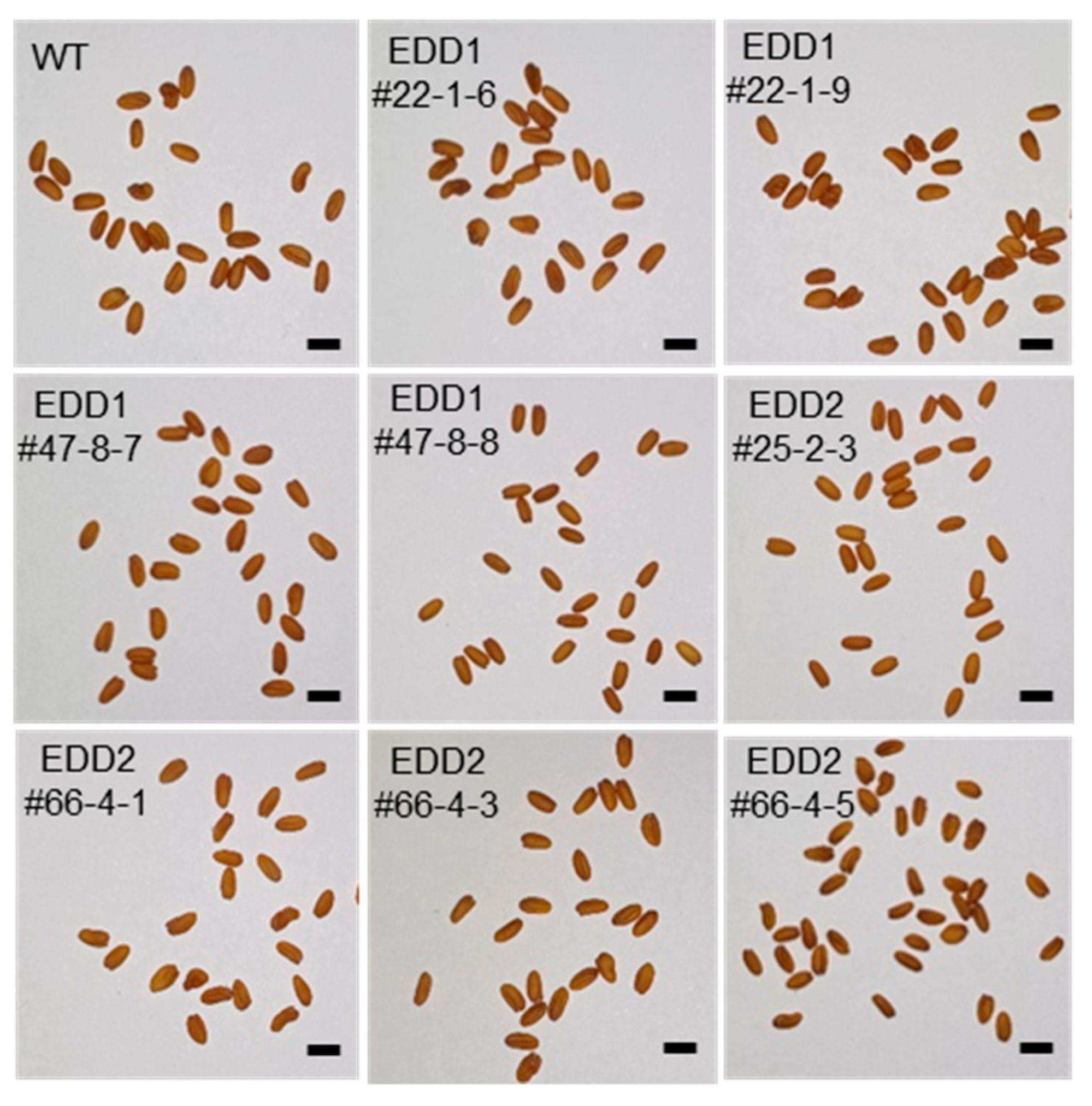
2.6. EDD1 and EDD2 Lines Were Phenotypically Similar to WT

Figure 5 shows the nucleotide and deduced amino acid changes of CsDGAT1s and CsDGAT2s from the EDD1 and EDD2 lines. For example, 3-nt and 27-nt deletions at each sgRNA target site in the CsDGAT1A of EDD1#22-1-6 caused 1-aa and 9-aa deletions, respectively. Meanwhile, a 121-nt deletion in the CsDGAT1A of EDD1#22-1-9 and a 1-nt insertion in the CsDGAT1A of #47-8-7 resulted in the early termination of translation at the 100-aa and 75-aa positions, respectively (Figure 5A). The CsDGAT1B of EDD1#22-1-6, #22-1-9, and #47-8 had a 121-nt deletion at the target site from sgRNA1 to sgRNA2, which resulted in the early termination of translation with altered amino acid residues at the 100-aa position (Figure 5B). The CsDGAT1C of EDD1#22-1-6 had the same 121-nt deletion as CsDGAT1B in the three lines as described above, resulting in the premature termination of translation. The CsDGAT1C of #47-8-7 had a 120-nt deletion, resulting in a 40-aa-deletion in CsDGAT1C (Figure 5C). In CsDGAT2A, EDD2#25-2-3 had 1-nt+8-nt deletion at each sgRNA site within a haploid, resulting in a 3-aa deletion and a change in 11 aa compared to the WT, but not early termination. EDD2#66-4-1 had a 4-nt deletion, resulting in early translation termination with aberrant amino acid residues (Figure 5D). The proteins encoded by CsDGAT2B in EDD2#25-2-3 and #66-4-1 were truncated due to a 10-nt deletion and a 2-nt deletion, respectively (Figure 5E). The CsDGAT2C of EDD2#25-2-3 and #66-4-1 had a 1-nt insertion and a 2-nt deletion, respectively (Figure 5F). The changes in the nucleotide sequences of CsDGAT1s and CsDGAT2s were also shown by Sanger sequencing (Supplementary Figures S8 and S9).
We investigated other seed phenotypes, such as seed morphology and germination rate, in the same lines analyzed in Figure 3 and Figure 4: EDD1#22-1-6, #22-1-9, #47-8-7, and #47-8-8; and EDD2#25-2-3, #66-4-1, #66-4-3, and #66-4-5. Their seed morphology was normal, similar to that of the WT (Figure 6). Some abnormal seeds were observed in the EDD1 and EDD2 lines, and even in the WT, but most seeds were normal. We observed whether there was any change in seed germination rate due to the KO of CsDGAT1 or CsDGAT2. Interestingly, all EDD1 and EDD2 lines, even EDD3#1-5-7 lines with about 19% decreased seed oil content [36], showed a 100% germination rate at 1 day after imbibition (DAI), identical to the WT. Thus, even though the functions of CsDGAT1 and CsDGAT2 were disrupted in the EDD1 and EDD2 lines, respectively, this did not affect their germination rates.

3. Discussion

As mentioned above, excellent agronomic traits such as cold tolerance, drought tolerance, low fertilizer requirements, and a short crop cycle make camelina an emerging and promising oilseed crop for industrial applications [24], whose triacylglycerol biosynthetic pathway and genes involved in seed oil accumulation have been gradually unveiled. Although Arabidopsis and camelina are closely related species belonging to the Brassicaceae family, the expression levels of the DGAT genes differ. DGAT1 is the main acyltransferase contributing to the seed oil content in Arabidopsis [7]. However, we previously showed that in camelina, the expression level of DGAT3 is the highest among the DGATs, and the Csdgat3 mutant camelina has reduced seed oil content, suggesting that CsDGAT3 is mainly involved in seed oil accumulation, more so than the other CsDGATs in this species [36]. Therefore, we wondered to what extent CsDGAT1 and CsDGAT2 are involved in seed oil accumulation, as well as which fatty acids CsDGAT1 and CsDGAT2 prefer during TAG biosynthesis. In this study, we determined that (1) CsDGAT1 and CsDGAT2 in camelina are expressed in a seed-specific manner, but have lower expression levels compared to CsDGAT3; (2) the three CsDGAT2 proteins localize to the ER; (3) the levels of seed oil in EDD1 and EDD2 lines were similar to those of the WT; (4) EDD1 lines had higher C18:3 and lower C20:1 contents in seed oil than in the WT; and (5) the seed fatty acid composition in the EDD2 lines was slightly reduced in the C18:3 content compared to that in the WT. These results indicate that CsDGAT1 and CsDGAT2 contribute less to seed oil content than CsDGAT3, but have sufficient activity to alter the seed fatty acid composition. Although these results suggest that CsDGAT1 and CsDGAT2 make only minor contributions to seed oil content, their contributions were sufficiently high that the seed oil content in Csdgat3 KO camelina plants was reduced by only 18.8% because of the functional redundancy of the three DGATs and PDATs involved in seed oil biosynthesis [36]. Therefore, CsDGAT1 and CsDGAT2 play minor but appropriate roles in seed oil biosynthesis. These findings show that the activity of seed oil-forming DGATs in camelina differs from that in Arabidopsis.
It is known that both DGAT1 and DGAT2 are ER-membrane-bound proteins and have the function of the TAG biosynthesis in the ER. CsDGAT1s have already been confirmed to be localized in the ER [11]. Therefore, in this study, it was important to determine whether CsDGAT2s were also localized in the ER. As shown in Figure 1, CsDGAT2s were confirmed to be localized and retained in the ER. According to McCartney et al. [46], the ER retrieval motif of plant ER membrane-bound proteins has a hydrophobic amino acid residue at the first and fifth positions and a K, R, D, or E residue at the fourth position (Φ-X-X-K/R/D/E-Φ, Φ hydrophobic amino acid residue). The ER retrieval motif of CsDGAT1 is YYHDL, similar to that of AtDGAT1 (Supplementary Figure S2A), and the ER retrieval motif of AtDGAT2 and VfDGAT2 is consistent with the LELKI consensus sequence [37]. In contrast, upon examining the amino acid sequences of DGAT2 of numerous plant species, we found that most of the fourth amino acid residues were K/R/E, but some were L/V. As in the case of CsDGAT2, the ER retrieval motif of Brassica napus BnDGAT2a and BnDGAT2 was LQLNI, which did not match the consensus motif [10,37]. McCartney et al. [46] showed that AtFAD2 with K/R/D/E at the fourth residue of the ER retrieval motif localized to the ER, but FAD2 with A or Q as the fourth residue did not. This suggests that having K/R/D/E as the fourth residue may not be an absolute requirement for the ER retrieval motif and that other, unknown factors affect the ER retention function of the ER retrieval motif.
In this study, we did not measure the transcript levels of each homoeolog of CsDGAT1 and CsDGAT2, but rather the overall transcript levels of CsDGAT1s and CsDGAT2s. To obtain amplifications specific to homoeologs with very high identity (>97%), it is thought that it would be helpful to design primers specific to regions of low identity, such as the 3′-UTR, or to combine the methods used by Hayashi et al. [42]. The expression levels of CsDGAT1 and CsDGAT2 were significantly lower than that of CsDGAT3 (Supplementary Figure S4A–C). This implies that while CsDGAT3 plays a critical role in determining seed oil content [36], the contribution of CsDGAT1 and CsDGAT2 to TAG biosynthesis might be lower than that that of CsDGAT3. Indeed, the seed oil content was not lower in the EDD1 lines than in the WT (Figure 3A), suggesting that CsDGAT1 has only a minor effect on seed oil biosynthesis, and the loss of its function can be compensated for by other CsDGATs. In Arabidopsis, AtDGAT1 is the most dominant DGAT and the dgat1 mutant (AS11) has a 25% reduced seed oil content [7,13]. Meanwhile, in camelina, CsDGAT3 is dominant [36]. At its highest expression level, CsDGAT2 expression is only 15% that of CsDGAT1 and 5% that of CsDGAT3 (Supplementary Figure S4B). Therefore, CsDGAT2 was predicted to have a smaller effect on seed oil biosynthesis than other CsDGATs. As expected, the seed oil content of the EDD2 lines did not differ from that of the WT (Figure 4A).
In Arabidopsis, AtDGAT1 has an acyl preference mainly for C18:1 but little preference for C18:3, whereas AtDGAT2 showed a preference for C18:1, C18:2, and C18:3 [41]. In camelina, CsDGAT1 showed a similar preference for all fatty acids constituting seed oil; in contrast, CsDGAT2 showed a much higher specificity for C18:3 than for other fatty acids in yeast heterologously expressing CsDGAT1 or CsDGAT2 [34]. However, it does not seem to occur in plant definitely because di-6:0-DAG, which does not occur naturally in plant seeds, was used as an acyl acceptor. When diverse acyl acceptors and C18 unsaturated acyl-CoAs (C18:1-, C18:2-, and C18:3-CoAs) were provided, CsDGAT1 preferred DAGs with relatively low degrees of unsaturation (di-C18:1-DAG and C16:0/C18:2-DAG) as the acyl acceptors and preferred all acyl-CoAs except when the acyl acceptors provided were di-C18:1-DAG and C18:1-CoA. Under the same condition, CsDGAT2 preferred DAGs with high degrees of unsaturation (di-C18:2-DAG and di-C18:3-DAG) as the acyl acceptors, with strongest preference for C18:3, followed by C18:2 [34]. CsDGAT1 may generate TAGs with 4 or 5 degrees of unsaturation (54:4 to 54:5), whereas CsDGAT2 may biosynthesize TAGs with 6 to 9 degrees of unsaturation (54:6 to 54:9). These data suggest that CsDGAT1 functions to increase the C18:1 and C18:2 content and that CsDGAT2 functions to increase the C18:3 content. These reports provide a good explanation for the increased C18:3 content in the Csdgat1 KO camelina and the decreased C18:3 content in the Csdgat2 KO camelina.
The EDD1 lines would retain the activities of CsDGAT2 and CsDGAT3 in regard to seed oil biosynthesis, and EDD2 lines would retain those of CsDGAT1 and CsDGAT3. CsDGAT2 was more highly expressed in developing seeds compared to CsPDAT1 (Supplementary Figure S4B,D), indicating that CsDGAT2 participates more in seed oil biosynthesis in the EDD1 lines, compensating for the absence of CsDGAT1 activity. However, given that the seed fatty acid composition of the EDD2 lines was unaltered, the activity of CsDGAT2 in the EDD1 lines is likely negligible. Therefore, it can be assumed that the seed fatty acid compositions of the EDD1 and EDD2 lines are dependent on the acyl preferences of CsDGAT3 (in the EDD1 lines) and of CsDGAT1 and CsDGAT3 (in the EDD2 lines), respectively. We predicted that in the EDD1 lines, the content of C18:3, which CsDGAT3 prefers, would be increased. Indeed, the EDD1 lines had an increased C18:3 content, but the C18:1 and C18:2 contents were also increased, which differed from the situation in the T2 seeds (Figure 2A and Figure 3C). Furthermore, the decreased C20:1 contents of the T3 seeds compared to the WT seeds suggested that CsDGAT1 prefers C20:1 as an acyl substrate (Figure 3C). The fact that the C20:1 contents in the EDD1 lines were decreased to a greater extent than those in the EDD2 lines [36] (Figure 3C) implies that CsDGAT1 contributes more to increasing the C20:1 content in seed oil than CsDGAT3 does. In the EDD2 lines, we expected to see a reduced C18:3 content because DGAT2 has a strong preference for C18:3 in plants [34,41]. However, there was only a slight alteration in the seed fatty acid composition, especially in the C18:3 content (Figure 4). This suggests that the activity of CsDGAT2 is much weaker than that of other CsDGATs, and only slightly affects fatty acid composition.
We observed reduced seed oil content and abnormal seed morphology in EDD2#9-2, #30-2, and #30-5 (Supplementary Figure S7). Initially, we thought that these phenotypes were the result of CsDGAT2 KO. Given the low expression level of CsDGAT2 (Supplementary Figure S4B) and the 1 to 3 missense mutations in their CsDGAT2 homoeologs (Table 2), it was unclear whether the phenotypes were caused by mutation of CsDGAT2. Furthermore, the fatty acid composition and content of three lines were very different from those of the rest of the EDD2 lines, which have less difference in fatty acid composition and content among them (Figure 2B and Supplementary Figure S6A). As a result, we concluded that there was an unknown problem with these lines, and we could not exclude an off-target effect. This should be investigated further to resolve the discrepancy.
The germination rates of the EDD1, EDD2, and EDD3 lines were all 100% at 1 DAI, similar to that of the WT. Considering that the seed oil content and seed weight of EDD1 and EDD2 lines did not change (Figure 3A,B and Figure 4A,B), this germination rate seems reasonable. In addition, EDD3#1-5-7, which had an 18.8% lower seed oil content, had a germination rate identical to that of the WT. Other studies have reported that the germination rate of plants with reduced seed oil content was similar to that of the WT. The germination of AS11, the Arabidopsis dgat1 mutant, which has a 25% reduced oil content [7,13], was delayed for 6 h compared to the WT, but its germination rate was similar to that of the WT [47].
In conclusion, we analyzed the biosynthetic functions and acyl preferences of CsDGAT1 and CsDGAT2, two seed-specific proteins in camelina, using Csdgat1 and Csdgat2 mutants generated by CRISPR/Cas9. Our results indicate that CsDGAT1 and CsDGAT2 participate in TAG biosynthesis, but their contributions are lower than that of CsDGAT3. Due to their functional redundancy in TAG biosynthesis, their exact contribution ratios were not determined, but the contributions of CsDGAT1 and CsDGAT2 were sufficient to affect the fatty acid composition. CsDGAT1 and CsDGAT2 prefer C20:1 and C18:3, respectively, as an acyl substrate. Collectively, these results showed that the activities of CsDGATs in camelina differ from those in Arabidopsis, although they have a very close phylogenetic relationship. These findings suggest that the overexpression or knockout of CsDGAT1 or CsDGAT2 could be applied to developing tailored oilseed plants, including camelina, with a high MUFA content or a high omega-3 fatty acid content in their seed oil. These findings, applying the diverse characteristics of camelina as an oilseed crop, along with gene editing techniques, are also poised to impact both agricultural and biotechnological industries, while enhancing the sustainability and efficiency of future agriculture. This study suggests that continuous exploration is warranted to fully harness the potential of camelina as a promising oilseed crop.

4. Materials and Methods

4.1. Biological Material

Camelina sativa cv. CAME was used as the wild-type parent for the mutagenesis of CsDGAT1 and CsDGAT2 genes. The camelina seeds were sown in soil at 4 °C for 2 days and then transferred to a culture room at 19 °C under long-day conditions (16-h light/8-h dark).

4.2. Subcellular Localization Analysis Using Nicotiana benthamiana Leaves

Each CsDGAT2 homoeolog was fused to yellow fluorescence protein (YFP) lacking the stop codon at its 5′-end. The expression cassette was constructed from the cauliflower mosaic virus (CaMV) 35S promoter with the 5′-untranslated region leader (Ω) sequence of the tobacco mosaic virus and the 35S terminator using the Golden Gate cloning method [48]. The monomeric red fluorescent protein (mRFP)-fused BrFAD2-1 and the tobacco bushy stunt virus p19 were used as an ER marker and a suppressor of virus-induced gene silencing, respectively [49,50]. After Agrobacterium tumefaciens (strain GV3101) was cultured at 28 °C overnight, the OD600 of Agrobacterium carrying YFP-fused CsDGAT2, mRFP-fused BrFAD2-1, and p19 was adjusted to 0.8, 0.8, and 0.4, respectively, with an infiltration buffer [10 mM 2-(N-morpholino)ethanesulfonic acid (MES) (pH 5.6), 10 mM MgCl2, 200 μM acetosyringone]. Mixed Agrobacterium was infiltrated into 5-week-old N. benthamiana leaves. Agroinfiltrated N. benthamiana leaves were cut off and the fluorescence signal was checked using a TCS SP8 laser scanning confocal microscope (Leica, Germany) 36–48 h after infiltration. YFP and mRFP were excited at 514 nm and 561 nm, respectively, and their fluorescence was observed at 518–553 nm and 573–616 nm, respectively.

4.3. Reverse-Transcription Quantitative PCR Analysis

To determine the expression level of CsDGAT1 and CsDGAT2 homoeologs in camelina, total RNAs were extracted from cotyledons from 5-day-old seedlings, roots from 2-week-old seedlings, stems from 6-week-old plants, leaves from 6-week-old plants, flowers from 7-week-old plants, and developing seeds at stage 1 (DS1), DS2, DS3, DS4, and DS5 using a SpectrumTM Plant Total RNA kit (Sigma-Aldrich, St. Louis, MO, USA). First-strand cDNAs were synthesized using 2 μg of total RNA and the RNA to cDNA EcoDry Premix (Clontech, Mountain View, CA, USA) at 42 °C for 40 min for reverse transcription. Primers for quantitative PCR (qPCR) were designed to specifically amplify each CsDGAT1 and CsDGAT2 homoeolog (Supplementary Table S6) using the method by Hayashi et al. [42], and the qPCR was conducted using 40 cycles of 94 °C for 30 s, 65 °C for 30 s, and 72 °C for 30 s, followed by 72 °C for 5 min, using a real-time PCR instrument StepOnePlus (Applied Biosystems, Waltham, MA, USA). CsActin2 was used as a reference transcript [28].

4.4. Guide RNA Design and Vector Construction

The first exons of the CsDGAT1A homoeolog (GenBank accession No. XM_010417066.2) and CsDGAT2A homoeolog (GenBank accession No. XM_010505546.2) were used as the target sequences for genome editing. The Cas Designer tool (http://rgenome.net/cas-designer/, accessed on 24 June 2024) [43] was employed to select two sgRNAs that matched the sequences of the CsDGAT1 and CsDGAT2 homoeologs. These sgRNAs were chosen based on the following three criteria recommended by Cas Designer: (1) a GC content of 40–60%, (2) an out-of-frame score of over 67 points, and (3) no 1–3 nt mismatches. The information on sgRNAs for CsDGAT1s and CsDGAT2s is listed in Supplementary Table S2.
To induce a mutation in CsDGAT1 and CsDGAT2 homoeologs, we used pHEE401E-Red vector [36], which replaced the hygromycin phosphotransferase gene with the DsRed marker gene, containing EC1.2 promoter-driven Zea mays codon-optimized Cas9 (zCas9) from Streptococcus pyogenes (enhancer:EC1.2P:zCas9). Two sgRNAs were inserted into the vector using the Golden Gate cloning method [36,51]. Briefly, the fragment containing two sgRNAs was twice amplified with different primer sets and pCBC-DT1T2 as a template. The destination vector and the fragment containing two sgRNAs were simultaneously digested with BsaI and ligated together using T4 DNA ligase at 37 °C for 5 h, 50 °C for 5 min, and 80 °C for 10 min. The ligated clones were transformed into Escherichia coli DH5α and the binary vectors harboring two sgRNAs were selected using Sanger sequencing. The completed vectors are depicted in Supplementary Figure S5. The sequences of primers used in this procedure are listed in Supplementary Table S6.

4.5. Camelina Transformation and Selection of Transformants

Agrobacterium tumefaciens (GV3101 strain) cells were transformed with each constructed vector using the freeze–thaw method [52]. Camelina was transformed using a simple floral dip method two or three times at 1-week intervals [30,36,53]. In brief, Agrobacterium GV3101 harboring the binary vector described above was inoculated into 1 L of LB broth containing 50 μg/mL of kanamycin and 50 μg/mL of rifampicin and incubated at 28 °C for 24 h. Transformed seeds that emitted orange-red fluorescence were screened using a green LED and a red filter in the dark [29] and grown on soil under the conditions described in Section 4.1.

4.6. Homoeolog-Specific PCR and Sanger Sequencing–Based Genotyping

Genomic DNA from 4-week-old camelina leaf samples was extracted using a magnetic bead-type Genomic DNA Prep Kit for Plants (BIOFACT, Daejeon, Republic of Korea), following the manufacturer’s instructions. Briefly, about 100 mg of leaf tissue was sampled from three different leaves of one camelina plant and placed in a 2 mL microcentrifuge tube containing 2.3 mm chrome beads (BioSpec Products, Bartlesville, OK, USA). The leaf samples were frozen in liquid nitrogen and ground with a Tissue Lyser (Qiagen, Hilden, Germany). Lysis buffer was added to the 2 mL microcentrifuge tube. Following 13,000 rpm centrifugation at room temperature for 3 min, the supernatant was transferred to a 96-deep-well plate. Isopropanol and magnetic beads were combined and transferred to a 96-deep-well plate. Magnetic beads attached to nucleic acids, proteins, and so on, were separated with a 96 multi-magnet pipet and washed three times in a 96-well round-bottom plate, and the genomic DNA was eluted. Using this DNA as a template, homoeolog-specific PCRs were performed for each individual. PCR was conducted by using 30 cycles of 94 °C for 20 s, 54 °C for 20 s, and 72 °C for 30 s, followed by 72 °C for 5 min. The PCR products were purified with a magnetic bead-type PCR Purification Kit (BIOFACT, Republic of Korea) for Sanger sequencing, following the manufacturer’s instructions. The sequences of homoeolog-specific primers are listed in Supplementary Table S6.
The results (ab1 file) of the homoeolog-specific PCR-based Sanger sequencing from Csdgat1 or Csdgat2 KO camelina plants were analyzed with the web-based tools TIDE (http://shinyapps.datacurators.nl/tide/, accessed on 24 June 2024) [44] or DSDecodeM (http://skl.scau.edu.cn/dsdecode/, accessed on 24 June 2024) [45].

4.7. Fatty Acid Analysis

For rapid analysis of fatty acid composition, about 10 mg of camelina seeds were crushed in a 2 mL microcentrifuge tube containing two to three 2.3 mm chrome beads and 0.5 mL of n-hexane using a Tissue Lyser (Qiagen). After centrifugation, the supernatant was transferred to a new 2 mL microcentrifuge tube and mixed with 0.5 mL of 0.5 M sodium methoxide in methanol. Then, 1 mL of 0.9% (w/v) NaCl was added to the sample, and fatty acid methyl esters (FAMEs) in supernatant were transferred to vials for gas chromatograph (GC). To quantify the camelina seed oil, weighed seed samples were crushed and mixed with 0.5 mL of 5% (v/v) H2SO4 in methanol containing pentadecanoic acid (15:0) as an internal standard. Fatty acids were transmethylated at 85 °C for 90 min, and FAMEs were extracted three times with 1 mL of n-hexane after 1.5 mL of 0.9% (w/v) NaCl was added to the sample and mixed. The FAMEs were dried with nitrogen gas and dissolved in 0.2 mL n-hexane. The fatty acid composition and seed oil content were determined by a GC-2010 plus (Shimadzu, Kyoto, Japan) gas chromatograph with a flame ionization detector (FID) and a 30 m × 0.25 mm (inner diameter) HP-FFAP column (Agilent, Santa Clara, CA, USA), while the oven temperature was increased from 150 °C to 220 °C at 30 °C/min and then held constant for 11 min. Ultrapure nitrogen gas was used as the carrier gas.

4.8. Phylogenetic Analysis and Multiple Alignment

To generate the phylogenetic tree, the deduced amino sequences of DGAT1 and DGAT2 from various plant species were obtained through the Basic Local Alignment Search Tool (BLAST) at the National Center for Biotechnology Information (NCBI) (http://blast.ncbi.nlm.nih.gov/Blast.cgi, accessed on 24 June 2024). The deduced amino sequences of CsDGAT1s and CsDGAT2s were obtained from Kim et al. [11] and the camelina genome database in EnsemblPlants (https://plants.ensembl.org/Camelina_sativa/Info/Index, accessed on 24 June 2024), respectively. The phylogenetic tree was constructed using MEGA6 software (Ver. 6.06) (https://www.megasoftware.net, accessed on 24 June 2024) using the maximum-likelihood method. The deduced amino acid sequences of DGAT1 and DGAT2 were aligned using DNASTAR15 MegAlin software (Ver. 15.1.0) with Clustal W and the resultant multiple alignments were shown using GENEDOC software (Ver. 2.6.002).

4.9. Seed Phenotyping

Seed weight was determined by weighing 100 seeds in triplicate. The germination rate was determined as follows. Seeds were surface-sterilized with 70% (v/v) ethanol for 1 min, and subsequently with a 20% (v/v) concentration of commercial bleach and 0.01% (v/v) Tween-20 for 10 min. One hundred surface-sterilized seeds were placed on a 90 × 20 mm petri dish containing a half-strength MS agar medium without sucrose. After synchronization at 4 °C for 2 days, the seeds were transferred to a 25 °C growth chamber with a 16 h light/8 h dark photoperiod. Three independent plates were checked at 1 day to 5 days after transferring to a 25 °C growth chamber.

Supplementary Materials

The following supporting information can be downloaded at: https://www.mdpi.com/article/10.3390/ijms25136944/s1.

Author Contributions

K.-R.L. and Y.Y. performed sgRNA design, binary vector construction and fatty acid composition analysis; Y.Y. transformed camelina, selected the transgenic seeds and managed the camelina plants from sowing to harvest; K.-R.L., Y.Y., J.L. (Juho Lee) and S.-K.L. analyzed the indel frequency from homoeolog-specific genomic PCR-Sanger sequencing; J.L. (Jihyea Lee) performed the phenotyping of Csdgat1 and Csdgat2 KO camelina seeds; S.K. analyzed the fatty acid contents; C.I. and I.K. collected camelina tissues and conducted quantitative RT-PCR; K.-R.L., M.C.S. and H.U.K. analyzed the data; K.-R.L. wrote the manuscript. All authors have read and agreed to the published version of the manuscript.

Funding

This research was supported by research grants from the Next-Generation BioGreen21 Program (SSAC, Project No. PJ01318501, K.-R.L.) and the Mid-Career Researcher Program of the National Research Foundation of Korea (NRF-2020R1A2C2008175 to H.U.K.; NRF-2021R1A2C1006049 to M.C.S.), Republic of Korea.

Data Availability Statement

The data presented in this study are available on request from the corresponding author.

Acknowledgments

We appreciate the technical advice of Ryeo Jin Kim (Sogang University, Republic of Korea) about subcellular localization and the technical assistance with confocal laser microscopy of Jun Oh (the National Institute of Agricultural Sciences, Republic of Korea).

Conflicts of Interest

The authors declare no conflicts of interest.

References

  1. Kennedy, E.P. Biosynthesis of complex lipids. Fed. Proc. 1961, 20, 934–940. [Google Scholar] [PubMed]
  2. Dahlqvist, A.; Ståhl, U.; Lenman, M.; Banaś, A.; Lee, M.; Sandager, L.; Ronne, H.; Stymne, S. Phospholipid:diacylglycerol acyltransferase: An enzyme that catalyzes the acyl-CoA-independent formation of triacylglycerol in yeast and plants. Proc. Natl. Acad. Sci. USA 2000, 97, 6487–6492. [Google Scholar] [CrossRef] [PubMed]
  3. Ståhl, U.; Carlsson, A.S.; Lenman, M.; Dahlqvist, A.; Huang, B.; Banaś, W.; Banaś, A.; Stymne, S. Cloning and functional characterization of a phospholipid:diacylglycerol acyltransferase from Arabidopsis. Plant Physiol. 2004, 135, 1324–1335. [Google Scholar] [CrossRef] [PubMed]
  4. Ichihara, K.; Takahashi, T.; Fujii, S. Diacylglycerol acyltransferase in maturing safflower seeds: Its influences on the fatty acid composition of triacylglycerol and on the rate of triacylglycerol synthesis. Biochim. Biophys. Acta 1988, 958, 125–129. [Google Scholar] [CrossRef]
  5. Cases, S.; Smith, S.J.; Zheng, Y.W.; Myers, H.M.; Lear, S.R.; Sande, E.; Novak, S.; Collins, C.; Welch, C.B.; Lusis, A.J.; et al. Identification of a gene encoding an acyl CoA:diacylglycerol acyltransferase, a key enzyme in triacylglycerol synthesis. Proc. Natl. Acad. Sci. USA 1998, 95, 13018–13023. [Google Scholar] [CrossRef] [PubMed]
  6. Routaboul, J.M.; Benning, C.; Bechtold, N.; Caboche, M.; Lepiniec, L. The TAG1 locus of Arabidopsis encodes for a diacylglycerol acyltransferase. Plant Physiol. Biochem. 1999, 37, 831–840. [Google Scholar] [CrossRef]
  7. Zou, J.; Wei, Y.; Jako, C.; Kumar, A.; Selvaraj, G.; Taylor, D.C. The Arabidopsis thaliana TAG1 mutant has a mutation in a diacylglycerol acyltransferase gene. Plant J. 1999, 19, 645–653. [Google Scholar] [CrossRef] [PubMed]
  8. Nykiforuk, C.L.; Furukawa-Stoffer, T.L.; Huff, P.W.; Sarna, M.; Laroche, A.; Moloney, M.M.; Weselake, R.J. Characterization of cDNAs encoding diacylglycerol acyltransferase from cultures of Brassica napus and sucrose-mediated induction of enzyme biosynthesis. Biochim. Biophys. Acta Mol. Cell Biol. Lipids 2002, 1580, 95–109. [Google Scholar] [CrossRef]
  9. Xu, J.; Francis, T.; Mietkiewska, E.; Giblin, E.M.; Barton, D.L.; Zhang, Y.; Zhang, M.; Taylor, D.C. Cloning and characterization of an acyl-CoA-dependent diacylglycerol acyltransferase 1 (DGAT1) gene from Tropaeolum majus, and a study of the functional motifs of the DGAT protein using site-directed mutagenesis to modify enzyme activity and oil content. Plant Biotechnol. J. 2008, 6, 799–818. [Google Scholar] [CrossRef]
  10. Cao, H. Structure-Function Analysis of Diacylglycerol Acyltransferase Sequences from 70 Organisms. BMC Res. Notes 2011, 4, 249. [Google Scholar] [CrossRef]
  11. Kim, H.; Park, J.H.; Kim, D.J.; Kim, A.Y.; Suh, M.C. Functional analysis of diacylglycerol acyltransferase 1 genes from Camelina sativa and effects of CsDGAT1B overexpression on seed mass and storage oil content in C. sativa. Plant Biotechnol. Rep. 2016, 10, 141–153. [Google Scholar] [CrossRef]
  12. Torabi, S.; Sukumaran, A.; Dhaubhadel, S.; Johnson, S.E.; LaFayette, P.; Parrot, W.A.; Rajcan, I.; Eskandari, M. Effects of type I Diacylglycerol O-acyltransferase (DGAT1) genes on soybean (Glycine max L.) seed composition. Sci. Rep. 2021, 11, 2556. [Google Scholar] [CrossRef] [PubMed]
  13. Katavic, V.; Reed, D.W.; Taylor, D.C.; Giblin, E.M.; Barton, D.L.; Zou, J.; Mackenzie, S.L.; Covello, P.S.; Kunst, L. Alteration of seed fatty acid composition by an ethyl methanesulfonate-induced mutation in Arabidopsis thaliana affecting diacylglycerol acyltransferase activity. Plant Physiol. 1995, 108, 399–409. [Google Scholar] [CrossRef] [PubMed]
  14. Lardizabal, K.D.; Mai, J.T.; Wagner, N.W.; Wyrick, A.; Voelker, T.; Hawkins, D.J. DGAT2 Is a New Diacylglycerol Acyltransferase Gene Family: Purification, cloning, and expression in insect cells of two polypeptides from mortierella ramanniana with diacylglycerol acyltransferase activity. J. Biol. Chem. 2001, 276, 38862–38869. [Google Scholar] [CrossRef] [PubMed]
  15. Zhang, M.; Fan, J.; Taylor, D.C.; Ohlrogge, J.B. DGAT1 and PDAT1 acyltransferases have overlapping functions in Arabidopsis triacylglycerol biosynthesis and are essential for normal pollen and seed development. Plant Cell 2009, 21, 3885–3901. [Google Scholar] [CrossRef] [PubMed]
  16. Li, R.; Yu, K.; Wu, Y.; Tateno, M.; Hatanaka, T.; Hildebrand, D.F. Vernonia DGATs can complement the disrupted oil and protein metabolism in epoxygenase expressing soybean seeds. Metab. Eng. 2012, 14, 29–38. [Google Scholar] [CrossRef]
  17. Shockey, J.; Gidda, S.; Chapital, D.; Kuan, J.; Dhanoa, P.; Bland, J.; Rothstein, S.; Mullen, R.; Dyer, J. Tung tree DGAT1 and DGAT2 have nonredundant functions in triacylglycerol biosynthesis and are localized to different subdomains of the endoplasmic reticulum. Plant Cell 2006, 18, 2294–2313. [Google Scholar] [CrossRef] [PubMed]
  18. Kroon, J.T.; Wei, W.; Simon, W.J.; Slabas, A.R. Identification and functional expression of a type 2 acyl-CoA:diacylglycerol acyltransferase (DGAT2) in developing castor bean seeds which has high homology to the major triglyceride biosynthetic enzyme of fungi and animals. Phytochemistry 2006, 67, 2541–2549. [Google Scholar] [CrossRef]
  19. Burgal, J.; Shockey, J.; Lu, C.; Dyer, J.; Larson, T.; Graham, I.; Browse, J. Metabolic engineering of hydroxy fatty acid production in plants: RcDGAT2 drives dramatic increases in ricinoleate levels in seed oil. Plant Biotechnol. J. 2008, 6, 819–831. [Google Scholar] [CrossRef]
  20. Saha, S.; Enugutti, B.; Rajakumari, S.; Rajasekharan, R. Cytosolic triacylglycerol biosynthetic pathway in oilseeds. molecular cloning and expression of peanut cytosolic diacylglycerol acyltransferase. Plant Physiol. 2006, 141, 1533–1543. [Google Scholar] [CrossRef]
  21. Hernández, M.L.; Whitehead, L.; He, Z.; Gazda, V.; Gilday, A.; Kozhevnikova, E.; Vaistij, F.E.; Larson, T.R.; Graham, I.A. A cytosolic acyltransferase contributes to triacylglycerol synthesis in sucrose-rescued Arabidopsis seed oil catabolism mutants. Plant Physiol. 2012, 160, 215–225. [Google Scholar] [CrossRef]
  22. Cao, H.; Shockey, J.M.; Klasson, K.T.; Chapital, D.C.; Mason, C.B.; Scheffler, B.E. Developmental Regulation of Diacylglycerol Acyltransferase Family Gene Expression in Tung Tree Tissues. PLoS ONE 2013, 8, e76946. [Google Scholar] [CrossRef] [PubMed]
  23. Zubr, J. Oil-seed crop: Camelina sativa. Ind. Crops Prod. 1997, 6, 113–119. [Google Scholar] [CrossRef]
  24. Iskandarov, U.; Kim, H.J.; Cahoon, E.B. Camelina: An Emerging oilseed platform for advanced biofuels and bio-based materials: In Plants and BioEnergy; Springer: New York, NY, USA, 2014; pp. 131–140. [Google Scholar]
  25. Berti, M.; Gesch, R.; Eynck, C.; Anderson, J.; Cermak, S. Camelina uses, genetics, genomics, production, and management. Ind. Crop. Prod. 2016, 94, 690–710. [Google Scholar] [CrossRef]
  26. Putnam, D.H.; Budin, J.T.; Field, L.A.; Breene, W.M. Camelina: A Promising Low Input Oilseed. New Crops; Wiley: New York, NY, USA, 1993; pp. 314–322. [Google Scholar]
  27. Kagale, S.; Koh, C.; Nixon, J.; Bollina, V.; Clarke, W.E.; Tuteja, R.; Spillane, C.; Robinson, S.J.; Links, M.G.; Clarke, C.; et al. The emerging biofuel crop Camelina sativa retains a highly undifferentiated hexaploid genome structure. Nat. Comm. 2014, 5, 3706. [Google Scholar] [CrossRef] [PubMed]
  28. Hutcheon, C.; Ditt, R.F.; Beilstein, M.; Comai, L.; Schroeder, J.; Goldstein, E.; Shewmaker, C.K.; Nguyen, T.; Rocher, J.D.; Kiser, J. Polyploid genome of Camelina sativa revealed by isolation of fatty acid synthesis genes. BMC Plant Biol. 2010, 10, 233–247. [Google Scholar] [CrossRef]
  29. Lu, C.; Kang, J. Generation of transgenic plants of a potential oilseed crop Camelina sativa by Agrobacterium-mediated transformation. Plant Cell Rep. 2008, 27, 273–278. [Google Scholar] [CrossRef]
  30. Liu, X.; Brost, J.; Hutcheon, C.; Guilfoil, R.; Wilson, A.K.; Leung, S.; Shewmaker, C.K.; Rooke, S.; Nguyen, T.; Kiser, J.; et al. Transformation of the oilseed crop Camelina sativa by Agrobacterium-mediated floral dip and simple large-scale screening of transformants. In Vitro Cell. Dev. Biol.–Plant 2012, 48, 462–468. [Google Scholar] [CrossRef]
  31. Clough, S.J.; Bent, A.F. Floral dip: A simplified method for Agrobacterium-mediated transformation of Arabidopsis thaliana. Plant J. 1998, 16, 735–743. [Google Scholar] [CrossRef]
  32. Bansal, S.; Durrett, T.P. Camelina sativa: An ideal platform for the metabolic engineering and field production of industrial lipids. Biochimie 2016, 120, 9–16. [Google Scholar] [CrossRef]
  33. Marmon, S.; Sturtevant, D.; Herrfurth, C.; Chapman, K.; Stymne, S.; Feussner, I. Two Acyltransferases Contribute Differently to Linolenic Acid Levels in Seed Oil. Plant Physiol. 2017, 173, 2081–2095. [Google Scholar] [CrossRef] [PubMed]
  34. Lager, I.; Jeppson, S.; Gippert, A.-L.; Feussner, I.; Stymne, S.; Marmon, S. Acyltransferases Regulate Oil Quality in Camelina sativa Through Both Acyl Donor and Acyl Acceptor Specificities. Front. Plant Sci. 2020, 11, 1144. [Google Scholar] [CrossRef] [PubMed]
  35. Gao, H.; Gao, Y.; Zhang, F.; Liu, B.; Ji, C.; Xue, J.; Yuan, L.; Li, R. Functional characterization of an novel acyl-CoA:diacylglycerol acyltransferase 3-3 (CsDGAT3-3) gene from Camelina sativa. Plant Sci. 2021, 303, 110752. [Google Scholar] [CrossRef] [PubMed]
  36. Lee, K.R.; Kim, Y.M.; Yeo, Y.; Kim, S.; Suh, M.C. Camelina cytosol-localized diacylglycerol acyltransferase 3 contributes to the accumulation of seed storage oils. Ind. Crop. Prod. 2022, 189, 115808. [Google Scholar] [CrossRef]
  37. Liu, Q.; Siloto, R.M.P.; Lehner, R.; Stone, S.J.; Weselake, R.J. Acyl-CoA:diacylglycerol acyltransferase: Molecular biology, biochemistry and biotechnology. Prog. Lipid Res. 2012, 51, 350–377. [Google Scholar] [CrossRef] [PubMed]
  38. Jako, C.; Kumar, A.; Wei, Y.; Zou, J.; Barton, D.L.; Giblin, E.M.; Covello, P.S.; Taylor, D.C. Seed-specific over-expression of an Arabidopsis cDNA encoding a diacylglycerol acyltransferase enhances seed oil content and seed weight. Plant Physiol. 2001, 126, 861–874. [Google Scholar] [CrossRef] [PubMed]
  39. Lopes, J.L.; Nobre, T.M.; Cilli, E.M.; Beltramini, L.M.; Araujo, A.P.; Wallace, B.A. Deconstructing the DGAT1 enzyme: Binding sites and substrate interactions. Biochim. Biophys. Acta 2014, 1838, 3145–3152. [Google Scholar] [CrossRef]
  40. Lopes, J.L.; Beltramini, L.M.; Wallace, B.A.; Araujo, A.P. Deconstructing the DGAT1 enzyme: Membrane interactions at substrate binding sites. PLoS ONE 2015, 10, e0118407. [Google Scholar] [CrossRef]
  41. Zhou, X.-R.; Shrestha, P.; Yin, F.; Petrie, J.R.; Singh, S.P. AtDGAT2 is a functional acyl-CoA:diacylglycerol acyltransferase and displays different acyl-CoA substrate preferences than AtDGAT1. FEBS Lett. 2013, 587, 2371–2376. [Google Scholar] [CrossRef]
  42. Hayashi, K.; Hashimoto, N.; Daigen, M.; Ashikawa, I. Development of PCR-based SNP markers for rice blast resistance genes at the Piz locus. Theor. Appl. Genet. 2004, 108, 1212–1220. [Google Scholar] [CrossRef]
  43. Park, J.; Bae, S.; Kim, J.S. Cas-Designer: A web-based tool for choice of CRISPR-Cas9 target sites. Bioinformatics 2015, 31, 4014–4016. [Google Scholar] [CrossRef] [PubMed]
  44. Brinkman, E.K.; Chen, T.; Amendola, M.; van Steensel, B. Easy quantitative assessment of genome editing by sequence trace decomposition. Nucleic Acids Res. 2014, 42, e168. [Google Scholar] [CrossRef]
  45. Liu, W.; Xie, X.; Ma, X.; Li, J. DSDecode: A Web-Based Tool for Decoding of Sequencing Chromatograms for Genotyping of Targeted Mutations. Mol. Plant 2015, 8, 1431–1433. [Google Scholar] [CrossRef] [PubMed]
  46. McCartney, A.W.; Dyer, J.M.; Dhanoa, P.K.; Kim, P.K.; Andrews, D.W.; McNew, J.A.; Mullen, R.T. Membrane-bound fatty acid desaturases are inserted co-translationally into the ER and contain different ER retrieval motifs at their carboxy termini. Plant J. 2004, 37, 156–173. [Google Scholar] [CrossRef] [PubMed]
  47. Lu, C.; Hills, M.J. Arabidopsis mutants deficient in diacylglycerol acyltransferase display increased sensitivity to abscisic acid, sugars, and osmotic stress during germination and seedling development. Plant Physiol. 2002, 129, 1352–1358. [Google Scholar] [CrossRef]
  48. Engler, C.; Kandzia, R.; Marillonnet, S. A One Pot, One Step, Precision Cloning Method with High Throughput Capability. PLoS ONE 2008, 3, e3647. [Google Scholar] [CrossRef] [PubMed]
  49. Jung, J.H.; Kim, H.; Go, Y.S.; Lee, S.B.; Hur, C.-G.; Kim, H.U.; Suh, M.C. Identification of functional BrFAD2-1 gene encoding microsomal delta-12 fatty acid desaturase from Brassica rapa and development of Brassica napus containing high oleic acid contents. Plant Cell Rep. 2011, 30, 1881–1892. [Google Scholar] [CrossRef]
  50. Qiu, W.; Park, J.W.; Scholthof, H.B. Tombusvirus P19-mediated suppression of virus-induced gene silencing is controlled by genetic and dosage features that influence pathogenicity. Mol. Plant Microbe Interact. 2002, 15, 269–280. [Google Scholar] [CrossRef]
  51. Wang, Z.P.; Xing, H.L.; Dong, L.; Zhang, H.Y.; Han, C.Y.; Wang, X.C.; Chen, Q.J. Egg cell-specific promoter-controlled CRISPR/Cas9 efficiently generates homozygous mutants for multiple target genes in Arabidopsis in a single generation. Genome Biol. 2015, 16, 144. [Google Scholar] [CrossRef]
  52. Höfgen, R.; and Willmitzer, L. Storage of competent cells for Agrobacterium transformation. Nucleic Acids Res. 1988, 16, 9877. [Google Scholar] [CrossRef]
  53. Lee, K.R.; Jeon, I.; Yu, H.; Kim, S.G.; Kim, H.S.; Ahn, S.J.; Lee, J.; Lee, S.K.; Kim, H.U. Increasing Monounsaturated Fatty Acid Contents in Hexaploid Camelina sativa Seed Oil by FAD2 Gene Knockout Using CRISPR-Cas9. Front. Plant Sci. 2021, 12, 702930. [Google Scholar] [CrossRef] [PubMed]
Figure 1. Subcellular localization of CsDGAT2A, CsDGAT2B, and CsDGAT2C in N. benthamiana leaf epidermal cells. (A) Binary vector construct for eYFP:CsDGAT2s and BrFAD2-1:mRFP. 35SP-Ω, cauliflower mosaic virus 35S promoter with 5′ leader sequence (Ω) of tobacco mosaic virus; LB, left border; RB, right border; 35ST, 35S terminator; YFP, yellow fluorescent protein; mRFP, monomeric red fluorescent protein. (BD) Agrobacterium harboring one of the eYFP:CsDGAT2s and the BrFAD2-1:mRFP construct was infiltrated into N. benthamiana leaves, and then the yellow and red fluorescence signals were visualized under laser confocal scanning microscopy. Bars, 20 μm.
Ijms 25 06944 g001
Figure 2. The composition of each fatty acid and fatty acid type from EDD1 and EDD2 T2 seeds. (A) Seed fatty acid composition of EDD1 mutants. (B) Saturated fatty acid (SFA), monounsaturated fatty acid (MUFA), and polyunsaturated fatty acid (PUFA) content of EDD1 mutant seeds. (C) Seed fatty acid composition of EDD2 mutants. (D) SFA, MUFA, and PUFA content of EDD2 mutant seeds. In (A,B), the minor fatty acids such as C20:0 and C20:2 were omitted. All experiments were performed in triplicate and the error bar is standard error. The values were statistically analyzed using one-way ANOVA followed by Tukey’s test (p < 0.05). Lowercase letters above the bars indicate the results of a one-way ANOVA statistical test.
Ijms 25 06944 g002
Figure 3. Seed phenotype analysis of T3 EDD1 mutants. (A) Total FAMEs per mg seeds. (B) Seed weight. (C) Seed fatty acid content. (D) Fatty acid content of each of the three different types: saturated fatty acid (SFA), monounsaturated fatty acid (MUFA), and polyunsaturated fatty acid (PUFA). (E) Biosynthetic pathway of TAG (triacylglycerol) from DAG (diacylglycerol) and C16:0 to C22:1 fatty acids by CsDGAT1. This indicates that CsDGAT1 prefers DAGs with MUFAs and MUFAs such as C18:1 and C20:1 as the acyl substrates, resulting in the biosynthesis of TAGs with a high MUFA content. In (C), minor fatty acids such as C20:0 and C20:2 were omitted. All experiments were performed in triplicate, and the error bar is the standard error. The values were statistically analyzed using one-way ANOVA followed by Tukey’s test (p < 0.05). Lowercase letters above the bars indicate the results of a one-way ANOVA statistical test.
Ijms 25 06944 g003
Figure 4. Seed phenotype analysis of T3 EDD2 mutants. (A) Total FAMEs per mg seeds. (B) Seed weight. (C) Seed fatty acid content. (D) Fatty acid content of each of the three different types: saturated fatty acid (SFA), monounsaturated fatty acid (MUFA), and polyunsaturated fatty acid (PUFA). (E) Biosynthetic pathway of TAG (triacylglycerol) from DAG (diacylglycerol) and C16:0 to C22:1 fatty acids by CsDGAT2. This indicates that CsDGAT2 prefers DAGs with PUFAs and PUFAs, especially C18:3, as the acyl substrates, resulting in the biosynthesis of TAGs with a high PUFA content. In (C), minor fatty acids such as C20:0 and C20:2 were omitted. All experiments were performed in triplicate, and the error bar is the standard error. The values were statistically analyzed using one-way ANOVA followed by Tukey’s test (p < 0.05). Lowercase letters above the bars indicate the results of a one-way ANOVA statistical test.
Ijms 25 06944 g004
Figure 5. Nucleotide sequences (pale-green background) and deduced amino acid sequences (sky-blue background) of CsDGAT1 and CsDGAT2 homoeologs from T2 mutants. (AC) CsDGAT1A to CsDGAT1C homoeologs from EDD1 mutants and (DF) CsDGAT2A to CsDGAT2C homoeologs from EDD2 mutants showed diverse mutation types. The indel patterns of CsDGAT1B and CsDGAT1C in EDD1#22-1-9 were identical to those in #22-1-6. Red letters indicate different nucleotides or amino acids compared to WT. Red dashes and asterisks represent gaps produced by deletions and the termination of translation, respectively. Blue nucleotide sequences are sgRNA1 and sgRNA2. Yellow box and pink box indicate the protospacer-adjacent motif (PAM) sequence of sgRNA1 and sgRNA2, respectively, and the blank box indicates the premature stop codon caused by deletion. Asterisks indicate the termination of the translation by a stop codon.
Ijms 25 06944 g005
Figure 6. Seed morphology of EDD1 and EDD2 T3 lines. Scale bar, 2 mm.
Ijms 25 06944 g006
Table 1. Patterns of indels, zygosity of three CsDGAT1 homoeologs, and predicted genotypes of Csdgat1 mutants (EDD1) generated by CRISPR/Cas9 system. D + number and I + number indicate the number of deleted and inserted nucleotides, respectively. A, B, and C in genotype represent CsDGAT1A, CsDGAT1B, and CsDGAT1C homoeologs, respectively. Lowercase letters and lowercase letters with ′ in genotypes show a nonsense mutation in CsDGAT1 and a missense mutation in CsDGAT1, respectively. Biallele, biallelic mutant; Chi, chimeric mutant; Hom, homozygous mutant; Het, heterozygous mutant, ?, unidentified genotype.
Line No.CsDGAT1ACsDGAT1BCsDGAT1CPredicted Genotype
IndelZygosityIndelZygosityIndelZygosity
EDD1#10-1, -3, -4, -10D6Het-WT-WTAa′BBCC
EDD1#10-5D39 + D6Het-WT-WTAa′BBCC
EDD1#10-6, -7D39 + D6Hom-WT-WTa′a′BBCC
EDD1#22-1, -4D3 + D27, D121BialleleD121HomD121Homaa′bbcc
EDD1#22-2D3 + D27, D121BialleleD121HetD121Hetaa′BbCc
EDD1#22-3D3 + D27HomD121HomD121Heta′a′bbCc
EDD1#22-5D3 + D27HomD121HetD121Homa′a′Bbcc
EDD1#22-6, -7D3 + D27, D121BialleleD121HomD121Hetaa′bbCc
EDD1#22-8D3 + D27, D121BialleleD121HetD121Homaa′Bbcc
EDD1#45-1, -3, -6-WT-WTD14HetAABBCc
EDD1#45-8, -9-WT-WTD14HomAABBcc
EDD1#47-1, -2, -3, -8I1HomD121HomD120Homaabbc′c′
EDD1#47-4, -9, -10Chi-D121HomD120Het??bbCc′
EDD1#47-5, -6Chi-D121HomD120Hom??bbc′c′
Table 2. Patterns of indels, zygosity of three CsDGAT2 homoeologs, and predicted genotypes of Csdgat2 mutants (EDD2) generated by CRISPR/Cas9 system. D + number and I + number indicate the number of deleted and inserted nucleotides, respectively. A, B, and C in genotype represent CsDGAT2A, CsDGAT2B, and CsDGAT2C homoeologs, respectively. Lowercase letters and lowercase letters with ′ in genotypes show a nonsense mutation in CsDGAT2 and a missense mutation in CsDGAT2, respectively. Biallele, biallelic mutant; Hom, homozygous mutant.
Line No.CsDGAT2ACsDGAT2BCsDGAT2CPredicted Genotype
IndelZygosityIndelZygosityIndelZygosity
EDD2#9-2, -4, -5 -8D37HomD2HomD9Homaabbc′c′
EDD2#9-3, -7, -9, -10D37HomD37, D2BialleleD9Homaabbc′c′
EDD2#24-1D9HomD2HomD10, D2Biallelea′a′bbcc
EDD2#24-2D9HomD45HomD10Homa′a′bbcc
EDD2#24-3, -4, -6, -8D9HomD45, D2BialleleD2Homa′a′bbcc
EDD2#24-7D9HomD45HomD10, D2Biallelea′a′bbcc
EDD2#24-9, -10D9HomD45, D2BialleleD10, D2Biallelea′a′bbcc
EDD2#25-1D1 + D8HomD5HomD2Homa′a′bbcc
EDD2#25-2, -5D1 + D8HomD5, D10BialleleI1Homa′a′bbcc
EDD2#25-3, -9 -10D1 + D8HomD5, D10BialleleD2, I1Biallelea′a′bbcc
EDD2#25-6D1 + D8HomD10HomD2, I1Biallelea′a′bbcc
EDD2#25-7, -8D1 + D8HomD5, D10BialleleD2Homa′a′bbcc
EDD2#30-1, -2 -4, -6, -9D36HomD9, D10BialleleD38, D9Biallelea′a′b′bcc′
EDD2#30-3D36HomD9, D10BialleleD9Homa′a′b′bc′c′
EDD2#30-5D36HomD10HomD38Homa′a′bbcc
EDD2#30-7D36HomD9HomD38, D9Biallelea′a′b′b′cc′
EDD2#30-8D36HomD9, D10BialleleD38Homa′a′b′bcc
EDD2#30-10D36HomD10HomD38, D9Biallelea′a′bbcc′
EDD2#42-1, -2D4HomI3, D5BialleleD38, D10Bialleleaab′bcc
EDD2#42-3, -5D2, D4BialleleI3, D5BialleleD38, D10Bialleleaab′bcc
EDD2#42-4D2HomI3, D5BialleleD38Homaab′bcc
EDD2#42-6D2, D4BialleleI3HomD38, D10Bialleleaab′b′cc
EDD2#42-7D4HomI3HomD38, D10Bialleleaab′b′cc
EDD2#42-8D2, D4BialleleD5HomD38, D10Bialleleaabbcc
EDD2#42-9D2HomD5+I1BialleleD38Homaabbcc
EDD2#42-10D4HomD5HomD38, D10Bialleleaabbcc
EDD2#44-1, -2, -3D2, D9BialleleD1, I1BialleleD3Homaa′bbc′c′
EDD2#44-4, -8D2, D9BialleleD1HomD3Homaa′bbc′c′
EDD2#44-5, -7D2HomD1, I1BialleleD10, D3Bialleleaabbcc′
EDD2#44-6D9HomD1, I1BialleleD10, D3Biallelea′a′bbcc′
EDD2#44-9D2HomD1, I1BialleleD3Homaabbc′c′
EDD2#44-10D2, D9BialleleD1, I1BialleleD10, D3Bialleleaa′bbcc′
EDD2#59-2, -3, -4, -6D4HomD37, D10BialleleD10, D2Bialleleaabbcc
EDD2#59-5D4HomD10HomD10, D2Bialleleaabbcc
EDD2#59-7, -10D4HomD10HomD2Homaabbcc
EDD2#59-8, -9D4HomD37, D10BialleleD2Homaabbcc
EDD2#66-1, -7, -9D2, D4BialleleD2HomD2Homaabbcc
EDD2#66-4, -6, -8D4HomD2HomD2Homaabbcc
EDD2#66-5, -10D2HomD2HomD2Homaabbcc
EDD2#137-2, -3, -4, -8, -9D36HomD2HomD4 + I1, D41Biallelea′a′bbc′c
EDD2#137-5D36HomD2HomD4 + I1Homa′a′bbc′c′
EDD2#137-6, -7D36HomD2HomD41Homa′a′bbcc
EDD2#147-2D9HomD37, D10BialleleD39, D2Biallelea′a′bbc′c
EDD2#147-3, -7, -9D9HomD37, D10BialleleD39Homa′a′bbc′c′
EDD2#147-4, -8D9HomD37HomD39Homa′a′bbc′c′
EDD2#147-5D9HomD37HomD2Homa′a′bbcc
EDD2#147-10D9HomD10HomD39, D2Biallelea′a′bbc′c
Disclaimer/Publisher’s Note: The statements, opinions and data contained in all publications are solely those of the individual author(s) and contributor(s) and not of MDPI and/or the editor(s). MDPI and/or the editor(s) disclaim responsibility for any injury to people or property resulting from any ideas, methods, instructions or products referred to in the content.

