Review

Cell Therapies for Acute Radiation Syndrome

by
Barbara A. Christy
1,2,*,
Maryanne C. Herzig
1,
Xiaowu Wu
1,
Arezoo Mohammadipoor
3,
Jennifer S. McDaniel
1 and
James A. Bynum
1,4,5
1
Blood and Shock Resuscitation, US Army Institute of Surgical Research, Joint Base San Antonio, Fort Sam Houston, TX 78234, USA
2
Department of Molecular Medicine, UT Health San Antonio, San Antonio, TX 78229, USA
3
Hemorrhage and Vascular Dysfunction, US Army Institute of Surgical Research, Joint Base San Antonio, Fort Sam Houston, TX 78234, USA
4
Department of Surgery, UT Health San Antonio, San Antonio, TX 78229, USA
5
Trauma Research and Combat Casualty Care Collaborative, UT Health San Antonio, San Antonio, TX 78229, USA
*
Author to whom correspondence should be addressed.
Int. J. Mol. Sci. 2024, 25(13), 6973; https://doi.org/10.3390/ijms25136973
Submission received: 10 May 2024 / Revised: 14 June 2024 / Accepted: 21 June 2024 / Published: 26 June 2024

Abstract

:
The risks of severe ionizing radiation exposure are increasing due to the involvement of nuclear powers in combat operations, the increasing use of nuclear power, and the existence of terrorist threats. Exposure to a whole-body radiation dose above about 0.7 Gy results in H-ARS (hematopoietic acute radiation syndrome), which is characterized by damage to the hematopoietic system; higher doses result in further damage to the gastrointestinal and nervous systems. Only a few medical countermeasures for ARS are currently available and approved for use, although others are in development. Cell therapies (cells or products produced by cells) are complex therapeutics that show promise for the treatment of radiation injury and have been shown to reduce mortality and morbidity in animal models. Since clinical trials for ARS cannot be ethically conducted, animal testing is extremely important. Here, we describe cell therapies that have been tested in animal models. Both cells and cell products appear to promote survival and lessen tissue damage after whole-body irradiation, although the mechanisms are not clear. Because radiation exposure often occurs in conjunction with other traumatic injuries, animal models of combined injury involving radiation and future countermeasure testing for these complex medical problems are also discussed.

1. Introduction

1.1. Background

Countering the threat of a chemical, biological, radiological, nuclear, or explosives (CBRNE) attack is a global challenge [1,2,3,4,5]. Radiological or nuclear threat is best exemplified by the consequences of the bombing of Hiroshima and Nagasaki and by the nuclear reactor disasters at Chernobyl and Fukushima and the mass casualties that resulted [6]. Other radiological threats exist in the form of dirty bombs, improvised explosive devices intended to disseminate radiological hazards [7,8,9,10,11]. The Radiation Injury Treatment Network (RITN) was begun in 2006 to provide a resource to help following a mass casualty incident (ritn.net). The U.S. military has a particular interest in medical countermeasures (MCMs) for radiation injury due to the increased exposure risk to military personnel, either in combat operations or in mass casualty events where active duty or National Guard troops may be called upon to provide assistance [12,13]. Currently, there are a small number of FDA-approved MCMs for the treatment of mild-to-moderate radiation-induced injuries, but the development of additional effective countermeasures or prophylactics to mitigate these increased risks is an urgent need.
Radiation injury frequently occurs in conjunction with other types of injury, such as burns and other types of trauma. This is especially true in terrorist attacks, industrial accidents, or combat involving nuclear weapons. There is currently a gap in knowledge regarding treatment for patients who have sustained a combined injury involving radiation along with other types of trauma simultaneously. One challenge for investigating combined injury in animal models is that it is difficult to mimic multiple injuries at the same time (such as those caused by blast). The experimental sequence and timing of injuries likely affect the outcome [14]. Regardless, it is important to investigate the effects of radiation exposure on the physiological response to different types of trauma including burn wounds, and, conversely, it is also necessary to investigate the effects of additional trauma on morbidity and mortality in patients with radiation injury. Future studies should inform clinical practice in radiation-combined injury patients by evaluating the impact on healing processes and timelines and by re-examining current guidelines for triage and treatment of traumatic injuries with the addition of radiation exposure.

1.2. Preclinical Models

Although the need for the development and approval of treatments for acute radiation exposure is urgent, this is a difficult and lengthy task. In developing treatments or prophylactics for FDA approval, reliable reproducible animal models are essential. Due to ethical and humanitarian concerns, no clinical trials are possible for acute radiation syndrome, meaning that the “animal efficacy rule” of the FDA applies here [15,16]. The “animal efficacy rule” states that “FDA’s regulations concerning the approval of new drugs when human efficacy studies are not ethical and field trials are not feasible are codified in 21 CFR 314.600 through 314.650 for drugs and 21 CFR 601.90 through 601.95 for biological products”. In the application of this rule, the FDA requires a well-understood pathophysiological mechanism, as well as a demonstration of the effects in at least two well-characterized animal models. No animal model completely mimics human anatomy and physiology; therefore, testing in more than one species is required. Although clinical trials are not needed, this high standard means that the path to approval is still lengthy and expensive. There are currently several well-characterized animal models in multiple species (Table 1; [17,18,19]). While none of these models can completely recreate human physiology, each has its own set of advantages and disadvantages.
Nonhuman primates (NHP) are the most obvious choice to most closely mimic human physiology, but they must be used judiciously due to ethical and cost considerations [20]. For practical reasons, small animal models are usually used to test medical countermeasures (MCM) initially, before moving on to testing in larger animal models with a much smaller subset of treatments [17,18].
Because of their small size, fast generation times, and low cost for maintenance, rodent models (mouse or rat) are commonly used. Most academic institutions are well-equipped to house and care for rodents. As a less sentient species, approval for use is more easily justified and obtained. Lastly, because of their lower cost to purchase and maintain, larger numbers can easily be utilized leading to more accuracy and consistency. The larger size of rats makes them a good model for trauma studies testing surgical interventions or for studies requiring more tissue or a greater volume of blood. On the other hand, mice are the most commonly utilized species and, as such, are better characterized, have more commercially available antibody reagents, and can be more easily manipulated genetically in order to determine the importance of particular molecular pathways for the radiation response.

1.3. Combined Injury Animal Models

There are multiple animal models available to begin to study the management of combined injury involving radiation exposure. Palmer et al. described a mouse model combining sublethal whole-body irradiation and significant (15% TBSA) scald burn wounds [21]. In this model, higher mortality was observed with the combined injury compared with radiation alone, as well as a greater decrease in circulating white blood cells at 48 h post injury. Higher circulating levels of the proinflammatory cytokines, interleukin-6, and tumor necrosis factor-a were also observed. Medhora et al. described a rat model for combined radiation and skin wound injuries that can be used to evaluate the effects of wound trauma on both acute and delayed radiation effects [22]. This model was built on a method used in a previous study [23] to evaluate combined whole thorax lung irradiation and nonlethal soft X-rays to the skin and measured the incidence of radiation pneumonitis (lung injury). Results from the study indicate that skin irradiation 3 h after thorax irradiation decreased radiation pneumonitis. A more recent study [22] used partial-body shielding of one leg to spare a minimal amount of bone marrow (~8%) from the highest dose of radiation, which allowed at least some of the animals to survive the acute hematopoietic syndrome so that later effects could be studied. Full-thickness skin wounds were generated by punch biopsy to mimic puncture wounds that might occur in a radiological incident. It was noted that the addition of skin wounds increased mortality in the animals with combined injury. Skin wound healing was also delayed in animals with combined injury compared with animals that received only skin wounds, but delayed radiation effects (pneumonitis and nephropathy) were not altered by the presence of skin wounds. Treatment with the drug lisinopril (started at 7 days post injury) did not affect wound healing or early mortality but did decrease morbidity due to delayed radiation effects. Additional mouse combined injury models have been described, including a model combining radiation injury with hemorrhage [24] and another combining radiation injury and a skin wound [24,25]. In these models, additional injuries in addition to radiation caused increased mortality and/or morbidity. When 15% TBSA skin wounds were combined with whole-body radiation exposure, significantly higher mortality occurred compared with radiation injury alone over a 30-day observation period [26,27]. Both radiation injury alone and the combined injury resulted in bone marrow cell depletion, weight loss, and pancytopenia. These and other combined injury models will be important for the determination of the interactions between radiation exposure and other types of trauma occurring in the same patient and will inform clinical practices for the treatment of these complicated situations.
It is surprising to note that skin injury prior to radiation exposure in mice decreases mortality rather than increases it, at least under some conditions [14,23,28,29]. This protection even extended to subcutaneous wounding shortly after X-irradiation and was termed “protective wounding” [30,31]. The survival benefit delivered by even a small subcutaneous cut (3 mm) was attributed to enhanced and accelerated recovery of hematopoiesis and increased production of some cytokines [31]. Using these and other yet-to-be-described models (for example, radiation plus cold injury or radiation plus blast injury), the interactions of various types of trauma and radiation injury can be studied, as well as the efficacy of cellular therapies and other potential radiomitigators in these settings. It is also clear that the complexities of the combined injury models will make the interpretation and development of countermeasures more difficult.

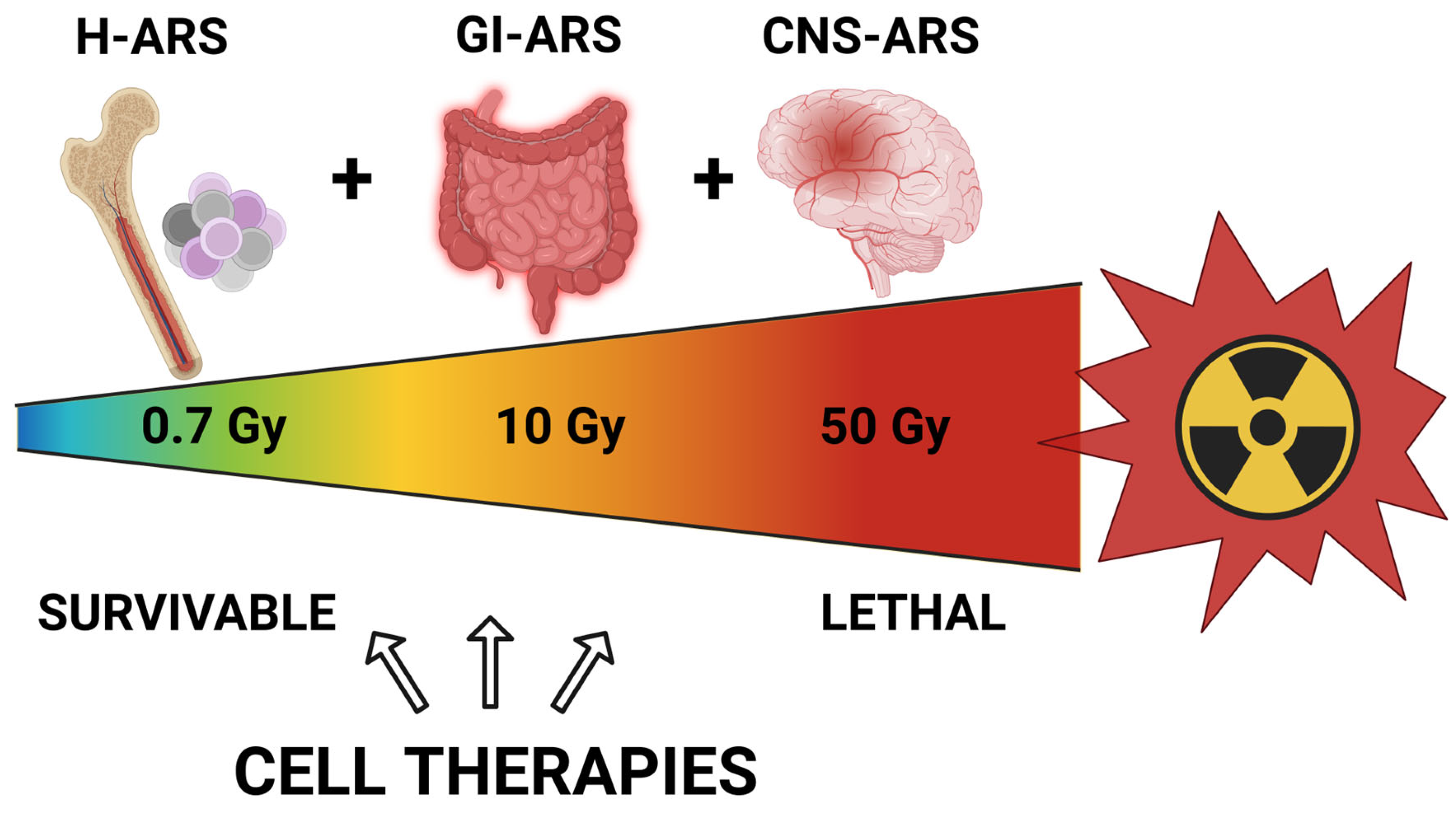
2. Acute Radiation Syndrome

Exposure to high doses of penetrating ionizing radiation, whether gamma rays, neutrons, or high-energy X-rays, is extremely damaging to biological systems. In a nuclear blast or following the use of a nuclear weapon, all or most of the body can be irradiated with a high dose in a relatively short period of time. If a whole-body radiation dose of more than approximately 0.7 Gy is delivered, this causes a severe acute illness called Acute Radiation Syndrome (ARS, also called radiation toxicity or radiation sickness). Due to natural repair processes, it is less toxic if the same overall dose is accumulated in smaller doses over a longer period of time. As illustrated in Figure 1, there are three defined but overlapping ARS subsyndromes that occur based on the dose received [32,33,34]. It should be noted that there is a gradient of damage seen with increasing doses. Although the injury response will vary due to individual patient differences (age, body size and composition, gender), even exposure to relatively low doses will likely produce harm. Patients exposed to at least 0.7 Gy develop symptoms reflective of bone marrow syndrome or hematopoietic syndrome (H-ARS). In this syndrome, the bone marrow and blood cells are primarily impacted. There is a drop in the numbers of all types of blood cells—pancytopenia—and the primary causes of death within this group are infection, coagulopathy, and hemorrhage. Some of the affected individuals will be expected to survive with supportive therapy allowing repopulation of the blood and bone marrow, especially at the lower end of the dose range, but recovery can take weeks to years. In fact, long-term residual bone marrow damage has been described in mouse model survivors of H-ARS with decreased white blood cells, lymphocytes, red blood cells, and platelets compared with nonirradiated controls even 20 months post irradiation [35,36,37,38]. Patients exposed to more than 10 Gy develop gastrointestinal syndrome (GI-ARS) in addition to blood and bone marrow damage. High doses of radiation cause endothelial and epithelial cell dysfunction and increased vascular permeability, which is especially damaging in the GI system [39]. Symptoms observed in GI-ARS include vomiting, diarrhea, electrolyte imbalance, and dehydration, and this syndrome is often lethal, especially at higher exposure levels. Supportive care may increase survival at the lower end of the dose range. A cardiovascular/central nervous system syndrome (CNS-ARS) will develop in addition to the damage to the hematopoietic and GI systems when patients are exposed to extremely high levels of whole-body radiation of more than 50 Gy. Individuals with this level of injury die within a few days, and no treatment options are currently available. A fourth syndrome has also been described, Cutaneous Radiation Syndrome (CRS) (https://www.cdc.gov/nceh/radiation/emergencies/pdf/ars.pdf; accessed on 24 May 2024) [40]. In CRS, exposure is limited to a smaller area of the body and causes mainly skin damage. Patients with cutaneous skin damage alone do not exhibit the severe life-threatening symptoms seen in ARS, but large skin doses can cause permanent damage to the affected area. Triage for radiation exposure includes severity scoring for prioritization for the use of medical countermeasures [41].

3. Management of Acute Radiation Syndrome

3.1. Background

Currently, there are a limited number of treatments beyond supportive therapies for the treatment of ARS [33,42]. The development of additional MCMs for the treatment of ARS is a priority [15,38,43,44,45,46,47,48,49,50,51]. MCMs for ARS fall into one of three categories: radioprotectors, radiomitigators, and radiotherapeutics [52]. Radioprotectors are treatments that are administered before radiation exposure and offer some protection against harmful effects. These interventions may be beneficial when the risk of significant exposure is likely, such as for medical and safety personnel responding to a nuclear incident, or for military personnel in combat situations where advanced weaponry is likely to be utilized. Radioprotectants are less useful when exposure is not anticipated prior to the incident, such as in a terrorist attack or nuclear facility incident. Radiomitigators are used after radiation exposure but prior to the development of ARS symptoms. To be classified as a radiomitigator, the treatment should be able to prevent or reduce eventual tissue and organ damage caused by exposure. Although administered after the initial insult has occurred, a radiomitigator might interrupt the cycle of immune, inflammatory, or other host responses that can lead to further damage. Alternatively, radiomitigators could be agents that act by initiating or accelerating regenerative processes to enable victims to survive the initial coagulopathy, immune dysfunction, and loss of intestinal barrier function. If a treatment of this type could be stockpiled and utilized rapidly in mass casualty situations, morbidity and mortality might be significantly reduced. Radiotherapeutics are administered once ARS symptoms have appeared to treat the symptoms. Maximum benefit to reduce mortality will likely be achieved by early treatment to prevent or reduce some of the damage before severe symptoms occur.

3.2. Approved Therapies

No agents have been fully approved by the FDA for use as radioprotectants at this time, but several recombinant growth factor agents have been approved for emergency use as radiomitigators, including Neupogen® Neulasta®, Nplate® (all from Amgen, Thousand Oaks, CA, USA), Leukine® (Partner Therapeutics, Lexington, MA, USA), Udenyca® (Coherus Biosciences, Redwood City, CA, USA), Stimufend® (Fresenius Kabi, Bad Homburg, Germany), and Ziextenzo® (Sandoz, Basil, Switzerland) (https://www.accessdata.fda.gov/emergency-preparedness-and-response/mcm-issues/radiological-and-nuclear-emergency-preparedness-information-fda#mcms, accessed on 5 May 2024). These are approved for H-ARS specifically and work by facilitating recovery of bone marrow precursors to generate neutrophils and white blood cells involved in immune function. They have a well-established history of clinical use for other indications and well-characterized safety profiles or are biosimilar to drugs with those features. There are limitations and side effects with all these agents: they do not work as radioprotectants; they may require additional supportive care which may not always be available; and they may cause delayed ARDS (Acute Respiratory Distress Syndrome). Another promising treatment that has not yet been approved, Entolimod (CBLB502), is currently under development. Entolimod (CBLB502) is a shortened version of the protein flagellin, which acts as a stimulatory ligand for host toll-like receptor (TLR) receptor signaling without the immunogenicity and toxicity of full-length flagellin [53]. TLR signaling stimulates immune function and induces NF-κB signaling, which in turn is antiapoptotic. CBLB502 has shown promise as a radiation countermeasure in murine and nonhuman primate (NHP) models [15]. Interestingly, the drug was shown to exert both radioprotective and radiomitigative effects in murine testing [54,55,56], making it even more attractive as a radiation MCM.

4. Cellular Therapeutics

4.1. Background

Cellular therapy describes the therapeutic use of not only live cells but also the use of cell products [57,58,59]. Transfusion with whole blood or blood products and hematopoietic stem cell transplantation (HSCT) can be classified as cellular therapies and these methods are already used in patients with radiation injury [42,60,61,62]. When the hematopoietic system is compromised in H-ARS, one of the earliest indicators of serious radiation damage is pancytopenia [34,63]. Bone marrow damage to precursor and stem cells blocks the replenishment of necessary blood cells, including white blood cells, red blood cells, and platelets, resulting in susceptibility to infection, clotting dysfunction, and diminished ability to recover from the damage. Blood transfusion is a temporary way to replenish some of the important cell types to buy time and allow the hematopoietic system to regenerate, assuming any stem and precursor cells remain in the bone marrow. A more comprehensive radiation treatment is bone marrow transplantation or hematopoietic stem cell transplantation [61,64,65,66]. Bone marrow contains hematopoietic stem cells (HSCs) involved in replenishing all the cell types in the blood. HSCs can also be found circulating in peripheral blood or umbilical cord blood. Umbilical cord blood may be a practical source for isolation based on availability due to its classification as medical waste. For radiation injury, HSCs need to come from an unirradiated donor source (allogeneic). Because of their immunogenicity, HSCs should be HLA-matched to reduce the chance of developing life-threatening graft v. host disease (GvHD) [67].
There is an intense interest in the use of cellular therapies beyond conventional blood transfusion for the mitigation or treatment of radiation injury [59]. Radiation injury can be complex, involving multiple organ systems, and as such may require a complex therapy consisting of multiple agents to address the damage [58]. The administration of either live cells or cell-produced biological products such as the secretome or extracellular vesicles (EVs) may provide the desired broad benefits (described below). Beyond the standard treatments of blood transfusion and hematopoietic stem cell transplantation, cellular therapies derived from mesenchymal stromal cells (MSCs) are at the forefront, especially for H-ARS.

Mesenchymal Stromal Cells—Background

Although they are not the only progenitor cell type under consideration for cell therapies, human MSCs have historically been the most studied cells for clinical use. Over 1000 human clinical trials have been conducted or are currently being conducted using MSCs for a variety of different diseases and injuries [68,69,70,71]. MSCs are adult stem-like cells and avoid the ethical considerations involved in the use of embryonic or neonatal cells. MSCs can be easily derived and expanded from readily available tissue sources such as bone marrow, adipose tissue, placental tissue, umbilical cord, or cord blood. Individual MSC populations show some differences reflective of their tissue source [72,73,74,75] and individual donor characteristics [76,77], but they share many similarities. MSCs are primary cells and are not immortalized cell “lines”; therefore, they have a limited lifespan and a reduced oncogenic risk. Although there is no one marker characteristic of MSCs, cells must conform to certain minimal criteria defined by a working group of the International Society for Cell and Gene Therapy (ISCT) to be classified as MSCs [78]. These criteria include expression of relevant surface proteins (≥95% positive for CD90, CD105, CD73) and a lack of expression of surface proteins characteristic of other cell types (≤2% positive for CD11b, CD34, CD14, CD45, CD31). The cells should also express undetectable levels of MHC Class II antigen on their surface under normal culture conditions; this property makes the cells less immunogenic (immune-privileged) and is important for safety reasons. However, under inflammatory conditions, likely similar to those found in patients with radiation injury, MHC Class II surface antigen expression increases in MSCs [79,80]. To be classified as MSCs, the cells must also be able to attach and proliferate on tissue culture plastic under standard conditions and retain the ability to differentiate into multiple lineages such as osteocytes, adipocytes, and chondrocytes under permissive conditions.
MSCs have anti-inflammatory properties and immunomodulatory activity and may also have antifibrotic and pro-regenerative properties, suggesting they may be beneficial in mitigation and/or treatment of ARS. MSCs and other cellular therapeutics secrete cytokines and other factors, proteins, RNA, and lipids, many of which are contained within EVs. These secreted components act in a paracrine manner to attenuate inflammation, augment angiogenesis, stimulate tissue repair, and modulate immune function (see [71] for an excellent review). Although not completely reflective of in vivo activity, a number of in vitro assays designed to assess the potency of different MSC preparations have been developed and are in common use [81,82,83,84,85]. Clinical trials have been performed for many different patient indications, including cardiovascular disease, immune disorders, neurological disease, osteoarthritis, and even as a treatment for COVID-19 [69]. MSCs have been approved for use in two inflammatory diseases, acute GvHD and Crohn’s disease [85,86]. Because they are relatively immune-privileged and immune-evasive, allogeneic MSCs can be used “off the shelf” in unrelated nonmatched donors, a property that dramatically increases their usefulness and cost-effectiveness. MSCs have shown an excellent safety profile, even when administered intravenously to patients [87,88,89,90,91]. Although there were concerns regarding potential procoagulant activity when MSCs or EVs are infused directly into the bloodstream of nonmatched recipients [92,93,94,95,96], these concerns have mostly been allayed by the addition of anticoagulants to the cells at the time of infusion [97,98].
Many different factors including immunomodulators have been identified as MSC-secreted factors, but it is not entirely clear what factors or combination of factors is responsible for activity in modulating immune function and inflammation [99,100,101,102,103]. The use of live MSCs rather than a mix of defined factors is more complicated but allows a more sophisticated response and the ability to adapt to different needs and conditions found in individual patients. Although MSCs secrete many factors responsible for much of their activity, they can also interact directly with immune cells in vitro [83,104,105,106,107]; interaction with host immune cells following infusion into animals likely provides additional functional activity. On the other hand, the use of live cells limits the applicability of the treatment method to well-equipped hospitals and clinics that have the necessary expertise and equipment to prepare and administer the cells under sterile conditions [70,80,108,109,110]. For this reason, investigation into the use of cell-free products derived from the cells is very important. Cell-free products produced by cells include secretome or conditioned medium and more purified EVs derived from conditioned medium [99,101,111,112,113,114,115]. It is possible that cell-free products such as secretome/MSC-conditioned medium or EVs could be packaged in a form (lyophilized or freeze-dried) that would be stable and readily available to stockpile for use in a future mass-casualty event or combat situation [114,116,117]. For that reason, investigation into the efficacy and optimal packaging and administration route for these products will be very important [118,119,120]. However, the choice of cell type and conditions for the cells producing the cell-free products will still influence the cell-free product and therefore remains an important issue.

4.2. Cellular Therapies for Radiation in Animal Models
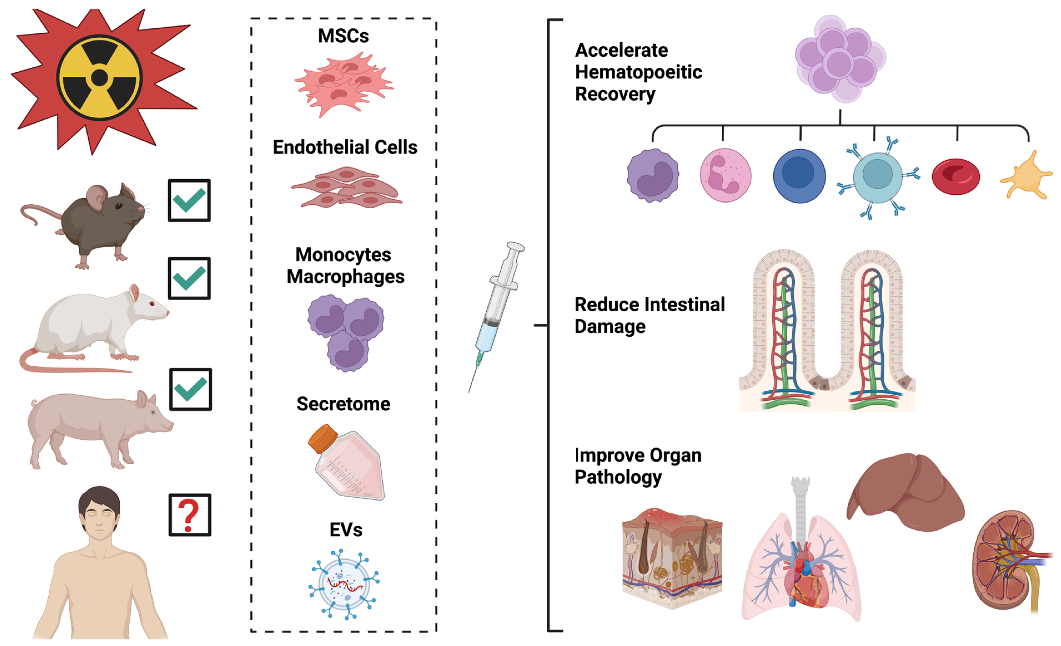
4.2.1. Cells

Multiple published studies in rodent models have shown the benefit of cellular therapies for the treatment of acute radiation injury (Table 2). In mouse models, several studies have demonstrated a survival benefit with live mouse MSCs using IV or IP administration following whole-body irradiation (gamma or X-ray) with a radiation dose that would normally be lethal [81,121,122,123]. Lange et al. showed 30–88% survival at 7 months vs 100% mortality of untreated animals irradiated with 9.5 Gy when treated 8 h after irradiation with 106 mouse bone-marrow-derived MSCs (BM-MSCs) [121]. Yang et al. found that while 7 Gy irradiation gave 20% survival at 45 days, infusion with 106 mouse MSCs resulted in improved 45-day survival of 50–60% [122]. Francois et al. determined that the MSCs need not be MHC-matched to be tolerated and mitigate radiation damage; MSCs derived from either syngeneic or allogeneic mice both showed positive effects [123]. The study by Chinnadurai et al. showed that “licensed” MSCs (activated by pretreatment with interferon gamma) were retained better than nonlicensed MSCs, although the difference was only seen in nonirradiated animals. The “licensed” MSCs protected irradiated mice from ARS, with 72% survival at day 30 versus 30% survival for vehicle-treated mice. In this study, male mice were noted to be more radiosensitive than female mice, and radiation treatment was performed at two doses of 4 Gy each. Two large doses of 107 MSCs were given at 24 h and 8 days after irradiation; IP injection was utilized to allow tolerance of the larger dose [81]. IP injection of mouse-adipose-derived stem cells also increased survival in lethally irradiated mice [124]. In this study, two different strains of mice were used to compare allogeneic (cross-strain) and syngeneic (same strain) MSCs. Thirty-day survival after 9.25 Gy irradiation increased with an IP injection of 5 × 10⁶ cells 24 h post irradiation. Beyond MSCs, other cell types have also shown benefits in lethally irradiated mice. Mouse endothelial cells (ECs) or their EV products (EC-EVs) increased both survival and bone marrow cellularity in irradiated mice [125].
Cells from human sources have also been tested in mouse radiation injury models. Increased survival of lethally irradiated mice was observed after retro-orbital administration of human BM-MSCs [64], umbilical-cord-blood-derived MSCs (UCB-MNCs) [127], or IV administration of human UCB-MSCs [128,131]. The study by Diaz et al. utilized a mouse total body irradiation model developed by Orschell’s group [36,137,138]. Mice were irradiated at 8 Gy for a lethal dose of 30% by 30 days. Human BM-MSCs (1.2 × 10⁷ per kg body weight) were administered by retro-orbital injection at either 3 h or 30 h post irradiation and monitored for 30-day survival, body weight change, and pancytopenia. Survival increased, although there was no change in body weight or pancytopenia, suggesting that recovery of the hematopoietic system was not the reason for increased survival. Similarly, the retro-orbital injection of human umbilical cord blood mononuclear cells (UCB-MNCs) increased 50-day survival in mice from 20% to 92% when given with antibiotics [127]. Antibiotics were given 4 h after irradiation with a lethal dose of 9 Gy; 2 × 10⁸ cells were administered retro-orbitally in four doses given between 24 and 52 h post irradiation. These benefits were not limited to the retro-orbital delivery of human cells. Kim et al. performed IV administration of human UCB-MSCs into mice at 1 × 10⁶ cells/dose at 3 and 30 h post 6 Gy irradiation and also achieved improved survival (from 0% in untreated animals at day 26 to approximately 40% survival in MSC-treated animals) [131]. Human macrophages or monocytes also act as radiomitigators in lethally irradiated mouse models. Interestingly, human macrophages that have been “educated” (preconditioned) by co-culturing with human MSCs are more potent at reducing radiation effects in a mouse model than either MSCs alone or “uneducated” macrophages [130]. It was subsequently shown that incubation of the macrophages [132] or even undifferentiated monocytes [133] with EVs derived from human MSCs could substitute for the MSC co-culture step. When the EVs used for “educating” the macrophages or monocytes were derived from MSCs that had been activated by treatment with the TLR4 receptor agonist lipopolysaccharide (LPS, a presumed inflammatory signal), the resulting “educated” macrophages were much more potent at increasing/prolonging survival, increasing expression of several key factors and reducing radiation damage to the spleen and bone marrow [132,133]. Administration of these cells was effective when given at either 4 h or 24 h post irradiation but was not effective at 48 h [133].
Although increased survival has been observed in these studies following the administration of MSCs or other cell types, there is debate about the mechanism. Lange [121] demonstrated that intravenous administration of mouse bone marrow MSCs within 8 h of irradiation promoted both overall survival and hematopoietic recovery in lethally irradiated mice; similar rescue of hematopoiesis was observed by Kim et al. [131] using human UCB-MSCs administered by IV infusion at either 3 h or 30 h post irradiation. In contrast, the recent study by Diaz et al. [64] demonstrated increased survival of lethally irradiated mice following retro-orbital injection of human BM-MSCs but observed no difference in pancytopenia or bone marrow hypocellularity in the treated animals. In that study, MSCs appeared to protect the gut from damage after irradiation. Although the studies use different models, injury conditions, cell types, doses, methods, and timing of cell administration, the consensus indicates that MSCs or other progenitor cells can confer survival benefits in rodents receiving potentially lethal doses of radiation. It also appears that cells from either the same species (either syngeneic or allogeneic) or from humans (xenogenic) are safe and show benefit. It is not yet clear what the optimal method of administration is. Although many studies utilize intravenous infusion, larger cell doses appear to be tolerated when administered intraperitoneally [81]. In the studies described here, cells administered by IV infusion, IP injection, or retro-orbital injection all decreased mortality due to radiation damage. The addition of prophylactic antibiotics has been suggested to confer additional survival benefit [123,127], likely by decreasing mortality due to infection during recovery of the hematopoietic system.
Interestingly, previous work has suggested that the inclusion of human MSCs along with HSCs in transplantation may facilitate engraftment and accelerate hematopoietic recovery [139,140]. In mice exposed to a lethal dose of radiation, the addition of mouse MSCs along with bone marrow cells (including HSCs) increased survival and promoted hematopoietic recovery [141]. However, the benefits were only seen up to a point; the addition of a large dose of MSCs was detrimental. More evaluation of these types of combination therapies is clearly warranted.

4.2.2. Cell Products

Conditioned medium collected from cultured cells, also called the secretome, contains many biologically active components, including EVs [99,101,113]. EVs are small membrane-enclosed vesicles secreted from cells; they can be found naturally in blood and other biological fluids or in the secretome of cultured cells. The contents of EVs include many biologically active molecules, including proteins, peptides, hormones, lipids, RNA, and other signaling molecules [101,103,113,115,142]. The composition of secreted EVs likely differs in different cell types and under different cellular conditions [101,108,111]. It is thought that much of the activity ascribed to MSCs is mediated through a paracrine mechanism via EV secretion. EVs can be further purified and concentrated from the secretome, and biological fluids and have been tested in multiple animal models of disease and have shown to be effective for the treatment of several conditions [143,144]. EVs are also being tested in a number of clinical trials [144]. Conditioned medium from rat MSC cultures has been tested in rat models of partial radiation injury. In one study, rats were administered conditioned medium from rat MSCs after abdominal irradiation (IP administration) in order to study intestinal damage. Rats receiving conditioned medium showed improved intestinal damage and survival, and medium from “activated” MSCs proved to be more potent [135]. Another study showed that conditioned medium from rat MSCs improved liver pathology when given prior to liver irradiation in rats [136]. In preclinical studies of radiation damage, EVs derived from “primed” MSCs showed protective activity [132,133]. In these studies, EVs derived from human MSCs can alter monocytes/macrophages to produce hematopoietic recovery benefits in a lethal mouse irradiation model for H-ARS. Additional evidence indicates that EVs derived from mouse bone marrow endothelial cells [125], human dental pulp stem cells [134], or human MSCs [129] increase survival, reduce bone marrow damage, and promote hematopoietic recovery in irradiated mice. Interestingly, Schoefinius et al. demonstrated that both mouse BM-MSCs and the EVs secreted by them conferred long-term survival benefits to lethally irradiated mice [126]. However, the MSCs were able to confer additional benefits in the short term. Their conclusion was that EVs protected irradiated hematopoietic stem cells but not progenitors. Finally, in a rat model partial-body knee irradiation, both rat BM-MSCs or their secreted EVs showed efficacy for reducing radiation-induced bone loss and bone marrow adiposity [145]. Although it is difficult to compare the efficacy of cells and their EV products because they have not often been compared in the same experiment, the reported benefits seen using EVs are encouraging and should be pursued further. Effective EV treatments would be much more practical for use on the battlefield or in military field hospitals, because of the potential for lyophilization for easier transport and storage. However, it remains to be seen whether EV function can be maintained in the lyophilized form. Another caveat is that EVs are not live cells; they cannot respond to environmental conditions. It will therefore be important to determine how to best induce production of the most clinically active EV products. For example, MSCs may secrete more potent anti-inflammatory EVs if they are activated by pretreatment with inflammatory mediators such as interferon-gamma or LPS [133,146].
The bulk of cellular therapeutic studies for acute radiation syndrome has been conducted in small animal models. Systemic administration of cells or EVs is more costly in larger animals due to the much larger body size and blood volume. However, porcine models are useful for the investigation of cellular therapeutics in the treatment of combined injuries that involve radiation and skin wounding. A minipig model was used to evaluate the efficacy of autologous adipose-derived regenerative cells (ADRCs, a heterogenous population including MSCs) on wound closure after sublethal whole-body irradiation combined with a full-thickness burn injury. Wound closure was accelerated in animals treated with ADRCs administered either by local injection or intravenously [147]. It is likely that cellular therapeutic treatments showing benefit in rodent models will continue to be tested and validated in large animals, including both porcine and nonhuman primate models. Thus far, cellular therapeutics have shown benefits in multiple animal models, but it remains to be seen if these agents will benefit human patients with radiation injury (Figure 2).

4.2.3. Countermeasure Testing in Combined Injury Models

Several countermeasures have been tested in combined injury models. In the mouse radiation + burn model described by Kiang et al., ciprofloxacin (given daily for 21 days) was successful in increasing survival [27]. Treatment significantly reduced early (Day 1) γ-H2AX focus formation indicative of DNA damage and later proinflammatory cytokine expression (Day 10). In another study, treatment of mice subjected to combined radiation + skin wound injury with the thrombopoietin receptor agonist Alxn4100TPO improved survival and mitigated weight loss, although wound healing was delayed [148,149]. Treatment with the hunger-stimulating peptide hormone ghrelin improved survival, mitigated weight loss, and accelerated wound healing in the mouse combined radiation + skin wound model [150]. Ghrelin treatment was also effective in mice subjected to radiation + 15% TBSA burn injury, increasing survival and mitigating pancytopenia and bone marrow cell depletion [150]. Cellular therapy using mouse bone marrow MSCs has also been tested using this model [26]. In this study, IV administration of MSCs grown under hypoxic conditions 24 h after combined injury led to a 30% increased 30-day survival, attenuated weight loss, accelerated wound healing, and decreased bone marrow cell loss. In contrast to other studies (Table 2), this group did not find the benefit of MSC treatment in mice treated with radiation alone in their model, but MSCs did appear to provide benefits to animals with combined injury [26]. Kiang et al. [151] also observed another difference between radiation injury alone and combined radiation + burn injury. The approved radiomitigator pegylated G-CSF improved survival, reduced weight loss, mitigated WBC and platelet depletion, and blocked splenomegaly in mice subjected to radiation injury alone but did not benefit mice with combined injury. This suggests that treatments that work for radiation injury alone may or may not work for combined injuries, and that treatments providing benefit in combined injury models may not provide benefit in animals with radiation injury alone. Determination of the best treatments for radiation alone and for different types of combined injury will require more careful study.

4.2.4. Delivery of Cellular Therapeutics

Cells or their products can be introduced by several means, including intravenously, intrathecally, topically, intraperitoneally, subcutaneously, intramuscularly, or by direct cardiac implantation [152]. It is not yet clear which route of administration is superior, and the choice of administration route may depend at least in part on patient indications [70,152]. For example, because IV-administered cells quickly become sequestered mainly in the lung, this may be the preferred route of administration for acute lung injury therapies. Conversely, topical application for burns or other skin wounds may be more suitable. At this time, the preferred administration route for ARS is unclear, and this topic will need further exploration. The most common routes of administration in preclinical animal testing for ARS caused by whole-body irradiation are IV and IP injection (Table 1). IV administration has the advantage of delivering therapeutics directly to the bloodstream with the potential for interaction with immune cells and systemic delivery to the entire body, but retention in the bloodstream is not long (especially for whole cells which get trapped in the lung). IP administration has the advantage of allowing tolerance of a larger dose of cells but could be difficult in the presence of abdominal injury. For practical use in military combat situations or for victims of a mass casualty involving radiation injury, the method of administration should be one that requires minimal expertise and equipment in order to benefit a large number of patients in potentially austere conditions.

5. Future Directions and Challenges

Despite the promise of cellular therapeutics for the treatment of acute radiation injury, many challenges remain before these therapies can be utilized routinely or stockpiled for emergency use. Future investigation will be necessary to determine how MSCs or other therapies can provide benefits for the treatment of radiation injury, including the level of injury (mild or severe) that might derive the greatest benefit. Careful side-by-side comparisons of live cells and the cell products derived from them (EVs) will be needed to determine potency and efficacy. Since a cell-free product will likely be simpler for use in military treatment facilities or for stockpiling for a mass casualty event, much of the effort could be focused on these products provided they show reasonable efficacy compared with live cell products. Additional work is needed to determine the optimal timing, dosing strategy, and administration methods for both cells and cell-free products. It will be imperative to determine the safety and efficacy of therapeutic agents in patients with different types of combined injury, as well as whether the use of these agents will interfere with the accepted clinical interventions for wound or burn trauma. Another important area for study concerns combination therapies; cellular therapies may be best used in conjunction with other agents, such as antibiotics or growth factors, to improve overall outcomes. More investigation into the mechanisms of action is also warranted. If specific properties of cells or specific combinations of cell-produced factors are identified as necessary and/or sufficient for activity, then cell-free treatments containing the important factors can be developed as a more consistent product, standardized for use in a mass casualty event or possibly tailored to patient-specific needs in less urgent conditions.

6. Conclusions

Cellular therapies to prevent, mitigate, or treat acute radiation injury could provide tremendous benefits based on the ability of MSCs or other progenitor cells to reduce inflammation, positively modulate immune function, enhance angiogenesis, and promote recipient regenerative processes. Live cells are very complex biological products that have yet to be optimized for growth, production, storage, administration, or patient selection. For military use or for stockpiling against future mass casualty events, cell-free products such as the secretome or EVs are needed, but the choice of cells, handling of cells during secretome/EV collection, processing methods, storage conditions, and administration methods also require optimization. It still remains to be seen whether secretome or EVs function similarly to live cells for the treatment of acute radiation injury, and this important question should be a priority for future research and development. The use of cells or the cell-free products they produce will likely provide a powerful tool for mitigation and treatment of serious radiation injury, saving lives and reducing long-term damage in those exposed to high radiation doses.

Author Contributions

Conceptualization, B.A.C. and M.C.H.; writing—original draft preparation, B.A.C. and M.C.H.; writing—review and editing, X.W., A.M. and J.S.M.; figure design/preparation, A.M.; project administration, B.A.C. and J.A.B.; funding acquisition, J.A.B., B.A.C. and M.C.H. All authors have read and agreed to the published version of the manuscript.

Funding

This research was funded by a grant from the Congressionally Directed Medical Research Program, grant number CDMRP BA190083.

Conflicts of Interest

The authors declare no conflicts of interest. The views expressed in this article are those of the authors and do not reflect the official policy or position of the US Army Medical Department, Department of the Army, DoD, or the US Government.

References

  1. CBRN EU. CBRN Risk Mitigation. 2024. Available online: https://cbrn-risk-mitigation.network.europa.eu/index_en (accessed on 5 May 2024).
  2. CBRNE Central. Asia-Pacific. Articles on Chemical, Biological, Radiological and Nuclear (CBRN) and Explosives Threats, Preparedness and Response in the Asia-Pacific Region. 2024. Available online: https://cbrnecentral.com/tag/asia-pacific (accessed on 1 January 2024).
  3. Homeland Security. National Strategy for Chemical, Biological, Radiological, Nuclear, and Explosives (CBRNE) Standards. U.S. Department of Homeland Security: Washington, DC, USA, 2022. Available online: https://www.dhs.gov/national-strategy-chemical-biological-radiological-nuclear-and-explosives-cbrne-standards (accessed on 20 June 2024).
  4. CBRNEC. Chemical, Biological, Radiological, Nuclear and Explosives Resilience Strategy for Canada. Public Safety Canada: Ottawa, ON, Canada, 2018. Available online: https://www.publicsafety.gc.ca/cnt/rsrcs/pblctns/rslnc-strtg/index-en.aspx (accessed on 1 May 2024).
  5. Coleman, C.N.; Bader, J.L.; Koerner, J.F.; Hrdina, C.; Cliffer, K.D.; Hick, J.L.; James, J.J.; Mansoura, M.K.; Livinski, A.A.; Nystrom, S.V.; et al. Chemical, Biological, Radiological, Nuclear, and Explosive (CBRNE) Science and the CBRNE Science Medical Operations Science Support Expert (CMOSSE). Disaster Med. Public Health Prep. 2019, 13, 995–1010. [Google Scholar] [CrossRef]
  6. Ende, N.; Azzam, E.I. Consideration for the treatment of mass casualties based on pathology of the fatalities of HiroCDCa and Nagasaki. Int. J. Radiat. Biol. 2011, 87, 443–444. [Google Scholar] [CrossRef] [PubMed]
  7. Brambilla, S.; Nelson, M.A.; Brown, M.J. Dirty bomb source term characterization and downwind dispersion: Review of experimental evidence. J. Environ. Radioact. 2023, 263, 107166. [Google Scholar] [CrossRef] [PubMed]
  8. Brambilla, S.; Nelson, M.A.; Brown, M.J. Review of particle deposition to and removal from clothing, skin, and hair after a radioactive airborne dispersal event. J. Environ. Radioact. 2023, 270, 107296. [Google Scholar] [CrossRef]
  9. Kugathasan, T.; Mothersill, C. Radiobiological and social considerations following a radiological terrorist attack; mechanisms, detection and mitigation: Review of new research developments. Int. J. Radiat. Biol. 2022, 98, 855–864. [Google Scholar] [CrossRef]
  10. Williams, G.; O’Malley, M. Surgical considerations in the management of combined radiation blast injury casualties caused by a radiological dirty bomb. Injury 2010, 41, 943–947. [Google Scholar] [CrossRef]
  11. Wolbarst, A.B.; Wiley, A.L.; Nemhauser, J.B., Jr.; Christensen, D.M.; Hendee, W.R. Medical response to a major radiologic emergency: A primer for medical and public health practitioners. Radiology 2010, 254, 660–677. [Google Scholar] [CrossRef]
  12. Eaton, E.B.; Varney, T.R., Jr. Mesenchymal stem cell therapy for acute radiation syndrome: Innovative medical approaches in military medicine. Mil. Med. Res. 2015, 2, 2. [Google Scholar] [CrossRef] [PubMed]
  13. Fukumoto, R. Mesenchymal stem cell therapy for acute radiation syndrome. Mil. Med. Res. 2016, 3, 17. [Google Scholar] [CrossRef]
  14. Ledney, G.D.; Stewart, D.A.; Exum, E.D.; Sheehy, P.A. Skin wound-enhanced survival and myelocytopoiesis in mice after whole-body irradiation. Acta Radiol. Oncol. 1981, 20, 29–38. [Google Scholar] [CrossRef]
  15. Singh, V.K.; Seed, T.M. A review of radiation countermeasures focusing on injury-specific medicinals and regulatory approval status: Part I. Radiation sub-syndromes, animal models and FDA-approved countermeasures. Int. J. Radiat. Biol. 2017, 93, 851–869. [Google Scholar] [CrossRef] [PubMed]
  16. Park, G.D.; Mitchel, J.T. Working with the U.S. Food and Drug Administration to obtain approval of products under the Animal Rule. Ann. N. Y. Acad. Sci. 2016, 1374, 10–16. [Google Scholar] [CrossRef] [PubMed]
  17. Singh, V.K.; Newman, V.L.; Berg, A.N.; MacVittie, T.J. Animal models for acute radiation syndrome drug discovery. Expert Opin. Drug Discov. 2015, 10, 497–517. [Google Scholar] [CrossRef] [PubMed]
  18. Hunter, K.S.; Carnell, L.S.; DiCarlo, A.L.; Hoffman, C.M.; Loelius, S.G.; Homer, M. Interagency approaches to animal models for acute radiation exposure. Int. J. Radiat. Biol. 2021, 97 (Suppl. S1), S2–S5. [Google Scholar] [CrossRef] [PubMed]
  19. Williams, J.P.; Brown, S.L.; Georges, G.E.; Hauer-Jensen, M.; Hill, R.P.; Huser, A.K.; Kirsch, D.G.; MacVittie, T.J.; Mason, K.A.; Medhora, M.M.; et al. Animal models for medical countermeasures to radiation exposure. Radiat. Res. 2010, 173, 557–578. [Google Scholar] [CrossRef] [PubMed]
  20. Singh, V.K.; Olabisi, A.O. Nonhuman primates as models for the discovery and development of radiation countermeasures. Expert Opin. Drug Discov. 2017, 12, 695–709. [Google Scholar] [CrossRef] [PubMed]
  21. Palmer, J.L.; Deburghgraeve, C.R.; Bird, M.D.; Hauer-Jensen, M.; Kovacs, E.J. Development of a combined radiation and burn injury model. J. Burn Care Res. 2011, 32, 317–323. [Google Scholar] [CrossRef] [PubMed]
  22. Medhora, M.; Gasperetti, T.; Schamerhorn, A.; Gao, F.; Narayanan, J.; Lazarova, Z.; Jacobs, E.R.; Tarima, S.; Fish, B.L. Wound Trauma Exacerbates Acute, but not Delayed, Effects of Radiation in Rats: Mitigation by Lisinopril. Int. J. Mol. Sci. 2020, 21, 3908. [Google Scholar] [CrossRef] [PubMed]
  23. Gao, F.; Fish, B.L.; Szabo, A.; Schock, A.; Narayanan, J.; Jacobs, E.R.; Moulder, J.E.; Lazarova, Z.; Medhora, M. Enhanced survival from radiation pneumonitis by combined irradiation to the skin. Int. J. Radiat. Biol. 2014, 90, 753–761. [Google Scholar] [CrossRef] [PubMed]
  24. Kiang, J.G.; Smith, J.T.; Anderson, M.N.; Swift, J.M.; Christensen, C.L.; Gupta, P.; Balakathiresan, N.; Maheshwari, R.K. Hemorrhage Exacerbates Radiation Effects on Survival, Leukocytopenia, Thrombopenia, Erythropenia, Bone Marrow Cell Depletion and Hematopoiesis, and Inflammation-Associated microRNAs Expression in Kidney. PLoS ONE 2015, 10, e0139271. [Google Scholar] [CrossRef]
  25. Kiang, J.G.; Garrison, B.R.; Burns, T.M.; Zhai, M.; Dews, I.C.; Ney, P.H.; Cary, L.H.; Fukumoto, R.; Elliott, T.B.; Ledney, G.D. Wound trauma alters ionizing radiation dose assessment. Cell Biosci. 2012, 2, 20. [Google Scholar] [CrossRef]
  26. Kiang, J.G.; Gorbunov, N.V. Bone Marrow Mesenchymal Stem Cells Increase Survival after Ionizing Irradiation Combined with Wound Trauma: Characterization and Therapy. J. Cell Sci. Ther. 2014, 5, 190. [Google Scholar] [CrossRef] [PubMed]
  27. Kiang, J.G.; Fukumoto, R. Ciprofloxacin increases survival after ionizing irradiation combined injury: Gamma-H2AX formation, cytokine/chemokine, and red blood cells. Health Phys. 2014, 106, 720–726. [Google Scholar] [CrossRef] [PubMed]
  28. Ledney, G.D.; Exum, E.D.; Jackson, W.E., 3rd. Wound-induced alterations in survival of 60Co irradiated mice: Importance of wound timing. Experientia 1985, 41, 614–616. [Google Scholar] [CrossRef] [PubMed]
  29. Ledney, G.D.; Exum, E.D.; Sheehy, P.A. Survival enhanced by skin-wound trauma in mice exposed to 60Co radiation. Experientia 1981, 37, 193–194. [Google Scholar] [CrossRef]
  30. Garrett, J.; Orschell, C.M.; Mendonca, M.S.; Bigsby, R.M.; Dynlacht, J.R. Subcutaneous wounding postirradiation reduces radiation lethality in mice. Radiat. Res. 2014, 181, 578–583. [Google Scholar] [CrossRef]
  31. Dynlacht, J.R.; Garrett, J.; Joel, R.; Lane, K.; Mendonca, M.S.; Orschell, C.M. Further Characterization of the Mitigation of Radiation Lethality by Protective Wounding. Radiat. Res. 2017, 187, 732–742. [Google Scholar] [CrossRef] [PubMed]
  32. A Brochure for Physicians Acute Radiation Syndrome. Available online: https://www.Cdc.gov/radiation-emergencies/media/pdfs/ARS.pdf (accessed on 23 November 2023).
  33. DiCarlo, A.L.; Maher, C.; Hick, J.L.; Hanfling, D.; Dainiak, N.; Chao, N.; Bader, J.L.; Coleman, C.N.; Weinstock, D.M. Radiation injury after a nuclear detonation: Medical consequences and the need for scarce resources allocation. Disaster Med. Public Health Prep. 2011, 5 (Suppl. S1), S32–S44. [Google Scholar] [CrossRef]
  34. Kiang, J.G.; Olabisi, A.O. Radiation: A poly-traumatic hit leading to multi-organ injury. Cell Biosci. 2019, 9, 25. [Google Scholar] [CrossRef]
  35. Chua, H.L.; Plett, P.A.; Fisher, A.; Sampson, C.H.; Vemula, S.; Feng, H.; Sellamuthu, R.; Wu, T.; Orschell, C.M. Lifelong Residual bone Marrow Damage in Murine Survivors of the Hematopoietic Acute Radiation Syndrome (H-ARS): A Compilation of Studies Comprising the Indiana University Experience. Health Phys. 2019, 116, 546–557. [Google Scholar] [CrossRef]
  36. Chua, H.L.; Plett, P.A.; Sampson, C.H.; Joshi, M.; Tabbey, R.; Katz, B.P.; MacVittie, T.J.; Orschell, C.M. Long-term hematopoietic stem cell damage in a murine model of the hematopoietic syndrome of the acute radiation syndrome. Health Phys. 2012, 103, 356–366. [Google Scholar] [CrossRef]
  37. MacVittie, T.J.; Farese, A.M.; Jackson, W.E., 3rd. A Systematic Review of the Hematopoietic Acute Radiation Syndrome (H-ARS) in Canines and Non-human Primates: Acute Mixed Neutron/Gamma vs. Reference Quality Radiations. Health Phys. 2020, 119, 527–558. [Google Scholar] [CrossRef] [PubMed]
  38. Wu, T.; Orschell, C.M. The delayed effects of acute radiation exposure (DEARE): Characteristics, mechanisms, animal models, and promising medical countermeasures. Int. J. Radiat. Biol. 2023, 99, 1066–1079. [Google Scholar] [CrossRef] [PubMed]
  39. Wang, J.; Boerma, M.; Fu, Q.; Hauer-Jensen, M. Significance of endothelial dysfunction in the pathogenesis of early and delayed radiation enteropathy. World J. Gastroenterol. 2007, 13, 3047–3055. [Google Scholar] [CrossRef]
  40. Iddins, C.J.; DiCarlo, A.L.; Ervin, M.D.; Herrera-Reyes, E.; Goans, R.E. Cutaneous and local radiation injuries. J. Radiol. Prot. 2022, 42, 011001. [Google Scholar] [CrossRef]
  41. Kenchegowda, D.; Bolduc, D.L.; Kurada, L.; Blakely, W.F. Severity scoring systems for radiation-induced GI injury—Prioritization for use of GI-ARS medical countermeasures. Int. J. Radiat. Biol. 2023, 99, 1037–1045. [Google Scholar] [CrossRef]
  42. Dainiak, N.; Albanese, J. Medical management of acute radiation syndrome. J. Radiol. Prot. 2022, 42, 031002. [Google Scholar] [CrossRef] [PubMed]
  43. Arora, R.; Chawla, R.; Marwah, R.; Kumar, V.; Goel, R.; Arora, P.; Jaiswal, S.; Sharma, R.K. Medical radiation countermeasures for nuclear and radiological emergencies: Current status and future perspectives. J. Pharm. Bioallied Sci. 2010, 2, 202–212. [Google Scholar] [CrossRef]
  44. Leiterer, A.; Bardot, I.; Ménétrier, F.; Bardot, S.; Grémy, O.; Bérard, P.; Pech, A.; Favaro, P. Medical countermeasures after a radiological event: An update from the CATO project. Int. J. Radiat. Biol. 2014, 90, 1043–1047. [Google Scholar] [CrossRef]
  45. Nicogossian, A.E.; Rummel, J.D.; Leveton, L.; Teeter, R. Development of countermeasures for medical problems encountered in space flight. Adv. Space Res. 1992, 12, 329–337. [Google Scholar] [CrossRef]
  46. Singh, V.K.; Hanlon, B.K.; Santiago, P.T.; Seed, T.M. A review of radiation countermeasures focusing on injury-specific medicinals and regulatory approval status: Part III. Countermeasures under early stages of development along with ‘standard of care’ medicinal and procedures not requiring regulatory approval for use. Int. J. Radiat. Biol. 2017, 93, 885–906. [Google Scholar] [PubMed]
  47. Singh, V.K.; Garcia, M.; Seed, T.M. A review of radiation countermeasures focusing on injury-specific medicinals and regulatory approval status: Part II. Countermeasures for limited indications, internalized radionuclides, emesis, late effects, and agents demonstrating efficacy in large animals with or without FDA IND status. Int. J. Radiat. Biol. 2017, 93, 870–884. [Google Scholar] [PubMed]
  48. Singh, V.K.; Newman, V.L.; Romaine, P.L.; Wise, S.Y.; Seed, T.M. Radiation countermeasure agents: An update (2011–2014). Expert Opin. Ther. Pat. 2014, 24, 1229–1255. [Google Scholar] [CrossRef] [PubMed]
  49. Singh, V.K.; Romaine, P.L.; Newman, V.L.; Seed, T.M. Medical countermeasures for unwanted CBRN exposures: Part II radiological and nuclear threats with review of recent countermeasure patents. Expert Opin. Ther. Pat. 2016, 26, 1399–1408. [Google Scholar] [CrossRef] [PubMed]
  50. Singh, V.K.; Romaine, P.L.; Seed, T.M. Medical Countermeasures for Radiation Exposure and Related Injuries: Characterization of Medicines, FDA-Approval Status and Inclusion into the Strategic National Stockpile. Health Phys. 2015, 108, 607–630. [Google Scholar] [CrossRef] [PubMed]
  51. Singh, V.K.; Seed, T.M. An update on romiplostim for treatment of acute radiation syndrome. Drugs Today 2022, 58, 133–145. [Google Scholar] [CrossRef]
  52. Micewicz, E.D.; Damoiseaux, R.D.; Deng, G.; Gomez, A.; Iwamoto, K.S.; Jung, M.E.; Nguyen, C.; Norris, A.J.; Ratikan, J.A.; Ruchala, P.; et al. Classes of Drugs that Mitigate Radiation Syndromes. Front. Pharmacol. 2021, 12, 666776. [Google Scholar] [CrossRef] [PubMed]
  53. Singh, V.K.; Seed, T.M. Entolimod as a radiation countermeasure for acute radiation syndrome. Drug Discov. Today 2021, 26, 17–30. [Google Scholar] [CrossRef] [PubMed]
  54. Burdelya, L.G.; Gleiberman, A.S.; Toshkov, I.; Aygun-Sunar, S.; Bapardekar, M.; Manderscheid-Kern, P.; Bellnier, D.; Krivokrysenko, V.I.; Feinstein, E.; Gudkov, A.V. Toll-like receptor 5 agonist protects mice from dermatitis and oral mucositis caused by local radiation: Implications for head-and-neck cancer radiotherapy. Int. J. Radiat. Oncol. Biol. Phys. 2012, 83, 228–234. [Google Scholar] [CrossRef]
  55. Krivokrysenko, V.I.; Toshkov, I.A.; Gleiberman, A.S.; Krasnov, P.; Shyshynova, I.; Bespalov, I.; Maitra, R.K.; Narizhneva, N.V.; Singh, V.K.; Whitnall, M.H.; et al. The Toll-Like Receptor 5 Agonist Entolimod Mitigates Lethal Acute Radiation Syndrome in Non-Human Primates. PLoS ONE 2015, 10, e0135388. [Google Scholar] [CrossRef]
  56. Krivokrysenko, V.I.; Shakhov, A.N.; Singh, V.K.; Bone, F.; Kononov, Y.; Shyshynova, I.; Cheney, A.; Maitra, R.K.; Purmal, A.; Whitnall, M.H.; et al. Identification of granulocyte colony-stimulating factor and interleukin-6 as candidate biomarkers of CBLB502 efficacy as a medical radiation countermeasure. J. Pharmacol. Exp. Ther. 2012, 343, 497–508. [Google Scholar] [CrossRef] [PubMed]
  57. Benderitter, M.; Caviggioli, F.; Chapel, A.; Coppes, R.P.; Guha, C.; Klinger, M.; Malard, O.; Stewart, F.; Tamarat, R.; Van Luijk, P.; et al. Stem cell therapies for the treatment of radiation-induced normal tissue side effects. Antioxid. Redox Signal. 2014, 21, 338–355. [Google Scholar] [CrossRef] [PubMed]
  58. DiCarlo, A.L.; Tamarat, R.; Rios, C.I.; Benderitter, M.; Czarniecki, C.W.; Allio, T.C.; Macchiarini, F.; Maidment, B.W.; Jourdain, J.-R. Cellular Therapies for Treatment of Radiation Injury: Report from a NIH/NIAID and IRSN Workshop. Radiat. Res. 2017, 188, e54–e75. [Google Scholar] [CrossRef] [PubMed]
  59. Rios, C.; Jourdain, J.R.; DiCarlo, A.L. Cellular Therapies for Treatment of Radiation Injury after a Mass Casualty Incident. Radiat. Res. 2017, 188, 242–245. [Google Scholar] [CrossRef] [PubMed]
  60. Bader, J.L.; Nemhauser, J.; Chang, F.; Mashayekhi, B.; Sczcur, M.; Knebel, A.; Hrdina, C.; Coleman, N. Radiation event medical management (REMM): Website guidance for health care providers. Prehospital Emerg. Care 2008, 12, 1–11. [Google Scholar] [CrossRef] [PubMed]
  61. Lopez, M.; Martin, M. Medical management of the acute radiation syndrome. Rep. Pract. Oncol. Radiother. 2011, 16, 138–146. [Google Scholar] [CrossRef] [PubMed]
  62. Dainiak, N. Medical management of acute radiation syndrome and associated infections in a high-casualty incident. J. Radiat. Res. 2018, 59 (Suppl S2), ii54–ii64. [Google Scholar] [CrossRef] [PubMed]
  63. Petrova, A.S. Blood platelet change in acute radiation sickness. Med. Radiol. 1956, 1, 52–56. [Google Scholar]
  64. Diaz, M.F.; Horton, P.L.D.; Dumbali, S.P.; Kumar, A.; Livingston, M.; Skibber, M.A.; Mohammadalipour, A.; Gill, B.S.; Zhang, S.; Cox, C.S.; et al. Bone marrow stromal cell therapy improves survival after radiation injury but does not restore endogenous hematopoiesis. Sci. Rep. 2020, 10, 22211. [Google Scholar] [CrossRef]
  65. Singh, V.K.; Brown, D.S.; Kao, T.C.; Seed, T.M. Preclinical development of a bridging therapy for radiation casualties. Exp. Hematol. 2010, 38, 61–70. [Google Scholar] [CrossRef]
  66. Weisdorf, D.; Chao, N.; Waselenko, J.K.; Dainiak, N.; Armitage, J.O.; McNiece, I.; Confer, D. Acute radiation injury: Contingency planning for triage, supportive care, and transplantation. Biol. Blood Marrow Transplant. 2006, 12, 672–682. [Google Scholar] [CrossRef] [PubMed]
  67. Schönberger, S.; Niehues, T.; Meisel, R.; Bernbeck, B.; Laws, H.J.; Kögler, G.; Enzmann, J.; Wernet, P.; Göbel, U.; Dilloo, D. Transplantation of haematopoietic stem cells derived from cord blood, bone marrow or peripheral blood: A single centre matched-pair analysis in a heterogeneous risk population. Klin. Padiatr. 2004, 216, 356–363. [Google Scholar] [CrossRef] [PubMed]
  68. Galderisi, U.; Peluso, G.; Di Bernardo, G. Clinical Trials Based on Mesenchymal Stromal Cells are Exponentially Increasing: Where are We in Recent. Years? Stem Cell Rev. Rep. 2022, 18, 23–36. [Google Scholar] [CrossRef] [PubMed]
  69. Jovic, D.; Yu, Y.; Wang, D.; Wang, K.; Li, H.; Xu, F.; Liu, C.; Liu, J.; Luo, Y. A Brief Overview of Global Trends in MSC-Based Cell Therapy. Stem Cell Rev. Rep. 2022, 18, 1525–1545. [Google Scholar] [CrossRef] [PubMed]
  70. Levy, O.; Kuai, R.; Siren, E.M.J.; Bhere, D.; Milton, Y.; Nissar, N.; De Biasio, M.; Heinelt, M.; Reeve, B.; Abdi, R.; et al. Shattering barriers toward clinically meaningful MSC therapies. Sci. Adv. 2020, 6, eaba6884. [Google Scholar] [CrossRef] [PubMed]
  71. Pittenger, M.F.; Discher, D.E.; Peault, B.M.; Phinney, D.G.; Hare, J.M.; Caplan, A.I. Mesenchymal stem cell perspective: Cell biology to clinical progress. npj Regen. Med. 2019, 4, 22. [Google Scholar] [CrossRef]
  72. Danev, N.; Li, G.; Duan, J.E.; Van de Walle, G.R. Comparative transcriptomic analysis of bovine mesenchymal stromal cells reveals tissue-source and species-specific differences. iScience 2024, 27, 108886. [Google Scholar] [CrossRef] [PubMed]
  73. Ma, J.; Wu, J.; Han, L.; Jiang, X.; Yan, L.; Hao, J.; Wang, H. Comparative analysis of mesenchymal stem cells derived from amniotic membrane, umbilical cord, and chorionic plate under serum-free condition. Stem Cell Res. Ther. 2019, 10, 19. [Google Scholar] [CrossRef]
  74. Sakaguchi, Y.; Sekiya, I.; Yagishita, K.; Muneta, T. Comparison of human stem cells derived from various mesenchymal tissues: Superiority of synovium as a cell source. Arthritis Rheum. 2005, 52, 2521–2529. [Google Scholar] [CrossRef]
  75. Wagner, W.; Wein, F.; Seckinger, A.; Frankhauser, M.; Wirkner, U.; Krause, U.; Blake, J.; Schwager, C.; Eckstein, V.; Ansorge, W.; et al. Comparative characteristics of mesenchymal stem cells from human bone marrow, adipose tissue, and umbilical cord blood. Exp. Hematol. 2005, 33, 1402–1416. [Google Scholar] [CrossRef]
  76. Jansen, B.J.H.; Gilissen, C.; Roelofs, H.; Schaap-Oziemlak, A.; Veltman, J.A.; Raymakers, R.A.P.; Jansen, J.H.; Kögler, G.; Figdor, C.G.; Torensma, R.; et al. Functional differences between mesenchymal stem cell populations are reflected by their transcriptome. Stem Cells Dev. 2010, 19, 481–490. [Google Scholar] [CrossRef] [PubMed]
  77. Kolliopoulos, V.; Tiffany, A.; Polanek, M.; Harley, B.A.C. Donor Variability in Human Mesenchymal Stem Cell Osteogenic Response as a Function of Passage Conditions and Donor Sex. bioRxiv 2023. [Google Scholar] [CrossRef]
  78. Dominici, M.; Le Blanc, K.; Mueller, I.; Slaper-Cortenbach, I.; Marini, F.C.; Krause, D.S.; Deans, R.J.; Keating, A.; Prockop, D.J.; Horwitz, E.M. Minimal criteria for defining multipotent mesenchymal stromal cells. The International Society for Cellular Therapy position statement. Cytotherapy 2006, 8, 315–317. [Google Scholar] [CrossRef] [PubMed]
  79. Christy, B.A.; Herzig, M.C.; Delavan, C.; Cantu, C.; Salgado, C.; Bynum, J.A.; Cap, A.P. Human primary fibroblasts perform similarly to MSCs in assays used to evaluate MSC safety and potency. Transfusion 2019, 59 (Suppl. S2), 1593–1600. [Google Scholar] [CrossRef] [PubMed]
  80. Galipeau, J.; Sensebe, L. Mesenchymal Stromal Cells: Clinical Challenges and Therapeutic Opportunities. Cell Stem Cell 2018, 22, 824–833. [Google Scholar] [CrossRef] [PubMed]
  81. Chinnadurai, R.; Bates, P.D.; Kunugi, K.A.; Nickel, K.P.; DeWerd, L.A.; Capitini, C.M.; Galipeau, J.; Kimple, R.J. Dichotomic Potency of IFNgamma Licensed Allogeneic Mesenchymal Stromal Cells in Animal Models of Acute Radiation Syndrome and Graft Versus Host Disease. Front. Immunol. 2021, 12, 708950. [Google Scholar] [CrossRef] [PubMed]
  82. Christy, B.A.; Herzig, M.C.; Delavan, C.P.; Abaasah, I.; Cantu, C.; Salgado, C.; Lovelace, S.; Garcia, L.; Jensen, K.; Montgomery, R.; et al. Use of multiple potency assays to evaluate human mesenchymal stromal cells. J. Trauma Acute Care Surg. 2020, 89 (Suppl. S2), S109–S117. [Google Scholar] [CrossRef] [PubMed]
  83. Herzig, M.C.; Christy, B.A.; Montgomery, R.K.; Delavan, C.P.; Jensen, K.J.; Lovelace, S.E.; Cantu, C.; Salgado, C.L.; Cap, A.P.; Bynum, J.A. Interactions of human mesenchymal stromal cells with peripheral blood mononuclear cells in a Mitogenic proliferation assay. J. Immunol. Methods 2021, 492, 113000. [Google Scholar] [CrossRef] [PubMed]
  84. Herzig, M.C.; Christy, B.A.; Montgomery, R.K.; Cantu-Garza, C.; Barrera, G.D.; Lee, J.H.; Mucha, N.; Talackine, J.R.; Abaasah, I.A.; Bynum, J.A.; et al. Short-term assays for mesenchymal stromal cell immunosuppression of T-lymphocytes. Front. Immunol. 2023, 14, 1225047. [Google Scholar] [CrossRef]
  85. Robb, K.P.; Fitzgerald, J.C.; Barry, F.; Viswanathan, S. Mesenchymal stromal cell therapy: Progress in manufacturing and assessments of potency. Cytotherapy 2019, 21, 289–306. [Google Scholar] [CrossRef]
  86. Cuende, N.; Rasko, J.E.J.; Koh, M.B.C.; Dominici, M.; Ikonomou, L. Cell, tissue and gene products with marketing authorization in 2018 worldwide. Cytotherapy 2018, 20, 1401–1413. [Google Scholar] [CrossRef] [PubMed]
  87. Abouzid, M.R.; Ali, K.; Kamel, I.; Esteghamati, S.; Saleh, A.; Ghanim, M. The Safety and Efficacy of Human Umbilical Cord-Derived Mesenchymal Stem Cells in Patients with Heart Failure and Myocardial Infarction: A Meta-Analysis of Clinical Trials. Cureus 2023, 15, e49645. [Google Scholar] [CrossRef] [PubMed]
  88. Hum, C.; Tahir, U.; Mei, S.H.; Champagne, J.; Fergusson, D.A.; Lalu, M.; Stewart, D.J.; Walley, K.; Marshall, J.; Dos Santos, C.C.; et al. Efficacy and Safety of Umbilical Cord-Derived Mesenchymal Stromal Cell Therapy in Preclinical Models of Sepsis: A Systematic Review and Meta-analysis. Stem Cells Transl. Med. 2024, 13, 346–361. [Google Scholar] [CrossRef] [PubMed]
  89. Liu, Q.; Ma, F.; Zhong, Y.; Wang, G.; Hu, L.; Zhang, Y.; Xie, J. Efficacy and safety of human umbilical cord-derived mesenchymal stem cells for COVID-19 pneumonia: A meta-analysis of randomized controlled trials. Stem Cell Res. Ther. 2023, 14, 118. [Google Scholar] [CrossRef] [PubMed]
  90. Long, Z.; Zhang, M.; Zhang, T.; Zeng, L.; Yang, K.; Yang, T.; Yu, G.; Li, J.; Wu, Y.; Chen, H. The Effectiveness and Safety of Mesenchymal Stem Cells in the Treatment of Osteoarthritis: A Systematic Review and Meta-analysis of 28 Randomized Controlled Trials. Stem Cells Int. 2022, 2022, 6151866. [Google Scholar] [CrossRef] [PubMed]
  91. Wang, F.; Li, Y.; Wang, B.; Li, J.; Peng, Z. The safety and efficacy of mesenchymal stromal cells in ARDS: A meta-analysis of randomized controlled trials. Crit. Care 2023, 27, 31. [Google Scholar] [CrossRef] [PubMed]
  92. Chance, T.C.; Rathbone, C.R.; Kamucheka, R.M.; Peltier, G.C.; Cap, A.P.; Bynum, J.A. The effects of cell type and culture condition on the procoagulant activity of human mesenchymal stromal cell-derived extracellular vesicles. J. Trauma Acute Care Surg. 2019, 87 (Suppl. S1), S74–S82. [Google Scholar] [CrossRef]
  93. Christy, B.A.; Herzig, M.C.; Montgomery, R.K.; Delavan, C.; Bynum, J.A.; Reddoch, K.M.; Cap, A.P. Procoagulant activity of human mesenchymal stem cells. J. Trauma Acute Care Surg. 2017, 83 (Suppl. S1), S164–S169. [Google Scholar] [CrossRef] [PubMed]
  94. George, M.J.; Prabhakara, K.; Toledano-Furman, N.E.; Wang, Y.-W.; Gill, B.S.; Wade, C.E.; Olson, S.D.; Cox, C.S. Clinical Cellular Therapeutics Accelerate Clot Formation. Stem Cells Transl. Med. 2018, 7, 731–739. [Google Scholar] [CrossRef]
  95. Gleeson, B.M.; Martin, K.; Ali, M.T.; Kumar, A.H.; Pillai, M.G.K.; Kumar, S.P.; O’Sullivan, J.F.; Whelan, D.; Stocca, A.; Khider, W.; et al. Bone Marrow-Derived Mesenchymal Stem Cells Have Innate Procoagulant Activity and Cause Microvascular Obstruction Following Intracoronary Delivery: Amelioration by Antithrombin Therapy. Stem Cells 2015, 33, 2726–2737. [Google Scholar] [CrossRef]
  96. Liao, L.; Shi, B.; Chang, H.; Su, X.; Zhang, L.; Bi, C.; Shuai, Y.; Du, X.; Deng, Z.; Jin, Y. Heparin improves BMSC cell therapy: Anticoagulant treatment by heparin improves the safety and therapeutic effect of bone marrow-derived mesenchymal stem cell cytotherapy. Theranostics 2017, 7, 106–116. [Google Scholar] [CrossRef] [PubMed]
  97. Schriner, J.B.; Triolo, F.; Gill, B.S.; Cardenas, J.C.; Olson, S.D.; Cox, C.S., Jr. Low molecular weight heparin decreases pro-coagulant activity in clinical MSC products. Cytotherapy 2024, 26, 194–200. [Google Scholar] [CrossRef] [PubMed]
  98. Wright, A.; Snyder, O.L.; He, H.; Christenson, L.K.; Fleming, S.; Weiss, M.L. Procoagulant Activity of Umbilical Cord-Derived Mesenchymal Stromal Cells’ Extracellular Vesicles (MSC-EVs). Int. J. Mol. Sci. 2023, 24, 9216. [Google Scholar] [CrossRef] [PubMed]
  99. Konala, V.B.; Mamidi, M.K.; Bhonde, R.; Das, A.K.; Pochampally, R.; Pal, R. The current landscape of the mesenchymal stromal cell secretome: A new paradigm for cell-free regeneration. Cytotherapy 2016, 18, 13–24. [Google Scholar] [CrossRef] [PubMed]
  100. Konala, V.B.R.; Bhonde, R.; Pal, R. Secretome studies of mesenchymal stromal cells (MSCs) isolated from three tissue sources reveal subtle differences in potency. Vitr. Cell. Dev. Biol. Anim. 2020, 56, 689–700. [Google Scholar] [CrossRef] [PubMed]
  101. Lavoie, J.R.; Rosu-Myles, M. Uncovering the secretes of mesenchymal stem cells. Biochimie 2013, 95, 2212–2221. [Google Scholar] [CrossRef]
  102. Lyamina, S.; Baranovskii, D.; Kozhevnikova, E.; Ivanova, T.; Kalish, S.; Sadekov, T.; Klabukov, I.; Maev, I.; Govorun, V. Mesenchymal Stromal Cells as a Driver of Inflammaging. Int. J. Mol. Sci. 2023, 24. [Google Scholar] [CrossRef] [PubMed]
  103. Wong, C.; Stoilova, I.; Gazeau, F.; Herbeuval, J.P.; Fourniols, T. Mesenchymal stromal cell derived extracellular vesicles as a therapeutic tool: Immune regulation, MSC priming, and applications to SLE. Front. Immunol. 2024, 15, 1355845. [Google Scholar]
  104. Quaedackers, M.E.; Baan, C.C.; Weimar, W.; Hoogduijn, M.J. Cell contact interaction between adipose-derived stromal cells and allo-activated T lymphocytes. Eur. J. Immunol. 2009, 39, 3436–3446. [Google Scholar] [CrossRef]
  105. Lohrmann, H.P.; Graw, C.M.; Graw, R.G., Jr. Stimulated lymphocyte cultures: Responder recruitment and cell cycle kinetics. J. Exp. Med. 1974, 139, 1037–1048. [Google Scholar] [CrossRef]
  106. Andreeva, E.; Bobyleva, P.; Gornostaeva, A.; Buravkova, L. Interaction of multipotent mesenchymal stromal and immune cells: Bidirectional effects. Cytotherapy 2017, 19, 1152–1166. [Google Scholar] [CrossRef] [PubMed]
  107. Mohammadipoor, A.; Lee, R.H.; Prockop, D.J.; Bartosh, T.J. Stanniocalcin-1 attenuates ischemic cardiac injury and response of differentiating monocytes/macrophages to inflammatory stimuli. Transl. Res. 2016, 177, 127–142. [Google Scholar] [CrossRef] [PubMed]
  108. Vizoso, F.J.; Costa, L.A.; Eiro, N. New era of mesenchymal stem cell-based medicine: Basis, challenges and prospects. Rev. Clin. Esp. 2023, 223, 619–628. [Google Scholar] [CrossRef]
  109. Oliva, J.; Pacini, S.; Canals, J.M.; Lim, M. Editorial: Mesenchymal Stromal Cells: Preclinical and Clinical Challenges. Front. Cell Dev. Biol. 2022, 10, 969178. [Google Scholar] [CrossRef]
  110. Najar, M.; Melki, R.; Khalife, F.; Lagneaux, L.; Bouhtit, F.; Agha, D.M.; Fahmi, H.; Lewalle, P.; Fayyad-Kazan, M.; Merimi, M. Therapeutic Mesenchymal Stem/Stromal Cells: Value, Challenges and Optimization. Front. Cell Dev. Biol. 2021, 9, 716853. [Google Scholar] [CrossRef] [PubMed]
  111. Sipos, F.; Muzes, G. Disagreements in the therapeutic use of mesenchymal stem cell-derived secretome. World J. Stem Cells 2022, 14, 365–371. [Google Scholar] [CrossRef]
  112. Mohammadipoor, A.; Hershfield, M.R.; Linsenbardt, H.R.; Smith, J.; Mack, J.; Natesan, S.; Averitt, D.L.; Stark, T.R.; Sosanya, N.M. Biological function of Extracellular Vesicles (EVs): A review of the field. Mol. Biol. Rep. 2023, 50, 8639–8651. [Google Scholar] [CrossRef]
  113. Goo, J.; Lee, Y.; Lee, J.; Kim, I.S.; Jeong, C. Extracellular Vesicles in Therapeutics: A Comprehensive Review on Applications, Challenges, and Clinical Progress. Pharmaceutics 2024, 16, 311. [Google Scholar] [CrossRef]
  114. Gemayel, J.; Chaker, D.; El Hachem, G.; Mhanna, M.; Salemeh, R.; Hanna, C.; Harb, F.; Ibrahim, A.; Chebly, A.; Khalil, C. Mesenchymal stem cells-derived secretome and extracellular vesicles: Perspective and challenges in cancer therapy and clinical applications. Clin. Transl. Oncol. 2023, 25, 2056–2068. [Google Scholar] [CrossRef]
  115. Forsberg, M.H.; Kink, J.A.; Hematti, P.; Capitini, C.M. Mesenchymal Stromal Cells and Exosomes: Progress and Challenges. Front. Cell Dev. Biol. 2020, 8, 665. [Google Scholar] [CrossRef]
  116. Bruno, S.; Deregibus, M.C.; Camussi, G. The secretome of mesenchymal stromal cells: Role of extracellular vesicles in immunomodulation. Immunol. Lett. 2015, 168, 154–158. [Google Scholar] [CrossRef] [PubMed]
  117. Lesage, F.; Thebaud, B. Mesenchymal Stromal Cell-Derived Extracellular Vesicles for Neonatal Lung Disease: Tiny Particles, Major Promise, Rigorous Requirements for Clinical Translation. Cells 2022, 11, 1176. [Google Scholar] [CrossRef] [PubMed]
  118. Pincela Lins, P.M.; Pirlet, E.; Szymonik, M.; Bronckaers, A.; Nelissen, I. Manufacture of extracellular vesicles derived from mesenchymal stromal cells. Trends Biotechnol. 2023, 41, 965–981. [Google Scholar] [CrossRef] [PubMed]
  119. Shaz, B.H.; Schäfer, R.; Fontaine, M.J.; Norris, P.J.; McKenna, D.H.; Jin, P.; Reems, J.-A.; Stroncek, D.; Tanashi, M.; Marks, D.; et al. Local manufacturing processes contribute to variability in human mesenchymal stromal cell expansion while growth media supplements contribute to variability in gene expression and cell function: A Biomedical Excellence for Safer Transfusion (BEST) collaborative study. Cytotherapy 2023, 26, 531–539. [Google Scholar] [PubMed]
  120. Stroncek, D.; Dinh, A.; Rai, H.; Zhang, N.; Somerville, R.; Panch, S. The need for uniform and coordinated practices involving centrally manufactured cell therapies. J. Transl. Med. 2022, 20, 184. [Google Scholar] [CrossRef] [PubMed]
  121. Lange, C.; Brunswig-Spickenheier, B.; Cappallo-Obermann, H.; Eggert, K.; Gehling, U.M.; Rudolph, C.; Schlegelberger, B.; Cornils, K.; Zustin, J.; Spiess, A.-N.; et al. Radiation rescue: Mesenchymal stromal cells protect from lethal irradiation. PLoS ONE 2011, 6, e14486. [Google Scholar] [CrossRef] [PubMed]
  122. Yang, X.; Balakrishnan, I.; Torok-Storb, B.; Pillai, M.M. Marrow Stromal Cell Infusion Rescues Hematopoiesis in Lethally Irradiated Mice despite Rapid Clearance after Infusion. Adv. Hematol. 2012, 2012, 142530. [Google Scholar] [CrossRef] [PubMed]
  123. Francois, M.; Birman, E.; Forner, K.A.; Gaboury, L.; Galipeau, J. Adoptive transfer of mesenchymal stromal cells accelerates intestinal epithelium recovery of irradiated mice in an interleukin-6-dependent manner. Cytotherapy 2012, 14, 1164–1170. [Google Scholar] [CrossRef]
  124. Chinnapaka, S.; Yang, K.S.; Samadi, Y.; Epperly, M.W.; Hou, W.; Greenberger, J.S.; Ejaz, A.; Rubin, J.P. Allogeneic adipose-derived stem cells mitigate acute radiation syndrome by the rescue of damaged bone marrow cells from apoptosis. Stem Cells Transl. Med. 2021, 10, 1095–1114. [Google Scholar] [CrossRef]
  125. Piryani, S.O.; Jiao, Y.; Kam, A.Y.; Liu, Y.; Vo-Dinh, T.; Chen, B.J.; Chao, N.J.; Doan, P.L. Endothelial Cell-Derived Extracellular Vesicles Mitigate Radiation-Induced Hematopoietic Injury. Int. J. Radiat. Oncol. Biol. Phys. 2019, 104, 291–301. [Google Scholar] [CrossRef]
  126. Schoefinius, J.S.; Brunswig-Spickenheier, B.; Speiseder, T.; Krebs, S.; Just, U.; Lange, C. Mesenchymal Stromal Cell-Derived Extracellular Vesicles Provide Long-Term Survival After Total Body Irradiation Without Additional Hematopoietic Stem Cell Support. Stem Cells 2017, 35, 2379–2389. [Google Scholar] [CrossRef] [PubMed]
  127. Kovalenko, O.A.; Azzam, E.I.; Ende, N. Human umbilical-cord-blood mononucleated cells enhance the survival of lethally irradiated mice: Dosage and the window of time. J. Radiat. Res. 2013, 54, 1010–1014. [Google Scholar] [CrossRef] [PubMed]
  128. Shim, S.; Lee, S.B.; Lee, J.G.; Jang, W.S.; Lee, S.J.; Park, S.; Lee, S.S. Mitigating effects of hUCB-MSCs on the hematopoietic syndrome resulting from total body irradiation. Exp. Hematol. 2013, 41, 346–353.e2. [Google Scholar] [CrossRef] [PubMed]
  129. Wen, S.; Dooner, M.; Cheng, Y.; Papa, E.; Del Tatto, M.; Pereira, M.; Deng, Y.; Goldberg, L.; Aliotta, J.; Chatterjee, D.; et al. Mesenchymal stromal cell-derived extracellular vesicles rescue radiation damage to murine marrow hematopoietic cells. Leukemia 2016, 30, 2221–2231. [Google Scholar] [CrossRef] [PubMed]
  130. Bouchlaka, M.N.; Moffitt, A.B.; Kim, J.; Kink, J.A.; Bloom, D.D.; Love, C.; Dave, S.; Hematti, P.; Capitini, C.M. Human Mesenchymal Stem Cell-Educated Macrophages Are a Distinct High IL-6-Producing Subset that Confer Protection in Graft-versus-Host-Disease and Radiation Injury Models. Biol. Blood Marrow Transplant. 2017, 23, 897–905. [Google Scholar] [CrossRef] [PubMed]
  131. Kim, A.; Shim, S.; Kim, M.J.; Myung, J.K.; Park, S. Mesenchymal stem cell-mediated Notch2 activation overcomes radiation-induced injury of the hematopoietic system. Sci. Rep. 2018, 8, 9277. [Google Scholar] [CrossRef] [PubMed]
  132. Kink, J.A.; Forsberg, M.H.; Reshetylo, S.; Besharat, S.; Childs, C.J.; Pederson, J.D.; Gendron-Fitzpatrick, A.; Graham, M.; Bates, P.D.; Schmuck, E.G.; et al. Macrophages Educated with Exosomes from Primed Mesenchymal Stem Cells Treat Acute Radiation Syndrome by Promoting Hematopoietic Recovery. Biol. Blood Marrow Transplant. 2019, 25, 2124–2133. [Google Scholar] [CrossRef] [PubMed]
  133. Forsberg, M.H.; Kink, J.A.; Thickens, A.S.; Lewis, B.M.; Childs, C.J.; Hematti, P.; Capitini, C.M. Exosomes from primed MSCs can educate monocytes as a cellular therapy for hematopoietic acute radiation syndrome. Stem Cell Res. Ther. 2021, 12, 459. [Google Scholar] [CrossRef] [PubMed]
  134. Kong, F.; Wu, C.T.; Geng, P.; Liu, C.; Xiao, F.; Wang, L.S.; Wang, H. Dental Pulp Stem Cell-Derived Extracellular Vesicles Mitigate Haematopoietic Damage after Radiation. Stem Cell Rev. Rep. 2021, 17, 318–331. [Google Scholar] [CrossRef]
  135. Chen, H.; Min, X.-H.; Wang, Q.-Y.; Leung, F.W.; Shi, L.; Zhou, Y.; Yu, T.; Wang, C.-M.; An, G.; Sha, W.-H.; et al. Pre-activation of mesenchymal stem cells with TNF-alpha, IL-1beta and nitric oxide enhances its paracrine effects on radiation-induced intestinal injury. Sci. Rep. 2015, 5, 8718. [Google Scholar] [CrossRef]
  136. Chen, Y.X.; Zeng, Z.C.; Sun, J.; Zeng, H.Y.; Huang, Y.; Zhang, Z.Y. Mesenchymal stem cell-conditioned medium prevents radiation-induced liver injury by inhibiting inflammation and protecting sinusoidal endothelial cells. J. Radiat. Res. 2015, 56, 700–708. [Google Scholar] [CrossRef]
  137. Plett, P.A.; Sampson, C.H.; Chua, H.L.; Joshi, M.; Booth, C.; Gough, A.; Johnson, C.S.; Katz, B.P.; Farese, A.M.; Parker, J.; et al. Establishing a murine model of the hematopoietic syndrome of the acute radiation syndrome. Health Phys. 2012, 103, 343–355. [Google Scholar] [CrossRef] [PubMed]
  138. Plett, P.A.; Sampson, C.H.; Chua, H.L.; Jackson, W.; Vemula, S.; Sellamuthu, R.; Fisher, A.; Feng, H.; Wu, T.; MacVittie, T.J.; et al. The H-ARS Dose Response Relationship (DRR): Validation and Variables. Health Phys. 2015, 109, 391–398. [Google Scholar] [CrossRef]
  139. Bernardo, M.E.; Ball, L.M.; Cometa, A.M.; Roelofs, H.; Zecca, M.; Avanzini, M.A.; Bertaina, A.; Vinti, L.; Lankester, A.; Maccario, R.; et al. Co-infusion of ex vivo-expanded, parental MSCs prevents life-threatening acute GVHD, but does not reduce the risk of graft failure in pediatric patients undergoing allogeneic umbilical cord blood transplantation. Bone Marrow Transplant. 2011, 46, 200–207. [Google Scholar] [CrossRef]
  140. Le Blanc, K.; Samuelsson, H.; Gustafsson, B.; Remberger, M.; Sundberg, B.; Arvidson, J.; Ljungman, P.; Lönnies, H.; Nava, S.; Ringdén, O. Transplantation of mesenchymal stem cells to enhance engraftment of hematopoietic stem cells. Leukemia 2007, 21, 1733–1738. [Google Scholar] [CrossRef] [PubMed]
  141. Hu, K.X.; Sun, Q.Y.; Guo, M.; Ai, H.S. The radiation protection and therapy effects of mesenchymal stem cells in mice with acute radiation injury. Br. J. Radiol. 2010, 83, 52–58. [Google Scholar] [CrossRef]
  142. Fichtel, P.; von Bonin, M.; Kuhnert, R.; Mobus, K.; Bornhauser, M.; Wobus, M. Mesenchymal Stromal Cell-Derived Extracellular Vesicles Modulate Hematopoietic Stem and Progenitor Cell Viability and the Expression of Cell Cycle Regulators in an Age-dependent Manner. Front. Bioeng. Biotechnol. 2022, 10, 892661. [Google Scholar] [CrossRef]
  143. Phinney, D.G.; Pittenger, M.F. Concise Review: MSC-Derived Exosomes for Cell-Free Therapy. Stem Cells 2017, 35, 851–858. [Google Scholar] [CrossRef]
  144. Shekari, F.; Nazari, A.; Assar Kashani, S.; Hajizadeh-Saffar, E.; Lim, R.; Baharvand, H. Pre-clinical investigation of mesenchymal stromal cell-derived extracellular vesicles: A systematic review. Cytotherapy 2021, 23, 277–284. [Google Scholar] [CrossRef]
  145. Zuo, R.; Liu, M.; Wang, Y.; Li, J.; Wang, W.; Wu, J.; Sun, C.; Li, B.; Wang, Z.; Lan, W. BM-MSC-derived exosomes alleviate radiation-induced bone loss by restoring the function of recipient BM-MSCs and activating Wnt/beta-catenin signaling. Stem Cell Res. Ther. 2019, 10, 30. [Google Scholar] [CrossRef]
  146. Kink, J.A.; Bellio, M.A.; Forsberg, M.H.; Lobo, A.; Thickens, A.S.; Lewis, B.M.; Ong, I.M.; Khan, A.; Capitini, C.M.; Hematti, P. Large-scale bioreactor production of extracellular vesicles from mesenchymal stromal cells for treatment of acute radiation syndrome. Stem Cell Res. Ther. 2024, 15, 72. [Google Scholar] [CrossRef]
  147. Foubert, P.; Doyle-Eisele, M.; Gonzalez, A.; Berard, F.; Weber, W.; Zafra, D.; Alfonso, Z.; Zhao, S.; Tenenhaus, M.; Fraser, J.K. Development of a combined radiation and full thickness burn injury minipig model to study the effects of uncultured adipose-derived regenerative cell therapy in wound healing. Int. J. Radiat. Biol. 2017, 93, 340–350. [Google Scholar] [CrossRef] [PubMed]
  148. Kiang, J.G.; Zhai, M.; Bolduc, D.L.; Smith, J.T.; Anderson, M.N.; Ho, C.; Lin, B.; Jiang, S. Combined Therapy of Pegylated G-CSF and Alxn4100TPO Improves Survival and Mitigates Acute Radiation Syndrome after Whole-Body Ionizing Irradiation Alone and Followed by Wound Trauma. Radiat. Res. 2017, 188, 476–490. [Google Scholar] [CrossRef] [PubMed]
  149. Kiang, J.G.; Zhai, M.; Liao, P.J.; Ho, C.; Gorbunov, N.V.; Elliott, T.B. Thrombopoietin Receptor Agonist Mitigates Hematopoietic Radiation Syndrome and Improves Survival after Whole-Body Ionizing Irradiation Followed by Wound Trauma. Mediators Inflamm. 2017, 2017, 7582079. [Google Scholar] [CrossRef] [PubMed]
  150. Kiang, J.G.; Zhai, M.; Liao, P.J.; Elliott, T.B.; Gorbunov, N.V. Ghrelin therapy improves survival after whole-body ionizing irradiation or combined with burn or wound: Amelioration of leukocytopenia, thrombocytopenia, splenomegaly, and bone marrow injury. Oxid. Med. Cell Longev. 2014, 2014, 215858. [Google Scholar] [CrossRef] [PubMed]
  151. Kiang, J.G.; Zhai, M.; Liao, P.J.; Bolduc, D.L.; Elliott, T.B.; Gorbunov, N.V. Pegylated G-CSF inhibits blood cell depletion, increases platelets, blocks splenomegaly, and improves survival after whole-body ionizing irradiation but not after irradiation combined with burn. Oxid. Med. Cell. Longev. 2014, 2014, 481392. [Google Scholar] [CrossRef]
  152. Giri, J.; Galipeau, J. Mesenchymal stromal cell therapeutic potency is dependent upon viability, route of delivery, and immune match. Blood Adv. 2020, 4, 1987–1997. [Google Scholar] [CrossRef]
Figure 1. Subsyndromes of acute radiation syndrome. Whole-body radiation doses above approximately 0.7 Gy (in humans) result in serious radiation injury, with the hematopoietic system being affected at the lower dose range. Above 10 Gy, damage to the gastrointestinal system is observed in addition to the hematopoietic system damage. Above 50 Gy, serious damage to the central nervous system also occurs. As indicated in the figure, cell therapies may provide benefits to patients with H-ARS and/or GI-ARS but are unlikely to benefit patients with CNS-ARS. ARS, acute radiation syndrome. H-ARS, hematopoietic ARS. GI-ARS, gastrointestinal ARS. CNS-ARS, central nervous system ARS.
Ijms 25 06973 g001
Figure 2. Cellular therapeutics (cells or cell products) have shown benefits in several organ systems in animal models of acute radiation syndrome.
Ijms 25 06973 g002
Table 1. Common animal species used for investigation of radiation injury.
SpeciesAdvantagesDisadvantages
MouseWell-characterizedPhysiology distinct from human
Multiple inbred strainsNo prodromal phase
Genetic modification feasibleSkin wound healing different from human
Many reagents available
Short generation time
Lower costs
RatLow costs similar to mouseFewer reagents available than for mouse
Larger size better for surgical studiesMore difficult to genetically modify
More blood and tissue availableLess genetically homogenous than mouse
Guinea PigRapid response to radiationLess characterized
Fewer reagents available than for mouse or rat
FerretGood model for prodromal phaseLess characterized
Fewer reagents available than for mouse or rat
RabbitEasy to handleLess characterized
Fewer reagents available than for mouse or rat
SwineGood model for skin wounds, burnsMore expensive to purchase and maintain
Good model for H-ARSMore difficult to use large number of animals
Fewer reagents available than for mouse or rat
CanineMedium sizePulmonary system different from human
Long lifespanCompanion animal status
Good model for GI-ARS
Immune system similarities to human
NHPPhysiology most comparable to humanMore sentient species
Well-characterizedMore expensive to purchase and maintain
Radiation response similar to humanMore difficult to use large number of animals
Gold standard model for FDA
Sources: Singh et al. [17], Hunter et al. [18], Williams et al. [19], Singh et al. [20].
Table 2. Rodent studies utilizing cellular therapies for the treatment of radiation injury. IV, intravenous; IP, intraperitoneal; RO, retro-orbital; MSC, mesenchymal stromal cell; BM, bone marrow; EV, extracellular vesicle; EC, endothelial cell; ASC, adipose stromal cell; UCB, umbilical cord blood; DPSC, dental pulp stromal cell; CM, conditioned medium.
ReferenceAnimal ModelCellular
Therapeutic
Admin. MethodTimingResults/Outcomes
Lange, 2011 [121]mousemouse BM-MSCsIVwithin 8 hIncreased survival, accelerated hematopoeitic recovery
whole body rad of irrad
Yang, 2012 [122]mousemouse MSCsIV16–24 hIncreased survival
whole body rad post-irrad
Francois, 2012 [123]mouse mouse MSCsIP1 dayIncreased survival, intestinal epith damage reduced;
whole body rad(non-MHC matched) post-irradantiobiotics improved survival
Schoefinius, 2017 [126]mousemouse BM-MSCs/MSC-EVsIVshortly afterMSCs increased short & long-term survival;
whole body rad post-irradEVs only increased long-term survival
Piryani, 2019 [125]mousemouse ECs/EC-EVsIV4 daily dosesIncreased survival, increased BM cellularity
whole body rad post-irrad
Chinnapaka, 2021 [124]mousemouse ASCsIP24 hIncreased survival, accelerated hematopoeitic recovery;
whole body rad post-irradcells migrated to BM
Chinnadurai, 2021 [81]mousemouse MSCs (activated)IP1 day & 8 daysIncreased survival
whole body rad post-irrad
Kovelenko, 2013 [127]mousehuman UCB-MNCsRO4 doses @24–52 hIncreased survival
whole body rad post-irrad
Shim, 2013 [128]mousehuman UCB-MSCsIV4 hAccelerated hematopoietic & BM recovery
whole body rad post-irrad
Wen, 2016 [129]mousehuman MSC-EVsIV6–72 hIncreased WBC & granulocyte numbers at 3 weeks
whole body rad post-irrad
Bouchlaka, 2017 [130]mousehuman macrophagesIV3 hIncreased/prolonged survival
whole body rad(after MSC co-culture) post-irrad
Kim, 2018 [131]mousehuman UCB-MSCsIV3 h or 30 hIncreased survival, accelerated hematopoeitic recovery;
whole body rad post-irradIncreased prolif in BM
Kink, 2019 [132]mousehuman macrophagesIV4 hIncreased survival, hematopoeitic recovery if macrophages
whole body rad(MSC-EV-educated) post-irradpretreated with EVs from LPS-primed human BM-MSCs
Diaz, 2020 [64]mousehuman BM-MSCsRO3 h or 30 hIncreased survival, improved gut recovery,
whole body rad post-irradno effect on pancytopenia
Forsberg, 2021 [133]mousehuman monocytesIV4 h, 24 h, 48 hIncreased survival, hematopoetic recovery if injected at 4–24 h
whole body rad(MSC-EV-educated) post-irradIL-6 required for female mice but not male mice
Kong, 2021 [134]mousehuman DPSC-EVsIV7 daily dosesInhibited decrease in WBCs, better recovery at 19–25 days
whole body rad post-irrad
Chen H, 2015 [135]ratrat MSC-CM ± activationIP/IVIP ContinuousImproved intestinal damage & survival;
partial rad (abd) & IV for 3 daysactivated MSC-CM more potent
Chen Y-X, 2015 [136]ratrat MSC-CMIVPre-irradiationImproved liver pathology
partial rad (liver)
Disclaimer/Publisher’s Note: The statements, opinions and data contained in all publications are solely those of the individual author(s) and contributor(s) and not of MDPI and/or the editor(s). MDPI and/or the editor(s) disclaim responsibility for any injury to people or property resulting from any ideas, methods, instructions or products referred to in the content.

