Article

Plastic Fly: What Drosophila melanogaster Can Tell Us about the Biological Effects and the Carcinogenic Potential of Nanopolystyrene

by
Massimo Aloisi
1,
Daniela Grifoni
1,
Osvaldo Zarivi
1,
Sabrina Colafarina
1,
Patrizia Morciano
1,2,† and
Anna Maria Giuseppina Poma
1,*,†
1
Department of Life, Health and Environmental Sciences, University of L’Aquila, 67100 L’Aquila, Italy
2
INFN Laboratori Nazionali del Gran Sasso, Assergi, 67100 L’Aquila, Italy
*
Author to whom correspondence should be addressed.
These authors share senior authorship.
Int. J. Mol. Sci. 2024, 25(14), 7965; https://doi.org/10.3390/ijms25147965 (registering DOI)
Submission received: 24 June 2024 / Revised: 15 July 2024 / Accepted: 16 July 2024 / Published: 21 July 2024
(This article belongs to the Section Molecular Biology)

Abstract

:
Today, plastic pollution is one of the biggest threats to the environment and public health. In the tissues of exposed species, micro- and nano-fragments accumulate, leading to genotoxicity, altered metabolism, and decreased lifespan. A model to investigate the genotoxic and tumor-promoting potential of nanoplastics (NPs) is Drosophila melanogaster. Here we tested polystyrene, which is commonly used in food packaging, is not well recycled, and makes up at least 30% of landfills. In order to investigate the biological effects and carcinogenic potential of 100 µm polystyrene nanoparticles (PSNPs), we raised Oregon [R] wild-type flies on contaminated food. After prolonged exposure, fluorescent PSNPs accumulated in the gut and fat bodies. Furthermore, PSNP-fed flies showed considerable alterations in weight, developmental time, and lifespan, as well as a compromised ability to recover from starvation. Additionally, we noticed a decrease in motor activity in DNAlig4 mutants fed with PSNPs, which are known to be susceptible to dietary stressors. A qPCR molecular investigation of the larval intestines revealed a markedly elevated expression of the genes drice and p53, suggesting a response to cell damage. Lastly, we used warts-defective mutants to assess the carcinogenic potential of PSNPs and discovered that exposed flies had more aberrant masses than untreated ones. In summary, our findings support the notion that ingested nanopolystyrene triggers metabolic and genetic modifications in the exposed organisms, eventually delaying development and accelerating death and disease.

1. Introduction

Plastic pollution is a topical global emergency, considering its increasing concentration in the environment [1]. Quantification of plastic residues in environmental matrixes can be complex, and there is still no standard procedure for conducting these measurements [2,3,4]. A new database that summarizes the results of field sampling from scientific literature is out, and it focuses on sea and ocean analysis [5]. Over 10 pieces/m3 the concentration is defined as very high, from 1 to 10 pieces/m3 high, from 0.005 to 1 pieces/m3 medium, low if between 0.0005 and 0.005 pieces/m3 and very low if under 0.0005 pieces/m3. The database archive is focused on macro- and micro-plastics (MPs) that can be sampled thanks to filters and specific nets. Of note, nanoplastics (NPs) are still more challenging because of their size (<100 nm). Nowadays, only few laboratories are equipped to quantify NPs concentration, but in the majority of cases, microscopy analysis is the most commonly used, even if it is not useful for large samples [6]. Informatic models were developed to try to predict global concentrations of NPs and plastic residues in general. We know that from 1950 to 2015, we produced 350 tons of plastic every year [7]. Eriksen M. et al. [8] calculated the total plastic particles floating in the oceans, deducing an amount of 170 trillion. Implementing extraction methodologies remains a fundamental aim to really understand the gravity of plastic pollution and to better design research studies. Sources of environmental pollution range from industrial production to vehicle emissions and domestic waste [9,10]. In the last category, Gui J. et al. [11] found around 2400 items/Kg of plastic in Chinese rural city waste. Depending on clothing materials, plastic residues are also released into the environment from washing machines and household appliances [12]. A sustainable textile industry is a valid choice to decrease plastic pollution. The friction of car wheels on asphalt is another source of plastic particles. Polymers from car wheels are today considered a class of plastic material based on their chemical properties [13]. Globally, a release of 0.81 kg/year of micro-gums is estimated to become an important fraction of air particulate matter [13,14,15,16]. The impact of MPs and NPs on human health is still controversial. It was seen that NPs accumulate into the cytoplasm of mammalian cells [17]. External membrane charge is crucial to the absorption process; positively charged- and uncharged particles are more easily internalized than negatively charged ones, probably because of the electric properties of cell membranes [18,19]. Particle shape is also important; indeed, MPs and NPs used in scientific works are generally spherical, but a wide spectrum of shapes can be found in the wild with different physical and aggregation properties [14,20]. The main degradation pathways from macro- to micro- and nano-plastics are induced by chemical and physical sources such as UV rays, heat, pH, and salinity through a radical-dependent process [21,22]. Biological pathways of degradation contribute either way. Increasingly, bacterial species are found living on plastic fragments, favoring their degradation [23,24]. Zooplankton can ingest them and release smaller digested plastic residues into aquatic environments [25]. Regarding in vitro studies, numerous papers are today available. Using Caco-2 cells as an intestinal model, cytotoxicity or permeability defects were not found, but only low oxidative stress [26,27,28,29]. Roursgaard et al. [30] using particles obtained from the artificial degradation of plastic bags to mimic environmental plastic mixtures, did not observe cytotoxicity either, but a reduction in lipid metabolism. Despite the fact that there was not oxidative stress, the comet assay showed an increase in double-strand breaks. In the HCT116 cell line, we observed a reduction in cell viability and micronuclei formation due to oxidative stress [31]. Caco-2 and HCT116 are the most commonly used cell lines for intestinal genotoxicity studies [32]. In addition, single-cell cultures are more affected than co-cultures or organoid cultures, showing the necessity of more complex models to really mimic human complexity [32]. Plastic residues can be found in the air too; therefore, using pulmonary cells is important to study the impact of MPs on the respiratory exposure route. Yang et al. [33] saw human pulmonary alveolar epithelial cells (HPAEpiCs) inflammation as a source of oxidative stress, alteration of the expression of genes involved in detoxification and apoptosis. Moreover, they observed a reduction in transepithelial resistance, leading to less functionality. The most common mechanisms are related to inflammation and apoptosis, especially with smaller NPs [34,35,36]. Given the relevance of MPs in cosmetic products, we also previously tested the HS27 fibroblast cell line, detecting genotoxicity and a dose-dependent reduction of cell viability [37]. A selection of in vivo studies on the main model organisms is summarized in Table 1. They allow us to study factors such as kinetics and systemic/multiorgan exposure and permit better translational results [38].
In addition, epigenetic alterations have been observed and recently reviewed in [52]. It is still a growing field of research; therefore, much more must be done, but it is relevant to understand if multi-generational and trans-generational effects are present. Scientific literature papers reported alterations in epigenes such as methyltransferases and modulation of genes involved in apoptosis, immune response, and lipid metabolism [52,53,54]. Drosophila melanogaster is a well-established model to study the effect of compounds, drugs, and nanomaterials on development and their genotoxic properties [55,56,57]. The fruit fly has a life cycle of around 10 days at 25 °C (from eggs to adults), and a single female lays hundreds of eggs, making it possible to follow a high number of generations and individuals. Moreover, a large number of behavioral and genetic assays are available to assess general biological effects and genotoxicity. The aim of this study is to exploit the genetic potential of Drosophila melanogaster to study the impact of polystyrene nanoparticles (PSNPs) in wild-type and mutant flies, detecting their biological effects, genotoxicity, and carcinogenic potential.

2. Results

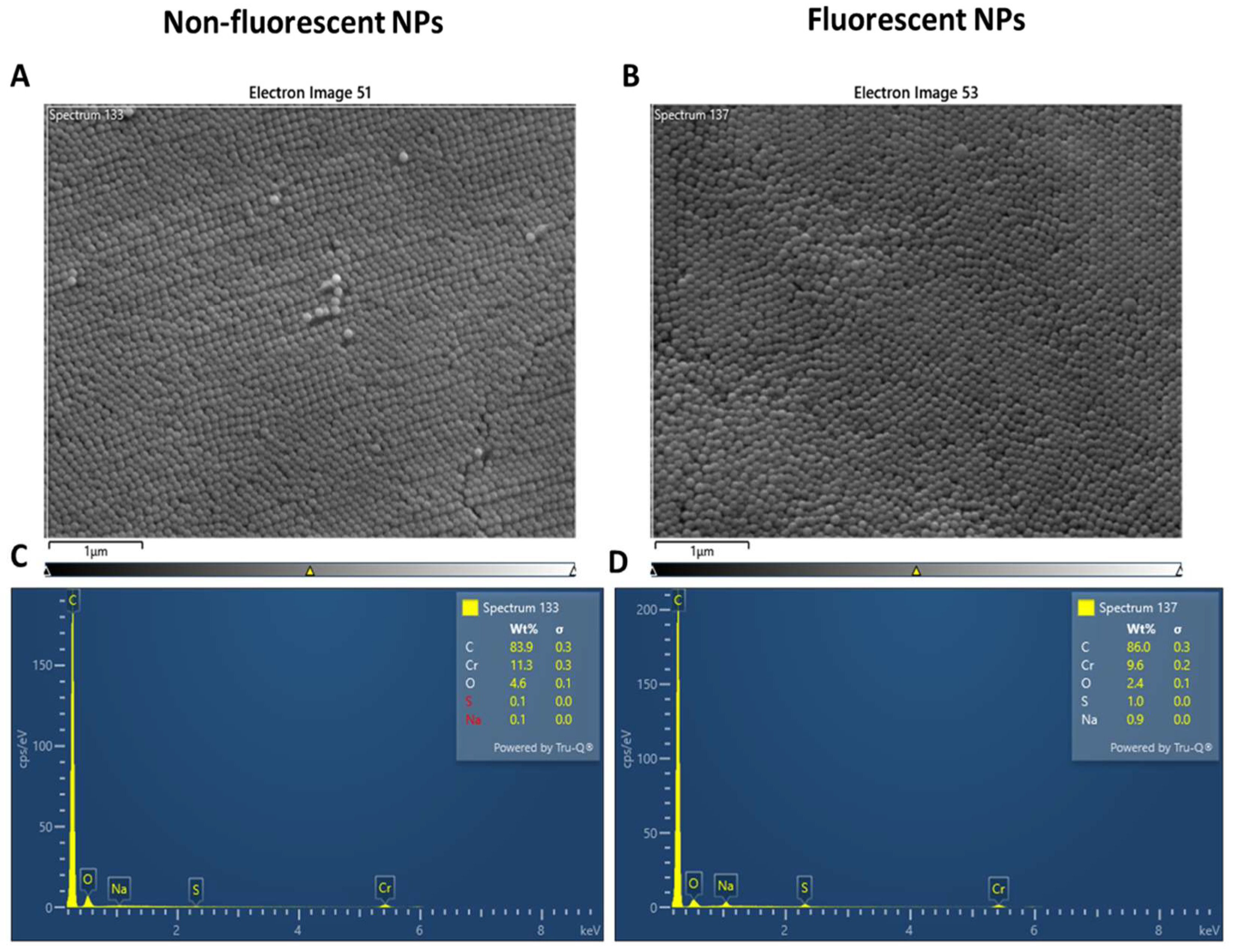
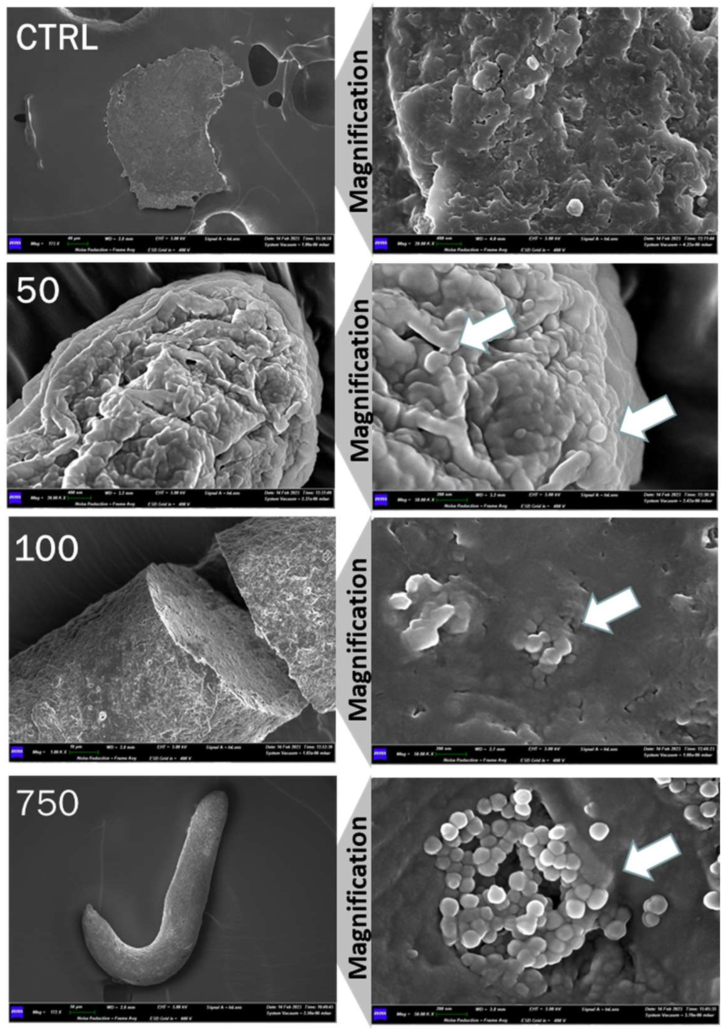
2.1. Analysis of Nanopolystyrene with Scanning Electron Microscopy (SEM)

Electron microscopy was used to characterize PSNPs. PSNPs had an average size of 100 nm, according to the manufacturer’s specifications (Figure 1A,B). Energy dispersive X-ray analysis (EDX) was used to confirm the absence of metals or other contaminants. The spectrum reveals only the presence of carbon, confirming the purity of the solution and the presence of oxygen due to the presence of water residue. Chromium is added to perform SEM analysis, and Na and S are needed as preservatives (Figure 1C,D).
Excrements were collected from the vial’s walls of treated and untreated wild-type flies and analyzed with SEM (Figure 2). The feces of the negative control were devoid of PSNPs, while those of the treated flies showed the presence of PSNPs averaging 100 nm, suggesting that the digestion process did not lead to an alteration of PSNPs size. At the highest concentration of PSNPs, the particles form aggregates in feces.

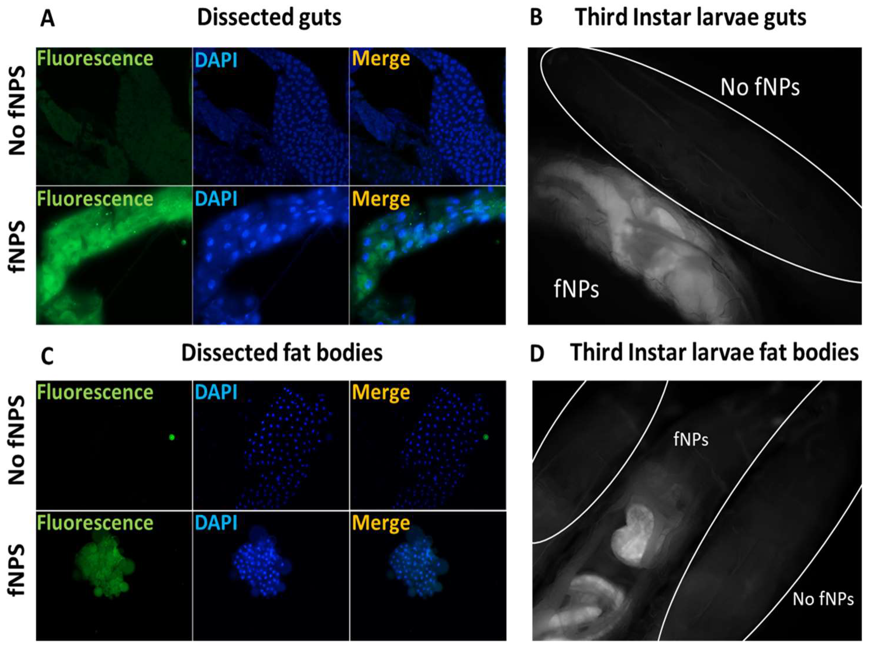
2.2. Biodistribution and Absorption

The ingestion of fluorescent PSNPs (fPSNPs) allowed the study of the biodistribution of plastics in larvae. Live and dissected OR-R larvae were observed under a fluorescence microscope (Figure 3). In living larvae, ingested fPSNPs are found in the gut (Figure 3B) and in body structures that are likely to be fat bodies. The analysis of dissected larvae confirms the presence of fNPs in guts and fat bodies (Figure 3A,C). The noticeable presence of PSNPs in fat bodies is probably due to the lipophilic nature of PSNPs (Figure 3D) and is indicative of the passage of PSNPs across the intestinal barrier and their release in the haemolymph (the fly blood).

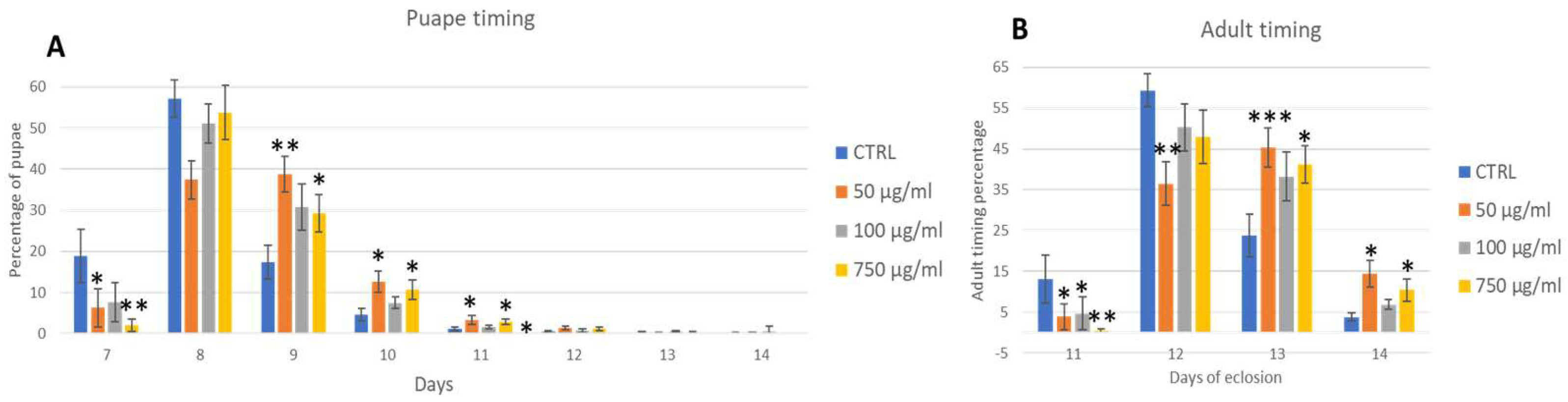
2.3. Development Traits (Life Cycle and Eclosion)

Wild-type parental flies were fed with PSNPs added to the instant food at various concentrations and left to lay eggs overnight on the PSNPs food. A significant reduction in the developmental traits of the offspring was observed. First, we assessed developmental parameters, such as the timing of pupae and adult emergence. Figure 4A,B shows the results. In untreated control, pupae begin to appear on day 7 after egg laying (AEL), and a significant reduction in pupae’s number in 50 µg/mL and 750 µg/mL conditions was found. On the 8th day, the difference disappears, even if the tendency remains, and from the 9th day on, the number of pupae increased in treatments, balancing the first-day difference. The result of this analysis thus shows a delay of 24 h in the larvae–pupae development. Adults’ emergence follows the same pattern except for 50 µg/mL, which remains significantly reduced on the second day. Interestingly, the concentration of 100 µg/mL does not affected this parameter. Eclosed adults show no differences in number (about 93% of pupae develop into adults in all conditions) or sex ratio. (Figure S1A,B in Supplementary Materials; Development traits of exposed adults). No obvious morphological defects were observed in adults.

2.4. Weight

Considering the delay in larvae–pupae development, the weight of adults was investigated. Figure 5 shows that the weight of treated male and female adults was significantly reduced with respect to the untreated flies.

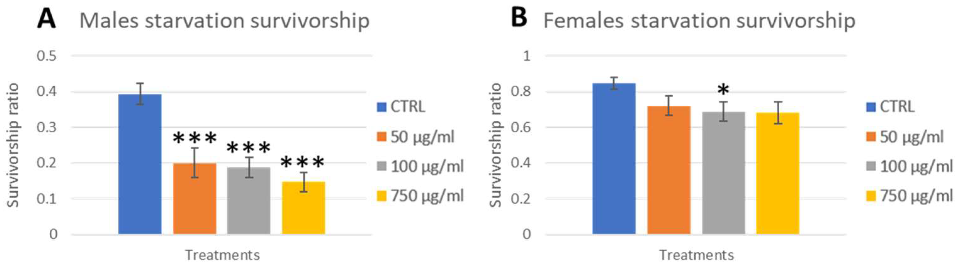
2.5. Stress Response

To assess if the developmental delay and reduced weight had any effects on adult fitness, we exposed flies to different secondary stresses (after PSNPs ingestion): starvation and exposure to high temperatures (seizure test). For the starvation assay, previously treated flies were left without food in empty vials for 24 h and scored for mortality every hour for the first 12 h (Figure S2A,B in Supplementary Materials; Starvation in exposed adults) and finally after 24 h (Figure 6A,B). No significant differences were observed during the first 12 h. After 24 h, males exposed showed significantly higher mortality at all concentrations. Females also showed a clear, although not significant, tendency to be less resistant to starvation at all concentrations.
For the seizure test, we exposed flies to 42 °C for 2 min and evaluated the number of fainting flies and recovery time. No significant differences were observed, suggesting the nervous system be not affected by PSNPs under our conditions.

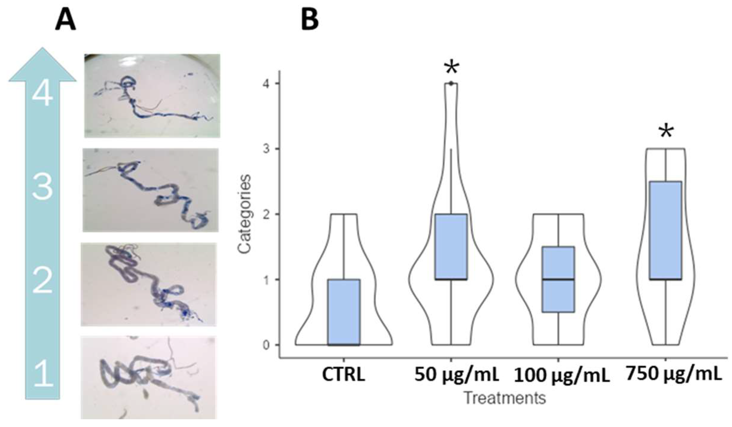
2.6. Larval Crawling and Wall Body Contraction

A larval crawling assay was performed on third instar OR-R larvae. No significant differences were found in terms of length of the crawls performed within a given time and body contractions per minute (Figure S3 in Supplementary Materials; crawling assay and wall body contraction screening in wild-type third instar larvae fed with PSNPs). These data showed that PSNPs do not affect movement ability or larval muscle functionality.

2.7. Adults Climbing

A climbing assay on wild-type and lig4 mutant adults was performed. DNA ligase 4 is a factor involved in DNA damage response and specifically catalyzes the final step of DNA end ligation in the NHEJ pathway [58]. In Drosophila, lig4 mutants are viable and sensitive to ionizing radiation during embryonic development. Interestingly, recently, lig4 mutant adults were found to be sensitive to nutrient stress, suggesting that lig4 is required for maintaining health and longevity in D. melanogaster [59]. The lig4 mutant was used to evaluate the effects of PSNPs in an already known stress-sensitive background. The assay was carried out on 3 days- (Figure 7, upper panel) and 18 days-old (Figure 7, lower panel) flies to evaluate the contribution of PSNPs toxicity to aging. While in OR-R no difference was found in the flies’ climbing ability, in lig4 mutants, after an initial decrease in climbing performance in young adults, the 18 days old male treated flies maintained higher locomotor activity than untreated flies. Note that the two age groups consisted of the same flies that were constantly maintained on food mixed with PSNP. Dead flies were recorded. In lig4 mutants, 3% control males, 13% 50 µg/mL, 4% 100 µg/mL, 1% 750 µg/mL, and 5% control females, 1% 50 µg/mL, 5% 100 µg/mL, and 10% 750 µg/mL, were found dead after 18 days. No OR-R flies died.

2.8. Trypan Blue and Smurf Assay

The Trypan Blue test was performed on third instar wild-type larvae to assess potential intestinal damage following PSNPs ingestion. Based on the extent of blue staining, the dissected guts were classified into five classes, from 0 to 4 (Figure 8A). A significant, though not drastic, increase in cell death was found at concentrations of 50 μg/mL and 750 μg/mL. To evaluate gut damage in adults, the Smurf assay was performed on w1118 flies fed with PSNPs. No significant results were recorded, but an interesting trend of smurf flies was found in both males and females, increasing with concentrations (Figure 8B). Considering the results, we continued to investigate the impact of PSNPs specifically on larval guts with the comet assay and qPCR on apoptotic gene expression.

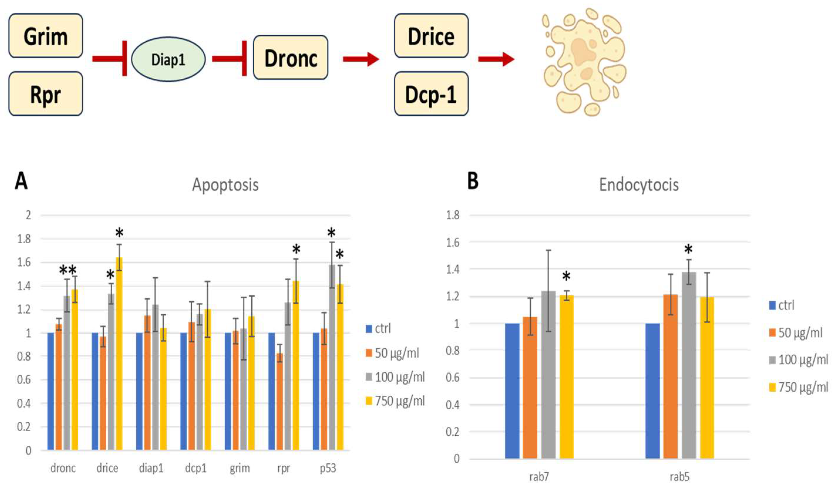
2.9. Pro-Apoptotic Gene Expression
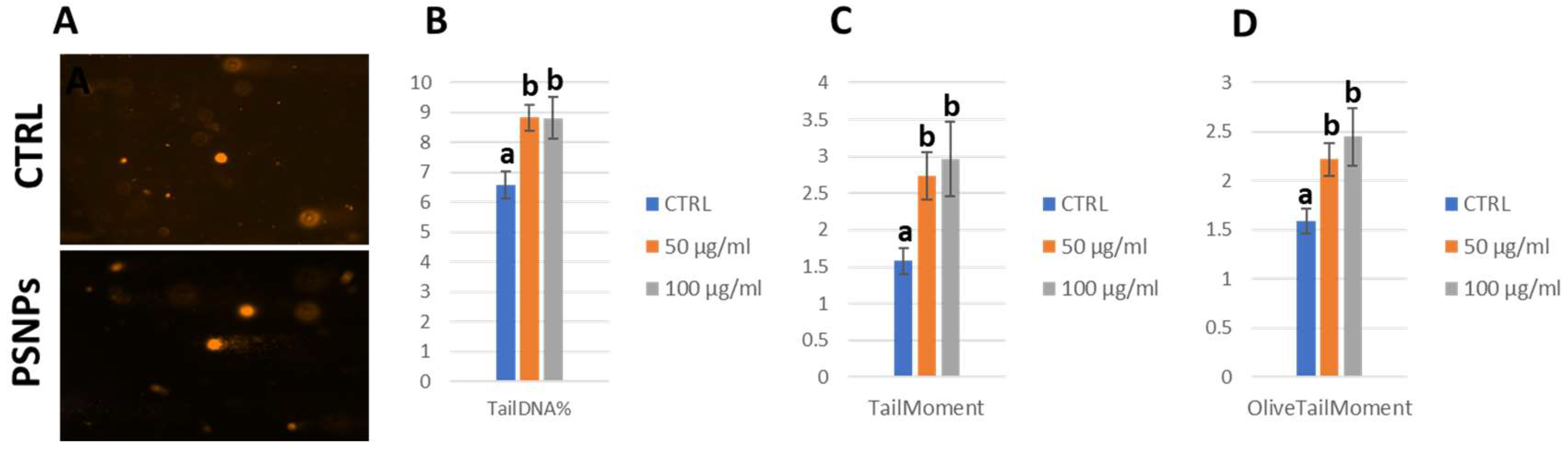
Given the presence of larval intestinal damage detected by the Trypan blue assay, a qPCR was carried out to evaluate the expression of apoptotic genes in larval guts. Caspases are the main executors of the apoptosis process. Important factors for the apoptotic response are Dronc (a caspase-9-like initiator caspase), Dcp-1, and Drice, which are caspase-3-like effector caspases. In the absence of apoptotic signals, inhibitors of apoptosis proteins (IAPs), such as Diap1, bind to caspases and inhibit their activity. In the presence of apoptotic stimuli, the pro-apoptotic proteins Reaper (Rpr), Hid, and Grim bind to Diap1 and release the caspases [60]. A schematic and simplified drawing of the described apoptotic process is shown in Figure 9. Another important player in the apoptotic process is the nuclear transcription factor p53, which regulates the expression of genes involved in apoptosis as well as growth arrest and senescence in response to genotoxic or cellular stress [61]. In our qPCR analysis, the expression of dronc, drice, diap1, dcp1, grim, rpr, and p53 was evaluated in larval guts.
Expression of the pro-apoptotic genes rpr, drice, and dronc was significantly increased in two concentrations (100 and 750 μg/mL) compared to the negative control, indicative of the presence of an apoptotic stimulus, as confirmed also by the higher expression of p53, a cellular stress response gene. The apoptotic stimulus detected is probably consequent to gut damage shown in Figure 8. The PSNPs also seem to stimulate endocytosis, as shown by the upregulation of the two markers of the endolysosomal pathway, rab5 and rab7, in treated flies. Rab5 is a marker for early endosomes, replaced by Rab7 during maturation [62]. Although only the highest concentrations of PSNP result in significant differences, there is a general trend toward an increased expression of endocytic markers in all the treated samples, indicating that transmembrane transport may participate in PSNP uptake.

2.10. Comet Assay

To further confirm that pro-apoptotic stimulation and overexpression of p53 in larval guts were associated with DNA damage, we carried out a comet assay in these tissues (Figure 10A). Over the past few years, comet assays have been successfully used in Drosophila to study genotoxicity as well as DNA repair [63]. For this analysis, we focused on the lower concentrations, 50 μg/mL and 100 μg/mL.
We scored specifically three indexes: tail DNA%, tail moment, and olive tail moment. Both concentrations were significant compared with the negative control, confirming the presence of DNA damage in gut cells. All indexes were significantly higher in treated flies with respect to the control (Figure 10B–D).

2.11. Carcinogenic Potential

To assess if PSNPs can favor carcinogenesis, we used genetically predisposed warts mutant flies and exposed them to 0 µg/mL, 50 µg/mL, 100 µg/mL, and 750 µg/mL of PSNP. warts encodes for a tumor suppressor factor of the Hippo pathway involved in tissue growth control. In Drosophila, mutations in the wts gene are recessive lethal, while viable heterozygous adults occasionally show large tumorous outgrowths in their bodies. This genetic background has been used to evaluate the carcinogenic potential of several compounds and can be used in toxicological studies. Heterozygous wts/+ flies were selected and scored for tumoral masses after PSNPs exposure (Figure 11A,B). As shown in Figure 11C, in two concentrations, we observed an increased number of abnormal masses, except for the concentration of 100 µg/mL.

3. Discussion

Plastic pollution is increasing every year and is considerable both in the oceans, seas, freshwater lands, and the atmosphere [64,65]. Chemical and physical factors can induce its degradation, leading to the formation of micro- and nano-fragments that multiply its toxicity on different scales that need to be deepened [66]. Their presence affects organisms, inducing oxidative stress, inflammation, fibrosis, and altered metabolism [67,68,69,70]. In this study, we decided to focus on polystyrene nanoparticles (PSNPs) for several reasons. PSNPs are one of the most prevalent NPs in the environment and are widely used in food packaging, posing a hazard to humans [71].
In our study, we used Drosophila melanogaster to study the impact of PSNPs exploiting their genetic similarity with humans and their well-recognized usage for biomedical research [54,55,56,57]. Initially, an overall characterization of PSNPs confirmed their dimensions and chemical composition. The PSNPs were administered to flies by ingestion mixed with standard fly food. Three different concentrations were used in the study based on previous papers found in the literature and our published data [31,37]. Parental individuals were crossed in the presence of PSNPs, and the laid embryos developed into adults on contaminated food to determine chronic exposure throughout development. In larvae, the distribution of fluorescent NPs indicates that PSNPs cross the intestinal barrier as they were found abundantly in fat bodies, perhaps as a consequence of their lipophilic nature. Such crossing is at least partly attributable to an endocytosis process, as suggested by the expression of endocytosis-related genes in this tissue. No fluorescence was evident in other organs.
Chronically exposed individuals showed increased pupal development time and adult emergence of about 24 h compared to control. Tu et al. [72] found a similar developmental retardation, but in later generations.
We can assume that PSNPs physically reduce the amount of actual ingested food, limiting storage. This can be confirmed by the fact that treated flies have a significant reduction in weight. Also, the trapped PSNPs in the fat bodies could provide a “second hit” when flies try to use their lipid reservoir. In fact, treated flies deprived of food showed less resistance to starvation, probably for the previous reasons. It can be concluded that the ingestion of NPs results in a general alteration of metabolism that leads to a fitness reduction in Drosophila.
Exposure to PSNPs in both wild-type larvae and adults showed no adverse effect on their movement ability. In contrast, Kauts et al. (2023) [73] found a reduction in larval crawling and adult climbing ability in the wild type, but using a different type of NPs, PET. Concerning the DNAlig4 mutants, we observed that aged flies exposed to PSNPs, after an initial decrease in their climbing performance, seemed to rescue their ability, surpassing untreated flies. Similar favorable results were obtained by Liang and colleagues [74], who studied the impact of PET microplastics of 2 µm on the lifespan of wild-type adult males. They observed that exposure to PET at a concentration of 1 g/L induced a slight increase in lifespan. This result is explained by the authors with the hormetic phenomenon that consists in the development of a positive response in individuals exposed to mild stress. It was already demonstrated with low dose radiation, heat shock, and hypergravity [75,76,77] and depends in part on DNA repair factors like ATR, ATM, and stress signal transducers (Sir2/SIRT1, JNK, p53, and dFOXO) in flies [78]. Moreover, in flies, the radioadaptive response, which is closely related to hormesis, relies on LIG4 [79]. For these reasons, in our experiment, it is difficult to assume that the beneficial effect observed in lig4 mutants can be attributed to hormesis. We can speculate that exposure to NPs increased the susceptibility of the lig4 mutants to mortality and resulted in the selection of the strongest individuals. The climbing test at 18 days was carried out on the lig4 flies that remained alive and resulted in better motor activity than the untreated control that did not undergo such selection. More studies are needed to understand this interesting phenomenon.
While adult guts showed only a light tendency to be affected by PSNPs, larvae showed a significant presence of cell death in the intestine. That prompted us to verify how PSNPs administration was translated into an apoptotic stimulus by evaluating the modulation of some genes associated with cellular death. We observed a significant increase in rpr expression, which is an initiator of apoptosis, and in dronc and drice, which actuate apoptosis [80,81]. Moreover, there was a significant increase in the p53 transcript, a cellular stress response protein, confirming the presence of some kind of perturbation in the gut and suggesting an apoptotic response. Future biochemical analysis is needed to verify whether modulation of gene expression results in modulation and/or activation at the protein level. A comet assay on larval guts showed a significant amount of DNA damage, possibly related to the cell death detected. Alaraby et al. [42] also observed increased DNA damage with the comet assay linked to oxidative stress in larvae using a true-to-life solution of PET nanoplastics. They used hemocytes, assessing that once absorbed, nanoplastics can induce DNA damage in other cellular types as well.
To assess the carcinogenic potential of PSNPs, we used mutants defective in warts. Warts is a serine/threonine kinase that phosphorylates Yorkie, thus controlling organ growth and cellular proliferation downstream of the Hippo pathway [82]. wts heterozygous flies develop melanotic masses spreading throughout their bodies, making them a suitable model to study the impact of the environment on cancer-predisposed individuals [83]. We found an increased number of tumoral masses, showing that, although wild types did not show any phenotypic anomalies, a predisposed organism can be more susceptible, as in the case of cancer. A possible explanation for the increased presence of tumors in adult warts heterozygote mutants could be LOH as a consequence of the DNA damage detected by the comet assay in larval gut tissue that contributes to genome instability [84].
In general, we found that male flies were more susceptible than females to NPs exposure. Sex-related differences were also found by Urbisz and colleagues [85] who observed histological alterations in the midgut that were more prominent in males after exposure to micro- and nano-polystyrene particles. This sex-dependent dimorphic response could be a consequence of the fact that some genes related to stress response evade the dosage compensation effect, resulting in more expression in females [86]. More generally speaking, several significant differences have been observed between Drosophila males and females in the regulation of metabolism, in response to nutritional challenges, and in immune response. This sex diversity is expected to result from differences in gene expression. However, the sex-specific differences are still underestimated and therefore poorly understood [87].
Differences in biological responses between sexes were also observed in other organisms. For instance, in the crustacean Macrobrachium nipponense, PSNPs of 75 nm can induce an initial increase followed by a decrease in sex hormones (estradiol, progesterone, and testosterone), causing alterations in the expression of sex-related genes in the gonads [88]. Moreover, in zebrafish, the co-exposure of 50 nm PS and triclosan, which is an antimicrobial agent found in personal care products, induced opposite effects in the two sexes. In males, the authors observed an increased presence of triclosan in the testes, while in females, the amount in the ovaries decreased. Following an opposite trend, male’s genes involved in sex-hormones decreased while females increased [89].
In conclusion, our findings showed that ingested PSNPs trigger metabolic and genetic modifications in the exposed organisms, eventually delaying development and accelerating death and disease. Moreover, we observed that the presence of mutations can induce different results, suggesting the genetic background may be relevant in determining how an organism responds to PSNPs exposure.

4. Materials and Methods

4.1. Flies Husbandry and Nanopolystyrene Exposure

All of the strains were obtained from Bloomington Stock Center and maintained at a constant temperature with a relative humidity of approximately 60%, a temperature of 25 °C, and fed with standard food (Nutri-Fly® Bloomington Formulation). In particular, the following strains were used: Oregon R (OR-R), w1118, Lig45, and st,in,kni,p,wts3–17/TM3, Sb mutants. The fruit flies were cultured in the dark. warts mutant was kindly sent by Dr. Fiammetta Vernì from Sapienza University (Rome, Italy).
For all the treatments, flies were kept at 25 °C and fed with instant food (Flystuff Nutri-Fly Food, Instant Formulation. This formulation requires no heating for preparation) with or without polystyrene nanoparticles (PSNPs) (oral exposure) and is allowed to lay eggs overnight in experimental vials. The resulting F1 generation was used as third-instar larvae or 3- and 18-days old adults. Concentrations: negative control (0 µg/mL); treatments: 50 µg/mL; 100 µg/mL; 750 µg/mL of PSNPs.

4.2. Poyistyrene Nanoparticles

Fluorescent (f) and non-fluorescent PSNPs of 100 nm of diameter were purchased from Polysciences Europe GmbH Badener Str. 13 69493 Hirschberg an der Bergstrasse Germany (fPSNPs; Fluoresbrite® Yellow Green Microspheres; cod. 17150-10; 0.10 µm 2.5% particles in water solution; 4.55 × 10¹³ particles/mL; Excitation max = 441 nm; Emission max = 486 nm; PSNPs; cod. 00876-15; 0.10 µm 2.5% particles in water solution; 4.55 × 10¹³ particles/mL).

4.3. Scannng Electron Microscopy and EDX Analysis

To characterize PSNPs and wild-type excrements, we used scanning electron microscopy (SEM) (Gemini Field Emission SEM 500, ZEISS, Milan, Italy) equipped with an X-ray microanalysis system (EDS Oxford Inca 250 x-act). The first one was used to assess PSNPs morphology from the stock solution to flies’ feces; the second one was used to conduct an elemental analysis of the particles. For SEM analysis, 1 µL of particle sample was deposited on a stub and left dehydrated in air; then, a 5 nm film of chromium was deposited on it with Sputter Quorum 150T ES to make it conductive. Flies’ excrements were collected from vial walls, left to dry in the air, and then the same protocol used for particles was followed. We used SEM analysis to obtain the morphological and chemical details of our samples.

4.4. Fluorescence Microscopy

To deepen PSNPs distribution in third instar larvae, we used fluorescence microscopy (ZeissAxio Imager M2) on living and dissected larvae. Third instar larvae were washed three times in PBS to remove external residues of food and then observed under the microscope. To assess if fPSNPs were absorbed by intestinal cells and bioaccumulated into other organs, we washed the larvae three times, dissected guts and fat bodies in a 0.7% physiological solution, fixed them in 4% formaldehyde, counterstained them with DAPI, and placed them on microscope slides for the observations.

4.5. Development Traits and Weight

To assess if PSNPs have an influence on the development of OR-R flies, we scored the timing of pupae formation and adult eclosion, viability, and sex differences. To do so, we counted every day at the same hour the number of pupae and adults, dividing them into males and females. The experiment was repeated 12 times (around 1200 pupae and adults scored). Adults were also observed under a stereomicroscope to detect eventual abnormalities in the eye, wings, and whole body. For weight, 100 3 days old OR-R adults were collected and divided into two groups of 50 flies, males and females, for three experiments (300 males and 300 females for each concentration).

4.6. Stress Tolerance

To evaluate if PSNPs exposure can influence flies’ survival mechanisms, we exposed non-treated and treated (0, 50, 100, and 750 μg/mL) OR-R flies to starvation and heat. For the starvation, we followed the Shaposhnikov protocol with some modifications [90]. Briefly, 3 days old flies were collected, split into males and females, and grouped ten by ten in empty vials. The assay was performed three times with 50 males and 50 females each (300 flies in each concentration). We scored the number of dead flies every hour for the first 12 h and then after 24 h. For the heat-shock assay (seizure assay), we followed the Mituzaite protocol with some modifications [91]. Briefly, 3 days old flies were collected and grouped for the starvation assay. The experiment was performed three times with 30 males and 30 females each (180 flies for every concentration). Every vial was immersed in water at 42.5 °C for 2 min, scoring how many flies faint every 20 s. Then, we scored the timing of recovery and how many flies needed more than 4 min to do so.

4.7. Climbing Assay

We performed a standard climbing assay on OR-R and DNAlig4 defective mutants following the Iuso et al. protocol with some modifications [92]. We collected 3- and 18-days old individuals and divided them into males and females. The experiment with the OR-R was repeated three times, scoring 50 males and 50 females each (300 flies for every concentration) grouped ten by ten in empty vials. The DNAlig4 experiment was repeated six times, scoring 50 males and 50 females each (600 flies in total). The assay consisted in measuring how many flies could reach a height of 7 cm in 20 s after being tapped down. The 3 days old and the 18 days old flies were the same; eventually, dead individuals were scored too.

4.8. Larval Crawling Assay and Wall Body Contraction

Wild-type larvae mobility was assessed by comparing treated and non-treated individuals. We used four conditions: 0, 50, 100, and 750 μg/mL. For the larval crawling assay, we followed Nichols and colleagues [93] protocol with some variations. A total of 36 OR-R larvae for each PSNPs concentration (three experiments, 12 each) were placed on a Petri dish with 1.5% agarose gel. We tracked the distance crossed per minute with graph paper. For the wall body contraction assay, the same number of larvae were placed singularly in physiological solution drops with a little bit of fresh waste. Then, we scored the number of contractions per minute under a stereomicroscope.

4.9. Smurf Assay (Intestinal Integrity Test)

To gain insight into gut damage and barrier dysfunction in adults, we performed a Smurf assay [94]. The experiment was repeated three times, dividing males and females with around 107 flies per concentration. Three days old w1118 flies grown in four different concentrations (0, 50, 100, and 750 μg/mL) were left on food added with FCF Brilliant blue for 24 h. Then, under a stereomicroscope, they were scored in no smurf and smurf based on the absorption of the dye. The w1118 flies were used because they are characterized by a lighter color of the abdomen, which allows a better view of the intestine [95].

4.10. Trypan Blue Assay

We performed the Trypan blue assay as a first sight into gut damage in larvae. We followed the Carmona et al. (2015) protocol with some light modifications [96]. Wild-type third instar larvae grown in four concentrations (0, 50, 100, and 750 μg/mL) were collected and washed three times in PBS. Then, guts were dissected, fixed in 4% formaldehyde, and stained in a 0.02% Trypan blue solution (diluted in PBS) for 30 min at 25 °C. A total of 15 guts per concentration were scored (5 per replicate). Finally, guts were placed on microscope slides and scored into five categories [96]: “no color = 0; light blue = 1; darkly stained nuclei = 2; large patches of darkly stained cells = 3; or complete staining of most cells in the tissue = 4”.

4.11. Total RNA Extraction and RT-qPCR

Variations in gene expression in treated and non-treated OR-R third instar larvae guts were evaluated by qPCR. We analyzed apoptosis-related genes (hid, rpr, dronc, drice, p53, diap1, and dcp1) and endocytosis-related genes (rab5 and rab7). Larvae were collected and washed three times in PBS; then, we dissected 40 guts per concentration. Total RNA was isolated from samples using TRIzol (Thermo Fisher Scientific, Waltham, MA, USA, Cat. 15596026) according to the manufacturer’s instructions.
The integrity of the RNA was immediately checked using 1.2% agarose gel electrophoresis (1 µg samples). The RNA concentration was assessed by the Qubit dsDNA BR Assay Kit (Invitrogen, Life Technologies, Carlsbad, CA, USA, cat. Q32850) used for the quantification of cfDNA on the Qubit 3.0 Fluorometer (Invitrogen, Life Technologies, Carlsbad, CA, USA), and RNA samples were stored at −80 °C until use.
First-strand cDNA synthesis was performed from 1 µg of total RNA using the Maxima H Minus First Strand cDNA Synthesis Kit with dsDNase (Thermo Fisher Scientific, Waltham, MA, USA, cat. K1681) according to the manufacturer’s instructions. The dsDNase enzyme was used in the first step to remove any contamination of the genomic DNA; at the same time, a no-RT control reaction was carried out.
RT-qPCR was performed in a final volume of 20 µL, including 15 ng of the cDNA product, specific forward and reverse primers (500 nM), and PowerUp SYBR Green Master Mix (Applied Biosystems cat. A25741, Waltham, MA, USA), which is a ready-to-use cocktail containing all components (including ROX Reference Dye at a final concentration of 500 nM).
The amplification reaction was performed in an Applied Biosystems 7300 system (ThermoFisher Scientific, Rockford, IL, USA), and all the PCRs were performed under the following conditions: 2 min at 50 °C, 2 min at 95 °C, and 40 cycles of 15 s at 95 °C and 1 min at 60 °C in 96-well optical reaction plates (Applied Biosystems, Waltham, MA, USA). The specificity of the qRT-PCR reactions was monitored through melting curve analysis (60–95 °C), after 40 cycles.
The relative expression ratio was analyzed using a delta–delta Ct (2−ΔΔCt) method [97]. actin and α tubulin were chosen as the reference genes, and the control was used as the calibrator. Three biological replicates were performed, and all samples were carried out in duplicate.
The genes analyzed and NCBI Reference Sequence were hid (NT_037436.4), rpr (NT_037436), dronc (NT_037436), drice (NT_037436), p53 (NT_033777.3), diap1 (NT_037436), dcp1 (NT_033778), rab5 (NT_033779), and rab7 (NT_033779). The primer sequences used in this study are listed in Table S1 in the Supplementary Materials (Sequence of the primers used for the qPCR analyses). Some primers were found in the literature, others were constructed on gene sequences with Primer Express 3.0 software (Applied Biosystems, USA); primers were synthesized and bought from Eurofins Genomics (Ebersberg, Germany). The validation of primers and analysis is consistent with the MIQE guidelines [98,99,100,101,102,103,104,105].

4.12. Comet Assay

We performed the alkaline comet assay on gut cells to verify the presence of DNA damage in treated (50 and 100 μg/mL) and untreated OR-R larvae. We followed the standard protocol with some modifications to adapt it to Drosophila [106,107]. Third instar larvae were collected and washed in PBS three times. Then, 40 guts per concentration were dissected (two experiments, 20 each) and incubated with 500 μL of 0.25% Trypsin, 2.21 mM EDTA, 1X (Corning cat.25-053-Cl) for 18 min at 40 °C.
To collect cells, we centrifuged (800× g) cell suspension, collected the supernatant, then recentrifuged again (3000× g) and recovered the pellet by resuspending in 100 uL of 0.7% low melting point agarose at 37 °C. Then, 50 uL were pipetted on Trevigen CometSlide (2 wells, catalog number 4250-200-03) and kept at 4 °C for 15 min. Then, slides were incubated for 1 h at 4 °C in the dark in a Lysis solution (NaCl 2.5 M, EDTA 0.1 M, Tris 10 mM, DMSO 10%, Triton X100 1%, pH). The alkaline treatment was in the running solution (NaOH 300 mM, EDTA 1 mM, pH) for 20 min, followed by an electrophoretic run for 30 min at 300 mA. Slides were washed three times (5 min each) with neutralization buffer (Tris 0.4 M pH 7.4) and then immersed in 70% ethanol at room temperature for 30 min and stored at 5 °C. The nuclei were stained with ethidium bromide for 5 min (2 μg/mL) and washed two times with dH2O. We performed two experiments with two technical replicates. Slides were observed with a fluorescence microscope (ZeissAxio Imager M2) and a 490 nm filter. For each condition, all cells on an area of the comet slide were analyzed with CASP ASP 1.2.3beta2 Image Analysis of Comet Assay.

4.13. Carcinogenic Potential

To assess the carcinogenic potential of PSNPs we exposed warts mutant flies following the same exposition protocol as the previous experiments. Parental flies (wts/TM3 X OR-R) were left mating on food contaminated with the different NPs concentrations overnight. Then, 3 days old adults were collected, selecting wts/+ flies based on the absence of the Stubble phenotype. The time of NPs’ exposure is about 13 days (from embryo to 3 days old in adults, 10 + 3 days) [108]. Then, we scored an average of 192 wts/+ flies per concentration, counting the number of tumoral masses and recording their location in the fly bodies (Head, Body, Wing, Leg, Halter).

4.14. Statistical Analysis

For the statistical analysis of data, we used Excel and Jamovi (The jamovi project (2024). jamovi (Version 2.5) [Computer Software]) to analyze significance. We evaluated the distribution of data with the Sahpiro–Wilk test to decide if to use parametric or not parametric tests based on results. Then, we applied two or one tailed Student t test or One way ANOVA (parametric or non-parametric). For statistically significant values, * = p < 0.05; ** = p < 0.005; *** = p < 0.001.

Supplementary Materials

The following supporting information can be downloaded at: https://www.mdpi.com/article/10.3390/ijms25147965/s1.

Author Contributions

Conceptualization, M.A., P.M., D.G. and A.M.G.P.; methodology, M.A., A.M.G.P., S.C., O.Z., D.G. and P.M.; software, M.A., O.Z.; validation, M.A., A.M.G.P. and D.G.; formal analysis, M.A., P.M., A.M.G.P. and D.G.; investigation, M.A.; resources, A.M.G.P. and D.G.; data curation, M.A., P.M., O.Z. and S.C.; writing—original draft preparation, M.A., A.M.G.P. and P.M.; writing-review and editing, MA. and A.M.G.P.; project administration, A.M.G.P. and D.G.; funding acquisition, A.M.G.P. and D.G. All authors have read and agreed to the published version of the manuscript.

Funding

This research was funded by Dept. MESVA, University of L’Aquila (FFO to A.M.G.P. and PSD2311 to D.G.). P.M. was supported by a fellowship from “INFN-Laboratori Nazionali del Gran Sasso” and M.A. was supported by a PhD fellowship from University of Teramo.

Institutional Review Board Statement

Not applicable.

Informed Consent Statement

Not applicable.

Data Availability Statement

Data is contained within the article and Supplementary Materials.

Acknowledgments

We thank Lorenzo Arizza and Centro di Microscopie-Università degli Studi dell’Aquila for the support for SEM analysis. We thank Fiammetta Vernì (Sapienza University) for warts mutant.

Conflicts of Interest

The authors declare no conflicts of interest.

References

  1. Ostle, C.; Thompson, R.C.; Broughton, D.; Gregory, L.; Wootton, M.; Johns, D.G. The rise in ocean plastics evidenced from a 60-year time series. Nat. Commun. 2019, 10, 1622. [Google Scholar] [CrossRef] [PubMed] [PubMed Central]
  2. De Frond, H.; Hampton, L.T.; Kotar, S.; Gesulga, K.; Matuch, C.; Lao, W.; Weisberg, S.B.; Wong, C.S.; Rochman, C.M. Monitoring microplastics in drinking water: An interlaboratory study to inform effective methods for quantifying and characterizing microplastics. Chemosphere 2022, 298, 134282. [Google Scholar] [CrossRef] [PubMed]
  3. Jambeck, J.R.; Geyer, R.; Wilcox, C.; Siegler, T.R.; Perryman, M.; Andrady, A.; Narayan, R.; Law, K.L. Marine pollution. Plastic waste inputs from land into the ocean. Science 2015, 347, 768–771. [Google Scholar] [CrossRef] [PubMed]
  4. Wurm, F.R.; Spierling, S.; Endres, H.J.; Barner, L. Plastics and the Environment-Current Status and Challenges in Germany and Australia. Macromol. Rapid Commun. 2020, 41, e2000351. [Google Scholar] [CrossRef] [PubMed]
  5. Čerkasova, N.; Enders, K.; Lenz, R.; Oberbeckmann, S.; Brandt, J.; Fischer, D.; Fischer, F.; Labrenz, M.; Schernewski, G. A Public Database for Microplastics in the Environment. Microplastics 2023, 2, 132–146. [Google Scholar] [CrossRef]
  6. Cerasa, M.; Teodori, S.; Pietrelli, L. Searching Nanoplastics: From Sampling to Sample Processing. Polymers 2021, 13, 3658. [Google Scholar] [CrossRef] [PubMed] [PubMed Central]
  7. Plastics—The Facts 2019 An Analysis of European Plastics Production, Demand and Waste Data. Available online: https://plasticseurope.org/wp-content/uploads/2023/10/Plasticsthefastfacts2023-1.pdf (accessed on 14 July 2024).
  8. Eriksen, M.; Cowger, W.; Erdle, L.M.; Coffin, S.; Villarrubia-Gómez, P.; Moore, C.J.; Carpenter, E.J.; Day, R.H.; Thiel, M.; Wilcox, C. A growing plastic smog, now estimated to be over 170 trillion plastic particles afloat in the world’s oceans-Urgent solutions required. PLoS ONE 2023, 8, e0281596. [Google Scholar] [CrossRef] [PubMed] [PubMed Central]
  9. Karbalaei, S.; Hanachi, P.; Walker, T.R.; Cole, M. Occurrence, sources, human health impacts and mitigation of microplastic pollution. Environ. Sci. Pollut. Res. Int. 2018, 25, 36046–36063. [Google Scholar] [CrossRef] [PubMed]
  10. van Wijnen, J.; Ragas, A.M.J.; Kroeze, C. Modelling global river export of microplastics to the marine environment: Sources and future trends. Sci. Total Environ. 2019, 673, 392–401. [Google Scholar] [CrossRef] [PubMed]
  11. Gui, J.; Sun, Y.; Wang, J.; Chen, X.; Zhang, S.; Wu, D. Microplastics in composting of rural domestic waste: Abundance, characteristics, and release from the surface of macroplastics. Environ. Pollut. 2021, 274, 116553. [Google Scholar] [CrossRef] [PubMed]
  12. Sharma, S.; Chatterjee, S. Microplastic pollution, a threat to marine ecosystem and human health: A short review. Environ. Sci. Pollut. Res. Int. 2017, 24, 21530–21547. [Google Scholar] [CrossRef] [PubMed]
  13. Kole, P.J.; Löhr, A.J.; Van Belleghem, F.G.A.J.; Ragas, A.M.J. Wear and Tear of Tyres: A Stealthy Source of Microplastics in the Environment. Int. J. Environ. Res. Public Health 2017, 14, 1265. [Google Scholar] [CrossRef] [PubMed] [PubMed Central]
  14. Abbasi, S.; Keshavarzi, B.; Moore, F.; Turner, A.; Kelly, F.J.; Dominguez, A.O.; Jaafarzadeh, N. Distribution and potential health impacts of microplastics and microrubbers in air and street dusts from Asaluyeh County, Iran. Environ. Pollut. 2019, 244, 153–164. [Google Scholar] [CrossRef] [PubMed]
  15. Liss, P.S. Microplastics: All up in the air? Mar. Pollut. Bull. 2020, 153, 110952. [Google Scholar] [CrossRef] [PubMed]
  16. Halle, L.L.; Palmqvist, A.; Kampmann, K.; Khan, F.R. Ecotoxicology of micronized tire rubber: Past, present and future considerations. Sci. Total Environ. 2020, 706, 135694. [Google Scholar] [CrossRef] [PubMed]
  17. Shin, H.; Kwak, M.; Lee, T.G.; Lee, J.Y. Quantifying the level of nanoparticle uptake in mammalian cells using flow cytometry. Nanoscale 2020, 12, 15743–15751. [Google Scholar] [CrossRef] [PubMed]
  18. Verma, A.; Stellacci, F. Effect of surface properties on nanoparticle-cell interactions. Small 2010, 6, 12–21. [Google Scholar] [CrossRef] [PubMed]
  19. Lunov, O.; Syrovets, T.; Loos, C.; Beil, J.; Delacher, M.; Tron, K.; Nienhaus, G.U.; Musyanovych, A.; Mailänder, V.; Landfester, K.; et al. Differential uptake of functionalized polystyrene nanoparticles by human macrophages and a monocytic cell line. ACS Nano 2011, 5, 1657–1669. [Google Scholar] [CrossRef] [PubMed]
  20. Alimi, O.S.; Budarz, J.F.; Hernandez, L.M.; Tufenkji, N. Microplastics and Nanoplastics in Aquatic Environments: Aggregation, Deposition, and Enhanced Contaminant Transport. Environ. Sci. Technol. 2018, 52, 1704–1724. [Google Scholar] [CrossRef] [PubMed]
  21. Yousif, E.; Haddad, R. Photodegradation and photostabilization of polymers, especially polystyrene: Review. SpringerPlus 2013, 2, 398. [Google Scholar] [CrossRef] [PubMed] [PubMed Central]
  22. Gewert, B.; Plassmann, M.M.; MacLeod, M. Pathways for degradation of plastic polymers floating in the marine environment. Environ. Sci. Process. Impacts 2015, 17, 1513–1521. [Google Scholar] [CrossRef] [PubMed]
  23. Jacquin, J.; Cheng, J.; Odobel, C.; Pandin, C.; Conan, P.; Pujo-Pay, M.; Barbe, V.; Meistertzheim, A.L.; Ghiglione, J.F. Microbial Ecotoxicology of Marine Plastic Debris: A Review on Colonization and Biodegradation by the “Plastisphere”. Front. Microbiol. 2019, 10, 865. [Google Scholar] [CrossRef] [PubMed] [PubMed Central]
  24. De Tender, C.; Devriese, L.I.; Haegeman, A.; Maes, S.; Vangeyte, J.; Cattrijsse, A.; Dawyndt, P.; Ruttink, T. Temporal Dynamics of Bacterial and Fungal Colonization on Plastic Debris in the North Sea. Environ. Sci. Technol. 2017, 51, 7350–7360. [Google Scholar] [CrossRef] [PubMed]
  25. Cui, R.; Kim, S.W.; An, Y.J. Polystyrene nanoplastics inhibit reproduction and induce abnormal embryonic development in the freshwater crustacean Daphnia galeata. Sci. Rep. 2017, 7, 12095. [Google Scholar] [CrossRef] [PubMed] [PubMed Central]
  26. Abdelkhaliq, A.; van der Zande, M.; Punt, A.; Helsdingen, R.; Boeren, S.; Vervoort, J.J.M.; Rietjens, I.M.C.M.; Bouwmeester, H. Impact of nanoparticle surface functionalization on the protein corona and cellular adhesion, uptake and transport. J. Nanobiotechnol. 2018, 16, 70. [Google Scholar] [CrossRef] [PubMed]
  27. Walczak, A.P.; Kramer, E.; Hendriksen, P.J.; Tromp, P.; Helsper, J.P.; van der Zande, M.; Rietjens, I.M.; Bouwmeester, H. Translocation of differently sized and charged polystyrene nanoparticles in in vitro intestinal cell models of increasing complexity. Nanotoxicology 2015, 9, 453–461. [Google Scholar] [CrossRef] [PubMed]
  28. Wu, B.; Wu, X.; Liu, S.; Wang, Z.; Chen, L. Size-dependent effects of polystyrene microplastics on cytotoxicity and efflux pump inhibition in human Caco-2 cells. Chemosphere 2015, 221, 333–341. [Google Scholar] [CrossRef] [PubMed]
  29. Lehner, R.; Wohlleben, W.; Septiadi, D.; Landsiedel, R.; Petri-Fink, A.; Rothen-Rutishauser, B. A novel 3D intestine barrier model to study the immune response upon exposure to microplastics. Arch. Toxicol. 2020, 94, 2463–2479. [Google Scholar] [CrossRef]
  30. Roursgaard, M.; Hezareh Rothmann, M.; Schulte, J.; Karadimou, I.; Marinelli, E.; Møller, P. Genotoxicity of Particles from Grinded Plastic Items in Caco-2 and HepG2 Cells. Front. Public Health 2022, 10, 906430. [Google Scholar] [CrossRef] [PubMed] [PubMed Central]
  31. Vecchiotti, G.; Colafarina, S.; Aloisi, M.; Zarivi, O.; Di Carlo, P.; Poma, A. Genotoxicity and oxidative stress induction by polystyrene nanoparticles in the colorectal cancer cell line HCT116. PLoS ONE 2021, 16, e0255120. [Google Scholar] [CrossRef] [PubMed] [PubMed Central]
  32. Bredeck, G.; Halamoda-Kenzaoui, B.; Bogni, A.; Lipsa, D.; Bremer-Hoffmann, S. Tiered testing of micro- and nanoplastics using intestinal in vitro models to support hazard assessments. Environ. Int. 2022, 158, 106921. [Google Scholar] [CrossRef] [PubMed]
  33. Yang, S.; Cheng, Y.; Chen, Z.; Liu, T.; Yin, L.; Pu, Y.; Liang, G. In vitro evaluation of nanoplastics using human lung epithelial cells, microarray analysis and co-culture model. Ecotoxicol. Environ. Saf. 2021, 226, 112837. [Google Scholar] [CrossRef] [PubMed]
  34. Xu, M.; Halimu, G.; Zhang, Q.; Song, Y.; Fu, X.; Li, Y.; Li, Y.; Zhang, H. Internalization and toxicity: A preliminary study of effects of nanoplastic particles on human lung epithelial cell. Sci. Total Environ. 2019, 694, 133794. [Google Scholar] [CrossRef] [PubMed]
  35. Shi, X.; Wang, X.; Huang, R.; Tang, C.; Hu, C.; Ning, P.; Wang, F. Cytotoxicity and Genotoxicity of Polystyrene Micro- and Nanoplastics with Different Size and Surface Modification in A549 Cells. Int. J. Nanomed. 2022, 17, 4509–4523. [Google Scholar] [CrossRef] [PubMed] [PubMed Central]
  36. Halimu, G.; Zhang, Q.; Liu, L.; Zhang, Z.; Wang, X.; Gu, W.; Zhang, B.; Dai, Y.; Zhang, H.; Zhang, C.; et al. Toxic effects of nanoplastics with different sizes and surface charges on epithelial-to-mesenchymal transition in A549 cells and the potential toxicological mechanism. J. Hazard. Mater. 2022, 430, 128485. [Google Scholar] [CrossRef] [PubMed]
  37. Poma, A.; Vecchiotti, G.; Colafarina, S.; Zarivi, O.; Aloisi, M.; Arrizza, L.; Chichiriccò, G.; Di Carlo, P. In Vitro Genotoxicity of Polystyrene Nanoparticles on the Human Fibroblast Hs27 Cell Line. Nanomaterials 2019, 9, 1299. [Google Scholar] [CrossRef] [PubMed] [PubMed Central]
  38. Xu, Y.; Shrestha, N.; Préat, V.; Beloqui, A. An overview of in vitro, ex vivo and in vivo models for studying the transport of drugs across intestinal barriers. Adv. Drug Deliv. Rev. 2021, 175, 113795. [Google Scholar] [CrossRef] [PubMed]
  39. Kelpsiene, E.; Torstensson, O.; Ekvall, M.T.; Hansson, L.A.; Cedervall, T. Long-term exposure to nanoplastics reduces life-time in Daphnia magna. Sci. Rep. 2020, 10, 5979. [Google Scholar] [CrossRef] [PubMed] [PubMed Central]
  40. Ekvall, M.T.; Gimskog, I.; Hua, J.; Kelpsiene, E.; Lundqvist, M.; Cedervall, T. Size fractionation of high-density polyethylene breakdown nanoplastics reveals different toxic response in Daphnia magna. Sci. Rep. 2022, 12, 3109. [Google Scholar] [CrossRef] [PubMed] [PubMed Central]
  41. Heinlaan, M.; Viljalo, K.; Richter, J.; Ingwersen, A.; Vija, H.; Mitrano, D.M. Multi-generation exposure to polystyrene nanoplastics showed no major adverse effects in Daphnia magna. Environ. Pollut. 2023, 323, 121213. [Google Scholar] [CrossRef] [PubMed]
  42. Alaraby, M.; Villacorta, A.; Abass, D.; Hernández, A.; Marcos, R. The hazardous impact of true-to-life PET nanoplastics in Drosophila. Sci. Total Environ. 2023, 863, 160954. [Google Scholar] [CrossRef] [PubMed]
  43. Tang, H.; Zhong, L.; Xu, Y.; Jin, Z.; Pan, Z.; Shen, J. Polypropylene microplastics affect the physiology in Drosophila model. Bull. Entomol. Res. 2023, 113, 355–360. [Google Scholar] [CrossRef] [PubMed]
  44. Shen, J.; Liang, B.; Zhang, D.; Li, Y.; Tang, H.; Zhong, L.; Xu, Y. Effects of PET microplastics on the physiology of Drosophila. Chemosphere 2021, 283, 131289, Erratum in Chemosphere 2022, 303, 134678. [Google Scholar] [CrossRef] [PubMed]
  45. Lu, Y.; Zhang, Y.; Deng, Y.; Jiang, W.; Zhao, Y.; Geng, J.; Ding, L.; Ren, H. Uptake and Accumulation of Polystyrene Microplastics in Zebrafish (Danio rerio) and Toxic Effects in Liver. Environ. Sci. Technol. 2016, 50, 4054–4060. [Google Scholar] [CrossRef] [PubMed]
  46. Qiao, R.; Sheng, C.; Lu, Y.; Zhang, Y.; Ren, H.; Lemos, B. Microplastics induce intestinal inflammation, oxidative stress, and disorders of metabolome and microbiome in zebrafish. Sci. Total Environ. 2019, 662, 246–253. [Google Scholar] [CrossRef] [PubMed]
  47. De Marco, G.; Oliveri Conti, G.; Giannetto, A.; Cappello, T.; Galati, M.; Iaria, C.; Pulvirenti, E.; Capparucci, F.; Angela Mauceri, A.; Ferrante, M.; et al. Embryotoxicity of polystyrene microplastics in zebrafish Danio rerio. Environ. Res. 2022, 208, 112552. [Google Scholar] [CrossRef] [PubMed]
  48. Lu, L.; Wan, Z.; Luo, T.; Fu, Z.; Jin, Y. Polystyrene microplastics induce gut microbiota dysbiosis and hepatic lipid metabolism disorder in mice. Sci. Total Environ. 2018, 631–632, 449–458. [Google Scholar] [CrossRef] [PubMed]
  49. Jin, Y.; Lu, L.; Tu, W.; Luo, T.; Fu, Z. Impacts of polystyrene microplastic on the gut barrier, microbiota and metabolism of mice. Sci. Total Environ. 2019, 649, 308–317. [Google Scholar] [CrossRef]
  50. Luo, T.; Zhang, Y.; Wang, C.; Wang, X.; Zhou, J.; Shen, M.; Zhao, Y.; Fu, Z.; Jin, Y. Maternal exposure to different sizes of polystyrene microplastics during gestation causes metabolic disorders in their offspring. Environ. Pollut. 2019, 255 Pt 1, 113122. [Google Scholar] [CrossRef]
  51. Yong, C.Q.Y.; Valiyaveettil, S.; Tang, B.L. Toxicity of Microplastics and Nanoplastics in Mammalian Systems. Int. J. Environ. Res. Public Health 2020, 17, 1509. [Google Scholar] [CrossRef] [PubMed] [PubMed Central]
  52. Poma, A.M.G.; Morciano, P.; Aloisi, M. Beyond genetics: Can micro and nanoplastics induce epigenetic and gene expression modifications? Front. Epigenet. Epigenom. 2023, 1, 1241583. [Google Scholar] [CrossRef]
  53. Cedervall, T.; Hansson, L.A.; Lard, M.; Frohm, B.; Linse, S. Food chain transport of nanoparticles affects behaviour and fat metabolism in fish. PLoS ONE 2012, 7, e32254. [Google Scholar] [CrossRef] [PubMed] [PubMed Central]
  54. Santos, D.; Luzio, A.; Bellas, J.; Monteiro, S.M. Microplastics and copper-induced changes in neurogenesis and DNA methyltransferases in the early life stages of zebrafish. Chem. Biol. Interact. 2022, 363, 110021. [Google Scholar] [CrossRef] [PubMed]
  55. Ong, C.; Yung, L.Y.; Cai, Y.; Bay, B.H.; Baeg, G.H. Drosophila melanogaster as a model organism to study nanotoxicity. Nanotoxicology 2015, 9, 396–403. [Google Scholar] [CrossRef] [PubMed]
  56. Ng, C.T.; Yu, L.E.; Ong, C.N.; Bay, B.H.; Baeg, G.H. The use of Drosophila melanogaster as a model organism to study immune-nanotoxicity. Nanotoxicology 2019, 13, 429–446. [Google Scholar] [CrossRef] [PubMed]
  57. Millet-Boureima, C.; Ennis, C.C.; Jamison, J.; McSweeney, S.; Park, A.; Gamberi, C. Empowering Melatonin Therapeutics with Drosophila Models. Diseases 2021, 9, 67. [Google Scholar] [CrossRef] [PubMed] [PubMed Central]
  58. DeLoriea, J.; Millet-Boureima, C.; Gamberi, C. Protocol to build a drug-testing pipeline using large populations of Drosophila melanogaster. STAR Protoc. 2023, 4, 102747. [Google Scholar] [CrossRef] [PubMed] [PubMed Central]
  59. Gorski, M.M.; Eeken, J.C.; de Jong, A.W.; Klink, I.; Loos, M.; Romeijn, R.J.; van Veen, B.L.; Mullenders, L.H.; Ferro, W.; Pastink, A. The Drosophila melanogaster DNA Ligase IV gene plays a crucial role in the repair of radiation-induced DNA double-strand breaks and acts synergistically with Rad54. Genetics 2003, 165, 1929–1941. [Google Scholar] [CrossRef] [PubMed] [PubMed Central]
  60. Joshi, R.; Banerjee, S.J.; Curtiss, J.; Ashley, A.K. DNA ligase IV mutations confer shorter lifespan and increased sensitivity to nutrient stress in Drosophila melanogaster. J. Appl. Genet. 2022, 63, 141–144. [Google Scholar] [CrossRef]
  61. Denton, D.; Kumar, S. Studying Apoptosis in Drosophila. Cold Spring Harb. Protoc. 2015, 7, 609–613. [Google Scholar] [CrossRef]
  62. Liu, Y.; Su, Z.; Tavana, O.; Gu, W. Understanding the complexity of p53 in a new era of tumor suppression. Cancer Cell 2024, 42, 946–967. [Google Scholar] [CrossRef] [PubMed] [PubMed Central]
  63. Borchers, A.C.; Langemeyer, L.; Ungermann, C. Who’s in control? Principles of Rab GTPase activation in endolysosomal membrane trafficking and beyond. J. Cell Biol. 2021, 220, e202105120. [Google Scholar] [CrossRef]
  64. Gaivão, I.; Sierra, L.M. Drosophila comet assay: Insights, uses, and future perspectives. Front. Genet. 2014, 5, 304. [Google Scholar] [CrossRef] [PubMed] [PubMed Central]
  65. Bhat, M.A.; Gedik, K.; Gaga, E.O. (Atmospheric micro (nano) plastics: Future growing concerns for human health. Air Qual. Atmos. Health 2023, 16, 233–262. [Google Scholar] [CrossRef] [PubMed]
  66. Liu, L.; Xu, M.; Ye, Y.; Zhang, B. On the degradation of (micro)plastics: Degradation methods, influencing factors, environmental impacts. Sci. Total Environ. 2022, 806 Pt 3, 151312. [Google Scholar] [CrossRef] [PubMed]
  67. Mouchet, F.; Rowenczyk, L.; Minet, A.; Clergeaud, F.; Silvestre, J.; Pinelli, E.; Ferriol, J.; Leflaive, J.; Ten-Hage, L.; Gigault, J.; et al. Ecotoxicity of Heteroaggregates of Polystyrene Nanospheres in Chironomidae and Amphibian. Nanomaterials 2022, 12, 2730. [Google Scholar] [CrossRef] [PubMed]
  68. Kahane-Rapport, S.R.; Czapanskiy, M.F.; Fahlbusch, J.A.; Friedlaender, A.S.; Calambokidis, J.; Hazen, E.L.; Goldbogen, J.A.; Savoca, M.S. Field measurements reveal exposure risk to microplastic ingestion by filter-feeding megafauna. Nat. Commun. 2022, 13, 6327. [Google Scholar] [CrossRef] [PubMed]
  69. Liu, W.; Zhang, B.; Yao, Q.; Feng, X.; Shen, T.; Guo, P.; Wang, P.; Bai, Y.; Li, B.; Wang, P.; et al. Toxicological effects of micro/nano-plastics on mouse/rat models: A systematic review and meta-analysis. Front. Public. Health 2023, 11, 1103289. [Google Scholar] [CrossRef]
  70. Charlton-Howard, H.S.; Bond, A.L.; Rivers-Auty, J.; Lavers, J.L. ‘Plasticosis’: Characterising macro- and microplastic-associated fibrosis in seabird tissues. J. Hazard. Mater. 2023, 450, 131090. [Google Scholar] [CrossRef]
  71. Kelpsiene, E.; Ekvall, M.T.; Lundqvist, M.; Torstensson, O.; Hua, J.; Cedervall, T. Review of ecotoxicological studies of widely used polystyrene nanoparticles. Environ. Sci. Process. Impacts 2022, 24, 8–16. [Google Scholar] [CrossRef]
  72. Tu, Q.; Deng, J.; Di, M.; Lin, X.; Chen, Z.; Li, B.; Tian, L.; Zhang, Y. Reproductive toxicity of polystyrene nanoplastics in Drosophila melanogaster under multi-generational exposure. Chemosphere 2023, 330, 138724. [Google Scholar] [CrossRef] [PubMed]
  73. Kauts, S.; Mishra, Y.; Yousuf, S.; Bhardwaj, R.; Singh, S.K.; Alshabrmi, F.M.; Abdurahman, M.; Vamanu, E.; Singh, M.P. Toxicological Profile of Polyethylene Terephthalate (PET) Microplastic in Ingested Drosophila melanogaster (Oregon R+) and Its Adverse Effect on Behavior and Development. Toxics 2023, 11, 782. [Google Scholar] [CrossRef]
  74. Liang, B.; Zhang, D.; Liu, X.; Xu, Y.; Tang, H.; Li, Y.; Shen, J. Sex-specific effects of PET-MPs on Drosophila lifespan. Arch. Insect Biochem. Physiol. 2022, 110, e21909. [Google Scholar] [CrossRef] [PubMed]
  75. Le Bourg, É.; Minois, N.; Bullens, P.; Bearet, P. A mild stress due to hypergravity exposure at youngage increases longevity in Drosophila melanogaster males. Biogerontology 2000, 1, 145–155. [Google Scholar] [CrossRef]
  76. Sørensen, J.; Kristensen, T.N.; Kristensen, K.; Loeschcke, V. Sex specific effects of heat induced hormesis in Hsf-deficient Drosophila melanogaster. Exp. Gerontol. 2007, 42, 1123–1129. [Google Scholar] [CrossRef]
  77. Vaiserman, A.M.; Koshel, N.M.; Litoshenko, A.Y.; Mozzhukhina, T.G.; Voitenko, V.P. Effect of X-irradiation in early ontogenesis on the longevity and amount of S1 nuclease-sensitive DNA sites in adult Drosophila melanogaster. Biogerontology 2003, 4, 9–14. [Google Scholar] [CrossRef]
  78. Moskalev, A.A.; Plyusnina, E.N.; Shaposhnikov, M.V. Radiation hormesis and radioadaptive response in Drosophila melanogaster flies with different genetic backgrounds: The role of cellular stress-resistance mechanisms. Biogerontology 2011, 12, 253–263. [Google Scholar] [CrossRef] [PubMed]
  79. Porrazzo, A.; Cipressa, F.; De Gregorio, A.; De Pittà, C.; Sales, G.; Ciapponi, L.; Morciano, P.; Esposito, G.; Tabocchini, M.A.; Cenci, G. Low dose rate γ-irradiation protects fruit fly chromosomes from double strand breaks and telomere fusions by reducing the esi-RNA biogenesis factor Loquacious. Commun. Biol. 2022, 5, 905. [Google Scholar] [CrossRef] [PubMed]
  80. Guruharsha, K.G.; Rual, J.F.; Zhai, B.; Mintseris, J.; Vaidya, P.; Vaidya, N.; Beekman, C.; Wong, C.; Rhee, D.Y.; Cenaj, O.; et al. A protein complex network of Drosophila melanogaster. Cell 2011, 147, 690–703. [Google Scholar] [CrossRef]
  81. Steller, H. Regulation of apoptosis in Drosophila. Cell Death Differ. 2008, 15, 1132–1138. [Google Scholar] [CrossRef]
  82. Polesello, C.; Tapon, N. Salvador-warts-hippo signaling promotes Drosophila posterior follicle cell maturation downstream of notch. Curr. Biol. CB 2007, 17, 1864–1870. [Google Scholar] [CrossRef] [PubMed]
  83. Minakhina, S.; Steward, R. Melanotic mutants in Drosophila: Pathways and phenotypes. Genetics 2006, 174, 253–263. [Google Scholar] [CrossRef] [PubMed]
  84. Cappucci, U.; Proietti, M.; Casale, A.M.; Schiavo, S.; Chiavarini, S.; Accardo, S.; Manzo, S.; Piacentini, L. Assessing genotoxic effects of plastic leachates in Drosophila melanogaster. Chemosphere 2024, 361, 142440. [Google Scholar] [CrossRef] [PubMed]
  85. Urbisz, A.Z.; Małota, K.; Chajec, L.; Marta, K.; Sawadro, M.K. Size-dependent and sex-specific negative effects of micro- and nano-sized polystyrene particles in the terrestrial invertebrate model Drosophila melanogaster. Micron 2024, 176, 103560. [Google Scholar] [CrossRef] [PubMed]
  86. Duan, J.; Larschan, E.N. Dosage Compensation: How to Be Compensated…Or Not? Curr. Biol. CB 2019, 29, R1229–R1231. [Google Scholar] [CrossRef] [PubMed]
  87. Catalán, A.; Hutter, S.; Parsch, J. Population and sex differences in Drosophila melanogaster brain gene expression. BMC Genom. 2012, 13, 654. [Google Scholar] [CrossRef] [PubMed] [PubMed Central]
  88. Li, Y.; Ye, Y.; Rihan, N.; Jiang, Q.; Liu, X.; Zhao, Y.; Che, X. Polystyrene nanoplastics decrease nutrient accumulation, disturb sex hormones, and inhibit reproductive development in juvenile Macrobrachium nipponense. Sci. Total Environ. 2023, 891, 164481. [Google Scholar] [CrossRef] [PubMed]
  89. Li, Z.; Xian, H.; Ye, R.; Zhong, Y.; Liang, B.; Huang, Y.; Dai, M.; Guo, J.; Tang, S.; Ren, X.; et al. Gender-specific effects of polystyrene nanoplastic exposure on triclosan-induced reproductive toxicity in zebrafish (Danio rerio). Sci. Total Environ. 2024, 932, 172876. [Google Scholar] [CrossRef] [PubMed]
  90. Shaposhnikov, M.; Proshkina, E.; Shilova, L.; Zhavoronkov, A.; Moskalev, A. Lifespan and Stress Resistance in Drosophila with Overexpressed DNA Repair Genes. Sci. Rep. 2015, 5, 15299. [Google Scholar] [CrossRef]
  91. Mituzaite, J.; Petersen, R.; Claridge-Chang, A.; Baines, R.A. Characterization of Seizure Induction Methods in Drosophila. eNeuro 2021, 8, ENEURO.0079-21.2021. [Google Scholar] [CrossRef]
  92. Iuso, A.; Sibon, O.C.; Gorza, M.; Heim, K.; Organisti, C.; Meitinger, T.; Prokisch, H. Impairment of Drosophila orthologs of the human orphan protein C19orf12 induces bang sensitivity and neurodegeneration. PLoS ONE 2014, 9, e89439. [Google Scholar] [CrossRef] [PubMed]
  93. Nichols, C.D.; Becnel, J.; Pandey, U.B. Methods to assay Drosophila behavior. J. Vis. Exp. JoVE 2012, 61, 3795. [Google Scholar] [CrossRef]
  94. Rera, M.; Bahadorani, S.; Cho, J.; Koehler, C.L.; Ulgherait, M.; Hur, J.H.; Ansari, W.S.; Lo, T., Jr.; Jones, D.L.; Walker, D.W. Modulation of longevity and tissue homeostasis by the Drosophila PGC-1 homolog. Cell Metab. 2011, 14, 623–634. [Google Scholar] [CrossRef] [PubMed]
  95. Staats, S.; Lüersen, K.; Wagner, A.E.; Rimbach, G. Drosophila melanogaster as a Versatile Model Organism in Food and Nutrition Research. J. Agric. Food Chem. 2018, 66, 3737–3753. [Google Scholar] [CrossRef] [PubMed]
  96. Carmona, E.R.; Escobar, B.; Vales, G.; Marcos, R. Genotoxic testing of titanium dioxide anatase nanoparticles using the wing-spot test and the comet assay in Drosophila. Mutat. Res. Genet. Toxicol. Environ. Mutagen. 2015, 778, 12–21. [Google Scholar] [CrossRef] [PubMed]
  97. Livak, K.J.; Schmittgen, T.D. Analysis of relative gene expression data using real-time quantitative PCR and the 2^DDCT method. Methods 2001, 25, 402–440. [Google Scholar] [CrossRef] [PubMed]
  98. Bustin, S.A.; Benes, V.; Garson, J.A.; Hellemans, J.; Huggett, J.; Kubista, M.; Mueller, R.; Nolan, T.; Pfaffl, M.W.; Shipley, G.L.; et al. The MIQE guidelines: Mini mum information for publication of quantitative real-time PCR experiments. Clin. Chem. 2009, 55, 61122. [Google Scholar] [CrossRef] [PubMed]
  99. Ponton, F.; Chapuis, M.P.; Pernice, M.; Sword, G.A.; Simpson, S.J. Evaluation of potential reference genes for reverse transcription-qPCR studies of physiological responses in Drosophila melanogaster. J. Insect Physiol. 2011, 57, 840–850. [Google Scholar] [CrossRef] [PubMed]
  100. Ihry, R.J.; Sapiro, A.L.; Bashirullah, A. Translational control by the DEAD Box RNA helicase belle regulates ecdysone-triggered transcriptional cascades. PLoS Genet. 2012, 8, e1003085. [Google Scholar] [CrossRef] [PubMed] [PubMed Central]
  101. Yang, P.; Yang, X.; Sun, L.; Han, X.; Xu, L.; Gu, W.; Zhang, M. Effects of cadmium on oxidative stress and cell apoptosis in Drosophila melanogaster larvae. Sci. Rep. 2022, 12, 4762. [Google Scholar] [CrossRef] [PubMed] [PubMed Central]
  102. Florentin, A.; Arama, E. Caspase levels and execution efficiencies determine the apoptotic potential of the cell. J. Cell Biol. 2012, 196, 513–527. [Google Scholar] [CrossRef] [PubMed] [PubMed Central]
  103. Wu, J.N.; Nguyen, N.; Aghazarian, M.; Tan, Y.; Sevrioukov, E.A.; Mabuchi, M.; Tang, W.; Monserrate, J.P.; White, K.; Brachmann, C.B. grim promotes programmed cell death of Drosophila microchaete glial cells. Mech Dev. 2010, 127, 407–417. [Google Scholar] [CrossRef] [PubMed] [PubMed Central]
  104. Tan, Y.; Yamada-Mabuchi, M.; Arya, R.; St Pierre, S.; Tang, W.; Tosa, M.; Brachmann, C.; White, K. Coordinated expression of cell death genes regulates neuroblast apoptosis. Development 2011, 138, 2197–2206. [Google Scholar] [CrossRef] [PubMed] [PubMed Central]
  105. Moon, N.S.; Di Stefano, L.; Morris, E.J.; Patel, R.; White, K.; Dyson, N.J. E2F and p53 induce apoptosis independently during Drosophila development but intersect in the context of DNA damage. PLoS Genet. 2008, 4, e1000153. [Google Scholar] [CrossRef] [PubMed]
  106. Collins, A.; Møller, P.; Gajski, G.; Vodenková, S.; Abdulwahed, A.; Anderson, D.; Bankoglu, E.E.; Bonassi, S.; Boutet-Robinet, E.; Brunborg, G.; et al. Measuring DNA modifications with the comet assay: A compendium of protocols. Nat. Protoc. 2023, 18, 929–989. [Google Scholar] [CrossRef] [PubMed]
  107. Poma, A.; Aloisi, M.; Bonfigli, A.; Colafarina, S.; Zarivi, O.; Aimola, P.; Vecchiotti, G.; Arrizza, L.; Di Cola, A.; Cesare, P. Particle Debris Generated from Passenger Tires Induces Morphological and Gene Expression Alterations in the Macrophages Cell Line RAW 264.7. Nanomaterials 2023, 13, 756. [Google Scholar] [CrossRef]
  108. Gnocchini, E.; Pilesi, E.; Schiano, L.; Vernì, F. Vitamin B6 Deficiency Promotes Loss of Heterozygosity (LOH) at the Drosophila warts (wts) Locus. Int. J. Mol. Sci. 2022, 23, 6087. [Google Scholar] [CrossRef]
Figure 1. Morphological analysis (A,B) and energy dispersive X-ray analysis (C,D) of polystyrene nanoparticles, non-fluorescent (A,C) and fluorescent (C,D).
Ijms 25 07965 g001
Figure 2. SEM analysis of PSNPs in feces from untreated flies (CTRL) and three different concentrations (50 μg/mL, 100 μg/mL, and 750 μg/mL). Left column has 172 X magnification for CTRL and 750 μg/mL, 20.00 K X for 50 μg/mL, and 1.00 K X for 100 μg/mL; the right column magnification is 50.00 K X for every condition. White arrows point to NPs.
Ijms 25 07965 g002
Figure 3. Fluorescence microscopy of fPSNPs in OR-R third instar larvae guts and fat bodies in two conditions: negative control (no fPSNPs, 0 μg/mL) and 750 μg/mL (fPSNPs). (A,C) dissected guts and fat bodies, respectively (400X); (B,D) whole living larvae (100X).
Ijms 25 07965 g003
Figure 4. Analysis of development traits of OR-R flies: pupae formation (A) and adult timing (B). Treatments: Control (0 µg/mL); 50 µg/mL, 100 µg/mL, 750 µg/mL. * p < 0.05; ** p < 0.005; *** p < 0.001.
Ijms 25 07965 g004
Figure 5. Weight distribution of 3 days old OR-R flies. * p < 0.05; ** p < 0.005.
Ijms 25 07965 g005
Figure 6. Starvation assay with OR-R flies. Three days-old adults chronically exposed were starved in empty vials and checked hourly for the first 12 h (Figure S2A,B Supplementary Materials) and after 24 h (A,B). Treatments: CTRL (0 µg/mL); 50 µg/mL; 100 µg/mL; 750 µg/mL. * p < 0.05; *** p < 0.001.
Ijms 25 07965 g006
Figure 7. Climbing assay of OR-R (A,B,E,F) and DNAlig4 (C,D,G,H) flies after 3 (AD) and 18 (EH) days from eclosion, divided into males (A,C,E,G) and females (B,D,F,H). * p < 0.05; ** p < 0.005; *** p < 0.001.
Ijms 25 07965 g007
Figure 8. Trypan blue assay in wild-type third instar larvae gut fed with PSNPs. (A) dissected guts were incubated with Trypan blue and then scored under a stereoscope based on blue intensity and spreading; (B) violin plot and box plot showing the final scoring of the four conditions used (0, 50, 100, and 750 μg/mL). * p < 0.05.
Ijms 25 07965 g008
Figure 9. A schematic drawing of the apoptotic process. Image realized with Biorender.com. Gene expression of apoptosis (A) and endocytosis (B) biomarkers in third instar larvae guts. Student’s t test was employed to compare the values of untreated (ctrl) and treated. * p < 0.05; ** p < 0.005.
Ijms 25 07965 g009
Figure 10. Dissected OR-R third instar larvae guts were homogenized to obtain a cell suspension that was subjected to an electrophoretic run and subsequent nuclear stain with ethidium bromide to observe DNA breaks under a fluorescence microscope (A). The percentage of DNA detected in comet tails, the tail moment and the olive tail moment are reported (BD). Non-parametric one-way ANOVA was used for significance analysis. Non-significant conditions were grouped with the same letter; different letters represent significance between conditions. Treatment: Control (0 µg/mL); 50 µg/mL; 100 µg/mL.
Ijms 25 07965 g010
Figure 11. warts assay showing carcinogenic potential of PSNPS. (A) representative images of tumors in 3 days old wts defective flies (white arrows); (B) table resuming the percentage of masses in the wts fly population; (C), bar plot showing the percentage of total alterations. Treatment: CTRL (0 µg/mL); 50 µg/mL, 100 µg/mL, 750 µg/mL. * p < 0.05.
Ijms 25 07965 g011
Table 1. MPs and NPs effects on in vivo models are resumed in the table. D. magna is the most commonly used organism for rapid ecotoxicity screenings, while D. melanogaster is the genetic model for excellence. Studies led in those organisms are consistent in showing a decrease in survival and behavioral alterations. D. rerio allowed the detection of development outcomes such as delay in hatching and anatomical abnormalities in the juvenile fish. Studies on M. musculus reported different and consistent effects on many organs, such as fibrosis, inflammation, and lipid metabolism alterations.
Animal ModelsParticle Type and SizeOutcomesReferences
Daphnia magnaPolystyrene,
53 nm to 200 nm
Reduced life-time and survival, decreased size, and decreased lipid content.[39,40,41]
Drosophila melanogasterPolystyrene and polyethylene terephthalate,
10 nm to 800 μm
Oxidative stress, genotoxicity, locomotor activity, egg deposition.[42,43,44]
Danio rerioPolystyrene,
70 nm to 10 μm
Oxidative stress, metabolic disorders, intestinal inflammation, development delay, and abnormalities.[45,46,47]
Mus musculusPolystyrene,
500 nm to 50 μm
Oxidative stress, metabolic disorders, dysbiosis, hepatic accumulation, inflammation in kidney and liver, reduction of intestinal mucosa.[48,49,50,51]
Disclaimer/Publisher’s Note: The statements, opinions and data contained in all publications are solely those of the individual author(s) and contributor(s) and not of MDPI and/or the editor(s). MDPI and/or the editor(s) disclaim responsibility for any injury to people or property resulting from any ideas, methods, instructions or products referred to in the content.

