Communication

The Activin Branch Ligand Daw Regulates the Drosophila melanogaster Immune Response and Lipid Metabolism against the Heterorhabditis bacteriophora Serine Carboxypeptidase

by
Sreeradha Mallick
,
Eric Kenney
and
Ioannis Eleftherianos
*
Infection and Innate Immunity Lab, Department of Biological Sciences, The George Washington University, Washington, DC 20052, USA
*
Author to whom correspondence should be addressed.
Int. J. Mol. Sci. 2024, 25(14), 7970; https://doi.org/10.3390/ijms25147970 (registering DOI)
Submission received: 11 June 2024 / Revised: 16 July 2024 / Accepted: 19 July 2024 / Published: 21 July 2024
(This article belongs to the Special Issue Innate Immunity: New Insights into Genetic and Signaling Networks)

Abstract

:
Despite impressive advances in the broad field of innate immunity, our understanding of the molecules and signaling pathways that control the host immune response to nematode infection remains incomplete. We have shown recently that Transforming Growth Factor-β (TGF-β) signaling in the fruit fly Drosophila melanogaster is activated by nematode infection and certain TGF-β superfamily members regulate the D. melanogaster anti-nematode immune response. Here, we investigate the effect of an entomopathogenic nematode infection factor on host TGF-β pathway regulation and immune function. We find that Heterorhabditis bacteriophora serine carboxypeptidase activates the Activin branch in D. melanogaster adults and the immune deficiency pathway in Activin-deficient flies, it affects hemocyte numbers and survival in flies deficient for Activin signaling, and causes increased intestinal steatosis in Activin-deficient flies. Thus, insights into the D. melanogaster signaling pathways and metabolic processes interacting with H. bacteriophora pathogenicity factors will be applicable to entomopathogenic nematode infection of important agricultural insect pests and vectors of disease.

1. Introduction

Insects have developed morphological, behavioral, and physiological defenses to combat parasitic nematode infection [1,2]. Most studies have focused on the immune response of natural hosts against entomopathogenic nematodes (EPNs) and the immune response of mosquitoes and black flies against filarial nematodes [3,4,5]. Insects activate both humoral and cellular immune responses to nematode infection as well as the phenoloxidase (PO) and coagulation cascades that lead to melanotic encapsulation [6,7,8,9,10,11,12,13,14,15,16,17]. Some nematode parasites have evolved strategies to evade or suppress the insect immune system by preventing or disrupting the activation of immune responses to promote their survival in the host [18,19,20,21,22,23,24,25,26].
Previous work has demonstrated the power of using the fruit fly model Drosophila melanogaster for dissecting the molecular/genetic basis of insect anti-nematode immune response [27,28,29,30,31,32,33,34]. Infection of D. melanogaster larvae with Heterorhabditis bacteriophora EPNs upregulates four antimicrobial peptide genes [27]. The antimicrobial peptide response is specific to their Photorhabdus luminescens mutualistic bacteria because axenic nematodes (those lacking their associated bacteria) fail to induce antimicrobial peptide genes. Also, we have found that H. bacteriophora nematodes upregulate several immune-related genes in adult D. melanogaster, but injection of P. luminescens bacteria alone results in lower levels of gene expression in the flies [30]. Inactivation of D. melanogaster transglutaminase, a conserved component of clotting cascades in insects and humans, results in decreased aggregation of zymosan beads and increased sensitivity of larvae to H. bacteriophora infection [33]. Of note, the clotting factors gp150 and fondue participate in the D. melanogaster anti-nematode response [33], and a homolog of thioester-containing complement protein 3, a basement membrane component (glutactin), a recognition protein (GNBP-like 3) and several small peptides contribute to the control of H. bacteriophora infection in D. melanogaster larvae [31]. Recently, we have identified differentially regulated genes in H. bacteriophora that are potentially involved in the process of infection and therefore are expected to interfere with D. melanogaster immune processes [35].
Our recent work has further demonstrated the participation of TGF-beta signaling in the D. melanogaster immune response against EPN infection and wounding [36]. More precisely, we have recently revealed a novel role for the TGF-β signaling pathway in the fly anti-nematode immunometabolic response [37]. We have shown that inactivation of Daw or Dpp regulates the survival of D. melanogaster flies to infection by two Heterorhabditis nematode species and their mutualistic bacteria whereas inactivation of Daw reduces nematode persistence in the mutant flies [38]. Also, the inactivation of Mad or Dpp promotes fly survival and increases antimicrobial peptide gene expression levels upon sterile injury or nematode infection, respectively, but not upon bacterial challenge [39]. Furthermore, extracellular ligand Scw and Type I receptor Sax in the BMP pathway as well as the Type I receptor Babo in the Activin pathway are substantially upregulated following Heterorhabditis gerrardi infection, which leads to activation of the intracellular component Mad [40]. Finally, we have demonstrated that crosstalk between TGF-β signaling and NF-κB immune signaling occurs at the extracellular level and is specific to H. gerrardi infection [41].
In the current work, we expand these findings by connecting the D. melanogaster TGF-β signaling regulation and immunometabolic function with H. bacteriophora nematode infection factor serine carboxypeptidase. This information will help us understand the molecular interplay between insect immune signaling and parasitic nematode effectors that promote infection.

2. Results

2.1. H. bacteriophora Recombinant Serine Carboxypeptidase (rSCP) Induces Activin and Imd Signaling Activity in Drosophila

To examine whether certain H. bacteriophora secreted proteins modulate the signaling capacity in the fly, we have injected 7 ng in 69 nl of H. bacteriophora rSCP (it corresponds to the amount produced by approx. 100 H. bacteriophora axenic nematodes during infection, unpublished data) into w1118 wild-type flies (Control) and used qRT-PCR and gene-specific primers to test the activation levels of TGF-β and Imd signaling in adult D. melanogaster. We assessed Imd signaling activity because H. bacteriophora produces molecules that facilitate infection through suppression of this pathway [42]. We have found that the Activin extracellular ligand Daw is significantly upregulated at 24 h post-injection with H. bacteriophora rSCP (Figure 1A). As before, we have found that H. bacteriophora infection upregulates Daw in D. melanogaster adults [38]. We have further shown that injection of H. bacteriophora rSCP significantly upregulates the expression of the antimicrobial peptide gene Diptericin-A in Daw loss-of-function mutant flies (line Pbac{XP}daw05680) compared to their background controls and the other treatments (Figure 1B). These results indicate that H. bacteriophora rSCP regulates TGF-β signaling in D. melanogaster and in the absence of the Activin branch also modulates innate immune signaling in the fly.

2.2. H. bacteriophora Recombinant Serine Carboxypeptidase Alters the Cellular Immunity and Survival Ability of Activin-Deficient Drosophila

To explore the participation of TGF-β signaling in the cellular immune response and survival ability of D. melanogaster in response to entomopathogenic nematode infection factors, we have injected 7 ng of H. bacteriophora rSCP into w1118 flies (Control) and Daw loss-of-function mutant flies (line Pbac{XP}daw05680), and 24 h later we have recorded numbers of circulating hemocytes. We have counted substantially fewer hemocytes in Daw mutants compared to w1118 controls and compared to Daw mutants injected with PBS or non-treated individuals (Figure 2A). We also estimated the survival response of the two D. melanogaster lines to injection with H. bacteriophora rSCP and found that background control flies are able to survive the challenge, whereas Daw mutant flies succumb at 6 days post injection (Figure 2B). Also, as we have shown before [38], Daw mutant flies are more sensitive to H. bacteriophora nematode infection compared to background controls. These findings indicate that H. bacteriophora rSCP confers pathogenicity to D. melanogaster in the absence of Activin signaling, and Daw can regulate the hemocyte population in the adult fly during response to an entomopathogenic nematode infection factor.

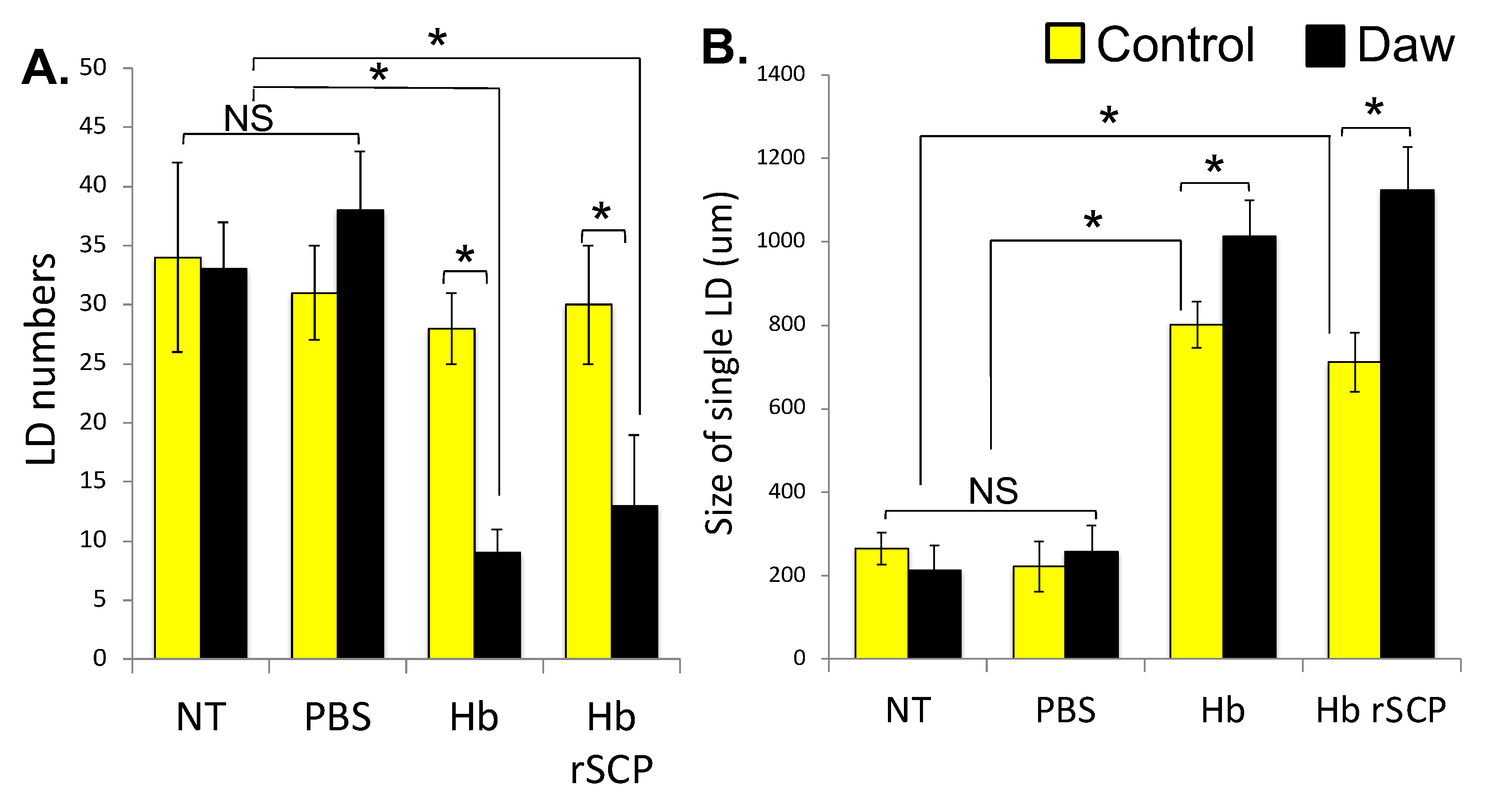
2.3. H. bacteriophora Infection of Activin-Deficient Drosophila Flies Results in Perturbed Intestinal Lipid Homeostasis

We have shown recently that intestinal lipid droplets in D. melanogaster mediate the antibacterial response [43]; therefore, here we first examined whether lipid droplets can also regulate anti-nematode immunity in the fly. For this, we infected single w1118 flies with approximately 100 H. bacteriophora axenic infective juveniles, or we injected w1118 individuals with rSCP from H. bacteriophora axenic nematodes and examined changes in numbers and size of lipid droplets 24 h later. No treatment or injection with PBS served as negative controls. We have found that H. bacteriophora-infected flies or those injected with H. bacteriophora rSCP have significantly reduced accumulation of lipid droplets in the gut and increased lipid droplet size as compared to the PBS-injected and non-treated individuals (Figure 3A and Figure 3B, respectively). In addition, this lipid droplet phenotype in the fly gut is exacerbated in Daw loss-of-function mutants (line Pbac{XP}daw05680) compared to w1118 background controls (Figure 3A,B). Thus, the Activin branch of the TGF-β signaling pathway in D. melanogaster regulates intestinal lipid homeostasis in response to entomopathogenic nematode infection or challenge with an entomopathogenic nematode infection factor.

3. Discussion

Our results reveal that Daw is substantially upregulated in wild-type flies upon injection of H. bacteriophora rSCP, and because it is a secreted signal [44], it may function systemically to regulate the expression of downstream genes in the Activin branch of TGF-β signaling. Also, because H. bacteriophora infection or injection of H. bacteriophora rSCP substantially upregulates Diptericin-A expression in Daw loss-of-function mutant flies, we postulate that certain entomopathogenic nematode infection factors, such as the H. bacteriophora SCP, are capable of altering the immune signaling in D. melanogaster when the Activin branch is inactivated.
The current data also suggest that Activin-deficient flies are sensitive to injection with the H. bacteriophora rSCP infection factor, and this phenotype is accompanied by increased Imd signaling activity and reduced hemocyte numbers. These findings strongly indicate a close interaction between this entomopathogenic nematode infection factor and the regulation of immune signaling and function in D. melanogaster deficient for the TGF-β Activin branch. Therefore, we speculate that the Activin pathway in the fly regulates cellular and humoral immune processes against challenge with the H. bacteriophora SCP.
Our results further indicate that infection of D. melanogaster wild-type flies with H. bacteriophora nematodes or injection with H. bacteriophora rSCP results in perturbed intestinal lipid metabolism marked by intestinal steatosis. These findings point out a relationship between Activin signaling activity and lipid metabolism in the context of entomopathogenic nematode infection.
Future work will study the conservation of these processes by expanding to other entomopathogenic nematodes. Our recent work indicates that excreted-secreted products from Steinernema carpocapsae manipulate the D. melanogaster immune response and they may also affect TGF-β signaling [45]. In future studies, we will perform time-course experiments to elucidate the causal relationships between Activin signaling activity and regulation of hemocyte numbers and lipid metabolism, which will provide a more complete picture on the dynamics of TGF-β signaling activation and immune responses. More precisely, we will focus on analyzing the tissue-specific transcript levels of TGF-β signaling molecules and exploring the immune signaling activity in various tissues of wild-type flies as well as TGF-β signaling mutant flies upon injection with the H. bacteriophora SCP infection factor. Another possibility will be to explore the involvement of other Activin branch members in cellular immune reactions of D. melanogaster against the H. bacteriophora SCP and identify whether (and how) cellular immune reactions in Activin mutant flies interfere or coordinate with humoral immunity. Also, investigating potential feedback loops between immune activation and metabolic changes in the context of entomopathogenic nematode infection factors may reveal interesting host-parasite dynamics. Finally, it will be interesting to investigate whether inactivating the Activin branch in H. bacteriophora-infected flies or flies injected with the H. bacteriophora SCP leads to changes in lipogenesis regulation in various fly tissues.
The fruit fly D. melanogaster is able to mount rapid and efficient reactions in response to diverse pathogens, including entomopathogenic nematodes [46]. Many of these anti-pathogen immune responses are remarkably conserved across insect species. The study of D. melanogaster is highly relevant to other Diptera, such as mosquitoes and leaf miners, given the evolutionary conservation of many of the key signal transduction pathways and transcriptional regulators that control innate immunity [47]. Deciphering the molecular and functional basis of the D. melanogaster immune response against insect parasitic nematodes and the infection factors they produce during the different stages of infection will provide a better understanding on the mechanisms that have evolved to oppose nematode attacks [2,48]. This information will in turn allow us to design alternative approaches to tackle agricultural insect pests and disease vectors effectively.

4. Materials and Methods

4.1. Fly Stocks

Drosophila melanogaster stocks were maintained and amplified at 25 °C and a 12:12-h light:dark cycle on Bloomington Drosophila Stock Center cornmeal food (Labexpress) supplemented with yeast (Carolina Biological Supply, Burlington, NC, USA). Adult flies (5–10 days old) carrying P-bac insertion Pbac{XP}daw05680 (strain d05680, Exelixis, Boston, MA, USA) and background control line w1118 (strain 3605, Bloomington, IL, USA) were used in all experiments. Both fly strains have been used consistently in our previous research [37,38,49].

4.2. Nematode and Bacterial Stocks

H. bacteriophora strain TT01 infective juveniles were kept in tissue culture flasks and multiplied through Galleria mellonella larvae that were placed on water traps [50]. This method involved the infection of G. mellonella larvae (kept in Petri dishes) with nematode infective juveniles (kept in tissue culture flasks) at 25 °C, the preparation of water traps with folded filter paper, the placement of the dead G. mellonella containing infective juveniles in the water trap 12 days after nematode infection, the migration of the new generation of infective juveniles from the dead insects to the water, and the storage of the new infective juveniles into tissue culture flasks [51]. One- to four-week-old nematodes were used in the experiments. Axenic nematodes without their symbiotic P. luminescens bacteria were generated using a previously developed method, where infective juveniles were amplified in fifth or sixth instar G. mellonella caterpillars that had been previously infected with the RET16 derivative of Photorhabdus temperata strain NC1 [42]. Bacterial cultures involved Escherichia coli (strain DH5a) and Micrococcus luteus (strain CIP A270). The bacteria were cultured on petri dishes containing 2.5% Luria-Bertani (LB) and 1.5% agar (Difco Laboratories, Detroit, MI, USA). Bacterial liquid cultures were prepared in sterile tubes containing 10 mL of 2.5% LB and incubated for 24 h on a rotary shaker at 265 rpm at 37 °C for E. coli and at 30 °C for M. luteus. Bacterial cultures were centrifuged at 4 °C and resuspended in phosphate-buffered saline (PBS) before their optical density (600 nm) was determined with a spectrophotometer (NanoDrop 2000c; Thermo Fisher Scientific, Waltham, MA, USA).

4.3. Production of Nematode Recombinant Serine Carboxypeptidase

The protocol for production and purification of recombinant H. bacteriophora serine carboxypeptidase has been described in detail before [52].

4.4. Fly Infections

Drosophila melanogaster Daw mutants and w1118 adult flies were infected with 100 infective juveniles of H. bacteriophora suspended in 250 μL of sterile water. Nematode suspensions were added to plastic vials containing four filter papers (Whatman Grade 1, 20 mm) at the bottom and 250 μL of 1% sucrose. Uninfected control flies were kept in vials containing sterile water and 1% sucrose only. All vials were covered with plugs, which were lowered close to the bottom of the vials to bring flies and nematodes in close proximity and facilitate infection. Fly infections with bacteria were performed by delivering 18.4 nl of a PBS suspension containing approximately 500 cells of E. coli or M. luteus into the hemocoel of D. melanogaster adults at the lateral anterior part of the thorax using a Nanoject II apparatus (Drummond Scientific, Broomall, PA, USA). Injections with PBS alone served as negative controls. All infected and control flies were kept in vials containing cornmeal food at 25 °C and a 12:12-h light:dark cycle. Each replicate included five male and five female 5–10 days old adult flies. Each infection was repeated three times with three replicates per treatment and different batches of flies, nematodes, and bacteria. Fly survival was estimated daily and up to six days post-infection.

4.5. Gene Expression Analysis

RNA was isolated from 4–5 D. melanogaster Daw mutants and w1118 adult flies (5–10 days old) using the Trizol reagent (Life Technologies, Frederick MD, USA) protocol. Reverse transcription was carried out following the instructions in the High-Capacity cDNA Reverse Transcription Kit (Applied Biosystems, Foster City, CA, USA). Real time PCR assays were performed in a CFX96 Real-Time System, C1000 Thermal Cycler (Bio-Rad, Philadelphia, PA, USA) using the GreenLink qPCR Mix (BioLink, Cary, NC, USA) and the following primers: Daw (CG16987) Forward GGTGGATCAGCAGAAGGACT, Daw Reverse GCCACTGATCCAGTGTTTGA, Diptericin-A (CG12763) Forward GCTGCGCAATCGCTTCTACT, Diptericin-A Reverse TGGTGGAGTTGGGCTTCATG, RpL32 (CG7939) Forward GATGACCATCCGCCCAGCA, RpL32 Reverse CGGACCGACAGCTGCTTGGC. The cycling conditions were 95 °C for 2 min, 40 repetitions of 95 °C for 15 s followed by 61 °C for 30 s, and then one round of 95 °C for 15 s, 65 °C for 5 s, and finally 95 °C for 5 s. Gene expression from the RT-qPCR experiments was analyzed in accordance with the 2−ΔΔCT method [53,54]. Each experiment involved biological duplicates and three technical replicates per sample and was repeated three times with different batches of flies and nematodes.

4.6. Hemocyte Count Estimation

Hemolymph was extracted from D. melanogaster Daw mutants and w1118 adult flies 24 h after infection with H. bacteriophora axenic nematodes or injection with rSCP, E. coli, or PBS. Untreated flies were used as negative controls. For hemocyte count estimation, hemolymph from 10 flies was first collected in 30 μL of 2.5× protease inhibitor cocktail (Sigma-Aldrich, Burlington, MA, USA), hemolymph samples were pipetted on a hemocytometer, and hemocyte numbers were recorded on a compound microscope (Olympus CX21, Center Valley, PA, USA) at 40× magnification. Fly hemocyte counting experiments were run three times with different batches of flies and nematodes, bacteria, and each experiment involved biological and technical triplicates.

4.7. Lipid Droplet Count and Size Estimation

Drosophila melanogaster Daw mutants and w1118 5–10-day old adult flies were infected with H. bacteriophora axenic nematodes or injected with rSCP or PBS, and 24 h later their gut tissues were dissected, stained in Nile red, and mounted in ProLong™ Diamond AntiFade Mountant with DAPI (Life Technologies) before imaging and lipid droplet counting on a Zeiss (White Plains, NY, USA) LSM 510 confocal microscope, as previously described [37]. Lipid droplet size quantification was carried out using ImageJ software 1.54e (National Institutes of Health). The experiment was replicated three times with different batches of flies and nematodes, and each experiment involved biological and technical triplicates.

4.8. Statistical Analysis

GraphPad Prism 8 was used for statistical analysis of the data and construction of the figures. Results from the survival experiments were analyzed using the log-rank (Mantel-Cox) test. Results from the rest of the experiments were analyzed using one-way analysis of variance (ANOVA) and Tukey post-hoc tests. A p-value < 0.05 was considered statistically significant.

Author Contributions

Conceptualization, I.E.; methodology, E.K., I.E. and S.M.; software, I.E. and S.M.; validation, E.K., I.E. and S.M.; formal analysis, I.E. and S.M.; investigation, E.K., I.E., and S.M.; data curation, I.E., and S.M.; writing—original draft preparation, I.E.; writing—review and editing, E.K. and S.M.; visualization, I.E. and S.M.; supervision, I.E.; project administration, I.E.; funding acquisition, I.E. All authors have read and agreed to the published version of the manuscript.

Funding

This research was funded by NSF, grant number IOS 2019869. S.M. was funded through a Wilbur V. Harlan summer research fellowship from the Department of Biological Sciences at George Washington University.

Institutional Review Board Statement

Not applicable.

Informed Consent Statement

Not applicable.

Data Availability Statement

The data supporting the findings of this study are available within the article.

Acknowledgments

We thank members of the Department of Biological Sciences at George Washington University (GWU) for critical reading of the manuscript. We also thank members of the Eleftherianos lab for maintaining the fly stocks and entomopathogenic nematodes.

Conflicts of Interest

The authors declare no conflict of interest.

References

  1. Brivio, M.F.; Mastore, M.; Pagani, M. Parasite-host relationship: A lesson from a professional killer. Invertebr. Surv. J. 2005, 2, 41–53. [Google Scholar]
  2. Castillo, J.C.; Reynolds, S.E.; Eleftherianos, I. Insect immune responses to nematode parasites. Trends Parasitol. 2011, 27, 537–547. [Google Scholar] [CrossRef] [PubMed]
  3. Eleftherianos, I.; Joyce, S.; Ffrench-Constant, R.H.; Clarke, D.J.; Reynolds, S.E. Probing the tri-trophic interaction between insects, nematodes and Photorhabdus. Parasitology 2010, 137, 1695–1706. [Google Scholar] [CrossRef] [PubMed]
  4. Erickson, S.M.; Xi, Z.; Mayhew, G.F.; Ramirez, J.L.; Aliota, M.T.; Christensen, B.M.; Dimopoulos, G. Mosquito infection responses to developing filarial worms. PLoS Negl. Trop. Dis. 2009, 3, e529. [Google Scholar] [CrossRef]
  5. Cupp, M.S.; Chen, Y.; Cupp, E.W. Cellular hemolymph response of Simulium vittatum (Diptera: Simuliidae) to intrathoracic injection of Onchocerca lienalis (Filarioidea: Onchocercidae) microfilariae. J. Med. Entomol. 1997, 34, 56–63. [Google Scholar] [CrossRef]
  6. Bartholomay, L.C.; Waterhouse, R.M.; Mayhew, G.F.; Campbell, C.L.; Michel, K.; Zou, Z.; Ramirez, J.L.; Das, S.; Alvarez, K.; Arensburger, P.; et al. Pathogenomics of Culex quinquefasciatus and meta-analysis of infection responses to diverse pathogens. Science 2010, 330, 88–90. [Google Scholar] [CrossRef] [PubMed]
  7. Kambris, Z.; Cook, P.E.; Phuc, H.K.; Sinkins, S.P. Immune activation by life-shortening Wolbachia and reduced filarial competence in mosquitoes. Science 2009, 326, 134–136. [Google Scholar] [CrossRef]
  8. Christensen, B.M.; Li, J.; Chen, C.C.; Nappi, A.J. Melanization immune responses in mosquito vectors. Trends Parasitol. 2005, 21, 192–199. [Google Scholar] [CrossRef] [PubMed]
  9. Wang, X.; Fuchs, J.F.; Infanger, L.C.; Rocheleau, T.A.; Hillyer, J.F.; Chen, C.C.; Christensen, B.M. Mosquito innate immunity: Involvement β1,3-glucan recognition protein in melanotic encapsulation immune responses in Armigeres subalbatus. Mol. Biochem. Parasitol. 2005, 139, 65–73. [Google Scholar] [CrossRef]
  10. Armer, C.A.; Rao, S.; Berry, R.E. Insect cellular and chemical limitations to pathogen development: The Colorado potato beetle, the nematode Heterorhabditis marelatus, and its symbiotic bacteria. J. Invertebr. Pathol. 2004, 87, 114–122. [Google Scholar] [CrossRef]
  11. van Sambeek, J.; Wiesner, A. Successful parasitation of locusts by entomopathogenic nematodes is correlated with inhibition of insect phagocytes. J. Invertebr. Pathol. 2009, 73, 154–161. [Google Scholar] [CrossRef]
  12. Liu, C.T.; Hou, R.F.; Chen, C.C. Formation of basement membrane-like structure terminates the cellular encapsulation of microfilariae in the haemocoel of Anopheles quadrimaculatus. Parasitology 1998, 116, 511–518. [Google Scholar] [CrossRef]
  13. Taylor, D.B.; Szalanski, A.L.; Adams, B.J.; Peterson, R.D., II. Susceptibility of house fly (Diptera: Muscidae) larvae to entomopathogenic nematodes (Rhabditida: Heterorhabditidae, Steinernematidae). Environ. Entomol. 1998, 27, 1514–1519. [Google Scholar] [CrossRef]
  14. Zhao, X.; Ferdig, M.T.; Li, J.; Christensen, B.M. Biochemical pathway of melanotic encapsulation of Brugia malayi in the mosquito, Armigeres subalbatus. Dev. Comp. Immunol. 1995, 19, 205–215. [Google Scholar] [CrossRef] [PubMed]
  15. Chalk, R.; Townson, H.; Natori, S.; Desmond, H.; Ham, P.J. Purification of an insect defensin from the mosquito, Aedes aegypti. Insect Biochem. Mol. Biol. 1994, 24, 403–410. [Google Scholar] [CrossRef] [PubMed]
  16. Vegni Talluri, M.; Cancrini, G. An ultrastructural study on the early cellular response to Dirofilaria immitis (Nematoda) in the Malpighian tubules of Aedes aegypti (refractory strains). Parasite 1994, 1, 343–348. [Google Scholar] [CrossRef] [PubMed]
  17. Christensen, B.M.; Forton, K.F. Hemocyte-mediated melanization of microfilariae in Aedes aegypti. J. Parasitol. 1986, 72, 220–225. [Google Scholar] [CrossRef]
  18. Lu, D.; Macchietto, M.; Chang, D.; Barros, M.M.; Baldwin, J.; Mortazavi, A.; Dillman, A.R. Activated entomopathogenic nematode infective juveniles release lethal venom proteins. PLoS Pathog. 2017, 13, e1006302. [Google Scholar] [CrossRef]
  19. Brivio, M.F.; Mastore, M.; Nappi, A.J. A pathogenic parasite interferes with phagocytosis of insect immunocompetent cells. Dev. Comp. Immunol. 2010, 34, 991–998. [Google Scholar] [CrossRef]
  20. Mastore, M.; Brivio, M.F. Cuticular surface lipids are responsible for disguise properties of an entomoparasite against host cellular responses. Dev. Comp. Immunol. 2008, 32, 1050–1062. [Google Scholar] [CrossRef]
  21. Li, X.Y.; Cowles, R.S.; Cowles, E.A.; Gaugler, R.; Cox-Foster, D.L. Relationship between the successful infection by entomopathogenic nematodes and the host immune response. Int. J. Parasitol. 2007, 37, 365–374. [Google Scholar] [CrossRef] [PubMed]
  22. Brivio, M.F.; Moro, M.; Mastore, M. Down-regulation of antibacterial peptide synthesis in an insect model induced by the body-surface of an entomoparasite (Steinernema feltiae). Dev. Comp. Immunol. 2006, 30, 627–638. [Google Scholar] [CrossRef] [PubMed]
  23. Brivio, M.F.; Mastore, M.; Moro, M. The role of Steinernema feltiae body-surface lipids in host-parasite immunological interactions. Mol. Biochem. Parasitol. 2004, 135, 111–121. [Google Scholar] [CrossRef] [PubMed]
  24. Brivio, M.F.; Pagani, M.; Restelli, S. Immune suppression of Galleria mellonella (Insecta, Lepidoptera) humoral defenses induced by Steinernema feltiae (Nematoda, Rhabditida): Involvement of the parasite cuticle. Exp. Parasitol. 2002, 101, 149–156. [Google Scholar] [CrossRef] [PubMed]
  25. Wang, Y.; Gaugler, R. Steinernema glaseri surface coat protein suppresses the immune response of Popillia japonica (Coleoptera: Scarabaeidae) larvae. Biol. Control 1999, 14, 45–50. [Google Scholar] [CrossRef]
  26. Jarosz, J. Active resistance of entomophagous rhabditid Heterorhabditis bacteriophora to insect immunity. Parasitology 1998, 117, 201–208. [Google Scholar] [CrossRef] [PubMed]
  27. Hallem, E.A.; Rengarajan, M.; Ciche, T.A.; Sternberg, P.W. Nematodes, bacteria, and flies: A tripartite model for nematode parasitism. Curr. Biol. 2007, 17, 898–904. [Google Scholar] [CrossRef] [PubMed]
  28. Peña, J.M.; Carrillo, M.A.; Hallem, E.A. Variation in the susceptibility of Drosophila to different entomopathogenic nematodes. Infect. Immun. 2015, 83, 1130–1138. [Google Scholar] [CrossRef]
  29. Castillo, J.C.; Shokal, U.; Eleftherianos, I. A novel method for infecting Drosophila adult flies with insect pathogenic nematodes. Virulence 2012, 3, 339–347. [Google Scholar] [CrossRef]
  30. Castillo, J.C.; Shokal, U.; Eleftherianos, I. Immune gene transcription in Drosophila adult flies infected by entomopathogenic nematodes and their mutualistic bacteria. J. Insect Physiol. 2013, 59, 179–185. [Google Scholar] [CrossRef]
  31. Arefin, B.; Kucerova, L.; Dobes, P.; Markus, R.; Strnad, H.; Wang, Z.; Hyrsl, P.; Zurovec, M.; Theopold, U. Genome-wide transcriptional analysis of Drosophila larvae infected by entomopathogenic nematodes shows involvement of complement, recognition and extracellular matrix proteins. J. Innate Immun. 2014, 6, 192–204. [Google Scholar] [CrossRef] [PubMed]
  32. Dobes, P.; Wang, Z.; Markus, R.; Theopold, U.; Hyrsl, P. An improved method for nematode infection assays in Drosophila larvae. Fly 2012, 6, 75–79. [Google Scholar] [CrossRef] [PubMed]
  33. Hyrsl, P.; Dobes, P.; Wang, Z.; Hauling, T.; Wilhelmsson, C.; Theopold, U. Clotting factors and eicosanoids protect against nematode infections. J. Innate Immun. 2011, 3, 65–70. [Google Scholar] [CrossRef]
  34. Wang, Z.; Wilhelmsson, C.; Hyrsl, P.; Loof, T.G.; Dobes, P.; Klupp, M.; Loseva, O.; Mörgelin, M.; Iklé, J.; Cripps, R.M.; et al. Pathogen entrapment by transglutaminase—A conserved early innate immune mechanism. PLoS Pathog. 2010, 6, e1000763. [Google Scholar] [CrossRef]
  35. Vadnal, J.; Ratnappan, R.; Keaney, M.; Kenney, E.; Eleftherianos, I.; O’Halloran, D.; Hawdon, J.M. Identification of candidate infection genes from the model entomopathogenic nematode Heterorhabditis bacteriophora. BMC Genom. 2017, 18, 8. [Google Scholar] [CrossRef] [PubMed]
  36. Bastin, A.; Eleftherianos, I. Functional role of the TGF-β signaling in the Drosophila immune response. Curr. Res. Immunol. 2023, 4, 100071. [Google Scholar] [CrossRef]
  37. Ozakman, Y.; Eleftherianos, I. TGF-beta Signaling Interferes with the Drosophila Innate Immune and Metabolic Response to Parasitic Nematode Infection. Front. Physiol. 2019, 10, 716. [Google Scholar] [CrossRef]
  38. Eleftherianos, I.; Castillo, J.C.; Patrnogic, J. TGF-β regulates resistance to parasitic nematode infection in Drosophila melanogaster. Immunobiology 2016, 221, 1362–1368. [Google Scholar] [CrossRef]
  39. Patrnogic, J.; Heryanto, C.; Eleftherianos, I. Wounding-induced upregulation of the Bone Morphogenic Protein signaling pathway in Drosophila promotes survival against parasitic nematode infection. Gene 2018, 673, 112–118. [Google Scholar] [CrossRef]
  40. Patrnogic, J.; Heruanto, C.; Eleftherianos, I. Transcriptional up-regulation of the TGF-β intracellular signaling transducer Mad of Drosophila larvae in response to parasitic nematode infection. Innate Immun. 2018, 24, 349–356. [Google Scholar] [CrossRef]
  41. Patrnogic, J.; Heryanto, C.; Ozakman, Y.; Eleftherianos, I. Transcript analysis reveals the requirement of NF-κB transcription factors for the activation of TGF-β signaling in nematode infected Drosophila. Immunogenetics 2019, 71, 501–510. [Google Scholar] [CrossRef] [PubMed]
  42. Kenney, E.; Hawdon, J.M.; O’Halloran, D.; Eleftherianos, I. Heterorhabditis bacteriophora Excreted-Secreted Products Enable Infection by Photorhabdus luminescens Through Suppression of the Imd Pathway. Front. Immunol. 2019, 10, 2372. [Google Scholar] [CrossRef] [PubMed]
  43. Harsh, S.; Heryanto, C.; Eleftherianos, I. Intestinal lipid droplets as novel mediators of host-pathogen interaction in Drosophila. Biol. Open 2019, 8, bio039040. [Google Scholar]
  44. Clark, R.I.; Woodcock, K.J.; Geissmann, F.; Trouillet, C.; Dionne, M.S. Multiple TGF-β superfamily signals modulate the adult Drosophila immune response. Curr. Biol. 2011, 21, 1672–1677. [Google Scholar] [CrossRef]
  45. Jones, K.; Tafesh-Edwards, G.; Kenney, E.; Toubarro, N.; Simões, N.; Eleftherianos, I. Excreted secreted products from the parasitic nematode Steinernema carpocapsae manipulate the Drosophila melanogaster immune response. Sci. Rep. 2022, 12, 14237. [Google Scholar] [CrossRef] [PubMed]
  46. Ozakman, Y.; Eleftherianos, I. Nematode infection and antinematode immunity in Drosophila. Trends Parasitol. 2021, 37, 1002–1013. [Google Scholar] [CrossRef] [PubMed]
  47. Eleftherianos, I.; Heryanto, C. Transcriptomic Insights into the Insect Immune Response to Nematode Infection. Genes 2021, 12, 202. [Google Scholar] [CrossRef] [PubMed]
  48. Dziedziech, A.; Shivankar, S.; Theopold, U. Drosophila melanogaster Responses against Entomopathogenic Nematodes: Focus on Hemolymph Clots. Insects 2020, 11, 62. [Google Scholar] [CrossRef]
  49. Ozakman, Y.; Raval, D.; Eleftherianos, I. Activin and BMP Signaling Activity Affects Different Aspects of Host Anti-Nematode Immunity in Drosophila melanogaster. Front. Immunol. 2021, 12, 795331. [Google Scholar] [CrossRef]
  50. White, G.F. A Method for Obtaining Infective Nematode Larvae from Cultures. Science 1927, 66, 302–303. [Google Scholar] [CrossRef]
  51. Heryanto, C.; Ratnappan, R.; O’Halloran, D.M.; Hawdon, J.C.; Eleftherianos, I. Culturing and Genetically Manipulating Entomopathogenic Nematodes. J. Vis. Exp. 2022, 181, e63885. [Google Scholar]
  52. Kenney, E.; Yaparla, A.; Hawdon, J.M.; O’Halloran, D.M.; Grayfer, L.; Eleftherianos, I. A putative lysozyme and serine carboxypeptidase from Heterorhabditis bacteriophora show differential virulence capacities in Drosophila melanogaster. Dev. Comp. Immunol. 2021, 114, 103820. [Google Scholar] [CrossRef] [PubMed]
  53. Livak, K.J.; Schmittgen, T.D. Analysis of relative gene expression data using real-time quantitative PCR and the 2−ΔΔCT method. Methods 2001, 25, 402–408. [Google Scholar] [CrossRef] [PubMed]
  54. Schmittgen, T.D.; Livak, K.J. Analyzing real-time PCR data by the comparative CT method. Nat. Protoc. 2008, 3, 1101–1108. [Google Scholar] [CrossRef] [PubMed]
Figure 1. Heterorhabditis bacteriophora recombinant serine carboxypeptidase (rSCP) activates Activin and Imd signaling in Drosophila melanogaster. Injection of H. bacteriophora (Hb) rSCP into: (A). Daw (Activin pathway) is upregulated in w1118 background control flies (ANOVA, F = 1.222, * p < 0.05), (B). Diptericin-A (Imd pathway) is upregulated in Daw mutants (ANOVA, F = 1.137, * p < 0.05). Each experiment was repeated three times and each experimental condition involved approximately 30 adult (5–10 days old) flies. NT: Non-Treated flies; Time-point: 24 h; NS: Non-Significant. Error bars represent standard errors.
Ijms 25 07970 g001
Figure 2. Effect of Heterorhabditis bacteriophora recombinant serine carboxypeptidase (rSCP) on hemocyte numbers and survival of Drosophila melanogaster Activin deficient flies. Injection of purified rSCP from H. bacteriophora (Hb) nematodes into Daw mutant flies reduces (A). hemocyte numbers (24 h) (ANOVA, F = 1.115, * p < 0.05) and (B). survival ability compared to the w1118 background flies (Control). Each experiment was replicated three times with 90 adult (5–10 days old) flies per experimental condition. (Mantel-Cox, df = 1, * p < 0.05); NS: Non-Significant. Error bars represent standard errors.
Ijms 25 07970 g002
Figure 3. Increased intestinal steatosis in Activin-deficient Drosophila melanogaster. (A). Lipid droplet (LD) numbers decrease (ANOVA, F = 1.301, * p < 0.05), and (B). LD size increases (ANOVA, F = 1.107, * p < 0.05) in the gut of Daw mutant flies infected with Heterorhabditis bacteriophora (Hb) nematodes or injected with Hb recombinant serine carboxypeptidase (rSCP). The experiment was performed three times with each experimental treatment including in total 90 adult (5–10 days old) flies. NS: Non-Significant. Error bars represent standard errors.
Ijms 25 07970 g003
Disclaimer/Publisher’s Note: The statements, opinions and data contained in all publications are solely those of the individual author(s) and contributor(s) and not of MDPI and/or the editor(s). MDPI and/or the editor(s) disclaim responsibility for any injury to people or property resulting from any ideas, methods, instructions or products referred to in the content.

