Article

Intrinsic Mechanism of CaCl2 Alleviation of H2O2 Inhibition of Pea Primary Root Gravitropism

1
College of Life Sciences and Technology, Gansu Agricultural University, Lanzhou 730070, China
2
Agronomy College, Gansu Agricultural University, Lanzhou 730070, China
3
Laboratory and Site Management Center, Gansu Agricultural University, Lanzhou 730070, China
4
State Key Laboratory of Arid-land Crop Science, Gansu Agricultural University, Lanzhou 730070, China
*
Authors to whom correspondence should be addressed.
Int. J. Mol. Sci. 2024, 25(16), 8613; https://doi.org/10.3390/ijms25168613
Submission received: 27 June 2024 / Revised: 29 July 2024 / Accepted: 5 August 2024 / Published: 7 August 2024
(This article belongs to the Special Issue Advance in Plant Abiotic Stress)

Abstract

:
Normal root growth is essential for the plant uptake of soil nutrients and water. However, exogenous H2O2 inhibits the gravitropic growth of pea primary roots. It has been shown that CaCl2 application can alleviate H2O2 inhibition, but the exact alleviation mechanism is not clear. Therefore, the present study was carried out by combining the transcriptome and metabolome with a view to investigate in depth the mechanism of action of exogenous CaCl2 to alleviate the inhibition of pea primordial root gravitropism by H2O2. The results showed that the addition of CaCl2 (10 mmol·L−1) under H2O2 stress (150 mmol·L−1) significantly increased the H2O2 and starch content, decreased peroxidase (POD) activity, and reduced the accumulation of sugar metabolites and lignin in pea primary roots. Down-regulated genes regulating peroxidase, respiratory burst oxidase, and lignin synthesis up-regulated PGM1, a key gene for starch synthesis, and activated the calcium and phytohormone signaling pathways. In summary, 10 mmol·L−1 CaCl2 could alleviate H2O2 stress by modulating the oxidative stress response, signal transduction, and starch and lignin accumulation within pea primary roots, thereby promoting root gravitropism. This provides new insights into the mechanism by which CaCl2 promotes the gravitropism of pea primary roots under H2O2 treatment.

Graphical Abstract

1. Introduction

Pea (Pisum sativum L.) is a strategic crop that ensures global food security and is favored for its versatility, nutrient richness, and adaptability [1]. The seedling stage of pea plants usually occurs during the low-rainfall season; thus, drought affects the growth of pea seedlings and, consequently, their yield and quality [2]. Roots absorb nutrients and water directly into the soil and act as sensors to sense and respond to a variety of external stresses [3]. As the only source of water and nutrient uptake for the seedling, the primary root must enter the soil without other support in order to be better adapted to the living environment [4]. Therefore, improving root growth capacity is a strategy to increase pea yield. It has been found that positive root geotropism is necessary for plant access to nutrients and water [5]. However, exogenous signals can change the plant’s root system architecture (RSA) and root growth direction, thus inhibiting plant growth [6]. Therefore, it is important to study the pathways that regulate the alteration of RSA and root growth direction.
Reactive oxygen species (ROS) are not only components that induce oxidative damage in the plant body but also act as signaling molecules to regulate plant growth [7]. ROS can alter the RSA by regulating lateral root formation in Arabidopsis thaliana [8]. In contrast, intracellular ROS homeostasis is regulated by a series of enzymes that include antioxidant enzymes such as NADPH/NADH, superoxide dismutase (SOD) (EC 1.15.1.1), oxidases, and peroxidase (POD) (EC 1.11.1.7) [9,10]. It has been found that ROS are involved in the signaling of the plant hormones abscisic acid (ABA) and auxin (IAA), which regulate seed germination, root growth, and differentiation [11]. H2O2 mediates NADPH oxidase to enable ABA activation of lateral root development, the synthesis of H2O2, and cell wall expansion [12,13,14]. Interaction between ROS and IAA can regulate root geotropism, lateral and adventitious root formation, and lignification [15]. Synergistic interactions occur between IAA and gibberellin (GA), wherein IAA induces the degradation of the DELLA protein, which inhibits GA signaling and is involved in the promotion of GA biosynthesis gene expression in terms of root elongation and root division [16].
In addition, H2O2 is involved in the regulation of calcium signaling pathways. It has been found that the H2O2 produced by tobacco cell inducers may activate H2O2-sensitive Ca2+ channels in the plasma membrane, resulting in an increase in cytoplasmic Ca2+ concentrations [17]. Ca2+ channels can be activated by NADPH oxidase to regulate plant cell expansion and, thus, cell development [18]. External signals stimulate the amyloplasts to settle on the endoplasmic reticulum, causing Ca2+ to be effluxed into the cytoplasm. This may cause a transient increase in cytoplasmic Ca2+ concentration, which, upon binding to calmodulin, either directly or indirectly activates the downstream signal transduction mechanisms [19]. Whereas starch is a key substance in the perception of gravity by the root system [20], changes in Ca2+ concentration caused by starch deposition regulate the direction of root growth.
It was found that cell wall accumulation limits cell elongation, thereby altering the RSA [21]. Cell wall metabolism is a key factor in plant response to environmental stresses and it is mediated by a variety of cell wall-modifying proteins. Soluble arabinogalactan proteins (AGPs) and insoluble extensions (EXTs) are associated with each other in the cell wall, and the EXT/AGP complex acts on the structure of the cell wall to regulate changes in the shape of the cell wall [22,23]. Increased H+ concentrations activate extensin activity and activate the cell wall, which leads to cell elongation [24]. Therefore, it is important to investigate the mechanism of action of exogenous H2O2 and CaCl2 on the cell walls of the primary roots of pea plants.
From the above, it is clear that H2O2 and Ca2+ are key signaling molecules that regulate the gravity-oriented nature of plants. It has been shown that exogenous H2O2 inhibits the gravity-oriented force of pea primary roots, while CaCl2 can alleviate this inhibition to a certain extent, but the specific alleviation mechanism has not been clarified [25]. Therefore, it is crucial to study in depth the alleviating effect of CaCl2 on H2O2 stress. Thus, this study combined transcriptomics and metabolomics to comprehensively analyze the pathways regulating exogenous CaCl2 to alleviate the inhibition of pea root gravitropism by H2O2. We also verified the roles of key metabolites and genes by combining the relevant physiological indicators with fluorescence quantification. This study provides a theoretical basis for the gravity-oriented nature of plant roots in adversity.

2. Results

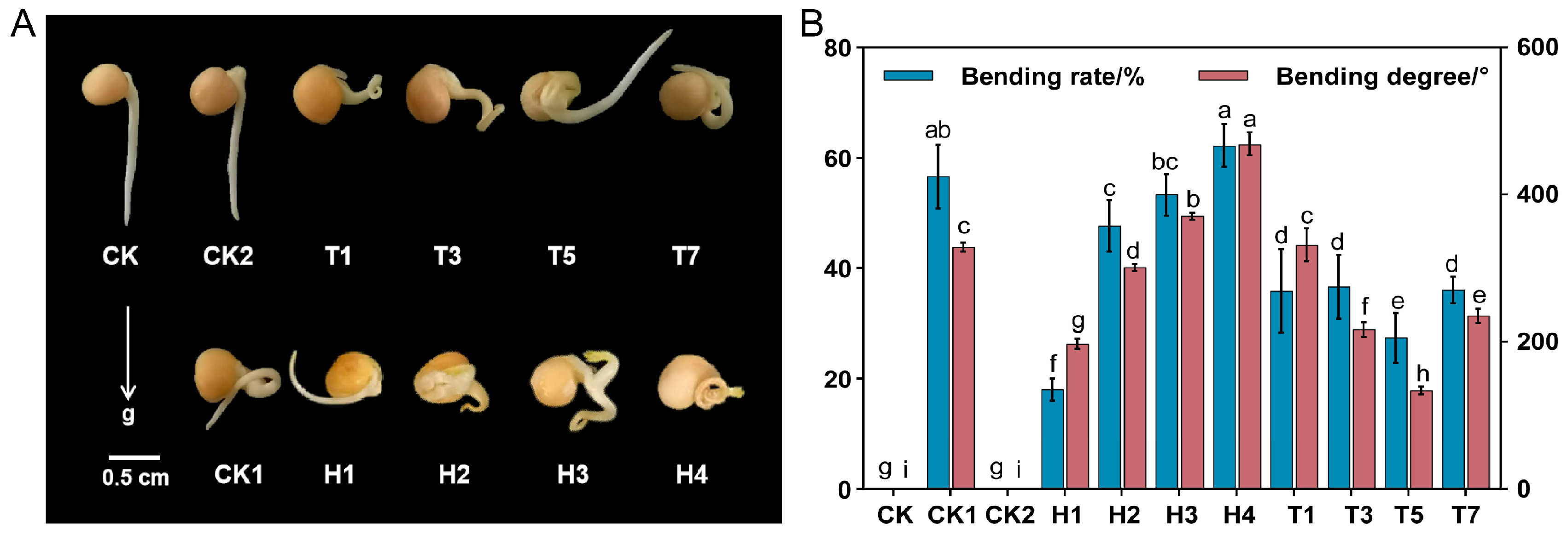
2.1. Root Non-Geostrophic Validation Experiments

In this study, we counted the bending rate (Table S1 and Figure S1 in the Supplementary Materials) and bending degree (Table S2 and Figure S2 in the Supplementary Materials) of pea primordial roots after 24, 36, 54, and 72 h of different treatments. The results showed that the shorter the treatment time, the greater the difference between replicates. Therefore, the main discussion in Section 3 is on the changes seen in the growth of pea primary roots at 72 h and the intrinsic mechanism.
Pea primary roots grew curved under exogenous H2O2 treatment (Figure 1A), and the bending rate and bending degree of primary roots (Figure 1B and Table S3) gradually increased with increasing concentration. The bending rate of primary roots under 150 mmol·L−1 H2O2 treatment was 3.3-fold higher than that of 20 mmol·L−1 H2O2. The subsequent application of different concentrations of CaCl2 on top of the 150 mmol·L−1 H2O2 treatment (Figure 1) revealed that CaCl2 had a mitigating effect on the bending growth of pea primordial roots under the H2O2 treatment (Figure 1A). With increasing CaCl2 concentration, the bending rate and bending degree of primordial roots had minimum values at the 10 mmol·L−1 CaCl2 and were significantly lower than for the 150 mmol·L−1 H2O2 treatment (Table S3). The application of 10 mmol·L−1 CaCl2 to pea primordial roots alone revealed no significant difference in growth status with CK treatment.
To further verify the important role of Ca2+ in mitigating the H2O2 inhibition of root growth toward gravitropism, CaSO4 and KCl, used at the same ionic (Ca2+, Cl) concentrations as the CaCl2 solution, were applied exogenously (Table S4 and Figure S3). It was found that the pea bending rate (Table S1 and Figure S1) and bending degree (Table S2 and Figure S2) were significantly reduced under the CaSO4 treatment compared with the 150 mmol·L−1 H2O2 treatment, and that these reached a minimum at a Ca2+ concentration of 10 mmol·L−1, while KCl treatment had no significant mitigating effect on H2O2 inhibition.
Based on these statistical results, pea primary roots under the 150 mmol·L−1 H2O2 and 150 mmol·L−1 H2O2 + 10 mmol·L−1 CaCl2 treatments were selected for the study of transcriptome and metabolome indexes.

2.2. Transcriptome Analysis and Validation of Key DGEs in Pea Primary Roots under H2O2 and CaCl2 Treatment

To investigate the mechanism of action of pea root growth under the CaCl2 mitigation of H2O2 application, transcriptome sequencing was performed on pea primordial roots under different treatments for 72 h. Eukaryotic reference transcriptome (RNA-seq) analyses of nine samples with PCA (Figure S4A) showed high similarity among the biological replicates. FC ≥ 1.5 and FDR < 0.05 were used as screening criteria in the comparison groups of CK vs. CK1, CK vs. T5, and CK1 vs. T5. In these analyses, 2701 differentially expressed genes (1200 up-regulated and 1501 down-regulated), 6857 differentially expressed genes (3242 up-regulated and 3615 down-regulated), and 6683 differentially expressed genes (3139 up-regulated and 3544 down-regulated) were found, respectively (Figure S4B). The Venn diagram of all differential genes (Figure S4C) showed that the total number of differentially expressed genes in the three control groups was 16,848. In addition, there were 456, 630, and 187 specifically expressed differential genes in the CK vs. CK1, CK vs. T5, and CK1 vs. T5 comparison groups, respectively. Nine DEGs were selected based on this transcriptome analysis (Figure 2), including two IAA-related genes (GAT1, IAA26), two ABA-related genes PYL4, the GA-related gene PAT1, the IAA-binding gene ABP19A in the cell wall, one lignin synthesis-related gene CCR1, and three genes related to starch and sucrose metabolism (PGM1, SUS, and BAM3). These RNA-Seq FPKM values of the genes showed a similar trend to the relative expression by qRT-PCR, confirming the authenticity of the transcriptome data.

2.3. Metabolomic Analysis of Pea Primary Roots under H2O2 and CaCl2 Treatments

To further elucidate the mechanism of root growth under the effect of H2O2 alleviated by CaCl2, we performed qualitative and quantitative metabolomic analyses on nine samples, and a total of 647 metabolites were detected. In the CK vs. CK1, CK vs. T5, and CK1 vs. T5 comparison groups, 303 differential metabolites (176 up-regulated and 127 down-regulated), 257 differential metabolites (81 up-regulated and 176 down-regulated), and 280 differential metabolites (124 up-regulated and 156 down-regulated) were found, respectively (Figure S4D). Correlation analysis showed that the correlation coefficients (R2) of the biological replicates of the samples were all greater than 0.9 (Figure S4E), and the PCA showed that the samples from different treatments were better separated (Figure S4F), suggesting that the metabolomic data were highly credible.

2.4. GO Enrichment Analysis of DEGs

The enrichment of DEGs in GO classification was analyzed at a threshold of p < 0.05 for the CK vs. CK1 and CK1 vs. T5 comparison groups, with 2133 and 5132 DEGs, respectively (Table S5). The results showed that the DEGs from both comparison groups were co-enriched in biological processes such as the “response to oxidative stress (GO:0006979)”, “plant-type cell wall organization (GO:0009664)”, and the “hydrogen peroxide catabolic process (GO:004274)”, and enriched in such cellular components as “plant-type cell wall (GO:0009505)”, “cell wall (GO:0005618)”, “integral component of membrane (GO:0016021)”, etc. The molecular functions were enriched in “heme binding (GO:0020037)”, “peroxidase activity (GO:0004601)”, “DNA-binding transcription factor activity (GO:0020039)”, etc. (Figure S5). The biological processes specifically enriched for CK vs. CK1 were the “defense response (GO:0006952)”, “abscisic acid-activated signaling pathway (GO:0009738)”, and “glutamine metabolic process (GO:0006541)”.
The results of the GO enrichment analysis indicated that the alteration of pea primary root growth by exogenous CaCl2 and H2O2 may be achieved through oxidative stress, the alteration of cell wall components, and the activation of phytohormone signaling.

2.5. CaCl2 and H2O2 Affect Oxidative Stress within Pea Primary Roots

The “hydrogen peroxide catabolic process (GO:004274)” and “response to oxidative stress (GO:0006979)”, which were significantly enriched in the GO biological processes, were analyzed for KEGG enrichment to further refine the DEGs-enriched pathways. It was found that the above DEGs were heavily enriched in the “phenylpropanoid biosynthesis” (ko00940) pathway (Figure S6A,B). A total of 24 DEGs were expressed in the “phenylpropanoid biosynthesis” pathway in the CK vs. CK1 and CK1 vs. T5 comparative groups, which were mainly the key genes regulating peroxidase. The heat-map visualization of DEGs co-expressed in the CK vs. CK1 and CK1 vs. T5 comparison groups (Figure 3A) revealed that 9 were up-regulated and 15 were down-regulated in the CK group, 15 were up-regulated and 9 were down-regulated in the CK1 group, and all of them were down-regulated in the T5 group, compared with the 3 treatment groups. By determining the POD (Figure 3C) and SOD (Figure 3D) activities of pea primary roots at 36-, 54-, and 72-h time intervals, it was found that the trends of the enzyme activities were basically the same at the different time intervals. Among them, the SOD and POD activities of the CK1 group were significantly higher than those of the CK group after 72 h. Compared with the CK1 group, the SOD activity of the T5 group was elevated, while the POD activity was significantly lower.
Oxidative stress in the root system alters the endogenous ROS content. DEG enrichment showed (Figure 3B) that the respiratory burst oxidase gene (RBOH) was activated by CaCl2 and H2O2 treatment, and the endogenous H2O2 content of the root system was significantly reduced by exogenous H2O2 treatment (Figure 3E). In contrast, the endogenous H2O2 content after the application of exogenous CaCl2 on the basis of H2O2 stress increased significantly compared with CK1, but was lower than that of the CK treatment, and the trends of the H2O2 content were basically the same at different time intervals. After the DAB staining of pea primary roots cultured for 72 h in the CK, CK1, and T5 treatment groups, the results showed (Figure 3F) that the primary roots were more lightly colored than the CK group under the action of exogenous H2O2, whereas the CaCl2 alleviation treatment resulted in deeper primary root coloration than the H2O2 treatment. This further indicated that CaCl2 might alleviate the inhibition of exogenous H2O2 on pea primary root growth by regulating endogenous H2O2.

2.6. Effect of CaCl2 and H2O2 on the Contents of Starch and Soluble Sugar

The transcriptome and metabolome analyses revealed that some DEGs and DAMs were enriched in the “starch and sucrose metabolic pathway”. Further analysis of this metabolic pathway showed that the expression of 11 DEGs (VCINV, ISA2, PGM1, ISA2, LECRKS7, LECRKS5, LECRKS4, LECRK71, BAM3, and BMY1) was significantly down-regulated by exogenous H2O2 (Figure 4A). After alleviation by CaCl2, the expression of three DEGs (ISA2, LECRKS7, and BMY1) was significantly down-regulated and BAM3 was significantly up-regulated. This suggests that CaCl2 and H2O2 may induce starch synthesis and metabolism in the primary roots of pea plants by regulating the expression of genes related to the “starch and sucrose metabolic pathway”.
Five key soluble sugars were detected in the metabolome (Figure 4B), and the results showed that “D-Melibiose, Sucrose, Turanose, D-(+)-Maltose Monohydrate, and Maltose” abundance was up-regulated in response to H2O2 and up-regulated after CaCl2 alleviation compared to the CK1 group. By determining the contents of starch (Figure 4C) and soluble sugar (Figure 4D) in the primary roots at different time intervals, it was found that the differences in the starch and soluble sugar contents at 36 h and 54 h were smaller. In contrast, the soluble sugar content in pea primary roots significantly increased by 1.87-fold, while starch content significantly decreased under the effect of H2O2 at 72 h. The soluble sugar and starch contents in the T5 group almost recovered to the level of the CK group. It suggests that it is possible for CaCl2 administration to alleviate the inhibition of root gravitropism by H2O2 by regulating the content of starch.
To investigate the effects of exogenous H2O2 and CaCl2 treatments on the amount and distribution of starch accumulation, we stained pea primary roots with Lugol’s iodine solution. The results showed (Figure 3G) that the starch in the CK group was mainly distributed in the root tip and the coloring was darker; in the CK1 group, the starch was uniformly distributed throughout the primordial roots and the coloring became lighter than that of the CK group; in the T5 group, the starch was distributed throughout the primordial roots and the coloring became darker than that of the CK1 group. Overall observation, followed by the freehand sectioning of transverse sections of primordial roots (Figure S7), revealed that the starch granules were significantly enlarged after H2O2 treatment, and the size of the starch granules was restored to the CK level after CaCl2 alleviation.

2.7. Effect of CaCl2 and H2O2 on Calcium Signaling in Pea Primary Roots

The starch in pea primary roots responds to gravity by activating the calcium signaling pathways. The transcriptome results showed that some DEGs are associated with calcium signaling. Calcium-transporting ATPase (ACA12/13), calcium-dependent protein kinase (CPK1/17), calcium-binding protein (KIC), calmodulin-3 (CAM3), and probable calcium-binding protein (CML25) with calmodulin-binding transcription activator 5 (CAMTA5) were activated under H2O2 and CaCl2 treatment (Figure 5). The predicted subcellular localization of the corresponding proteins of the above eight DEGs by the (https://wolfpsort.hgc.jp/, accessed on 6 November 2023) website revealed that these proteins were mainly distributed on the cytoplasmic membrane (ACA12/13), cytoplasm (CPK1/17, CAM3, and CML25), mitochondrion (KIC), and endoplasmic reticulum (CAMTA5).

2.8. Effects of CaCl2 and H2O2 on Phytohormone Signal Transduction in Pea Primary Roots

Transcriptome analysis showed that a large number of DEGs were enriched in “Plant hormone signal transduction (ko04075)”, and the key genes regulating phytohormone signaling under different treatments will be analyzed in the following.
A total of 35 DEGs were detected in the IAA signaling pathway, and 7 DEGs were co-expressed in the CK vs. CK1 and CK1 vs. T5 comparison groups (Table S6). GABA transporter 1 (GAT1), auxin-responsive protein IAA26 (IAA26), and auxin response factor 5 (ARF5) were up-regulated by 2.668, 2.008, and 2.375-fold, respectively, under H2O2 treatment, and down-regulated by 11.756, 2.040, and 2.438-fold, respectively, in the treatment group of applied CaCl2. Probable auxin-responsive protein 1 (IAA1) and indole-3-acetic acid-amido synthetase (GH3.1) were down-regulated by 2.303 and 2.058-fold, respectively, upon H2O2 treatment, and CaCl2 imposition alleviated the expression of the down-regulated genes (Figure 6A).
A total of 18 DEGs were detected in the ABA signaling pathway, and 3 DEGs were co-expressed in the CK vs. CK1 versus CK1 vs. T5 comparison group (Table S6). Among them (Figure 6B), the abscisic acid receptor (PYL4) was up-regulated 1.874- and 2.818-fold in CK vs. CK1 and CK1 vs. T5, respectively. In addition, protein phosphatase 2C (PP2CA) was down-regulated 1.794-fold and 1.297-fold in CK vs. CK1 and CK1 vs. T5, respectively, while serine/threonine-protein kinase (SAPK2) was down-regulated by 2.236-fold in CK vs. CK1 and up-regulated by 2.359-fold in CK1 vs. T5.
A total of 31 DEGs were detected in the GA signaling pathway, and 6 DEGs were co-expressed in the CK vs. CK1 and CK1 vs. T5 comparison groups (Table S6). Among them, the scarecrow-like transcription factor (PAT1), scarecrow-like protein (SCL14/33), and transcription factor phytochrome-interacting factor-like 15 (PIL15) were down-regulated by 2.330, 1.417, 1.546, and 1.476-fold, respectively, in the CK vs. CK1 comparison group, and were up-regulated by 4.103, 1.591, 1.616, and 2.082-fold, respectively, in the CK1 vs. T5 comparison group (Figure 6C). This shows that pea primordial roots with applied CaCl2 under exogenous H2O2 treatment significantly regulated the expression of key genes in phytohormone signaling.

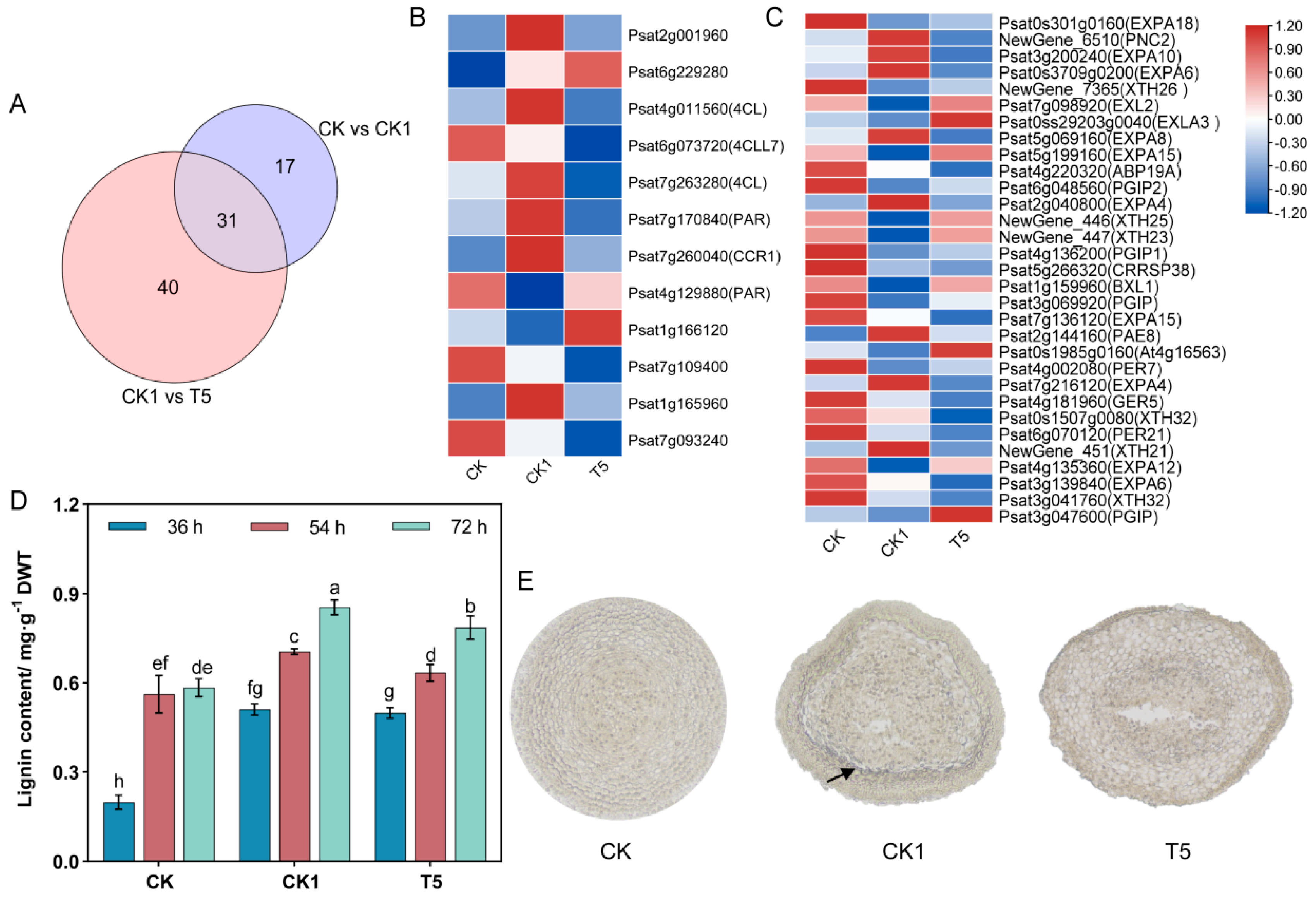
2.9. CaCl2 and H2O2 Affect Pea Primary Root Cell Walls

In the “plant-type cell wall (GO:0009505)” and “cell wall (GO:0005618)” pathways, the CK vs. CK1 and CK1 vs. T5 groups were significantly enriched in 48 and 71 DEGs, respectively (Figure 7A), with a total of 31 genes expressed. Exogenous H2O2 and CaCl2 activated the cell wall relaxation factors: α-expansin (EXPA) and xyloglucan endotransglucosylase/hydrolase (XTH). A heat-map visualization of the key genes showed (Figure 7C) that EXPA18/10/6/3/8/15/4/12 and XTH26/25/23/32/21 were differentially expressed in the CK vs. CK1 and CK1 vs. T5 treatment groups. In the CK vs. CK1 group, EXPA10/6/8/4 and XTH21 were up-regulated and EXPA18/12/15/6 and XTH26/25/23/32 were down-regulated; in the CK1 vs. T5 group, EXPA3/15/2/12 and XTH23/25 were up-regulated and EXPA10/6/8/4 and XTH21 were down-regulated.
Exogenous H2O2 and CaCl2 treatments induced the differential expression of key genes for lignin synthesis in pea primary roots. A total of 37 DEGs were detected in the lignin synthesis pathway in phenylpropane metabolism, and 12 DEGs were co-expressed in the CK vs. CK1 and CK1 vs. T5 comparison groups (Figure 7B). Among them, phenylalanine ammonia-lyase class 3 (Psat2g001960), 4-coumarate--CoA ligase (Psat4g011560) (Psat7g263280), cinnamoyl-CoA reductase 1 (CCR1), phenylacetaldehyde reductase (Psat7g170840), and berberine bridge enzyme-like 21 (Psat1g165960) were up-regulated 2.064-, 2.074-, 2.118-, 6.071-, 2.118-, and 2.809-fold under H2O2 treatment and down-regulated 1.975-, 2.861-, 3.630-, 3.688-, 3.630-, and 2.143-fold after CaCl2 mitigation, respectively. By determining the lignin content of primary roots in the different treatment groups in different time periods, it was found that lignin significantly accumulated under the effect of H2O2 (Figure 7D), and the lignin accumulation increased with time. To investigate the effects of exogenous H2O2 and CaCl2 treatments on the amount and distribution of lignin accumulation, we stained pea primordial roots with resorcinol. The results showed (Figure 7E) that the CK group had lighter coloring and less lignin accumulation; the coloring of primary roots in the CK1 and T5 groups was deeper than that in the CK group. By determining the lignin content of the primordial roots in different treatment groups, it was found that lignin significantly accumulated under the action of H2O2, and its content was reduced after CaCl2 alleviation (Figure 7E).

3. Discussion

The growth morphology of pea primary roots was altered under the action of exogenous H2O2, while the application of CaCl2 could alleviate the phenomenon [25]. In order to investigate the intrinsic mechanism, this study was carried out by applying different concentrations of H2O2 and CaCl2, counting the relevant growth indexes (germination potential, bending rate, and bending degree), and determining the relevant physiological indexes (POD, SOD, lignin content, etc.). Through transcriptome sequencing and metabolome assays, we analyzed the key pathways of DEG and DAM enrichment in pea primary roots under different treatments, with a view to finding the key genes and metabolite interactions of H2O2 and CaCl2 that regulate root growth and development.

3.1. CaCl2 and H2O2 Treatments Affect Pea Primary Root Growth toward Gravitropism

The growth and development of the primary root is essential for the early growth of peas [26]. In this study, the germination potential of pea seeds increased in the presence of high concentrations of H2O2 (Table S7 and Figure S8), suggesting that H2O2 could promote the germination rate of pea seeds, thereby verifying the findings of Barba-Espin et al. [27]. Jiang et al. [28] found that H2O2 caused the non-directional growth of the primary roots of Lathyrus quinquenervius and wavy growth of the primary roots of Arabidopsis, which may be caused by the uneven distribution of calcium ions and IAA [29]. In our study, we found that the geotropism-related growth of pea primordial roots was inhibited under H2O2 treatment, while the bending rate and bending degree of pea primordial roots increased as the concentration of exogenous H2O2 increased (Figure 1). This inhibition was relieved by the application of CaCl2, which is consistent with the findings of Li et al. [25]. To further investigate whether the alleviation was caused by Ca2+ or Cl, this study demonstrated that Ca2+ was a key factor in alleviating the inhibition of pea gravitropism by H2O2 by replacing the counterion to apply CaSO4 and KCl with the same particle concentration (Ca2+, Cl) as CaCl2. However, the exact manner of its mitigation is currently unknown. Root tips usually have more gravity-sensing signals; however, it has been found that plants can still sense gravity after the root crown has been removed [30,31], suggesting that the root tip is not the only gravity-sensing site [32]. The gravity-oriented nature of the roots was weakened after the removal of the medial columella cells from the roots of maize seedlings, but the tendency of gravity-oriented growth was maintained [33]. Therefore, the whole primary root was selected for analysis in this study, in order to screen the key factors of exogenous H2O2 and the CaCl2 regulation of root growth more comprehensively.

3.2. Activation of Oxidative Stress in Primary Roots by CaCl2 and H2O2 Treatments

ROS play an important role in shaping the RSA by regulating root growth and lateral root formation [34]. Transcriptome analysis showed that the applied H2O2 induced oxidative stress in the primary roots, and the enriched pathway was the “phenylpropane metabolic pathway”. The DEGs in this pathway mainly regulate peroxidase. The study by Wan et al. [35] showed that exogenous H2O2 treatment could enhance the cold resistance of oilseed rape seedlings by inducing the accumulation of antioxidant substances and activating the activity of antioxidant enzymes. In this study, we found that 11 peroxidase genes (including GSVIVT00023967001, PER12, PNC2, PRX112, POD, PER52, Psat5g250040, PER55, PNC1, PER10, and PER25) were up-regulated in response to H2O2, whereas all of them were down-regulated under CaCl2 treatment. Barley root growth under high Cd stress was inhibited, while cationic POD isozymes accumulated Cd and were concentration-dependent [36]. In this study, the POD and SOD activities in primary roots were significantly elevated by H2O2, while POD activity was reduced by CaCl2 alleviation. SOD is at the core of antioxidant enzymes, and its elevated activity scavenges free radicals and enhances membrane permeability [37]. It was found that exogenous Ca2+ could improve the antioxidant capacity of black algae to enhance its resistance to Cd [38].
ROS have a dual role in the plant body, one as stressors that trigger oxidative stress and the other as signaling molecules that are involved in plant development [39]. It was found that respiratory burst oxidase D (RBOHD) induces the production of cytoplasmic ectodomain ROS [40]. In this study, exogenous H2O2 stress led to a decrease in endogenous H2O2 content (Figure 3E), whereas the content of endogenous H2O2 in the root system increased after CaCl2 application. This suggests that Ca2+ may be regulating the endogenous H2O2 to alleviate the non-gravitropic nature of primary roots due to exogenous H2O2 stress, which is similar to the findings of Liu et al. [41]. It was found that endogenous H2O2 synthesis was reduced by the exogenous H2O2 inhibition of sallow bean [28] root vigor, while the endogenous H2O2 content of pea primordial roots [41] was significantly reduced by the activation of antioxidant enzyme systems. We further confirmed that exogenous H2O2 inhibited endogenous H2O2 accumulation using DAB staining (Figure 3F), and hypothesized that this was possibly because exogenous H2O2 accelerated the clearance of endogenous H2O2 by POD; the specific mechanism of action needs to be further explored. Transcriptome analysis showed that the expression of RBOH-related genes (RBOHA, RBOHB, RBOHC, RBOHE, and RBOHH) was activated upon the application of H2O2. This suggests that RBOH may be a key gene in the exogenous H2O2 regulation of endogenous H2O2 content changes. It was found that the expression of AtRBOHC regulates root development in Arabidopsis [42], and it can be hypothesized that the RBOH gene in this study may be related to pea root development, which needs further verification.

3.3. Effect of CaCl2 and H2O2 on Starch Metabolism in Primary Roots

According to the starch-equilibrium body hypothesis, starch-filled amyloplasts are asymmetrically distributed in the root system during gravity perception, thus inducing asymmetric growth signal transmissions [43,44]. In this study, exogenous H2O2 decreased the starch content and increased the soluble sugar content in primary roots. Exogenous H2O2 also decreased the distribution of starch in the root tip, which finding is similar to the findings of Zhou et al. [29]. It suggests that H2O2 may attenuate root gravitropism by converting starch to sugar in the root tip. The application of CaCl2 could also alleviate the acceleration of starch metabolism induced by H2O2, thus restoring the gravity-oriented nature of roots to some extent. Interestingly, the starch granules were significantly enlarged under the H2O2 treatment compared to the CK treatment, which may also be a key factor leading to the change in gravitropism, the exact mechanism of which remains to be further verified.
Starch consists of straight-chain starch and branched-chain starch, in which straight-chain starch is synthesized by granule-bound starch synthase (gss1) activity [45,46]. It was found that the reduced expression of the PGM1 gene, a key starch-synthesizing gene in the root tip of Arabidopsis, resulted in diminished root geotropism [47]. In contrast, in pgm1 mutants, the deposition of amyloid-free plastids is blocked, leading to a slowing of the gravitropic response in roots and shoots [48]. In this study, the transcriptome and fluorescence quantification results showed that H2O2 down-regulated the expression of PGM1 (Figure 2 and Figure 4A), a key gene for starch synthesis, and its expression was up-regulated after CaCl2 alleviation. It has been found that Ca2+ stabilizes α-amylase activity modulating gravitational sensitivity; therefore, Ca2+ and amylase are the controlling factors in stabilizing starch content in cells [49,50,51]. Combined with the results of starch content measurements, we can speculate that PGM1 is a key gene in the H2O2-regulated changes seen in starch content in pea primary roots.

3.4. Effects of CaCl2 and H2O2 on Calcium Signaling in Primary Roots

Instantaneous changes in Ca2+ are early events in the plant’s response to a variety of environmental signals [52]. It was found that cold stress induces Ca2+ signaling in plant cells, involving the activation of Ca2+ channels and Ca2+ pumps [53,54]. Water stress causes hypoxia in plant roots, and by knocking down CAX (Ca2+/H+ exchanger) and ACA (Ca2+-ATPase), it was found that the harmful effects of water stress on roots were alleviated by ACA knockdown [55]. In plant cells, CaM, calmodulin neurophosphatase b-like proteins (CBLs), CMLs, and CDPKs (CPKs) can bind to free calcium in the cytoplasm, triggering a conformational change of the proteins that can lead to downstream physiological and biochemical responses [56,57,58]. Ca2+ is involved in plant root geotropism. An earlier study found that gravity leads to the asymmetric distribution of Ca2+ gradients within pea and maize roots [59], and that the application of Ca2+ chelating agents resulted in the retardation of root geotropism [60]. Gravity-stimulated Ca2+ is involved in regulating differential changes in extracellular pH in the elongation zones of Arabidopsis roots on both the ground-oriented and far-ground sides in response to auxin, resulting in a change in root orientation [61]. In addition, primary Arabidopsis roots were less gravity-oriented in the presence of exogenous H2O2. The expression of MCA1, which encodes a Ca2+-permeable mechanosensitive channel, was significantly increased and Ca2+ levels were higher in cells on the inner side of bent roots than in those on the outer side [29]. Through transcriptome analysis, we predicted the subcellular localization of key genes regulating calcium signaling and found that ACA12/13, CPK1/17, KIC, CAM3, CML25, and CAMTA5 were activated by H2O2 and CaCl2. It was further demonstrated that root geotropism was regulated by Ca2+.

3.5. Effects of CaCl2 and H2O2 on Phytohormone Signal Transduction

The phytohormones IAA, ABA, and GA, which are key regulators of cell elongation and division [62], are essential in the physiology of plant resistance to adversity [20]. The RSA is affected by the crosstalk of different hormones, and external signaling molecules alter the plant signaling pathways [63,64]. In Arabidopsis, auxin early and fast-response genes, including auxin/indole-3-acetic acid (Aux/IAA), ARF, SAUR, and GH3, are key genes for auxin signaling [65,66]. The auxin-induced GH3 gene can mediate IAA inactivation through coupling, which, in turn, attenuates auxin signaling [67]. MP/AtARF5 can control embryonic root initiation by interacting with transcription factors [68]. AtARF10/16 binds to microRNA160, which regulates root-cap cell formation [69]. In the ABA signaling pathway, protein phosphatase 2Cs (PP2Cs) are negative regulators, PYR/PYL/RCARs are ABA receptors, and snf1-related protein kinase 2s (SnRKs) are positive regulators, which collectively mediate the stimulatory response of the plant body to exogenous signaling molecules [70,71]. In this study, during the action of H2O2 with CaCl2, the hormones regulating IAA (GAT1, IAA26, ARF5, IAA1, AUX22B, AUX22D, and GH3.1), ABA (PYL4, PP2CA, and SAPK2), and GA (SCL32, PAT1, RSL1, SCL14, PIL15, and SCL33) key genes for signaling were activated. This may be the reason for the differential growth of pea primary roots induced by H2O2 and CaCl2.

3.6. Effect of CaCl2 and H2O2 on the Cell Wall of Primary Roots

Under water stress, the genes regulating those enzymes related to maize isoflavone biosynthesis are up-regulated and lignin is increased in the elongation zone [72]. The phenylpropane pathway is one of the sources of the lignin found in plant cells. POD is the last enzyme in the lignin synthesis pathway and high POD activity increases lignification [73]. Large accumulations of lignin under drought stress limit cell-wall extension in soybean roots [74]. The EXPA and XET proteins play important roles in cell wall expansion [75]. In our study, the genes regulating EXPA and XET showed 20 DEGs up-regulated and 11 DEGs down-regulated in the CK treatment group, 8 DEGs up-regulated and 23 DEGs down-regulated in the CK1 treatment group, and 9 DEGs up-regulated and 22 DEGs down-regulated in the T5 treatment group. This indicates that the curved growth of pea primary roots under H2O2 treatment may be related to the activities of EXPA and XET proteins. Cell wall extension depends on the deposition of cell wall components and the modification of cell wall structure to balance rigidity and extensibility. It was shown that the cytoskeletal network, the deposition of cell wall components, Ca2+ homeostasis, ROS, ectoplasmic pH changes, and cell-wall-modifying proteins regulate cell wall extension [76,77,78]. To better adapt to the external environment, the plant body must establish the correct cell shape and size [79]. Therefore, it can be hypothesized that exogenous H2O2 may promote lignin accumulation by increasing POD activity [80], thus altering the cell wall extensibility of pea primary roots and affecting the normal growth of root cells. After the application of CaCl2, there were no significant changes in the genes regulating the expression of EXPA and XET proteins compared to the CK1 group, while the lignin content was reduced compared to the CK group.

4. Materials and Methods

4.1. Plant Materials and Treatment

The Longwan 1 pea was used as the experimental material. Pea seeds of uniform size and full grains were selected; they were first rinsed with running water for 30 min, then sterilized with 75% alcohol for 30 s, and finally rinsed with sterile water 4–5 times. The seeds were placed in petri dishes with two layers of filter paper, then 20 seeds were placed in each petri dish. Finally, 20 mL of culture solution was added and the seeds were incubated in the incubator (Zhejiang Topu Yunnong Technology Co., Ltd.; Zhejiang, China) at a constant temperature of 25 °C for 72 h in the dark.
The culture solution concentration was screened with reference to the method used by Li et al. [25]. H2O2 concentration was screened with six concentration gradients of 0, 20, 80, 150, 200, and 300 mmol·L−1 H2O2 (Sinopharm Group Chemical reagent Co., Ltd.; Shanghai, China). Each petri dish was considered as one replicate, and three replicates were set up. At the four time points of 24, 36, 54, and 72 h, the germination potential, bending rate, and bending degree of peas in different experimental groups were counted; 150 mmol·L−1 was determined to be the optimal inhibitory concentration.
For CaCl2 concentration screening, a control group with CK (deionized water), CK1 (150 mmol·L−1 H2O2), CK2 (10 mmol·L−1 CaCl2 (Tianjin Guangfu Technology Development Co., Ltd.; Tianjin, China)), and a mitigation group (H2O2 + different concentrations of CaCl2) (Table 1) were set up to carry out the experiment. The specific treatment concentrations of CaCl2 are given in Table 1. Each petri dish was considered as one replicate, and three replicates were set up. At the four time points of 24, 36, 54, and 72 h, the germination potential, bending rate, and bending degree of peas in the different experimental groups were measured. T5 (150 mmol·L−1 H2O2 + 10 mmol·L−1 CaCl2) was finally determined as the optimal mitigation concentration. The primary roots of the CK, CK1, and T5 groups treated for 36, 54, and 72 h were taken and stored at −80 °C for subsequent index measurements.

4.2. Root Germination Potential, Bending Rate and Bending Degree Statistics

The germination potential, bending rate, and bending degree of pea seeds under different treatments were measured and 3 replications for each were performed (1 replicate per petri dish, 20 seeds per dish).
The statistical criterion for germination potential is as follows [80]: the length of the radicle of the seed is equal to the length of the seed.
Germination potential (%) = (number of seeds germinating normally at 72 h/total number of seeds per dish).
The statistical criterion for bending primary roots is as follows [41]: primary roots were considered to be bent if the angle between the tip growth angle and the direction of gravity was greater than 180°, i.e., bent, and the bending angle was measured using ImageJ 1.53q software (https://imagej.net/, accessed on 17 July 2023) and Java 1.8.0_322 (64-bit) [81].

4.3. Measurement of Physiological Indices

Starch content, soluble sugar content, lignin content, superoxide dismutase (SOD) (EC 1.15.1.1) activity, peroxidase (POD) (EC 1.11.1.7) activity, and hydrogen peroxide content were determined using assay kits (Suzhou Keming Biotechnology Co.; Ltd.; Suzhou, China).

4.4. H2O2, Starch, and Lignin Staining

The diaminobenzidine (DAB) method was used to stain the primary roots of pea plants to detect H2O2 changes [82,83]. Primary roots from CK, CK1, and T5 specimens treated for 72 h were taken and placed in 1 mg·mL−1 DAB (Hefei Bomei Biotechnology Co., Ltd.; Hefei, China) staining solution, then treated in the dark at a constant temperature of 25 °C for 2 h. The DAB solution was poured out, and the primordial roots were rinsed with distilled water 4–5 times to remove the staining solution on the surface of the material, and the staining was observed; the darker the yellow color, the greater the H2O2 content.
The pea primary roots were stained using Lugo’s iodine solution [84] (iodine 4.5–5.5% (Tianjin Guangfu Technology Development Co., Ltd.; Tianjin, China) and potassium iodide 9.5–10.5% (Tianjin Guangfu Technology Development Co., Ltd.; Tianjin, China)) method to detect starch changes. Primary roots from the groups with CK, CK1, and T5 treatment for 72 h were placed in 9-millimeter disposable Petri dishes, to which 20 mL of staining solution was added (the concentration of staining solution could be adjusted according to the starch content of the material), and the staining was photographed and observed after 10 min.
Pea root tips were stained using the phloroglucinol method [85] to detect changes in lignin at 72 h under the CK, CK1, and T5 treatments. Pea root tips of 1 cm were placed in 2 mL centrifuge tubes and fixed for more than 24 h by adding 1.8 mL of FAA 70% fixative. Paraffin sections (apical transverse sections) were completed by Wuhan Xavier Biotechnology Co. (Wuhan, China). A total of 1% phloroglucinol (Yuanye Biotechnology technology company, Shanghai, China) solution (95% ethanol) was added dropwise to the sections and stained for 2 min. Then, 25% HCl was added dropwise for 2 min to develop the color. The samples with completed color development were quickly placed under a microscope (Leica Microsystems, Wetzlar, Germany) to observe the lignin distribution throughout the cross-section of the root tip and photographed.

4.5. Transcriptome Sequencing

Three biological replicates of pea primary roots from each of the CK, CK1, and T5 treatment groups were made. RNA sequencing analysis was performed by Biomarker Technologies Co., Ltd. (Beijing, China). Total RNA from the pea primary roots was extracted using the RNA Prep Pure Plant Kit (Tiangen, Beijing, China) according to the instructions provided by the manufacturer, and RNA sequencing analysis was performed using the Hieff NGS Ultima Dual-mode mRNA Library Prep Kit for Illumina (Yeasen Biotechnology (Shanghai) Co., Ltd., Shanghai, China). The libraries were finally sequenced on the Illumina NovaSeq platform. The raw data were further processed using the bioinformatics analysis platform BMKCloud (www.biocloud.net, accessed on 5 October 2023). Reads containing adapters were removed from the raw data, and these clean reads were mapped to the reference genome of Pisum_sativum_v1a (GCA_900700895.2, NCBI).
Genes with a DESeq2 corrected p-value < 0.05 and a fold change of ≥1.5 [86] were designated as differentially expressed. Differentially expressed genes (DEGs) were functionally annotated using the GO [87] and KEGG [88] databases.

4.6. Quantitative Real-Time PCR

To verify the reliability of the transcriptome sequencing data, nine DEGs were selected from the transcriptome data for qRT-PCR. Three replications were made for each treatment. The qRT-PCR primer sequence information is given in Table S8. qRT-PCR was performed after RNA reverse transcription using a LightCycler®96 Real time-PCR machine (Roche, Mannheim, Germany). The reaction system used a total of 20 μL: 10 μL of 2 × Talent qPCR premix, 6.8 μL of RNase-Free ddH2O, 2 μL of 75 ng/μL cDNA, and the forward and reverse primers of 10 μmol·L−1 were 0.6 μL each. The qRT-PCR reaction program was used as referenced by Lu et al. [86]. The relative expression of nine genes was calculated by the 2−ΔΔCt method, using β-tubulin [89] as the housekeeping gene.

4.7. LC–MS/MS Analysis

The same samples were used for the metabolome as for the transcriptome. Metabolome assays was performed with the UPLC-ESI-MS/MS system (UPLC, Waters Acquity I-Class PLUS; MS, Applied Biosystems QTRAP 6500+) (Waters, Milford, MA, America). The total peak area was normalized to the original peak area information for subsequent analysis. The screening criteria were: FC > 1, p-value < 0.05, and VIP > 1 [86]. Spearman correlation analysis and principal component analysis (PCA) were used to determine the reproducibility of within-group and quality-control samples.

4.8. Statistical Analysis

The experimental data were analyzed for statistical significance using Excel 2010 (Microsoft Corporation, Redmond, WA, America) and IBM SPSS Statistics 26 software (IBM Corporation, Armonk, NY, America). Graphs were generated using TBtools v2.096 (Dr. Chengjie Chen, Guangzhou, China, https://github.com/CJ-Chen/TBtools/releases, accessed on 4 August 2024), GraphPad Prism 9 (Dr. Harvey Motulsky, USA), Adobe Illustrator CC 2018 (Adobe Corporation, SAN Jose, CA, America), and Adobe Photoshop 2021.

5. Conclusions

In this study, we comprehensively revealed the key pathways through transcriptomics and metabolomics that function to alleviate the inhibition of pea primary root gravitropism by H2O2 with exogenous CaCl2 (Figure 8). The transcriptome results indicated that CaCl2 may alleviate H2O2 stress by regulating the pea primary root oxidative stress response, starch and sucrose metabolism, calcium signaling and phytohormone signaling, and cell wall composition under a gravity field. Among them, PGM1, which regulates starch synthesis, is a key gene for gravity perception in pea primary roots. The metabolome and physiological and biochemical results showed that CaCl2 might alleviate the inhibition of H2O2 on the gravitropic movement of pea primary roots by regulating POD and SOD activities, increasing the content of endogenous H2O2 and starch, and decreasing the accumulation of lignin to alleviate the inhibition of H2O2 on the gravitropic movement of pea primary roots. This study not only contributes to the study of the pathways of pea root growth under adversity stress but also provides theoretical references for the study of root vectorial movement.

Supplementary Materials

The following supporting information can be downloaded at: https://www.mdpi.com/article/10.3390/ijms25168613/s1.

Author Contributions

Software, T.M. and J.D.; formal analysis, R.W.; resources, S.M. and Q.C.; data curation, R.W., L.M., L.X. and Y.M.; writing—original draft, R.W.; writing—review and editing, L.M., S.L. and Q.C.; visualization, T.M. and Z.C.; project administration, S.L.; funding acquisition, S.L. All authors have read and agreed to the published version of the manuscript.

Funding

This work was supported by the Natural Science Foundation of Gansu Province (24JRRA651) (Shaoying Ma), National Green Fertilizer Industry Technology System (CARS-22-G-12) (Qiang Chai), Gansu Provincial Major Project (20ZD7NA007) (Sheng Li), and College Student Innovation and Entrepreneurship Project (202310733007) (Sheng Li).

Institutional Review Board Statement

Not applicable.

Informed Consent Statement

Not applicable.

Data Availability Statement

To support the results, the RNA-seq raw data set has been deposited in the NCBI database (https://www.ncbi.nlm.nih.gov/bioproject/, accessed on 27 September 2023), with alogin accession of PRJNA1021333.

Conflicts of Interest

The authors declare no conflicts of interest.

References

  1. Pavan, S.; Delvento, C.; Nazzicari, N.; Ferrari, B.; D’Agostino, N.; Taranto, F.; Lotti, C.; Ricciardi, L.; Annicchiarico, P. Merging genotyping-by-sequencing data from two ex situ collections provides insights on the pea evolutionary history. Hortic. Res. 2022, 9, uhab062. [Google Scholar] [CrossRef] [PubMed]
  2. Lei, B.; Zhang, F.Y. Responses of seed germination and seeding growth physiology of three pea variants to drought stress. J. Jianghan Univ. Nat. Sci. Ed. 2022, 50, 22–29. [Google Scholar]
  3. Villordon, A.Q.; Ginzberg, I.; Firon, N. Root architecture and root and tuber crop productivity. Trends Plant Sci. 2014, 19, 419–425. [Google Scholar] [CrossRef] [PubMed]
  4. Taylor, I.; Lehner, K.; McCaskey, E.; Nirmal, N.; Ozkan-Aydin, Y.; Murray-Cooper, M.; Jain, R.; Hawkes, E.W.; Ronald, P.C.; Goldman, D.I.; et al. Mechanism and function of root circumnutation. Proc. Natl. Acad. Sci. USA 2021, 118, e2018940118. [Google Scholar] [CrossRef] [PubMed]
  5. Toal, T.W.; Ron, M.; Gibson, D.; Kajala, K.; Splitt, B.; Johnson, L.S. Regulation of root angle and gravitropism. G3 2018, 8, 3841–3855. [Google Scholar] [CrossRef] [PubMed]
  6. Kim, H.J.; Kobayashi, A.; Fujii, N.; Miyazawa, Y.; Takahashi, H. Gravitropic response and circumnutation in pea (Pisum sativum) seedling roots. Physiol. Plant 2016, 157, 108–118. [Google Scholar] [CrossRef] [PubMed]
  7. Gorgues, L.; Li, X.; Maurel, C.; Martinière, A.; Nacry, P. Root osmotic sensing from local perception to systemic responses. Stress Biol. 2022, 2, 36. [Google Scholar] [CrossRef]
  8. Manzano, C.; Pallero-Baena, M.; Casimiro, I.; Rybel, D.B.; Orman-Ligeza, B.; Isterdael, G.V.; Isterdael, G.V.; Beeckman, T.; Draye, X.; Casero, P.; et al. The emerging role of reactive oxygen species signaling during lateral root development. Plant Physiol. 2014, 165, 1105–1119. [Google Scholar] [CrossRef] [PubMed]
  9. Mostofa, M.G.; Ghosh, A.; Li, Z.G.; Siddiqui, M.N.; Fujita, M.; Tran, L.S.H. Methylglyoxal-a signaling molecule in plant abiotic stress responses. Free Radic. Biol. Med. 2018, 122, 96–109. [Google Scholar] [CrossRef]
  10. Wan, J.; Wang, R.; Zhang, P.; Sun, L.; Ju, Q.; Huang, H.; Lü, S.Y.; Tran, L.S.; Xu, J. MYB70 modulates seed germination and root system development in Arabidopsis. iScience 2021, 24, 103228. [Google Scholar] [CrossRef]
  11. Devireddy, A.R.; Zandalinas, S.I.; Fichman, Y.; Mittler, R. Integration of reactive oxygen species and hormone signaling during abiotic stress. Plant J. 2021, 105, 459–476. [Google Scholar] [CrossRef] [PubMed]
  12. Su, G.X.; Zhang, W.H.; Liu, Y.L. Involvement of hydrogen peroxide generated by polyamine oxidative degradation in the development of lateral roots in soybean. J. Integr. Plant Biol. 2006, 48, 426–432. [Google Scholar] [CrossRef]
  13. Pei, Z.M.; Murata, Y.; Benning, G.; Thomine, S.; Klüsener, B.; Allen, G.J.; Grill, E.; Schroeder, J.I. Calcium channels activated by hydrogen peroxide mediate abscisic acid signalling in guard cells. Nature 2000, 406, 731–734. [Google Scholar] [CrossRef] [PubMed]
  14. Potikha, T.S.; Collins, C.C.; Johnson, D.I.; Delmer, D.P.; Levine, A. The involvement of hydrogen peroxide in the differentiation of secondary walls in cotton fibers. Plant Physiol. 1999, 119, 849–858. [Google Scholar] [CrossRef]
  15. Tognetti, V.B.; Bielach, A.; Hrtyan, M. Redox regulation at the site of primary growth: Auxin, cytokinin and ROS crosstalk. Plant Cell Environ. 2017, 40, 2586–2605. [Google Scholar] [CrossRef]
  16. Frigerio, M.; Alabadí, D.; Pérez-Gómez, J.; García-Cárcel, L.; Phillips, A.L.; Hedden, P.; Lázquez, M.A. Transcriptional regulation of gibberellin metabolism genes by auxin signaling in Arabidopsis. Plant Physiol. 2006, 142, 553–563. [Google Scholar] [CrossRef]
  17. Lecourieux, D.; Mazars, C.; Pauly, N.; Ranjeva, R.; Pugin, A. Analysis and effects of cytosolic free calcium increases in response to elicitors in Nicotiana plumbaginifolia cells. Plant Cell 2002, 14, 2627–2641. [Google Scholar] [CrossRef]
  18. Foreman, J.; Demidchik, V.; Bothwell, J.H.; Mylona, P.; Miedema, H.; Torres, M.A.; Linstead, P.; Costa, S.; Brownlee, C.; Jones, J.D.G.; et al. Reactive oxygen species produced by NADPH oxidase regulate plant cell growth. Nature 2003, 422, 442–446. [Google Scholar] [CrossRef]
  19. Sanders, D.; Pelloux, J.; Brownlee, C.; Harper, J.F. Calcium at the crossroads of signaling. Plant Cell 2002, 14, S401–S417. [Google Scholar] [CrossRef]
  20. Wei, R.N.; Ma, L.; Lu, X.; Xu, L.; Feng, X.J.; Ma, Y.T. Research advances in plant root geotropism. Plant Growth Regul. 2024, 102, 237–250. [Google Scholar] [CrossRef]
  21. Wolf, S.; Hématy, K.; Höfte, H. Growth control and cell wall signaling in plants. Annu. Rev. Plant Biol. 2012, 63, 381–407. [Google Scholar] [CrossRef] [PubMed]
  22. Nguema-Ona, E.; Vicré-Gibouin, M.; Gotté, M.; Plancot, B.; Lerouge, P.; Bardor, M.; Driouich, A. Cell wall O-glycoproteins and N-glycoproteins: Aspects of biosynthesis and function. Front. Plant Sci. 2014, 5, 499. [Google Scholar] [CrossRef] [PubMed]
  23. Ganie, S.A.; Ahammed, G.J. Dynamics of cell wall structure and related genomic resources for drought tolerance in rice. Plant Cell Rep. 2021, 40, 437–459. [Google Scholar] [CrossRef] [PubMed]
  24. McQueen-Mason, S.; Durachko, D.M.; Cosgrove, D.J. Two endogenous proteins that induce cell wall extension in plants. Plant Cell 1992, 4, 1425–1433. [Google Scholar]
  25. Li, S.; Su, L.R.; Ma, S.Y.; Shi, Z.Z.; Yang, X.M. Initial exploration of the mechanism underlying H2O2-induced root horizontal bending in pea. Sci. Bull. 2015, 60, 1298–1300. [Google Scholar] [CrossRef]
  26. Wang, Y.; Sun, H.; Wang, H.; Yang, X.; Xu, Y.; Yang, Z.F.; Xu, C.W.; Li, P.C. Integrating transcriptome, co-expression and QTL-seq analysis reveals that primary root growth in maize is regulated via flavonoid biosynthesis and auxin signal transduction. J. Exp. Bot. 2021, 72, 4773–4795. [Google Scholar] [CrossRef]
  27. Barba-Espin, G.; Diaz-Vivancos, P.; Clemente-Moreno, M.; Albacete, A.; Faize, L.; Faize, M.; Pérez-Alfocea, F.; Hernández, J.A. Interaction between hydrogen peroxide and plant hormones during germination and the early growth of pea seedlings. Plant Cell Environ. 2010, 33, 981–994. [Google Scholar] [CrossRef]
  28. Jiang, J.; Su, M.; Wang, L.; Jiao, C.; Sun, Z.; Cheng, W.; Li, F.M.; Wang, C.Y. Exogenous hydrogen peroxide reversibly inhibits root gravitropism and induces horizontal curvature of primary root during grass pea germination. Plant Physiol. Biochem. 2012, 53, 84–93. [Google Scholar] [CrossRef]
  29. Zhou, L.; Hou, H.; Yang, T.; Lian, Y.; Sun, Y.; Bian, Z.; Wang, C.Y. Exogenous hydrogen peroxide inhibits primary root gravitropism by regulating auxin distribution during Arabidopsis seed germination. Plant Physiol. Biochem. 2018, 128, 126–133. [Google Scholar] [CrossRef]
  30. Richter, P.; Strauch, S.M.; Lebert, M. Disproval of the Starch-Amyloplast Hypothesis? Trends Plant Sci. 2019, 24, 291–293. [Google Scholar] [CrossRef]
  31. Blancaflor, E.B.; Fasano, J.M.; Gilroy, S. Mapping the role of cap cells in root gravitropism. Plant Physiol. 1998, 116, 213–222. [Google Scholar] [CrossRef]
  32. Ishikawa, H.; Evans, M.L. Gravity-induced changes in intracelular potentials inelongation cortical cells of mung bean roots. Plant Cell Physiol. 1990, 31, 457–462. [Google Scholar] [PubMed]
  33. Edelmann, H.G. Graviperception in maize plants: Is amyloplast sedimentation a red herring? Protoplasma 2018, 255, 1877–1881. [Google Scholar] [CrossRef] [PubMed]
  34. Silva-Navas, J.; Moreno-Risueno, M.A.; Manzano, C.; Téllez-Robledo, B.; Navarro-Neila, S.; Carrasco, V.; Pollmann, S.; Gallego, F.J.; Pozo, J.C.D. Flavonols mediate root phototropism and growth through regulation of proliferation-to-differentiation transition. Plant Cell 2016, 28, 1372–1387. [Google Scholar] [CrossRef] [PubMed]
  35. Wan, L.; Zhang, M.; Zhong, F.Y.; Zhou, Q.; Jiang, H.D. Effects of seed soaking with hydrogen peroxide on seedling cold tolerance of late direct-sowing rape. Chin. J. Oil Crop Sci. 2015, 37, 811–819. [Google Scholar]
  36. Tamás, L.; Durceková, K.; Huttová, L.; Huttová, J.; Mistrík, I.; Ollé, M. Rhizosphere localized cationic peroxidase from barley roots is strongly activated by cadmium and correlated with root growth inhibition. Chemosphere 2007, 66, 1292–1300. [Google Scholar] [CrossRef] [PubMed]
  37. Chen, H.P.; Tan, X.F. Literature review of researches on superoxide dismutase. Non-Wood For. Res. 2007, 1, 59–65. [Google Scholar]
  38. Min, H.L.; Cai, S.J.; Xu, J.S.; Shi, X.G. Effects of exogenous calcium on resistance of Hydrilla verticillata (L.f.) Rovle to cadmium stress. Acta Ecol. Sin. 2012, 32, 256–264. [Google Scholar]
  39. Apel, K.; Hirt, H. Reactive oxygen species: Metabolism, oxidative stress, and signal transduction. Annu. Rev. Plant Biol. 2004, 55, 373–399. [Google Scholar] [CrossRef]
  40. Chu, J.; Monte, I.; DeFalco, T.A.; Köster, P.; Derbyshire, P.; Menke, F.L.; Zipfel, C. Conservation of the PBL-RBOH immune module in land plants. Curr. Biol. 2023, 33, 1130–1137.e5. [Google Scholar] [CrossRef]
  41. Liu, H.J.; Li, S.; Ma, S.Y.; Zhang, P.N.; Shi, Z.Z.; Yang, X.M. Responses of primary root and antioxidase system to exogenous Ca2+ in pea under H2O2, stress. Acta Prataculturae Sin. 2014, 23, 189–197. [Google Scholar]
  42. Jing, H.J.; Li, C.X.; Wang, J.F.; Guo, X.D.; Wang, S.L.; Tian, H.Y.; Wang, L.F. Research advances in regulation of noxs-derived ROS on root growth and development. Plant Physiol. J. 2013, 49, 417–424. [Google Scholar]
  43. Kiss, J.Z.; Hertel, R.; Sack, F.D. Amyloplasts are necessary for full gravitropic sensitivity in roots of Arabidopsis thaliana. Planta 1989, 177, 198–206. [Google Scholar] [CrossRef] [PubMed]
  44. Christie, J.M.; Murphy, A.S. Shoot phototropism in higher plants: New light through old concepts. Am. J. Bot. 2013, 100, 35–46. [Google Scholar] [CrossRef]
  45. Gámez-Arjona, F.M.; Mérida, Á. Interplay between the N-terminal domains of Arabidopsis starch synthase 3 determines the interaction of the enzyme with the starch granule. Front. Plant Sci. 2021, 12, 704161. [Google Scholar] [CrossRef] [PubMed]
  46. Sala, J.; Mosesso, N.; Isono, E.; Schwechheimer, C. Arabidopsis thaliana B-GATA factors repress starch synthesis and gravitropic growth responses. New Phytol. 2023, 239, 979–991. [Google Scholar] [CrossRef]
  47. Li, Y.; Yuan, W.; Li, L.; Miao, R.; Dai, H.; Zhang, J.; Xu, W.F. Light-dark modulates root hydrotropism associated with gravitropism by involving amyloplast response in Arabidopsis. Cell Rep. 2020, 32, 108198. [Google Scholar] [CrossRef]
  48. Chen, J.Y.; Yu, R.B.; Li, N.; Deng, A.G.; Zhang, X.X.; Zhao, Y.R.; Qu, C.F.; Yuan, Y.F.; Pan, Z.X.; Zhou, Y.Y.; et al. Amyloplast sedimentation repolarizes LAZYs to achieve gravity sensing in plants. Cell 2023, 186, 4788–4802. [Google Scholar] [CrossRef] [PubMed]
  49. Kordyum, E.L. A role for the cytoskeleton in plant cell gravisensitivity and Ca(2+)-signaling in microgravity. Cell Biol. Int. 2003, 27, 219–221. [Google Scholar] [CrossRef]
  50. Kashem, M.A.; Itoh, K.; Iwabuchi, S.; Mitsui, T. Possible involvement of phosphoinositide-Ca2+ signaling in the regulation of alpha-amylase expression and germination of rice seed (Oryza sativa L.). Plant Cell Physiol. 2000, 41, 399–407. [Google Scholar] [CrossRef]
  51. Hasenstein, K.H.; Park, M.R.; John, S.P.; Ajala, C. High-gradient magnetic fields and starch metabolism: Results from a space experiment. Sci. Rep. 2022, 12, 18256. [Google Scholar] [CrossRef]
  52. Plieth, C. Calcium: Just another regulator in the machinery of life? Ann. Bot. 2005, 96, 1–8. [Google Scholar] [CrossRef]
  53. Mori, K.; Renhu, N.; Naito, M.; Nakamura, A.; Shiba, H.; Yamamoto, T.; Suzaki, T.; Iida, H.; Miura, K. Ca2+-permeable mechanosensitive channels MCA1 and MCA2 mediate cold-induced cytosolic Ca2+ increase and cold tolerance in Arabidopsis. Sci. Rep. 2018, 8, 550. [Google Scholar] [CrossRef]
  54. Yuan, P.; Yang, T.; Poovaiah, B.W. Calcium signaling-mediated plant response to cold stress. Int. J. Mol. Sci. 2018, 19, 3896. [Google Scholar] [CrossRef] [PubMed]
  55. Wang, F.; Chen, Z.H.; Liu, X.; Colmer, T.D.; Zhou, M.; Shabala, S. Tissue-specific root ion profiling reveals essential roles of the CAX and ACA calcium transport systems in response to hypoxia in Arabidopsis. J. Exp. Bot. 2016, 67, 3747–3762. [Google Scholar] [CrossRef]
  56. DeFalco, T.A.; Bender, K.W.; Snedden, W.A. Breaking the code: Ca2+ sensors in plant signalling. Biochem. J. 2010, 425, 27–40. [Google Scholar] [CrossRef]
  57. Durian, G.; Sedaghatmehr, M.; Matallana-Ramirez, L.P.; Schilling, S.M.; Schaepe, S.; Guerra, T.; Herde, M.; Witte, C.P.; Mueller-Roeber, B.; Schulze, W.X.; et al. Calcium-dependent protein kinase CPK1 controls cell death by in vivo phosphorylation of senescence master regulator ORE1. Plant Cell 2020, 32, 1610–1625. [Google Scholar] [CrossRef]
  58. Tian, W.; Wang, C.; Gao, Q.; Li, L.; Luan, S. Calcium spikes, waves and oscillations in plant development and biotic interactions. Nat. Plants 2020, 6, 750–759. [Google Scholar] [CrossRef]
  59. Lee, J.S.; Mulkey, T.J.; Evans, M.L. Gravity-Induced polar transport of calcium across root tips of maize. Plant Physiol. 1983, 73, 874–876. [Google Scholar] [CrossRef]
  60. Lee, J.S.; Mulkey, T.J.; Evans, M.L. Inhibition of polar calcium movement and gravitropism in roots treated with auxin-transport inhibitors. Planta 1984, 160, 536–543. [Google Scholar] [CrossRef]
  61. Su, S.H.; Gibbs, N.M.; Jancewicz, A.L.; Masson, P.H. Molecular mechanisms of root gravitropism. Curr. Biol. 2017, 27, R964–R972. [Google Scholar] [CrossRef]
  62. Petricka, J.J.; Winter, C.M.; Benfey, P.N. Control of Arabidopsis root development. Annu. Rev. Plant Biol. 2012, 63, 563–590. [Google Scholar] [CrossRef]
  63. Sengupta, D.; Reddy, A.R. Simplifying the root dynamics: From complex hormone–environment interactions to specific root architectural modulation. Plant Growth Regul. 2018, 85, 337–349. [Google Scholar] [CrossRef]
  64. Sharma, M.; Singh, D.; Saksena, H.B.; Sharma, M.; Tiwari, A.; Awasthi, P.; Botta, H.K.; Shukla, B.N.; Laxmi, A. Understanding the intricate web of phytohormone signalling in modulating root system architecture. Int. J. Mol. Sci. 2021, 22, 5508. [Google Scholar] [CrossRef] [PubMed]
  65. Hagen, G.; Guilfoyle, T. Auxin-responsive gene expression: Genes, promoters and regulatory factors. Plant Mol. Biol. 2002, 49, 373–385. [Google Scholar] [CrossRef] [PubMed]
  66. Vanneste, S.; Friml, J. Auxin: A trigger for change in plant development. Cell 2009, 136, 1005–1016. [Google Scholar] [CrossRef]
  67. Staswick, P.E.; Serban, B.; Rowe, M.; Tiryaki, I.; Maldonado, M.T.; Maldonado, M.C.; Suza, W. Characterization of an Arabidopsis enzyme family that conjugates amino acids to indole-3-acetic acid. Plant Cell 2005, 17, 616–627. [Google Scholar] [CrossRef]
  68. Schlereth, A.; Möller, B.; Liu, W.; Kientz, M.; Flipse, J.; Rademacher, E.H.; Schmid, M.; Jürgens, G.; Weijers, D. MONOPTEROS controls embryonic root initiation by regulating a mobile transcription factor. Nature 2010, 464, 913–916. [Google Scholar] [CrossRef] [PubMed]
  69. Li, Y.; Han, S.; Qi, Y.H. Advances in structure and function of auxin response factor in plants. J. Integr. Plant Biol. 2023, 65, 617–632. [Google Scholar] [CrossRef]
  70. Raghavendra, A.S.; Gonugunta, V.K.; Christmann, A.; Grill, E. ABA perception and signalling. Trends Plant Sci. 2010, 157, 395–401. [Google Scholar] [CrossRef]
  71. Danquah, A.; De Zélicourt, A.; Colcombet, J.; Hirt, H. The role of ABA and MAPK signaling pathways in plant abiotic stress responses. Biotechnol. Adv. 2014, 32, 40–52. [Google Scholar] [CrossRef] [PubMed]
  72. Yamaguchi, M.; Sharp, R.E. Complexity and coordination of root growth at low water potentials: Recent advances from transcriptomic and proteomic analyses. Plant Cell Environ. 2010, 33, 590–603. [Google Scholar] [CrossRef] [PubMed]
  73. Long, G.; Wu, P.; Fu, J.; Lu, H.; Zhang, R. Research progress on regulation of peroxidase on lignin synthesis. Mod. Agric. Sci. Technol. 2021, 23, 47–54. [Google Scholar]
  74. Yamaguchi, M.; Valliyodan, B.; Zhang, J.; Lenoble, M.E.; Yu, O.; Rogers, E.E.; Nguyen, H.T.; Sharp, R.E. Regulation of growth response to water stress in the soybean primary root. I. Proteomic analysis reveals region-specific regulation of phenylpropanoid metabolism and control of free iron in the elongation zone. Plant Cell Environ. 2010, 33, 223–243. [Google Scholar] [CrossRef] [PubMed]
  75. Dalal, M.; Sahu, S.; Tiwari, S.; Rao, A.R.; Gaikwad, K. Transcriptome analysis reveals interplay between hormones, ROS metabolism and cell wall biosynthesis for drought-induced root growth in wheat. Plant Physiol. Biochem. 2018, 130, 482–492. [Google Scholar] [CrossRef] [PubMed]
  76. Braidwood, L.; Breuer, C.; Sugimoto, K. My body is a cage: Mechanisms and modulation of plant cell growth. New Phytol. 2013, 201, 388–402. [Google Scholar] [CrossRef] [PubMed]
  77. Bashline, L.; Lei, L.; Li, S.D.; Gu, Y. Cell wall, cytoskeleton, and cell expansionin higher plants. Mol. Plant 2014, 7, 586–600. [Google Scholar] [CrossRef] [PubMed]
  78. Wolf, S.; Höfte, H. Growth control: A saga of cell walls ROS, and peptide receptors. Plant Cell 2014, 26, 1848–1856. [Google Scholar] [CrossRef] [PubMed]
  79. Nissen, K.S.; Willats, W.G.T.; Malinovsky, F.G. Understanding CrRLK1L function: Cell walls and growth control. Trends Plant Sci. 2016, 21, 516–527. [Google Scholar] [CrossRef]
  80. Qian, F.; Zeng, F.; Gao, Y.; Pan, X.; Sun, Y.X.; Wu, X.Y. Effect of different exogenous hormone on germination characteristics in quinoa. Mol. Plant Breed. 2014, 1–14. Available online: http://kns.cnki.net/kcms/detail/46.1068.s.20240220.1714.013.html (accessed on 4 August 2024).
  81. Lee, H.J.; Kim, H.S.; Park, J.M.; Cho, H.S.; Jeon, J.H. PIN-mediated polar auxin transport facilitates root-obstacle avoidance. New Phytol. 2020, 225, 1285–1296. [Google Scholar] [CrossRef]
  82. Watanabe, N.; Lam, E. BAX inhibitor-1 modulates endoplasmic reticulum stress-mediated programmed cell death in Arabidopsis. J. Biol. Chem. 2008, 283, 3200–3210. [Google Scholar] [CrossRef]
  83. You, J.; Fang, Y.J.; Xiong, L.Z. Reactive oxygen detection. Bio-101 2018, e1010170. Available online: https://bio-protocol.org/bio101/e1010170 (accessed on 4 August 2024).
  84. Yin, J.; Xie, M.H.; Wu, X.H.; Wang, X.X.; Chen, B.; Wang, Y.C.; Wang, Q.Q.; Zhang, F.F. Establishment of starch atlas of Tianshui ‘huaniu’ apple. J. Gansu Agric. Univ. 2021, 56, 176–182. [Google Scholar]
  85. Li, Y.Y.; Zhao, C.T.; Qi, Y.Y.; Liu, M.; Zhang, S.R.; Liu, Z.H.; Li, W.B.; Jiang, Z.F. Analysis on the accumulation of lignin in the stem of soybean. Acta Agric. Boreali-Sin. 2022, 37, 77–85. [Google Scholar]
  86. Lu, X.; Ma, L.; Zhang, C.; Yan, H.; Bao, J.; Gong, M.S.; Wang, W.H.; Li, S.; Ma, S.Y.; Chen, B.H. Grapevine (Vitis vinifera) responses to salt stress and alkali stress: Transcriptional and metabolic profiling. BMC Plant Biol. 2022, 22, 528. [Google Scholar] [CrossRef]
  87. Young, M.D.; Wakefield, M.J.; Smyth, G.K.; Oshlack, A. Gene ontology analysis for RNA-seq: Accounting for selection bias. Genome Biol. 2010, 11, R14. [Google Scholar] [CrossRef]
  88. Mao, X.; Cai, T.; Olyarchuk, J.G.; Wei, L.P. Automated genome annotation and pathway identification using the KEGG Orthology (KO) as a controlled vocabulary. Bioinformatics 2005, 21, 3787–3793. [Google Scholar] [CrossRef]
  89. Die, J.V.; Román, B.; Nadal, S.; González-Verdejo, C.I. Evaluation of candidate reference genes for expression studies in Pisum sativum under different experimental conditions. Planta 2010, 232, 145–153. [Google Scholar] [CrossRef]
Figure 1. Effect of H2O2 and CaCl2 on the growth of pea primary roots. (A) Phenotypes of pea primary root growth under different treatments at 72 h. (B) Bending rate and bending degree of pea primordial roots under CaCl2-mitigated H2O2 treatment at 72 h. The growth phenotypes of pea primordial roots under different treatments at 72 h were as shown. Data are the mean ± SD of three replicates, and the error bars indicate the standard deviation of three replicates. Different lower-case letters are the results of significance analyses of Duncan’s multiple range test, indicating statistically significant differences (p < 0.05). CK: deionized water; CK1: 150 mmol·L−1 H2O2; CK2: 10 mmol·L−1 CaCl2; H1/H2/H3/H4: 20/80/200/300 mmol·L−1; T1/T3/T5/T7: 150 mmol·L−1 H2O2 + 1/5/10/15 mmol·L−1 CaCl2.
Ijms 25 08613 g001
Figure 2. qRT-PCR analysis of DEGs in the primary roots under H2O2 and CaCl2 treatments. We selected nine DEGs regulating key metabolic pathways for qRT-PCR validation. The qRT-PCR values were compared with gene FPKM values to validate the reliability of the transcriptomic data. Different lower-case letters are the results of significance analyses of Duncan’s multiple range test, indicating statistically significant differences (p < 0.05). What is compared here is the significance of the same indicator between different treatments. Genes included GAT2.1, IAA26, PYL4, PGM1, PAT1, ABP19A, CCR1, SUS2, and BAM3. CK: water only, CK1: 150 mmol·L−1 H2O2, and T5: mmol·L−1 H2O2 + 10 mmol·L−1 CaCl2; the same apply in the figures below.
Ijms 25 08613 g002
Figure 3. Oxidative stress regulates pea RSA growth. (A) Heat map of peroxidase gene expression in the “phenylpropane metabolic pathway”. (B) Heat map of respiratory burst oxidase (RBOH) gene expression. (C) POD activity. (D) SOD activity. (E) Endogenous H2O2 content. Different lower-case letters are the results of significance analyses of Duncan’s multiple range test, indicating statistically significant differences (p < 0.05). What is compared here is the significance between different treatments at all times. (F) H2O2 staining of primary roots, wherein pea primary roots cultured for 72 h were stained in DAB staining solution for 2 h. Photographs were taken to observe the staining results. (G) Primary root starch staining, wherein pea primary roots cultured for 72 h were stained in Lugol’s iodine solution for 10 min, and photographs were taken to observe the staining results.
Ijms 25 08613 g003
Figure 4. Starch metabolism regulates pea RSA growth. (A) The transcriptome and metabolome were combined to analyze the “starch and sucrose metabolic pathway”. (B) Heat map of the relative sugar contents as detected by the metabolome. (C) Starch content. (D) Soluble sugar content. Different lower-case letters are the results of significance analyses of Duncan’s multiple range test, indicating statistically significant differences (p < 0.05). What is compared here is the significance between different treatments at all times. Red and blue boxes indicate up- and down-regulated genes, respectively. The main stem of the graph represents the key metabolites obtained based on the KEGG database (DAMs detected by the metabolome have an orange background). The normalized mean expression of each gene is represented by a colored cell, based on a color scale.
Ijms 25 08613 g004
Figure 5. Calcium signaling is involved in pea RSA growth. Red and blue boxes indicate up- and down-regulated genes, respectively. Black arrows: the putative regulatory relationships of key genes for calcium signaling (calcium in the cytoplasm in the presence of exogenous H2O2 may bind to calmodulin proteins such as CaM, CML25, and CPK1/17, thereby activating calcium signaling). The normalized mean expression of each gene is represented by a color-scale-based colored cell. The ICONS on organelles and cell membranes represent calcium transporters. The purple hexagon (ACA12/13) and orange rectangle (CAMTA5) are calcium channel proteins, and the blue icon is calcium ion binding protein (KIC).
Ijms 25 08613 g005
Figure 6. Key genes regulating IAA, ABA, and GA signaling. (A) IAA signaling. (B) ABA signaling. (C) GA signaling. Red and blue boxes indicate up- and down-regulated genes, respectively. The trunk of the graph represents key genes obtained based on the KEGG database. The normalized mean expression of each gene is represented by a color cell based on the color scale. All blue backgrounds in the figure represent key protein regulatory pathways of signaling pathways.
Ijms 25 08613 g006
Figure 7. Cell wall involvement in pea RSA growth. (A) Venn diagram of the DEGs associated with the “plant cell wall synthesis” pathway. (B) Heat map of the expression of the key genes for lignin synthesis. (C) Heat map of the DEGs regulating the cell wall relaxation factors. (D) Lignin content. Different lower-case letters are the results of significance analyses of Duncan’s multiple range test, indicating statistically significant differences (p < 0.05). What is compared here is the significance between different treatments at all times. (E) Resorcinol lignin staining. Sections of 72-h pea primary roots were taken and stained with resorcinol, then quickly placed under a light microscope (×100) to observe the degree of lignin accumulation. The darker the color, the greater the lignin accumulation. The black arrows point to layers of cells where lignin accumulates more.
Ijms 25 08613 g007
Figure 8. Model of the mechanism by which CaCl2 mitigates the H2O2 inhibition of pea primordial roots toward gravitropism. By summarizing previous research along with the results of the present study, it can be hypothesized that CaCl2 may alleviate the mechanism of H2O2 inhibition of pea primary root growth toward gravitropism. Under the gravitational field, exogenous H2O2 induced oxidative stress responses in pea primary roots, which mainly consisted of elevated regulatory POD and SOD activities and reduced endogenous H2O2 content. Meanwhile, the reduced H2O2 content induced the down-regulation of PGM1 expression and reduced the starch content. This altered the gene expression pattern of the downstream calcium signaling and phytohormone signaling pathways. We hypothesized that phytohormone signaling may regulate the down-regulation of the cell wall extensin EXPA/XTH gene and the increase in lignin accumulation, while the application of CaCl2 could alleviate the inhibition of pea primary roots toward gravitropism by exogenous H2O2. In the present study, CaCl2 may have alleviated the inhibition of pea primary root gravitropism by H2O2 by regulating the level of oxidative stress in primary roots (a decrease in POD activity and an increase in SOD activity), increasing the endogenous H2O2 and starch content, and decreasing the lignin accumulation to mitigate the inhibition of pea primary root gravitropism by H2O2. Black text: body content (signaling molecules, signaling pathways, metabolites, etc.) as obtained from the summary of this study. Black arrows: regulatory pathways. Black dashed arrows: possible regulatory pathways. Purple arrows: the regulatory pathways of H2O2. Red arrows: the regulatory pathway of CaCl2.
Ijms 25 08613 g008
Table 1. Concentrations of the H2O2 and CaCl2 treatments.
TreatmentH2O2 (mmol·L−1)CaCl2 (mmol·L−1)Ca2+ (mmol·L−1)Cl (mmol·L−1)
CK0000
CK1150000
CK20101020
H120000
H280000
H3200000
H4300000
T1150112
T31505510
T5150101020
T7150151530
Note: Deionized water was used for the configuration of all culture solutions in the experiment, excluding ionic forms other than water molecules.
Disclaimer/Publisher’s Note: The statements, opinions and data contained in all publications are solely those of the individual author(s) and contributor(s) and not of MDPI and/or the editor(s). MDPI and/or the editor(s) disclaim responsibility for any injury to people or property resulting from any ideas, methods, instructions or products referred to in the content.

