Review

Primary Progressive Multiple Sclerosis—A Key to Understanding and Managing Disease Progression

1
Department of Neurology, Regional Hospital in Legnica, Iwaszkiewicza 5, 59-220 Legnica, Poland
2
Clinical Department of Neurology, University Centre of Neurology and Neurosurgery, Faculty of Medicine, Wroclaw Medical University, Borowska 213, 50-556 Wroclaw, Poland
3
Department of Basic Medical Sciences, Wroclaw Medical University, Borowska 211, 50-556 Wroclaw, Poland
4
Department of General and Interventional Radiology and Neuroradiology, Wroclaw Medical University, Borowska 213, 50-556 Wroclaw, Poland
*
Author to whom correspondence should be addressed.
Int. J. Mol. Sci. 2024, 25(16), 8751; https://doi.org/10.3390/ijms25168751
Submission received: 18 July 2024 / Revised: 4 August 2024 / Accepted: 8 August 2024 / Published: 11 August 2024

Abstract

:
Primary progressive multiple sclerosis (PPMS), the least frequent type of multiple sclerosis (MS), is characterized by a specific course and clinical symptoms, and it is associated with a poor prognosis. It requires extensive differential diagnosis and often a long-term follow-up before its correct recognition. Despite recent progress in research into and treatment for progressive MS, the diagnosis and management of this type of disease still poses a challenge. Considering the modern concept of progression “smoldering” throughout all the stages of disease, a thorough exploration of PPMS may provide a better insight into mechanisms of progression in MS, with potential clinical implications. The goal of this study was to review the current evidence from investigations of PPMS, including its background, clinical characteristics, potential biomarkers and therapeutic opportunities. Processes underlying CNS damage in PPMS are discussed, including chronic immune-mediated inflammation, neurodegeneration, and remyelination failure. A review of potential clinical, biochemical and radiological biomarkers is presented, which is useful in monitoring and predicting the progression of PPMS. Therapeutic options for PPMS are summarized, with approved therapies, ongoing clinical trials and future directions of investigations. The clinical implications of findings from PPMS research would be associated with reliable assessments of disease outcomes, improvements in individualized therapeutic approaches and, hopefully, novel therapeutic targets, relevant for the management of progression.

1. Introduction

Multiple sclerosis (MS) is a long-lasting, multifocal demyelinating disease of the central nervous system (CNS). MS-related lesions, developing in the course of the disease, are disseminated throughout the brain and spinal cord and result in a wide range of symptoms (i.a., motor deficit, sensory impairment, visual deficit, bladder dysfunction or cognitive decline), which contribute to accumulating disability and substantially affect psychosocial aspects of patients’ functioning and their life quality [1]. The complex etiology of MS comprises autoimmune/inflammatory and neurodegenerative components. Originally, exacerbating immune-mediated demyelination was assumed as the background for early stages of the disease, clinically manifested in relapsing–remitting course, followed by neurodegeneration underlying the later, progressive stage [1]. Due to current research concepts, acute events of focal CNS injury are superimposed on the long-term process of progressive damage to CNS, slowly expanding throughout disease stages (“smouldering MS”) [2]. The pathology of this progressive damage, apart from the neurodegenerative process, also involves chronic and diffuse inflammation and failure of the repair capacity [3]. The emerging neurological deficit and its dynamics result from an interplay of relapses and progression across the entire range of the MS course [4].
Recent progress in understanding MS patophysiology has allowed substantial improvements in diagnostic and therapeutic opportunities. Several disease-modifying therapies (DMTs) are available, which can effectively control MS activity. However, the prevention and management of progression still pose a challenge to clinicians and thus remain subjects of extensive research.
Primary progressive multiple sclerosis (PPMS) is the least frequent type of the disease, characterized by a specific course and clinical symptoms, and associated with a poor prognosis. It requires extensive differential diagnosis and often a long-term follow up before its correct recognition. Due to this specificity, PPMS has sometimes been suggested to be a separate clinical entity. On the other hand, pathologic findings in CNS tissues and corresponding lesions shown in magnetic resonance imaging (MRI) of the brain and spinal cord are similar in patients with PPMS and the secondary progressive type of disease (SPMS), evolving from the relapsing–remitting one. Considering the modern concept of “smoldering” progression, already present but occult in the early stage of disease, a thorough exploration of PPMS may provide a better insight into mechanisms of progression in MS, with potential clinical implications.
The goal of this study was to review the current evidence from the investigation of PPMS, in the more general context of disease progression. We aimed at the integration of recent findings about cellular and molecular mechanisms underlying the PPMS background with a wide panel of clinical aspects, including diagnostic issues, potential monitoring and prognostic biomarkers and therapeutic opportunities.

2. Method—Literature Search

3. Terms and Definitions

The standardized definitions for clinical courses of MS, widely used in clinical practice, included relapsing–remitting (RRMS), secondary progressive (SPMS) and primary progressive (PPMS) type of disease, with the relapsing–progressive (RPMS) type usually distinguished during transition between RRMS and SPMS. On the basis of further research findings and clinical observations, a new classification was proposed in 2013 [5]. According to this, the two main phenotypes of MS (relapsing and progressive) can be additionally modified by the temporary presence of activity or progression. Thus, the active progressive type of MS is characterized by occasional relapses and/or new CNS lesions shown in MRIs, superimposed on periods of stable condition or the gradual accumulation of disability.
Furthermore, two types of clinical deterioration have been distinguished throughout all stages of the disease: relapse-associated worsening (RAW)—an increase in neurological deficit emerging from recent exacerbation—and progression independent of relapses (PIRA), slowly developing in the long-term course [4,6]. Data from clinical trials indicate that PIRA is responsible for 70–90% of all disability accrual events within 2–10 years of follow up [7,8]. Due to the variable dynamics of the disease, clinical worsening (especially RAW) may be temporary (with symptoms at least partly resolved after treatment) or persistent. Confirmed/sustained disability worsening, often used as the outcome measure in clinical trials, is usually defined as the increase in disability level (assessed in dedicated scales), which persists without improvement within three or six months of follow-up.

4. Primary Progressive Multiple Sclerosis (PPMS)

4.1. Clinical Characteristics

PPMS is the least common type of disease, diagnosed in about 10–15% of MS patients. Unlike with RRMS, both men and women are equally affected, and the mean age at onset is usually older (between 37 and 43 years). The familial rate of PPMS occurrence tends to be lower than in the overall MS population [9].
The onset of symptoms is often difficult to discern because of their insidious and slow development over time. Thus, the identification of progression is often retrospective, and the diagnostic process may be challenging. The most common clinical manifestation at onset includes motor deficit and sensory impairment with a pattern suggestive of myelopathy, often accompanied with bowel and bladder dysfunction. Cerebellar and brainstem symptoms may occur at early or later stage of disease, while optic neuritis is quite uncommon. Early and gradual deterioration in cognitive performance is often observed in PPMS [9,10].
Less than 30% of PPMS patients experience a distinct relapse throughout the disease (active type of PPMS), so the clinical worsening almost exclusively results from progression. However, its rate seems more rapid than in RRMS, with a shorter median time to milestones in the Expanded Disability Status Scale (EDSS) [11], corresponding with severe disability (mobility limitation, using the wheelchair). The initial increase in disability and the number of neurological functional systems involved at early stage of PPMS predict further rate of progression and time of survival [10].
Patients with PPMS have fewer and smaller brain lesions revealed in T2/FLAIR MR sequences but tend to have more lesions in the spinal cord. The inflammatory component is less prominent in PPMS; thus, gadolinium contrast-enhanced (Gd+)-lesions (active plaques associated with profound leakage of the blood–brain barrier) also occur much less frequently. On the contrary, diffuse atrophy of the brain and spinal cord is extensive and develops faster than in RRMS. Advanced neuroimaging techniques allow more specific aspects of PPMS-related CNS damage to be revealed [9,10].
The presence of oligoclonal IgG bands (OCBs) in the cerebrospinal fluid (CSF) and their absence in serum, indicating intrathecal IgG synthesis (OCB type 2), is considered typical for MS, both of relapsing–remitting and progressive types. OCBs in the CSF are present in up to 90% of patients with PPMS, and they have been included in the current version of McDonald diagnostic criteria for this type of disease [12]. However, Villar et al. [13] showed that in the majority of patients with PPMS, additional oligoclonal immunoglobulin G bands (OCGBs) can be found also in the serum (type III), unlike in those with RRMS and SPMS. Similar findings were observed in late-onset MS. It was suggested that this phenomenon may be due to the greater likelihood of systemic infections and blood–CSF barrier dysfunction in older patients, therefore related rather to aging than disease-specific mechanism [13,14,15].

4.2. Diagnostic Criteria

According to the separate chapter of the 2017 revision to the McDonald diagnostic criteria for MS [16], the recognition of PPMS requires confirmation of continued clinical progression, independent of relapse activity, observed retrospectively or prospectively for at least one year, with concomitance of at least two of the following categories: ≥one lesion detected in MRI of the brain (in typical regions: periventricular, (juxta)cortical, infratentorial), ≥two lesions detected in MRI of the spinal cord and presence of OCB in CSF.
Exclusion of alternative diagnoses can be particularly demanding challenge in case of PPMS.
Differential diagnosis should include CNS diseases with progressive course and predominant involvement of the spinal cord, cerebellum and brainstem. Among hereditary disorders with late onset, familial spastic paraparesis, X-linked adrenoleukodystophy, Wilson’s disease and hereditary ataxias should be taken into account. A similar clinical manifestation may occur in infectious (syphilis, brucellosis, HIV or HTLV-1-associated CNS involvement) or other systemic inflammatory disorders (neurosarcoidosis, lupus erythematosus, or Behcet disease), as well as in neurodegenerative diseases (multiple system atrophy). Finally, metabolic disturbances (e.g., vit. B12 deficiency) should be excluded [9,17,18].

5. PPMS—Background for Progression

Pathomechanisms involved in the background for CNS damage in PPMS comprise chronic inflammation, neurodegeneration with axonal loss and the failure of repair/compensation processes. Immunocompetent cells (T- and B-cells, NK) and their interactions with the cells residing in the CNS (such as astrocytes and microglia) contribute to inflammatory components, while the dysfunction of glial cells is also associated with neurodegeneration (including oxidative stress, excitotoxicity and apoptosis), and impaired proliferation and differentiation of oligodendrocytes results in remyelination failure [19,20,21] (Figure 1).
In pathological CNS findings from PPMS patients, chronic demyelinating lesions are predominant and mostly inactive, although some may display radial expansion and signs of activity, including the peripheral infiltration of microglia and macrophages, with demyelination at their edges. The specific types of lesions are characterized as smoldering or slowly expanding ones (with a thin rim of activated microglia ingesting iron and few myelin-containing macrophages) and shadow plaques, indicating partial remyelination. These lesions can be detected in white and gray matter, with cortical location regarded as most typical and correlating with clinical manifestation in PPMS. With the duration of the disease, the number of inactive lesions tends to increase, but their size decreases [22].

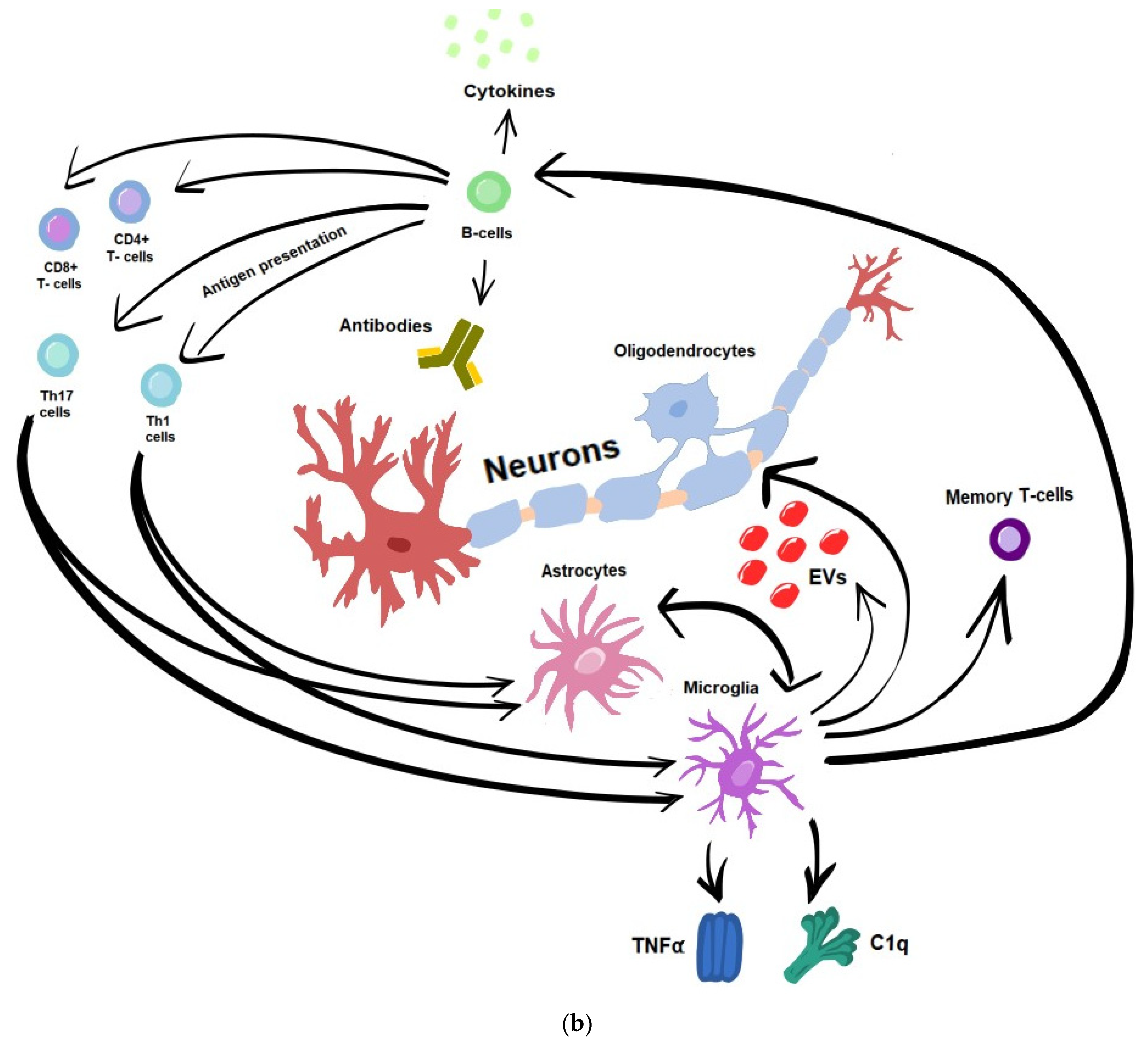
5.1. Immune-Mediated Inflammation and Neurodegeneration

Neuroinflammation in PPMS seems to be less dependent on systemic immune activity and compartmentalized within the CNS. Apart from focal chronic injury within demyelinative lesions, there are inflammatory infiltrates mainly located in the leptomeninges and diffuse alterations in apparently normal white and grey matter [22]. The key players of these processes include T- and B-cells, while CNS-resident cells provide links between inflammation and neuronal damage.

5.1.1. T-Cells

The T-cell-mediated autoimmune response, crucial for initiation of MS-related CNS injury, is also relevant for progressive types of disease. The T-cell activation marker sCD27 was found to be elevated in the CSF of PPMS patients [23].
On the contrary to early stages of MS (when CD4+ T-cells are mostly implicated), CD8+ T-cells are the predominant subset found in chronic lesions, and their presence correlates with the degree of axonal damage [19,24,25,26]. Furthermore, Th1 and Th17 cells are involved in chronic inflammation underlying progression via interaction with astrocytes and microglia (promoting its proinflammatory phenotype), the upregulation of cytokines (e.g., IL-17) and the inhibition of neurotrophic factor production [27,28].

5.1.2. B-Cells

Recently, it has been proposed that the interplay of T- and B-cells is a core feature of MS pathogenesis, especially in progressive types of disease [19,29]. Different mechanisms associated with B-cell activity in CNS include the production of auto-antibodies targeted against myelin, antigen presentation to Th17 and Th1 cells and driving the autoproliferation of brain-homing T cells, the secretion of inflammatory cytokines and chemokines (e.g., IL-6, il-12, IL-15, TNF-α, and INF-γ), as well as the secretion of soluble toxic factors that induce oligodendrocyte and neuronal injury [24,29,30]. It was also suggested that B-cells of MS patients are less capable of downregulating immune responses due to their deficiency in IL-10 production [29]. The depletion of B-cells using anti-CD20 monoclonal antibodies resulted in significantly diminished pro-inflammatory responses of CD4+ and CD8+ T-cells as well as myeloid cells, and provided evidence of the use of these agents as highly effective therapies both in relapsing and progressive MS.
The retention of pro-inflammatory B-cells within CNS is supposed to be mediated by activated leukocyte cell adhesion molecule (ALCAM) and their activity by B-cell maturation antigen (BCMA) and transmembrane activator and CAML interactor (TACI), two survival receptors liberated into CSF [29,31].
B-cells also contribute to the pathology of MS by producing autoantibodies against different CNS antigens presented on neurons, oligodendrocytes and astrocytes. These antibodies, reactive against myelin and other CNS antigens, can be detected as already mentioned OCB in CSF [32].
Furthermore, B-cells have been recognized as the main component of ectopic lymphoid follicles localized subpial or adjacent to the cortex, identified in brain tissue samples from subjects with progressive MS, and associated with a loss of cortical neurons and activation of microglia. Although these findings seemed more typical for a secondary progressive type of disease (and presumably for its longer duration), subpial and cortical inflammatory lesions were often demonstrated in MRI in PPMS patients [33].

5.1.3. Microglia

Microglia represent a specialized population of macrophage-like cells in the CNS, acting as mediators between the peripheral and CNS immune system and creating a microenvironment for neurons via interaction with astrocytes and oligodendrocytes.
In physiological conditions, microglia provide trophic support for neurons and promote repair processes, while in the course of MS (particularly during its progressive stage), there is a transition from a homeostatic state of microglia to their altered phenotypes, dynamically changing in morphological and functional aspects in response to pathological conditions [34,35,36]. This detrimental phenotype of microglia is responsible for neuroaxonal damage via different mechanisms, linking inflammation and degeneration [36]. The presence of such activated microglia (also assigned as “microglia inflamed in multiple sclerosis—MIMS”) has been demonstrated in brain tissues from patients with progressive MS in chronic demyelinating lesions (especially smoldering/slowly expanding ones) [24,35,37], as well as within diffuse alterations in normal-appearing white and grey matter [37]. MIMS contains the two main clusters: the first one is characterized by lipoprotein reactivity and associated with lysosomal activity, the clearance of myelin debris and the regulation of the inflammatory response. The second one, with typical accumulation of iron, plays a role in antigen presentation (causing restimulation of autoreactive memory T-cells) and the propagation of inflammation (as a source of complement components, mediating antibody-dependent cytotoxicity) [37,38]. The proinflammatory effect is also associated with the production of cytokines, e.g., tumor necrosis factor-alpha (TNF-α) and interleukin 1 beta (IL-1β), found in extracellular vesicles released from activated microglia in progressive MS [39]. The chemoattraction of immunocompetent cells from the periphery and positive feedback interaction between microglia and astrocytes further enhance the inflammatory response in CNS [36].
Microglia-derived cytokines provide a link between inflammation and neurodegeneration, i.a., by facilitating glutamatergic transmission and potentiating excitotoxicity—another detrimental effect of altered microglia upon neurons (increased vulnerability due to dysregulation of ion homeostasis and metabolic intermediates) [36,40]. Moreover, excessive iron accumulation in activated microglia may promote the transition of T-cells to their pro-inflammatory and pathogenic phenotype, as well as lead to iron-mediated cell death, i.e., ferroptosis [41,42]. In addition, due to metabolic pathways associated with an increased production of ATP, altered microglia become a source of reactive oxygen and nitrogen species, contributing to oxidative stress (OS) and mitochondrial dysfunction. Iron may further enhance oxidative damage in the presence of ROS produced by the oxidative burst [41,42]. These processes, together with impaired antioxidant mechanisms, result in an increase in energy demand from neurons, with concomitant insufficient ability to supply it [36,40]. OS-mediated mitochondrial dysfunction is supposed to affect the activity of enzyme complexes and promote apoptosis [43]. It has been demonstrated in experimental models that CSF from patients with PPMS applied to healthy rat neurons induced mitochondrial dysfunction, energetic failure and axonal damage with neuronal apoptosis, which was inhibited by caspase inhibitors [41,42,43,44,45,46,47,48,49,50,51,52]. Other pathways involved in neuronal loss in progressive MS include the activation of necroptosis signaling in cortical neurons and the induction of apoptotic cascades by microglia-derived inflammatory and cytotoxic mediators (TNFα, IL-1β, IL-6 and ROS) [50].
Apart from the aforementioned mechanisms of neuronal injury, energetic deficit, lack of growth factors and cytotoxic mediators from activated microglia may specifically impair axonal growth and cause the degeneration of synapses (synaptopathy), as well as (coupled with inadequate clearance of myelin debris) contribute to the failure of remyelination [36,53].

5.1.4. Astrocytes

Astrocytes provide structural and metabolic support for neurons, control the neuronal microenvironment, and participate in the immunomodulation and regulation of BBB function [54].
Similarly to microglia, in the course of progressive MS, astrocytes become activated (gathering at the edge of chronic active lesions, particularly in the cortex), which is indicated, i.a., by the upregulated expression of glial fibrillary acidic protein (GFAP). Activated astrocytes undergo a range of transcriptional and functional alterations [55], including the dysregulation of their homeostatic functions (glutamate uptake, lactate shuttling and elimination of potassium). Furthermore, astrocytes secrete cytotoxic factors, mainly associated with OS (reactive oxygen species and nitric oxide). Adverse effects of excitotoxity and OS-mediated injury comprise the impairment of neuronal metabolism and damage to the axonal cytoskeleton [36,54].
Reactive astrocytes also promote inflammation in CNS—directly (by the production of interleukins, e.g., IL-6) or chemoattracting immunocompetent cells (e.g., via CCL2). They support the survival and activity of B-cells (via B-cell-activating factor—BAFF), which reciprocally maintain the proinflammatory activation of astrocytes [36]. Moreover, microglia–astrocytes crosstalk plays an important role in the regulation of inflammatory signaling within a CNS environment [38,56].
Astrocyte alteration in progressive stages of MS may contribute to a subtle, chronic dysfunction of BBB, supposed to compromise protection of the CNS from peripheral toxic factors. In addition, the formation of a glial scar by astrocytes (isolating areas of MS-related damage) can inhibit remyelination and axonal regeneration [40,54].

5.2. Remyelination Failure

The loss and dysfunction of oligodendrocytes (OL) and their precursors (OPC), responsible for the production of myelin in CNS, is crucial for the balance between the processes of myelin injury and repair in the course of MS. The results of this impaired balance can be particularly observed in chronic active or “smoldering” demyelinating lesions. A range of extrinsic and intrinsic factors have an adverse impact on OPC migration, proliferation and differentiation [57].
As mentioned above, remyelination may be impeded by OL/OPC interactions with microglia and astrocytes on multiple levels (including the effect of released mediators, shaping extracellular matrix, insufficient amount of trophic support or inadequate clearance of myelin debris). Glutamate excitotoxicity and proinflammatory cytokines may directly cause the loss or dysfunction of OL, while chemokines mainly modulate the migration and proliferation of OPC [36,50,56,58]. OL and OPC, due to their poor antioxidant defense, are most susceptible to adverse effects of OS, including their selective damage [41].
Relevant impact upon the impaired proliferation and differentiation of OPC into OL is associated with aging. Aging-related alterations, displayed in animal models of demyelination, include the dysregulation of transcription factors, decreased expression of metabolic activity and the accumulation of cells with particular senescence-associated secretory phenotypes (SASPs) [59]. Neural progenitor cells (NPCs) (derived from induced pluripotent stem cell lines) acquired from PPMS patients or from white matter chronic lesions in autopsy brain tissues failed to promote OPC maturation and showed the expression of high-mobility group box-1 (HMGB1), which are considered cellular senescence hallmarks [60].
The investigation of dynamic changes in the chronic lesion microenvironment, as well as determining the endogenous components required for OL lineage cell progression, would enable a better understanding of remyelination regulation with potential therapeutic targets [57].

6. Monitoring and Predicting Progression in PPMS—Potential Biomarkers

One of the “hot topics” in MS research, especially with regard to progressive types of disease, is associated with putative biomarkers, useful for diagnostic, prognostic and monitoring purposes. Reliable and sensitive tools seem necessary to identify patients with higher risks of disease progression and to follow-up its course, including the assessment of the response to treatment.

6.1. Clinical Biomarkers

The Expanded Disability Status Scale (EDSS) [11] is the basic tool for the quantification of MS-related disability, most commonly used in routine clinical practice and as the primary outcome measure in clinical trials. An increase in the EDSS score (persistent or continuing over time) can reflect sustained disability worsening or PIRA. Although standardized, relatively simple and time-saving, EDSS has its limitations, particularly as a measure for early/silent progression. In spite of the detailed subscales evaluating different CNS functional systems, the motor function/gait ability remains the major factor affecting the total EDSS score and defining its milestones (limited walking distance and need for assistive devices). Thus, the EDSS score appears less sensitive to other disability aspects, both in patients within low and high ranges. To overcome this and to provide measures for particular aspects of disability, more refined tests are proposed, evaluating, e.g., manual dexterity, visual acuity and—particularly important—cognitive performance [61,62].
The timed 25-foot walk (T25FW) is considered one of the best-characterized measures of walking in MS, frequently used in the assessment of mobility impairment. The nine-hole peg test (NHPT) is a standard quantitative test of upper extremity function, applied for dominant and non-dominant hands, with a 20% change in the test score considered relevant for clinically meaningful worsening. Koch et al. determined the significance of progression rates in PPMS patients based on EDSS, T25FW and NHPT scales over a 3-year follow-up. Their results showed significant worsening in the EDSS and T25FW results, while only a few patients worsened in the NHPT. Although NHPT seems less appropriate as an individual primary endpoint in PPMS, it might be useful as the additional measure for the individual assessment of patients’ disability [63].
Considering the importance and frequency of cognitive failure in PPMS, adequate neuropsychological tests are indispensable in its recognition and quantification. Particular tools can be applied to evaluate performance in selected cognitive domains (e.g., California Verbal Learning Test (CVLT2) [64] for verbal memory, and Symbol Digit Modality Test (SDMT) [64,65] for attention and executive function) or Paced Auditory Serial Addition Test (PASAT), evaluating calculation ability, but also the speed and flexibility of information processing. Neuropsychological tests batteries, dedicated to MS patients, have been also developed for the more complex evaluation of cognitive performance (e.g., Brief International Cognitive Assessment for Multiple Sclerosis (BICAMS) or Brief Repeatable Battery of Neuropsychological Tests (BRB-N) [65].
Furthermore, integrated measures of physical and mental aspects of disability have been designed. The Multiple Sclerosis Functional Composite (MSFC) consists of T25FW, 9HPT and PASAT. The MSFC has been proven to be a sensitive tool for measuring the accumulation of disability throughout the disease, and is thus potentially useful in monitoring progression [8,64,65]. Similarly to the status of NEDA (“No Evidence of Disease Activity”), used as the primary outcome in clinical trials, NEP (“No Evidence of Progression”) has been proposed, defined as the absence of 24-week sustained clinical progression (measured by an increase in EDSS score; ≥20% increase in 25FWT; and ≥20% increase in 9HPT) [8,64,65].
Recently, increased attention has been paid to patient-reported outcome measures (PROMs), which provide patients’ perspective of their functioning in different spheres of life and therefore allow the evaluation of the impact of their disease. PROMs can capture particular domains, including depression, anxiety, pain, bladder control, fatigue or sleep disturbances, as well as more general issues, including overall disease burden, quality of life or social participation (e.g., PROMIS 29 questionnaire, MS Quality of Life—54-MSQOL). Due to the guidelines of EMA, these measures are being increasingly used in research and randomized clinical studies.
In multi-center three-phase trials with ocrelizumab in PPMS patients (ORATORIO, CONSONANCE), the main outcome measures included the composite evaluation of progression (NEP), SDMT and Brief Visuospatial Memory Test—Revised for the assessment of cognitive performance and 36-Item Short-Form Health Survey as a measure of health-related quality of life [66,67,68].
Regular and quantified monitoring of performance in the functional tests and PROMs seems essential for monitoring progression. Due to the increasing popularity of digital biosensors with easily administered applications (smartphones or smartwatches), frequent objective assessment in daily functioning and relevant review of the measures at follow-up visits becomes more accessible.

6.2. Biochemical Biomarkers

Among body fluids biomarkers of activity or progression in MS, the most relevant evidence supports the use of serum neurofilament light chain (NfL) and glial fibrillary acidic protein (GFAP).
NfL is a cytoskeletal protein that is released after axonal damage to extracellular space, penetrating to CSF and (in a much smaller but proportionate amount) to peripheral blood. Its concentration can be reliably measured both in CSF and serum [69,70,71]. Elevated NfL amount in MS patients is associated with recent inflammatory activity. However, as a sensitive marker of axonal loss, NfL can also be applied for monitoring progression. NfL concentration was found to correlate with EDSS score and its increase over time [72,73,74]. Used as the outcome measure in clinical trials with PPMS patients (INFORMS—fingolimod, ORATORIO—ocrelizumab), NfL level decreased during several months of treatment, parallel to clinical effects (slowed rate of disability accumulation) [72,73,74]. Furthermore, the predictive value of this potential marker has been shown in PPMS patients treated with high-efficacy therapies; high levels of NfL at baseline predicted the risk of confirmed disability progression or occurrence of PIRA within few years of follow-up [74].
GFAP is a monomer intermediate filament protein, mainly expressed in astrocytes; its breakdown products reflect astrocyte injury and reactive astrogliosis. GFAP seems to be an even better indicator of progression than activity, as its level not only clearly differentiates between MS patients and healthy controls, but is also significantly higher in progressive forms and inactive stages of disease [73,74]. Furthermore, in progressive MS, an elevated level of GFAP correlates with disease duration, EDSS score and its dynamics, as well as with decreased white and gray matter volumes.
Concomitant analysis of serum GFAP and NfL shows their significant relationship with each other, and increases the predictive value of these combined markers with regard to confirmed disability worsening and PIRA [70,71,72,73]. On the other hand, little specificity of both NfL and GFAP (their increased concentration occurs in a range of CNS diseases with various etiology–inflammatory, degenerative, metabolic, post-traumatic, etc.) as well as a lack of a consistent measurement methodology and normative values are limitations to their use as biomarkers in this field.
Recent studies on biomarkers of progression in MS focused on chitinase -3-like-1protein (CHI3L1). CHI3L1 is expressed, i.a., in neutrophils, macrophages, endothelial cells, astrocytes and microglia, and is involved in tissue remodeling and modulating inflammation. This secretory protein can be detected in both CSF and serum. The studies revealed elevated levels of CHI3L1 in CSF in MS patients compared to healthy controls, and in PPMS in comparison with RRMS [75]. Correlations were also found between increased levels of CHI3L1 and baseline EDSS score as well as the subsequent progression of disability, with less of a relationship with age and disease duration [75,76,77,78].
Studies on putative biomarkers associated with OS concerned, i.a., total oxidation status, level of 8-iso-prostaglandin, malondialdehyde or superoxide dismutase (SOD) as indices of pro-oxidative mechanisms. Total antioxidant status (TAS), glutathione levels and glutathione peroxidase activity have been investigated in turn as indices of antioxidant capacity [51,78]. The findings from investigations of OS indices in body fluids from MS patients suggest a disturbed balance between pro- and antioxidant mechanisms, to the detriment of the latter [43,51]. With regard to indices relevant for the progression of disease, increased level of 8-iso-prostaglandin, the product of lipid peroxidation of arachidonic acid, was demonstrated in the patients with progressive types of MS. Platelet hemostatic function, also found to be advanced in progressive MS, positively correlated with increased production of the superoxide radicals, as well as with the level of disability. The levels of advanced oxidation protein products and nitric oxide metabolites were higher in the MS subjects with a greater increase in disability over 5 years of follow-up [37,52].
Sphingolipids also seem promising candidates for the role of the markers for progression in MS. The accumulation of ceramide derivatives has been demonstrated in MS lesions and CSF, and their level correlated with clinical deterioration in progressive types of the disease. Ceramide was also suggested as a factor produced by the meningeal inflammatory infiltrates and associated with microglia activation. Sphingolipids are supposed to impair balance between signaling pathways involved in neuronal damage and neuroprotection, as well as oligodendrocyte homeostasis [3,79].
In recent years, there has been increasing interest in extracellular vesicles (EVs)—small structures released by potentially all cell types, carrying a range of molecules and acting as mediators of intercellular communication—as potential biomarkers in neurodegenerative and autoimmune diseases, including MS [80]. EVs can be revealed in all body fluids, including plasma, serum and CSF, and evaluated by means of so-called liquid biopsy [81,82]. In the microenvironment of neuroinflammation, the amount of EVs produced by immune cells increases, indicating inflammatory activity [83]. EVs released by T-cells are able to activate endothelial cells and monocytes, while EVs derived from endothelial cells in turn activate CD4+ and CD8+ T-cells [84]. In addition, EVs seem to be involved in endothelial dysfunction and BBB disruption; the amount of CNS endothelial-derived EVs has been suggested as an indicator of BBB permeability and integrity in the course of MS [85]. Furthermore, as already mentioned, EVs derived from activated microglia have a distinct proteomic profile and carry cytokines like TNF-α and IL-1, acting as mediators propagating inflammation, as well as preventing remyelination [83]. Thus, EVs released by microglia and other CNS-resident cells have been proposed as markers of these cells’ activity in vivo, which could be directly quantified in liquid biopsy from CSF by flow cytometry [86,87,88].
In the investigation of the molecular basis for the MS background, attention has been recently paid to the role of non-coding RNAs (ncRNAs), including microRNAs (miRNAs) and long non-coding RNAs (lncRNAs) [89]. miRNAs are small single-stranded molecules that regulate gene expression at the post-transcriptional level, relevant for numerous cellular processes (including apoptosis or stress response) [89,90,91]. Long (more than 200 nucleotides) non-coding RNAs (lncRNAs) take part in the regulation of transcription, post-transcription, and translation [86,92] and have a substantial impact on the development of the immune system and nervous tissue. The dysregulation of lncRNAs has been associated with processes of neuronal death in the course of neurodegenerative diseases [93]. A number of aberrantly expressed miRNAs and lncRNAs have been identified in body fluids (plasma, serum, CSF) and CNS cells from brain tissues in MS patients. Preliminary findings of these investigations suggested the possible role of ncRNA as biomarkers differentiating the progressive from the relapsing–remitting phenotype of disease or predicting the severity of its course [94,95]. Further studies using in vitro or in vivo models seem necessary to verify the relevance of these observations.

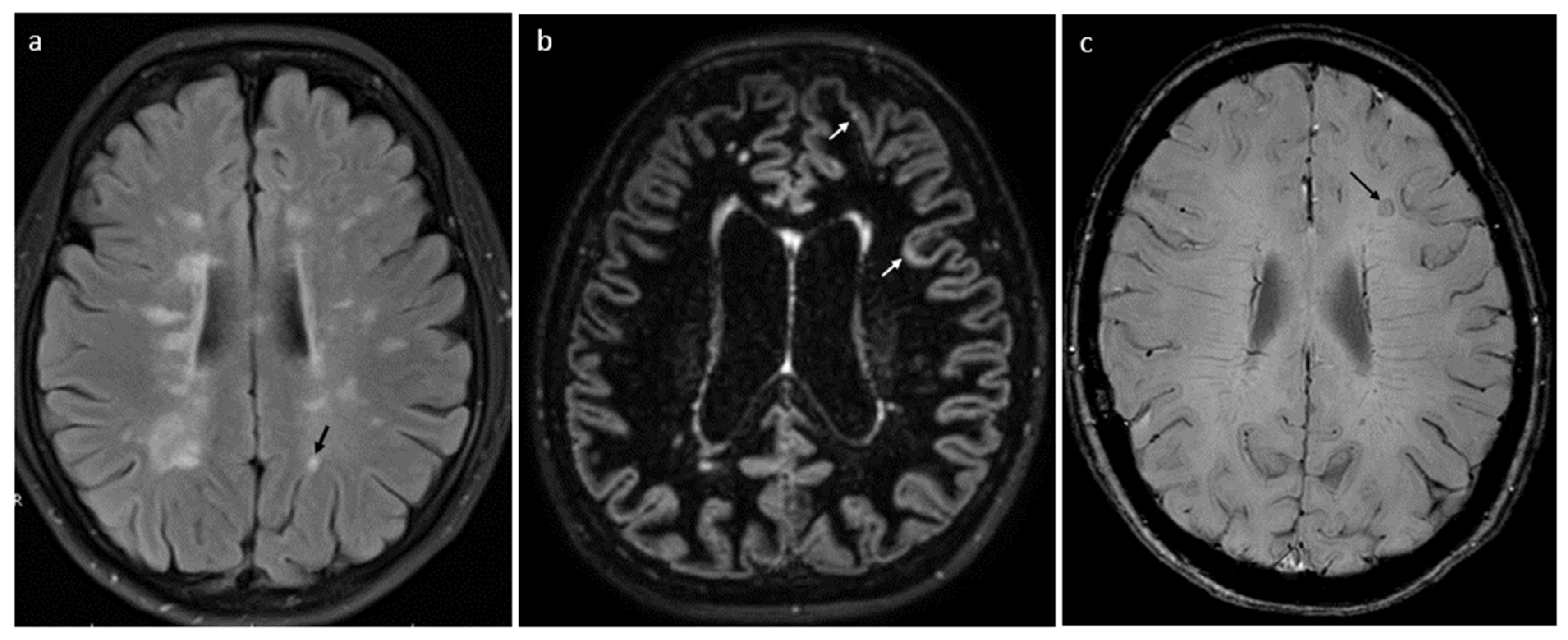
6.3. Radiological Biomarkers

As stated above, the number and load of new/active lesions in standard MRIs of the brain and spinal cord, which may occur especially in the early stage of PPMS, are considered appropriate markers for MS activity. However, identifying and monitoring progression requires other, more specific radiological indices. Evidence-based data support mainly the use of the following: slowly expanding lesions (SELs), paramagnetic rim lesions (PRLs), cortical lesions, and measures of global and regional CNS atrophy.
SELs and PRLs correspond with process of multifocal, chronic CNS inflammation, a substantial compound of background for progression in MS. SELs are characterized by constant and concentric volumetric expansion in T2, with concurrent reduction in T1-weighted sequence, revealed in two to three subsequent MRI scans [22,96,97,98,99]. PRLs have a typical rim surrounding at least 75% of lesion, which reflects the layer of iron-laden microglia and macrophages, accompanying demyelination and axonal transection and can be shown in phase imaging, SWI or multi-gradient echo sequences (Figure 2).
SELs and (even more) PRLs are usually scarce in number but more frequent in progressive MS.
Their presence and amount show significant correlation with degree of disability and cognitive impairment and their deterioration over time. Furthermore, the co-localization of SELs and PRLs is strongly associated with the prediction of PIRA within further few years of follow-up [96,100].
Chronic neuroinflammation in the form of subpial infiltrates of lymphocytes, together with retrograde degeneration from axons transected within white matter lesions, contribute to cortical demyelination. Until recently, cortical lesions have been difficult to reveal by standard MRI sequences. New techniques with the use of ultra-high-magnetic field resolution (>7 T), double inversion recovery (DIR) and phase-sensitive inversion (PSIR) allow the visualization of cortical damage and ribbon-like subpial demyelination [96,101,102,103]. They appear to be more common and extensive in progressive MS and are mainly localized in the parietal lobe, followed by the frontal, occipital, temporal lobes and insula. Their total volume correlates with the level and deterioration rate of physical disability and cognitive performance [96,101,104].
Cerebral atrophy is a hallmark of diffuse neurodegeneration with irreversible axonal and neuronal loss, particularly relevant in progressive MS. Increased brain volume loss in PPMS significantly correlated with disability progression, independent of the number of previous or new T2 lesions [2,51,100]. Relationships were shown between global and regional cortical atrophy and cognitive functioning, as well as fatigue and mood disorders in patients with progressive MS. Volumetric analysis of serial MRIs in PPMS revealed the highest rate of atrophy for cortical but also deep grey matter structures (including thalamus, putamen, hippocampus and cingulate gyrus). Atrophy of the thalamus has been considered as the most important measure to predict clinical worsening (including cognitive decline) within a few years’ observation [97,100,101,105]. Moreover, spinal cord volume loss is another marker of disease progression and seems to be independent of the number of spinal cord lesions (Figure 3). Spinal cord atrophy occurs at higher rates than cerebral atrophy, particularly in progressive forms of disease, and correlates with increase in disability [103] (Figure 2 and Figure 3).
Diffuse neuronal and axonal damage typical for PPMS can be depicted as “dirty-appearing white matter”—ill-defined hyperintense areas (in T2-weighted or proton density-weighted images), located mainly around the lateral ventricles. Microstructural brain tissue injury within normal-appearing white or grey matter can be even better demonstrated in diffusion MRI techniques (diffusion tensor imaging—DTI). Indices of these microstructural abnormalities—increased mean diffusivity (MD) (a measure of the degree of the restriction to diffusion) and decreased fractional anisotropy (FA)—have been shown as predictive factors for disability worsening (including cognitive performance) in progressive MS [104,106].
Another advanced neuroimaging technique, which allows the visualization of diffuse CNS damage in progressive MS, is positron emission tomography (PET). PET radioligand binding to 18kD translator protein (TSPO) corresponds with the function of activated microglia and macrophages in the brains of people with MS. Increased uptake of TSPO tracers, such as 11C-(R) -PK11195 and F- PBR111, was found in all MS phenotypes, but predominantly in progressive forms. PET turned out to also be a sensitive tool in the functional imaging of chronic demyelinative lesions (e.g., PRL) [100,101].
The integration of different radiological measures (e.g., indicating focal and diffuse MS-related CNS damage or combining structural with functional brain imaging) may provide more sensitive monitoring and predictive markers for progression in MS.
Table 1 summarizes the biomarkers of progression in MS currently in use or under investigation.

7. PPMS—Therapeutic Options

Ocrelizumab (OCR) is currently the only disease-modifying therapy (DMT) approved by FDA (2017) and EMA (2018) for use in the active type of PPMS (besides its application in RRMS). OCR is a humanized monoclonal antibody, targeted at CD20 antigen in B-cells. Its mode of action includes selective (excluding pro-B-cells and long-term immune memory plasmatic cells) and rapid depletion of B-cells via multiple mechanisms (apoptosis, antibody-dependent cellular cytotoxicity, antibody-dependent cell-mediated phagocytosis and complement-dependent cytotoxicity). The use of OCR in PPMS was feasible, considering the role of B-cells in chronic neuroinflammation (e.g., subpial infiltration and diffuse grey matter injury) underlying progression in MS. The results of ORATORIO, phase 3 randomized clinical trial testing OCR vs. placebo in PPMS patients, demonstrated beneficial effect of the drug on clinical and radiological measures of progression and activity of disease. During at least 24 weeks of treatment, OCR was associated with a significant reduction in the hazard for confirmed disability progression (measured with EDSS score). With regard to particular disability aspects, treatment with OCR lowered the risk of confirmed worsening in walking ability and upper limb dexterity (measured with T25FW and 9HPT scores), decreased level of physical and cognitive fatigue and improved overall quality of life [107,108,109,110,111].
Analysis of MRI parameters showed that OCR, in comparison to placebo, caused a significant decrease in total volume of T1 and T2 brain lesions, as well as lowering the rate of global and regional cerebral atrophy in the patients with PPMS, including volume of cerebellum, cerebral cortex and (most significantly) thalamus. Overall, among those treated with OCR, there was significant increase in percentage of patients who showed no evidence of progression or activity [108,109,110,111]. The safety of OCR in the PPMS cohort turned out to be similar to that of the RRMS population in the other trials, with infusion-related reactions, upper respiratory tract infections, and oral herpes infections as the most frequent adverse events, and with insignificant frequency of serious infections or malignancies.
Interesting findings from ORATORIO were associated with the use of different markers of progression and suggested their monitoring and prognostic utility. During the treatment, there was a significant difference in the decrease in serum NFl level, favoring OCR treatment to placebo. Higher baseline NFl level correlated with number and volume of SEL and with subsequent rate of reduction in thalamic volume during the treatment [108,109,110,111,112].
Long-term observations of patients from the ORATORIO cohort demonstrated that after 10 years, more than 1/3 of those continuously treated with OCR were progression-free, and more than 80% did not require a walking aid or wheelchair. Those who started the drug early maintained lower risk of confirmed disability worsening in comparison with the patients initially receiving a placebo and switching to OCR after 3 years. The safety profile of the drug in long-term data remained stable and satisfactory [112,113].
Nowadays, there are ongoing multicenter studies (eg CONFIDENCE, VERISMO or CONSONANCE), assessing the safety, adherence and persistence of OCR in newly treated MS cohorts (including PPMS ones) in a real-world setting [2,109,110,111,114].
Encouraged by these findings, another anti-CD20-antibody, rituximab, was evaluated for its efficacy and safety in PPMS in the OLYMPUS trial. Although pre-specified analyses revealed that RTX significantly slowed progression in younger patients with active plaques in MRI, the trial failed to meet its main endpoint—lowering the proportion of confirmed disability progression in the study cohort.
Unlike OCR, several other DMTs effectively used in RRMS (i.a. interferon beta, fingolimod, natalizumab, alemtuzumab) were not proved to be beneficial in PPMS. Corresponding clinical trials (e.g., INFORMS and ARPEGGIO) showed that these drugs did not significantly reduce disability progression or indices of brain atrophy in MRI [113,115].
Bruton’s tyrosine kinase inhibitors (BTKi) seem to be another promising approach for the treatment of progressive MS. BTKi exert a multidirectional effect on both innate and adaptive immune system, including activation of B-cells and activated phenotype of microglia. Furthermore, due to their small size, they are able to cross the BBB and directly modify CNS-compartmentalized inflammatory processes (with parenchymal and leptomeningeal lymphocyte infiltration), important in the progression of the disease. They are also supposed to promote myelin repair activities. Supported by the results of phase II trials, there are ongoing studies (PERSEUS for PPMS and HERCULES for SPMS) investigating the efficacy and safety of tolebrutinib in progressive types of MS [115,116,117,118,119].
Apart from targeting chronic neuroinflammation, attempts have been made to prevent progression in MS by promoting the protection of axons and myelin repair (mainly through affecting the proliferation, differentiation and maturation of oligodendrocytes).
Ibudilast is an inhibitor of cyclic nucleotide phosphodiesterases, also targeting macrophage migration inhibitory factor and Toll-like receptor 4, and penetrating through the blood–brain barrier. A phase 2 clinical trial involving patients with progressive MS demonstrated that ibudilast, in comparison with placebo, was associated with a slower rate of brain atrophy, as well as a reduced volume and magnetization transfer ratio of slowly expanding lesions (SELs). These radiological findings may indicate neuroprotective effects of ibudilast and support their further investigation [120,121,122].
High-dose biotin (HDB) activates carboxylases and enhances the production of ATP; thus, it was supposed to prevent hypoxia-driven axonal degeneration and was considered a novel therapeutic target in progressive types of MS [76]. Among disability measures in PPMS, randomized controlled trials only showed some beneficial effect of long-term intake of HDB upon walking ability, and there was strong evidence for increased laboratory test interference as a potential bias in the results [123].
Another object of investigation in this field has been the opicinumab-monoclonal antibody against LINGO-1 (inhibitor of oligodendrocyte differentiation and axonal regeneration). However, trials conducted on patients with optic neuritis or RRMS have failed to confirm the efficacy of opicinumab with regard to clinical outcomes; thus, the remyelination potential of the drug (as well as its putative impact on progressive MS) remains unclear [115,124,125].
Elezanumab is a monoclonal antibody that neutralizes repulsive guidance molecule (RGMa), an inhibitor of neurite outgrowth. In animal models of autoimmune CNS demyelination, elenazumab has been shown to decrease the area of inflammation, promote axonal regeneration and remyelination and improve functional recovery. Due to these preclinical data supporting the neuroregenerative and neuroprotective potential of the drug, the two phase 2 studies were designed to evaluate the safety and efficacy of elezanumab in patients with relapsing or progressive types of MS [126,127,128].
Oligodendrocyte precursor cells (OPC) seem a promising target for therapies stimulating remyelination. Preclinical evidence suggests a specific role for clobetasol (anti-inflammatory synthetic glucocorticoid) in the regulation of differentiation and increased viability of OPC. Studies on animal models showed that miconazole (anti-fungal, a synthetic derivative of imidazole) reduced disease activity and improved motor functionality by enhancing OPC differentiation, but did not significantly affect the remeylination. Metformine in preclinical studies appeared to have a potential impact on oxidative stress and induced the formation of new myelin [86]. There is also some evidence that muscarinic antagonists (clemastine fumarate, benztropine, and quetiapine) may exert a similar effect. Although treatment with clemastine fumarate resulted in a reduction in prolonged visual evoked potential latency (electrophysiological measure for remyelination), no radiological indices of remyelination were detected in MRI [129,130,131].
Mesenchymal stem-cell-based (MSC) therapy also has been investigated as a promising candidate for capturing multiple targets in the treatment of progressive MS. The role of MSCs is associated with the modulation of pathogenic immune responses via the production of anti-inflammatory cytokines, the inhibition of pro-inflammatory ones (by suppressing Th1 and Th17 lymphocytes) and the upregulation of regulatory processes. MSCs are also responsible for the secretion of neurotrophic growth factors, which promote the proliferation and survival of neurons, counteracting neurodegeneration and axonal loss. Moreover, MSC therapy is supposed to activate oligodendrogenesis and suppress the apoptosis of oligodendrocytes, contributing to processes of myelin repair. The results of the first placebo-controlled trial using intrathecal administration of MSCs in patients with active progressive MS demonstrated a beneficial effect of therapy upon the disease outcomes—the achievement of status without evidence of disease activity, stabilization or improvement in disability scores and decreased level of NfLs [85,102,103]. Further randomized trials are necessary to evaluate the effectiveness and a safety profile of MSC therapy in long-term observations [114,131,132].
Relevant evidence from successfully completed and ongoing clinical trials on therapeutic agents targeting progression in PPMS is summarized in Table 2.
The therapeutic approach in PPMS should also include symptomatic treatment for predominating signs of neurological deficit (e.g., relieving spasticity or bladder dysfunction), as well as physiotherapeutic interventions and psychological support (with regard to cognitive problems or mood disturbances). Due to the usually older age at PPMS onset compared to the overall MS population, aspects of CNS aging and appropriate treatment of comorbidities also have to be taken into account. Furthermore, complex therapeutic strategies, currently recommended in MS, assume the modification of lifestyle factors (e.g., balanced diet, encouraging physical and mental activity, and avoiding smoking or alcohol abuse), aimed at enhancing “neurological reserve” (the compensatory capability of CNS) and thus preventing progression [113,114].

8. Summary

Despite recent progress in clinical studies and the advent of the first approved therapies for PPMS (Table 3), the diagnosis and management of this type of disease still pose a challenge. In view of current concepts of “smoldering MS”, with the interplay of relapses and progression throughout the whole range of the disease course, thorough investigations of PPMS with multidimensional evaluation of progression seem necessary to elucidate the aspects not addressed so far. These investigations would provide a better insight into the processes of CNS damage underlying progression, with identification of their potential monitoring and prognostic biomarkers. Clinical implications of these findings would be associated with the reliable assessment of disease outcomes, improvement in individualized therapeutic approach and hopefully also novel therapeutic targets, relevant for the management of progression.
Table 2 summarizes chronological milestones in research on primary progressive multiple sclerosis, including treatment strategies.

Supplementary Materials

The following supporting information can be downloaded at: https://www.mdpi.com/article/10.3390/ijms25168751/s1.

Author Contributions

I.S., conceptualization, writing—original draft preparation; E.D., conceptualization, writing—review; H.M.—writing part of immune dysregulation; A.Z.—writing part of radiological biomarkers; and A.P.-D. conceptualization, writing—review and editing. All authors have read and agreed to the published version of the manuscript.

Funding

Supported by Wroclaw Medical University SUBZ.C220.24.053.2024.

Acknowledgments

Special thanks to Eliza Dehacq for the graphics.

Conflicts of Interest

The authors declare no conflicts of interest.

Abbreviations

ALCAMActivated leukocyte cell-adhesion molecule
APCAntigen-presenting cells
BICAMSBrief International Cognitive Assessment for Multiple Sclerosis
BBBBlood–brain barrier
BCMAB-cell maturation antigen
BRB-NBrief Repeatable Battery of Neuropsychological Tests
CHI3L1chitinase -3-like-1protein
CNSCentral nervous system
CSFCerebrospinal fluid
CVLT2California Verbal Learning
DMTDisease-modifying therapies
EDSSExpanded Disability Status Scale
EVsExtracellular vesicles
GFAPGlial fibrillary acidic protein
lncRNAsLong non-coding RNAs
miRNAsmicroRNAs
MSMultiple sclerosis
MSFCMultiple Sclerosis Functional Composite
NEDA“No Evidence of Disease Activity”
NEP“No Evidence of Progression”
NHPTThe nine-hole peg test
ncRNAsNon-coding RNAs
OCBOligoclonal IgG bands
OCGBOligoclonal immunoglobulin G bands
OLOligodendrocytes
OSOxidative stress
PASATPaced Auditory Serial Addition Test
PIRAProgression independent of relapses
PPMSPrimary progressive multiple sclerosis
PRLParamagnetic rim lesions
PROMsPatient-reported outcome measures
RAWRelapse-associated worsening
RNSReactive nitrogen species
ROSReactive oxygen species
RPMSRelapsing–progressive multiple sclerosis
RRMSRelapsing–remitting multiple sclerosis
SDMTSymbol Digit Modality Test
SELSlowly expanding lesions
SPMSSecondary progressive multiple sclerosis
TACITransmembrane activator and CAML interactor
TASTotal Antioxidant Status

References

  1. Ghasemi, N.; Razavi, S.; Nikzad, E. Multiple Sclerosis: Pathogenesis, Symptoms, Diagnoses and Cell-Based Therapy. Cell J. 2017, 19, 1–10. [Google Scholar] [CrossRef] [PubMed] [PubMed Central]
  2. Giovannoni, G.; Popescu, V.; Wuerfel, J.; Hellwig, K.; Iacobaeus, E.; Jensen, M.B.; García-Domínguez, J.M.; Sousa, L.; De Rossi, N.; Hupperts, R.; et al. Smouldering multiple sclerosis: The ‘real MS’. Ther. Adv. Neurol. Disord. 2022, 15, 17562864211066751. [Google Scholar] [CrossRef] [PubMed] [PubMed Central]
  3. Lassmann, H. Multiple Sclerosis Pathology. Cold Spring Harb. Perspect. Med. 2018, 8, a02893. [Google Scholar] [CrossRef] [PubMed]
  4. Müller, J.; Cagol, A.; Lorscheider, J.; Tsagkas, C.; Benkert, P.; Yaldizli, Ö.; Kuhle, J.; Derfuss, T.; Sormani, M.P.; Thompson, A.; et al. Harmonizing Definitions for Progression Independent of Relapse Activity in Multiple Sclerosis: A Systematic Review. JAMA Neurol. 2023, 80, 1232–1245. [Google Scholar] [CrossRef] [PubMed]
  5. Lublin, F.D.; Reingold, S.C.; Cohen, J.A.; Cutter, G.R.; Sørensen, P.S.; Thompson, A.J.; Wolinsky, J.S.; Balcer, L.J.; Banwell, B.; Barkhof, F.; et al. Defining the clinical course of multiple sclerosis: The 2013 revisions. Neurology 2014, 83, 278–286. [Google Scholar] [CrossRef] [PubMed] [PubMed Central]
  6. Kappos, L.; Wolinsky, J.S.; Giovannoni, G.; Arnold, D.L.; Wang, Q.; Bernasconi, C.; Model, F.; Koendgen, H.; Manfrini, M.; Belachew, S.; et al. Contribution of Relapse-Independent Progression vs Relapse-Associated Worsening to Overall Confirmed Disability Accumulation in Typical Relapsing Multiple Sclerosis in a Pooled Analysis of 2 Randomized Clinical Trials. JAMA Neurol. 2020, 77, 1132–1140. [Google Scholar] [CrossRef] [PubMed]
  7. Zanghì, A.; Galgani, S.; Bellantonio, P.; Zaffaroni, M.; Borriello, G.; Inglese, M.; Romano, S.; Conte, A.; Patti, F.; Trojano, M.; et al. Relapse-associated worsening in a real-life multiple sclerosis cohort: The role of age and pyramidal phenotype. Eur. J. Neurol. 2023, 30, 2736–2744. [Google Scholar] [CrossRef] [PubMed]
  8. Meca-Lallana, V.; Berenguer-Ruiz, L.; Carreres-Polo, J.; Eichau-Madueño, S.; Ferrer-Lozano, J.; Forero, L.; Higueras, Y.; Lara, N.T.; Vidal-Jordana, A.; Pérez-Miralles, F.C. Deciphering Multiple Sclerosis Progression. Front. Neurol. 2021, 12, 608491. [Google Scholar] [CrossRef] [PubMed] [PubMed Central]
  9. Rice, C.M.; Cottrell, D.; Wilkins, A.; Scolding, N.J. Primary progressive multiple sclerosis: Progress and challenges. J. Neurol. Neurosurg. Psychiatry 2013, 84, 1100–1106. [Google Scholar] [CrossRef] [PubMed]
  10. Cottrell, D.A.; Kremenchutzky, M.; Rice, G.P.A.; Koopman, W.J.; Hader, W.; Baskerville, J.; Ebers, G.C. The natural history of multiple sclerosis: A geographically based study. 5. The clinical features and natural history of primary progressive multiple sclerosis. Brain 1999, 122 Pt 4, 625–639. [Google Scholar] [CrossRef] [PubMed]
  11. Kurtzke, J.F. Rating neurologic impairment in multiple sclerosis: An expanded disability status scale (EDSS). Neurology 1983, 33, 1444–1452. [Google Scholar] [CrossRef] [PubMed]
  12. Katsarogiannis, E.; Landtblom, A.-M.; Kristoffersson, A.; Wikström, J.; Semnic, R.; Berntsson, S.G. Absence of Oligoclonal Bands in Multiple Sclerosis: A Call for Differential Diagnosis. J. Clin. Med. 2023, 12, 4656. [Google Scholar] [CrossRef] [PubMed]
  13. Villar, L.M.; Masterman, T.; Casanova, B.; Gómez-Rial, J.; Espiño, M.; Sádaba, M.C.; González-Porqué, P.; Coret, F.; Álvarez-Cermeño, J.C. CSF oligoclonal band patterns reveal disease heterogeneity in multiple sclerosis. J. Neuroimmunol. 2009, 211, 101–104. [Google Scholar] [CrossRef] [PubMed]
  14. Konen, F.F.; Hannich, M.J.; Schwenkenbecher, P.; Grothe, M.; Gag, K.; Jendretzky, K.F.; Gingele, S.; Sühs, K.-W.; Witte, T.; Skripuletz, T.; et al. Diagnostic Cerebrospinal Fluid Biomarker in Early and Late Onset Multiple Sclerosis. Biomedicines 2022, 10, 1629. [Google Scholar] [CrossRef]
  15. Pannewitz-Makaj, K.; Wurster, U.; Jendretzky, K.F.; Gingele, S.; Sühs, K.-W.; Stangel, M.; Skripuletz, T.; Schwenkenbecher, P. Evidence of Oligoclonal Bands Does Not Exclude Non-Inflammatory Neurological Diseases. Diagnostics 2020, 11, 37. [Google Scholar] [CrossRef] [PubMed]
  16. Thompson, A.J.; Banwell, B.L.; Barkhof, F.; Carroll, W.M.; Coetzee, T.; Comi, G.; Correale, J.; Fazekas, F.; Filippi, M.; Freedman, M.S.; et al. Diagnosis of multiple sclerosis: 2017 revisions of the McDonald criteria. Lancet Neurol. 2018, 17, 162–173. [Google Scholar] [CrossRef] [PubMed]
  17. Karaaslan, Z. Hereditary Disorders Mimicking Progressive Multiple Sclerosis. Noro Psikiyatr. Arsivi 2019, 56, 1–2. [Google Scholar] [CrossRef] [PubMed] [PubMed Central]
  18. Omerhoca, S.; Akkas, S.Y.; Icen, N.K. Multiple Sclerosis: Diagnosis and Differential Diagnosis. Noro Psikiyatr. Arsivi 2018, 55 (Suppl. S1), S1–S9. [Google Scholar] [CrossRef] [PubMed] [PubMed Central]
  19. Abdelhak, A.; Weber, M.S.; Tumani, H. Primary Progressive Multiple Sclerosis: Putting Together the Puzzle. Front. Neurol. 2017, 8, 234. [Google Scholar] [CrossRef]
  20. Fernandes, M.G.F.; Mohammadnia, A.; Pernin, F.; Schmitz-Gielsdorf, L.E.; Hodgins, C.; Cui, Q.-L.; Yaqubi, M.; Blain, M.; Hall, J.; Dudley, R.; et al. Mechanisms of metabolic stress induced cell death of human oligodendrocytes: Relevance for progressive multiple sclerosis. Acta Neuropathol. Commun. 2023, 11, 108. [Google Scholar] [CrossRef]
  21. Vasić, M.; Topić, A.; Marković, B.; Milinković, N.; Dinčić, E. Oxidative stress-related risk of the multiple sclerosis development. J. Med. Biochem. 2023, 42, 1–8. [Google Scholar] [CrossRef] [PubMed] [PubMed Central]
  22. Absinta, M.; Lassmann, H.; Trapp, B.D. Mechanisms underlying progression in multiple sclerosis. Curr. Opin. Neurol. 2020, 33, 277–285. [Google Scholar] [CrossRef] [PubMed] [PubMed Central]
  23. Komori, M.; Blake, A.; Greenwood, M.; Lin, Y.C.; Kosa, P.; Ghazali, D.; Winokur, P.; Natrajan, M.; Wuest, S.C.; Romm, E.; et al. Cerebrospinal fluid markers reveal intrathecal inflammation in progressive multiple sclerosis. Ann. Neurol. 2015, 78, 3–20. [Google Scholar] [CrossRef] [PubMed]
  24. Pukoli, D.; Vécsei, L. Smouldering Lesion in MS: Microglia, Lymphocytes and Pathobiochemical Mechanisms. Int. J. Mol. Sci. 2023, 24, 12631. [Google Scholar] [CrossRef]
  25. Dendrou, C.A.; Fugger, L.; Friese, M.A. Immunopathology of multiple sclerosis. Nat. Rev. Immunol. 2015, 15, 545–558. [Google Scholar] [CrossRef] [PubMed]
  26. Arneth, B. Activated CD4+ and CD8+ T Cell Proportions in Multiple Sclerosis Patients. Inflammation 2016, 39, 2040–2044. [Google Scholar] [CrossRef] [PubMed]
  27. Prajeeth, C.K.; Kronisch, J.; Khorooshi, R.; Knier, B.; Toft-Hansen, H.; Gudi, V.; Floess, S.; Huehn, J.; Owens, T.; Korn, T.; et al. Effectors of Th1 and Th17 cells act on astrocytes and augment their neuroinflammatory properties. J. Neuroinflamm. 2017, 14, 1–14. [Google Scholar] [CrossRef] [PubMed]
  28. Jadidi-Niaragh, F.; Mirshafiey, A. Th17 cell, the new player of neuroinflammatory process in multiple sclerosis. Scand. J. Immunol. 2011, 74, 1–13. [Google Scholar] [CrossRef]
  29. Comi, G.; Bar-Or, A.; Lassmann, H.; Uccelli, A.; Hartung, H.P.; Montalban, X.; Sørensen, P.S.; Hohlfeld, R.; Hauser, S.L. Expert Panel of the 27th Annual Meeting of the European Charcot Foundation. Role of B Cells in Multiple Sclerosis and Related Disorders. Ann. Neurol. 2021, 89, 13–23. [Google Scholar] [CrossRef]
  30. Greenfield, A.L.; Hauser, S.L. B-cell Therapy for Multiple Sclerosis: Entering an era. Ann. Neurol. 2018, 83, 13–26. [Google Scholar] [CrossRef]
  31. Thaler, F.S.; A Laurent, S.; Huber, M.; Mulazzani, M.; Dreyling, M.; Ködel, U.; Kümpfel, T.; Straube, A.; Meinl, E.; von Baumgarten, L. Soluble TACI and soluble BCMA as biomarkers in primary central nervous system lymphoma. Neuro Oncol. 2017, 19, 1618–1627. [Google Scholar] [CrossRef] [PubMed]
  32. Bankoti, J.; Apeltsin, L.; Hauser, S.L.; Allen, S.; Albertolle, M.E.; Witkowska, H.E.; Von Büdingen, H.-C. In multiple sclerosis, oligoclonal bands connect to peripheral B-cell responses. Ann. Neurol. 2014, 75, 266–276. [Google Scholar] [CrossRef] [PubMed] [PubMed Central]
  33. Magliozzi, R.; Howell, O.; Vora, A.; Serafini, B.; Nicholas, R.; Puopolo, M.; Reynolds, R.; Aloisi, F. Meningeal B-cell follicles in secondary progressive multiple sclerosis associate with early onset of disease and severe cortical pathology. Brain 2007, 130 Pt 4, 1089–1104. [Google Scholar] [CrossRef] [PubMed]
  34. Bachiller, S.; Jiménez-Ferrer, I.; Paulus, A.; Yang, Y.; Swanberg, M.; Deierborg, T.; Boza-Serrano, A. Microglia in Neurological Diseases: A Road Map to Brain-Disease Dependent-Inflammatory Response. Front. Cell. Neurosci. 2018, 12, 488. [Google Scholar] [CrossRef] [PubMed]
  35. Paolicelli, R.C.; Sierra, A.; Stevens, B.; Tremblay, M.-E.; Aguzzi, A.; Ajami, B.; Amit, I.; Audinat, E.; Bechmann, I.; Bennett, M.; et al. Microglia states and nomenclature: A field at its crossroads. Neuron 2022, 110, 3458–3483. [Google Scholar] [CrossRef] [PubMed] [PubMed Central]
  36. Husseini, L.; Geladaris, A.; Weber, M.S. Toward identifying key mechanisms of progression in multiple sclerosis. Trends Neurosci. 2024, 47, 58–70. [Google Scholar] [CrossRef] [PubMed]
  37. Absinta, M.; Maric, D.; Gharagozloo, M.; Garton, T.; Smith, M.D.; Jin, J.; Fitzgerald, K.C.; Song, A.; Liu, P.; Lin, J.-P.; et al. A lymphocyte-microglia-astrocyte axis in chronic active multiple sclerosis. Nature 2021, 597, 709–714. [Google Scholar] [CrossRef] [PubMed] [PubMed Central]
  38. Healy, L.M.; Stratton, J.A.; Kuhlmann, T.; Antel, J. The role of glial cells in multiple sclerosis disease progression. Nat. Rev. Neurol. 2022, 18, 237–248. [Google Scholar] [CrossRef] [PubMed]
  39. Lawrence, J.M.; Schardien, K.; Wigdahl, B.; Nonnemacher, M.R. Roles of neuropathology-associated reactive astrocytes: A systematic review. Acta Neuropathol. Commun. 2023, 11, 42. [Google Scholar] [CrossRef]
  40. Robinson, R.R.; Dietz, A.K.; Maroof, A.M.; Asmis, R.; Forsthuber, T.G. The role of glial-neuronal metabolic cooperation in modulating progression of multiple sclerosis and neuropathic pain. Immunotherapy 2019, 11, 129–147. [Google Scholar] [CrossRef] [PubMed] [PubMed Central]
  41. Tobore, T.O. Oxidative/Nitroxidative Stress and Multiple Sclerosis. J. Mol. Neurosci. 2021, 71, 506–514. [Google Scholar] [CrossRef] [PubMed]
  42. Tang, C.; Yang, J.; Zhu, C.; Ding, Y.; Yang, S.; Xu, B.; He, D. Iron metabolism disorder and multiple sclerosis: A comprehensive analysis. Front. Immunol. 2024, 15, 1376838. [Google Scholar] [CrossRef] [PubMed]
  43. Adamczyk, B.; Adamczyk-Sowa, M. New Insights into the Role of Oxidative Stress Mechanisms in the Pathophysiology and Treatment of Multiple Sclerosis. Oxid. Med. Cell Longev. 2016, 2016, 1973834. [Google Scholar] [CrossRef] [PubMed] [PubMed Central]
  44. Qi, X.; Lewin, A.S.; Sun, L.; Hauswirth, W.W.; Guy, J. Mitochondrial Protein Nitration Primes Neurodegeneration in Experimental Autoimmune Encephalomyelitis. J. Biol. Chem. 2006, 281, 31950–31962. [Google Scholar] [CrossRef] [PubMed]
  45. Vidaurre, O.G.; Haines, J.D.; Sand, I.K.; Adula, K.P.; Huynh, J.L.; McGraw, C.A.; Zhang, F.; Varghese, M.; Sotirchos, E.; Bhargava, P.; et al. Cerebrospinal fluid ceramides from patients with multiple sclerosis impair neuronal bioenergetics. Brain 2014, 137 Pt 8, 2271–2286, Erratum in: Brain 2015, 138 Pt 7, e367. [Google Scholar] [CrossRef] [PubMed]
  46. Alcázar, A.; Regidor, I.; Masjuan, J.; Salinas, M.; Álvarez-Cermeño, J.C. Induction of apoptosis by cerebrospinal fluid from patients with primary-progressive multiple sclerosis in cultured neurons. Neurosci. Lett. 1998, 255, 75–78. [Google Scholar] [CrossRef] [PubMed]
  47. Alcázar, A.; Regidor, I.; Masjuan, J.; Salinas, M.; Álvarez-Cermeño, J.C. Axonal damage induced by cerebrospinal fluid from patients with relapsing-remitting multiple sclerosis. J. Neuroimmunol. 2000, 104, 58–67. [Google Scholar] [CrossRef] [PubMed]
  48. Cid, C.; Alvarez-Cermeño, J.C.; Regidor, I.; Plaza, J.; Salinas, M.; Alcázar, A. Caspase inhibitors protect against neuronal apoptosis induced by cerebrospinal fluid from multiple sclerosis patients. J. Neuroimmunol. 2003, 136, 119–124. [Google Scholar] [CrossRef] [PubMed]
  49. Wong, J.K.; Lin, J.; Kung, N.J.; Tse, A.L.; E Shimshak, S.J.; Roselle, A.K.; Cali, F.M.; Huang, J.; Beaty, J.M.; Shue, T.M.; et al. Cerebrospinal fluid immunoglobulins in primary progressive multiple sclerosis are pathogenic. Brain 2023, 146, 1979–1992. [Google Scholar] [CrossRef] [PubMed] [PubMed Central]
  50. Kennedy, P.G.E.; George, W.; Yu, X. The Possible Role of Neural Cell Apoptosis in Multiple Sclerosis. Int. J. Mol. Sci. 2022, 23, 7584. [Google Scholar] [CrossRef]
  51. Hollen, C.; Neilson, L.E.; Barajas RFJr Greenhouse, I.; Spain, R.I. Oxidative stress in multiple sclerosis-Emerging imaging techniques. Front. Neurol. 2023, 13, 1025659. [Google Scholar] [CrossRef] [PubMed] [PubMed Central]
  52. Kallaur, A.P.; Reiche, E.M.V.; Oliveira, S.R.; Simão, A.N.C.; Pereira, W.L.C.J.; Alfieri, D.F.; Flauzino, T.; Proença, C.M.; Lozovoy, M.A.B.; Kaimen-Maciel, D.R.; et al. Immune-Inflammatory, and Oxidative Stress Biomarkers as Predictors for Disability and Disease Progression in Multiple Sclerosis. Mol. Neurobiol. 2017, 54, 31–44. [Google Scholar] [CrossRef] [PubMed]
  53. Sen, M.K.; Mahns, D.A.; Coorssen, J.R.; Shortland, P.J. The roles of microglia and astrocytes in phagocytosis and myelination: Insights from the cuprizone model of multiple sclerosis. Glia 2022, 70, 1215–1250. [Google Scholar] [CrossRef] [PubMed] [PubMed Central]
  54. Salles, D.; Samartini, R.S.; Alves, M.T.S.; Malinverni, A.C.M.; Stávale, J.N. Functions of astrocytes in multiple sclerosis: A review. Mult. Scler. Relat. Disord. 2022, 60, 103749. [Google Scholar] [CrossRef] [PubMed]
  55. Kerkering, J.; Muinjonov, B.; Rosiewicz, K.S.; Diecke, S.; Biese, C.; Schiweck, J.; Chien, C.; Zocholl, D.; Conrad, T.; Paul, F.; et al. iPSC-derived reactive astrocytes from patients with multiple sclerosis protect cocultured neurons in inflammatory conditions. J. Clin. Investig. 2023, 133, e164637. [Google Scholar] [CrossRef] [PubMed]
  56. Charabati, M.; Wheeler, M.A.; Weiner, H.L.; Quintana, F.J. Multiple sclerosis: Neuroimmune crosstalk and therapeutic targeting. Cell 2023, 186, 1309–1327. [Google Scholar] [CrossRef] [PubMed] [PubMed Central]
  57. Zhao, X.; Jacob, C. Mechanisms of Demyelination and Remyelination Strategies for Multiple Sclerosis. Int. J. Mol. Sci. 2023, 24, 6373. [Google Scholar] [CrossRef] [PubMed] [PubMed Central]
  58. Macchi, B.; Marino-Merlo, F.; Nocentini, U.; Pisani, V.; Cuzzocrea, S.; Grelli, S.; Mastino, A. Role of inflammation and apoptosis in multiple sclerosis: Comparative analysis between the periphery and the central nervous system. J. Neuroimmunol. 2015, 287, 80–87. [Google Scholar] [CrossRef] [PubMed]
  59. Melchor, G.S.; Khan, T.; Reger, J.F.; Huang, J.K. Remyelination Pharmacotherapy Investigations Highlight Diverse Mechanisms Underlying Multiple Sclerosis Progression. ACS Pharmacol. Transl. Sci. 2019, 2, 372–386. [Google Scholar] [CrossRef] [PubMed] [PubMed Central]
  60. Nicaise, A.M.; Wagstaff, L.J.; Willis, C.M.; Paisie, C.; Chandok, H.; Robson, P.; Fossati, V.; Williams, A.; Crocker, S.J. Cellular senescence in progenitor cells contributes to diminished remyelination potential in progressive multiple sclerosis. Proc. Natl. Acad. Sci. USA 2019, 116, 9030–9039. [Google Scholar] [CrossRef] [PubMed] [PubMed Central]
  61. Meca-Lallana, J.E.; Casanova, B.; Rodríguez-Antigüedad, A.; Eichau, S.; Izquierdo, G.; Durán, C.; Río, J.; Hernández, M.Á.; Calles, C.; Prieto-González, J.M.; et al. Consensus on early detection of disease progression in patients with multiple sclerosis. Front. Neurol. 2022, 13, 931014. [Google Scholar] [CrossRef] [PubMed] [PubMed Central]
  62. Ontaneda, D.; Thompson, A.J.; Fox, R.J.; Cohen, J.A. Progressive multiple sclerosis: Prospects for disease therapy, repair, and restoration of function. Lancet 2017, 389, 1357–1366. [Google Scholar] [CrossRef] [PubMed]
  63. Koch, M.W.; Mostert, J.P.; Wolinsky, J.S.; Lublin, F.D.; Uitdehaag, B.; Cutter, G.R. Comparison of the EDSS, Timed 25-Foot Walk, and the 9-Hole Peg Test as Clinical Trial Outcomes in Relapsing-Remitting Multiple Sclerosis. Neurology 2021, 97, e1560–e1570. [Google Scholar] [CrossRef] [PubMed] [PubMed Central]
  64. Niino, M.; Fukazawa, T.; Kira, J.I.; Okuno, T.; Mori, M.; Sanjo, N.; Ohashi, T.; Fukaura, H.; Fujimori, J.; Shimizu, Y.; et al. Validation of the Brief International Cognitive Assessment for Multiple Sclerosis in Japan. Mult. Scler. J. Exp. Transl. Clin. 2017, 3, 2055217317748972. [Google Scholar] [CrossRef] [PubMed] [PubMed Central]
  65. Meca-Lallana, V.; Gascón-Giménez, F.; Ginestal-López, R.C.; Higueras, Y.; Téllez-Lara, N.; Carreres-Polo, J.; Eichau-Madueño, S.; Romero-Imbroda, J.; Vidal-Jordana, Á.; Pérez-Miralles, F. Cognitive impairment in multiple sclerosis: Diagnosis and monitoring. Neurol. Sci. 2021, 42, 5183–5193. [Google Scholar] [CrossRef] [PubMed] [PubMed Central]
  66. Comi, G.; Bermel, R.; Bar-Or, A.; McGinley, M.; Arnold, D.; Henry, R.; Benedict, R.; Bhargava, P.; Butzkueven, H.; Chard, D.; et al. A multicenter, open label, single-arm, phase 3b study (CONSONANCE) to assess efficacy of ocrelizumab in patients with primary and secondary progressive multiple sclerosis: Year 1 interim analysis of cognition outcomes. In Proceedings of the AAN Annual Meeting, Seattle, WA, USA, 2–7 April 2022. [Google Scholar]
  67. Montalban, X.; Hauser, S.L.; Kappos, L.; Arnold, D.L.; Bar-Or, A.; Comi, G.; de Seze, J.; Giovannoni, G.; Hartung, H.-P.; Hemmer, B.; et al. ORATORIO Clinical Investigators. Ocrelizumab versus Placebo in Primary Progressive Multiple Sclerosis. N. Engl. J. Med. 2017, 376, 209–220. [Google Scholar] [CrossRef] [PubMed]
  68. Van Munster, C.E.; Uitdehaag, B.M. Outcome Measures in Clinical Trials for Multiple Sclerosis. CNS Drugs. 2017, 31, 217–236. [Google Scholar] [CrossRef] [PubMed]
  69. Yang, J.; Hamade, M.; Wu, Q.; Wang, Q.; Axtell, R.; Giri, S.; Mao-Draayer, Y. Current and Future Biomarkers in Multiple Sclerosis. Int. J. Mol. Sci. 2022, 23, 5877. [Google Scholar] [CrossRef] [PubMed] [PubMed Central]
  70. Barro, C.; Healy, B.C.; Liu, Y.; Saxena, S.; Paul, A.; Polgar-Turcsanyi, M.; Guttmann, C.R.G.; Bakshi, R.; Kropshofer, H.; Weiner, H.L.; et al. Serum GFAP and NfL Levels Differentiate Subsequent Progression and Disease Activity in Patients with Progressive Multiple Sclerosis. Neurol. Neuroimmunol. Neuroinflamm. 2022, 10, e200052. [Google Scholar] [CrossRef] [PubMed] [PubMed Central]
  71. Sun, M.; Liu, N.; Xie, Q.; Li, X.; Sun, J.; Wang, H.; Wang, M. A candidate biomarker of glial fibrillary acidic protein in CSF and blood in differentiating multiple sclerosis and its subtypes: A systematic review and meta-analysis. Mult. Scler. Relat. Disord. 2021, 51, 102870. [Google Scholar] [CrossRef] [PubMed]
  72. Ayrignac, X.; Le Bars, E.; Duflos, C.; Hirtz, C.; Maceski, A.M.; Carra-Dallière, C.; Charif, M.; Pinna, F.; Prin, P.; de Champfleur, N.M.; et al. Serum GFAP in multiple sclerosis: Correlation with disease type and MRI markers of disease severity. Sci. Rep. 2020, 10, 10923. [Google Scholar] [CrossRef] [PubMed] [PubMed Central]
  73. Abdelhak, A.; Huss, A.; Kassubek, J.; Tumani, H.; Otto, M. Serum GFAP as a biomarker for disease severity in multiple sclerosis. Sci. Rep. 2018, 8, 14798, Erratum in: Sci Rep. 2019, 9, 8433. [Google Scholar] [CrossRef] [PubMed] [PubMed Central]
  74. Bar-Or, A.; Thanei, G.-A.; Harp, C.; Bernasconi, C.; Bonati, U.; Cross, A.H.; Fischer, S.; Gaetano, L.; Hauser, S.L.; Hendricks, R.; et al. Blood neurofilament light levels predict non-relapsing progression following anti-CD20 therapy in relapsing and primary progressive multiple sclerosis: Findings from the ocrelizumab randomised, double-blind phase 3 clinical trials. EBioMedicine 2023, 93, 104662. [Google Scholar] [CrossRef] [PubMed] [PubMed Central]
  75. Floro, S.; Carandini, T.; Pietroboni, A.M.; De Riz, M.A.; Scarpini, E.; Galimberti, D. Role of Chitinase 3-like 1 as a Biomarker in Multiple Sclerosis: A Systematic Review and Meta-analysis. Neurol Neuroimmunol. Neuroinflamm. 2022, 9, e1164. [Google Scholar] [CrossRef] [PubMed]
  76. Talaat, F.; Abdelatty, S.; Ragaie, C.; Dahshan, A. Chitinase-3-like 1-protein in CSF: A novel biomarker for progression in patients with multiple sclerosis. Neurol. Sci. 2023, 44, 3243–3252. [Google Scholar] [CrossRef] [PubMed] [PubMed Central]
  77. Pérez-Miralles, F.; Prefasi, D.; García-Merino, A.; Gascón-Giménez, F.; Medrano, N.; Castillo-Villalba, J.; Cubas, L.; Alcalá, C.; Gil-Perotín, S.; Gómez-Ballesteros, R.; et al. CSF chitinase 3-like-1 association with disability of primary progressive MS. Neurol. Neuroimmunol. Neuroinflamm. 2020, 7, e815. [Google Scholar] [CrossRef] [PubMed] [PubMed Central]
  78. Nowak-Kiczmer, M.; Niedziela, N.; Zalejska-Fiolka, J.; Adamczyk-Sowa, M. Evaluation of antioxidant parameters of multiple sclerosis patients’ serum according to the disease course. Mult. Scler. Relat. Disord. 2023, 77, 104875. [Google Scholar] [CrossRef]
  79. Podbielska, M.; O’Keeffe, J.; Pokryszko-Dragan, A. New Insights into Multiple Sclerosis Mechanisms: Lipids on the Track to Control Inflammation and Neurodegeneration. Int. J. Mol. Sci. 2021, 22, 7319. [Google Scholar] [CrossRef] [PubMed] [PubMed Central]
  80. Gutiérrez-Fernández, M.; de la Cuesta, F.; Tallón, A.; Cuesta, I.; Fernández-Fournier, M.; Laso-García, F.; Gómez-de Frutos, M.C.; Díez-Tejedor, E.; Otero-Ortega, L. Potential Roles of Extracellular Vesicles as Biomarkers and a Novel Treatment Approach in Multiple Sclerosis. Int. J. Mol. Sci. 2021, 22, 9011. [Google Scholar] [CrossRef] [PubMed] [PubMed Central]
  81. Irmer, B.; Chandrabalan, S.; Maas, L.; Bleckmann, A.; Menck, K. Extracellular Vesicles in Liquid Biopsies as Biomarkers for Solid Tumors. Cancers 2023, 15, 1307. [Google Scholar] [CrossRef]
  82. Prajjwal, P.; Shree, A.; Das, S.; Inban, P.; Ghosh, S.; Senthil, A.; Gurav, J.; Kundu, M.; Marsool, M.D.M.; Gadam, S.; et al. Vascular multiple sclerosis: Addressing the pathogenesis, genetics, pro-angiogenic factors, and vascular abnormalities, along with the role of vascular intervention. Ann. Med. Surg. 2023, 85, 4928–4938. [Google Scholar] [CrossRef] [PubMed]
  83. Pistono, C.; Osera, C.; Cuccia, M.; Bergamaschi, R. Roles of Extracellular Vesicles in Multiple Sclerosis: From Pathogenesis to Potential Tools as Biomarkers and Therapeutics. Sclerosis 2023, 1, 91–112. [Google Scholar] [CrossRef]
  84. Kaskow, B.J.; Baecher-Allan, C. Effector T Cells in Multiple Sclerosis. Cold Spring Harb. Perspect. Med. 2018, 8, a029025. [Google Scholar] [CrossRef] [PubMed]
  85. Roig-Carles, D.; Willms, E.; Fontijn, R.D.; Martinez-Pacheco, S.; Mäger, I.; de Vries, H.E.; Hirst, M.; Sharrack, B.; Male, D.K.; Hawkes, C.A.; et al. Endothelial-Derived Extracellular Vesicles Induce Cerebrovascular Dysfunction in Inflammation. Pharmaceutics 2021, 13, 1525. [Google Scholar] [CrossRef] [PubMed] [PubMed Central]
  86. Verderio, C.; Muzio, L.; Turola, E.; Bergami, A.; Novellino, L.; Ruffini, F.; Riganti, L.; Corradini, I.; Francolini, M.; Garzetti, L.; et al. Myeloid microvesicles are a marker and therapeutic target for neuroinflammation. Ann. Neurol. 2012, 72, 610–624. [Google Scholar] [CrossRef] [PubMed]
  87. Gelibter, S.; Pisa, M.; Croese, T.; Finardi, A.; Mandelli, A.; Sangalli, F.; Colombo, B.; Martinelli, V.; Comi, G.; Filippi, M.; et al. Spinal Fluid Myeloid Microvesicles Predict Disease Course in Multiple Sclerosis. Ann. Neurol. 2021, 90, 253–265. [Google Scholar] [CrossRef] [PubMed]
  88. Blonda, M.; Amoruso, A.; Grasso, R.; Di Francescantonio, V.; Avolio, C. Multiple Sclerosis Treatments Affect Monocyte-Derived Microvesicle Production. Front. Neurol. 2017, 8, 422. [Google Scholar] [CrossRef] [PubMed]
  89. Yousuf, A.; Qurashi, A. Non-coding RNAs in the Pathogenesis of Multiple Sclerosis. Front. Genet. 2021, 12, 717922. [Google Scholar] [CrossRef] [PubMed]
  90. van Wijk, N.; Zohar, K.; Linial, M. Challenging Cellular Homeostasis: Spatial and Temporal Regulation of miRNAs. Int. J. Mol. Sci. 2022, 23, 16152. [Google Scholar] [CrossRef]
  91. Dziadkowiak, E.; Baczyńska, D.; Wieczorek, M.; Olbromski, M.; Moreira, H.; Mrozowska, M.; Budrewicz, S.; Dzięgiel, P.; Barg, E.; Koszewicz, M. miR-31-5p as a Potential Circulating Biomarker and Tracer of Clinical Improvement for Chronic Inflammatory Demyelinating Polyneuropathy. Oxid. Med. Cell Longev. 2023, 2023, 2305163. [Google Scholar] [CrossRef] [PubMed] [PubMed Central]
  92. Jalaiei, A.; Asadi, M.R.; Sabaie, H.; Dehghani, H.; Gharesouran, J.; Hussen, B.M.; Taheri, M.; Ghafouri-Fard, S.; Rezazadeh, M. Long Non-Coding RNAs, Novel Offenders or Guardians in Multiple Sclerosis: A Scoping Review. Front. Immunol. 2021, 12, 774002. [Google Scholar] [CrossRef] [PubMed] [PubMed Central]
  93. Riva, P.; Ratti, A.; Venturin, M. The Long Non-Coding RNAs in Neurodegenerative Diseases: Novel Mechanisms of Pathogenesis. Curr. Alzheimer Res. 2016, 13, 1219–1231. [Google Scholar] [CrossRef] [PubMed]
  94. Martín, M.M.-S.; Gómez, I.; Quiroga-Varela, A.; Río, M.G.-D.; Cedeño, R.R.; Álvarez, G.; Buxó, M.; Miguela, A.; Villar, L.M.; Castillo-Villalba, J.; et al. miRNA Signature in CSF From Patients with Primary Progressive Multiple Sclerosis. Neurol. Neuroimmunol. Neuroinflamm. 2022, 10, e200069. [Google Scholar] [CrossRef] [PubMed] [PubMed Central]
  95. Gupta, M.; Martens, K.; Metz, L.M.; de Koning, A.J.; Pfeffer, G. Long noncoding RNAs associated with phenotypic severity in multiple sclerosis. Mult. Scler. Relat. Disord. 2019, 36, 101407. [Google Scholar] [CrossRef] [PubMed]
  96. Tiu, V.E.; Enache, I.; Panea, C.A.; Tiu, C.; Popescu, B.O. Predictive MRI Biomarkers in MS-A Critical Review. Medicina 2022, 58, 377. [Google Scholar] [CrossRef] [PubMed] [PubMed Central]
  97. Calvi, A.; Tur, C.; Chard, D.; Stutters, J.; Ciccarelli, O.; Cortese, R.; Battaglini, M.; Pietroboni, A.; De Riz, M.; Galimberti, D.; et al. Slowly expanding lesions relate to persisting black-holes and clinical outcomes in relapse-onset multiple sclerosis. Neuroimage Clin. 2022, 35, 103048. [Google Scholar] [CrossRef] [PubMed] [PubMed Central]
  98. Miller, D.H.; Lublin, F.D.; Sormani, M.P.; Kappos, L.; Yaldizli, Ö.; Freedman, M.S.; Cree, B.A.C.; Weiner, H.L.; Lubetzki, C.; Hartung, H.; et al. Brain atrophy and disability worsening in primary progressive multiple sclerosis: Insights from the INFORMS study. Ann. Clin. Transl. Neurol. 2018, 5, 346–356. [Google Scholar] [CrossRef] [PubMed] [PubMed Central]
  99. Macaron, G.; Ontaneda, D. Diagnosis and Management of Progressive Multiple Sclerosis. Biomedicines 2019, 7, 56. [Google Scholar] [CrossRef] [PubMed] [PubMed Central]
  100. Andravizou, A.; Dardiotis, E.; Artemiadis, A.; Sokratous, M.; Siokas, V.; Tsouris, Z.; Aloizou, A.-M.; Nikolaidis, I.; Bakirtzis, C.; Tsivgoulis, G.; et al. Brain atrophy in multiple sclerosis: Mechanisms, clinical relevance and treatment options. Auto. Immun. Highlights 2019, 10, 7. [Google Scholar] [CrossRef] [PubMed] [PubMed Central]
  101. Filippi, M.; Preziosa, P.; Langdon, D.; Lassmann, H.; Paul, F.; Rovira, À.M.D.; Schoonheim, M.M.; Solari, A.; Stankoff, B.; Rocca, M.A. Identifying Progression in Multiple Sclerosis: New Perspectives. Ann. Neurol. 2020, 88, 438–452. [Google Scholar] [CrossRef] [PubMed]
  102. Colasanti, A.; Guo, Q.; Muhlert, N.; Giannetti, P.; Onega, M.; Newbould, R.D.; Ciccarelli, O.; Rison, S.; Thomas, C.; Nicholas, R.; et al. In Vivo Assessment of Brain White Matter Inflammation in Multiple Sclerosis with (18)F-PBR111 PET. J. Nucl. Med. 2014, 55, 1112–1118. [Google Scholar] [CrossRef] [PubMed]
  103. Casserly, C.; Seyman, E.E.; Alcaide-Leon, P.; Guenette, M.; Lyons, C.; Sankar, S.; Svendrovski, A.; Baral, S.; Oh, J. Spinal Cord Atrophy in Multiple Sclerosis: A Systematic Review and Meta-Analysis. J. Neuroimaging 2018, 28, 556–586. [Google Scholar] [CrossRef] [PubMed]
  104. Siger, M. Magnetic Resonance Imaging in Primary Progressive Multiple Sclerosis Patients: Review. Clin. Neuroradiol. 2022, 32, 625–641. [Google Scholar] [CrossRef] [PubMed]
  105. Cao, Y.; Diao, W.; Tian, F.; Zhang, F.; He, L.; Long, X.; Zhou, F.; Jia, Z. Gray Matter Atrophy in the Cortico-Striatal-Thalamic Network and Sensorimotor Network in Relapsing-Remitting and Primary Progressive Multiple Sclerosis. Neuropsychol. Rev. 2021, 31, 703–720. [Google Scholar] [CrossRef] [PubMed]
  106. Ge, Y.; Grossman, R.I.; Babb, J.S.; He, J.; Mannon, L.J. Dirty-appearing white matter in multiple sclerosis: Volumetric MR imaging and magnetization transfer ratio histogram analysis. Am. J. Neuroradiol. 2003, 24, 1935–1940. [Google Scholar] [PubMed] [PubMed Central]
  107. Chisari, C.G.; Bianco, A.; Morra, V.B.; Calabrese, M.; Capone, F.; Cavalla, P.; Chiavazza, C.; Comi, C.; Danni, M.; Filippi, M.; et al. Effectiveness of Ocrelizumab in Primary Progressive Multiple Sclerosis: A Multicenter, Retrospective, Real-world Study (OPPORTUNITY). Neurotherapeutics 2023, 20, 1696–1706. [Google Scholar] [CrossRef] [PubMed] [PubMed Central]
  108. Portaccio, E.; Fonderico, M.; Iaffaldano, P.; Pastò, L.; Razzolini, L.; Bellinvia, A.; De Luca, G.; Ragonese, P.; Patti, F.; Morra, V.B.; et al. Disease-Modifying Treatments and Time to Loss of Ambulatory Function in Patients with Primary Progressive Multiple Sclerosis. JAMA Neurol. 2022, 79, 869–878. [Google Scholar] [CrossRef] [PubMed] [PubMed Central]
  109. Rose, D.R.; Cohen, J.A. Long-term ocrelizumab in progressive multiple sclerosis. Lancet Neurol. 2020, 19, 966–969. [Google Scholar] [CrossRef] [PubMed]
  110. Weber, M.S.; Buttmann, M.; Meuth, S.G.; Dirks, P.; Rouzic, E.M.-L.; Eggebrecht, J.C.; Hieke-Schulz, S.; Leemhuis, J.; Ziemssen, T. Safety, Adherence and Persistence in a Real-World Cohort of German MS Patients Newly Treated with Ocrelizumab: First Insights from the CONFIDENCE Study. Front. Neurol. 2022, 13, 863105. [Google Scholar] [CrossRef] [PubMed] [PubMed Central]
  111. Kappos, L.; Traboulsee, A.; Li, D.K.B.; Bar-Or, A.; Barkhof, F.; Montalban, X.; Leppert, D.; Baldinotti, A.; Schneble, H.-M.; Koendgen, H.; et al. Ocrelizumab exposure in relapsing-remitting multiple sclerosis: 10-year analysis of the phase 2 randomized clinical trial and its extension. J. Neurol. 2024, 271, 642–657. [Google Scholar] [CrossRef] [PubMed] [PubMed Central]
  112. Weber, M.S.; Kappos, L.; Hauser, S.L.; Nicholas, J.A.; Schneble, H.M.; Wang, Q.; Giovannoni, G.; Filippi, M. The Patient Impact of 10 Years Ocrelizumab Treatment in Multiple Sclerosis: Long Term Data from Phase III OPERA and ORATORIO Studies—ECTRIMS 2023. In Proceedings of the 9th Joint ECTRIMS-ACTRIMS Meeting, Milan, Italy, 11–13 October 2023. [Google Scholar]
  113. Kremer, D.; Akkermann, R.; Küry, P.; Dutta, R. Current advancements in promoting remyelination in multiple sclerosis. Mult. Scler. 2019, 25, 7–14. [Google Scholar] [CrossRef] [PubMed] [PubMed Central]
  114. Amin, M.; Hersh, C.M. Updates and advances in multiple sclerosis neurotherapeutics. Neurodegener. Dis. Manag. 2023, 13, 47–70. [Google Scholar] [CrossRef] [PubMed] [PubMed Central]
  115. Allanach, J.R.; Farrell, J.W., 3rd; Mésidor, M.; Karimi-Abdolrezaee, S. Current status of neuroprotective and neuroregenerative strategies in multiple sclerosis: A systematic review. Mult. Scler. 2022, 28, 29–48. [Google Scholar] [CrossRef] [PubMed] [PubMed Central]
  116. Krämer, J.; Bar-Or, A.; Turner, T.J.; Wiendl, H. Bruton tyrosine kinase inhibitors for multiple sclerosis. Nat. Rev. Neurol. 2023, 19, 289–304. [Google Scholar] [CrossRef] [PubMed] [PubMed Central]
  117. Martin, E.; Aigrot, M.S.; Grenningloh, R.; Stankoff, B.; Lubetzki, C.; Boschert, U.; Zalc, B. Bruton’s Tyrosine Kinase Inhibition Promotes Myelin Repair. Brain Plast. 2020, 5, 123–133. [Google Scholar] [CrossRef] [PubMed] [PubMed Central]
  118. Saberi, D.; Geladaris, A.; Dybowski, S.; Weber, M.S. Bruton’s tyrosine kinase as a promising therapeutic target for multiple sclerosis. Expert Opin. Ther. Targets 2023, 27, 347–359. [Google Scholar] [CrossRef] [PubMed]
  119. Geladaris, A.; Torke, S.; Weber, M.S. Bruton’s Tyrosine Kinase Inhibitors in Multiple Sclerosis: Pioneering the Path Towards Treatment of Progression? CNS Drugs 2022, 36, 1019–1030. [Google Scholar] [CrossRef] [PubMed] [PubMed Central]
  120. Goodman, A.D.; Fedler, J.K.; Yankey, J.; Klingner, E.A.; Ecklund, D.J.; Goebel, C.V.; Bermel, R.A.; Chase, M.; Coffey, C.S.; Klawiter, E.C.; et al. Response to ibudilast treatment according to progressive multiple sclerosis disease phenotype. Ann. Clin. Transl. Neurol. 2021, 8, 111–118. [Google Scholar] [CrossRef] [PubMed] [PubMed Central]
  121. Ehrhardt, H.; Lambe, J.; Moussa, H.; Vasileiou, E.S.; Kalaitzidis, G.; Murphy, O.C.; Filippatou, A.G.; Pellegrini, N.; Douglas, M.; Davis, S.; et al. Effects of Ibudilast on Retinal Atrophy in Progressive Multiple Sclerosis Subtypes: Post Hoc Analyses of the SPRINT-MS Trial. Neurology 2023, 101, e1014–e1024. [Google Scholar] [CrossRef] [PubMed] [PubMed Central]
  122. Nakamura, K.; Thoomukuntla, B.; Bena, J.; Cohen, J.A.; Fox, R.J.; Ontaneda, D. Ibudilast reduces slowly enlarging lesions in progressive multiple sclerosis. Mult. Scler. 2024, 30, 369–380. [Google Scholar] [CrossRef] [PubMed] [PubMed Central]
  123. Espiritu, A.I.; Remalante-Rayco, P.P.M. High-dose biotin for multiple sclerosis: A systematic review and meta-analyses of randomized controlled trials. Mult. Scler. Relat. Disord. 2021, 55, 103159. [Google Scholar] [CrossRef] [PubMed]
  124. Ahmed, Z.; Fulton, D.; Douglas, M.R. Opicinumab: Is it a potential treatment for multiple sclerosis? Ann. Transl. Med. 2020, 8, 892. [Google Scholar] [CrossRef] [PubMed]
  125. Cadavid, D.; Mellion, M.; Hupperts, R.; Edwards, K.R.; A Calabresi, P.; Giovannoni, G.; Hartung, H.-P.; Arnold, D.L.; Fisher, E.; Rudick, R.; et al. Safety and efficacy of opicinumab in patients with relapsing multiple sclerosis (SYNERGY): A randomised, placebo-controlled, phase 2 trial. Lancet Neurol. 2019, 18, 845–856. [Google Scholar] [CrossRef] [PubMed]
  126. Gharagozloo, M.; Bannon, R.; Calabresi, P.A. Breaking the barriers to remyelination in multiple sclerosis. Curr. Opin. Pharmacol. 2022, 63, 102194. [Google Scholar] [CrossRef] [PubMed] [PubMed Central]
  127. Huang, L.; Fung, E.; Bose, S.; Popp, A.; Böser, P.; Memmott, J.; Kutskova, Y.A.; Miller, R.; Tarcsa, E.; Klein, C.; et al. Elezanumab, a clinical stage human monoclonal antibody that selectively targets repulsive guidance molecule A to promote neuroregeneration and neuroprotection in neuronal injury and demyelination models. Neurobiol. Dis. 2021, 159, 105492. [Google Scholar] [CrossRef] [PubMed]
  128. Havla, J.; Hohlfeld, R. Antibody Therapies for Progressive Multiple Sclerosis and for Promoting Repair. Neurotherapeutics 2022, 19, 774–784. [Google Scholar] [CrossRef] [PubMed] [PubMed Central]
  129. Antel, J.P.; Kennedy, T.E.; Kuhlmann, T. Seeking neuroprotection in multiple sclerosis: An ongoing challenge. J. Clin. Investig. 2023, 133, e168595. [Google Scholar] [CrossRef] [PubMed] [PubMed Central]
  130. Green, A.J.; Gelfand, J.M.; Cree, B.A.; Bevan, C.; Boscardin, W.J.; Mei, F.; Inman, J.; Arnow, S.; Devereux, M.; Abounasr, A.; et al. Clemastine fumarate as a remyelinating therapy for multiple sclerosis (ReBUILD): A randomised, controlled, double-blind, crossover trial. Lancet 2017, 390, 2481–2489. [Google Scholar] [CrossRef] [PubMed]
  131. Islam, M.A.; Alam, S.S.; Kundu, S.; Ahmed, S.; Sultana, S.; Patar, A.; Hossan, T. Mesenchymal Stem Cell Therapy in Multiple Sclerosis: A Systematic Review and Meta-Analysis. J. Clin. Med. 2023, 12, 6311. [Google Scholar] [CrossRef] [PubMed] [PubMed Central]
  132. Gavasso, S.; Kråkenes, T.; Olsen, H.; Evjenth, E.C.; Ytterdal, M.; Haugsøen, J.B.; Kvistad, C.E. The Therapeutic Mechanisms of Mesenchymal Stem Cells in MS-A Review Focusing on Neuroprotective Properties. Int. J. Mol. Sci. 2024, 25, 1365. [Google Scholar] [CrossRef] [PubMed] [PubMed Central]
  133. Lublin, F.D.; Reingold, S.C. Defining the clinical course of multiple sclerosis: Results of an international survey. National Multiple Sclerosis Society (USA) Advisory Committee on Clinical Trials of New Agents in Multiple Sclerosis. Neurology 1996, 46, 907–911. [Google Scholar] [CrossRef] [PubMed]
  134. Wolinsky, J.S.; Arnold, D.L.; Brochet, B.; Hartung, H.P.; Montalban, X.; Naismith, R.T.; Manfrini, M.; Overell, J.; Koendgen, H.; Sauter, A.; et al. Long-term follow-up from the ORATORIO trial of ocrelizumab for primary progressive multiple sclerosis: A post-hoc analysis from the ongoing open-label extension of the randomised, placebo-controlled, phase 3 trial. Lancet Neurol. 2020, 19, 998–1009. [Google Scholar] [CrossRef] [PubMed]
Figure 1. Background for disease progression in PPMS: (a) basic components, (b) key players involved in neuroinflammation and neurodegeneration.
Ijms 25 08751 g001aIjms 25 08751 g001b
Figure 2. Patient with PPMS. MR images of the brain showing the following: (a) multiple demyelinating plaques in periventricular and juxtacortical (arrow) locations on FLAIR sequence, (b) cortical plaques (arrows) on DIR sequence and (c) a paramagnetic rim lesion (arrow) on SWI sequence; own resources.
Ijms 25 08751 g002
Figure 3. Patient with PPMS. MR images of the spine showing: (a) multiple demyelinating plaques within the cervical spinal cord (arrows) on a sagittal fat-saturated T2 weighted image and (b) in the axial plane on a T2-weighted image (arrow). Fat-saturated T2-weighted image (c) showing atrophy of the lower thoracic spinal cord in the course of the disease (arrow); own resources.
Ijms 25 08751 g003
Table 1. Currently used or investigated biomarkers of progression in MS.
BiomarkersRelevanceReferences
Clinical
(repeated assessment indicates worsening in particular aspects of disability)
Functional scalesEDSSoverall degree of disability (including several functional systems)[63]
T25FWmobility of gait[63]
NHPTmanual dexterity[63]
Neuropsychological testsCVLT2verbal memory[64]
SDMTattention and executive functions[64,65]
PASATcomputing capacity, speed and flexibility of information processing[65]
BICAMS BRB-NTcognitive performance in multiple domains[65]
PROMsPROMIS 29 questionnaire, MS Quality of Life–54-MSQOLpatients’ perspective of their functioning in different spheres of life[65]
Biochemical–body fluid (serum, blood cells, CSF)NfLindicates neuronal/axonal injury[69,70,71]
GFAPreflects astrocyte injury and reactive astrogliosis[73,74]
CHI3L1reflects chronic inflammatory activity[75,76,77,78]
Indices of oxidative stress8-iso-prostaglandinpredominance of pro-oxidative over anti-oxidative mechanisms[37,52]
platelet hemostatic function [37,52]
advanced oxidation protein products and nitric oxide metabolites[37,52]
Sphingolipidsceramide derivativesindices for microglia activation, signaling pathways linking inflammation with neurodegeneration[79]
EVsmarkers of microglia and other CNS-resident cell activity (mediators propagating inflammation and preventing remyelination)[83]
ncRNAregulation of gene expression relevant for chronic inflammation and neuronal injury[89,90,91,92,93,94,95]
Radiological
(MRI)
SELconstant and concentric volumetric expansion in T2, with concurrent reduction in T1-weighted sequence, revealed in subsequent 2–3 MRI scansspecific demyelinative lesions–sites of chronic inflammation[96,97,98,99,100]
PRLtypical rim surrounding at least 75% of lesion, which reflects the layer of iron-laden microglia and macro-phages, accompanying demyelination and axonal transection (phase imaging, SWI or multi-gradient echo sequences)specific demyelinative lesions—sites of chronic inflammation[96,97,98,99,100]
cortical lesionsvisualization of cortical damage and ribbon-like subpial demyelination (ultra-high magnetic field resolution (>7 T)chronic neuroinflammation[96,102,103]
measures of global and regional CNS atrophyloss of cerebral structure volume diffuse neurodegeneration with axonal loss[100,101,102,103,104,105]
“dirty-appearing white matter”ill-defined hyperintense areas, located mainly around the lateral ventriclesdiffuse neuronal damage with chronic inflammation[104,106]
The abbreviations: The Expanded Disability Status Scale (EDSS), the timed 25-foot walk (T25FW), the 9-hole peg test (NHPT), California Verbal Learning Test (CVLT2), Symbol Digit Modality Test (SDMT), Paced Auditory Serial Addition Test (PASAT), Brief International Cognitive Assessment for Multiple Sclerosis (BICAMS), Brief Repeatable Battery of Neuropsychological Tests (BRB-N), patient-reported outcome measures (PROMs), neurofilament light chain (NfL), glial fibrillary acidic protein (GFAP), chitinase-3-like-1protein (CHI3L1), slowly expanding lesions (SEL), paramagnetic rim lesions (PRL), extracellular vesicles (EVs), non-coding RNAs (ncRNAs).
Table 2. Evidence from completed and ongoing clinical trials on therapeutic agents targeting progression in PPMS.
Clinical StudyDescriptionReferences
2012–2016The objective of this clinical trial was to evaluate the safety of a single intravenous infusion of autologous bone marrow-derived mesenchymal stem cells (MSCs) in multiple sclerosis (MS) with progressive disease.ClinicalTrials.gov
(accessed 3 August 2024)
-
Clinical trials, Phase 1
-
Completed
2013–2017This study reported that the treatment effect of ibudilast, in comparison with placebo, was associated with slower rate of brain atrophy in patients with PPMS.[120]
ClinicalTrials.gov
(accessed 3 August 2024)
-
Clinical trials (SPRINT-MS), Phase 2
-
No longer looking for participants
2014–2019The hypothesis of this project was based on the results of animal model studies in which quetiapine fumarate was shown to have remyelinating and neuroprotective effects in models of inflammatory and non-inflammatory demyelination.[130,131]
ClinicalTrials.gov
(accessed 3 August 2024)
-
Clinical trials, Phase 1/Phase 2
-
Completed
2016–2016This study confirms the efficacy of opicinumab with regard to clinical outcomes; thus, the remyelination potential of the drug (as well as its putative impact on progressive MS) remains unclear.[115,124,125]
ClinicalTrials.gov
(accessed 3 August 2024)
-
Clinical trials, Phase 1
-
No longer looking for participants
2017–2026The aim of this study is to assess the safety and efficacy of Glatiramer Acetate (GA) Depot in slowing the progression of disability in patients with PPMS.ClinicalTrials.gov
(accessed 3 August 2024)
-
Clinical trials, Phase 2
-
No longer looking for participants
-
Active, not recruiting
2020–2025The efficacy of SAR442168 (Tolebrutinib) compared to placebo in delaying disability progression in PPMS. SAR442168, a CNS-penetrant Bruton’s tyrosine kinase (BTK) inhibitor, has the potential for a dual mechanism of action: modulation and consequent inhibition of antigen-induced B-cell activation responsible for inflammation, and modulation of macrophages and dysfunctional microglial cells linked to neuroinflammation in the brain and spinal cord.ClinicalTrials.gov
(accessed 3 August 2024)
-
Clinical trials (PERSEUS), Phase 3
-
Looking for participants
-
Recruiting
2021–2025An assessment of the safety of intrathecal administration of DUOC-01 cells to adults with PPMS. DUOC-01 is a population of cells isolated from donated human umbilical cord blood mononuclear cells. DUOC-01 cells are derived from CB CD14+ monocytes.ClinicalTrials.gov
(accessed 3 August 2024)
-
Clinical trials, Phase 1A
-
Looking for participants
-
Recruiting
2022–2029Observation of patients who completed the CONSONANCE trial after a 4-year study period to assess the effect of ocrelizumab on disability such as the need to use an assistive device or a wheelchair.ClinicalTrials.gov
(accessed 3 August 2024)
-
Clinical trials (CONSONANCE EXTENSION STUDY), Phase 3
-
Not recruiting
2022–2025This study evaluates the efficacy and safety of oral masitinib, which acts as a selective tyrosine kinase inhibitor targeting immune cells (mast cells and microglia). The study involves the treatment of patients with primary progressive or secondary progressive forms of multiple sclerosis without relapsing–remitting disease.ClinicalTrials.gov
(accessed 3 August 2024)
-
Clinical trials (MAXIMS), Phase 3
-
Looking for participants
-
Recruiting
2024–2026Remyelination is a promising therapeutic strategy in PPMS—remote ischemic conditioning (RIC) at a dose of four cycles per day prevents gait deterioration in patients with PPMS.ClinicalTrials.gov
(accessed 3 August 2024)
-
Clinical trials, phase not applicable
-
Looking for participants
-
Not yet recruiting
Table 3. Chronological summary of milestones in PPMS research.
MilestoneDescriptionReferences
1996—standardized definition of PPMSDefinition of clinical course of MS, based on an international survey (USA National Multiple Sclerosis Society—NMSS).
PPMS—separate subtype of disease with continuous progression from onset of clinical manifestation
[9,10,133]
2013—modified definitions of the clinical course in MSPPMS treatment remains a separate clinical course because of the absence of exacerbations prior to clinical progression;
PPMS can be additionally modified by the temporary presence of activity or progression.
[5]
2017—recent revision of McDonald diagnostic criteriaThe recognition of PPMS requires the confirmation of continued clinical progression, independent of relapse activity, observed retrospectively or prospectively for at least one year,
with concomitance of at least two of the following categories:
≥one lesion detected in MRI of the brain (in typical regions), ≥two lesions detected in MRI of the spinal cord,
the presence of OCB in CSF.
[16]
2017–18—the approval of ocrelizumab (OCR) as the first disease-modifying therapy for PPMSOCR-humanized monoclonal antibody targeted at CD20 antigen on B-cells.
Beneficial effects of OCR on clinical and radiological measures of progression and activity in PPMS confirmed by ORATORIO clinical trial.
Approved by FDA (2017) and EMA (2018) for use in the active type of PPMS.
[66,67,134]
Disclaimer/Publisher’s Note: The statements, opinions and data contained in all publications are solely those of the individual author(s) and contributor(s) and not of MDPI and/or the editor(s). MDPI and/or the editor(s) disclaim responsibility for any injury to people or property resulting from any ideas, methods, instructions or products referred to in the content.

