Article

Albiflorin Decreases Glutamate Release from Rat Cerebral Cortex Nerve Terminals (Synaptosomes) through Depressing P/Q-Type Calcium Channels and Protein Kinase A Activity

1
Department of Anesthesiology, Far-Eastern Memorial Hospital, New Taipei City 22060, Taiwan
2
Department of Mechanical Engineering, Yuan Ze University, Taoyuan 32003, Taiwan
3
International Program in Engineering for Bachelor, Yuan Ze University, Taoyuan 32003, Taiwan
4
Division of Cardiovascular Surgery, Cardiovascular Center, Far-Eastern Memorial Hospital, New Taipei City 22060, Taiwan
5
Department of Electrical Engineering, Yuan Ze University, Taoyuan 32003, Taiwan
6
Department of Medical Research, Far-Eastern Memorial Hospital, New Taipei City 22060, Taiwan
7
School of Medicine, Fu Jen Catholic University, New Taipei City 24205, Taiwan
8
Research Center for Chinese Herbal Medicine, College of Human Ecology, Chang Gung University of Science and Technology, Taoyuan 33303, Taiwan
*
Author to whom correspondence should be addressed.
These authors contributed equally to this work.
Int. J. Mol. Sci. 2024, 25(16), 8846; https://doi.org/10.3390/ijms25168846
Submission received: 26 June 2024 / Revised: 9 August 2024 / Accepted: 13 August 2024 / Published: 14 August 2024
(This article belongs to the Section Biochemistry)

Abstract

:
The purpose of this study was to investigate whether and how albiflorin, a natural monoterpene glycoside, affects the release of glutamate, one of the most important neurotransmitters involved in neurotoxicity, from cerebrocortical nerve terminals (synaptosomes) in rats. The results showed that albiflorin reduced 4-aminopyridine (4-AP)-elicited glutamate release from synaptosomes, which was abrogated in the absence of extracellular Ca2+ or in the presence of the vesicular glutamate transporter inhibitor or a P/Q-type Ca2+ channel inhibitor, indicating a mechanism of action involving Ca2+-dependent depression of vesicular exocytotic glutamate release. Albiflorin failed to alter the increase in the fluorescence intensity of 3,3-diethylthiacarbocyanine iodide (DiSC3(5)), a membrane-potential-sensitive dye. In addition, the suppression of protein kinase A (PKA) abolished the effect of albiflorin on glutamate release. Albiflorin also reduced the phosphorylation of PKA and synaptosomal-associated protein of 25 kDa (SNAP-25) and synapsin I at PKA-specific residues, which correlated with decreased available synaptic vesicles. The results of transmission electron microscopy (TEM) also observed that albiflorin reduces the release competence of synaptic vesicles evoked by 4-AP in synaptosomes. In conclusion, by studying synaptosomally released glutamate, we suggested that albiflorin reduces vesicular exocytotic glutamate release by decreasing extracellular Ca2+ entry via P/Q-type Ca2+ channels and reducing PKA-mediated synapsin I and SNAP-25 phosphorylation.

1. Introduction

The neurotransmitter glutamate has an important role in excitatory neurotransmission in the mammalian central nervous system (CNS), but excessive glutamate leads to excitotoxicity. This event is present in several neurological disorders, such as epilepsy, traumatic brain injury, stroke, and Alzheimer’s disease [1]. A high concentration of glutamate in the synaptic cleft leads to the overstimulation of glutamate receptors, which in turn leads to increased calcium levels, mitochondrial abnormalities, oxidative stress, and eventually cell atrophy and death [1]. Therefore, a normal level of synaptic glutamate is necessary for the prevention of excitotoxicity, and this may potentially be achieved through the regulation of synaptic glutamate release [2].
Natural chemicals from food or herbs play a crucial role in the development of safe and effective treatments for CNS disease therapy. Albiflorin (C23H28O11, MW 480.5; Figure 1A), a monoterpene glycoside, is an active ingredient of the roots of Paeonia albiflora that is used in traditional Chinese medicine and has medicinal properties [3,4]. Albiflorin has antioxidant, anti-inflammatory, immunoregulatory, analgesic, as well as neuroprotective effects, without causing overt toxicity [5,6,7,8]. Regarding its neuroprotective effects, albiflorin has been demonstrated to reduce neuronal loss, attenuate brain damage, and alleviate mood and memory deficits in several experimental models of neurological disorders, including spinal cord injury, cerebral ischemia, depression, post-traumatic stress disorder, and Alzheimer’s disease [9,10,11,12,13,14,15]. However, to the best of our knowledge, no research has examined the effect of albiflorin on the regulation of synaptic glutamate release, which is a crucial mechanism for neuroprotective action [2,16,17]. Therefore, the aim of this study was to examine the effect of albiflorin on the release of glutamate and the underlying mechanism using rat cerebral cortex nerve terminals (synaptosomes). Synaptosomes can accumulate, store, and release neurotransmitters and do not suffer from any postsynaptic interactions. Therefore, the preparation and modulation of synaptosomes is a well-established in vitro technique for investigating synaptic neurotransmitter release [18].

2. Results

2.1. Albiflorin Reduced Ca2+-Dependent Vesicular Exocytotic Glutamate Release from Cerebral Cortical Synaptosomes in Rats

Experiments were conducted to investigate whether albiflorin can modify 4-AP (1 mM)-elicited glutamate release from cerebral cortical synaptosomes. 4-AP is a K+ channel blocker that mimics the physiological mechanisms of terminal depolarization and the Ca2+-dependent vesicular exocytotic glutamate release [19]. As shown in Figure 1B, the exposure of synaptosomes to 4-AP (1 mM) elicited glutamate overflow of 7.6 ± 0.4 nmol/mg/5 min. Glutamate efflux from synaptosomes elicited by 4-AP was lower when the synaptosomes were preincubated with 10 μM albiflorin for 10 min than when they were treated with 4-AP alone [t(8) = 23.1, p < 0.0001]. Albiflorin did not alter the basal, pre-depolarization glutamate level (p > 0.05). The effect of albiflorin was dose-dependent, with 3, 5, 10, and 30 μM albiflorin depressing glutamate release to 86% (p < 0.0001), 74% (p < 0.0001), 52% (p < 0.0001), and 50% (p < 0.0001) of the control values, respectively (Figure 1C). The IC50 value was 18 μM. In addition, the effect of albiflorin was efficiently prevented by the concomitant presence of bafilomycin A1 [F(2,12) = 355.6, p < 0.0001], an inhibitor of vesicular glutamate transporters, which blocks the uptake of glutamate into synaptic vesicles, as well as by the application of EGTA to synaptosomes (in the extracellular Ca2+-free medium) [F(2,12) = 470.5, p < 0.0001] (Figure 1D). Bafilomycin A1 and Ca2+-free medium significantly affected glutamate efflux elicited by 4-AP (p < 0.0001; Figure 1D). However, no statistical difference was observed between the release after bafilomycin A1 or Ca2+-free medium alone and after bafilomycin A1 + albiflorin or Ca2+-free medium + albiflorin treatment (p > 0.05).

2.2. Reduced Glutamate Release from Albiflorin Is Mediated through Ca2+ Channel Suppression

The ability of albiflorin to depress glutamate overflow from synaptosomes was further assessed. As shown in Figure 2, the release of glutamate elicited by 4-AP was greatly reduced by ω-conotoxin GVIA, an N-type Ca2+ channel blocker [F(2,12) = 256.7, p < 0.0001], or ω-agatoxin IVA, a P/Q-type Ca2+ channel blocker [F(2,12) = 233.3, p < 0.0001]. In the presence of ω-conotoxin GVIA, albiflorin continued to significantly reduce the 4-AP-induced release of glutamate (p < 0.0001). In contrast to the ω-conotoxin GVIA, the effect of albiflorin was prevented by the ω-agatoxin IVA pretreatment, and no significant difference was observed between the glutamate release after ω-agatoxin IVA treatment alone and after the ω-agatoxin IVA and albiflorin treatment (p = 0.8).

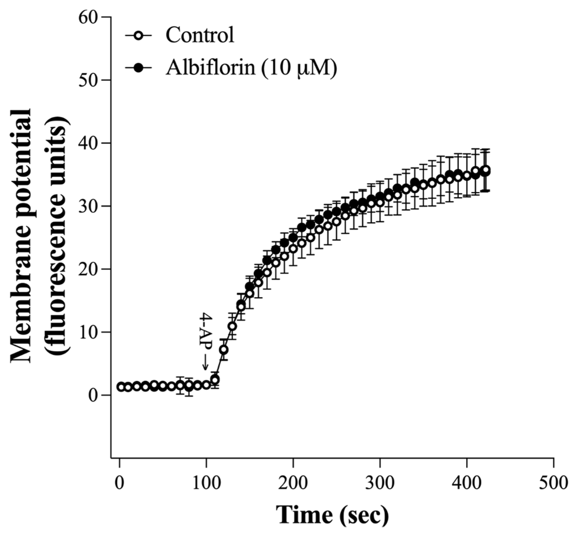
2.3. Albiflorin Failed to Affect the Synaptosomal Membrane Potential

The suppression of Ca2+ channels by albiflorin might be due to an alteration in the synaptosomal plasma membrane potential, which consequently modulates Ca2+ influx into the terminal. Therefore, the effect of albiflorin on the synaptosomal plasma membrane potential was assessed with the membrane-potential-sensitive dye DiSC3(5). As shown in Figure 3, 4-AP (1 mM) increased the fluorescence intensity of DiSC3(5). Compared with that of 4-AP alone, the presence of albiflorin did not significantly affect the increase in DiSC3(5) fluorescence intensity caused by 4-AP [t(8) = 0.08, p = 0.94].

2.4. The Albiflorin-Mediated Inhibition of Glutamate Release Involved the Suppression of the Protein Kinase A (PKA) Pathway

One of the important modulators of vesicular exocytotic glutamate release is presynaptic PKA [20]. As shown in Figure 4A, the PKA inhibitor H89 significantly reduced the glutamate release elicited by 4-AP [F(2,12) = 132.8, p < 0.0001]. The effect of albiflorin was efficiently prevented by the concomitant presence of H89, there being no statistical difference between the release after H89 alone and after albiflorin + H89 treatment (p = 0.94). Similar results were observed with another PKA inhibitor, KT5720 (1 μM) [F(2,12) = 100.4, p < 0.0001]. Albiflorin only reduced glutamate release by an additional 1% in synaptosome pretreated with KT5720 (p = 0.98). Furthermore, 4-AP (1 mM) increased the phosphorylation of PKA [F(2,12) = 632.7, p < 0.0001], which was also markedly decreased after exposure to albiflorin (p < 0.001). The levels of PKA were not significantly different among the groups [F(2,12) = 0.003, p = 1] (Figure 4B–D).
To further explore the regulatory effect of albiflorin on presynaptic PKA pathway, we assessed the phosphorylation of SNAP-25 and synapsin 1, which are presynaptic substrates for PKA [19]. PKA phosphorylates SNAP-25 on threonine 138 (T138) and synapsin I on serine 9 (S9), promoting the trafficking and mobilization of synaptic vesicles to nerve terminals and consequently increasing the size of the releasable vesicle pools [21,22]. As shown in Figure 5A–E, compared to the control, 1 mM 4-AP increased the levels of p-138-SNAP-25 [F(2,12) = 347.3, p < 0.0001] and p-S9-synapsin I [F(2,12) = 139.4, p < 0.0001] without altering the total amount of SNAP-25 [F(2,12) = 0.06, p = 0.94] and synapsin I [F(2,12) = 0.07, p = 0.93]. We also showed that 10 μM albiflorin reduced the 4-AP-evoked increase in p-138-SNAP-25 and p-S9-synapsin I level (p < 0.001).

2.5. Albiflorin Reduces the Release Competence of Synaptic Vesicles Evoked by 4-AP in Synaptosomes

The regulation of synaptic vesicles by albiflorin was further evaluated using transmission electron microscopy (TEM). As shown in Figure 6A,B, the synaptosome contained numerous synaptic vesicles (control group). Synaptosomes treated with 1 mM 4-AP showed significantly fewer synaptic vesicles than control synaptosomes [F(2,6) = 65.3, p < 0.001], indicating that 4-AP evokes the release of all released vesicles. However, the number of synaptic vesicles was greater in synaptosomes preincubated with 10 μM albiflorin for 10 min before 4-AP application than in those treated with 4-AP alone (p < 0.001).

3. Discussion

This work focused on evaluating the effects of albiflorin on the release of glutamate from synaptosomes acutely prepared from the cerebral cortex of adult rats. We report here that albiflorin reduced vesicular exocytotic glutamate release from cerebrocortical synaptosomes via the depression of Ca2+ channel activation and the PKA pathway.

3.1. The Mechanism by Which Albiflorin Inhibited the 4-AP-Evoked Glutamate Release

The K+ channel blocker 4-AP, which mimics the physiological mechanisms of terminal depolarization and the Ca2+-dependent release of neurotransmitters, is considered a quasi-physiological stimulus for investigating the characteristics of the induced release of glutamate [23]. In the present study, 1 mM 4-AP elicited the release of a large amount of glutamate from nerve terminals, consistent with previous studies [24,25]. Albiflorin reduced this 4-AP-elicited glutamate release in a dose-dependent manner (3–30 μM) and reached a maximum effect at 18 μM. Furthermore, the albiflorin-mediated decrease in glutamate release was abolished by a blockade of vesicular glutamate transporters. This suggests that the albiflorin-mediated inhibition of 4-AP-elicited glutamate release is mediated by a decrease in the exocytotic pool available for release. The mechanisms possibly involved in the ability of albiflorin to inhibit vesicular exocytotic glutamate release were further assessed. In particular, the involvement of voltage-dependent Ca2+ channels, which are primarily contributed to the activation of vesicle exocytosis, was investigated [26]. Ca2+ channels, which are primarily supported for glutamate exocytosis, are N-type and P/Q-type [19,27,28]. Notably, we found that the depression of P/Q-type Ca2+ channels was involved in the glutamate-release-inhibiting effect of albiflorin, as indicated by the blockage of the action of albiflorin by the P/Q-type Ca2+ channel inhibitor, while the N-type Ca2+ channel inhibitor was ineffective. The glutamate release measured in the presence of both ω-agatoxin IVA and albiflorin was not significantly different from that observed in the presence of ω-agatoxin IVA alone. The lack of additivity in the actions of albiflorin and ω-agatoxin IVA can be explained by the inhibition of the same release pathway by both compounds. Moreover, the effect of albiflorin on 4-AP-elicited glutamate release was also significantly affected in the absence of extracellular Ca2+. These results suggest that albiflorin depresses vesicular exocytotic glutamate release from synaptosomes by decreasing extracellular Ca2+ entry via P/Q-type Ca2+ channels.
Additionally, the inhibition of Na+ channels or activation of K+ channels causes presynaptic inhibition resulting from the hyperpolarization of nerve terminals. This results in a reduction in Ca2+ influx and a consequent decrease in neurotransmitter release [29,30]. This mechanism did not involve the inhibitory effect of 4-AP-elicited glutamate release by albiflorin observed in the present study. This notion is supported by our observation that albiflorin did not influence 4-AP-evoked plasma membrane depolarization in synaptosomes. Although no direct evidence has indicated that albiflorin acts on presynaptic Ca2+ channels, our tentative conclusion is that the inhibitory effect of glutamate release by albiflorin occurs primarily through direct regulation of Ca2+ channels to affect Ca2+ entry. Further investigations are needed to better understand how albiflorin leads to the suppression of Ca2+ channels.
In glutamatergic nerve terminals, PKA increases glutamate release, and the phosphorylation of synaptic proteins in the release machinery, such as SNAP-25 and synapsin I, could be involved [20]. SNAP-25 and synapsin I are phosphorylated by PKA at T138 and S9, respectively. This phosphorylation promotes synaptic vesicle trafficking and increases the size of the releasable vesicle pools [22,31]. In the present study, when albiflorin was combined with the PKA inhibitor H89, the inhibitory effect of 4-AP-elicited glutamate release observed was not different from that observed with H89 alone. The lack of synergy in the effects of albiflorin and H89 can be explained by the notion that both factors inhibit the same pathway. In addition, we also observed increased levels of p-PKA, p-138-SNAP-25, and p-S9-synapsin I in 4-AP-treated cortical synaptosomes, which may involve a 4-AP-induced increase in the number of releasable vesicles and therefore increasing glutamate exocytosis. Furthermore, we found that albiflorin significantly decreased the levels of p-PKA, p-T138-SNAP-25, and p-S9-synapsin I in 4-AP-treated cortical synaptosomes. Therefore, it seems that the suppression of PKA by albiflorin reduces the phosphorylation of synapsin I and SNAP-25, resulting in attenuation of the number of releasable synaptic vesicles and reduced glutamate release. Consistent with this hypothesis, TEM revealed that 4-AP-induced synaptic vesicle changes were significantly reduced in albiflorin-treated cortical synaptosomes.

3.2. Therapeutic Implications

Albiflorin has no obvious acute oral toxicity and is a safe natural drug ingredient [7]. The ability of albiflorin to inhibit glutamate release from nerve terminals in the present study is of special interest, considering that excess glutamate is involved in many brain disorders, including neurodegenerative diseases (e.g., Alzheimer’s disease and Parkinson’s disease), and in mood disorders (e.g., depression) [32,33]. The reduction in glutamate release by albiflorin needs further investigation, and this observation represents a crucial mechanism of action of albiflorin that might contribute to its therapeutic effect on brain disorders. We infer that the glutamate-release-inhibiting effects of albiflorin might contribute to the reported effects on Alzheimer’s disease and depression [9,11,34].

4. Materials and Methods

4.1. Animals

Male Sprague Dawley rats weighing 180–200 g were purchased from the BioLASCO Taiwan Co., Ltd. (Taipei, Taiwan). The animal care and treatment were conducted in accordance with the Guide for the Care and Use of Laboratory Animals of the National Institutes of Health. The experimental protocol was approved by the Fu Jen Catholic University Animal Care Committee (A11208). In brief, the animals were kept in a room with controlled environmental conditions, maintaining a steady temperature of 23 ± 3 °C, a relative humidity of 50 ± 10%, and a 12 h light/dark cycle. The animals were provided with a standard rodent diet and had unrestricted access to purified water. All efforts were made to minimize the number of animals used and to reduce their suffering. A total of 25 animals were utilized in the study.

4.2. Preparation of Synaptosomes

Synaptosomes were isolated using Percoll density gradient centrifugation [24]. The experimental rats were sacrificed by decapitation. Their brains were quickly extracted and placed in a chilled medium containing 320 mM sucrose with a pH of 7.4. The cerebral cortex was then dissected and homogenized in the sucrose medium. The homogenate was centrifuged at 3000× g for 2 min at 4 °C. The resulting supernatant was then centrifuged again at 14,500× g for 12 min at 4 °C. The pellet was resuspended in a HEPES-buffered medium. The suspension was then separated using a layered, discontinuous Percoll gradient, which contained 320 mM sucrose, 1 mM EDTA, 0.25 mM dl-dithiothreitol, and Percoll concentrations of 3%, 10%, and 23%. The gradients were then centrifuged at 32,500× g for 7 min at 4 °C. Synaptosomes were recovered from the 10 and 23% Percoll bands and diluted in a HEPES buffer medium (HBM). This medium contained 140 mM NaCl, 5 mM KCl, 5 mM NaHCO3, 1 mM MgCl2·6H2O, 1.2 mM Na2HPO4, 10 mM glucose, and 10 mM HEPES, with a pH of 7.4. Following further centrifugation at 27,000× g for 10 min at 4 °C, the synaptosomal pellets were resuspended in HBM, and the protein content was determined by the Bradford assay (Thermo Fisher Scientific, Waltham, MA, USA). A volume of 0.5 mg of synaptosomal suspension was diluted in HBM and centrifugated (3000× g at 4 °C) for 10 min. The synaptosomal pellet was divided into three independent fractions used for the following purposes: (a) analysis of the glutamate release, plasma membrane potential, and Na+ concentration; (b) evaluation of the expression of p-PKA, p-T138-SNAP-25, and p-S9-synapsin I by Western blot; and (c) evaluation of the changes in synaptic vesicles by TEM.

4.3. Glutamate Release Assay

The release of glutamate from purified cerebrocortical synaptosomes was monitored in real time using an assay. Exogenous glutamate dehydrogenase (GDH) and NADP+ were used in this assay to couple the oxidative deamination of the released glutamate to the generation of NADPH. This process was detected using fluorometric methods [35,36]. Synaptosomal pellets were resuspended in HBM containing 16 M bovine serum albumin to bind any free fatty acids. A 2 mL aliquot was transferred to a stirred, thermostatted cuvette at 37 °C and incubated in the presence of 1 mM NADP+, 50 units of glutamate dehydrogenase, and 1.2 mM CaCl2. The released glutamate undergoes oxidative deamination by GDH to produce NADPH, resulting in an increase in fluorescence. This increase in fluorescence, with excitation and emission wavelengths of 340 and 460 nm, respectively, was measured to monitor glutamate release. The fluorescence of NADPH was detected using LS-55B (PerkinElmer, Waltham, MA, USA) model spectrofluorimeter. At the end of each experiment, 5 nmol of exogenous glutamate was added as a standard. Data were obtained at 2 s intervals. The fluorescence response used to quantify the released glutamate after a 5 min depolarization with 1 mM 4-AP was expressed as nanomoles of glutamate per milligram of synaptosomal protein per 5 min (nmol/mg/5 min).

4.4. Plasma Membrane Potential

The plasma membrane potential was evaluated using DiSC3(5), a dye that is advantageous due to its ability to alter fluorescence intensity in response to changes in membrane potential [37]. Synaptosomes were preincubated and resuspended following the protocol used for the glutamate release experiments. After 3 min of incubation, 5 μM DiSC3(5) was added and allowed to equilibrate. Following 4 min of incubation, CaCl2 (1.2 mM) was introduced. At the 10 min mark, 4-AP (1 mM) was added to depolarize the synaptosomes. The fluorescence of DiSC3(5) was then monitored at excitation and emission wavelengths of 646 nm and 674 nm, respectively. The results were expressed in fluorescence units.

4.5. TEM

For ultrastructural analysis, purified cortical synaptosomes were fixed in 4% paraformaldehyde and 0.1% glutaraldehyde for 12 h at 4 °C. The fixed cortical synaptosomes were rinsed in phosphate-buffered saline (PBS), then post-fixed in 1% osmium tetroxide for 2 h. Afterward, they were dehydrated and embedded in epoxy resin. Next, ultrathin sections were cut to a thickness of 70 nm using an ultramicrotome (EM UC7, Leica Microsystems, Wetzlar, Germany). A transmission electron microscope (JEM-1400, JEOL, Tokyo, Japan) was used to examine the ultrastructure of the cortical synaptosomes.

4.6. Western Blotting

The method and conditions for preparing samples for Western blot analysis have been described previously [38]. Synaptosomal lysates (20 mg/lane) were subjected to sodium dodecylsulfate polyacrylamide gel electrophoresis (SDS-PAGE) in 10% gel under reducing conditions and electroblotted onto polyvinylidene fluoride (PVDF) membranes (Bio-Rad Laboratories, Hercules, CA, USA). After electroblotting, non-specific binding sites in membranes were blocked with Tris-buffered saline containing Tween-20 (TBS-T: 20 mM Tris, 500 mM NaCl, 0.1% Tween 20, pH 7.5) and 5% skimmed milk at room temperature for 1 h. Subsequently, membranes were incubated overnight at 4 °C with the primary antibody. Membranes were washed with TBS-T and incubated for 2 h with horseradish peroxidase–coupled secondary antibody at room temperature. Enhanced chemiluminescence (Amersham, Buckinghamshire, UK) was used to visualize the protein bands, which were exposed to X-ray film. Films were scanned, and the level of proteins was assessed using the Image J software (version 1.45 J, National Institutes of Health, Bethesda, Rockville, MD, USA). The density of the band of interest was normalized against β-actin to determine the relative band density ratio. The primary antibodies used in this study included β-actin (1:5000), PKA (1:2000), p-PKA (1:2000), SNAP-25 (1:50,000), p-T138-SNAP-25 (1:500), synapsin I (1:50,000), and p-S9-synapsin I (1:700). β-actin, PKA, p-PKA, synapsin I, and p-S9-synapsin I were purchased from Cell Signaling (Danvers, MA, USA). SNAP-25 was obtained from Abcam (Cambridge, UK). p-T138-SNAP-25 was obtained from Aviva Systems Biology Corporation (San Diego, CA, USA).

4.7. Data Analysis

The Shapiro–Wilk test was used to test the data normality. Data analysis and graph creation were performed using GraphPad Prism version 8.0 (GraphPad Software, San Diego, CA, USA). The results were analyzed using one-way analysis of variance (ANOVA), followed by Tukey’s post hoc test when comparing more than two groups. A two-tailed Student’s t-test was used to determine whether there was a significant difference between the means of two groups. Data were presented as mean ± standard error of the mean (SEM). The statistical significance was set at p < 0.05.

4.8. Chemicals

Albiflorin (purity > 98%) was purchased from ChemFaces (Wuhan, Hubei, China). The calcium channel blockers ω-conotoxin GVIA and ω-agatoxin IVA were purchased from Alomone (Jerusalem, Israel). DiSC3(5) was acquired from Invitrogen (Carlsbad, CA, USA). Bafilomycin A1 and KT5720 were obtained from Tocris Cookson (Bristol, UK). 4-AP, H89, Percoll, sucrose, EDTA, dl-dithiothreitol, HEPES, GDH, NADP+, SDS and general reagents were acquired from Sigma-Aldrich (St. Louis, MO, USA). Albiflorin, DiSC3(5), and H89 were dissolved in 0.1% (14.3 mM) dimethylsulfoxide (DMSO). 4-AP, ω-conotoxin GVIA, and ω-agatoxin IVA were dissolved in distilled water.

5. Conclusions

Our data demonstrated that by decreasing Ca2+ entry via P/Q-type Ca2+ channels and reducing PKA-mediated synapsin I and SNAP-25 phosphorylation, albiflorin inhibits vesicular exocytotic glutamate release from rat cerebral cortical synaptosomes. This investigation enhances the understanding of albiflorin action in the brain and suggests that albiflorin is valuable for treating glutamate-induced neurological disorders.

Author Contributions

Conceptualization, C.-W.L. and T.-Y.L.; data curation, T.-Y.L., Y.-Y.C., K.-M.C. and M.-Y.L.; funding acquisition, C.-W.L., T.-Y.L. and S.-J.W.; investigation, C.-W.L., T.-Y.L., Y.-Y.C., K.-M.C. and S.-J.W.; project administration, C.-W.L. and T.-Y.L.; supervision, S.-J.W.; writing—original draft, S.-J.W.; writing—review and editing, M.-Y.L. and S.-J.W. All authors have read and agreed to the published version of the manuscript.

Funding

This work was supported by the Far Eastern Memorial Hospital (112-FEMH-FJU-01; FEMH-2023-C-075; FEMH-2023-C-076) and the National Science and Technology Council (111-2314-B-418-009-MY2; 112-2320-B-030-010-MY3), Taiwan.

Institutional Review Board Statement

The animal care and experimental procedures were conducted in accordance with the Guide for the Care and Use of Laboratory Animals of the National Institutes of Health and were approved by the Fu Jen Catholic University Animal Care Committee (A11208).

Informed Consent Statement

Not applicable.

Data Availability Statement

Data will be made available on request.

Conflicts of Interest

The authors have no competing financial interests to declare.

References

  1. Choi, D.W. Glutamate neurotoxicity and diseases of the nervous system. Neuron 1988, 1, 623–634. [Google Scholar] [CrossRef]
  2. Lazarevic, V.; Yang, Y.; Ivanova, D.; Fejtova, A.; Svenningsson, P. Riluzole attenuates the efficacy of glutamatergic transmission by interfering with the size of the readily releasable neurotransmitter pool. Neuropharmacology 2018, 143, 38–48. [Google Scholar] [CrossRef]
  3. Wu, S.H.; Wu, D.G.; Chen, Y.W. Chemical constituents and bioactivities of plants from the genus Paeonia. Chem. Biodivers. 2010, 7, 90–104. [Google Scholar] [CrossRef]
  4. He, D.Y.; Dai, S.M. Anti-inflammatory and immunomodulatory effects of paeonia lactiflora pall., a traditional chinese herbal medicine. Front. Pharmacol. 2011, 2, 10. [Google Scholar] [CrossRef]
  5. Fei, F.; Yang, H.; Peng, Y.; Wang, P.; Wang, S.; Zhao, Y.; Huang, J.; Yu, X.; Feng, S.; Sun, R.; et al. Sensitive analysis and pharmacokinetic study of the isomers paeoniflorin and albiflorin after oral administration of Total Glucosides of White Paeony Capsule in rats. J. Chromatogr. B Analyt. Technol. Biomed. Life Sci. 2016, 1022, 30–37. [Google Scholar] [CrossRef]
  6. Zhou, J.; Wang, L.; Wang, J.; Wang, C.; Yang, Z.; Wang, C.; Zhu, Y.; Zhang, J. Paeoniflorin and albiflorin attenuate neuropathic pain via MAPK pathway in chronic constriction injury rats. Evid. Based Complement. Alternat Med. 2016, 1, 8082753. [Google Scholar] [CrossRef] [PubMed]
  7. Han, J.; Xia, Y.; Lin, L.; Zhang, Z.; Tian, H.; Li, K. Next-generation metabolomics in the development of new antidepressants: Using albiflorin as an example. Curr. Pharm. Des. 2018, 24, 2530–2540. [Google Scholar] [CrossRef]
  8. Wang, X.; Su, L.; Tan, J.; Ding, T.; Yue, Y. Albiflorin alleviates DSS-induced ulcerative colitis in mice by reducing inflammation and oxidative stress. Iran. J. Basic. Med. Sci. 2023, 26, 48–56. [Google Scholar] [CrossRef] [PubMed]
  9. Wang, Y.L.; Wang, J.X.; Hu, X.X.; Chen, L.; Qiu, Z.K.; Zhao, N.; Yu, Z.D.; Sun, S.Z.; Xu, Y.Y.; Guo, Y.; et al. Antidepressant-like effects of albiflorin extracted from Radix paeoniae Alba. J. Ethnopharmacol. 2016, 179, 9–15. [Google Scholar] [CrossRef] [PubMed]
  10. Qiu, Z.K.; He, J.L.; Liu, X.; Zeng, J.; Chen, J.S.; Nie, H. Anti-PTSD-like effects of albiflorin extracted from Radix paeoniae Alba. J. Ethnopharmacol. 2017, 198, 324–330. [Google Scholar] [CrossRef] [PubMed]
  11. Xu, Y.J.; Mei, Y.; Shi, X.Q.; Zhang, Y.F.; Wang, X.Y.; Guan, L.; Wang, Q.; Pan, H.F. Albiflorin ameliorates memory deficits in APP/PS1 transgenic mice via ameliorating mitochondrial dysfunction. Brain Res. 2019, 1719, 113–123. [Google Scholar] [CrossRef]
  12. Wang, Q.S.; Yan, K.; Li, K.D.; Gao, L.N.; Wang, X.; Liu, H.; Zhang, Z.; Li, K.; Cui, Y.L. Targeting hippocampal phospholipid and tryptophan metabolism for antidepressant-like effects of albiflorin. Phytomedicine 2021, 92, 153735. [Google Scholar] [CrossRef]
  13. Lee, S.; Ryu, S.M.; Kim, D.H.; Lee, Y.E.; Lee, S.J.; Kang, S.; Kim, J.S.; Lee, S.I. Neuroprotective effect of Geijigadaehwang-tang against trimethyltin-induced hippocampal neurodegeneration: An in vitro and in vivo study. J. Ethnopharmacol. 2022, 296, 115451. [Google Scholar] [CrossRef]
  14. Zhu, F.; Xiong, J.; Yi, F.; Luo, E.; Huang, C.; Li, R. Albiflorin relieves cerebral ischemia-reperfusion injury by activating Nrf2/HO-1 pathway. Histol. Histopathol. 2023, 38, 233–245. [Google Scholar] [CrossRef]
  15. Fang, P.; Wang, Y.; Sun, F.; Lin, H.; Zhang, X. Effects of albiflorin on oxidative stress and inflammatory responses in rats with acute spinal cord injury. Immun. Inflamm. Dis. 2023, 11, e1015. [Google Scholar] [CrossRef]
  16. Lazarevic, V.; Mantas, I.; Flais, I.; Svenningsson, P. Fluoxetine suppresses glutamate- and GABA-mediated neurotransmission by altering SNARE complex. Int. J. Mol. Sci. 2019, 20, 4247. [Google Scholar] [CrossRef] [PubMed]
  17. Mdzinarishvili, A.; Sumbria, R.; Lang, D.; Klein, J. Ginkgo extract EGb761 confers neuroprotection by reduction of glutamate release in ischemic brain. J. Pharm. Pharm. Sci. 2012, 15, 94–102. [Google Scholar] [CrossRef]
  18. Dunkley, P.R.; Jarvie, P.E.; Robinson, P.J. A rapid Percoll gradient procedure for preparation of synaptosomes. Nat. Protoc. 2008, 3, 1718–1728. [Google Scholar] [CrossRef]
  19. Nicholls, D.G. Presynaptic modulation of glutamate release. Prog. Brain Res. 1998, 116, 15–22. [Google Scholar] [CrossRef]
  20. Greengard, P.; Valtorta, F.; Czernik, A.J.; Benfenati, F. Synaptic vesicle phosphoproteins and regulation of synaptic function. Science 1993, 259, 780–785. [Google Scholar] [CrossRef]
  21. Menegon, A.; Bonanomi, D.; Albertinazzi, C.; Lotti, F.; Ferrari, G.; Kao, H.T.; Benfenati, F.; Baldelli, P.; Valtorta, F. Protein kinase A-mediated synapsin I phosphorylation is a central modulator of Ca2+-dependent synaptic activity. J. Neurosci. 2006, 26, 11670–11681. [Google Scholar] [CrossRef]
  22. Nagy, G.; Reim, K.; Matti, U.; Brose, N.; Binz, T.; Rettig, J.; Neher, E.; Sørensen, J.B. Regulation of releasable vesicle pool sizes by protein kinase A-dependent phosphorylation of SNAP-25. Neuron 2004, 41, 417–429. [Google Scholar] [CrossRef]
  23. Tibbs, G.R.; Dolly, J.O.; Nicholls, D.G. Dendrotoxin, 4-aminopyridine, and beta-bungarotoxin act at common loci but by two distinct mechanisms to induce Ca2+-dependent release of glutamate from guinea-pig cerebrocortical synaptosomes. J. Neurochem. 1989, 52, 201–206. [Google Scholar] [CrossRef]
  24. Lu, C.W.; Yeh, K.C.; Chiu, K.M.; Lee, M.Y.; Lin, T.Y.; Wang, S.J. The effect of isosaponarin derived from wasabi leaves on glutamate release in rat synaptosomes and its underlying mechanism. Int. J. Mol. Sci. 2022, 23, 8752. [Google Scholar] [CrossRef]
  25. Lin, T.Y.; Chen, I.Y.; Lee, M.Y.; Lu, C.W.; Chiu, K.M.; Wang, S.J. Inhibition of glutamate release from rat cortical nerve terminals by dehydrocorydaline, an alkaloid from Corydalis yanhusuo. Molecules 2022, 27, 960. [Google Scholar] [CrossRef]
  26. Jarvis, S.E.; Zamponi, G.W. Interactions between presynaptic Ca2+ channels, cytoplasmic messengers and proteins of the synaptic vesicle release complex. Trends Pharmacol. Sci. 2001, 22, 519–525. [Google Scholar] [CrossRef]
  27. Vázquez, E.; Sánchez-Prieto, J. Presynaptic modulation of glutamate release targets different calcium channels in rat cerebrocortical nerve terminals. Eur. J. Neurosci. 1997, 9, 2009–2018. [Google Scholar] [CrossRef]
  28. Millán, C.; Sánchez-Prieto, J. Differential coupling of N- and P/Q-type calcium channels to glutamate exocytosis in the rat cerebral cortex. Neurosci. Lett. 2002, 330, 29–32. [Google Scholar] [CrossRef]
  29. Li, M.; West, J.W.; Numann, R.; Murphy, B.J.; Scheuer, T.; Catterall, W.A. Convergent regulation of sodium channels by protein kinase C and cAMP-dependent protein kinase. Science 1993, 261, 1439–1442. [Google Scholar] [CrossRef]
  30. Wu, L.G.; Saggau, P. Presynaptic inhibition of elicited neurotransmitter release. Trends Neurosci. 1997, 20, 204–212. [Google Scholar] [CrossRef]
  31. Risinger, C.; Bennett, M.K. Differential phosphorylation of syntaxin and synaptosome-associated protein of 25 kDa (SNAP-25) isoforms. J. Neurochem. 1999, 72, 614–624. [Google Scholar] [CrossRef] [PubMed]
  32. Sanacora, G.; Treccani, G.; Popoli, M. Towards a glutamate hypothesis of depression: An emerging frontier of neuropsychopharmacology for mood disorders. Neuropharmacology 2012, 62, 63–77. [Google Scholar] [CrossRef] [PubMed]
  33. Henter, I.D.; Park, L.T.; Zarate, C.A., Jr. Novel glutamatergic modulators for the treatment of mood disorders: Current status. CNS Drugs 2021, 35, 527–543. [Google Scholar] [CrossRef] [PubMed]
  34. Wang, C.C.; Hsieh, P.W.; Kuo, J.R.; Wang, S.J. Rosmarinic Acid, a Bioactive Phenolic Compound, Inhibits Glutamate Release from Rat Cerebrocortical Synaptosomes through GABA(A) Receptor Activation. Biomolecules 2021, 11, 1029. [Google Scholar] [CrossRef]
  35. Chiu, K.M.; Lee, M.Y.; Lu, C.W.; Lin, T.Y.; Wang, S.J. Plantainoside D reduces depolarization-evoked glutamate release from rat cerebral cortical synaptosomes. Molecules 2023, 28, 1313. [Google Scholar] [CrossRef]
  36. Lu, C.W.; Wu, C.C.; Chiu, K.M.; Lee, M.Y.; Lin, T.Y.; Wang, S.J. Inhibition of Synaptic Glutamate Exocytosis and Prevention of Glutamate Neurotoxicity by Eupatilin from Artemisia argyi in the Rat Cortex. Int. J. Mol. Sci. 2022, 23, 13406. [Google Scholar] [CrossRef] [PubMed]
  37. Akerman, K.E.; Scott, I.G.; Heikkilä, J.E.; Heinonen, E. Ionic dependence of membrane potential and glutamate receptor-linked responses in synaptoneurosomes as measured with a cyanine dye, DiS-C2-(5). J. Neurochem. 1987, 48, 552–559. [Google Scholar] [CrossRef] [PubMed]
  38. Lu, C.W.; Lin, C.J.; Hsieh, P.W.; Chiu, K.M.; Lee, M.Y.; Lin, T.Y.; Wang, S.J. An Anthranilate Derivative Inhibits Glutamate Release and Glutamate Excitotoxicity in Rats. Int. J. Mol. Sci. 2022, 23, 2641. [Google Scholar] [CrossRef]
Figure 1. Albiflorin suppresses the release of glutamate induced by 4-AP from rat cerebrocortical nerve terminals. (A) Albiflorin molecular structure. (B) Glutamate release triggered by 4-AP (1 mM) in both the control condition and in the presence of albiflorin. (C) Dose–response curves of 4-AP-evoked glutamate release for the calculation of IC50 values. The concentration of albiflorin ranges from 1 to 30 μM. (D) Effects of Ca2+-free medium containing 0.3 mM EGTA, 0.2 μM bafilomycin A1, and 10 μM albiflorin alone or concomitantly added on the 4-AP (1 mM)-elicited glutamate release. Albiflorin or bafilomycin A1 was added 10 min before 4-AP addition. Results are the means ± SEM (n = 5 rats/group). *** p < 0.001 versus control group.
Ijms 25 08846 g001
Figure 2. The inhibition of 4-AP-induced glutamate release by albiflorin is blocked by the P/Q-type Ca2+ channel inhibitor ω-agatoxin IVA but not by the N-type Ca2+ channel inhibitor ω-conotoxin GVIA. Effects of neuronal voltage-sensitive Ca2+ channel blockers, ω-conotoxin GVIA or ω-agatoxin IVA, in combination with or without albiflorin, on the 1 mM 4-AP-elicited glutamate release. Albiflorin, ω-conotoxin GVIA or ω-agatoxin IVA was added 10 min before the addition of 4-AP. Results are the means ± SEM (n = 5 rats/group). *** p < 0.001 versus control group, # p < 0.001 versus ω-conotoxin GVIA-treated group.
Ijms 25 08846 g002
Figure 3. Albiflorin does not affect the synaptosomal membrane potential. The membrane potential of synaptosomes was measured using DiSC3(5) during depolarization with 4-AP (1 mM), either without (control) or with albiflorin, which was added 10 min prior to the 4-AP. Results are the means ± SEM (n = 5 rats/group).
Ijms 25 08846 g003
Figure 4. Albiflorin-mediated inhibition of 4-AP-evoked glutamate release is prevented by PKA inhibitors H89 and KT5720. (A) Effects of H89, KT5720, and albiflorin alone or concomitantly added on the 4-AP (1 mM)-elicited glutamate release. (B) Representative images and (C,D) quantification of PKA and p-Thr197-PKA protein levels in the absence (control) or presence of 4-AP or 4-AP + albiflorin. H89 and KT5720 were added 30 min before 4-AP addition. Results are the means ± SEM (n = 5 rats/group). *** p < 0.001 versus control group, # p < 0.001 versus 4-AP-treated group.
Ijms 25 08846 g004
Figure 5. Albiflorin decreases 4-AP-elicited phosphorylation of synapsin I and SNAP-25. (A) Representative images and (BE) quantification of synapsin-1, p-Ser9-synapsin I, SNAP-25, and p-T138-SNAP-25 protein levels in the absence (control) or presence of 4-AP or 4-AP + albiflorin. Albiflorin was added 10 min before 4-AP addition. Results are the means ± SEM (n = 5 rats/group). *** p < 0.001 versus control group, # p < 0.001 versus 4-AP-treated group.
Ijms 25 08846 g005
Figure 6. The effect of albiflorin on 4-AP-evoked changes in synaptic vesicles in synaptosomes. The synapse ultrastructure (A) and the number of synaptic vesicles (B) in each synaptosome were observed using TEM. Scale bar: 200 nm, magnification: 60,000×. Results are the means ± SEM (n = 3 rats/group). *** p < 0.001 versus control group, # p < 0.001 versus 4-AP-treated group.
Ijms 25 08846 g006
Disclaimer/Publisher’s Note: The statements, opinions and data contained in all publications are solely those of the individual author(s) and contributor(s) and not of MDPI and/or the editor(s). MDPI and/or the editor(s) disclaim responsibility for any injury to people or property resulting from any ideas, methods, instructions or products referred to in the content.

