Article

Circular RNA Profile in Atherosclerotic Disease: Regulation during ST-Elevated Myocardial Infarction

1
Institute of Clinical Medicine, University of Oslo (UiO), 0372 Oslo, Norway
2
Research Institute for Internal Medicine, Oslo University Hospital, Rikshospitalet, 0372 Oslo, Norway
3
Department of Medicine, Cardiovascular Division, Brigham and Women’s Hospital, Harvard Medical School, Boston, MA 02115, USA
4
Department of Cardiology, Oslo University Hospital, Rikshospitalet, 0372 Oslo, Norway
5
Department of Cardiology, Oslo University Hospital, Ullevål, 0450 Oslo, Norway
6
Clinic of Cardiology, St. Olav’s Hospital, Trondheim University Hospital, 7030 Trondheim, Norway
7
Department of Circulation and Medical Imaging, Norwegian University of Science and Technology (NTNU), 7030 Trondheim, Norway
8
Department of Microbiology, Oslo University Hospital, Rikshospitalet, 0372 Oslo, Norway
*
Author to whom correspondence should be addressed.
Int. J. Mol. Sci. 2024, 25(16), 9014; https://doi.org/10.3390/ijms25169014
Submission received: 20 June 2024 / Revised: 15 August 2024 / Accepted: 16 August 2024 / Published: 19 August 2024

Abstract

:
Circular (circ) RNAs are non-coding RNAs with important functions in the nervous system, cardiovascular system, and cancer. Their role in atherosclerosis and myocardial infarction (MI) remains poorly described. We aim to investigate the potential circRNAs in immune cells during atherogenesis and examine the most regulated during MI and the modulation by interleukin (IL)-6 receptor inhibition by tocilizumab. Wild-type (WT) and ApoE−/− mice were fed an atherogenic diet for 10 weeks, and the circRNA profile was analyzed by circRNA microarray. Whole blood from patients with ST-elevated MI (STEMI) and randomized to tocilizumab (n = 21) or placebo (n = 19) was collected at admission, 3–7 days, and at 6 months, in addition to samples from healthy controls (n = 13). Primers for human circRNA were designed, and circRNA levels were measured using RT-qPCR. mRNA regulation of predicted circRNA targets was investigated by RNA sequencing. The expression of 867 circRNAs differed between atherogenic and WT mice. In STEMI patients, circUBAC2 was significantly lower than in healthy controls. CircANKRD42 and circUBAC2 levels were inversely correlated with troponin T, and for circUBAC2, an inverse correlation was also seen with final infarct size at 6 months. The predicted mRNA targets for circUBAC2 and circANKRD42 were investigated and altered levels of transcripts involved in the regulation of inflammatory/immune cells, apoptosis, and mitochondrial function were found. Finally, tocilizumab induced an up-regulation of circANKRD42 and circUBAC2 3–7 days after percutaneous coronary intervention. CircRNA levels were dysregulated in STEMI, potentially influencing the immune system, apoptosis, and mitochondrial function.

Graphical Abstract

1. Introduction

Atherosclerosis is the major underlying cause of cardiovascular disease (CVD), characterized by the formation of fibro-fatty, lipid-rich, and inflammatory lesions within the arterial wall. A crucial pathogenic factor in atherosclerosis is the bidirectional interaction between persistent low-grade inflammation and lipid abnormalities [1]. Treatment and prevention strategies for CVD have improved over the last 40 years [2]. Nevertheless, CVD remains one of the leading causes of morbidity and mortality worldwide [3]. Thus, there is still a need for the development of novel treatments and prevention strategies. An improved understanding of the pathogenic mechanisms of atherosclerosis and its clinical consequences, such as myocardial infarction (MI), is a prerequisite to achieve this goal.
Circular (circ) RNAs are non-coding RNAs in which the 3′ and the 5′ ends have been joined in a covalently closed loop through a process called back-splicing [4]. The lack of free terminal ends prevents the degradation of circRNA by exonucleases, making them more stable than their linear counterparts [5]. Levels of circRNAs vary at slow rates and are thought to be affected minimally by acute events [5]. These traits could make circRNAs potential biomarkers for chronic diseases, as suggested in cancer, Parkinson’s disease, and kidney disorders [6,7].
CircRNAs are shown to have regulatory roles for both transcription and translation [8]. The best-characterized mechanism in regulating transcription is by decreasing the levels of micro RNAs (miRNAs) by a process known as sponging. This binding of specific miRNAs to the circRNA reduces the amount of available miRNA and consequently increases the level of targeted messenger RNA (mRNA) [9]. Another suggested role is regulating their linear counterpart in cis, as back-splicing competes with linear splicing [10]. CircRNAs are suggested to regulate several pathophysiological processes with relevance to CVD, such as apoptosis [11], autophagy [12], inflammation [13], oxidative stress [14], and angiogenesis [15,16], potentially being therapeutic targets and biomarkers also in these disorders [16].
Several recent studies have investigated the effect of targeting inflammation in atherosclerosis and its clinical consequences, such as ST-elevated MI (STEMI) [17,18]. STEMI is one of the most serious outcomes of atherosclerotic disease and is associated with, and potentially also triggered by, enhanced inflammation and immune activation [19]. Several studies have shown a pathogenic role for interleukin (IL)-6-related pathways in STEMI by contributing to plaque inflammation, ischemia-reperfusion injury, and maladaptive myocardial remodeling [20,21,22]. Indeed, we have recently shown that treatment with the IL-6 receptor inhibitor tocilizumab in conjunction with percutaneous coronary intervention (PCI) improved the myocardial salvage index (MSI) in STEMI. This beneficial effect was associated with a downregulation of genes related to neutrophil function and, in particular, neutrophil degranulation [23]. If these tocilizumab-mediated effects also include modulation of circRNAs is still unknown.
In recent years, research has focused on describing the different circRNAs in human tissues and, in particular, their expression levels in different cancer types. However, although there are reports regarding circRNAs in atherosclerosis and MI, the profile of circRNAs in ischemic heart disease is still unclear. Additionally, the effect of anti-inflammatory therapy on circRNA levels in MI has so far not been investigated. To fill this knowledge gap, we first examined an atherogenic mouse model to discover circRNAs associated with atherogenesis. To examine the relevance of these findings to humans, we examined the expression of selected circRNAs in whole blood from patients with STEMI to examine (i) how circRNAs are regulated during and after STEMI; (ii) whether these molecules are related to outcome, i.e., MSI, troponin T (TnT) levels, and infarct size; (iii) the potential indirect effect of the regulated circRNAs on mRNA targets in the same patients; and finally, (iv) if and how levels of circRNAs are modulated by tocilizumab.

2. Results

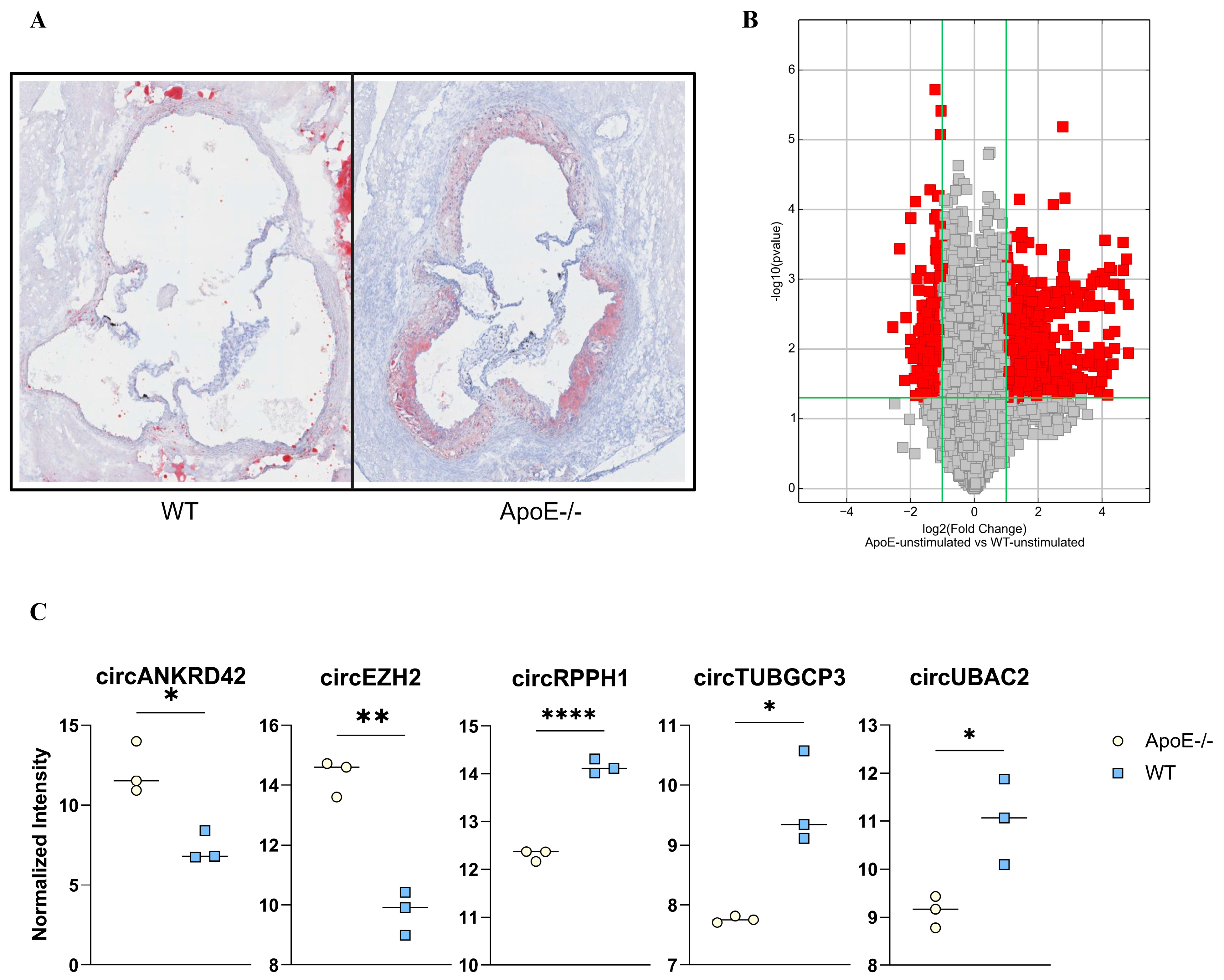
2.1. Atherogenesis Alters the circRNA Expression: Pre-Clinical Explorative Studies

After 10 weeks on an atherogenic diet, ApoE−/− mice (n = 3) developed atherosclerotic plaques in the aortic root, while no visible signs of atherosclerosis were seen in the WT mice (n = 3) (Figure 1A). Splenocytes, representing relevant immune cells in relation to atherosclerosis, analyzed for a total of 13.488 circRNAs, showed that a total of 867 circRNAs differed between atherogenic mice and WT mice (filtered by fold change ≥ 2 and p < 0.05). Levels of 596 circRNAs were significantly higher in ApoE−/− mice than in the WT mice, and 271 circRNAs were significantly lower in the ApoE−/− mice (Figure 1B). The 10 circRNAs with the largest enrichment in ApoE−/− immune cells were more than 20-fold higher than in splenocytes from WT mice, and the 10 circRNAs with the largest suppression in ApoE−/− splenocytes were 3.6–5.9-fold lower (Supplemental Table S2).

2.2. Atherogenic circRNA Translated to Human Homologs

To examine the relevance of these findings to humans, we next aimed to investigate the human analogs of the 10 most enriched and the 10 most suppressed circRNAs in the murine ApoE−/− immune cells. Two had an intron origin and were excluded; five had already been identified, and the primer sequences were available (Figure 1C). For the remaining 13 circRNAs, primer pairs for the human homologs were designed with an assumption of similar back-splicing as in mice (Supplemental Table S1). Two primer pairs gave specific products only in macrophages (circKIF21A, circCACNB2), one only in whole blood (circRPPH1), and four both in macrophages and whole blood (circANKRD42, circEZH2, circTUBGCP3, circUBAC2) (Supplemental Figure S1 and Supplemental Table S2). Those that gave specific products in whole blood were used for further investigations in the STEMI patients (Figure 2).

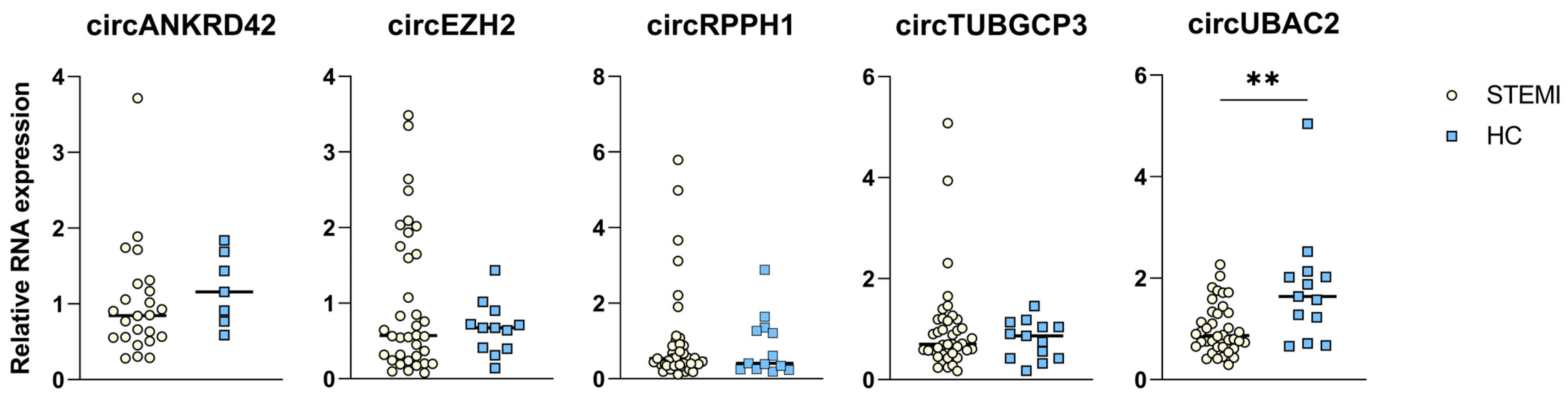
2.3. circRNA Expression in Patients with STEMI

In the ASSAIL-MI trial, a single dose of intravenous tocilizumab was compared with placebo in patients with acute STEMI. The study drug was allocated in a 1:1 fashion. A total of 40 patients, 19 from the placebo arm and 21 from the tocilizumab arm, were chosen for circRNA analysis. The patients were selected based on age, sex, and clinical variables to obtain an equal distribution for the whole population.
Table 1 shows admission characteristics of the 40 patients who participated in this sub-study. At hospital admission, before administration of tocilizumab or placebo, circUBAC2 was significantly lower in the STEMI patients than in healthy controls (fold gene expression (FGE) 1.0 vs. 1.8; p = 0.001) (Figure 2). For circEZH2, circANKRD42, circRPPH1, and circTUBGCP3, however, there were no significant differences between STEMI patients at admission and healthy controls (Figure 2).

2.4. Negative Correlation between circRNA Levels and Peak TnT Levels and Final Infarct Size

In the whole study group, there were statistically significant correlations between peak TnT levels and circRNA levels at admission. As can be seen in Figure 3A, high levels of circANKRD42 and circUBAC2 were associated with lower levels of peak TnT (r = −0.530, p = 0.008 and r = −0.415, p = 0.009, respectively). A similar pattern was seen between the admission levels of circUBAC2 and the final infarction size at 6 months as assessed by CMR (r = −0.346, p = 0.033; Figure 3B), but not between circANKRD42 and final infarction size (r = 0.233, p = 0.284). In contrast, there were no significant correlations between these circRNAs and MSI (circANKRD42: r = 0.202, p = 0.344, and circUBAC2: r = 0.234, p = 0.151, respectively).

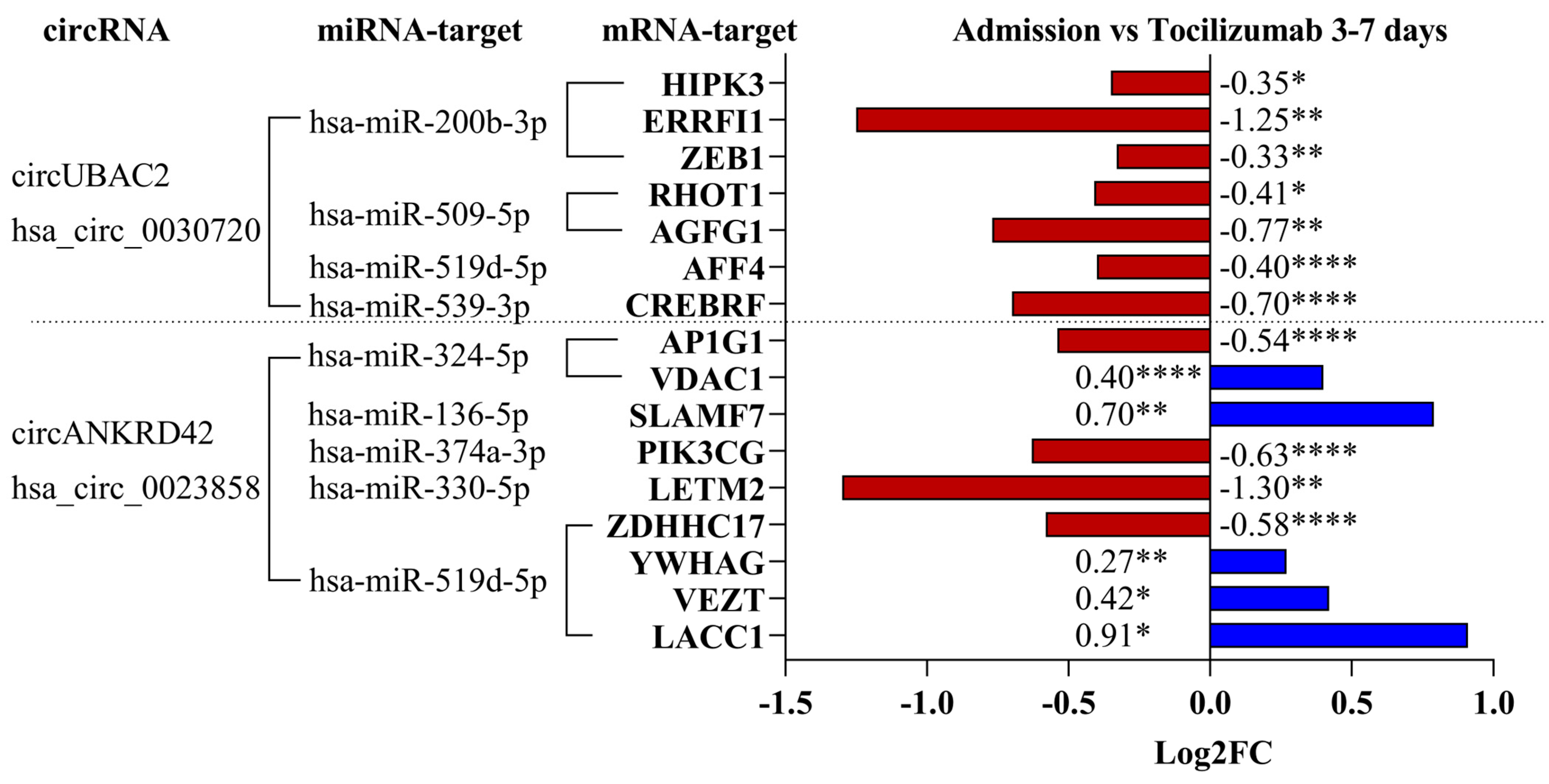
2.5. Regulation of mRNA Targets for the circRNA–miRNA Interaction

A proposed role for circRNAs is to sponge specific miRNAs and thus indirectly regulate gene expression in the cells [9,24]. According to published data, circUBAC2 [25] and circANKRD42 [26] are predicted to work as sponges for certain miRNAs with known functions relevant to the immune response in CVD [27].
The top 5 transcript targets for each miRNA were identified using the miRDB database [28,29] (Supplemental Table S3), and the transcript levels were investigated in the RNA-seq data from whole blood from STEMI patients [23]. Seven of the twenty-five investigated mRNA targets of circUBAC2-related miRNAs were differentially regulated, and all were lower at days 3–7 compared to the time at admission and tocilizumab treatment (Figure 4). Some of the most downregulated mRNAs targeted by circUBAC2 have been related to cell adhesion and inflammatory processes (ERRFI1) [30], regulation of apoptosis (CREBRF), intracellular regulation of cholesterol metabolism (AGFG1) [31], and cell migration (AFF4) [32]. For the circANKRD42-related miRNA, nine of the twenty-five investigated targets were significantly regulated. Five of these were significantly higher and four were lower at days 3–7 vs. admission and tocilizumab treatment (Figure 4). Several of these targets have functions related to a proper immune response. VDAC1 is known as a gatekeeper of mitochondrial function [33], while SLAMF7 is related to B-cell and macrophage activation [34], LETM2 is known to regulate the PI3K-AKT pathway [35], and LACC1 is highly expressed in inflammatory macrophages [36]. Only one mRNA, LETM2, showed significant regulation between the placebo and tocilizumab-treated groups at 3–7 days (Log2FC −1.03, p = 0.0008).
Finally, another proposed role of circRNAs is to regulate their linear counterpart by competing with the linear splicing. However, there were no regulations of either linear ANKRD42 or linear UBAC2 in whole blood from STEMI patients at days 3–7 compared to admission.

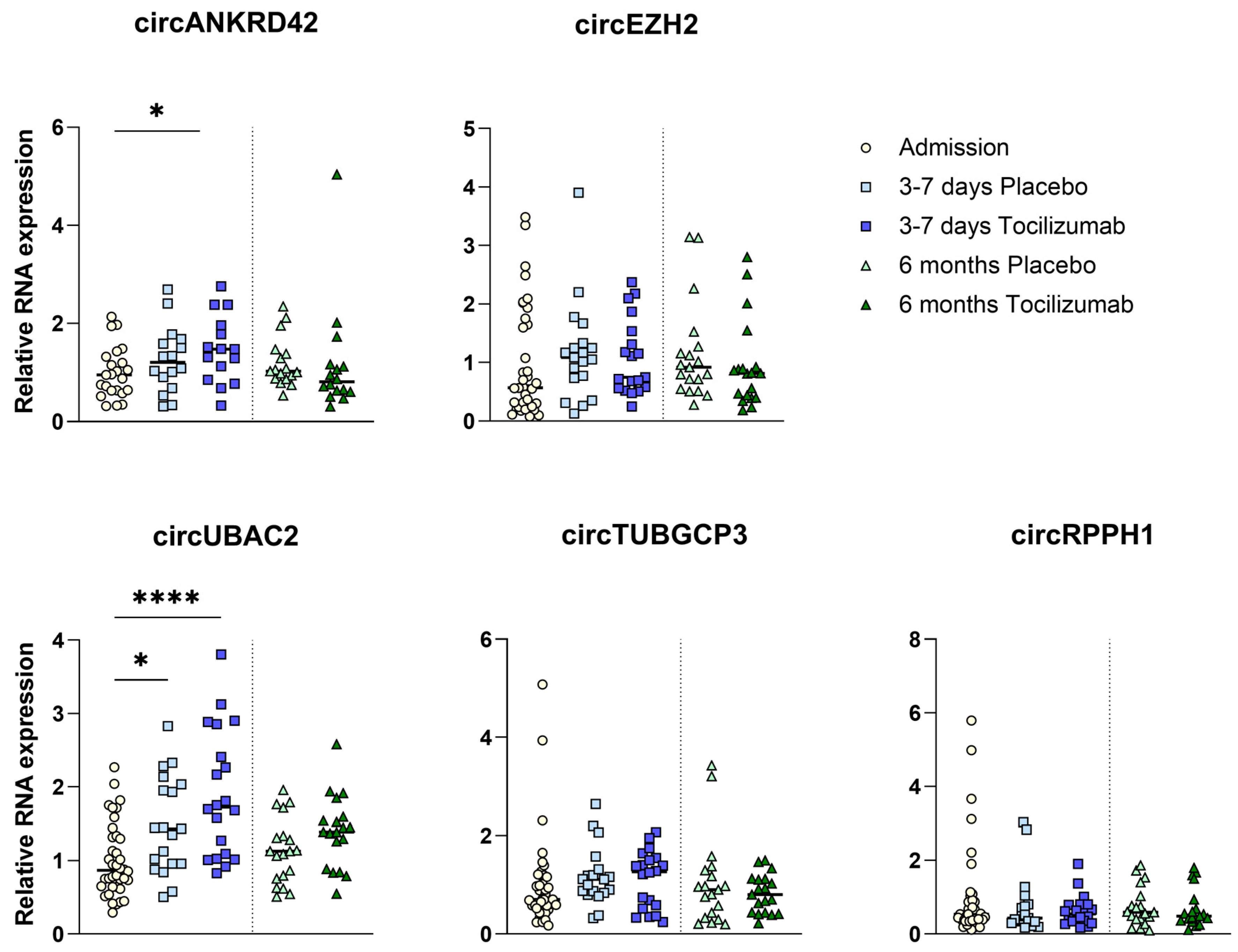
2.6. circRNA Expression with IL-6R Inhibition

Levels of circANKRD42 increased significantly from admission to 3–7 days in the tocilizumab group (FGE 1.0 vs. 1.47; p < 0.05). In contrast, the modest increase in the placebo group did not reach statistical significance (FGE 1.0 vs. 1.26; p = 0.18) (Figure 5). circUBAC2 levels increased from admission to 3–7 days in the placebo arm (FGE 1.0 vs. 1.48; p < 0.05) as well as in the tocilizumab arm (1.0 vs. 1.91; p < 0.001). The increase was more pronounced in the tocilizumab group (FGE 1.91 vs. 1.48; p = 0.09) (Figure 5). Levels of circEZH2, circRPPH1, and circTUBGCP3 did not increase from admission in either the tocilizumab or the placebo group (Figure 5).
In the patients from the ASSAIL study that were selected for the circRNA analyses, there was a similar proportion of males and females. Notably, whereas circANKRD42 levels were slightly higher in females compared with males at 3–7 days in the placebo group, this difference reached statistical significance in the tocilizumab group (p = 0.012) (Supplemental Figure S2).

3. Discussion

We show that the profile of circRNAs in splenocytes from atherosclerosis-prone ApoE−/− mice treated with a high-fat diet differs from that observed in WT mice. To translate these findings to a human setting, we analyzed whether the human counterparts exist in whole blood from a STEMI population, randomized to receive IL-6 receptor inhibition by tocilizumab or placebo at hospital admission prior to/during PCI. We found that in STEMI patients, tocilizumab induced a marked up-regulation of two of the circRNAs that were highly differentially expressed between the ApoE−/− mice and WT mice (circANKRD42 and circUBAC2). At the time of admission, the circUBAC2 level was lower in patients with STEMI than in healthy controls, suggesting that an up-regulation could be beneficial. It has previously been suggested that circRNAs are relatively stable and not influenced by acute events [5]. However, we show that certain circRNA are regulated during STEMI and modulated by anti-inflammatory therapy (i.e., tocilizumab).
Data from the preclinical study and the data from the clinical trial suggest some patterns of regulation of circRNAs. Levels of circUBAC2 were lower in the mice with atherosclerosis (ApoE−/−) than in the WT mice. Interestingly, patients with STEMI had significantly lower levels of circUBAC2 at admission compared to healthy controls, and intriguingly, tocilizumab induced a rise in circUBAC2 towards the levels observed in healthy controls. Whereas the levels of circUBAC2 rose in both treatment groups, the levels in the tocilizumab arm were closer to what we found in the healthy controls. All these results suggest that high levels of circUBAC2 could be beneficial. Additionally, admission levels of both circUBAC2 and circANKRD42 were inversely correlated with peak TnT, and for circUBAC2, a similar inverse correlation was seen with final infarct size at 6 months. It is therefore tempting to hypothesize that the more marked increase of these circRNAs in the tocilizumab as compared with the placebo group may be beneficial for the restoration of the myocardium and that levels of circUBAC2 could be used as a putative biomarker for myocardial inflammation and damage in the future.
In STEMI, circANKRD42 levels at admission did not differ from those of healthy controls. However, like circUBAC2, the levels of circANKRD42 rose during treatment with tocilizumab. Xu et al. suggest that circANKRD42 functions as a profibrotic factor in patients with idiopathic pulmonary fibrosis [26]. Although persistent pro-fibrotic activity may promote myocardial dysfunction, the rise in circANKRD42 soon after PCI could also contribute to tissue repair within the myocardium. Moreover, the predicted up-regulation of mRNA for VDAC1 during tocilizumab treatment may improve mitochondrial function in MI [33]. In contrast to our results, Li et al. found up-regulation of circUBAC2 in whole blood from patients with MI. The two studies rely on different patient selections, i.e., only first-time MI in the ASSAIL-MI trial and only <6 h after symptom onset in the ASSAIL-MI trial. Nonetheless, there is a need for well-designed studies with strict protocols and validation cohorts to further elucidate the role of circRNAs in MI.
Previous research has shown that high levels of circRNA lead to more sponging of miRNA [9,24,37]. miRNA usually downregulates corresponding mRNAs [9]. In other words, high levels of circRNA lead to higher levels of mRNA due to less degradation. However, when comparing circRNA levels to mRNA levels in the ASSAIL-MI study, the results revealed that the 10 predicted miRNA targets only showed a total of five mRNA targets that were significantly higher, while 11 of the mRNA targets were significantly lower. Thirty-four of the investigated mRNA targets were not regulated. It is therefore tempting to hypothesize that circRNA sponging of miRNAs in these patients is deviating from what has been shown in vitro studies and that other roles of circRNA could be more relevant in vivo. Some of the predicted mRNAs that were regulated may be involved in the recruitment and activation of inflammatory cells (ERRFI1, AFF4, SLAMF7, LACC1) [30,32,34,36], apoptosis (CREBRF), intracellular regulation of cholesterol metabolism (AGFG1) [31], and mitochondrial function (VDAC1) [33]. All these processes are relevant in STEMI [38,39]. However, whether this regulation is due to or in coherent to the circUBAC2 and circANKRD42 levels needs to be further investigated. Regulation through circRNA protein binding is also a relevant pathway that needs attention [40]. Further studies are therefore needed to clarify the clinical relevance of the regulation of circRNAs in STEMI and the relevance of tocilizumab treatment.
Our study has some limitations. While we have an equal proportion of males and females in the STEMI population, we only conducted the pre-clinical trial on male mice, which potentially could have induced some bias in the selection of regulated circRNAs. The number of patients, and in particular healthy controls and mice, was relatively low, and we lack patients with chronic coronary atherosclerosis as a control group. Moreover, the pattern between male and female patients in the clinical part of the study should also be interpreted with caution due to the low number of patients in each group. The associations of regulated circRNA with mRNA targets are only predictive and should be interpreted with caution. The circRNAs in the pre-clinical study were examined in splenocytes that do not necessarily reflect the composition of immune cells as a whole, where neutrophils are dominating.

4. Materials and Methods

4.1. Ethics

The ASSessing the effect of Anti-IL-6 treatment in Myocardial Infarction trial (ASSAIL-MI) was approved by the Regional Ethics Committee (REK South-East 2016/1223) and the Norwegian Medicines Agency. All study participants provided written consent. The trial was conducted in compliance with the Declaration of Helsinki and Good Clinical Practice. The animal experiment was approved by the Norwegian National Animal Research Authority with FOTS project license numbers 7927, 8395, 5336, and 21681. All animal experiments were performed following the European Directive 2010/63/EU and conducted in accordance with the Animals in Research: Reporting in vivo Experiments (ARRIVE) guidelines.

4.2. Mouse Models

Male C57BL/6NTac (WT) (n = 3) and ApoE−/− (n = 3) mice obtained from Taconic Biosciences (Cambridge City, IN, USA) were fed an atherogenic high-fat diet (40% fat, 0.27% cholesterol, Research Diets Inc., New Brunswick, NJ, USA, Diet #D16031603) ad libitum for 10 weeks before harvest. The atherosclerotic plaque burden was evaluated by histological analysis of the aortic root as described elsewhere [41]. Briefly, frozen hearts were embedded in the Tissue-Tek OCT compound and sectioned from the caudal to the cranial direction at 10 µm intervals on a cryostat. The presence of atherosclerosis was determined in paraformaldehyde-fixed sections collected at 100 µm intervals, stained with Oil-Red-O (Sigma-Aldrich, Darmstadt, Germany), and counterstained using hematoxylin (H-3404, Vector Laboratories, Newmak, CA, USA).

4.3. Murine Circular RNA Array

Total RNA was isolated from 20 million spleen immune cells from each WT and ApoE−/− mice using the miRNeasy Mini Kit (Qiagen, Hilden, Germany). Isolated RNA was treated with DNase I (Qiagen) and stored at −80 °C until further analysis. RNA concentrations and purity based on the 260/280 and the 260/230 ratios were assessed by spectrophotometer absorbance (NanoDrop ND-1000, Thermo Fisher Scientific, Waltham, MA, USA). CircRNA microarray was performed using Arraystar Mouse circRNA Array V2 (Arraystar Inc., Rockville, MD, USA) based on Arraystar’s standard protocols. Briefly, RNase R (Epicentre, Inc., Lindenhurst, IL, USA) digested the total RNA to remove all linear RNAs to enrich the circRNAs. The enriched circRNAs were amplified and transcribed into fluorescent circRNA utilizing a random priming method (Arraystar Super RNA Labeling Kit; Arraystar, Rockville, MD, USA) and hybridized onto the Arraystar Mouse circRNA Array V2 (8 × 15K, Arraystar). After washing, the arrays were scanned by the Agilent Scanner G2505C. Agilent Feature Extraction software (version 11.0.1.1) was used to analyze the acquired array images. The R software (version 4.4), limma package, was used for quantile normalization and data processing. Differentially expressed circRNAs were visualized with Volcano Plot filtering. Differentially expressed circRNAs between two samples were identified through fold change (FC) filtering (FC > 5 and p-value < 0.05).

4.4. Primer Design

For human circRNA primers, we searched for human homologues based on the murine sequences found in the circRNA array. After comparing the sequences (Jalview, Oxford, GB, 2.11.3.2), we used the Primer-Blast tool: https://circinteractome.nia.nih.gov/circular_rna.html (accessed on 7 December 2022) to design the primer pairs. The primer pairs for circRNA analysis were also found in the literature: circUBAC2 [25], circANKRD42 [26], circEZH2 [42], circRPPH1 [43], circTUBGCP3 [44]. The specificities of the primers were assessed by gel electrophoresis and Sanger sequencing of the quantitative reverse transcription PCR (RT-qPCR) products (Eurofins Genomics, Ebersberg, Germany). The bands were extracted from the gel by using the QIAquick Gel Extraction Kit and protocol (Qiagen) (Supplemental Figure S1).

4.5. Human Total RNA-Isolation and cDNA Synthesis

Total RNA was isolated from the BD PAXgeneTM Blood RNA tubes using MagMaxTM for Stabilized Blood Tubes RNA Isolation Kit (InvitrogenTM, Waltham, MA, USA) (ThermoFisher, Waltham, MA, USA) according to the manufacturer’s instructions. cDNA synthesis was carried out as described by Panda et al. [45]. Briefly, we mixed 650 ng total RNA, RiboLock (Thermo Scientific Lot, Waltham, MA, USA), dNTP mix (Thermo Scientific), random primers (Roche, Basel, Switzerland), 5x RT buffer (Thermo Scientific), and Maxima RT (Thermo Scientific). We incubated the samples for 10 min at 25 °C, followed by 30 min at 50 °C, and 5 min at 85 °C on a Veriti 96-well thermocycler (Applied Biosystems, Waltham, MA, USA).

4.6. Quantitative Reverse Transcriptase PCR

RT-qPCR was performed using the Bio-Rad CFX384 Maestro, C1000 Touch, qPCR machine (Bio-Rad Labratories Inc., Hercules, CA, USA) The optimal RT-qPCR protocol for our primer pairs was 3-step PCR, amplification + melt, 45 cycles with an annealing temperature of 56.6 °C. The RT-qPCR was performed on a CFX 384 real-time PCR system using 5 µL of cDNA and SYBR Green real-time master mix (Quantabio, Beverly, MA, USA). The expression levels of targeted circRNAs were normalized by the 2−∆∆CT method. Beta-ACTIN and circHIPK3 mRNA levels were used as stable internal controls for the circRNA. The primers used in the PCR are listed in Supplemental Table S1.

4.7. miRNA and mRNA Targets

We predicted miRNA targets for circUBAC2 and circANKRD42 using miRanda [46], RNAhybrid [47], and TargetScan [48] algorithms. miRNA targets were also verified by the literature [25,26]. We investigated mRNA targets for miRNAs using the database miRDB (https://mirdb.org/mirdb/index.html, accessed on 30 January 2024) [28]. The top 5 mRNA targets for each miRNA were investigated in RNA sequencing from whole blood from patients in ASSAIL-MI.

4.8. RNA Sequencing

RNA sequencing from full blood was previously done and has been described in [23]. The sequencing was reanalyzed; briefly, the RNA samples were analyzed by Novogene (UK) Company Limited, Milton, Cambridge, GB. The ribosomal RNA depletion library preparation was used for the RNA isolated from whole blood with PAXgene tubes. We used the fastp software (v0.23.0) to remove contaminated adapters and low-quality reads with a phred score below 30 in the pair-end mode [49]. Filtered reads were mapped to the human transcriptome (Gencode Human Release H37, Heidelberg, Germany), and transcripts were quantified with 200 bootstrap iterations by Salmon (v1.5.2) [50,51]. We summarized the Salmon outputs to gene level and imported them into DESeq2 (v1.34.0) via tximeta (v.1.12.3) [52,53].
Descriptions of the mouse model, murine circular RNA array, primer design for human homologs, human RNA isolation, cDNA synthesis, quantitative reverse transcriptase PCR, prediction of miRNA and mRNA targets, and RNA sequencing are given in the Supplemental Materials.

4.9. Patients and Study Design of ASSAIL-MI

In the phase 2 ASSAIL-MI trial (Clinicaltrials.gov: NCT03004703), a single dose of intravenous tocilizumab was compared with placebo in patients with acute STEMI. The study drugs were allocated in a 1:1 fashion to 199 patients between March 2017 and February 2020, as previously described [17]. The key inclusion criteria were STEMI and symptom onset less than 6 h before PCI. Exclusion criteria were previous MI; chronic infection, or chronic autoimmune or inflammatory disease; uncontrolled inflammatory bowel disease; ongoing infectious or immunologic disease; major surgery within the past eight weeks; or treatment with immunosuppressants other than low-dose steroids (equivalent to systemic exposure to 5 mg prednisone per day) [23]. A total of 40 patients, 19 from the placebo arm and 21 from the tocilizumab arm, were chosen out for circRNA and mRNA analysis. The patients were selected based on age, gender, and clinical parameters to obtain equal distribution as for the whole population. Admission characteristics of the 40 patients who participated in this sub-study are described in Table 1.
All the patients received dual antiplatelet therapy (DAPT) and unfractionated heparin (5000–7500 IE) intravenously before PCI was performed, and 76% of the patients had received unfractionated heparin (5000 IE) before arrival at the hospital.

4.10. Blood Sampling Protocol

We collected whole blood samples prior to the intra-arterial administration of unfractionated heparin and PCI. Blood samples were repeated 14–33 h after PCI and at 3–7 days, 3 months, and 6 months. We used BD PAXgeneTM Blood RNA tubes (BD, Franklin Lakes, NJ, USA) for RNA analysis of whole blood. For comparison of circRNA levels, we also collected venous blood samples from 13 sex and age-matched self-reported healthy controls.

4.11. Measurement of Cardiac Markers in the ASSAIL-MI Trial

High-sensitivity troponin T (TnT) was measured by electrochemiluminescence immunoassay (Elecsys 2010 analyzer, Roche Diagnostics, Basel, Switzerland). The myocardial salvage index (MSI) (%), defined as: a r e a   a t   r i s k i n f a r c t   s i z e a r e a   a t   r i s k · 100 and final infarct size (% of left ventricular mass), was measured by cardiac magnetic resonance (CMR) imaging 3 to 7 days after the intervention as previously described [17].

4.12. Statistics

Statistical analyses were performed using SPSS version 25 (IBM Corp., Armonk, NY, USA) and GraphPad Prism 9.4.1 (GraphPad Software, Boston, MA, USA). Fold gene expression (FGE) was normalized toward the admission group, containing all patients admitted to the hospital, to get a fold change value of 1. An unpaired t-test was run to reveal significant differences in admission versus healthy controls. An unpaired t-test was also conducted for comparison of admission versus the different treatments (placebo vs. tocilizumab) and timepoints (admission vs. 3–7 days). A Pearson and Spearman correlation analysis was performed for admission circRNA levels and peak TnT, final infarct size, and MSI. For continuous variables, we used a two-sided t-test. Non-parametric variables were tested by using a Mann–Whitney test. We tested categorical variables using Fisher’s exact test. p-values < 0.05 (two-sided) were considered statistically significant.

5. Conclusions

To the best of our knowledge, this is the first study to investigate circRNA expression in immune cells from patients receiving anti-inflammatory treatment. We show that circRNA levels are altered in patients with STEMI. We also show an inverse correlation between investigated circRNAs and TnT and left ventricular mass. The circRNA levels are altered by tocilizumab. Our data suggest that the increase of circUBAC2 and circANKRD42 associated with treatment with tocilizumab can result in altered levels of mRNAs influencing the activation of the immune system, apoptosis, and mitochondrial function. However, further experiments and larger studies are needed to better understand the complex function of circRNAs in MI and how they could be modulated by therapy.

Supplementary Materials

The following supporting information can be downloaded at: https://www.mdpi.com/article/10.3390/ijms25169014/s1.

Author Contributions

Conceptualization, F.A.H., C.H., P.A., I.A., B.H. and T.B.D.; methodology, F.A.H., C.H., X.Y.K., A.Q.-J., S.H., I.A. and T.B.D.; formal analysis, F.A.H., C.H., K.B., L.G., A.K.A., G.Ø.A., B.H.A., O.K. and S.H.; investigation, F.A.H., C.H., X.Y.K., A.Q.-J., S.H. and T.B.D.; resources, X.Y.K., K.B., L.G., A.K.A., G.Ø.A., B.H.A., O.K., P.A., I.A., B.H. and T.B.D.; writing—original draft preparation, F.A.H., C.H., P.A. and T.B.D.; project administration, P.A., B.H., I.A. and T.B.D.; funding acquisition, B.H. and I.A. All authors have read and agreed to the published version of the manuscript.

Funding

This study was granted by the Medical Research Program (MSRP) at the University of Oslo (UiO), the Research Council of Norway (nos. 271555/F20 and 283867), Health Authority South-Eastern Regional Health Authority (nos. 2019067 and 2018084) and the Central Norway Regional Health Authority.

Institutional Review Board Statement

The ASSessing the effect of Anti-IL-6 treatment in Myocardial Infarction trial (ASSAIL-MI) was approved by the Regional Ethics Committee (REK South-East 2016/1223) and the Norwegian Medicines Agency. All study participants provided written consent. The trial was conducted in compliance with the Declaration of Helsinki and Good Clinical Practice. The animal experiment was approved by the Norwegian National Animal Research Authority with FOTS project license numbers 7927, 8395, 5336, and 21681. All animal experiments were performed following the European Directive 2010/63/EU and conducted in accordance with the Animals in Research: Re-porting in vivo Experiments (ARRIVE) guidelines.

Informed Consent Statement

All study participants provided written consent to be included in the study.

Data Availability Statement

Ethical restrictions from the Regional Committee for Medical and Research Ethics in South-East Norway, prohibits data from individual patients to be made available on publicly available repository. However, an institutional data transfer agreement can be established, and data can be shared if the aims of data use are covered by ethical approval and patient consent. The procedure will involve an update to the ethical approval as well as review by legal departments at both institutions, and the process will typically take 2 to 4 months from initial contact.

Conflicts of Interest

Kaspar Broch has received lecture fees from NovoNordisk, Norvartis Pharmacosmos, AstraZeneca, Boehringer Ingelheim, Pfizer, Orion Pharma, and Vifor Pharma, and has been on advisory boards for Pharmacosmos, Pfizer, and AstraZeneca. Geir Ø. Andersen has received lecture fees from Orion Pharma. Lars L. Gullestad has received lecture fees from AstraZeneca, Boehringer Ingelheim, Novartis, and Amgen. He has also been a member of the local advisory board in AstraZeneca and Boehringer Ingelheim. The remaining authors have no possible conflicts of interest.

References

  1. Libby, P. The Changing Landscape of Atherosclerosis. Nature 2021, 592, 524–533. [Google Scholar] [CrossRef]
  2. Stewart, J.; Manmathan, G.; Wilkinson, P. Primary Prevention of Cardiovascular Disease: A Review of Contemporary Guidance and Literature. JRSM Cardiovasc. Dis. 2017, 6, 2048004016687211. [Google Scholar] [CrossRef]
  3. The Top 10 Causes of Death. Available online: https://www.who.int/news-room/fact-sheets/detail/the-top-10-causes-of-death (accessed on 21 February 2023).
  4. Conn, S.J.; Pillman, K.A.; Toubia, J.; Conn, V.M.; Salmanidis, M.; Phillips, C.A.; Roslan, S.; Schreiber, A.W.; Gregory, P.A.; Goodall, G.J. The RNA Binding Protein Quaking Regulates Formation of circRNAs. Cell 2015, 160, 1125–1134. [Google Scholar] [CrossRef]
  5. Enuka, Y.; Lauriola, M.; Feldman, M.E.; Sas-Chen, A.; Ulitsky, I.; Yarden, Y. Circular RNAs Are Long-Lived and Display Only Minimal Early Alterations in Response to a Growth Factor. Nucleic Acids Res. 2016, 44, 1370–1383. [Google Scholar] [CrossRef]
  6. Yu, J.; Xie, D.; Huang, N.; Zhou, Q. Circular RNAs as Novel Diagnostic Biomarkers and Therapeutic Targets in Kidney Disease. Front. Med. 2021, 8, 714958. [Google Scholar] [CrossRef]
  7. Zhang, Y.; Luo, J.; Yang, W.; Ye, W.-C. CircRNAs in Colorectal Cancer: Potential Biomarkers and Therapeutic Targets. Cell Death Dis. 2023, 14, 353. [Google Scholar] [CrossRef]
  8. Wang, X.; Li, H.; Lu, Y.; Cheng, L. Regulatory Effects of Circular RNAs on Host Genes in Human Cancer. Front. Oncol. 2021, 10, 586163. [Google Scholar]
  9. Sakshi, S.; Jayasuriya, R.; Ganesan, K.; Xu, B.; Ramkumar, K.M. Role of circRNA-miRNA-mRNA Interaction Network in Diabetes and Its Associated Complications. Mol. Ther. Nucleic Acids 2021, 26, 1291–1302. [Google Scholar] [CrossRef] [PubMed]
  10. Patop, I.L.; Wüst, S.; Kadener, S. Past, Present, and Future of circRNAs. EMBO J. 2019, 38, e100836. [Google Scholar] [CrossRef]
  11. Chen, W.; Zhang, Y.; Yin, M.; Cheng, Z.; Li, D.; Luo, X.; Liu, X.; Tu, J. Circular RNA circPRDX3 Mediates Neuronal Survival Apoptosis in Ischemic Stroke by Targeting miR-641 and NPR3. Brain Res. 2022, 1797, 148114. [Google Scholar] [CrossRef]
  12. Yang, Z.; Huang, C.; Wen, X.; Liu, W.; Huang, X.; Li, Y.; Zang, J.; Weng, Z.; Lu, D.; Tsang, C.K.; et al. Circular RNA Circ-FoxO3 Attenuates Blood-Brain Barrier Damage by Inducing Autophagy during Ischemia/Reperfusion. Mol. Ther. 2022, 30, 1275–1287. [Google Scholar] [CrossRef]
  13. Huang, R.; Zhang, W.; Li, W.; Gao, Y.; Zheng, D.; Bi, G. Overexpressing Circ_0000831 Is Sufficient to Inhibit Neuroinflammation and Vertigo in Cerebral Ischemia through a miR-16-5p-Dependent Mechanism. Exp. Neurol. 2022, 353, 114047. [Google Scholar] [CrossRef]
  14. Cheng, Q.; Wang, J.; Li, M.; Fang, J.; Ding, H.; Meng, J.; Zhang, J.; Fang, X.; Liu, H.; Ma, C.; et al. CircSV2b Participates in Oxidative Stress Regulation through miR-5107-5p-Foxk1-Akt1 Axis in Parkinson’s Disease. Redox Biol. 2022, 56, 102430. [Google Scholar] [CrossRef]
  15. Xie, Q.; Ma, Y.; Ren, Z.; Gu, T.; Jiang, Z. Circular RNA: A New Expectation for Cardiovascular Diseases. J. Cell. Biochem. 2024, 125, e30512. [Google Scholar] [CrossRef]
  16. Xu, G.; Liu, G.; Wang, Z.; Li, Y.; Fang, W. Circular RNAs: Promising Treatment Targets and Biomarkers of Ischemic Stroke. Int. J. Mol. Sci. 2024, 25, 178. [Google Scholar] [CrossRef]
  17. Broch, K.; Anstensrud, A.K.; Woxholt, S.; Sharma, K.; Tøllefsen, I.M.; Bendz, B.; Aakhus, S.; Ueland, T.; Amundsen, B.H.; Damås, J.K.; et al. Randomized Trial of Interleukin-6 Receptor Inhibition in Patients with Acute ST-Segment Elevation Myocardial Infarction. J. Am. Coll. Cardiol. 2021, 77, 1845–1855. [Google Scholar] [CrossRef] [PubMed]
  18. Abbate, A.; Trankle, C.R.; Buckley, L.F.; Lipinski, M.J.; Appleton, D.; Kadariya, D.; Canada, J.M.; Carbone, S.; Roberts, C.S.; Abouzaki, N.; et al. Interleukin-1 Blockade Inhibits the Acute Inflammatory Response in Patients with ST-Segment-Elevation Myocardial Infarction. J. Am. Heart Assoc. 2020, 9, e014941. [Google Scholar] [CrossRef]
  19. Hjerteinfarkt. Available online: https://legehandboka.no/handboken/kliniske-kapitler/hjertekar/tilstander-og-sykdommer/koronarsykdom/hjerteinfarkt/ (accessed on 22 February 2023).
  20. Feng, Y.; Ye, D.; Wang, Z.; Pan, H.; Lu, X.; Wang, M.; Xu, Y.; Yu, J.; Zhang, J.; Zhao, M.; et al. The Role of Interleukin-6 Family Members in Cardiovascular Diseases. Front. Cardiovasc. Med. 2022, 9, 818890. [Google Scholar] [CrossRef]
  21. Huang, M.; Yang, D.; Xiang, M.; Wang, J. Role of Interleukin-6 in Regulation of Immune Responses to Remodeling after Myocardial Infarction. Heart Fail. Rev. 2015, 20, 25–38. [Google Scholar] [CrossRef]
  22. Ridker, P.M.; Rane, M. Interleukin-6 Signaling and Anti-Interleukin-6 Therapeutics in Cardiovascular Disease. Circ. Res. 2021, 128, 1728–1746. [Google Scholar] [CrossRef] [PubMed]
  23. Huse, C.; Anstensrud, A.K.; Michelsen, A.E.; Ueland, T.; Broch, K.; Woxholt, S.; Yang, K.; Sharma, K.; Tøllefsen, I.M.; Bendz, B.; et al. Interleukin-6 Inhibition in ST-Elevation Myocardial Infarction: Immune Cell Profile in the Randomised ASSAIL-MI Trial. eBioMedicine 2022, 80, 104013. [Google Scholar] [CrossRef]
  24. Panda, A.C. Circular RNAs Act as miRNA Sponges. In Circular RNAs: Biogenesis and Functions; Xiao, J., Ed.; Advances in Experimental Medicine and Biology; Springer: Singapore, 2018; pp. 67–79. ISBN 9789811314261. [Google Scholar]
  25. Li, Q.; Wang, Y.; An, Y.; Wang, J.; Gao, Y. The Particular Expression Profiles of Circular RNA in Peripheral Blood of Myocardial Infarction Patients by RNA Sequencing. Front. Cardiovasc. Med. 2022, 9, 810257. [Google Scholar] [CrossRef]
  26. Xu, P.; Zhang, J.; Wang, M.; Liu, B.; Li, R.; Li, H.; Zhai, N.; Liu, W.; Lv, C.; Song, X. hnRNPL-Activated circANKRD42 Back-Splicing and circANKRD42-Mediated Crosstalk of Mechanical Stiffness and Biochemical Signal in Lung Fibrosis. Mol. Ther. 2022, 30, 2370–2387. [Google Scholar] [CrossRef] [PubMed]
  27. Roberts, L.B.; Kapoor, P.; Howard, J.K.; Shah, A.M.; Lord, G.M. An Update on the Roles of Immune System-Derived microRNAs in Cardiovascular Diseases. Cardiovasc. Res. 2021, 117, 2434–2449. [Google Scholar] [CrossRef]
  28. Chen, Y.; Wang, X. miRDB: An Online Database for Prediction of Functional microRNA Targets. Nucleic Acids Res. 2020, 48, D127–D131. [Google Scholar] [CrossRef]
  29. Liu, W.; Wang, X. Prediction of Functional microRNA Targets by Integrative Modeling of microRNA Binding and Target Expression Data. Genome Biol. 2019, 20, 18. [Google Scholar] [CrossRef]
  30. Chen, B.; Dong, L.; Zhang, J.; Hao, Y.; Chi, W.; Song, D. Exploring Shared Pathways and the Shared Biomarker ERRFI1 in Obstructive Sleep Apnoea and Atherosclerosis Using Integrated Bioinformatics Analysis. Sci. Rep. 2023, 13, 15103. [Google Scholar] [CrossRef]
  31. Duan, Z.; Yang, M.; Yang, J.; Wu, Z.; Zhu, Y.; Jia, Q.; Ma, X.; Yin, Y.; Zheng, J.; Yang, J.; et al. AGFG1 Increases Cholesterol Biosynthesis by Disrupting Intracellular Cholesterol Homeostasis to Promote PDAC Progression. Cancer Lett. 2024, 598, 217130. [Google Scholar] [CrossRef]
  32. Fang, Y.; Cao, H.; Gong, X.; Chen, Y.; Zhuang, Y.; Zhou, S.; Chen, Y.; Jiang, Y.; Ji, X.; Peng, H.; et al. AFF4 Predicts the Prognosis of Colorectal Cancer Patients and Suppresses Colorectal Cancer Metastasis via Promoting CDH1 Expression. Front. Oncol. 2022, 12, 797392. [Google Scholar] [CrossRef]
  33. Camara, A.K.S.; Zhou, Y.; Wen, P.-C.; Tajkhorshid, E.; Kwok, W.-M. Mitochondrial VDAC1: A Key Gatekeeper as Potential Therapeutic Target. Front. Physiol. 2017, 8, 460. [Google Scholar] [CrossRef]
  34. O’Connell, P.; Blake, M.K.; Godbehere, S.; Amalfitano, A.; Aldhamen, Y.A. SLAMF7 Modulates B Cells and Adaptive Immunity to Regulate Susceptibility to CNS Autoimmunity. J. Neuroinflamm. 2022, 19, 241. [Google Scholar] [CrossRef]
  35. Zhou, S.; Zhong, Z.; Lu, Y.; Li, Y.; Yao, H.; Zhao, Y.; Guo, T.; Yang, K.; Li, Y.; Chen, S.; et al. A LETM2-Regulated PI3K-Akt Signaling Axis Reveals a Prognostic and Therapeutic Target in Pancreatic Cancer. Cancers 2022, 14, 4722. [Google Scholar] [CrossRef] [PubMed]
  36. Wei, Z.; Oh, J.; Flavell, R.A.; Crawford, J.M. LACC1 Bridges NOS2 and Polyamine Metabolism in Inflammatory Macrophages. Nature 2022, 609, 348–353. [Google Scholar] [CrossRef]
  37. Huang, V. Endogenous miRNAa: miRNA-Mediated Gene Upregulation. In RNA Activation; Li, L.-C., Ed.; Advances in Experimental Medicine and Biology; Springer: Singapore, 2017; pp. 65–79. ISBN 978-981-10-4310-9. [Google Scholar]
  38. Matter, M.A.; Paneni, F.; Libby, P.; Frantz, S.; Stähli, B.E.; Templin, C.; Mengozzi, A.; Wang, Y.-J.; Kündig, T.M.; Räber, L.; et al. Inflammation in Acute Myocardial Infarction: The Good, the Bad and the Ugly. Eur. Heart J. 2024, 45, 89–103. [Google Scholar] [CrossRef] [PubMed]
  39. Ramachandra, C.J.A.; Hernandez-Resendiz, S.; Crespo-Avilan, G.E.; Lin, Y.-H.; Hausenloy, D.J. Mitochondria in Acute Myocardial Infarction and Cardioprotection. eBioMedicine 2020, 57, 1–9. [Google Scholar] [CrossRef]
  40. Zhao, H.; Tan, Z.; Zhou, J.; Wu, Y.; Hu, Q.; Ling, Q.; Ling, J.; Liu, M.; Ma, J.; Zhang, D.; et al. The Regulation of circRNA and lncRNAprotein Binding in Cardiovascular Diseases: Emerging Therapeutic Targets. Biomed. Pharmacother. 2023, 165, 115067. [Google Scholar] [CrossRef]
  41. Kong, X.Y.; Huse, C.; Yang, K.; Øgaard, J.; Berges, N.; Vik, E.S.; Nawaz, M.S.; Quiles-Jiménez, A.; Abbas, A.; Gregersen, I.; et al. Endonuclease V Regulates Atherosclerosis through C-C Motif Chemokine Ligand 2-Mediated Monocyte Infiltration. J. Am. Heart Assoc. 2021, 10, e020656. [Google Scholar] [CrossRef] [PubMed]
  42. Yao, B.; Zhang, Q.; Yang, Z.; An, F.; Nie, H.; Wang, H.; Yang, C.; Sun, J.; Chen, K.; Zhou, J.; et al. CircEZH2/miR-133b/IGF2BP2 Aggravates Colorectal Cancer Progression via Enhancing the Stability of m6A-Modified CREB1 mRNA. Mol. Cancer 2022, 21, 140. [Google Scholar] [CrossRef]
  43. Huang, Y.; Zheng, W.; Ji, C.; Wang, X.; Yu, Y.; Deng, X.; Zhou, X.; Fang, L. Circular RNA circRPPH1 Promotes Breast Cancer Progression via circRPPH1-miR-512-5p-STAT1 Axis. Cell Death Discov. 2021, 7, 376. [Google Scholar] [CrossRef]
  44. Yang, Y.; Fan, X.; Nie, Y.; Liu, D.; Zhu, D.; Wu, K.; Zhang, Y.; Li, W.; Tian, X.; Wang, H.; et al. CircTUBGCP3 Facilitates the Tumorigenesis of Lung Adenocarcinoma by Sponging miR-885-3p. Cancer Cell Int. 2021, 21, 651. [Google Scholar] [CrossRef]
  45. Panda, A.C.; Gorospe, M. Detection and Analysis of Circular RNAs by RT-PCR. Bio Protocol. 2018, 8, e2775. [Google Scholar] [CrossRef]
  46. Enright, A.J.; John, B.; Gaul, U.; Tuschl, T.; Sander, C.; Marks, D.S. MicroRNA Targets in Drosophila. Genome Biol. 2003, 5, R1. [Google Scholar] [CrossRef]
  47. Rehmsmeier, M.; Steffen, P.; Höchsmann, M.; Giegerich, R. Fast and Effective Prediction of microRNA/Target Duplexes. RNA 2004, 10, 1507–1517. [Google Scholar] [CrossRef]
  48. McGeary, S.E.; Lin, K.S.; Shi, C.Y.; Pham, T.M.; Bisaria, N.; Kelley, G.M.; Bartel, D.P. The Biochemical Basis of microRNA Targeting Efficacy. Science 2019, 366, eaav1741. [Google Scholar] [CrossRef]
  49. Chen, S.; Zhou, Y.; Chen, Y.; Gu, J. Fastp: An Ultra-Fast All-in-One FASTQ Preprocessor. Bioinformatics 2018, 34, i884–i890. [Google Scholar] [CrossRef] [PubMed]
  50. Patro, R.; Duggal, G.; Love, M.I.; Irizarry, R.A.; Kingsford, C. Salmon Provides Fast and Bias-Aware Quantification of Transcript Expression. Nat. Methods 2017, 14, 417–419. [Google Scholar] [CrossRef] [PubMed]
  51. Frankish, A.; Diekhans, M.; Ferreira, A.-M.; Johnson, R.; Jungreis, I.; Loveland, J.; Mudge, J.M.; Sisu, C.; Wright, J.; Armstrong, J.; et al. GENCODE Reference Annotation for the Human and Mouse Genomes. Nucleic Acids Res. 2019, 47, D766–D773. [Google Scholar] [CrossRef] [PubMed]
  52. Love, M.I.; Huber, W.; Anders, S. Moderated Estimation of Fold Change and Dispersion for RNA-Seq Data with DESeq2. Genome Biol. 2014, 15, 550. [Google Scholar] [CrossRef]
  53. Love, M.I.; Soneson, C.; Hickey, P.F.; Johnson, L.K.; Pierce, N.T.; Shepherd, L.; Morgan, M.; Patro, R. Tximeta: Reference Sequence Checksums for Provenance Identification in RNA-Seq. PLoS Comput. Biol. 2020, 16, e1007664. [Google Scholar] [CrossRef]
Figure 1. Murine circRNA profile in atherogenic ApoE−/− mice. (A) Atherosclerotic lesions in the aortic root from wild type (WT) and ApoE−/− after 10 weeks on an atherogenic diet, stained with Oil-Red-O. (B) Volcano-plot showing the circRNA regulation. Significantly differentially expressed circRNAs, with an FC > 5 and p-value < 0.05, from splenic cells in WT and ApoE−/− mice marked in red. (C) Normalized intensity of the circRNAs from the circRNA murine array between ApoE−/− vs. WT. * p < 0.05, ** p < 0.01, **** p < 0.0001.
Ijms 25 09014 g001
Figure 2. Relative circRNA expression of whole blood from STEMI patients at admission vs. healthy controls. The figure presents the FGE regulation of the circRNA targets in a subgroup of patients from the ASSAIL-MI. Samples were collected at admission before the patients were randomized to tocilizumab or placebo. Comparison between STEMI patients (n = 40) and healthy HC (n = 13) were made by an unpaired t-test. ** p < 0.01.
Ijms 25 09014 g002
Figure 3. Correlation analysis between circRNA and clinical parameters from STEMI patients. (A) Inverse correlation with peak TnT and admission circRNA levels for circANKRD42 and circUBAC2 was conducted with a Spearman correlation analysis. (B) Inverse correlation between admission circRNA levels for circUBAC2 and the finale infarct size at 6 months, measured by % of left ventricular mass. * p < 0.05, ** p < 0.01.
Ijms 25 09014 g003
Figure 4. Overview of circRNA, miRNA, and mRNA interactions in patient samples. An overview of the miRNAs regulated by circUBAC2 and circANKRD42. We also show the mRNA targets for the different miRNAs and how they are regulated. * p < 0.05, ** p < 0.01, **** p < 0.0001.
Ijms 25 09014 g004
Figure 5. Fold gene expression of circRNA targets in immune cells in STEMI patients treated with tocilizumab (n = 21) or placebo (n = 19). The figure shows the regulation of the circRNAs after ST-elevation myocardial infarction (STEMI) at different time points in the tocilizumab and the placebo group. Unpaired t-test between the different groups. * p < 0.05, **** p < 0.0001.
Ijms 25 09014 g005
Table 1. Overview of patient characteristics. Values are mean ± SD, n (%), or median (interquartile range). Admission characteristics stratified by treatment allocation and gender. ACE = angiotensin-converting enzyme; ARB = angiotensin receptor blocker; CK-MB = creatine kinase myocardial band; DAPT = dual anti-platelet therapy; HDL = high-density lipoprotein; LDL = low-density lipoprotein; PCI = percutaneous coronary intervention; NT-proBNP = N-terminal pro-B-type natriuretic peptide. The healthy controls (n = 13) were matched with age, sex, and ethnicity.
Placebo
(n = 19) A
Tocilizumab
(n = 21) B
Demographics
  Age, years64 ± 665 ± 6
  Body mass index, kg/m224.4 ± 2.825.2 ± 3.3
  Male9 (47)11 (52)
  White19 (100)21 (100)
Smoking status
  Never smokers6 (32)7 (33)
  Previous smokers4 (21)7 (33)
  Current smokers9 (47)7 (33)
Prior conditions
  Angina pectoris1 (5)0
  Cerebrovascular disease1 (5)1 (5)
  Hypertension7 (37)7 (33)
  Treatment
  ACE inhibitor or ARB4 (21)1 (5)
  Oral anticoagulants01 (5)
  Platelet inhibitor1 (5)3 (14)
  Beta-blocker1 (5)4 (19)
  Calcium antagonist2 (10)3 (14)
  Diuretic1 (5)0
  Statin3 (16)5 (24)
  Up-front DAPT19 (100)21 (100)
Clinical characteristics
  Blood pressure at admission, mm Hg
   Systolic124 ± 22130 ± 23
   Diastolic77 ± 1477 ± 21
  Heart rate at admission, beats/min70 ± 1670 ± 15
  Time from symptom onset to arrival at PCI center, min184 ± 80144 ± 64
  Door-to-balloon time, min27 ± 1521 ± 8
  Killip class
   I18 (95)20 (95)
   II1 (5)1 (5)
  GRACE risk score147 ± 20147 ± 24
  Infarct location
   Left anterior descending branch3 (16)5 (24)
   Circumflex or marginal5 (26)2 (10)
   Right coronary artery11 (58)13 (62)
Laboratory values
  Hemoglobin, g/dL14.1 ± 1.313.9 ± 1.5
  Platelet count, 109/L251 ± 52261 ± 64
  Total white blood cell count, 109/L10.5 ± 3.411.7 ± 4.0
  Aspartate transaminase, U/L29 (24–39)29 (23–38)
  Troponin T, ng/L58 (28–95)33 (19–62)
  CK-MB, μg/L6.0 (2.8–23.8)4.4 (2.7–6.3)
  NT-proBNP, ng/L155 (58–347)107 (52–212)
  Creatinine, mmol/L64 ± 1370 ± 13
  Glucose, mmol/L8.1 ± 2.08.2 ± 1.3
  HbA1c, mmol/mol36 (33–48)38 (36–39)
  Total cholesterol, mmol/L5.5 ± 1.15.4 ± 1.1
  HDL cholesterol, mmol/L1.3 (1.1–1.7)1.2 (1.0–1.3)
  LDL cholesterol, mmol/L3.8 ± 0.93.9 ± 1.0
  C-reactive protein, mg/L4.1 (2.1–5.0)1.7 (0.9–5.0)
  Albumin, g/L42 ± 640 ± 3
Disclaimer/Publisher’s Note: The statements, opinions and data contained in all publications are solely those of the individual author(s) and contributor(s) and not of MDPI and/or the editor(s). MDPI and/or the editor(s) disclaim responsibility for any injury to people or property resulting from any ideas, methods, instructions or products referred to in the content.

