Updates on Mechanisms of Cytochrome P450 Catalysis of Complex Steroid Oxidations
Abstract
:1. Introduction
2. P450 11A1
3. P450 11B2
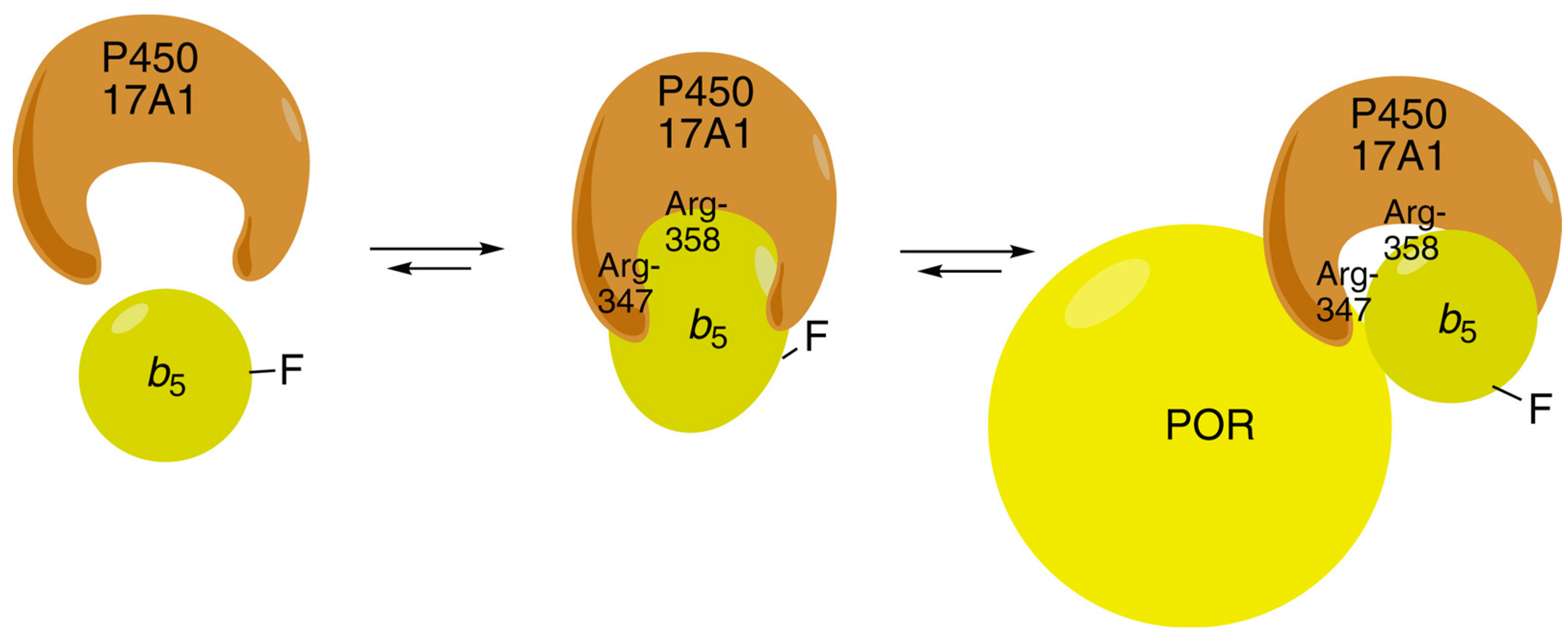
4. P450 17A1
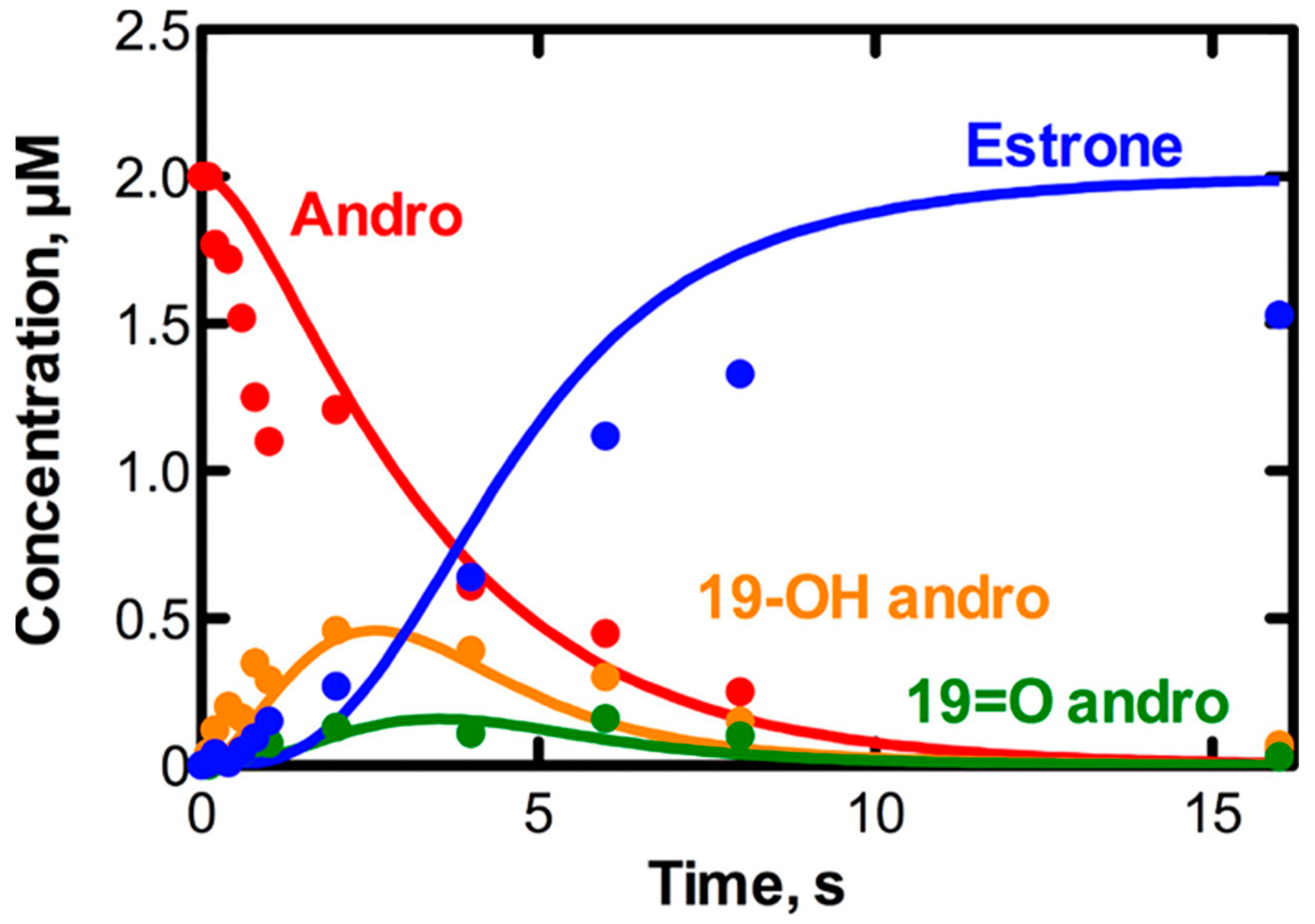
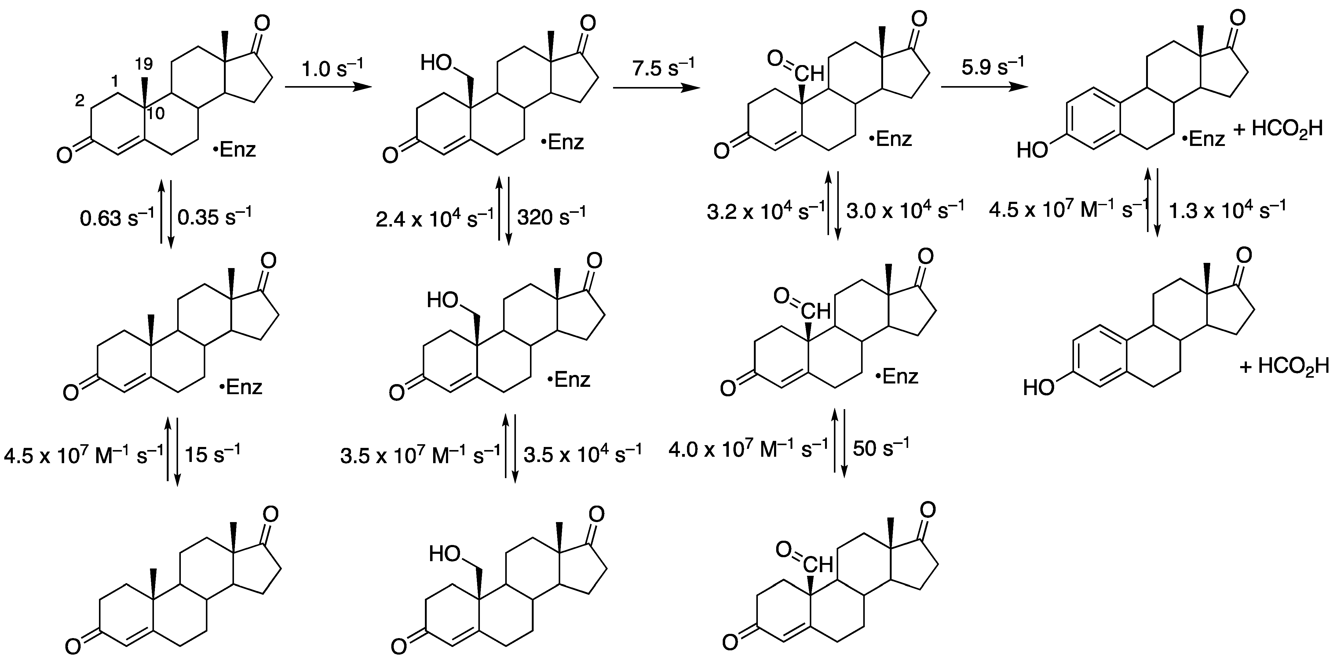
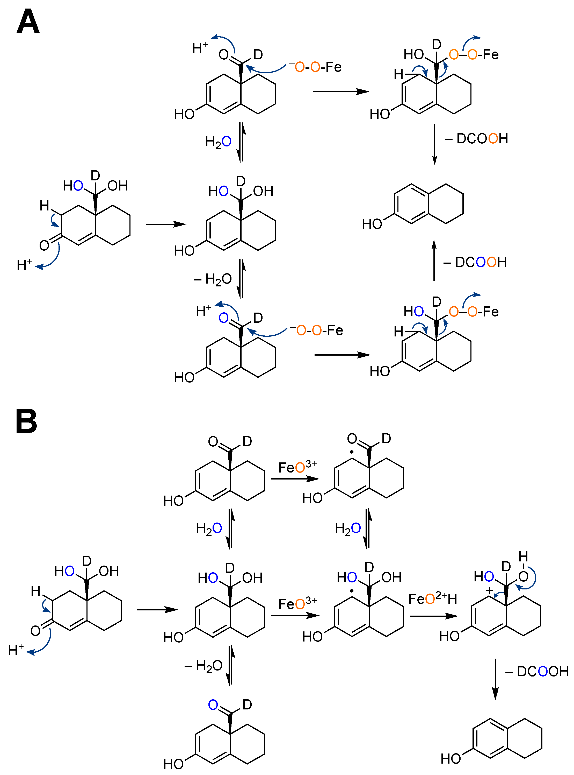
5. P450 19A1
6. P450 51A1
7. Summary and Conclusions
Author Contributions
Funding
Institutional Review Board Statement
Informed Consent Statement
Data Availability Statement
Acknowledgments
Conflicts of Interest
References
- McLean, K.J.; Leys, D.; Munro, A.W. Microbial cytochromes P450. In Cytochrome P450. Structure, Mechanism, and Biochemistry, 4th ed.; Ortiz de Montellano, P.R., Ed.; Springer: New York, NY, USA, 2015; pp. 261–407. [Google Scholar]
- McLean, K.J.; Dunford, A.J.; Neeli, R.; Driscoll, M.D.; Munro, A.W. Structure, function and drug targeting in Mycobacterium tuberculosis cytochrome P450 systems. Arch. Biochem. Biophys. 2007, 464, 228–240. [Google Scholar] [CrossRef]
- Driscoll, M.D.; McLean, K.J.; Levy, C.; Mast, N.; Pikuleva, I.A.; Lafite, P.; Rigby, S.E.; Leys, D.; Munro, A.W. Structural and biochemical characterization of Mycobacterium tuberculosis CYP142: Evidence for multiple cholesterol 27-hydroxylase activities in a human pathogen. J. Biol. Chem. 2010, 285, 38270–38282. [Google Scholar] [CrossRef]
- Lamb, D.C.; Skaug, T.; Song, H.-L.; Jackson, C.J.; Podust, L.M.; Waterman, M.R.; Kell, D.B.; Kelly, D.E.; Kelly, S.L. The cytochrome P450 complement (CYPome) of Streptomyces coelicolor A3(2). J. Biol. Chem. 2002, 277, 24000–24005. [Google Scholar] [CrossRef]
- Lamb, D.C.; Ikeda, H.; Nelson, D.R.; Ishikawa, J.; Skaug, T.; Jackson, C.; Omura, S.; Waterman, M.R.; Kelly, S.L. Cytochrome P450 complement (CYPome) of the avermectin-producer Streptomyces avermitilis and comparison to that of Streptomyces coelicolor A3(2). Biochem. Biophys. Res. Commun. 2003, 307, 610–619. [Google Scholar] [CrossRef]
- Auchus, R.J.; Miller, W.L. P450 enzymes in steroid processing. In Cytochrome P450: Structure, Mechanism, and Biochemistry, 4th ed.; Ortiz de Montellano, P.R., Ed.; Springer: New York, NY, USA, 2015; Volume 2, pp. 851–879. [Google Scholar]
- Li, Y.C.; Chiang, J.Y.L. The expression of a catalytically active cholesterol 7α-hydroxylase cytochrome P-450 in Escherichia coli. J. Biol. Chem. 1991, 266, 19186–19191. [Google Scholar] [CrossRef]
- Norlin, M.; Andersson, U.; Bjorkhem, I.; Wikvall, K. Oxysterol 7α-hydroxylase activity by cholesterol 7α-hydroxylase (CYP7A). J. Biol. Chem. 2000, 275, 34046–34053. [Google Scholar] [CrossRef]
- Norlin, M.; Toll, A.; Bjorkhem, I.; Wikvall, K. 24-Hydroxycholesterol is a substrate for hepatic cholesterol 7α-hydroxylase (CYP7A). J. Lipid Res. 2000, 41, 1629–1639. [Google Scholar] [CrossRef] [PubMed]
- Pikuleva, I.A. Cholesterol-metabolizing cytochromes P450. Drug Metab. Dispos. 2006, 34, 513–520. [Google Scholar] [CrossRef]
- Pikuleva, I.A. Cytochrome P450s and cholesterol homeostasis. Pharmacol. Ther. 2006, 112, 761–773. [Google Scholar] [CrossRef] [PubMed]
- Wang, H.P.; Kimura, T. Purification and characterization of adrenal cortex mitochondrial cytochrome P-450 specific for cholesterol side chain cleavage activity. J. Biol. Chem. 1976, 251, 6068–6074. [Google Scholar] [CrossRef] [PubMed]
- Bureik, M.; Lisurek, M.; Bernhardt, R. The human steroid hydroxylases CYP11B1 and CYP11B2. Biol. Chem. 2002, 383, 1537–1551. [Google Scholar] [CrossRef] [PubMed]
- Chung, B.; Picado-Leonard, J.; Haniu, M.; Bienkowski, M.; Hall, P.F.; Shively, J.E.; Miller, W.L. Cytochrome P450c17 (steroid 17α-hydroxylase/17,20 lyase): Cloning of human adrenal and testis cDNAs indicates the same gene is expressed in both tissues. Proc. Natl. Acad. Sci. USA 1987, 84, 407–411. [Google Scholar] [CrossRef]
- Praporski, S.; Ng, S.M.; Nguyen, A.D.; Corbin, C.J.; Mechler, A.; Zheng, J.; Conley, A.J.; Martin, L.L. Organization of cytochrome P450 enzymes involved in sex steroid synthesis: Protein-protein interactions in lipid membranes. J. Biol. Chem. 2009, 284, 33224–33232. [Google Scholar] [CrossRef]
- Wikvall, K. Cytochrome P450 enzymes in the bioactivation of vitamin D to its hormonal form. Int. J. Mol. Med. 2001, 7, 201–209. [Google Scholar] [CrossRef]
- Pikuleva, I.A.; Björkhelm, I.; Waterman, M.R. Expression, purification, and enzymatic properties of recombinant human cytochrome P450c27 (CYP27). Arch. Biochem. Biophys. 1997, 343, 123–130. [Google Scholar] [CrossRef]
- Li-Hawkins, J.; Lund, E.G.; Bronson, A.D.; Russell, D.W. Expression cloning of an oxysterol 7a-hydroxylase selective for 24-hydroxycholesterol. J. Biol. Chem. 2000, 275, 16543–16549. [Google Scholar] [CrossRef]
- Russell, D.W.; Halford, R.W.; Ramirez, D.M.; Shah, R.; Kotti, T. Cholesterol 24-hydroxylase: An enzyme of cholesterol turnover in the brain. Annu. Rev. Biochem. 2009, 78, 1017–1040. [Google Scholar] [CrossRef]
- Aoyama, Y.; Funae, Y.; Noshiro, M.; Horiuchi, T.; Yoshida, Y. Occurrence of a P450 showing high homology to yeast lanosterol 14-demethylase (P45014DM) in rat liver. Biochem. Biophys. Res. Commun. 1994, 201, 1320–1326. [Google Scholar] [CrossRef]
- Guengerich, F.P. Human cytochrome P450 enzymes. In Cytochrome P450: Structure, Mechanism, and Biochemistry, 4th ed.; Ortiz de Montellano, P.R., Ed.; Springer: New York, NY, USA, 2015; Volume 2, pp. 523–785. [Google Scholar]
- Waxman, D.J.; Lapenson, D.P.; Aoyama, T.; Gelboin, H.V.; Gonzalez, F.J.; Korzekwa, K. Steroid hormone hydroxylase specificities of eleven cDNA-expressed human cytochrome P450s. Arch. Biochem. Biophys. 1991, 290, 160–166. [Google Scholar] [CrossRef] [PubMed]
- Olesti, E.; Boccard, J.; Visconti, G.; Gonzalez-Ruiz, V.; Rudaz, S. From a single steroid to the steroidome: Trends and analytical challenges. J. Steroid Biochem. Mol. Biol. 2021, 206, 105797. [Google Scholar] [CrossRef] [PubMed]
- Rege, J.; Turcu, A.F.; Else, T.; Auchus, R.J.; Rainey, W.E. Steroid biomarkers in human adrenal disease. J. Steroid Biochem. Mol. Biol. 2019, 190, 273–280. [Google Scholar] [CrossRef]
- Zhu, B.T.; Lee, A.J. NADPH-dependent metabolism of 17β-estradiol and estrone to polar and nonpolar metabolites by human tissues and cytochrome P450 isoforms. Steroids 2005, 70, 225–244. [Google Scholar] [CrossRef]
- Lee, A.J.; Conney, A.H.; Zhu, B.T. Human cytochrome P450 3A7 has a distinct high catalytic activity for the 16α-hydroxylation of estrone but not 17β-estradiol. Cancer Res. 2003, 63, 6532–6536. [Google Scholar]
- Auchus, R.J. Steroid 17-hydroxylase and 17,20-lyase deficiencies, genetic and pharmacologic. J. Steroid Biochem. Mol. Biol. 2017, 165, 71–78. [Google Scholar] [CrossRef]
- Pullinger, C.R.; Eng, C.; Salen, G.; Shefer, S.; Batta, A.K.; Erickson, S.K.; Verhagen, A.; Rivera, C.R.; Mulvihill, S.J.; Malloy, M.J.; et al. Human cholesterol 7α-hydroxylase (CYP7A1) deficiency has a hypercholesterolemic phenotype. J. Clin. Investig. 2002, 110, 109–117. [Google Scholar] [CrossRef]
- Chen, Z.; Wang, O.; Nie, M.; Elison, K.; Zhou, D.; Li, M.; Jiang, Y.; Xia, W.; Meng, X.; Chen, S.; et al. Aromatase deficiency in a Chinese adult man caused by novel compound heterozygous CYP19A1 mutations: Effects of estrogen replacement therapy on the bone, lipid, liver and glucose metabolism. Mol. Cell Endrocinol 2015, 399, 32–42. [Google Scholar] [CrossRef] [PubMed]
- Parajes, S.; Chan, A.O.; But, W.M.; Rose, I.T.; Taylor, A.E.; Dhir, V.; Arlt, W.; Krone, N. Delayed diagnosis of adrenal insufficiency in a patient with severe penoscrotal hypospadias due to two novel P450 side-change cleavage enzyme (CYP11A1) mutations (p.R360W; p.R405X). Eur. J. Endcrinol 2012, 167, 881–885. [Google Scholar] [CrossRef]
- Simonetti, L.; Bruque, C.D.; Fernández, C.S.; Benavides-Mori, B.; Delea, M.; Kolomenski, J.E.; Espeche, L.D.; Buzzalino, N.D.; Nadra, A.D.; Dain, L. CYP21A2 mutation update: Comprehensive analysis of databases and published genetic variants. Hum. Mut. 2018, 39, 5–22. [Google Scholar] [CrossRef]
- Köroğlu, M.; Karakaplan, M.; Gündüz, E.; Kesriklioğlu, B.; Ergen, E.; Aslantürk, O.; Özdemir, Z.M. Cerebrotendinous Xanthomatosis patients with late diagnosed in single orthopedic clinic: Two novel variants in the CYP27A1 gene. Orphanet J Rare Dis. 2024, 19, 53. [Google Scholar] [CrossRef]
- Stiles, A.R.; McDonald, J.G.; Bauman, D.R.; Russell, D.W. CYP7B1: One cytochrome P450, two human genetic diseases, and multiple physiological functions. J. Biol. Chem. 2009, 284, 28485–28489. [Google Scholar] [CrossRef] [PubMed]
- Tang, Y.P.; Gong, J.Y.; Setchell, K.D.R.; Zhang, W.; Zhao, J.; Wang, J.S. Successful treatment of infantile oxysterol 7α-hydroxylase deficiency with oral chenodeoxycholic acid. BMC Gastroenterol. 2021, 21, 163. [Google Scholar] [CrossRef]
- Chumsri, S.; Howes, T.; Bao, T.; Sabnis, G.; Brodie, A. Aromatase, aromatase inhibitors, and breast cancer. J. Steroid Biochem. Mol. Biol. 2011, 125, 13–22. [Google Scholar] [CrossRef]
- Ryan, C.J.; Smith, M.R.; de Bono, J.S.; Molina, A.; Logothetis, C.J.; de Souza, P.; Fizazi, K.; Mainwaring, P.; Piulats, J.M.; Ng, S.; et al. Abiraterone in metastatic prostate cancer without previous chemotherapy. N. Engl. J. Med. 2013, 368, 138–148. [Google Scholar] [CrossRef]
- Wrobel, T.M.; Jorgensen, F.S.; Pandey, A.V.; Grudzinska, A.; Sharma, K.; Yakubu, J.; Bjorkling, F. Non-steroidal CYP17A1 inhibitors: Discovery and assessment. J. Med. Chem. 2023, 66, 6542–6566. [Google Scholar] [CrossRef]
- Scott, L.J. Abiraterone acetate: A review in metastatic castration-resistant prostrate cancer. Drugs 2017, 77, 1565–1576. [Google Scholar] [CrossRef]
- Bird, I.M.; Abbott, D.H. The hunt for a selective 17,20 lyase inhibitor; learning lessons from nature. J. Steroid Biochem. Mol. Biol. 2016, 163, 136–146. [Google Scholar] [CrossRef]
- Pignatti, E.; Kollar, J.; Hafele, E.; Schuster, D.; Steele, R.E.; Vogt, B.; Schumacher, C.; Groessl, M. Structural and clinical characterization of CYP11B2 inhibition by dexfadrostat phosphate. J. Steroid Biochem. Mol. Biol. 2023, 235, 106409. [Google Scholar] [CrossRef]
- Mendieta, M.; Hu, Q.Z.; Engel, M.; Hartmann, R.W. Highly potent and selective nonsteroidal dual inhibitors of CYP17/CYP11B2 for the treatment of prostate cancer to reduce risks of cardiovascular diseases. J. Med. Chem. 2013, 56, 6101–6107. [Google Scholar] [CrossRef]
- Hu, Q.Z.; Yin, L.N.; Hartmann, R.W. Selective dual inhibitors of CYP19 and CYP11B2: Targeting cardiovascular diseases hiding in the shadow of breast cancer. J. Med. Chem. 2012, 55, 7080–7089. [Google Scholar] [CrossRef]
- Isin, E.M. Unusual biotransformation reactions of drugs and drug candidates. Drug Metab. Dispos. 2023, 51, 413–426. [Google Scholar] [CrossRef]
- Varfaj, F.; Zulkifli, S.N.; Park, H.G.; Challinor, V.L.; De Voss, J.J.; Ortiz de Montellano, P.R. Carbon-carbon bond cleavage in activation of the prodrug nabumetone. Drug Metab. Dispos. 2014, 42, 828–838. [Google Scholar] [CrossRef]
- Ortiz de Montellano, P.R.; De Voss, J.J. Substrate oxidation by cytochrome P450 enzymes. In Cytochrome P450: Structure, Mechanism, and Biochemistry, 3rd ed.; Ortiz de Montellano, P.R., Ed.; Plenum Publishers: New York, NY, USA, 2005; pp. 183–245. [Google Scholar]
- Ortiz de Montellano, P.R.; De Voss, J.J. Oxidizing species in the mechanism of cytochrome P450. Nat. Prod. Rep. 2002, 19, 477–493. [Google Scholar] [CrossRef] [PubMed]
- Guengerich, F.P. Cytochrome P450 catalysis in natural product biosynthesis. In Comprehensive Natural Products, III: Chemistry and Biology; Bollinger, M., Booker, S., Bandarian, V., Liu, H.-W., Begley, T., Eds.; Comprehensive Natural Products, III; Elsevier: New York, NY, USA, 2020; Volume 5, Radicals and Metalloenzymology; pp. 96–113. [Google Scholar]
- Farrow, S.C.; Kamileen, M.O.; Meades, J.; Ameyaw, B.; Xiao, Y.; O’Connor, S.E. Cytochrome P450 and O-methyltransferase catalyze the final steps in the biosynthesis of the anti-addictive alkaloid ibogaine from Tabernanthe iboga. J. Biol. Chem. 2018, 293, 13821–13833. [Google Scholar] [CrossRef]
- Dang, T.T.T.; Franke, J.; Tatsis, E.; O’Connor, S.E. Dual catalytic activity of a cytochrome P450 controls bifurcation at a metabolic branch point of alkaloid biosynthesis in Rauwolfia serpentina. Angew. Chem. Int. Ed. 2017, 56, 9440–9444. [Google Scholar] [CrossRef]
- Guengerich, F.P.; Yoshimoto, F.K. Formation and cleavage of C-C Bonds by enzymatic oxidation-reduction reactions. Chem. Rev. 2018, 118, 6573–6655. [Google Scholar] [CrossRef] [PubMed]
- Guengerich, F.P.; Tateishi, Y.; McCarty, K.D. Mechanisms of cytochrome P450 catalysis: C-C bond cleavage reactions and roles of iron-oxygen complexes. Med. Chem. Res. 2023, 32, 1263–1277. [Google Scholar] [CrossRef]
- Acimovic, J.; Goyal, S.; Kosir, R.; Golicnik, M.; Perse, M.; Belic, A.; Urlep, Z.; Guengerich, F.P.; Rozman, D. Cytochrome P450 metabolism of the post-lanosterol intermediates explains enigmas of cholesterol synthesis. Sci. Rep. 2016, 6, 28462. [Google Scholar] [CrossRef] [PubMed]
- Tuckey, R.C.; Nguyen, M.N.; Chen, J.; Slominski, A.T.; Baldisseri, D.M.; Tieu, E.W.; Zjawiony, J.K.; Li, W. Human cytochrome P450scc (CYP11A1) catalyzes epoxide formation with ergosterol. Drug Metab. Dispos. 2012, 40, 436–444. [Google Scholar] [CrossRef]
- Slominski, A.T.; Li, W.; Kim, T.-K.; Semak, I.; Wang, J.; Zjawiony, J.K.; Tuckey, R.C. Novel activities of CYP11A1 and their potential physiological significance. J. Steroid Biochem. Mol. Biol. 2015, 151, 25–37. [Google Scholar] [CrossRef]
- Slominski, A.T.; Kim, T.K.; Li, W.; Yi, A.K.; Postlethwaite, A.; Tuckey, R.C. The role of CYP11A1 in the production of vitamin D metabolites and their role in the regulation of epidermal functions. J. Steroid Biochem. Mol. Biol. 2014, 144 Pt A, 28–39. [Google Scholar] [CrossRef]
- Tuckey, R.C.; Li, W.; Shehabi, H.Z.; Janjetovic, Z.; Nguyen, M.N.; Kim, T.K.; Chen, J.; Howell, D.E.; Benson, H.A.; Sweatman, T.; et al. Production of 22-hydroxy metabolites of vitamin D3 by cytochrome P450scc (CYP11A1) and analysis of their biological activities on skin cells. Drug Metab. Dispos. 2011, 39, 1577–1588. [Google Scholar] [CrossRef]
- Slominski, A.; Semak, I.; Wortsman, J.; Zjawiony, J.; Li, W.; Zbytek, B.; Tuckey, R.C. An alternative pathway of vitamin D metabolism. Cytochrome P450scc (CYP11A1)-mediated conversion to 20-hydroxyvitamin D2 and 17,20-dihydroxyvitamin D2. FEBS J. 2006, 273, 2891–2901. [Google Scholar] [CrossRef]
- Zhang, D.; Flint, O.; Wang, L.; Gupta, A.; Westhouse, R.A.; Zhao, W.; Raghavan, N.; Caceres-Cortes, J.; Marathe, P.; Shen, G.; et al. Cytochrome P450 11A1 bioactivation of a kinase inhibitor in rats: Use of radioprofiling, modulation of metabolism, and adrenocortical cell lines to evaluate adrenal toxicity. Chem. Res. Toxicol. 2012, 25, 556–571. [Google Scholar] [CrossRef]
- Mast, N.; Annalora, A.J.; Lodowski, D.T.; Palczewski, K.; Stout, C.D.; Pikuleva, I.A. Structural basis for three-step sequential catalysis by the cholesterol side chain cleavage enzyme CYP11A1. J. Biol. Chem. 2011, 286, 5607–5613. [Google Scholar] [CrossRef] [PubMed]
- Strushkevich, N.; MacKenzie, F.; Cherkesova, T.; Grabovec, I.; Usanov, S.; Park, H.W. Structural basis for pregnenolone biosynthesis by the mitochondrial monooxygenase system. Proc. Natl. Acad. Sci. USA 2011, 108, 10139–10143. [Google Scholar] [CrossRef]
- Davydov, R.; Strushkevich, N.; Smil, D.; Yantsevich, A.; Gilep, A.; Usanov, S.; Hoffman, B.M. Evidence that Compound I is the active species in both the hydroxylase and lyase steps by which P450scc converts cholesterol to pregnenolone: EPR/ENDOR/cryoreduction/annealing studies. Biochemistry 2015, 54, 7089–7097. [Google Scholar] [CrossRef]
- Davydov, R.; Gilep, A.A.; Strushkevich, N.V.; Usanov, S.A.; Hoffman, B.M. Compound I is the reactive intermediate in the first monooxygenation step during conversion of cholesterol to pregnenolone by cytochrome P450scc: EPR/ENDOR/cryoreduction/annealing studies. J. Am. Chem. Soc. 2012, 134, 17149–17156. [Google Scholar] [CrossRef] [PubMed]
- Yoshimoto, F.K.; Jung, I.J.; Goyal, S.; Gonzalez, E.; Guengerich, F.P. Isotope-labeling studies support the electrophilic Compound I iron active species, FeO3+, for the carbon-carbon bond cleavage reaction of the cholesterol side-chain cleavage enzyme, cytochrome P450 11A1. J. Am. Chem. Soc. 2016, 138, 12124–12141. [Google Scholar] [CrossRef] [PubMed]
- Ortiz de Montellano, P.R. Substrate oxidation. In Cytochrome P450: Structure, Mechanism, and Biochemistry, 4th ed.; Ortiz de Montellano, P.R., Ed.; Springer: New York, NY, USA, 2015; pp. 111–176. [Google Scholar]
- Su, H.; Wang, B.; Shaik, S. Quantum-mechanical/molecular-mechanical studies of CYP11A1-catalyzed biosynthesis of pregnenolone from cholesterol reveal a C-C bond cleavage reaction that occurs by a Compound I-mediated electron transfer. J. Am. Chem. Soc. 2019, 141, 20079–20088. [Google Scholar] [CrossRef]
- Morisaki, M.; Sato, S.; Ikekawa, N.; Shikita, M. Stereochemical specificity at carbon-20 and -22 of hydroxylated cholesterols for side-chain cleavage by adrenocortical cytochrome P-450scc. FEBS Lett. 1976, 72, 337–340. [Google Scholar] [CrossRef]
- McCarty, K.D.; Liu, L.; Tateishi, Y.; Wapshott-Stehli, H.L.; Guengerich, F.P. The multistep oxidation of cholesterol to pregnenolone by human cytochrome P450 11A1 is highly processive. J. Biol. Chem. 2024, 300, 105495. [Google Scholar] [CrossRef] [PubMed]
- Morisaki, M.; Duque, C.; Takane, K.; Ikekawa, N.; Shikita, M. Substrate specificity of adrenocortical cytochrome P-450scc. II. Effect of structural modification of cholesterol A/B ring on their side chain cleavage reaction. J. Steroid Biochem. 1982, 16, 101–105. [Google Scholar] [CrossRef] [PubMed]
- Morisaki, M.; Duque, C.; Ikekawa, N.; Shikita, M. Substrate specificity of adrenocortical cytochrome P-450scc. I. Effect of structural modification of cholesterol side-chain on pregnenolone production. J. Steroid Biochem. 1980, 13, 545–550. [Google Scholar] [CrossRef]
- Johnson, K.A.; Simpson, Z.B.; Blom, T. Global Kinetic Explorer: A new computer program for dynamic simulation and fitting of kinetic data. Anal. Biochem. 2009, 387, 20–29. [Google Scholar] [CrossRef] [PubMed]
- Johnson, K.A. Kinetic Analysis for the New Enzymology, 1st ed.; KinTek: Austin, TX, USA, 2019. [Google Scholar]
- Walsh, C. Enzymatic Reaction Mechanisms; W. H. Freeman Co.: San Francisco, CA, USA, 1979; pp. 34–35. [Google Scholar]
- Northrop, D.B. Deuterium and tritium kinetic isotope effects on initial rates. Methods Enzymol. 1982, 87, 607–625. [Google Scholar]
- Schiffer, L.; Anderko, S.; Hannemann, F.; Eiden-Plach, A.; Bernhardt, R. The CYP11B subfamily. J. Steroid Biochem. Mol. Biol. 2015, 151, 38–51. [Google Scholar] [CrossRef] [PubMed]
- Xiong, Y.; Zeng, Z.; Liang, T.T.; Yang, P.P.; Lu, Q.X.; Yang, J.Y.; Zhang, J.; Fang, W.; Luo, P.Y.; Hu, Y.; et al. Unequal crossing over between CYP11B2 and CYP11B1 causes 11β -hydroxylase deficiency in a consanguineous family. J. Steroid Biochem. Mol. Biol. 2023, 233, 106375. [Google Scholar] [CrossRef] [PubMed]
- Azizi, M. Decreasing the effects of aldosterone in resistant hypertension—A success story. N. Engl. J. Med. 2023, 388, 461–463. [Google Scholar] [CrossRef] [PubMed]
- Hayashi, T.; Zhang, Z.; Al-Eyd, G.; Sasaki, A.; Yasuda, M.; Oyama, M.; Gomez-Sanchez, C.E.; Asakura, H.; Seki, T.; Mukai, K.; et al. Expression of aldosterone synthase CYP11B2 was inversely correlated with longevity. J. Steroid Biochem. Mol. Biol. 2019, 191, 105361. [Google Scholar] [CrossRef]
- Reddish, M.J.; Guengerich, F.P. Human cytochrome P450 11B2 produces aldosterone by a processive mechanism due to the lactol form of the intermediate 18-hydroxycorticosterone. J. Biol. Chem. 2019, 294, 12975–12991. [Google Scholar] [CrossRef]
- Mulatero, P.; di Cella, S.M.; Monticone, S.; Schiavone, D.; Manzo, M.; Mengozzi, G.; Rabbia, F.; Terzolo, M.; Gomez-Sanchez, E.P.; Gomez-Sanchez, C.E.; et al. 18-Hydroxycorticosterone, 18-hydroxycortisol, and 18-oxocortisol in the diagnosis of primary aldosteronism and its subtypes. J. Clin. Endocrinol. Metab. 2012, 97, 881–889. [Google Scholar] [CrossRef] [PubMed]
- Damasco, M.C.; Lantos, C.P. The existence of two interconvertible forms of 18-hydroxycorticosterone: Is one of them an active precursor of aldosterone? J. Steroid Biochem. 1975, 6, 69–74. [Google Scholar] [CrossRef] [PubMed]
- Valentín-Goyco, J.; Im, S.C.; Auchus, R.J. Kinetics of intermediate release enhances P450 11B2-catalyzed aldosterone synthesis. Biochemistry 2024, 63, 1026–1037. [Google Scholar] [CrossRef]
- Crawford, E.D.; Shore, N.D.; Petrylak, D.P.; Higano, C.S.; Ryan, C.J. Abiraterone acetate and prednisone in chemotherapy-naïve prostate cancer patients: Rationale, evidence and clinical utility. Ther. Adv. Med. Oncol. 2017, 9, 319–333. [Google Scholar] [CrossRef]
- Gonzalez, E.; Guengerich, F.P. Kinetic processivity of the two-step oxidations of progesterone and pregnenolone to androgens by human cytochrome P450 17A1. J. Biol. Chem. 2017, 292, 13168–13185. [Google Scholar] [CrossRef] [PubMed]
- Miller, S.L.; Wright, J.N.; Corina, D.L.; Akhtar, M. Mechanistic studies on pregnene side-chain cleavage enzyme (17α-hydroxylase-17,20-lyase) using 18O. J. Chem. Soc. Chem. Commun. 1991, 3, 157–159. [Google Scholar] [CrossRef]
- Yoshimoto, F.K.; Gonzalez, E.; Auchus, R.J.; Guengerich, F.P. Mechanism of 17α,20-lyase and new hydroxylation reactions of human cytochrome P450 17A1: 18O labeling and oxygen surrogate evidence for a role of a perferryl oxygen. J. Biol. Chem. 2016, 291, 17143–17164. [Google Scholar] [CrossRef]
- Gonzalez, E.; Johnson, K.M.; Pallan, P.S.; Phan, T.T.N.; Zhang, W.; Lei, L.; Wawrzak, Z.; Yoshimoto, F.K.; Egli, M.; Guengerich, F.P. Inherent steroid 17α,20-lyase activity in defunct cytochrome P450 17A enzymes. J. Biol. Chem. 2018, 293, 541–556. [Google Scholar] [CrossRef]
- Petrunak, E.M.; DeVore, N.M.; Porubsky, P.R.; Scott, E.E. Structures of human steroidogenic cytochrome P450 17A1 with substrates. J. Biol. Chem. 2014, 289, 32952–32964. [Google Scholar] [CrossRef]
- Mak, A.Y.; Swinney, D.C. 17-O-Acetyltestosterone formation from progesterone in microsomes from pig testes—Evidence for the Baeyer-Villiger rearrangement in androgen formation catalyzed by Cyp17. J. Am. Chem. Soc. 1992, 114, 8309–8310. [Google Scholar] [CrossRef]
- Mak, P.J.; Gregory, M.C.; Denisov, I.G.; Sligar, S.G.; Kincaid, J.R. Unveiling the crucial intermediates in androgen production. Proc. Natl. Acad. Sci. USA 2015, 112, 15856–15861. [Google Scholar] [CrossRef]
- Khatri, Y.; Gregory, M.C.; Grinkova, Y.V.; Denisov, I.G.; Sligar, S.G. Active site proton delivery and the lyase activity of human CYP17A1. Biochem. Biophys. Res. Commun. 2014, 443, 179–184. [Google Scholar] [CrossRef] [PubMed]
- Liu, Y.; Denisov, I.G.; Grinkova, Y.V.; Sligar, S.G.; Kincaid, J.R. P450 CYP17A1 variant with a disordered proton shuttle assembly retains peroxo-mediated lyase efficiency. Chem. A Eur. J. 2020, 26, 16846–16852. [Google Scholar] [CrossRef] [PubMed]
- Swinney, D.C.; Mak, A.Y. Androgen formation by cytochrome P450 CYP17. Solvent isotope effect and pL studies suggest a role for protons in the regulation of oxene versus peroxide chemistry. Biochemistry 1994, 33, 2185–2190. [Google Scholar] [CrossRef]
- Gregory, M.C.; Denisov, I.G.; Grinkova, Y.V.; Khatri, Y.; Sligar, S.G. Kinetic solvent isotope effect in human P450 CYP17A1-mediated androgen formation: Evidence for a reactive peroxoanion intermediate. J. Am. Chem. Soc. 2013, 135, 16245–16247. [Google Scholar] [CrossRef]
- Miller, J.C.; Lee, J.H.Z.; McLean, M.A.; Chao, R.R.; Stone, I.S.J.; Pukala, T.L.; Bruning, J.B.; De Voss, J.J.; Schuler, M.A.; Sligar, S.G.; et al. Engineering C-C bond cleavage activity into a P450 monooxygenase enzyme. J. Am. Chem. Soc. 2023, 145, 9207–9222. [Google Scholar] [CrossRef]
- Kresge, A.J. Solvent isotope effects and the mechanism of chymotrysin action. J. Am. Chem. Soc. 1973, 95, 3065–3067. [Google Scholar] [CrossRef]
- Jencks, W.P. Catalysis in Chemistry and Enzymology; McGraw-Hill: New York, NY, USA, 1969; pp. 274–279. [Google Scholar]
- Fersht, A. Structure and Mechanism in Protein Science; Freeman: New York, NY, USA, 1999; p. 110. [Google Scholar]
- Abeles, R.H.; Frey, P.A.; Jencks, W.P. Biochemistry; Jones and Bartlett: New York, NY, USA, 1992; p. 115. [Google Scholar]
- Hermans, J., Jr.; Scheraga, H.A. The thermally induced configurational change of ribonuclease in water and deuterium. Biochim. Biophys. Acta 1959, 36, 534–535. [Google Scholar] [CrossRef]
- Katagiri, M.; Suhara, K.; Shiroo, M.; Fujimura, Y. Role of cytochrome b5 in the cytochrome P-450-mediated C21-steroid 17,20-lyase reaction. Biochem. Biophys. Res. Commun. 1982, 108, 379–384. [Google Scholar] [CrossRef]
- Katagiri, M.; Kagawa, N.; Waterman, M.R. The role of cytochrome b5 in the biosynthesis of androgens by human P450c17. Arch. Biochem. Biophys. 1995, 317, 343–347. [Google Scholar] [CrossRef] [PubMed]
- Kim, D.; Kim, V.; McCarty, K.D.; Guengerich, F.P. Tight binding of cytochrome b5 to cytochrome P450 17A1 is a critical feature of stimulation of C21 steroid lyase activity and androgen synthesis. J. Biol. Chem. 2021, 296, 100571. [Google Scholar] [CrossRef] [PubMed]
- Peng, H.M.; Im, S.C.; Pearl, N.M.; Turcu, A.F.; Rege, J.; Waskell, L.; Auchus, R.J. Cytochrome b5 activates the 17,20-lyase activity of human cytochrome P450 17A1 by increasing the coupling of NADPH consumption to androgen production. Biochemistry 2016, 55, 4356–4365. [Google Scholar] [CrossRef]
- Lee-Robichaud, P.; Wright, J.N.; Akhtar, M.E.; Akhtar, M. Modulation of the activity of human 17a-hydroxylase-17,20-lyase (CYP17) by cytochrome b5: Endocrinological and mechanistic implications. Biochem. J. 1995, 308 Pt 3, 901–908. [Google Scholar] [CrossRef] [PubMed]
- Bhatt, M.R.; Khatri, Y.; Rodgers, R.J.; Martin, L.L. Role of cytochrome b5 in the modulation of the enzymatic activities of cytochrome P450 17α-hydroxylase/17,20-lyase (P450 17A1). J. Steroid Biochem. Mol. Biol. 2016, 170, 2–18. [Google Scholar] [CrossRef] [PubMed]
- Yamazaki, H.; Johnson, W.W.; Ueng, Y.F.; Shimada, T.; Guengerich, F.P. Lack of electron transfer from cytochrome b5 in stimulation of catalytic activities of cytochrome P450 3A4. Characterization of a reconstituted cytochrome P450 3A4/NADPH-cytochrome P450 reductase system and studies with apo-cytochrome b5. J. Biol. Chem. 1996, 271, 27438–27444. [Google Scholar] [CrossRef] [PubMed]
- Yamazaki, H.; Nakamura, M.; Komatsu, T.; Ohyama, K.; Hatanaka, N.; Asahi, S.; Shimada, N.; Guengerich, F.P.; Shimada, T.; Nakajima, M.; et al. Roles of NADPH-P450 reductase and apo- and holo-cytochrome b5 on xenobiotic oxidations catalyzed by 12 recombinant human cytochrome P450s expressed in membranes of Escherichia coli. Protein Exp. Purif. 2002, 24, 329–337. [Google Scholar] [CrossRef] [PubMed]
- Duggal, R.; Liu, Y.; Gregory, M.C.; Denisov, I.G.; Kincaid, J.R.; Sligar, S.G. Evidence that cytochrome b5 acts as a redox donor in CYP17A1 mediated androgen synthesis. Biochem. Biophys. Res. Commun. 2016, 477, 202–208. [Google Scholar] [CrossRef]
- Auchus, R.J.; Lee, T.C.; Miller, W.L. Cytochrome b5 augments the 17,20-lyase activity of human P450c17 without direct electron transfer. J. Biol. Chem. 1998, 273, 3158–3165. [Google Scholar] [CrossRef] [PubMed]
- Guengerich, F.P.; Wilkey, C.J.; Glass, S.M.; Reddish, M.J. Conformational selection dominates binding of steroids to human cytochrome P450 17A1. J. Biol. Chem. 2019, 294, 10028–10041. [Google Scholar] [CrossRef]
- Simonov, A.N.; Holien, J.K.; Yeung, J.C.; Nguyen, A.D.; Corbin, C.J.; Zheng, J.; Kuznetsov, V.L.; Auchus, R.J.; Conley, A.J.; Bond, A.M.; et al. Mechanistic scrutiny identifies a kinetic role for cytochrome b5 regulation of human cytochrome P450c17 (CYP17A1, P450 17A1). PLoS ONE 2015, 10, e0141252. [Google Scholar] [CrossRef]
- Tateishi, Y.; Webb, S.N.; Li, B.; Liu, L.; Lindsey Rose, K.; Leser, M.; Patel, P.; Guengerich, F.P. Proteomics, modeling, and fluorescence assays delineate cytochrome b5 residues involved in binding and stimulation of cytochrome P450 17A1 17,20-lyase. J. Biol. Chem. 2024, 300, 105688. [Google Scholar] [CrossRef] [PubMed]
- Peng, H.M.; Liu, J.; Forsberg, S.E.; Tran, H.T.; Anderson, S.M.; Auchus, R.J. Catalytically relevant electrostatic interactions of cytochrome P450c17 (CYP17A1) and cytochrome b5. J. Biol. Chem. 2014, 289, 33838–33849. [Google Scholar] [CrossRef] [PubMed]
- Estrada, D.F.; Laurence, J.S.; Scott, E.E. Substrate-modulated cytochrome P450 17A1 and cytochrome b5 interactions revealed by NMR. J. Biol. Chem. 2013, 288, 17008–17018. [Google Scholar] [CrossRef]
- Richard, A.M.; Estrada, D.F.; Flynn, L.; Pochapsky, S.S.; Scott, E.E.; Pochapsky, T.C. Tracking protein-protein interactions by NMR: Conformational selection in human steroidogenic cytochrome P450 CYP17A1 induced by cytochrome b5. Phys. Chem. Chem. Phys. 2024, 26, 16980–16988. [Google Scholar] [CrossRef]
- Ryan, K.J. Conversion of androstenedione to estrone by placental microsomes. Biochim. Biophys. Acta 1958, 27, 658–662. [Google Scholar] [CrossRef] [PubMed]
- Ryan, K.J. Metabolism of C-16-oxygenated steroids by human placenta: The formation of estriol. J. Biol. Chem. 1959, 234, 2006–2008. [Google Scholar] [CrossRef]
- Ryan, K.J. Biological aromatization of steroids. J. Biol. Chem. 1959, 234, 268–272. [Google Scholar] [CrossRef]
- Barbieri, R.L.; Canick, J.A.; Ryan, K.J. Estrogen 2-hydroxylase: Activity in rat tissues. Steroids 1978, 32, 529–538. [Google Scholar] [CrossRef]
- Miyairi, S.; Sugita, O.; Sassa, S.; Fishman, J. Aromatization and 19-hydroxylation of androgens by rat brain cytochrome P-450. Biochem. Biophys. Res. Commun. 1988, 150, 311–315. [Google Scholar] [CrossRef]
- Fishman, J. Aromatic hydroxylation of estrogens. Annu. Rev. Physiol 1983, 45, 61–72. [Google Scholar] [CrossRef]
- Fishman, J.; Guzik, H.; Hellman, L. Aromatic ring hydroxylation of estradiol in man. Biochemistry 1970, 9, 1593–1598. [Google Scholar] [CrossRef]
- Fishman, J. Biochemical mechanism of aromatization. Cancer Res. 1982, 42, 3277s–3280s. [Google Scholar] [PubMed]
- Simpson, E.R.; Michael, M.D.; Agarwal, V.R.; Hinshelwood, M.M.; Bulun, S.E.; Zhao, Y. Expression of the CYP19 (aromatase) gene: An unusual case of alternative promoter usage. FASEB J. 1997, 11, 29–36. [Google Scholar] [CrossRef]
- Zhao, Y.; Nichols, J.E.; Bulun, S.E.; Mendelson, C.R.; Simpson, E.R. Aromatase P450 gene expression in human adipose tissue: Role of a Jak/STAT pathway in regulation of the adipose-specific promoter. J. Biol. Chem. 1995, 270, 16449–16457. [Google Scholar] [CrossRef]
- Waterman, M.R.; Simpson, E.R. Regulation of steroid hydroxylase gene expression is multifactorial in nature. Recent. Prog. Horm. Res. 1989, 45, 533–566. [Google Scholar] [PubMed]
- Evans, C.T.; Corbin, C.J.; Saunders, C.T.; Merrill, J.C.; Simpson, E.R.; Mendelson, C.R. Regulation of estrogen biosynthesis in human adipose stromal cells: Effects of dibutyryl cyclic AMP, epidermal growth factor, and phorbol esters on the synthesis of aromatase cytochrome P-450. J. Biol. Chem. 1987, 262, 6914–6920. [Google Scholar] [CrossRef] [PubMed]
- Namkung, M.J.; Porubek, D.J.; Nelson, S.D.; Juchau, M.R. Regulation of aromatic oxidation of estradiol-17β in maternal hepatic, fetal hepatic and placental tissues: Comparative effects of a series of inducing agents. J. Steroid Biochem. 1985, 22, 563–567. [Google Scholar] [CrossRef]
- Jiang, B.; Kamat, A.; Mendelson, C.R. Hypoxia prevents induction of aromatase expression in human trophoblast cells in culture: Potential inhibitory role of the hypoxia-inducible transcription factor Mash-2 (mammalian achaete-scute homologous protein-2). Mol. Endocrinol. 2000, 14, 1661–1673. [Google Scholar] [CrossRef]
- Mendelson, C.R.; Jiang, B.; Shelton, J.M.; Richardson, J.A.; Hinshelwood, M.M. Transcriptional regulation of aromatase in placenta and ovary. J. Steroid Biochem. Mol. Biol. 2005, 95, 25–33. [Google Scholar] [CrossRef]
- Means, G.D.; Kilgore, M.W.; Mahendroo, M.S.; Mendelson, C.R.; Simpson, E.R. Tissue-specific promoters regulate aromatase cytochrome P450 gene expression in human ovary and fetal tissues. Mol. Endocrinol. 1991, 5, 2005–2013. [Google Scholar] [CrossRef]
- Kamat, A.; Smith, M.E.; Shelton, J.M.; Richardson, J.A.; Mendelson, C.R. Genomic regions that mediate placental cell-specific and developmental regulation of human Cyp19 (aromatase) gene expression in transgenic mice. Endocrinology 2005, 146, 2481–2488. [Google Scholar] [CrossRef]
- Clyne, C.D.; Kovacic, A.; Speed, C.J.; Zhou, J.; Pezzi, V.; Simpson, E.R. Regulation of aromatase expression by the nuclear receptor LRH-1 in adipose tissue. Mol. Cell Endrocinol 2004, 215, 39–44. [Google Scholar] [CrossRef] [PubMed]
- Yanase, T.; Suzuki, S.; Goto, K.; Nomura, M.; Okabe, T.; Takayanagi, R.; Nawata, H. Aromatase in bone: Roles of vitamin D3 and androgens. J. Steroid Biochem. Mol. Biol. 2003, 86, 393–397. [Google Scholar] [CrossRef] [PubMed]
- Lou, Y.R.; Murtola, T.; Tuohimaa, P. Regulation of aromatase and 5a-reductase by 25-hydroxyvitamin D3, 1a,25-dihydroxyvitamin D3, dexamethasone and progesterone in prostate cancer cells. J. Steroid Biochem. Mol. Biol. 2005, 94, 151–157. [Google Scholar] [CrossRef]
- Stillman, S.C.; Evans, B.A.; Hughes, I.A. Androgen dependent stimulation of aromatase activity in genital skin fibroblasts from normals and patients with androgen insensitivity. Clin. Endocrinol. 1991, 35, 533–538. [Google Scholar] [CrossRef] [PubMed]
- Zhao, Y.; Nichols, J.E.; Valdez, R.; Mendelson, C.R.; Simpson, E.R. Tumor necrosis factor-alpha stimulates aromatase gene expression in human adipose stromal cells through use of an activating protein-1 binding site upstream of promoter 1.4. Mol. Endocrinol. 1996, 10, 1350–1357. [Google Scholar] [CrossRef]
- Enjuanes, A.; Garcia-Giralt, N.; Supervia, A.; Nogues, X.; Ruiz-Gaspa, S.; Bustamante, M.; Mellibovsky, L.; Grinberg, D.; Balcells, S.; Diez-Perez, A. Functional analysis of the I.3, I.6, pII and I.4 promoters of CYP19 (aromatase) gene in human osteoblasts and their role in vitamin D and dexamethasone stimulation. Eur. J. Endcrinol 2005, 153, 981–988. [Google Scholar] [CrossRef] [PubMed]
- Laville, N.; Balaguer, P.; Brion, F.; Hinfray, N.; Casellas, C.; Porcher, J.M.; Ait-Aissa, S. Modulation of aromatase activity and mRNA by various selected pesticides in the human choriocarcinoma JEG-3 cell line. Toxicology 2006, 228, 98–108. [Google Scholar] [CrossRef] [PubMed]
- Hathi, D.; Goswami, S.; Sengupta, N.; Baidya, A. A novel homozygous CYP19A1 gene mutation causing aromatase deficiency. Cureus 2022, 14, e22059. [Google Scholar] [CrossRef]
- Patel, S. Disruption of aromatase homeostasis as the cause of a multiplicity of ailments: A comprehensive review. J. Steroid Biochem. Mol. Biol. 2017, 168, 19–25. [Google Scholar] [CrossRef] [PubMed]
- Wang, L.; Ellsworth, K.A.; Moon, I.; Pelleymounter, L.L.; Eckloff, B.W.; Martin, Y.N.; Fridley, B.L.; Jenkins, G.D.; Batzler, A.; Suman, V.J.; et al. Functional genetic polymorphisms in the aromatase gene CYP19 vary the response of breast cancer patients to neoadjuvant therapy with aromatase inhibitors. Cancer Res. 2010, 70, 319–328. [Google Scholar] [CrossRef] [PubMed]
- Zirilli, L.; Rochira, V.; Diazzi, C.; Caffagni, G.; Carani, C. Human models of aromatase deficiency. J. Steroid Biochem. Mol. Biol. 2008, 109, 212–218. [Google Scholar] [CrossRef]
- Gervasini, G.; Jara, C.; Olier, C.; Romero, N.; Martinez, R.; Carrillo, J.A. Polymorphisms in ABCB1 and CYP19A1 genes affect anastrozole plasma concentrations and clinical outcomes in postmenopausal breast cancer patients. Br. J. Clin. Pharmacol. 2017, 83, 562–571. [Google Scholar] [CrossRef] [PubMed]
- Straume, A.H.; Knappskog, S.; Lonning, P.E. Effect of CYP19 rs6493497 and rs7176005 haplotype status on in vivo aromatase transcription, plasma and tissue estrogen levels in postmenopausal women. J. Steroid Biochem. Mol. Biol. 2012, 128, 69–75. [Google Scholar] [CrossRef]
- Cai, Q.; Kataoka, N.; Li, C.; Wen, W.; Smith, J.R.; Gao, Y.T.; Shu, X.O.; Zheng, W. Haplotype analyses of CYP19A1 gene variants and breast cancer risk: Results from the Shanghai Breast Cancer Study. Cancer Epidemiol. Biomark. Prev. 2008, 17, 27–32. [Google Scholar] [CrossRef]
- Setiawan, V.W.; Doherty, J.A.; Shu, X.O.; Akbari, M.R.; Chen, C.; De Vivo, I.; Demichele, A.; Garcia-Closas, M.; Goodman, M.T.; Haiman, C.A.; et al. Two estrogen-related variants in CYP19A1 and endometrial cancer risk: A pooled analysis in the Epidemiology of Endometrial Cancer Consortium. Cancer Epidemiol. Biomark. Prev. 2009, 18, 242–247. [Google Scholar] [CrossRef]
- Brueggemeier, R.W. Aromatase inhibitors in breast cancer therapy. Expert. Rev. Anticancer. Ther. 2002, 2, 181–191. [Google Scholar] [CrossRef]
- Brodie, A.M.H.; Banks, P.K.; Inskster, S.E.; Dowsett, M.; Coombes, R.C. Aromatase inhibitors and hormone-dependent cancers. J. Steroid Biochem. Mol. Biol. 1990, 37, 327–333. [Google Scholar] [CrossRef] [PubMed]
- Bhatnagar, A.S.; Häusler, A.; Schieweck, K.; Browne, L.J.; Bowman, R.; Steele, R.E. Novel aromatase inhibitors. J. Steroid Biochem. Mol. Biol. 1990, 37, 363–367. [Google Scholar] [CrossRef]
- Sherwin, P.F.; McMullan, P.C.; Covey, D.F. Effects of steroid D-ring modification on suicide inactivation and competitive inhibition of aromatase by anologues of androsta-1,4-diene-3,17-dione. J. Med. Chem. 1989, 32, 651–658. [Google Scholar] [CrossRef]
- Kellis, J.T., Jr.; Childers, W.E.; Robinson, C.H.; Vickery, L.E. Inhibition of aromatase cytochrome P-450 by 10-oxirane and 10-thiirane substituted androgens: Implications for the structure of the active site. J. Biol. Chem. 1987, 262, 4421–4426. [Google Scholar] [CrossRef] [PubMed]
- Snider, C.E.; Brueggemeier, R.W. Potent enzyme-activated inhibition of aromatase by a 7α-substituted C19 steroid. J. Biol. Chem. 1987, 262, 8685–8689. [Google Scholar] [CrossRef] [PubMed]
- Covey, D.F.; Hood, W.F.; McMullan, P.C. Studies of the inactivation of human placental aromatase by 17α-ethynyl-substituted 10β-hydroperoxy and related 19-nor steroids. Biochem. Pharmacol. 1986, 35, 1671–1674. [Google Scholar] [CrossRef] [PubMed]
- Wing, L.Y.; Garrett, W.M.; Brodie, A.M.H. Effects of aromatase inhibitors, aminoglutethimide, and 4-hydroxyandrostenedione on cyclic rats and rats with 7,12-dimethylbenz[a]anthracene-induced mammary tumors. Cancer Res. 1985, 45, 2425–2428. [Google Scholar] [PubMed]
- Marcotte, P.A.; Robinson, C.H. Design of mechanism-based inactivators of human placental aromatase. Cancer Res. 1982, 42, 3322s–3326s. [Google Scholar]
- Santen, R.J.; Santner, S.J.; Tilsen-Mallett, N.; Rosen, H.R.; Samojlik, E.; Veldhuis, J.D. In vivo and in vitro pharmacological studies of aminoglutethimide as an aromatase inhibitor. Cancer Res. 1982, 42, 3353s–3359s. [Google Scholar] [PubMed]
- Teslenko, I.; Watson, C.J.W.; Chen, G.; Lazarus, P. Inhibition of the aromatase enzyme by exemestane cysteine conjugates. Mol. Pharmacol. 2022, 102, 216–222. [Google Scholar] [CrossRef] [PubMed]
- Chumsri, S.; Thompson, E.A. Carryover effects of aromatase inhibitors in prevention. Lancet 2020, 395, 91–92. [Google Scholar] [CrossRef]
- Dowsett, M.; Forbes, J.F.; Bradley, R.; Ingle, J.; Aihara, T.; Bliss, J.; Boccardo, F.; Coates, A.; Coombes, R.C.; Cuzick, J.; et al. Aromatase inhibitors versus tamoxifen in early breast cancer: Patient-level meta-analysis of the randomised trials. Lancet 2015, 386, 1341–1352. [Google Scholar] [CrossRef]
- Amaral, C.; Varela, C.; Azevedo, M.; da Silva, E.T.; Roleira, F.M.F.; Chen, S.; Correia-da-Silva, G.; Teixeira, N. Effects of steroidal aromatase inhibitors on sensitive and resistant breast cancer cells: Aromatase inhibition and autophagy. J. Steroid Biochem. Mol. Biol. 2013, 135, 51–59. [Google Scholar] [CrossRef]
- Chumsri, S.; Sabnis, G.J.; Howes, T.; Brodie, A.M. Aromatase inhibitors and xenograft studies. Steroids 2011, 76, 730–735. [Google Scholar] [CrossRef] [PubMed]
- Ta, N.; Walle, T. Aromatase inhibition by bioavailable methylated flavones. J. Steroid Biochem. Mol. Biol. 2007, 107, 127–129. [Google Scholar] [CrossRef] [PubMed]
- Nagar, S.; Saha, A. Exploring benzcyclo derivatives as potent aromatase inhibitors using ligand-based modeling studies. Eur. J. Med. Chem. 2010, 45, 4307–4315. [Google Scholar] [CrossRef]
- Bakker, J.; Honda, S.; Harada, N.; Balthazart, J. The aromatase knockout (ArKO) mouse provides new evidence that estrogens are required for the development of the female brain. Ann. N. Y. Acad. Sci. 2003, 1007, 251–262. [Google Scholar] [CrossRef]
- Mowa, C.N.; Jesmin, S.; Miyauchi, T. The penis: A new target and source of estrogen in male reproduction. Histol. Histopathol. 2006, 21, 53–67. [Google Scholar]
- Carreau, S.; Delalande, C.; Silandre, D.; Bourguiba, S.; Lambard, S. Aromatase and estrogen receptors in male reproduction. Mol. Cell Endrocinol 2006, 246, 65–68. [Google Scholar] [CrossRef]
- Osawa, Y.; Higashiyama, T.; Shimizu, Y.; Yarborough, C. Multiple functions of aromatase and the active site structure; aromatase is the placental estrogen 2-hydroxylase. J. Steroid Biochem. Mol. Biol. 1993, 44, 469–480. [Google Scholar] [CrossRef]
- Cheng, Q.; Sohl, C.D.; Yoshimoto, F.K.; Guengerich, F.P. Oxidation of dihydrotestosterone by human cytochromes P450 19A1 and 3A4. J. Biol. Chem. 2012, 287, 29554–29567. [Google Scholar] [CrossRef] [PubMed]
- Yoshimoto, F.K.; Guengerich, F.P. Mechanism of the third oxidative step in the conversion of androgens to estrogens by cytochrome P450 19A1 steroid aromatase. J. Am. Chem. Soc. 2014, 136, 15016–15025. [Google Scholar] [CrossRef]
- Sohl, C.D.; Guengerich, F.P. Kinetic analysis of the three-step steroid aromatase reaction of human cytochrome P450 19A1. J. Biol. Chem. 2010, 285, 17734–17743. [Google Scholar] [CrossRef]
- Cole, P.A.; Robinson, C.H. A peroxide model reaction for placental aromatase. J. Am. Chem. Soc. 1988, 110, 1284–1285. [Google Scholar] [CrossRef]
- Watanabe, Y.; Ishimura, Y. A model study on aromatase cytochrome P-450 reaction: Transformation of androstene-3,17,19-trione to 10β-hydroxyestr-4-ene-3,17-dione. J. Am. Chem. Soc. 1989, 111, 8047–8049. [Google Scholar] [CrossRef]
- Cole, P.A.; Robinson, C.H. Synthesis of and reactivity studies with 19-peroxide-androstenedione derivatives: Analogues of a proposed aromatase intermediate. J. Chem. Soc. Perk Trans. 1 1990, 2119–2125. [Google Scholar] [CrossRef]
- Wertz, D.L.; Sisemore, M.F.; Selke, M.; Driscoll, J.; Valentine, J.S. Mimicking cytochrome P-450 2B4 and aromatase: Aromatization of a substrate analogue by a peroxo Fe(III) porphyrin complex. J. Am. Chem. Soc. 1998, 120, 5331–5332. [Google Scholar] [CrossRef]
- Korzekwa, K.R.; Trager, W.F.; Smith, S.J.; Osawa, Y.; Gillette, J.R. Theoretical studies on the mechaism of conversion of androgens to estrogens by aromatase. Biochemistry 1991, 30, 6155–6162. [Google Scholar] [CrossRef] [PubMed]
- Korzekwa, K.R.; Trager, W.F.; Mancewicz, J.; Osawa, Y. Studies on the mechanism of aromatase and other cytochrome P450 mediated deformylation reactions. J. Steroid Biochem. Mol. Biol. 1993, 44, 367–373. [Google Scholar] [CrossRef]
- Hackett, J.C.; Brueggemeier, R.W.; Hadad, C.M. The final catalytic step of cytochrome P450 aromatase: A density functional theory study. J. Am. Chem. Soc. 2005, 127, 5224–5237. [Google Scholar] [CrossRef] [PubMed]
- Sen, K.; Hackett, J.C. Coupled electron transfer and proton hopping in the final step of CYP19-catalyzed androgen aromatization. Biochemistry 2012, 51, 3039–3049. [Google Scholar] [CrossRef]
- Xu, K.; Wang, Y.; Hirao, H. Estrogen formation via H-abstraction from the O–H bond of gem-diol by Compound I in thereaction of CYP19A1: Mechanistic scenario derived from multiscale QM/MM calculations. ACS Catal. 2015, 5, 4175–4179. [Google Scholar] [CrossRef]
- Mak, P.J.; Luthra, A.; Sligar, S.G.; Kincaid, J.R. Resonance Raman spectroscopy of the oxygenated intermediates of human CYP19A1 implicates a Compound I intermediate in the final lyase step. J. Am. Chem. Soc. 2014, 136, 4825–4828. [Google Scholar] [CrossRef]
- Gantt, S.L.; Denisov, I.G.; Grinkova, Y.V.; Sligar, S.G. The critical iron-oxygen intermediate in human aromatase. Biochem. Biophys. Res. Commun. 2009, 387, 169–173. [Google Scholar] [CrossRef]
- Akhtar, M.; Corina, D.; Pratt, J.; Smith, T. Studies on the removal of C-19 in oestrogen biosynthesis using 18O2. J. Chem. Soc. Chem. Commun. 1976, 854–856. [Google Scholar] [CrossRef]
- Akhtar, M.; Calder, M.R.; Corina, D.L.; Wright, J.N. Mechanistic studies on C-19 demethylation in oestrogen biocynthesis. Biochem. J. 1982, 201, 569–580. [Google Scholar] [CrossRef] [PubMed]
- Caspi, E.; Wicha, J.; Arunachalam, T.; Nelson, P.; Spiteller, G. Estrogen biosynthesis: Concerning the obligatory intermediacy of 2β-hydroxy-10β-formyl androst-4-ene-3,17-dione. J. Am. Chem. Soc. 1984, 106, 7282–7283. [Google Scholar] [CrossRef]
- Khatri, Y.; Luthra, A.; Duggal, R.; Sligar, S.G. Kinetic solvent isotope effect in steady-state turnover by CYP19A1 suggests involvement of Compound I for both hydroxylation and aromatization steps. FEBS Lett. 2014, 588, 3117–3122. [Google Scholar] [CrossRef] [PubMed]
- Graham-Lorence, S.; Khalil, M.W.; Lorence, M.C.; Mendelson, C.R.; Simpson, E.R. Structure-function relationships of human aromatase cytochrome P-450 using molecular modeling and site-directed mutagenesis. J. Biol. Chem. 1991, 266, 11939–11946. [Google Scholar] [CrossRef]
- Zhang, C.; Gilardi, G.; Di Nardo, G. Depicting the proton relay network in human aromatase: New insights into the role of the alcohol-acid pair. Protein Sci. 2022, 31, e4389. [Google Scholar] [CrossRef]
- St. John, P.C.; Guan, Y.; Kim, Y.; Etz, B.D.; Kim, S.; Paton, R.S. Quantum chemical calculations for over 200,000 organic radical species and 40,000 associated closed-shell molecules. Sci. Data 2020, 7, 244. [Google Scholar] [CrossRef] [PubMed]
- St. John, P.C.; Guan, Y.; Kim, Y.; Kim, S.; Paton, R.S. Prediction of organic homolytic bond dissociation enthalpies at near chemical accuracy with sub-second computational cost. Nat. Commun. 2020, 11, 2328. [Google Scholar] [CrossRef]
- Lichtenberger, F.; Nastainczyk, W.; Ullrich, V. Cytochrome P-450 as an oxene transferase. Biochem. Biophys. Res. Commun. 1976, 70, 939–946. [Google Scholar] [CrossRef]
- Guengerich, F.P.; Vaz, A.D.; Raner, G.N.; Pernecky, S.J.; Coon, M.J. Evidence for a role of a perferryl-oxygen complex, FeO3+, in the N-oxygenation of amines by cytochrome P450 enzymes. Mol. Pharmacol. 1997, 51, 147–151. [Google Scholar] [CrossRef]
- Gustafsson, J.-Å.; Rondahl, L.; Bergman, J. Iodosylbenzene derivatives as oxygen donors in cytochrome P-450 catalyzed steroid hydroxylations. Biochemistry 1979, 18, 865–870. [Google Scholar] [CrossRef]
- Kelly, W.G.; Stolee, A.H. Stabilization of placental aromatase by dithiothreitol in the presence of oxidizing agents. Steroids 1978, 31, 533–539. [Google Scholar] [CrossRef]
- Tateishi, Y.; McCarty, K.D.; Martin, M.V.; Yoshimoto, F.K.; Guengerich, F.P. Role of a ferric peroxide anion intermediate (Fe3+O2−) in cytochrome P450 19A1 steroid aromatization and a cytochrome P450 2B4 secosteroid model. Angew. Chem. Int. Ed. 2024, 63, e202406542. [Google Scholar] [CrossRef]
- Rittle, J.; Green, M.T. Cytochrome P450 Compound I: Capture, characterization, and C-H bond activation kinetics. Science 2010, 330, 933–937. [Google Scholar] [CrossRef]
- Akhtar, M.; Wright, J.N. A review of O-18 labelling Studies to probe the mechanism of aromatase (CYP19A1). J. Steroid Biochem. Mol. Biol. 2022, 216, 106010. [Google Scholar] [CrossRef] [PubMed]
- Akhtar, M.; Lee-Robichaud, P.; Akhtar, M.E.; Wright, J.N. The impact of aromatase mechanism on other P450s. J. Steroid Biochem. Mol. Biol. 1997, 61, 127–132. [Google Scholar] [CrossRef]
- Lee-Robichaud, P.; Shyadehi, A.Z.; Wright, J.N.; Akhtar, M.E.; Akhtar, M. Mechanistic kinship between hydroxylation and desaturation reactions: Acyl-carbon bond cleavage promoted by pig and human CYP17 (P-45017a; 17a-hydroxylase-17,20-lyase). Biochemistry 1995, 34, 14104–14113. [Google Scholar] [CrossRef] [PubMed]
- Akhtar, M.; Corina, D.L.; Miller, S.L.; Shyadehi, A.Z.; Wright, J.N. Incorporation of label from 18O2 into acetate during side-chain cleavage catalysed by cytochrome P45017a (17α-hydroxylase-17,20-lyase). J. Chem. Soc. Perk Trans. 1 1994, 263–267. [Google Scholar] [CrossRef]
- Tateishi, Y.; McCarty, K.D.; Martin, M.V.; Guengerich, F.P. Oxygen-18 labeling defines a ferric peroxide (Compound 0) mechanism in the oxidative deformylation of aldehydes by cytochrome P450 2B4. ACS Catal. 2024, 14, 2388–2394. [Google Scholar] [CrossRef] [PubMed]
- McCarty, K.D.; Tateishi, Y.; Hargrove, T.Y.; Lepesheva, G.I.; Guengerich, F.P. Oxygen-18 labeling reveals a mixed Fe-O mechanism in cytochrome P450 51A1 sterol 14α-demethylation. Angew. Chem. Int. Ed. 2024, 63, e202317711. [Google Scholar] [CrossRef] [PubMed]
- Caspi, E.; Arunachalam, T.; Nelson, P.A. Biosynthesis of estrogens: Aromatization of (19R)-, (19S)-, and (19S)-[19-3H,2H,1H]-3β-hydroxyandrost-5-en-17-ones by human placental aromatase. J. Am. Chem. Soc. 1986, 108, 1847–1852. [Google Scholar] [CrossRef]
- Vaz, A.D.N.; Kessell, K.J.; Coon, M.J. Aromatization of a bicyclic steroid analog, 3-oxodecalin-4-ene-10-carboxaldehyde, by liver microsomal cytochrome P450 2B4. Biochemistry 1994, 33, 13651–13661. [Google Scholar] [CrossRef] [PubMed]
- Houghton, E.; Teale, P.; Dumasia, M.C. Studies related to the origin of C18 neutral steroids isolated from extracts of urine from the male horse: The identification of urinary 19-oic acids and their decarboxylation to produce estr-4-en-17β-ol-3-one (19-nortestosterone) and estr-4-ene-3,17-dione (19-norandrost-4-ene-3,17-dione) during sample processing. Anal. Chim. Acta 2007, 586, 196–207. [Google Scholar] [CrossRef]
- Van Eenoo, P.; Delbeke, F.T.; de Jong, F.H.; De Backer, P. Endogenous origin of norandrosterone in female urine: Indirect evidence for the production of 19-norsteroids as by-products in the conversion from androgen to estrogen. J. Steroid Biochem. Mol. Biol. 2001, 78, 351–357. [Google Scholar] [CrossRef]
- Le Bizec, B.; Monteau, F.; Gaudin, I.; André, F. Evidence for the presence of endogenous 19-norandrosterone in human urine. J. Chromatogr. B Biomed. Sci. Appl. 1999, 723, 157–172. [Google Scholar] [CrossRef] [PubMed]
- Lepesheva, G.I.; Waterman, M.R. Sterol 14α-demethylase cytochrome P450 (CYP51), a P450 in all biological kingdoms. Biochim. Biophys. Acta 2007, 1770, 467–477. [Google Scholar] [CrossRef]
- Liu, W.X.; Liu, Y.T.; Fan, H.Y.; Liu, M.; Han, J.; An, Y.F.; Dong, Y.; Sun, B. Design, Synthesis, and biological evaluation of dual-target COX-2/ CYP51 inhibitors for the treatment of fungal infectious diseases. J. Med. Chem. 2022, 65, 12219–12239. [Google Scholar] [CrossRef]
- Han, G.Y.; Liu, N.; Li, C.L.; Tu, J.; Li, Z.; Sheng, C.Q. Discovery of novel fungal lanosterol 14a-demethylase (CYP51)/histone deacetylase dual inhibitors to treat azole-resistant Candidiasis. J. Med. Chem. 2020, 63, 5341–5359. [Google Scholar] [CrossRef]
- Emami, S.; Tavangar, P.; Keighobadi, M. An overview of azoles targeting sterol 14α-demethylase for antileishmanial therapy. Eur. J. Med. Chem. 2017, 135, 241–259. [Google Scholar] [CrossRef]
- De Vita, D.; Moraca, F.; Zamperini, C.; Pandolfi, F.; Di Santo, R.; Matheeussen, A.; Maes, L.; Tortorella, S.; Scipione, L. In vitro screening of 2-(1H-imidazol-1-yl)-1-phenylethanol derivatives as antiprotozoal agents and docking studies on Trypanosoma cruzi CYP51. Eur. J. Med. Chem. 2016, 113, 28–33. [Google Scholar] [CrossRef] [PubMed]
- Gowri, M.; Beaula, W.S.; Biswal, J.; Dhamodharan, P.; Saiharish, R.; Prasad, S.R.; Pitani, R.; Kandaswamy, D.; Raghunathan, R.; Jeyakanthan, J.; et al. β-Lactam substituted polycyclic fused pyrrolidine/pyrrolizidine derivatives eradicate C. albicans in an ex vivo human dentinal tubule model by inhibiting sterol 14-a demethylase and cAMP pathway. Biochim. Biophys. Acta Gen. Subj. 2016, 1860, 636–647. [Google Scholar] [CrossRef]
- Lepesheva, G.I.; Friggeri, L.; Waterman, M.R. CYP51 as drug targets for fungi and protozoan parasites: Past, present and future. Parasitology 2018, 145, 1820–1836. [Google Scholar] [CrossRef]
- Friggeri, L.; Hargrove, T.Y.; Rachakonda, G.; Williams, A.D.; Wawrzak, Z.; Di Santo, R.; De Vita, D.; Waterman, M.R.; Tortorella, S.; Villalta, F.; et al. Structural basis for rational design of inhibitors targeting Trypanosoma cruzi sterol 14α-demethylase: Two regions of the enzyme molecule potentiate its inhibition. J. Med. Chem. 2014, 57, 6704–6717. [Google Scholar] [CrossRef]
- Bellamine, A.; Lepesheva, G.I.; Waterman, M.R. Fluconazole binding and sterol demethylation in three CYP51 isoforms indicate differences in active site topology. J. Lipid Res. 2004, 45, 2000–2007. [Google Scholar] [CrossRef]
- Sharma, V.; Madia, V.N.; Tudino, V.; Nguyen, J.V.; Debnath, A.; Messore, A.; Ialongo, D.; Patacchini, E.; Palenca, I.; Basili Franzin, S.; et al. Miconazole-like scaffold is a promising lead for Naegleria fowleri-specific CYP51 Inhibitors. J. Med. Chem. 2023, 66, 17059–17073. [Google Scholar] [CrossRef]
- McCarty, K.D.; Sullivan, M.E.; Tateishi, Y.; Hargrove, T.Y.; Lepesheva, G.I.; Guengerich, F.P. Processive kinetics in the three-step lanosterol 14α-demethylation reaction catalyzed by human cytochrome P450 51A1. J. Biol. Chem. 2023, 299, 104841. [Google Scholar] [CrossRef] [PubMed]
- Fischer, R.T.; Trzaskos, J.M.; Magolda, R.L.; Ko, S.S.; Brosz, C.S.; Larsen, B. Lanosterol 14a-methyl demethylase: Isolation and characterization of the third metabolically generated oxidative demethylation intermediate. J. Biol. Chem. 1991, 266, 6124–6132. [Google Scholar] [CrossRef]
- Sen, K.; Hackett, J.C. Molecular oxygen activation and proton transfer mechanisms in lanosterol 14α-demethylase catalysis. J. Phys. Chem. B 2009, 113, 8170–8182. [Google Scholar] [CrossRef] [PubMed]
- Sen, K.; Hackett, J.C. Peroxo-iron mediated deformylation in sterol 14α-demethylase catalysis. J. Am. Chem. Soc. 2010, 132, 10293–10305. [Google Scholar] [CrossRef]
- Hargrove, T.Y.; Wawrzak, Z.; Guengerich, F.P.; Lepesheva, G.I. A requirement for an active proton delivery network supports a Compound I-mediated C–C bond cleavage in CYP51 catalysis. J. Biol. Chem. 2020, 295, 9998–10007. [Google Scholar] [CrossRef] [PubMed]
- Kalita, S.; Shaik, S.; Dubey, K.D. Mechanistic conundrum of C-C bond cleavage by CYP51. ACS Catal. 2022, 12, 5673–5683. [Google Scholar] [CrossRef]
- Shyadehi, A.Z.; Lamb, D.C.; Kelly, S.L.; Kelly, D.E.; Schunck, W.H.; Wright, J.N.; Corina, D.; Akhtar, M. Mechanism of the acyl-carbon bond cleavage reaction catalyzed by recombinant sterol 14α-demethylase of Candida albicans (other names are: Lanosterol 14a-demethylase, P-45014DM, and CYP51). J. Biol. Chem. 1996, 271, 12445–12450. [Google Scholar] [CrossRef] [PubMed]
- Furst, M.; Gran-Scheuch, A.; Aalbers, F.S.; Fraaije, M.W. Baeyer-Villiger monooxygenases: Tunable oxidative biocatalysts. ACS Catal. 2019, 9, 11207–11241. [Google Scholar] [CrossRef]
- Chanique, A.M.; Polidori, N.; Sovic, L.; Kracher, D.; Assil-Companioni, L.; Galuska, P.; Parra, L.P.; Gruber, K.; Kourist, R. A cold-active flavin-dependent monooxygenase from Janthinobacterium svalbardensis unlocks applications of Baeyer-Villiger monooxygenases at low temperature. ACS Catal. 2023, 13, 3549–3562. [Google Scholar] [CrossRef] [PubMed]
- Matsumoto, K.; Hasegawa, T.; Ohara, K.; Kamei, T.; Koyanagi, J.; Akimoto, M. Role of human flavin-containing monooxygenase (FMO) 5 in the metabolism of nabumetone: Baeyer-Villiger oxidation in the activation of the intermediate metabolite, 3-hydroxy nabumetone, to the active metabolite, 6-methoxy-2-naphthylacetic acid in vitro. Xenobiotica 2020, 51, 155–166. [Google Scholar] [CrossRef]
- Fiorentini, F.; Romero, E.; Fraaije, M.W.; Faber, K.; Hall, M.; Mattevi, A. Baeyer-Villiger monooxygenase FMO5 as entry point in drug metabolism. ACS Chem. Biol. 2017, 12, 2379–2387. [Google Scholar] [CrossRef]
- Lai, W.G.; Farah, N.; Moniz, G.A.; Wong, Y.N. A Baeyer-Villiger oxidation specifically catalyzed by human flavin-containing monooxygenase 5. Drug Metab. Dispos. 2011, 39, 61–70. [Google Scholar] [CrossRef] [PubMed]
- van der Werf, M.J. Purification and characterization of a Baeyer-Villiger mono-oxygenase from Rhodococcus erythropoplis DCL14 involved in three different monocyclic monoterpene degradation pathways. Biochem. J. 2000, 347, 693–701. [Google Scholar] [CrossRef]
- Churchman, L.R.; Salisbury, L.J.; De Voss, J.J. Synthesis of obtusifoliol and analogues as CYP51 substrates. Org. Biomol. Chem. 2022, 20, 7316–7324. [Google Scholar] [CrossRef]
- Lepesheva, G.I.; Nes, W.D.; Zhou, W.; Hill, G.C.; Waterman, M.R. CYP51 from Trypanosoma brucei is obtusifoliol-specific. Biochemistry 2004, 43, 10789–10799. [Google Scholar] [CrossRef] [PubMed]
- Guengerich, F.P. Common and uncommon cytochrome P450 reactions related to metabolism and chemical toxicity. Chem. Res. Toxicol. 2001, 14, 611–650. [Google Scholar] [CrossRef] [PubMed]
- Rendic, S.; Guengerich, F.P. Survey of human oxidoreductases and cytochrome P450 enzymes involved in the metabolism of xenobiotic and natural chemicals. Chem. Res. Toxicol. 2015, 28, 38–42. [Google Scholar] [CrossRef]
- Guengerich, F.P.; Isin, E.M. Unusual metabolic reactions and pathways. In Handbook of Metabolic Pathways of Xenobiotics; Lee, P., Aizawa, H., Gau, L., Prakash, C., Zhong, D., Eds.; John Wiley & Sons: Chichester, UK, 2014; Volume 1, pp. 147–197. [Google Scholar]
- Garrett, W.M.; Hoover, D.J.; Shackleton, C.H.; Anderson, L.D. Androgen metabolism by porcine granulosa cells during the process of luteinization in vitro: Identification of 19-oic-androstenedione as a major metabolite and possible precursor for the formation of C18 neutral steroids. Endocrinology 1991, 129, 2941–2950. [Google Scholar] [CrossRef] [PubMed]
- Kem, D.C.; Tang, K.; Hanson, C.S.; Brown, R.D.; Painton, R.; Weinberger, M.H.; Hollifield, J.W. The prediction of anatomical morphology of primary aldosteronism using serum 18-hydroxycorticosterone levels. J. Clin. Endocrinol. Metab. 1985, 60, 67–73. [Google Scholar] [CrossRef] [PubMed]
- Oelkers, W.; Boelke, T.; Bähr, V. Dose-response relationships between plasma adrenocorticotropin (ACTH), cortisol, aldosterone, and 18-hydroxycorticosterone after injection of ACTH-(1-39) or human corticotropin-releasing hormone in man. J. Clin. Endocrinol. Metab. 1988, 66, 181–186. [Google Scholar] [CrossRef] [PubMed]
- Sivaramakrishnan, S.; Ouellet, H.; Matsumura, H.; Guan, S.; Moenne-Loccoz, P.; Burlingame, A.L.; Ortiz de Montellano, P.R. Proximal ligand electron donation and reactivity of the cytochrome P450 ferric-peroxo anion. J. Am. Chem. Soc. 2012, 134, 6673–6684. [Google Scholar] [CrossRef]
- Ouellet, H.; Guan, S.; Johnston, J.B.; Chow, E.D.; Kells, P.M.; Burlingame, A.L.; Cox, J.S.; Podust, L.M.; Ortiz de Montellano, P.R. Mycobacterium tuberculosis CYP125A1, a steroid C27 monooxygenase that detoxifies intracellularly generated cholest-4-en-3-one. Mol. Microbiol. 2010, 77, 730–742. [Google Scholar] [CrossRef]












Disclaimer/Publisher’s Note: The statements, opinions and data contained in all publications are solely those of the individual author(s) and contributor(s) and not of MDPI and/or the editor(s). MDPI and/or the editor(s) disclaim responsibility for any injury to people or property resulting from any ideas, methods, instructions or products referred to in the content. |
© 2024 by the authors. Licensee MDPI, Basel, Switzerland. This article is an open access article distributed under the terms and conditions of the Creative Commons Attribution (CC BY) license (https://creativecommons.org/licenses/by/4.0/).
Share and Cite
Guengerich, F.P.; Tateishi, Y.; McCarty, K.D.; Yoshimoto, F.K. Updates on Mechanisms of Cytochrome P450 Catalysis of Complex Steroid Oxidations. Int. J. Mol. Sci. 2024, 25, 9020. https://doi.org/10.3390/ijms25169020
Guengerich FP, Tateishi Y, McCarty KD, Yoshimoto FK. Updates on Mechanisms of Cytochrome P450 Catalysis of Complex Steroid Oxidations. International Journal of Molecular Sciences. 2024; 25(16):9020. https://doi.org/10.3390/ijms25169020
Chicago/Turabian StyleGuengerich, F. Peter, Yasuhiro Tateishi, Kevin D. McCarty, and Francis K. Yoshimoto. 2024. "Updates on Mechanisms of Cytochrome P450 Catalysis of Complex Steroid Oxidations" International Journal of Molecular Sciences 25, no. 16: 9020. https://doi.org/10.3390/ijms25169020


