Abstract
Facioscapulohumeral muscular dystrophy (FSHD) is an inherited myopathy, characterized by progressive and asymmetric muscle atrophy, primarily affecting muscles of the face, shoulder girdle, and upper arms before affecting muscles of the lower extremities with age and greater disease severity. FSHD is a disabling condition, and patients may also present with various extramuscular symptoms. FSHD is caused by the aberrant expression of double homeobox 4 (DUX4) in skeletal muscle, arising from compromised epigenetic repression of the D4Z4 array. DUX4 encodes the DUX4 protein, a transcription factor that activates myotoxic gene programs to produce the FSHD pathology. Therefore, sequence-specific oligonucleotides aimed at reducing DUX4 levels in patients is a compelling therapeutic approach, and one that has received considerable research interest over the last decade. This review aims to describe the current preclinical landscape of oligonucleotide therapies for FSHD. This includes outlining the mechanism of action of each therapy and summarizing the preclinical results obtained regarding their efficacy in cellular and/or murine disease models. The scope of this review is limited to oligonucleotide-based therapies that inhibit the DUX4 gene, mRNA, or protein in a way that does not involve gene editing.
1. Introduction
1.1. FSHD Overview
Facioscapulohumeral muscular dystrophy (FSHD, MIM: 158900 and 158901) is an autosomal dominant myopathy with a global incidence of approximately 1 in 8000–22,000, making it one of the most common forms of muscular dystrophy worldwide [,,]. FSHD is characterized by progressive muscle weakness and atrophy that develops in a left–right asymmetric fashion, primarily affecting muscles of the face, shoulder girdle, and upper arms. Additional muscle groups can be affected with age, such as the ankle dorsiflexors and proximal leg muscles, resulting in obligate wheelchair use for approximately 20% of patients [,]. Some FSHD patients also experience extramuscular symptoms such as hearing loss, retinal vasculopathy, and/or cardiac conduction defects. FSHD is highly variable in terms of disease onset and severity []. There are no curative treatments available for FSHD, with current interventions limited to managing symptoms [].
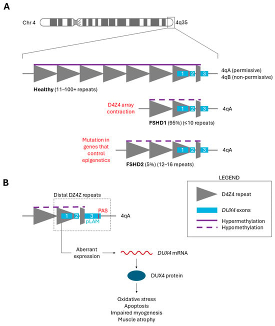
FSHD is a genetic condition with two distinct types. Patients can be classified as having either FSHD1 or FSHD2 depending on the genetic mechanism that results in the de-repression of the D4Z4 macrosatellite repeat array, located in the subtelomeric region 4q35 (Figure 1A). Healthy individuals have 11–100 + 3.3 kb D4Z4 repeat units. FSHD1, affecting 95% of patients, is caused by array contraction to ≤10 repeats []. FSHD2, affecting 5% of patients, is caused by a mutation in genes involved in epigenetic methylation of the D4Z4 array (e.g., SMCHD1, DNMT3B, and LRIF1) [,,]. FSHD1 is diagnosed by assessing the size of the repeat contraction, while FSHD2 diagnosis also requires testing for a mutation in SMCHD1, DNMT3B, and/or LRIF1 []. Curiously, FSHD2 patients also tend to have fewer D4Z4 repeats than healthy individuals (12–16), demonstrating the complexity of this condition and how the distinction between FSHD1 and FSHD2 may not be as straightforward as initially thought []. Regardless, the shared outcome in both types of FSHD is the loss of repressive methylation in the D4Z4 array. Therefore, the clinical presentation is identical between FSHD1 and FSHD2 [].

Figure 1.
(A) A schematic representation of the D4Z4 region in healthy individuals and FSHD patients. The D4Z4 macrosatellite tandem repeat array is found in the subtelomeric region 4q35. Each gray triangle indicates a 3.3 kb D4Z4 repeat unit, within each of which a DUX4 retrogene is contained. The 1st and 2nd DUX4 exons (blue boxes) occur in each full D4Z4 unit. A partial D4Z4 (gray trapezoid) occurs after the most distal complete unit, followed by the 3rd DUX4 exon. Healthy individuals have 11–100+ repeats and full epigenetic repression (purple line). FSHD patients have fewer repeats and compromised epigenetic repression (purple dotted line) arising from one of two genetic changes indicated in red text. (B) A schematic representation of aberrant DUX4 expression from the most distal complete D4Z4 repeat within the FSHD-permissive 4qA haplotype. The 1st and 2nd DUX4 exons are within each D4Z4 unit. The 3rd DUX4 exon and PAS site are found directly downstream of the most distal D4Z4 unit. Compromised repression results in low-level DUX4 protein expression existing within the skeletal muscle, perturbing downstream gene expression to cause the FSHD pathology (oxidative stress, apoptosis, impaired myogenesis, muscle atrophy, etc.).
1.2. DUX4 Is the Central Cause of FSHD
FSHD is caused by the aberrant expression of the double homeobox 4 (DUX4) protein in skeletal muscle, arising from the loss of chromatin repression at the D4Z4 array. The DUX4 gene encodes a transcription factor that is normally involved in zygotic genome activation during the four-cell stage of early embryonic development [,]. Afterward, DUX4 is epigenetically silenced in all adult tissues apart from the limited expression of unrelated DUX4 isoforms in the testis and thymus [,]. Alternative splicing is known to occur for the DUX4 transcript; however, only mis-expression of the full-length isoform in muscle tissue is relevant to FSHD. Any mention hereafter of DUX4 mRNA refers only to this full-length, pathogenic isoform.
Narrowing in on the genetic region from which FSHD arises, we return to the D4Z4 repeat array and the DUX4 gene therein. Within each 3.3 kb D4Z4 repeat unit is a retrogene containing exons 1 and 2 of the DUX4 open reading frame. A partial D4Z4 unit occurs after the most distal complete unit, followed by the 3rd DUX4 exon (Figure 1B) []. Aberrant DUX4 expression occurs from this distal D4Z4 unit in the FSHD-permissive 4qA haplotype. FSHD can only manifest in one of two major 4q allele variants: 4qA and 4qB. Unlike the non-permissive 4qB haplotype, the 4qA haplotype contains the pLAM region with a polyadenylation site (PAS), allowing for the transcription of a stable DUX4 mRNA when epigenetic repression is compromised in the D4Z4 array [,].
Following transcription of the DUX4 gene, stochastic, low-level DUX4 protein expression occurs in the myofiber nuclei of the skeletal muscle. Inappropriate DUX4 protein expression in adult skeletal muscle is highly toxic, driving gene programs that result in oxidative stress, dysregulated transcript quality control, protein aggregation, inflammation, apoptosis, impaired myogenesis, and muscle atrophy (Figure 1B) [,,]. The DUX4 protein directly activates various genes including TRIM43, ZSCAN4, MBD3L2, WFDC3, PRAMEF1, RFPL2, and KHDC1 [,]. Disruption of these signaling pathways by reactivated DUX4 produces the FSHD pathology that we see in patients, manifesting primarily in the muscle tissue. This makes mis-expressed DUX4 the primary therapeutic target for treating FSHD. Sequence-specific oligonucleotides comprise a large portion of targeted therapies that are currently under investigation for FSHD.
1.3. DUX4-Related Challenges for Preclinical Research
While DUX4 is a convenient therapeutic target for FSHD, certain DUX4 characteristics can make it challenging to evaluate treatment efficacy in preclinical experiments. One notable challenge is that the DUX4 protein is very difficult to detect in patient muscle tissue due to its low expression level and infrequent DUX4-positive myonuclei []. As a result, outcome measures concerning treatment efficacy tend to vary between studies. While DUX4 mRNA levels are usually measured to determine post-treatment knockdown, studies often use indirect measures as well, such as DUX4 target gene expression [].
Another important consideration is that the D4Z4 macrosatellite region and the DUX4 gene are restricted to Old World primates, making animal models, murine and otherwise, incapable of exactly recapitulating the expression characteristics and disease phenotype seen in human FSHD patients []. To address this, DUX4 expression must be artificially introduced in these mammalian models. Similarly, non-patient cell models must also introduce DUX4 to evaluate its knockdown by targeted oligonucleotides. However, patient-derived primary or immortalized cell lines, being the most common cellular FSHD model, maintain low-level DUX4 expression [].
1.4. Delivery Methods for DUX4-Targeting Oligonucleotides
As of the writing of this review, several delivery strategies have been used in preclinical studies with DUX4-targeting oligonucleotides. These include Vivo conjugation, fatty acid conjugation (palmitoyl), and adeno-associated virus (AAV) vectors. Firstly, Vivo octa-guanidine dendrimers are a synthetic, cell-penetrating molecule used for the delivery of antisense phosphorodiamidate morpholino oligomers (PMOs) []. Palmitoyl, a type of fatty acid conjugation, is used on antisense oligonucleotides to improve muscle delivery and potency []. Lastly, AAV vectors are small, non-pathogenic DNA viruses capable of efficiently transducing cells for sustained expression of unmodified oligos, such as shRNAs or miRNAs [].
2. Oligonucleotide Therapies Targeting DUX4
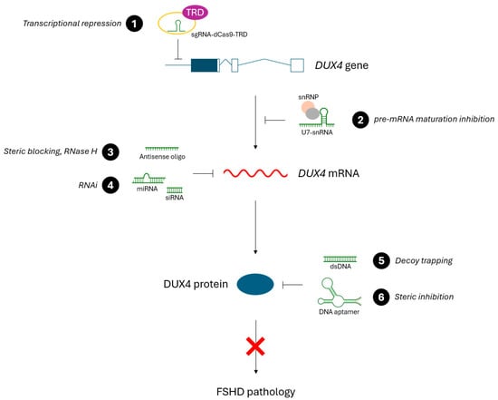
FSHD is a condition arising solely from the aberrant reactivation of a dormant gene: DUX4. Therefore, therapies that directly target aberrant DUX4 expression present a compelling treatment option. This review focuses on preclinical FSHD therapies that use a sequence-specific approach for targeting DUX4. This includes oligonucleotides with sequence complementarity to either the DUX4 gene, DUX4 mRNA, or the DUX4 protein. This complementarity is used to inhibit DUX4 somewhere along its gene > mRNA > protein expression axis, thereby preventing DUX4 transactivation and the resulting FSHD pathology (Figure 2). In this review, we discuss the different types of targeted oligonucleotide therapies for FSHD, summarize the preclinical results obtained so far, and discuss further considerations for these treatment approaches. Notably, the scope of this review is limited to only non-gene-editing approaches that target DUX4.

Figure 2.
An overview of oligonucleotide therapies for FSHD and where they inhibit DUX4 expression (gene, mRNA, or protein) to ameliorate FSHD symptoms. (1) sgRNA-dCas9-TRD targets the promoter or coding region of the DUX4 gene, resulting in transcriptional repression. (2) U7-snRNA alters the specificity of a small nuclear ribonucleoprotein complex (snRNP) to inhibit DUX4 pre-mRNA maturation. (3) Antisense oligonucleotides bind to DUX4 mRNA, causing steric blocking (and downstream effects) or RNase H-mediated degradation, depending on the AO type. (4) Various RNA molecules (miRNA, siRNA, etc.) degrade DUX4 mRNA through the RNA interference pathway. (5) Decoy dsDNA molecules have DUX4-binding motifs that trap the DUX4 protein in a binding sink, inhibiting the transactivation of downstream DUX4 targets. (6) DNA aptamers bind to the DUX4 protein, inhibiting DUX4 activity through steric inhibition.
The targeted oligonucleotide therapies discussed in this review are divided into three categories: antisense oligonucleotides (AOs), RNA interference (RNAi), and other oligonucleotides. These therapies have shown promising preclinical results in cellular and murine models of FSHD, primarily in their ability to lower DUX4 mRNA levels, reduce DUX4 target gene expression, and alleviate FSHD symptoms (Table 1, Table 2 and Table 3).

Table 1.
Overview of preclinical studies investigating antisense oligonucleotides to treat FSHD.

Table 2.
Overview of preclinical studies investigating RNAi-based oligonucleotides to treat FSHD.

Table 3.
Overview of preclinical studies investigating other oligonucleotides to treat FSHD.
2.1. Antisense Oligonucleotides (AOs)
Antisense oligonucleotides (AOs) are synthetic, single-stranded nucleic acids that target a complementary mRNA molecule, dictated by Watson–Crick base pairing, to initiate post-transcriptional gene silencing. AOs were first identified in 1978 by Zamecnik and Stephenson, who found that complementary oligonucleotides inhibited the translation of Rous sarcoma virus mRNA []. AOs bind to a target mRNA in a sequence-dependent manner and prevent its translation, thereby reducing the amount of the target protein [].
AOs are known to utilize various chemistries, a fact that makes them distinct from other oligonucleotide therapies. Modern AO drugs have chemical modifications to improve their pharmacological properties like tolerability, target affinity, nuclease resistance, and intracellular uptake [,]. Commonly used AO chemistries involve modifying the phosphate backbone (PS, phosphorothioate; PMO, phosphorodiamidate morpholino oligomer) or ribose sugar (2′OMe, 2′-O-methyl; 2′-MOE, 2′-O-methoxyethyl; LNA, locked nucleic acid) []. In addition, dendrimer and fatty acid conjugate modifications have been used to facilitate the delivery of DUX4-targeting AOs [,]. AOs can also be synthesized as gapmers, a chimeric molecule comprised of a central DNA region and a flanking region of modified RNA [].
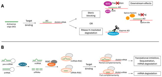
The formation of an AO-mRNA duplex results in (1) RNase H-mediated degradation of the target mRNA or (2) steric blocking of the target mRNA (Figure 3A) [,,]. Only gapmer AOs recruit RNase H to cleave the target mRNA. Gapmer AOs produce a DNA–RNA substrate that when bound to mRNA is recognizable by RNase H [,]. Steric-blocking AOs inhibit proper mRNA translation, splicing, and/or stability in an RNase H-independent manner [,,]. Additionally, these steric-blocking AOs can initiate further downstream degradation pathways, such as nonsense-mediated decay and no-go decay [,].

Figure 3.
The mechanism of action for (A) AO and (B) RNAi therapies. (A) Antisense oligonucleotides (AOs) can degrade target mRNA transcripts by recruiting RNase H, or by inducing steric blocking. AO-mediated steric blocking of a target mRNA transcript can inhibit proper translation, splicing, and/or stability. Further downstream degradation pathways can be initiated on steric-blocked mRNA transcripts. (B) RNAi-based therapies (siRNAs, miRNAs, etc.) degrade target mRNA transcripts using the RNA interference (RNAi) pathway. RNAi can be induced by miRNAs or siRNAs complementary to an mRNA transcript. DICER processes the precursor molecules to produce miRNA or siRNAs, which then are loaded into the Argonaute (AGO) protein of the RNA-induced silencing complex (RISC). Using the guide strand, the RISC targets a complementary mRNA transcript and induces translational inhibition, sequestration, and/or mRNA degradation (miRNA-RISC) or simply mRNA degradation (siRNA-RISC).
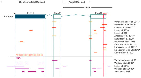
As summarised in Table 1, numerous preclinical studies have demonstrated that AO therapies can effectively reduce the amount of DUX4 mRNA and DUX4 target gene expression both in vitro and in vivo [,,,,,,,,,,,,,]. In particular, demonstrating DUX4 knockdown in vivo represents a notable milestone in the preclinical development of AO therapies, something that has been recently achieved by several research groups [,,,,,,,,]. Other measures were also used to evaluate AO treatment efficacy, such as DUX4 protein levels, muscle fiber health, and murine functional performance. Most groups used primary or immortalized myoblasts/myocytes (often differentiated into myotubes) as an in vitro FSHD model, and FLExDUX4 mice as an in vivo FSHD model [,,]. Earlier studies used local (intramuscular) injection for in vivo AO treatment, while studies after 2021 evaluated systemic (intraperitoneal, subcutaneous) injection routes. Systemic injection, being more clinically viable, managed to yield similarly efficient DUX4 knockdown compared with local injection. Regarding the DUX4 target site, the >10 studies produced since 2011 tend to target exons 2 and 3, often with an emphasis on the polyadenylations site (PAS), pre-mRNA cleavage sites, and/or splice sites therein (Figure 4). Exons 2 and 3 are preferred DUX4 target sites because this region differs from the largely homologous DUX4c gene. This DUX4–DUX4c sequence homology concerns the N-terminal region at the DNA-binding homeobox domains, encoded by exon 1 [].

Figure 4.
An overview of the oligonucleotide target sites on DUX4 mRNA. Orange (antisense oligonucleotides) and purple (RNAi) lines indicate oligo target sites. Partially overlapping target sites are simply represented as a continuous line. All attempted target sites are included, even oligos that showed poor indications in corresponding studies. The figure is not to scale and approximate oligo sites/sizes are shown. (Top) A schematic representation of the DUX4 gene (downwards arrow indicates start codon; blue, open reading frame; boxes, exons; lines, introns; red line, polyadenylation signal). The distal D4Z4 unit, partial D4Z4 unit, and pLAM region are indicated by double-sided arrows. Note that the following groups used some or all of the same oligonucleotides (indicated by *): Vanderplanck et al. (2011), Ansseau et al. (2017), and Derenne et al. (2020) [,,]; Marsollier et al. (2016) and Chen et al. (2016) [,]; Marsollier et al. (2016) and Falzarano et al. (2021) [,]; Lu-Nguyen et al. (2021), Lu-Nguyen et al. (2022a), and Lu-Nguyen et al. (2022b) [,,]; Wallace et al. (2012) and Wallace et al. (2018) [,].
2.2. RNA Interference (RNAi)
Like AOs, RNAi-based oligonucleotides act at the RNA level, binding to a target mRNA according to antisense sequence complementarity to initiate post-transcriptional gene silencing. Where these classifications differ is that RNAi-based oligonucleotides initiate the RNA interference (RNAi) pathway to knockdown target mRNAs. First defined by Fire et al. in 1998, RNAi is a conserved, biological mechanism by which double-stranded RNA triggers the loss of homologous mRNA []. RNAi can be induced by miRNAs or siRNAs complementary to an mRNA transcript. DICER endonucleases cleave precursor molecules (pre-miRNA or shRNA) to produce mature microRNA (miRNA) or small-interfering RNA (siRNA), which then are loaded into the Argonaute (AGO) protein of the RNA-induced silencing complex (RISC). Using the guide strand, the RISC targets a complementary mRNA transcript and induces translational inhibition, sequestration, and/or mRNA degradation (miRNA-RISC), or simply mRNA degradation (siRNA-RISC) (Figure 3B) [,].
The two types of RNAi-based oligonucleotide therapies for FSHD are miRNAs (natural or artificial) and siRNAs. Since 2011, several studies have shown that these RNAi-based oligos can knockdown DUX4 mRNA and reduce DUX4 transactivation, in addition to improving other markers of FSHD symptom reversal (e.g., DUX4 protein levels, muscle fiber health, murine functional performance, etc.) (Table 2) [,,,,,,,,,,,]. Notably, the amount of published work is less for RNAi-based approaches compared with AOs. These studies commonly used primary or immortalized myoblasts/myocytes (often differentiated into myotubes) as an in vitro FSHD model, and AAV-DUX4 mice as an in vivo FSHD model [,,]. FLExDUX4 mice were also used as an in vivo model for testing RNAi therapies, but to a lesser extent []. Most studies used local (intramuscular) injection to evaluate preclinical efficacy in vivo, except for three groups with partially released findings using systemic (intravenous) injection [,,,,]. All current preclinical studies opted for adeno-associated virus (AAV)-mediated delivery of siRNAs or miRNAs in vivo, except for the partially released findings from Avidity Biosciences, Inc. and Dyne Therapeutics, Inc., which each describe a proprietary anti-mTfR1 mAb conjugate for delivery [,,,]. Lastly, as summarized in Figure 4, these miRNAs and siRNAs target DUX4 mRNA at all three of its exons, especially exon 1. Other sites like upstream D4Z4 regions, intronic regions, and pre-mRNA cleavage sites have also been tested.
2.3. Other Oligonucleotides
Other non-gene-editing, preclinical oligonucleotide therapies have also been investigated as potential treatments for FSHD (Table 3). Unlike AO and RNAi therapies which target DUX4 mRNA, these oligos tend to target DUX4 expression at the gene or protein level (Figure 2). These approaches present further compelling options for treating FSHD, in addition to the antisense approaches previously discussed.
2.3.1. CRISPR/dCas9 Transcriptional Repression
Multiple research groups have explored CRISPR/dCas9-mediated transcriptional repression of the DUX4 gene as a targeted therapy for FSHD. This is a form of CRISPR inhibition (CRISPRi) which uses the sequence specificity of the sgRNA-Cas9 complex to target the DUX4 promoter, but with a catalytically inactive ‘dead’ Cas9 (dCas9) fused to a transcriptional repressor domain (TRD) []. This allows for specific re-silencing of the D4Z4 region, reducing DUX4 expression and the resulting FSHD pathology. While various TRDs have been used, most studies opted for the Krüppel-associated box (KRAB) domain. Evaluation of this treatment approach has been largely conducted in primary FSHD myoblasts, myocytes, or myotubes, with the only in vivo testing performed by Himeda et al. (2021) using AAV delivery and local (intramuscular) injection in FLExDUX4 mice []. These studies have all reported a reduction in DUX4 mRNA following treatment, as well as a reduction in DUX4 target gene expression and/or increased H3K9 tri-methylation at the D4Z4 array [,,,,]. Notably, rather than directly repressing the DUX4 gene, Himeda et al. (2018) used the CRISPR/dCas9-KRAB system to repress epigenetic activators of DUX4 (ASH1L, BRD2, KDM4C, SMARCA5). A similar reduction in DUX4 mRNA was observed for this approach [].
Abstracts proposing other non-gene-editing, CRISPR-based approaches have been recently published. The results are preliminary and have not been fully released, however. One group is developing CRISPR/Cas13-mediated cleavage of DUX4 mRNA, reporting effective DUX4 knockdown in vivo []. Another group suggests using CRISPR/Cas13-ADAR (adenosine deaminase acting on RNA)-mediated editing of DUX4 mRNA to create a C > U nonsense mutation []. No definitive results have been published at this time.
2.3.2. DNA Aptamers
Aptamers are single-stranded oligonucleotides that can bind to a specific protein or protein family thanks to their secondary and tertiary folding structure. The unique 3D conformation of an aptamer allows for target interaction like that of an antigen and antibody []. Therefore, aptamers can be used to specifically target a protein of interest and, in the case of FSHD, bind to and inhibit DUX4. Klingler et al. (2020) designed DNA aptamers with a high affinity to the DUX4 protein []. While not evaluated, these DNA aptamers could be used to treat FSHD by sterically inhibiting the DUX4 protein in skeletal muscle.
2.3.3. dsDNA Decoy Trapping
Mariot et al. (2020) demonstrated a unique approach to prevent DUX4 transactivation known as decoy trapping []. Decoy trapping uses double-stranded DNA fragments whose sequence corresponds to DUX4 binding motifs, akin to the DNA regions that DUX4 normally binds to as a transcription factor. By saturating the cellular environment with dsDNA decoy binding sites, the DUX4 protein is trapped in a binding sink and unable to activate its normal target genes. Mariot et al. (2020) found that dsDNA treatment was able to reduce the expression of downstream DUX4 target genes in vitro and in vivo [].
2.3.4. U7-snRNA Pre-mRNA Inhibition
Rashnonejad et al. (2021) describe a strategy to inhibit DUX4 mRNA expression using U7-small nuclear RNA (snRNA) antisense expression cassettes []. U7-snRNA is a part of the small nuclear ribonucleoprotein complex (snRNP), which is involved in 3′ end processing of histone pre-mRNAs in the nucleus. This therapeutic approach uses modified U7-snRNA with antisense sequence specificity to DUX4, capable of inhibiting pre-mRNA production or maturation. Rashnonejad et al. (2021) showed that these U7-snRNA expression cassettes, delivered by AAV, effectively reduced DUX4 mRNA, the DUX4 protein, and DUX4 target gene expression in immortalized FSHD myotubes [].
3. Further Considerations
Antisense therapies for FSHD, meaning AOs or RNAi drugs that target DUX4 mRNA, are the furthest along in preclinical development compared to other oligonucleotide approaches. Many AO and RNAi therapies have shown promising indications both in vitro and in vivo, as discussed previously. Therefore, this discussion of certain advantages and disadvantages of DUX4-targeting oligonucleotide therapies will focus on antisense approaches only.
3.1. Advantages of Antisense Approaches
Antisense therapies are ideal for monogenic diseases that can be attributed to a single root cause. In the case of FSHD, this is aberrant DUX4 expression in skeletal muscle. Another advantage of antisense therapies is that they are highly specific and employ potent molecules with a relatively simple mechanism of action, often taking advantage of conserved cellular processes [].
Additionally, compared with gene editing approaches that prevent DUX4 expression, antisense therapies involve no changes to genomic DNA, acting only at the RNA level [,]. This makes them more acceptable from a regulatory standpoint, unburdened by the moral concern surrounding CRISPR/Cas9 editing of the human genome, even if for therapeutic purposes. For this reason, it may be fair to suggest that antisense therapies are a more clinically viable form of targeted, genetic therapy for FSHD.
3.2. Disadvantages of Antisense Approaches
Efficient delivery to muscle tissue is a considerable challenge for antisense therapies, often hindering the clinical utility of oligonucleotides that otherwise demonstrate good preclinical efficacy. AOs and RNAi oligonucleotides are relatively large nucleic acids that tend to be negatively charged and hydrophilic []. Molecules with such properties do not readily pass through the plasma membrane. Furthermore, upon systemic injection, these molecules must avoid nuclease degradation, mononuclear phagocyte system entrapment, protein entrapment, and high renal clearance [,,]. If these can be overcome, there remains the issue of inefficient cellular uptake, as these oligonucleotides are also prone to endosomal entrapment within the cell [,,]. All this means that only a small percentage of the injected drug becomes bioavailable to provide therapeutic benefit to a patient. However, various strategies to improve delivery are currently being explored for AOs and RNAi oligos, including chemical modification, delivery conjugates, and carrier molecules [].
The persistence of the therapeutic effect is another notable disadvantage of antisense therapies. Given that antisense therapies act on mRNA, a transient and replenishable molecule, regular lifelong administrations may be necessary to offer long-term reversal of FSHD symptoms for patients. This problem is not shared by other proposed genetic therapies that would permanently inactivate the toxic DUX4 gene (e.g., CRISPR/Cas9 editing) such that multiple treatments are not needed.
3.3. Early-Stage Clinical Trials for Select FSHD Therapies
Two RNAi-based oligonucleotide therapies for FSHD are currently recruiting for Phase 1/2 clinical trials to evaluate their safety, tolerability, pharmacokinetics, pharmacodynamics, and efficacy in adult patients. First, ARO-DUX4, developed by Arrowhead Pharmaceuticals, is a DUX4-specific siRNA using an unspecified and proprietary delivery method (Phase 1/2 NCT06131983) [,]. Second, AOC 1020, developed by Avidity Biosciences, is a DUX4-specific siRNA using a proprietary anti-mTfR1 mAb delivery conjugate (Phase 1/2 FORTITUDE™ NCT0574792) [,]. Both therapies have previously demonstrated preclinical efficacy in cellular and murine models of FSHD [,,,].
4. Conclusions
Since DUX4 was identified as the central cause of FSHD, numerous targeted oligonucleotide therapies have been proposed, many of which have shown promising results in preclinical stages. However, despite DUX4 presenting itself as an ideal therapeutic target, there are still considerable challenges that may prevent these therapies from reaching clinical use and benefiting patients. First, more progress towards fully characterizing FSHD is needed, as it remains an incredibly complicated condition with many unanswered questions. Further research into the molecular underpinnings of FSHD may offer additional therapeutic targets amenable to oligonucleotide therapies. Similarly, it is important to continue investigating the normal physiological role of DUX4 in the testis and thymus, as this is not fully understood and could impact decisions made when targeting DUX4 in skeletal muscle, possibly in terms of off-target effects.
Another consideration would be the potential synergistic effect of combining multiple therapies, particularly those that target DUX4 expression via different modes of action. While this has not yet been attempted for FSHD, combined therapies have been explored considerably for Duchenne muscular dystrophy (DMD), with certain combinations improving treatment efficiency. In particular, proposed combination treatment strategies for DMD often use one therapy that corrects the genetic defect (e.g., AO) and another that addresses secondary disease manifestations [].
Overall, with a strong pipeline of candidate oligos from many different research groups and two siRNA drugs entering early clinical trials, the future appears hopeful for a targeted treatment option for patients with FSHD.
Author Contributions
Conception and design, S.L.B.; literature review and writing—original draft preparation, S.L.B.; writing—review and editing, S.L.B. and T.Y.; supervision and funding acquisition, T.Y. All authors have read and agreed to the published version of the manuscript.
Funding
No specific grant support was received for this study. T.Y. is supported by Muscular Dystrophy Canada (MD423), the Friends of Garrett Cumming Research Fund (MD423), the HM Toupin Neurological Science Research Fund (MD423), the Canadian Institutes of Health Research (CIHR) (PS 169193, PS 175261, PS 180495, PS 183719, PS 183733), the Women and Children’s Health Research Institute (WCHRI) (RES0068823), US Department of Defense (USDD W81XWH2210801), Gilbert K Winter Fund (RES0053826), Heart and Stroke Foundation Canada (HSFC GIA G-21-0031582).
Data Availability Statement
Not applicable.
Acknowledgments
We would like to acknowledge Saeed Anwar (Department of Medical Genetics, University of Alberta, Edmonton, AB, Canada) for providing advice and guidance throughout the writing of this review.
Conflicts of Interest
T.Y. is a co-founder and shareholder of OligomicsTx Inc., which aims to commercialize antisense technology. S.L.B. declares that this study was conducted in the absence of any commercial or financial relationships that could be construed as potential conflicts of interest.
References
- Wang, L.H.; Tawil, R. Facioscapulohumeral Dystrophy. Curr. Neurol. Neurosci. Rep. 2016, 16, 66. [Google Scholar] [CrossRef]
- Mostacciuolo, M.; Pastorello, E.; Vazza, G.; Miorin, M.; Angelini, C.; Tomelleri, G.; Galluzzi, G.; Trevisan, C. Facioscapulohumeral Muscular Dystrophy: Epidemiological and Molecular Study in a North-east Italian Population Sample. Clin. Genet. 2009, 75, 550–555. [Google Scholar] [CrossRef]
- Deenen, J.C.W.; Arnts, H.; Van Der Maarel, S.M.; Padberg, G.W.; Verschuuren, J.J.G.M.; Bakker, E.; Weinreich, S.S.; Verbeek, A.L.M.; Van Engelen, B.G.M. Population-Based Incidence and Prevalence of Facioscapulohumeral Dystrophy. Neurology 2014, 83, 1056–1059. [Google Scholar] [CrossRef]
- Mul, K.; Lassche, S.; Voermans, N.C.; Padberg, G.W.; Horlings, C.G.; Van Engelen, B.G. What’s in a Name? The Clinical Features of Facioscapulohumeral Muscular Dystrophy. Pract. Neurol. 2016, 16, 201–207. [Google Scholar] [CrossRef] [PubMed]
- Aguirre, A.S.; Astudillo Moncayo, O.M.; Mosquera, J.; Muyolema Arce, V.E.; Gallegos, C.; Ortiz, J.F.; Andrade, A.F.; Oña, S.; Buj, M.J. Treatment of Facioscapulohumeral Muscular Dystrophy (FSHD): A Systematic Review. Cureus 2023, 15, e39903. [Google Scholar] [CrossRef] [PubMed]
- Lemmers, R.J.L.F.; Van Der Vliet, P.J.; Klooster, R.; Sacconi, S.; Camaño, P.; Dauwerse, J.G.; Snider, L.; Straasheijm, K.R.; Jan Van Ommen, G.; Padberg, G.W.; et al. A Unifying Genetic Model for Facioscapulohumeral Muscular Dystrophy. Science 2010, 329, 1650–1653. [Google Scholar] [CrossRef] [PubMed]
- Lemmers, R.J.L.F.; Tawil, R.; Petek, L.M.; Balog, J.; Block, G.J.; Santen, G.W.E.; Amell, A.M.; Van Der Vliet, P.J.; Almomani, R.; Straasheijm, K.R.; et al. Digenic Inheritance of an SMCHD1 Mutation and an FSHD-Permissive D4Z4 Allele Causes Facioscapulohumeral Muscular Dystrophy Type 2. Nat. Genet. 2012, 44, 1370–1374. [Google Scholar] [CrossRef]
- van den Boogaard, M.L.; Lemmers, R.J.L.F.; Balog, J.; Wohlgemuth, M.; Auranen, M.; Mitsuhashi, S.; van der Vliet, P.J.; Straasheijm, K.R.; van den Akker, R.F.P.; Kriek, M.; et al. Mutations in DNMT3B Modify Epigenetic Repression of the D4Z4 Repeat and the Penetrance of Facioscapulohumeral Dystrophy. Am. J. Hum. Genet. 2016, 98, 1020–1029. [Google Scholar] [CrossRef] [PubMed]
- Hamanaka, K.; Šikrová, D.; Mitsuhashi, S.; Masuda, H.; Sekiguchi, Y.; Sugiyama, A.; Shibuya, K.; Lemmers, R.J.L.F.; Goossens, R.; Ogawa, M.; et al. Homozygous Nonsense Variant in LRIF1 Associated with Facioscapulohumeral Muscular Dystrophy. Neurology 2020, 94, e2441–e2447. [Google Scholar] [CrossRef]
- Sacconi, S.; Briand-Suleau, A.; Gros, M.; Baudoin, C.; Lemmers, R.J.L.F.; Rondeau, S.; Lagha, N.; Nigumann, P.; Cambieri, C.; Puma, A.; et al. FSHD1 and FSHD2 Form a Disease Continuum. Neurology 2019, 92, e2273–e2285. [Google Scholar] [CrossRef]
- Hendrickson, P.G.; Doráis, J.A.; Grow, E.J.; Whiddon, J.L.; Lim, J.-W.; Wike, C.L.; Weaver, B.D.; Pflueger, C.; Emery, B.R.; Wilcox, A.L.; et al. Conserved Roles for Murine DUX and Human DUX4 in Activating Cleavage Stage Genes and MERVL/HERVL Retrotransposons. Nat. Genet. 2017, 49, 925–934. [Google Scholar]
- De Iaco, A.; Planet, E.; Coluccio, A.; Verp, S.; Duc, J.; Trono, D. DUX-Family Transcription Factors Regulate Zygotic Genome Activation in Placental Mammals. Nat. Genet. 2017, 49, 941–945. [Google Scholar] [CrossRef]
- Snider, L.; Geng, L.N.; Lemmers, R.J.L.F.; Kyba, M.; Ware, C.B.; Nelson, A.M.; Tawil, R.; Filippova, G.N.; Van Der Maarel, S.M.; Tapscott, S.J.; et al. Facioscapulohumeral Dystrophy: Incomplete Suppression of a Retrotransposed Gene. PLoS Genet. 2010, 6, e1001181. [Google Scholar] [CrossRef]
- Das, S.; Chadwick, B.P. Influence of Repressive Histone and DNA Methylation upon D4Z4 Transcription in Non-Myogenic Cells. PLoS ONE 2016, 11, e0160022. [Google Scholar] [CrossRef]
- Himeda, C.L.; Jones, P.L. The Genetics and Epigenetics of Facioscapulohumeral Muscular Dystrophy. Annu. Rev. Genom. Hum. Genet. 2019, 20, 265–291. [Google Scholar] [CrossRef]
- Lim, K.R.Q.; Nguyen, Q.; Yokota, T. DUX4 Signalling in the Pathogenesis of Facioscapulohumeral Muscular Dystrophy. Int. J. Mol. Sci. 2020, 21, 729. [Google Scholar] [CrossRef]
- Mocciaro, E.; Runfola, V.; Ghezzi, P.; Pannese, M.; Gabellini, D. DUX4 Role in Normal Physiology and in FSHD Muscular Dystrophy. Cells 2021, 10, 3322. [Google Scholar] [CrossRef]
- Geng, L.N.; Yao, Z.; Snider, L.; Fong, A.P.; Cech, J.N.; Young, J.M.; van der Maarel, S.M.; Ruzzo, W.L.; Gentleman, R.C.; Tawil, R.; et al. DUX4 Activates Germline Genes, Retroelements, and Immune Mediators: Implications for Facioscapulohumeral Dystrophy. Dev. Cell 2012, 22, 38–51. [Google Scholar] [CrossRef] [PubMed]
- Yao, Z.; Snider, L.; Balog, J.; Lemmers, R.J.L.F.; Van Der Maarel, S.M.; Tawil, R.; Tapscott, S.J. DUX4-Induced Gene Expression Is the Major Molecular Signature in FSHD Skeletal Muscle. Hum. Mol. Genet. 2014, 23, 5342–5352. [Google Scholar] [CrossRef] [PubMed]
- Clapp, J.; Mitchell, L.M.; Bolland, D.J.; Fantes, J.; Corcoran, A.E.; Scotting, P.J.; Armour, J.A.L.; Hewitt, J.E. Evolutionary Conservation of a Coding Function for D4Z4, the Tandem DNA Repeat Mutated in Facioscapulohumeral Muscular Dystrophy. Am. J. Hum. Genet. 2007, 81, 264–279. [Google Scholar] [CrossRef]
- DeSimone, A.M.; Cohen, J.; Lek, M.; Lek, A. Cellular and Animal Models for Facioscapulohumeral Muscular Dystrophy. Dis. Models Mech. 2020, 13, dmm046904. [Google Scholar] [CrossRef]
- Morcos, P.A.; Li, Y.; Jiang, S. Vivo-Morpholinos: A Non-Peptide Transporter Delivers Morpholinos into a Wide Array of Mouse Tissues. BioTechniques 2008, 45, 613–623. [Google Scholar] [CrossRef]
- Prakash, T.P.; Mullick, A.E.; Lee, R.G.; Yu, J.; Yeh, S.T.; Low, A.; Chappell, A.E.; Østergaard, M.E.; Murray, S.; Gaus, H.J.; et al. Fatty Acid Conjugation Enhances Potency of Antisense Oligonucleotides in Muscle. Nucleic Acids Res. 2019, 47, 6029–6044. [Google Scholar] [CrossRef] [PubMed]
- Borel, F.; Kay, M.A.; Mueller, C. Recombinant AAV as a Platform for Translating the Therapeutic Potential of RNA Interfer-ence. Mol. Ther. 2014, 22, 692–701. [Google Scholar] [CrossRef] [PubMed]
- Vanderplanck, C.; Ansseau, E.; Charron, S.; Stricwant, N.; Tassin, A.; Laoudj-Chenivesse, D.; Wilton, S.D.; Coppée, F.; Belayew, A. The FSHD Atrophic Myotube Phenotype Is Caused by DUX4 Expression. PLoS ONE 2011, 6, e26820. [Google Scholar] [CrossRef]
- Lim, J.-W.; Snider, L.; Yao, Z.; Tawil, R.; Van Der Maarel, S.M.; Rigo, F.; Bennett, C.F.; Filippova, G.N.; Tapscott, S.J. DICER/AGO-Dependent Epigenetic Silencing of D4Z4 Repeats Enhanced by Exogenous siRNA Suggests Mechanisms and Therapies for FSHD. Hum. Mol. Genet. 2015, 24, 4817–4828. [Google Scholar] [CrossRef]
- Marsollier, A.-C.; Ciszewski, L.; Mariot, V.; Popplewell, L.; Voit, T.; Dickson, G.; Dumonceaux, J. Antisense Targeting of 3′ End Elements Involved in DUX4 mRNA Processing Is an Efficient Therapeutic Strategy for Facioscapulohumeral Dystrophy: A New Gene-Silencing Approach. Hum. Mol. Genet. 2016, 25, 1468–1478. [Google Scholar] [CrossRef] [PubMed]
- Chen, J.C.; King, O.D.; Zhang, Y.; Clayton, N.P.; Spencer, C.; Wentworth, B.M.; Emerson, C.P.; Wagner, K.R. Morpholino-Mediated Knockdown of DUX4 Toward Facioscapulohumeral Muscular Dystrophy Therapeutics. Mol. Ther. 2016, 24, 1405–1411. [Google Scholar] [CrossRef]
- Ansseau, E.; Vanderplanck, C.; Wauters, A.; Harper, S.; Coppée, F.; Belayew, A. Antisense Oligonucleotides Used to Target the DUX4 mRNA as Therapeutic Approaches in FaciosScapuloHumeral Muscular Dystrophy (FSHD). Genes 2017, 8, 93. [Google Scholar] [CrossRef]
- Derenne, A.; Tassin, A.; Nguyen, T.H.; De Roeck, E.; Jenart, V.; Ansseau, E.; Belayew, A.; Coppée, F.; Declèves, A.-E.; Legrand, A. Induction of a Local Muscular Dystrophy Using Electroporation in Vivo: An Easy Tool for Screening Therapeutics. Sci. Rep. 2020, 10, 11301. [Google Scholar] [CrossRef]
- Lim, K.R.Q.; Maruyama, R.; Echigoya, Y.; Nguyen, Q.; Zhang, A.; Khawaja, H.; Sen Chandra, S.; Jones, T.; Jones, P.; Chen, Y.-W.; et al. Inhibition of DUX4 Expression with Antisense LNA Gapmers as a Therapy for Facioscapulohumeral Muscular Dystrophy. Proc. Natl. Acad. Sci. USA 2020, 117, 16509–16515. [Google Scholar] [CrossRef] [PubMed]
- Lim, K.R.Q.; Bittel, A.; Maruyama, R.; Echigoya, Y.; Nguyen, Q.; Huang, Y.; Dzierlega, K.; Zhang, A.; Chen, Y.-W.; Yokota, T. DUX4 Transcript Knockdown with Antisense 2′-O-Methoxyethyl Gapmers for the Treatment of Facioscapulohumeral Muscular Dystrophy. Mol. Ther. 2021, 29, 848–858. [Google Scholar] [CrossRef] [PubMed]
- Bouwman, L.F.; Den Hamer, B.; Van Den Heuvel, A.; Franken, M.; Jackson, M.; Dwyer, C.A.; Tapscott, S.J.; Rigo, F.; Van Der Maarel, S.M.; De Greef, J.C. Systemic Delivery of a DUX4-Targeting Antisense Oligonucleotide to Treat Facioscapulohumeral Muscular Dystrophy. Mol. Ther. Nucleic Acids 2021, 26, 813–827. [Google Scholar] [CrossRef]
- Falzarano, M.S.; Argenziano, M.; Marsollier, A.C.; Mariot, V.; Rossi, D.; Selvatici, R.; Dumonceaux, J.; Cavalli, R.; Ferlini, A. Chitosan-Shelled Nanobubbles Irreversibly Encapsulate Morpholino Conjugate Antisense Oligonucleotides and Are Ineffective for Phosphorodiamidate Morpholino-Mediated Gene Silencing of DUX4. Nucleic Acid. Ther. 2021, 31, 201–207. [Google Scholar] [CrossRef] [PubMed]
- Lu-Nguyen, N.; Malerba, A.; Herath, S.; Dickson, G.; Popplewell, L. Systemic Antisense Therapeutics Inhibiting DUX4 Expression Ameliorates FSHD-like Pathology in an FSHD Mouse Model. Hum. Mol. Genet. 2021, 30, 1398–1412. [Google Scholar] [CrossRef] [PubMed]
- Lu-Nguyen, N.; Malerba, A.; Antoni Pineda, M.; Dickson, G.; Popplewell, L. Improving Molecular and Histopathology in Diaphragm Muscle of the Double Transgenic ACTA1-MCM/FLExDUX4 Mouse Model of FSHD with Systemic Antisense Therapy. Hum. Gene Ther. 2022, 33, 923–935. [Google Scholar] [CrossRef] [PubMed]
- Lu-Nguyen, N.; Dickson, G.; Malerba, A.; Popplewell, L. Long-Term Systemic Treatment of a Mouse Model Displaying Chronic FSHD-like Pathology with Antisense Therapeutics That Inhibit DUX4 Expression. Biomedicines 2022, 10, 1623. [Google Scholar] [CrossRef]
- Kakimoto, T.; Ogasawara, A.; Ishikawa, K.; Kurita, T.; Yoshida, K.; Harada, S.; Nonaka, T.; Inoue, Y.; Uchida, K.; Tateoka, T.; et al. A Systemically Administered Unconjugated Antisense Oligonucleotide Targeting DUX4 Improves Muscular Injury and Motor Function in FSHD Model Mice. Biomedicines 2023, 11, 2339. [Google Scholar] [CrossRef] [PubMed]
- Wallace, L.M.; Liu, J.; Domire, J.S.; Garwick-Coppens, S.E.; Guckes, S.M.; Mendell, J.R.; Flanigan, K.M.; Harper, S.Q. RNA Interference Inhibits DUX4-Induced Muscle Toxicity In Vivo: Implications for a Targeted FSHD Therapy. Mol. Ther. 2012, 20, 1417–1423. [Google Scholar] [CrossRef]
- Wallace, L.M.; Saad, N.Y.; Pyne, N.K.; Fowler, A.M.; Eidahl, J.O.; Domire, J.S.; Griffin, D.A.; Herman, A.C.; Sahenk, Z.; Rodino-Klapac, L.R.; et al. Pre-Clinical Safety and Off-Target Studies to Support Translation of AAV-Mediated RNAi Therapy for FSHD. Mol. Ther. Methods Clin. Dev. 2018, 8, 121–130. [Google Scholar] [CrossRef]
- Saad, N.Y.; Al-Kharsan, M.; Garwick-Coppens, S.E.; Chermahini, G.A.; Harper, M.A.; Palo, A.; Boudreau, R.L.; Harper, S.Q. Human miRNA miR-675 Inhibits DUX4 Expression and May Be Exploited as a Potential Treatment for Facioscapulohumeral Muscular Dystrophy. Nat. Commun. 2021, 12, 7128. [Google Scholar] [CrossRef] [PubMed]
- Arrowhead Pharmaceuticals, Inc. Arrowhead Presents Preclinical Data on ARO-DUX4 at FSHD Society International Research Congress. 2021. Available online: https://ir.arrowheadpharma.com/news-releases/news-release-details/arrowhead-presents-preclinical-data-aro-dux4-fshd-society (accessed on 29 June 2024).
- Jagannathan, S.; De Greef, J.C.; Hayward, L.J.; Yokomori, K.; Gabellini, D.; Mul, K.; Sacconi, S.; Arjomand, J.; Kinoshita, J.; Harper, S.Q. Meeting Report: The 2021 FSHD International Research Congress. Skelet. Muscle 2022, 12, 1. [Google Scholar] [CrossRef] [PubMed]
- Mariot, V.; Sidlauskaite, E.; Gall, L.L.; Corbex, E.; Dumonceaux, J. VP. 61 An AAV-shRNA DUX4-Based Therapy to Treat Facioscapulohumeral Muscular Dystrophy (FSHD). Neuromuscul. Disord. 2022, 32, S107. [Google Scholar] [CrossRef]
- Malecova, B.; Sala, D.; Melikian, G.M.; Erdogan, G.; Johns, R.; Young, J.; Ventre, E.; Gatto, S.; Onorato, M.; Pavlicek, A.; et al. DUX4 siRNA Optimization for the Development of an Antibody-Oligonucleotide Conjugate (AOC) for the Treatment of FSHD (P17-13.009). Neurology 2022, 98, 1776. [Google Scholar] [CrossRef]
- Avidity Biosciences, Inc. Targeting DUX4 for Silencing with AOC for the Treatment of FSHD. 2024. Available online: https://www.aviditybiosciences.com/wp-content/uploads/2024/03/MDA-2024-FSHD-RNASeq_v6.0_STC.pdf (accessed on 29 June 2024).
- Dyne Therapeutics, Inc. Dyne Therapeutics Presents New Preclinical Data for its Facioscapulohumeral Muscular Dystrophy Program During the FSHD Society International Research Congress. 2024. Available online: https://investors.dyne-tx.com/news-releases/news-release-details/dyne-therapeutics-presents-new-preclinical-data-its (accessed on 14 July 2024).
- Dyne Therapeutics, Inc. The FORCETM Platform Achieves Robust and Durable DUX4 Suppression and Functional Benefit in FSHD Mouse Models. 2024. Available online: https://www.dyne-tx.com/wp-content/uploads/Natoli_FSHDIRC_June2024.pdf (accessed on 14 July 2024).
- Himeda, C.L.; Jones, T.I.; Jones, P.L. CRISPR/dCas9-Mediated Transcriptional Inhibition Ameliorates the Epigenetic Dysregulation at D4Z4 and Represses DUX4-Fl in FSH Muscular Dystrophy. Mol. Ther. 2016, 24, 527–535. [Google Scholar] [CrossRef]
- Himeda, C.L.; Jones, T.I.; Virbasius, C.-M.; Zhu, L.J.; Green, M.R.; Jones, P.L. Identification of Epigenetic Regulators of DUX4-Fl for Targeted Therapy of Facioscapulohumeral Muscular Dystrophy. Mol. Ther. 2018, 26, 1797–1807. [Google Scholar] [CrossRef]
- Klingler, C.; Ashley, J.; Shi, K.; Stiefvater, A.; Kyba, M.; Sinnreich, M.; Aihara, H.; Kinter, J. DNA Aptamers against the DUX4 Protein Reveal Novel Therapeutic Implications for FSHD. FASEB J. 2020, 34, 4573–4590. [Google Scholar] [CrossRef]
- Mariot, V.; Joubert, R.; Marsollier, A.-C.; Hourdé, C.; Voit, T.; Dumonceaux, J. A Deoxyribonucleic Acid Decoy Trapping DUX4 for the Treatment of Facioscapulohumeral Muscular Dystrophy. Mol. Ther. Nucleic Acids 2020, 22, 1191–1199. [Google Scholar] [CrossRef]
- Himeda, C.L.; Jones, T.I.; Jones, P.L. Targeted Epigenetic Repression by CRISPR/dSaCas9 Suppresses Pathogenic DUX4-Fl Expression in FSHD. Mol. Ther. Methods Clin. Dev. 2021, 20, 298–311. [Google Scholar] [CrossRef]
- Das, S.; Chadwick, B.P. CRISPR Mediated Targeting of DUX4 Distal Regulatory Element Represses DUX4 Target Genes Dysregulated in Facioscapulohumeral Muscular Dystrophy. Sci. Rep. 2021, 11, 12598. [Google Scholar] [CrossRef]
- Rashnonejad, A.; Amini-Chermahini, G.; Taylor, N.K.; Wein, N.; Harper, S.Q. Designed U7 snRNAs Inhibit DUX4 Expression and Improve FSHD-Associated Outcomes in DUX4 Overexpressing Cells and FSHD Patient Myotubes. Mol. Ther. Nucleic Acids 2021, 23, 476–486. [Google Scholar] [CrossRef]
- Rashnonejad, A.; Amini-Chermahini, G.; Taylor, N.; Fowler, A.; Kraus, E.; King, O.; Harper, S. FP. 29 AAV-CRISPR-Cas13 Gene Therapy for FSHD: DUX4 Gene Silencing Efficacy and Immune Responses to Cas13b Protein. Neuromuscul. Disord. 2022, 32, S103–S104. [Google Scholar] [CrossRef]
- Saljoughian, N.; Rizzotto, L.; Sezgin, Y.; Faraji, H.; Wallace, L.; Kararoudi, M.N.; Palmieri, D.; Harper, S.P. 144 Developing Cas13-ADAR-Mediated DUX4 mRNA Editing as a Prospective Therapy for FSHD. Neuromuscul. Disord. 2022, 32, S106. [Google Scholar] [CrossRef]
- Sasaki-Honda, M.; Jonouchi, T.; Arai, M.; He, J.; Okita, K.; Sakurai, S.; Yamamoto, T.; Sakurai, H. Hit-and-Run Silencing of Endogenous DUX4 by Targeting DNA Hypomethylation on D4Z4 Repeats in Facioscapulohumeral Muscular Dystrophy. bioRxiv 2022. [CrossRef]
- Stephenson, M.L.; Zamecnik, P.C. Inhibition of Rous Sarcoma Viral RNA Translation by a Specific Oligodeoxyribonucleotide. Proc. Natl. Acad. Sci. USA 1978, 75, 285–288. [Google Scholar] [CrossRef]
- Dias, N.; Stein, C.A. Antisense Oligonucleotides: Basic Concepts and Mechanisms. Mol. Cancer Ther. 2002, 1, 347–355. [Google Scholar] [PubMed]
- Khvorova, A.; Watts, J.K. The Chemical Evolution of Oligonucleotide Therapies of Clinical Utility. Nat. Biotechnol. 2017, 35, 238–248. [Google Scholar] [CrossRef]
- Shah, N.J. Antisense Oligonucleotides. In Introduction to Basics of Pharmacology and Toxicology; Raj, G.M., Raveendran, R., Eds.; Springer: Singapore, 2019; pp. 407–411. ISBN 978-981-329-778-4. [Google Scholar]
- Monia, B.P.; Lesnik, E.A.; Gonzalez, C.; Lima, W.F.; McGee, D.; Guinosso, C.J.; Kawasaki, A.M.; Cook, P.D.; Freier, S.M. Evaluation of 2′-Modified Oligonucleotides Containing 2‘-Deoxy Gaps as Antisense Inhibitors of Gene Expression. J. Biol. Chem. 1993, 268, 14514–14522. [Google Scholar] [CrossRef]
- Crooke, S.T.; Baker, B.F.; Crooke, R.M.; Liang, X. Antisense Technology: An Overview and Prospectus. Nat. Rev. Drug Discov. 2021, 20, 427–453. [Google Scholar] [CrossRef]
- Minshull, J.; Hunt, T. The Use of Single-Stranded DNA and RNase H to Promote Quantitative ‘Hybrid Arrest of Translation’ of mRNA/DNA Hybrids in Reticulocyte Lysate Cell-Free Translations. Nucl. Acids Res. 1986, 14, 6433–6451. [Google Scholar] [CrossRef]
- Nakamura, H.; Oda, Y.; Iwai, S.; Inoue, H.; Ohtsuka, E.; Kanaya, S.; Kimura, S.; Katsuda, C.; Katayanagi, K.; Morikawa, K. How Does RNase H Recognize a DNA.RNA Hybrid? Proc. Natl. Acad. Sci. USA 1991, 88, 11535–11539. [Google Scholar] [CrossRef]
- Ward, A.J.; Norrbom, M.; Chun, S.; Bennett, C.F.; Rigo, F. Nonsense-Mediated Decay as a Terminating Mechanism for Antisense Oligonucleotides. Nucleic Acids Res. 2014, 42, 5871–5879. [Google Scholar] [CrossRef]
- Liang, X.; Nichols, J.G.; Hsu, C.-W.; Vickers, T.A.; Crooke, S.T. mRNA Levels Can Be Reduced by Antisense Oligonucleotides via No-Go Decay Pathway. Nucleic Acids Res. 2019, 47, 6900–6916. [Google Scholar] [CrossRef]
- Stadler, G.; Chen, J.C.; Wagner, K.; Robin, J.D.; Shay, J.W.; Emerson, C.P.; Wright, W.E. Establishment of Clonal Myogenic Cell Lines from Severely Affected Dystrophic Muscles—CDK4 Maintains the Myogenic Population. Skelet. Muscle 2011, 1, 12. [Google Scholar] [CrossRef] [PubMed]
- Krom, Y.D.; Dumonceaux, J.; Mamchaoui, K.; Den Hamer, B.; Mariot, V.; Negroni, E.; Geng, L.N.; Martin, N.; Tawil, R.; Tapscott, S.J.; et al. Generation of Isogenic D4Z4 Contracted and Noncontracted Immortal Muscle Cell Clones from a Mosaic Patient. Am. J. Pathol. 2012, 181, 1387–1401. [Google Scholar] [CrossRef]
- Jones, T.; Jones, P.L. A Cre-Inducible DUX4 Transgenic Mouse Model for Investigating Facioscapulohumeral Muscular Dystrophy. PLoS ONE 2018, 13, e0192657. [Google Scholar] [CrossRef]
- Ansseau, E.; Eidahl, J.O.; Lancelot, C.; Tassin, A.; Matteotti, C.; Yip, C.; Liu, J.; Leroy, B.; Hubeau, C.; Gerbaux, C.; et al. Ho-mologous Transcription Factors DUX4 and DUX4c Associate with Cytoplasmic Proteins during Muscle Differentiation. PLoS ONE 2016, 11, e0146893. [Google Scholar] [CrossRef]
- Fire, A.; Xu, S.; Montgomery, M.K.; Kostas, S.A.; Driver, S.E.; Mello, C.C. Potent and Specific Genetic Interference by Double-Stranded RNA In. Nature 1998, 391, 806–811. [Google Scholar]
- Wilson, R.C.; Doudna, J.A. Molecular Mechanisms of RNA Interference. Annu. Rev. Biophys. 2013, 42, 217–239. [Google Scholar] [CrossRef] [PubMed]
- Wallace, L.M.; Garwick, S.E.; Mei, W.; Belayew, A.; Coppee, F.; Ladner, K.J.; Guttridge, D.; Yang, J.; Harper, S.Q. DUX4, a Candidate Gene for Facioscapulohumeral Muscular Dystrophy, Causes P53-dependent Myopathy in Vivo. Ann. Neurol. 2011, 69, 540–552. [Google Scholar] [CrossRef]
- Larson, M.H.; Gilbert, L.A.; Wang, X.; Lim, W.A.; Weissman, J.S.; Qi, L.S. CRISPR Interference (CRISPRi) for Sequence-Specific Control of Gene Expression. Nat. Protoc. 2013, 8, 2180–2196. [Google Scholar] [CrossRef]
- Di Mauro, V.; Lauta, F.C.; Modica, J.; Appleton, S.L.; De Franciscis, V.; Catalucci, D. Diagnostic and Therapeutic Aptamers. JACC Basic. Transl. Sci. 2024, 9, 260–277. [Google Scholar] [CrossRef] [PubMed]
- Roberts, T.C.; Langer, R.; Wood, M.J.A. Advances in Oligonucleotide Drug Delivery. Nat. Rev. Drug Discov. 2020, 19, 673–694. [Google Scholar] [CrossRef]
- Gagliardi, M.; Ashizawa, A.T. The Challenges and Strategies of Antisense Oligonucleotide Drug Delivery. Biomedicines 2021, 9, 433. [Google Scholar] [CrossRef] [PubMed]
- Guo, S.; Zhang, M.; Huang, Y. Three ‘E’ Challenges for siRNA Drug Development. Trends Mol. Med. 2024, 30, 13–24. [Google Scholar] [CrossRef] [PubMed]
- Guo, F.; Li, Y.; Yu, W.; Fu, Y.; Zhang, J.; Cao, H. Recent Progress of Small Interfering RNA Delivery on the Market and Clinical Stage. Mol. Pharm. 2024, 21, 2081–2096. [Google Scholar] [CrossRef]
- Study of ARO-DUX4 in Adult Patients with Facioscapulohumeral Muscular Dystrophy Type 1. Available online: https://clinicaltrials.gov/study/NCT06131983 (accessed on 6 July 2024).
- Halseth, A.; Ackermann, E.; Brandt, T.; Chen, C.; Cho, H.; Stahl, M.; DiTrapani, K.; Hughes, S.; Tawil, R.; Statland, J. P51 Phase 1/2 Study to Evaluate AOC 1020 for Adult Patients with Facioscapulohumeral Muscular Dystrophy: FORTITUDE Trial Design. Neuromuscul. Disord. 2023, 33, S71. [Google Scholar] [CrossRef]
- Phase 1/2 Study of AOC 1020 in Adults with Facioscapulohumeral Muscular Dystrophy (FSHD) (FORTITUDE). Available online: https://clinicaltrials.gov/study/NCT05747924 (accessed on 6 July 2024).
- Cordova, G.; Negroni, E.; Cabello-Verrugio, C.; Mouly, V.; Trollet, C. Combined Therapies for Duchenne Muscular Dystrophy to Optimize Treatment Efficacy. Front. Genet. 2018, 9, 114. [Google Scholar] [CrossRef]
Disclaimer/Publisher’s Note: The statements, opinions and data contained in all publications are solely those of the individual author(s) and contributor(s) and not of MDPI and/or the editor(s). MDPI and/or the editor(s) disclaim responsibility for any injury to people or property resulting from any ideas, methods, instructions or products referred to in the content. |
© 2024 by the authors. Licensee MDPI, Basel, Switzerland. This article is an open access article distributed under the terms and conditions of the Creative Commons Attribution (CC BY) license (https://creativecommons.org/licenses/by/4.0/).