Article

Polar-Region Soils as Novel Reservoir of Lactic Acid Bacteria from the Genus Carnobacterium

by
Katarzyna Kosiorek
,
Jakub Grzesiak
,
Jan Gawor
,
Agnieszka Sałańska
and
Tamara Aleksandrzak-Piekarczyk
*
Institute of Biochemistry and Biophysics, Polish Academy of Sciences, 02-106 Warsaw, Poland
*
Author to whom correspondence should be addressed.
Int. J. Mol. Sci. 2024, 25(17), 9444; https://doi.org/10.3390/ijms25179444
Submission received: 10 July 2024 / Revised: 26 August 2024 / Accepted: 28 August 2024 / Published: 30 August 2024
(This article belongs to the Special Issue Molecular Studies of Microbial Communities)

Abstract

:
Polar habitats offer excellent sites to isolate unique bacterial strains due to their diverse physical, geochemical, and biological factors. We hypothesize that the unique environmental conditions of polar regions select for distinct strains of lactic acid bacteria (LAB) with novel biochemical properties. In this study, we characterized ten strains of psychrotrophic LAB isolated from hitherto poorly described sources—High Arctic and maritime Antarctic soils and soil-like materials, including ornithogenic soils, cryoconites, elephant seal colonies, and postglacial moraines. We evaluated the physiological and biochemical properties of the isolates. Based on 16S rRNA and housekeeping genes, the four LAB strains were assigned to three Carnobacterium species: C. alterfunditum, C. maltaromaticum, and C. jeotgali. The remaining strains may represent three new species of the Carnobacterium genus. All isolates were neutrophilic and halophilic psychrotrophs capable of fermenting various carbohydrates, organic acids, and alcohols. The identified metabolic properties of the isolated Carnobacterium strains suggest possible syntrophic interactions with other microorganisms in polar habitats. Some showed antimicrobial activity against food pathogens such as Listeria monocytogenes and human pathogens like Staphylococcus spp. Several isolates exhibited unique metabolic traits with potential biotechnological applications that could be more effectively exploited under less stringent technological conditions compared to thermophilic LAB strains, such as lower temperatures and reduced nutrient concentrations. Analysis of extrachromosomal genetic elements revealed 13 plasmids ranging from 4.5 to 79.5 kb in five isolates, featuring unique genetic structures and high levels of previously uncharacterized genes. This work is the first comprehensive study of the biochemical properties of both known and new Carnobacterium species and enhances our understanding of bacterial communities in harsh and highly selective polar soil ecosystems.

1. Introduction

Lactic acid bacteria (LAB) are among the most studied microorganisms used in industry. They are Gram-positive, thermophilic or mesophilic aerotolerant anaerobes, non-sporulating cocci, or bacilli that produce lactic acid as the major fermentation product from various carbohydrates [1]. LAB have gained importance due to their metabolic activity, Generally Regarded as Safe (GRAS) status, and antimicrobial properties, leading to applications as probiotic and protective cultures in food production, pharmaceuticals, and biotechnology. LAB are widely distributed in nature, particularly in nutrient-rich habitats such as raw and fermented foods, decaying plant material, human and animal mucosal cavities, soil, and marine environments [2,3].
Despite extensive research on LAB, polar environments remain poorly characterized regarding these bacteria. Representatives of the genera Carnobacterium and Lactobacillus have only been identified in Antarctic lake sediments, Arctic permafrost, and Antarctic coastal sandy soils [4,5,6,7]. Little is known about the biology and ecology of psychrotolerant LAB in polar soils and soil-like materials.
The source materials for this study were various soils and soil-associated habitats from King George Island (maritime Antarctica) and Spitsbergen Island (High Arctic). Both islands have similar microbial habitats [8]. Coastal sites frequented by sea birds and marine mammals have developed rich ornithogenic soils due to chemical interactions between animal excreta and underlying rocks [9,10]. Both locations also contain glaciers that have retreated and thinned over the past five decades due to climate warming [11]. As a result, vast deglaciated areas have emerged, mostly devoid of vegetation and prone to erosion [12,13]. Glaciers also harbor soil-like microbial habitats, most notably cryoconite holes—shallow surface melt holes with dark debris at the bottom [14,15]. Although ephemeral, these contribute to periglacial microbial biodiversity [16]. Freshwater lakes are also common in polar regions, often hosting extensive microbial mat biomes [17,18].
Polar-region soils experience extremely harsh conditions, leading to simplified ecosystem structures. Limited organic matter, frequent freeze–thaw and wet–dry cycles, low humidity, low thermal capacity of the substrate, rapid drainage, and strong radiation are detrimental to most terrestrial life forms [8]. While non-ice-covered polar terrains are more abundant in nutrients from marine aerosol, seabirds, and mammalian rookeries [19,20], recently ice-free soils are oligotrophic with limited nutrients [13,21].
The highly selective environments of the High Arctic and maritime Antarctica, influenced by dynamic trophic conditions, can be sources of bacteria with high biotechnological potential. Psychrophilic and psychrotolerant microorganisms are being used as sources of new enzymes of industrial importance, including in pharmaceutical synthesis, production of “green″ chemicals, food processing, and agricultural development [22]. Although there is a general understanding of the microbial community in Arctic and Antarctic soils, most bacteria in these regions belong to the phyla Acidobacteriota, Actinobacteriota, Pseudomonadota, and Bacteroidota, while Bacillota (parent taxon of LAB) is found in minuscule amounts [23,24]. Therefore, the lack of information on LAB in polar soils is not surprising, and there are also few studies identifying the genetic and phenotypic mechanisms of LAB adaptation to the polar conditions.
This study investigates the genetic and metabolic properties of Carnobacterium spp. isolates from Arctic and Antarctic soils and soil-like materials to identify adaptive traits and biotechnological potential. Our results indicate that postglacial polar soils are rich sources of both known and new Carnobacterium species with unique characteristics, including the use of uncommon carbon sources, reduced nutritional requirements, resistance to high salt concentrations, growth at low temperatures, as well as a wide pH spectrum, and production of cold-active substances inhibiting other bacteria species. Identifying these properties may facilitate the future use of these isolates in biotechnology applications, many of which can be conducted under more suitable industrial conditions than those involving thermophilic or mesophilic LAB strains.2. Results

2. Results

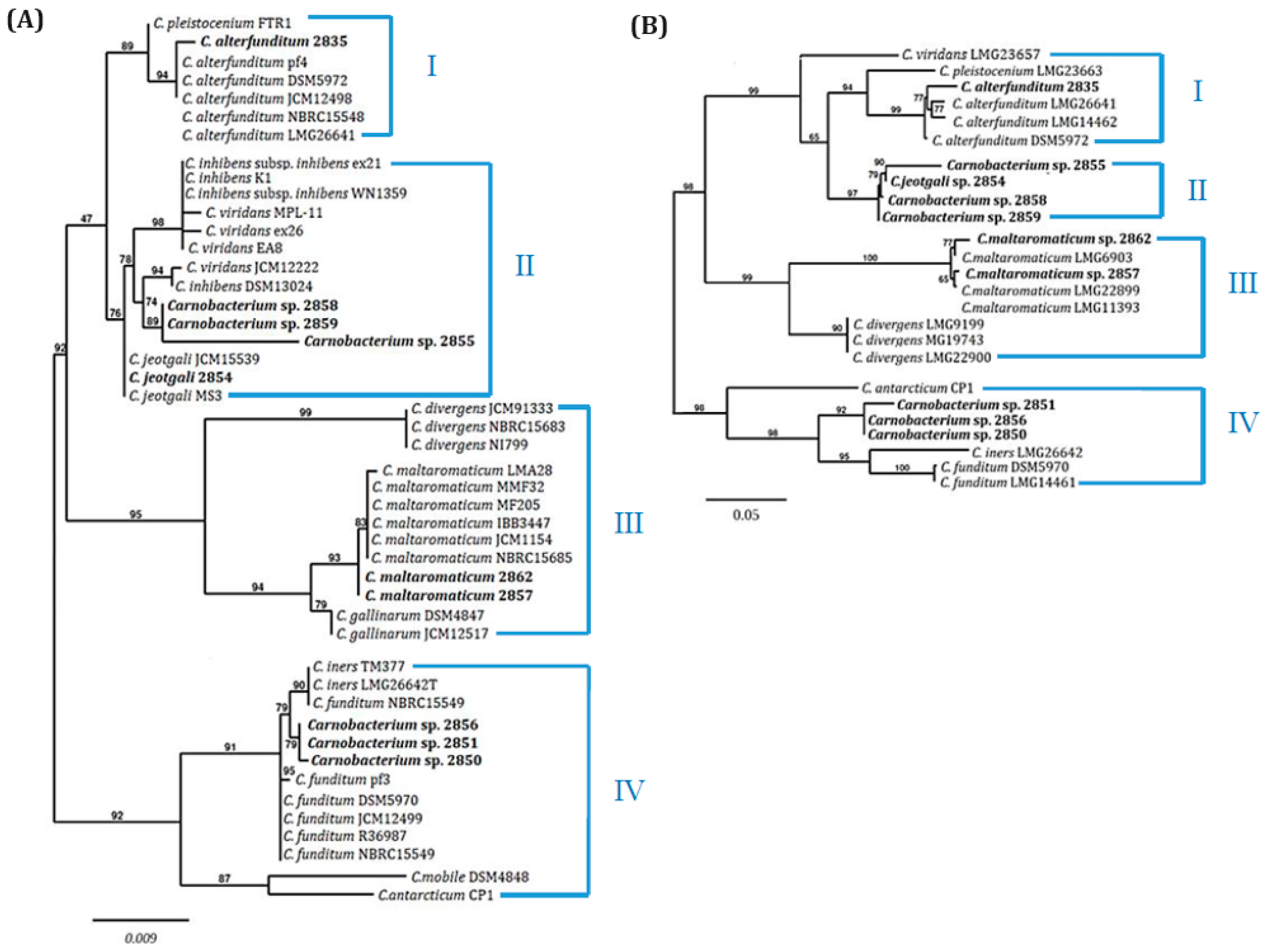
2.1. Taxonomy of Carnobacterium spp. Isolates

Ten LAB strains were isolated from two polar regions: nine from terrestrial environments on King George Island in Antarctica and one from Hans Glacier in the Arctic (Table 1). Identification to the genus Carnobacterium was determined by 16S rRNA gene sequencing and BLASTn searches. The Arctic isolate 2857 showed 100% sequence identity with C. maltaromaticum, while Antarctic strains 2835, 2854, and 2862 aligned with C. alterfunditum, C. jeotgali, or C. maltaromaticum, respectively (Table 1). The remaining isolates had sequence coverage and identity ranges of 93–100% and 98–100%, respectively, relative to various Carnobacterium species.
High similarity in 16S rDNA sequences across multiple species impeded precise species determination for six isolates, prompting further phylogenetic analysis using a dendrogram based on 16S rDNA sequences from GenBank and study isolates (Figure 1A). Four main groups (I–IV) were distinguished: groups I and IV included polar environmental strains, while groups II and III comprised microbiota from meat, fish, and seafood. Isolate 2835 grouped with C. alterfunditum and C. pleistocenium from permafrost and polar lake sediments in group I. Group II, including isolates 2854, 2855, 2858, and 2859, contained strains similar to C. alterfunditum, C. inhibens, C. jeotgali, and C. viridans, primarily isolated from fish or fermented foods. Among them, isolate 2854 clustered together with C. jeotgali strains, while 2855, 2858, and 2859 were in a separate clade with C. viridans and C. inhibens as the most related species. Group III included C. divergens, C. gallinarum, and C. maltaromaticum from meat and seafood microbiome, with isolates 2857 and 2862 clustering with C. maltaromaticum. Group IV included isolates 2850, 2851, and 2856, clustering with C. funditum and C. iners from Antarctic aquatic environments.
A dendrogram based on concatenated housekeeping genes pheS and rpoA (Figure 1B) showed similar topology to the 16S rRNA tree, with four major groups. However, isolates 2850, 2851, and 2856 remained unassigned to specific species. Isolates 2854, 2855, 2858, and 2859 also formed a separate clade without related known species, potentially due to a lack of deposited pheS and rpoA genes from C. jeotgali.
Further species identification using 16S-23S rRNA spacer regions (ITS) pattern analysis confirmed species affiliations for isolates 2835, 2857, and 2862, while isolates 2850, 2851, 2855, and 2859 exhibited unique ITS patterns, indicating novel species (Supplementary Figure S1). HindIII restriction analysis of ITS fragments corroborated these findings.

2.2. Physiological Characteristics

Isolates exhibited typical Carnobacterium spp. traits: Gram-positive, non-spore-forming, non-motile facultative anaerobes, occurring in single rods, in pairs, or in small chains. Strains assigned to C. jeotgali or C. maltaromaticum (2854, 2857, 2862) displayed distinct physiological properties compared to reference mesophilic strains (C. jeotgali MS3, C. maltaromaticum IBB3447 and LMA28) (Table 2) [28,29,30]. For eight polar isolates, growth temperatures ranged from 4 to 20 °C with an optimum at 16 °C. Notably, two C. maltaromaticum strains (2857 and 2862) continued to grow at 37 °C. In contrast, mesophilic strains MS3, IBB3447, and LMA28 had higher temperature optima at 30 °C. Most strains grew in BHI and GM17 media, but not in MRS. Optimal growth for all polar Carnobacterium isolates was achieved in 50% BHI at 16 °C. NaCl tolerance ranged from 2 to 8%, with lower salinity resistance observed in mesophilic isolates (MS3, IBB3447, LMA28); however, C. alterfunditum 2835 and C. maltaromaticum 2857 grew even in 10% NaCl. All isolates demonstrated efficient growth at high pH (8.0–9.0) with a significant lag at pH 5.0–6.0. Mesophilic reference strains C. maltaromaticum IBB3447 and LMA28 grew at pH 3.0, indicating a broader pH tolerance compared to polar isolates. Optimal pH levels for all isolates were between 6.0 and 8.0 (Table 2).

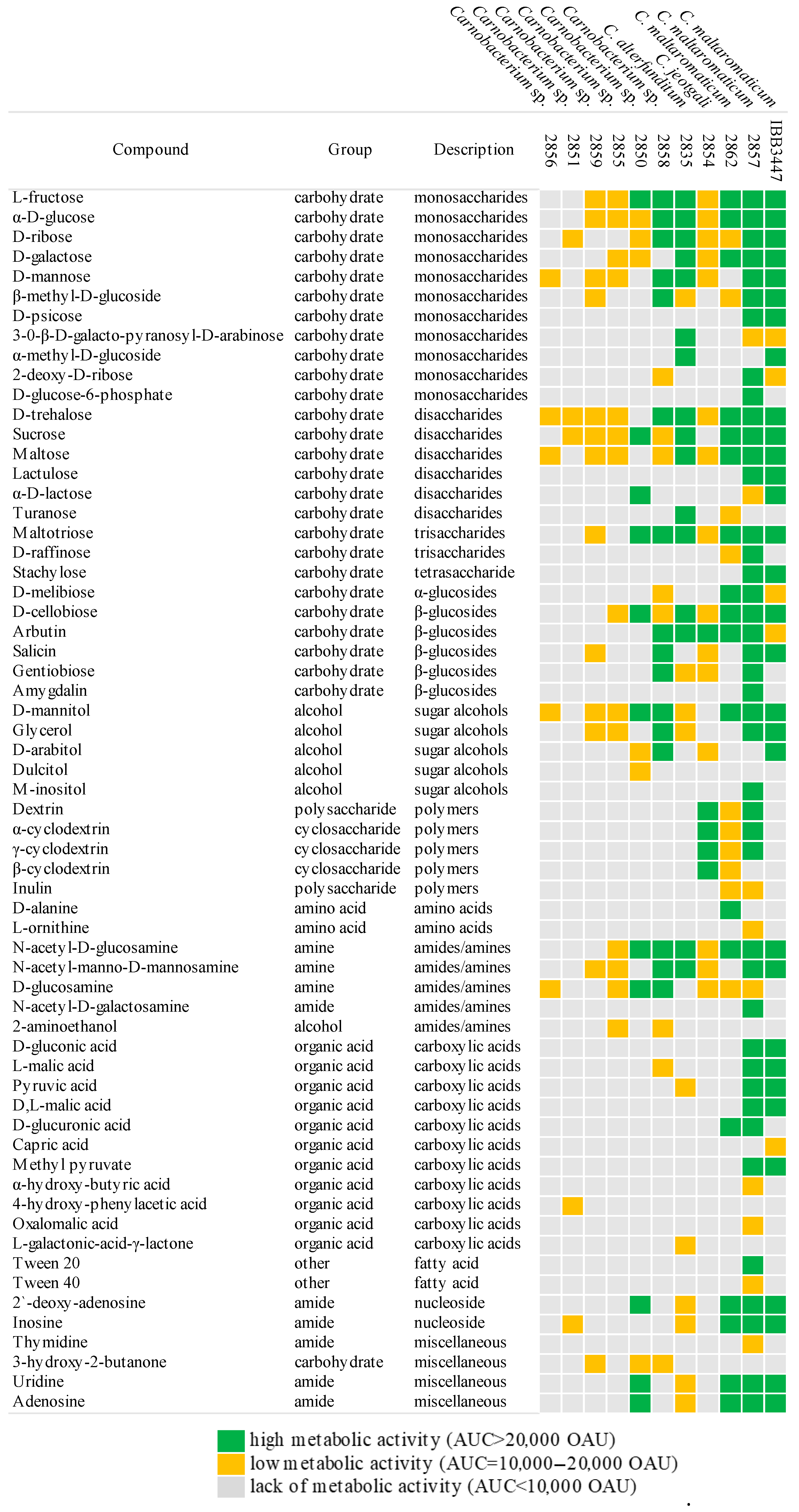
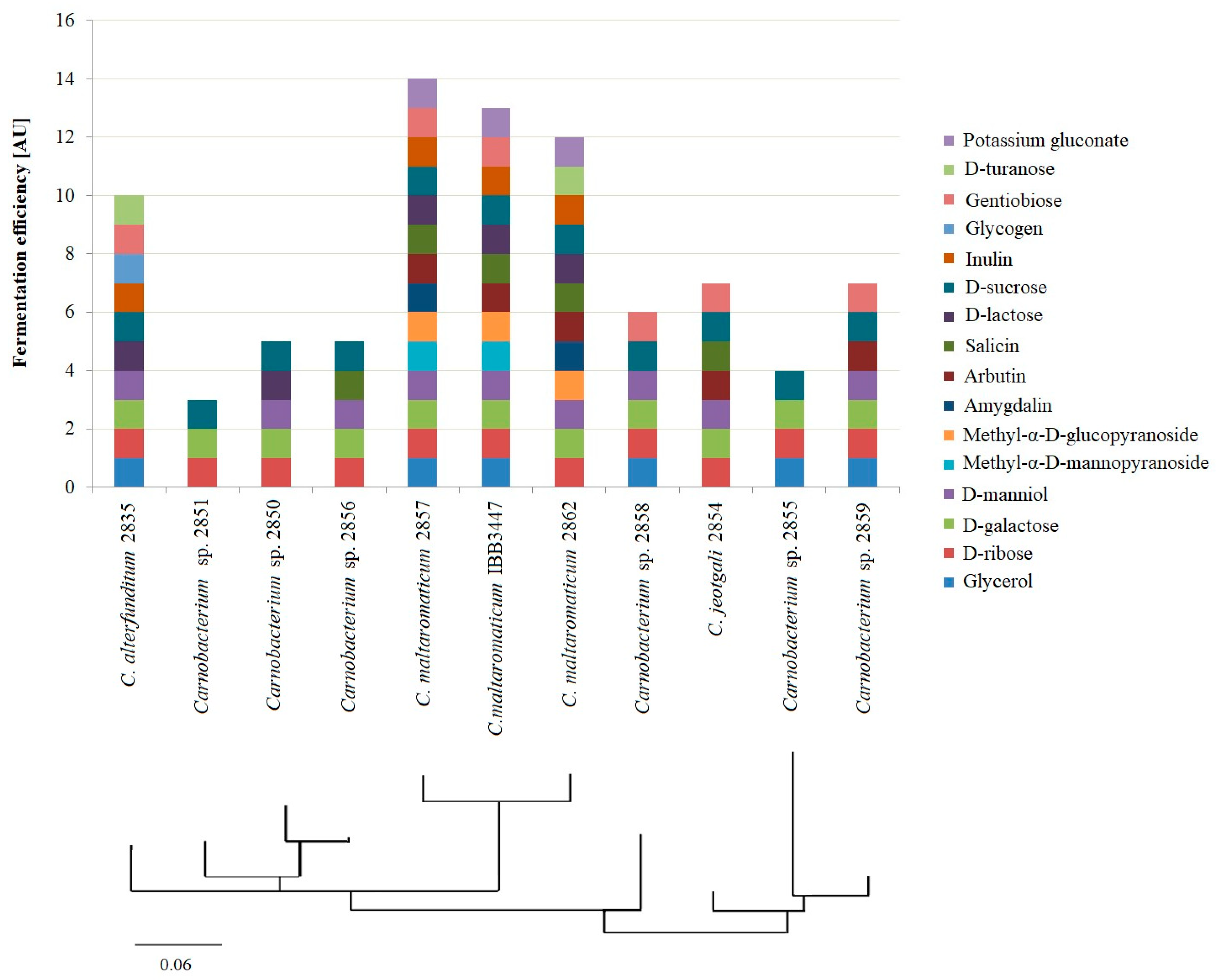
2.3. Carbon Assimilation

Most strains metabolized several monosaccharides (D-glucose, D-galactose, L-fructose, D-mannose) and disaccharides (D-trehalose, D-cellobiose, sucrose). Significant differences in the ability to utilize individual carbohydrates were noted, particularly in the metabolism of β-glycosides and their derivatives (arbutin, salicin, lactose, amygdalin, and gentiobiose), as well as the α-disaccharide D-melibiose. All strains were metabolically active on several amides (N-acetyl-D-glucosamine, N-acetyl-manno-D-mannosamine) and sugar alcohols (D-mannitol), with limited activity in the presence of amines, amino acids, or organic acids.
C. jeotgali 2854 exhibited a metabolic pattern similar to the reference MS3 strain, with minor differences in pentoses and β-glycosides. Polar isolates of C. maltaromaticum metabolized the same carbohydrates as their reference strains but with less efficiency than those from milk or cheese. Carnobacterium spp. from unassigned species (2851, 2855, 2856, 2859) showed weaker metabolic activity, utilizing only a few pentoses and hexoses (Table 2, Figure 2). Strains such as Carnobacterium spp. (2850, 2858), C. alterfunditum 2835, and especially C. maltaromaticum utilized available carbon sources more efficiently, including di- and polysaccharides (Table 2). Interestingly, C. alterfunditum sp. 2835, 2859, 2858, and C. maltaromaticum 2857, 2862 could assimilate glycerol as a carbon source (Table 2, Figure 2).
Phylogenetic analysis based on combined API50CH and Phenotype MicroArrays (Figure 3) further highlighted similarities and differences in metabolic activity. The strains with the highest metabolic activity (C. maltaromaticum 2857 and 2862) clustered with the reference strain C. maltaromaticum IBB3447 (Figure 3). Carnobacterium sp. 2850, 2851, and 2856 formed a separate cluster, indicating a lack of affinity with known species, while isolates 2855 and 2859 grouped close to C. jeotgali 2854, suggesting evolutionary affinity.

2.4. Lactic Acid Production

All isolates produced lactic acid (LA) with yields ranging from 4.3 to 5.9 g/L (Table 2). C. jeotgali 2854 had the highest average LA titer (5.9 g/L), followed by Carnobacterium sp. 2851 (5.7 g/L) and Carnobacterium sp. 2859 (5.6 g/L). C. maltaromaticum 2862 showed the lowest titer (4.3 g/L). Most strains produced a mixture of L- and D-Las, with the L-LA isoform constituting 96–100% of the total LA produced. C. maltaromaticum strains exclusively produced the L form of LA.

2.5. Antimicrobial Activity

Carnobacterium spp. isolates were tested for antimicrobial activity against 17 pathogenic (Bacillus sp., Campylobacter sp., Candida sp., Salmonella sp., Staphylococcus spp., Streptococcus spp.) and six non-pathogenic indicator strains (Bacillus sp., Lactococcus sp., and Staphylococcus spp.) (Table 3). C. jeotgali 2854 and Carnobacterium sp. 2855 exhibited the broadest antimicrobial activity, being bactericidal against seven human pathogens or commensals: C. jejuni, S. aureus, S. caprae, S. epidermidis, S. hyicus, S. intermedius, and S. lugdunensis. Carnobacterium sp. 2858 was active against four staphylococci species, while Carnobacterium sp. 2859 was effective against only S. epidermidis. C. maltaromaticum 2857 and IBB3447 showed inhibitory activity against L. monocytogenes LMGT2604 (Table 3). Bactericidal activity was observed for pure cultures, ammonium sulphate precipitates, and proteinase K-treated supernatants (2854, 2855, 2858, 2859). However, only compounds produced by 2857 and IBB3447 lost bactericidal activity after protease treatment, indicating a bacteriocin-like nature.

2.6. Plasmid Characterization

Thirteen plasmids were identified across five Carnobacterium spp. isolates: five in C. maltaromaticum 2862, three each in 2835 and 2851, and one each in 2856 and 2859 (Table 4). The complete circular nucleotide sequences of these plasmids range from 4.8 to 79.5 kbp with a GC content of 28.9–33.9% (Table 4), aligning with known carnobacterial plasmids but slightly lower that the chromosomal DNA average (38.4%). The plasmids exhibited low sequence similarity (2–58%) to those in GenBank, including plasmids from Carnobacteriaceae and Jeotgalibaca strains in fish food additives and polar marine sediments (Supplementary Figure S2).
A total of 560 plasmid genes were identified and annotated, with only 35% encoding proteins with known functions, while 65% were of unknown function. Genes encoding membrane and extracellular proteins accounted for 38% and 13%, respectively, with cytoplasmic protein comprising 49%. Detailed gene annotations for each plasmid, including their position and potential functions, are provided in Supplementary Table S1a–m.
Seventeen replicons were identified across the plasmids, with some plasmids containing multiple replicons (Table 4, Supplementary Figure S3). Comparison of these replication proteins with those from other Carnobacterium spp. plasmids revealed three phylogenetic groups. Group I includes most Rep proteins (10/17) with Rep_3 (Pfam: PF01051; clade IA) or Rep_trans (Pfam: PF06970; clade IB) domains and a downstream helix-turn-helix (HTH) motif. Group II includes proteins with only the Rep_3 domain (3/17). Group III includes Rep proteins with Rep_3 (clade IIIA) or RepA_N domains (Pfam: PF06970; clade IIIB) and HTH motifs at different positions (Table 4, Supplementary Figure S3).
Mobilization, conjugation, and partition system components were identified in the plasmids (Table 4, Supplementary Table S1a–m). Six plasmids (2851_p2, 2851_p3, 2856_p1, 2859_p1, 2862_p1, and 2862_p4) carry homologs of the mobilization gene mobA, suggesting potential for plasmid mobilization. Conjugal transfer genes (ArdC, TraG, TrwB, YukC) were also identified in some plasmids (2851_p2 and 2856_p1). Partitioning system genes (parA and/or parB) were present in eight plasmids, indicating stable inheritance mechanisms. Additionally, two plasmids (2835_p1 and 2862_p5) contained complete type II toxin–antitoxin systems (parD-parE and mazE-mazF), with other plasmids containing partial systems (phd-yefM and txe-yoeB).
Ten plasmids encoded genes involved in adaptive functions, affecting host cell phenotypes such as carbohydrate or peptide transport and metabolism, and protection against environmental stresses such as low temperatures, high salinity, UV radiation, metal ions, and antibiotics (Supplementary Table S1a–m). Nutrient transport systems included ABC-type peptide transport permeases (OppA, SapC), phosphotransferase system (CelB), and TRAP permeases (DctQ, DctR). Carbohydrate utilization genes encoded components for glucose, galactose, and cellobiose metabolism. Peptidase and protease genes (ArdC, NlpC, PepV, PepA, YddH, Clp) were also identified. Plasmid 2859_p1 included genes for D-glucuronate degradation (UxuA, UxaC).
Four plasmids (2835_p1, 2851_p1, 2851_p3, 2862_p3) contained genes for antibiotic resistance and environmental stress protection. Plasmid 2835_p1 had the highest number of genes related to metal transport and antibiotic resistance, including the kdpABCD operon for potassium ion transport and efflux genes (MacB, TcaB). DNA repair genes (umuD and uvrX) were found in plasmids 2851_p3 and 2851_p1. Plasmid 2862_p3 encoded AmaP, a membrane-anchoring protein for alkaline shock response. Additionally, plasmid 2862_p1 contained cspD, a cold shock protein gene with protective functions against low temperatures.

3. Discussion

This study presents the genotypic and phenotypic characterization of ten new Carnobacterium spp. strains isolated from poorly reported Arctic and Antarctic terrestrial habitats, including cryoconites, elephant seal colonies, and postglacial moraines. Polar-region environments, such as glacial forelands, cryoconite holes, and ornithogenic soils, are recognized for their unique microbiocenosis diversity due to harsh, rapidly changing conditions [13,16,31]. Our findings confirm these speculations, demonstrating that polar-region soils and soil-like materials can be a prolific source of LAB species with unique metabolic traits and antimicrobial properties, facilitating their adaption to polar habitats.
Members of the Carnobacteriaceae family have previously been isolated from various habitats, including vacuum-packaged and refrigerated foods, seafood, fish, humans, biofilms, tundra soil, and Antarctic water reservoirs and sediments [6,32,33]. However, there has been no information on their isolation from aerated soils affected by freezing and thawing. DNA sequencing and phylogenic analysis identified strains of C. jeotgali, C. alterfunditum, and C. maltaromaticum in various polar terrestrial structures. Meltwater runoff supplying organic and inorganic matter to soil structures may explain the presence of LAB in postglacial environments. This is supported by phylogenetic clustering of the 16S rRNA genes of the Carnobacterium spp. studied here, indicating a close relationship with strains from polar environments. C. alterfunditum 2835 and Carnobacterium spp. 2850, 2851, and 2856 are closely related to strains isolated from polar environments such as permafrost and Antarctic lake sediments. It has previously been suggested that the process of glacial surface ablation affects the development of cryoconite holes on Antarctic glaciers, and that on ice-covered surfaces, embedded sediments can migrate downward, providing a continuous supply of inorganic and organic material including microorganisms [16,34]. On the other hand, C. jeotgali 2854 and C. maltaromaticum 2862 showed similarity of 16S rRNA and ITSs to species dominating the microbiota of meat, fish, and seafood. This implies that migrating birds and animals, as well as inorganic and organic components of marine aerosol, should also be considered possible transmitters of microbiota in postglacial habitats. Indeed, Teixeira et al. highlighted the role of marine aerosol, birds, and mammals in the formation of soil microbial communities in coastal regions of the Antarctic environment [20]. Given the phylogenetic affinity of the Carnobacterium spp. strains isolated here, it cannot be ruled out that they may be of marine and animal origin. Interestingly, as many as five of the isolated strains may represent new species in the Carnobacterium genus, suggesting that polar soils may be an environment in which new LAB species differentiate. However, a definitive determination of the status of the species must follow more rigorous taxonomic characterization.
Polar Carnobacterium spp. strains isolated here are characterized by a repertoire of phenotypic traits that result from their adaptation to life in polar-region conditions and that distinguish them from mesophilic carnobacterial strains. Most of these psychrophilic isolates tolerated salinity in the optimal range of 2–6%, which classifies them as moderate halophiles, according to the standard reported for mesophilic LAB [35,36]. However, when it comes to coping with salinity limits, C. alterfunditum 2835, C. jeotgali 2854, and C. maltaromaticum 2857 showed increased tolerance compared to mesophiles, even to values such as 9–10% NaCl. This increased tolerance can be explained by the consequences of ablative processes in the emerging soils, where glacial meltwaters can transport salts from marine aerosol deep into the non-ice-covered area [37]. The presence of sea salt aerosol in Antarctic coastal regions and water evaporation in postglacial environments are also factors that may contribute to the enhanced resistance of the bacteria studied to high salinity [38,39]. The Carnobacterium strains presented here grew well in the pH range of 6–9, while one isolate from an Arctic glacier (C. maltaromaticum 2857) was able to grow at pH 5 and lower, which can be explained by the pH values of the surface of Hans glacier, where the ice and cryoconites had a pH of 3.3–4.8 [15]. On the other hand, mesophilic carnobacterial strains (C. maltaromaticum IBB3447 and LMA28) had a greater resistance to acidification, manifested by their ability to actively grow at pH 3, which may be due to adaptive features to fermentation processes. Mesophilic Carnobacterium spp. and other LAB actively acidify the milk or plant material, leading to a reduction in pH values from ca. 7 to below 4 [40]. The soils of the polar region mainly have pH close to neutral, and therefore the discussed differences in resistance to acidification may indicate a loss of adaptive functions towards low pH, resulting from adaptation to polar conditions [24].
LAB are generally fastidious microorganisms with complex nutrient requirements. However, optimal growth conditions for the psychrophiles studied here occurred mainly at reduced nutrient concentrations, while a more rich nutrient solution resulted in ineffective growth or its inhibition. This may be related to the low nutrient content of recently ice-uncovered soil structures or coastal areas where soils remain nutrient-poor despite the influence of organic and inorganic inputs from marine aerosol, local meltwater streams, seabirds, and mammalian rookeries [13]. Such habitats can shape the metabolic properties of the residents and lead to a narrow profile of assimilated compounds. Indeed, simple sugars such as monosaccharides and glucose-containing disaccharides were mainly utilized by isolated Carnobacterium spp. strains as carbon sources, while other groups of compounds such as carboxylic acids, amines, amides, and amino acids were assimilated poorly or not at all. This phenomenon can be explained by adaptation to polar environments, where soil structures exposed to freezing and thawing are rich in simple carbohydrates and polyols, whereas complex sugars are not often identified. A preference for simple carbohydrates may also indicate symbiotic interactions with other microorganisms found in these habitats. In polar soil structures, soluble simple sugars are the main source of nutrients, which are provided by algae, mosses, and lichens [41]. Underwood et al. showed that algae under stress conditions can secrete simple carbohydrates that are immediately consumed by bacteria, while mosses and lichens provide disaccharides (mostly sucrose) and several alcohols (such as arabitol or mannitol) [42]. Strains of Carnobacterium spp. have been reported in a postglacial microcommunity with a phototrophic partner [41],indicating that symbiotic interactions within this genus may serve as an important adaptive mechanism. Interestingly, three isolated strains (Carnobacterium sp. 2858, C. maltaromaticum 2857, and C. alterfunditum 2835) efficiently metabolized glycerol, which is a unique feature among LAB and could result in potential industrial applications, as this compound is a major waste in biofuel production, and bacteria that utilize it can be used to produce various organic acids (e.g., propionic acid) or in bioremediation processes.
Five carnobacterial isolates examined here carried between one and five plasmids. Such abundance is quite exceptional, as most Carnobacterium spp. strains are devoid of such mobile elements [32], and consequently, a limited number of complete carnobacterial plasmid sequences have been deposited in the GenBank database so far. Moreover, plasmids from Carnobacterium spp. had a unique structure and low similarity (2–58%) to other bacterial plasmids, the closest of which were pMA1X17-3 of Jeotgalibaca sp. MA1X17-3 isolated from polar marine sediment and plasmid 2 of C. maltaromaticum 18ISCm from diseased Korean trout [43]. The fact that these five strains were isolated from neighboring sites suggests that the similarities between their plasmids may be due to horizontal gene transfer. Indeed, in support of this hypothesis, in plasmid 2856_p1 of Carnobacterium sp. 2856 with the highest homology to pMA1X17-3, the components for a sufficient mobilization system (mobA, trwB, yukC) were identified. The basis for the compatible coexistence of such a multiplicity of distinct plasmids in a single bacterial cell is also worthy of interest. In silico predictions of the affinity of the replication initiation proteins encoded in the 13 plasmids sequenced in this work classified them into three families: Rep3, Rep_trans, and RepA_N. Plasmids encoding Rep proteins from the Rep_3, RepA_N, or Rep_trans families have been shown to replicate via the theta replication mechanism in several lactococcal strains [44,45], suggesting the same mode of replication of carnobacterial plasmids. Phylogenetic analysis of the replication proteins of plasmids from the same strains separated them into distinct groups, indicating a lack of relatedness between them. This may suggest that each ori interacts specifically only with the corresponding Rep protein, allowing several different plasmids to coexist in a single bacterial cell. Another characteristic feature of the plasmids identified here is the fairly abundant occurrence of systems for their stable persistence in the cell, both in the form of parAB genes responsible for equal partitioning of plasmids into daughter cells and TA systems that eliminate plasmid-deficient cells. Consequently, many newly acquired plasmid-encoded adaptive traits are stably maintained, conditioning bacterial survival in harsh polar environments. Moreover, genetic determinants for adaptation can be actively transmitted to other bacteria due to the presence of mobilization and conjugation transfer genes in some plasmids of the carnobacterial strains identified here.
Plasmid determinants supporting host adaptation to polar environmental conditions are quite widely described in the literature and are distinguished primarily by the presence of proteins with protection functions against cold and UV radiation, including the cold shock protein, the UmuD subunit of DNA polymerase V, and the UV damage repair protein UvrX [46]. The enhanced UV radiation and oxygen solubility present in ice-uncovered polar soils promote the formation of reactive oxygen species (ROS), leading to the risk of damage to cellular macromolecules, including DNA, RNA, proteins, and lipids [12,47]. As such, plasmid genes may play an important adaptive role by providing antioxidant defense mechanisms, including enzymes for DNA repair and reducing levels of toxic ROS. Plasmids identified in polar isolates of Carnobacterium spp. encode over 50% of membrane or extracellular proteins, which is quite high compared to other LAB species, such as L. lactis IL594 isolated from cheese, which contains only up to 35% of genes encoding such proteins [46,48]. Such a large number of membrane/extracellular proteins encoded in the plasmids may also be related to LAB’s adaptive mechanisms to polar-region environments by adjusting cell membrane fluidity or producing exopolymeric substances that enhance growth during successive freeze–thaw cycles [49]. Two of the identified plasmids (2862_p2 and 2862_p5) carry genes encoding enzymes responsible for lipoprotein and exopolysaccharide (EPS) synthesis and homologous to those of C. maltaromaticum LMA28 and C. funditum DSM5970, respectively. Previously, it was shown that lipoproteins in polar bacteria determine the maintenance of cell membrane structure and permeability at low temperatures [50], while the formation of EPS can provide a cell coating that enables protection against freezing [51]. This may indicate important plasmid-based mechanism for LAB strains to adapt to polar environments by enhancing the expression of cellular integrity proteins under conditions of low or fluctuating water potential leading to desiccation and freeze–thaw stress in cold habitats.
Identification of plasmid-encoded resistance to macrolides and peptide antibiotics (macB, tcaB in 2835_p1) represents a novel feature within carnobacterial adaptive traits. As previously reported, the C. maltaromaticum strain possesses chromosomally-encoded antibiotic resistance genes, including several aminoglycosides, β-lactams, and tetracyclines [52]. mac genes encoding extracellular efflux systems were previously reported to transmit between environmental bacterial species [53], whereas glycopeptide-resistant associated tca operon was identified mostly in human pathogenic strains of. S. aureus [54]. So far, there is no information on bacterial species with neither mac nor tca operons in polar environments. Polar regions are still environments with minimal human impact, but human activities at Antarctic research stations can generate macrolide- and aminoglycoside-resistant bacteria [55]. The analyses in this work also showed that Antarctic plasmids are sources of genes that may be beneficial to the metabolic properties of their host. In this group, genes encoding proteins involved in carbohydrate and peptide metabolism are particularly important, since the use of sugars (galactose) and proteins (casein) present in milk affects the exploitation of LAB in the food industry. Indeed, the plasmids described here encoded ABC-type peptide transporters (OppA, SapC), cellobiose-specific permeases CelB of the PTS system [56], as well as mutarotase involved in galactose metabolism (GalM) [57]. Most polar Carnobacterium isolates showed moderate or no ability to utilize galactose and lactose compared to mesophilic reference strains isolated from milk and its products. This feature indicates the loss or lack of ability of polar carnobacteria to hydrolyze lactose, the main milk sugar, absent in a polar environment. It is worth mentioning that the plasmidome of polar carnobacteria contains 18% to 100% of genes encoding proteins of unknown function. This is considerably higher compared to the plasmidomes (5–75%) (GenBank, NCBI, last accessed 10 November 2023) of strains isolated from more temperate environments, suggesting their native origin more than transfer from inhabited areas. Therefore, an in-depth analysis of this hitherto undescribed gene pool would be essential for a comprehensive evaluation of LAB plasmidomes from the polar region described in this work.
LAB strains and their metabolites are used industrially as natural preservatives to control the growth of pathogenic and food spoilage bacteria. Unlike thermophilic LAB species, Carnobacterium strains isolated from polar environments are capable of thriving and functioning in cold environments, making them particularly suitable for applications that require or benefit from low temperatures. This ability could be especially advantageous for the production of lactic acid and antimicrobial compounds under more favorable technological conditions, including lower temperatures and nutrient concentrations. Several structurally diverse bacteriocins (i.e., bacterial ribosomally synthetized antimicrobial peptides) from Carnobacterium spp. with considerable antimicrobial potential have already been characterized [58,59,60], but this feature is not very common among this group of bacteria [61]. The C. maltaromaticum 2857 and IBB3447 tested here were active against L. monocytogenes with loss of activity after proteinase treatment, strongly suggesting that the compounds they produce may be bacteriocins. Other strains such as C. jeotgali 2854 and Carnobacterium sp. 2855, 2858, and 2859 showed antagonistic activity against Staphylococcus spp., but the active compounds they secreted were resistant to proteolysis, suggesting their non-bacteriocin nature or cyclic structure, which may result in insensitivity to some proteases, as is the case with carnocyclin A [58]. These proteinase-insensitive compounds were active against C. jejuni, reinforcing the hypothesis that the antimicrobial compounds produced by strains 2854, 2855, and 2858 are not bacteriocins, as LAB are thought to exhibit anti-Campylobacter potential based on the production of non-peptide compounds (e.g., organic acids) [59,62]. This assumption requires further research, as the ability of cold-tolerant and halotolerant LAB strains to produce antimicrobials may be important for their potential industrial applications. Moreover, the production of these compounds appears to be chromosomally encoded, as no plasmids were detected in most of the producer strains, indicating stability in the maintenance of this trait.
LA produced by LAB is an important preservative in food production, also ensuring proper acid and flavor conditions [63]. In comparison to thermophilic LAB species, Carnobacterium strains isolated from olar environments possess the ability of lactic acid and antimicrobial compound production in more suitable technological conditions, including lower temperatures and nutrient concentrations. Polar isolates of Carnobacterium spp. produced total LA at similar concentrations in the range of 4.3–7.6 g/L, which is not a very efficient titer, but which can be increased by modifying oxygen availability, pH, culture temperature, or nutrient abundance [64]. Significantly, psychrophilic Carnobacterium spp. strains produced L(+)-lactic acid as the main end product from glucose, which is more favored in food and pharmaceutical industries, where high purity of LA monomers (≥98%) is critical in the synthesis of polylactides [65]. Furthermore, carnobacteria, by secreting lactic acid into the surrounding soil, can increase the bioavailability of labile phosphorus and biogenic metals such as iron or zinc [66,67]. Therefore, by increasing the availability of essential nutrients, they are likely to contribute to soil biodiversity in polar regions [23,68].

4. Materials and Methods

4.1. Isolation of Carnobacterium spp. Strains

Soil from polar areas (ornithogenic soils, elephant seal colonies, and postglacial moraines) and soil-like materials (cryoconites and microbial mats from a freshwater reservoir) were collected using sterile spatulas. Cryoconite holes were emptied using a sterile 160 mL plastic syringe. Samples were stored in 50 mL Falcon-type tubes (Fisher Scientific, Waltham, MA, USA) at −20 °C until further study in Poland (see Table 1 for details). Approximately 10 g of each sample was placed in a sterilized 100 mL Simax-type glass bottle with 90 mL of cool (ca. 4 °C) YGLPB semi-strength broth containing peptone (5 g/L), beef extract (4 g/L), glucose (2.5 g/L), lactose (2.5 g/L), yeast extract (1.5 g/L), KH2PO4 (1.25 g/L), K2HPO4 (1.25 g/L), MgSO4 (0.1 g/L), and MnSO4 (0.025 g/L). The semi-closed bottles were incubated anaerobically (7 days; 10 °C; N2/CO2 atmosphere) in a GenBox 7.0 L container (Biomerieux, Marcy-l′Étoile, France). Cultures (10 μL) were streaked on solid YGLPB medium and incubated (14 days; 10 °C; N2/CO2). Colonies were transferred to fresh medium and incubated aerobically (7 days; 10 °C). Cultures were checked for catalase activity with 3% H2O2, Gram’s reaction with KOH, and cell morphology using light microscope with crystal violet staining. Spores were not observed. Isolates were cultured in 50% Brain Heart Infusion (BHI) medium (Oxoid, Basingstoke, UK), solidified with 2% w/v agar (Merck, Darmstadt, Germany) as needed, and incubated at 16 °C. Reference strain culture conditions are listed in Supplementary Table S2.

4.2. Genetic Analyses

Primers used in the study are listed in Supplementary Table S2. The 16S rRNA sequences were amplified as described previously [69]. Amplification of house-keeping genes (pheS, rpoA) followed [70]. PCR amplifications were performed using Takara ExTaq™ polymerase (TaKaRa, Kusatsu shi, Japan) in a C1000 Thermal Cycler (Bio-Rad, Hercules, CA, USA). PCR product sizes (~1500 bp for 16S rRNA, ~300 bp for pheS, ~600 bp for rpoA) were confirmed and sequenced using BigDye Terminator v.3.1 chemistry (Applied Biosystems, San Francisco, CA, USA) on an ABI3730xl genetic analyzer (Life Technologies, Carlsbad, CA, USA) at the DNA Sequencing and Synthesis Facility (IBB PAS, Poland). Sequencing reads were assembled using Clone Manager Professional 9 (S&E Software, version 9.2, Edison, NJ, USA) and aligned against GenBank references using BLAST (NCBI, Bethesda, MD, USA) [71]. The 16S rRNA, rpoA, and pheS gene sequences were deposited in GenBank (accession numbers in Supplementary Table S3).
Phylogenetic trees, based on the neighbor-joining method with bootstrap analysis (1000 repetitions), were prepared using Clustal Omega [72] and Phylogeny.fr [73,74]. Internal transcribed spacer (ITS)-PCR regions were amplified according to [75] using Phusion® High-Fidelity DNA Polymerase (NEB, Hitchin, UK) and KIL1 and GIL1 primers (Supplementary Table S2). ITS-PCR restriction analysis was based on a theoretical restriction analysis of Carnobacterium ITS sequences deposited in GenBank [71] and used HindIII, EcoRV, EcoRI, SacI, and BamHI enzymes (Thermo Fisher Scientific, Branchburg, NJ, USA).

4.3. Plasmids Isolation and Sequencing

Carnobacterium spp. cells were harvested by centrifugation (4 °C; 10 min; 8000 rpm) from a 10 mL culture at mid-log phase (OD600 0.6–0.8). Cell pellets were resuspended in 1 mL of TEG buffer (25 mM Tris pH 8.0, 10 mM EDTA, 50 mM glucose) with lysozyme (10 mg/mL, NEB, UK) and incubated (37 °C; 30 min). Plasmids were isolated using a plasmid isolation kit according to the manufacturer’s instructions (A&A Biotechnology, Poland) and sequenced at the DNA Sequencing and Synthesis Facility of the Institute of Biochemistry and Biophysics of the Polish Academy of Science (IBB PAS, Warsaw, Poland) on an Illumina MiSeq instrument (Illumina, San Diego, CA, USA) using the v3 chemistry kit (Applied Biosystems, San Francisco, CA, USA). Sequences were assembled and annotated using Unicycler v0.4.6. [76] and DFAST 1.2.18 [77]. Regulatory and metabolic gene sequences were compared with NCBI database homologs using BLASTn and BLASTp (NIH, Bethesda, MD, USA; accessed on 2 January 2024). Protein domains were analyzed using PFAM (http://pfam.xfam.org/; accessed on 5 January 2024), CDD (NCBI, NIH), InterPro (EMBL-EBI), and HHpred [78]. Transmembrane helices in proteins were predicted using TMHMM2.0 (DTU Health Tch, Kongens Lyngby, Denmark). The complete nucleotide sequences of the Carnobacterium spp. plasmids were deposited in GeneBank (accession numbers in Table 4).
Optimal growth conditions were tested as described previously [32]. Carnobacterium spp. strains were inoculated from deep-freeze in YGLPB, incubated (14 days; 10 °C), transferred to the desired media (50% BHI, BHI, LB, MRS), and incubated at various temperatures (4 °C, 16 °C, 20 °C, 30 °C, 37 °C) for 72 h under aerobic conditions. Optical density (OD600) was measured using a plate reader (Bioscreen C, Helsinki, Finland). Salinity tolerance was assessed in 50% BHI with 2%, 4%, 6%, 8%, 9%, or 10% NaCl (Merck, Germany). pH tolerance was tested in 50% BHI at 5.0, 6.0, 7.0, 8.0, or 9.0, adjusted with H2SO4 or NaOH. Single colonies were transferred to 50% BHI and incubated (12 h; 16 °C), and 2 μL of each culture was added to 100-well plates with 198 μL of medium at desired salinity or pH. OD600 was measured using a plate reader (Bioscreen C, Finland) for 72 h at 16 °C. Experiments were performed in triplicate.

4.4. Metabolic Analyses

Carbon source fermentation patterns were determined using API50CHL (BioMérieux, France) and Phenotype MicroArrays (Biolog, Hayward, CA, USA). Colonies were scraped from 50% BHI agar plates, titrated in IF-0a fluid (Biolog, USA) to 65% transmittance, and supplemented with growth supplements and Biolog redox tetrazolium G dye (Biolog, USA), according to standard protocols recommended by Biolog for Streptococcus species. Then, 100 μL aliquots were added to PM1 and PM2 plates and incubated in an OmniLog incubator-reader for 72 h at 16 °C and 30 °C for polar and dairy isolates, respectively. OmniLog arbitrary units (OAUs) were recorded and areas under the curve (AUC) of metabolic activity were calculated and averaged and presented in OAUs.
Swimming and swarming assays were performed as described by [79]. Strains were streaked on 50% BHI agar plates and incubated (48 h; 16 °C), and single colonies were transferred to swimming and swarming plates. Results were recorded after 24 and 48 h.
Lactic acid (LA) detection followed [80]. Total LA and concentrations of D- and L-lactate were assayed in triplicate using a D-/L-lactic acid determination kit (Megazyme International, Wicklow, Ireland).

4.5. Antimicrobial Activity and Bacteriocin Purification

Antimicrobial activity of overnight cultures and ammonium sulfate precipitates of post-culture liquids was tested against selected Gram-positive and Gram-negative bacteria using the spot-on-lawn method [81]. Crude extracts were treated with proteinase K (NEB, USA) at 1 mg/mL for 1 h to test susceptibility to proteolysis. Bacteriocins were precipitated with ammonium sulphate as described previously [82] and stored at −20 °C.

4.6. Statistical Analysis

Data on genetic and physiological traits of isolated Carnobacterium spp. strains were regressed to estimate a 95% confidence level (p-value ≤ 0.05) using Microsoft Excel (Excel 2021 for Windows, Microsoft). Experiments were conducted in triplicate unless otherwise specified.

5. Conclusions

The study presented here provides the first in-depth insight into the characterization of Carnobacterium spp. isolated from less explored niches—soils and soil-like materials of the Arctic and Antarctic regions—sources of LAB that have not been previously reported in the scientific literature. Performed analyses provide:
  • Identification of metabolic properties of isolated Carnobacterium strains that may suggest an ecological role involving various syntrophic interactions with other microorganisms in polar habitats, such as algae, mosses, and lichens, through the secretion and fermentation of carbohydrates and alcohols.
  • Presentation of methodology and data providing guidelines for the isolation of psychrophilic LAB from environmental resources of polar regions; identification of new Carnobacterium species; and characterization of metabolic traits and antimicrobial properties that determine the adaptive potential of LAB strains to permanently cold habitats, as well as for biotechnological application.
  • Description of important metabolic features of industrial importance in cold-adapted Carnobacterium strains, such as the production of fermented foods, probiotics, and antimicrobial compounds, that could be more efficiently harnessed under conditions that are less demanding than those required by thermophilic LAB strains.
  • Detailed characterization of carnobacterial plasmids and identification of certain genetic elements that may be useful in targeted engineering of suitable Carnobacteriaceae strains.
The findings underscore the under-explored biodiversity of polar regions and their potential for biotechnological applications.

Supplementary Materials

The following supporting information can be downloaded at: https://www.mdpi.com/article/10.3390/ijms25179444/s1.

Author Contributions

Conceptualization, K.K., J.G. (Jakub Grzesiak) and T.A.-P.; methodology, K.K., J.G. (Jan Gawor), A.S., J.G. (Jakub Grzesiak) and T.A.-P.; validation, K.K., J.G. (Jakub Grzesiak) and T.A.-P.; formal analysis, K.K., J.G. (Jakub Grzesiak), A.S., J.G. (Jan Gawor) and T.A.-P.; investigation, K.K., J.G., (Jan Gawor), A.S., J.G. (Jakub Grzesiak) and T.A.-P.; resources, K.K., J.G. (Jakub Grzesiak) and T.A.-P.; data curation, K.K., J.G. (Jan Gawor) and T.A.-P.; writing—original draft preparation, K.K., J.G. (Jakub Grzesiak) and T.A.-P.; writing—review and editing, K.K. and T.A.-P.; visualization, K.K. and T.A.-P.; supervision, T.A.-P.; project administration, K.K. and T.A.-P.; funding acquisition, K.K. and T.A.-P. All authors have read and agreed to the published version of the manuscript.

Funding

This work was funded by grants no. 2018/29/B/NZ9/02278 and 2018/31/N/NZ9/03336 from the National Science Centre (Poland).

Institutional Review Board Statement

Not applicable.

Informed Consent Statement

Not applicable.

Data Availability Statement

The complete nucleotide sequences of the Carnobacterium spp. plasmids obtained in this work were deposited in GenBank under accession numbers OR786482 to OR786494. The nucleotide sequences of the 16S rRNA, rpoA, and pheS genes were deposited under accession numbers OQ266887, OQ445549 to OQ445559, and OQ865238 to OQ865259.

Acknowledgments

Samples and data were obtained through the facilities of the Polish Antarctic Station ARCTOWSKI and the Polish Polar Station HORNSUND. We thank John Bowman (University of Tasmania) for providing C. funditum DSM5970 and C. alterfunditum DSM5972 strains used in this research. Sequencing was carried out at the DNA Sequencing and Synthesis Facility of the Institute of Biochemistry and Biophysics, Polish Academy of Science.

Conflicts of Interest

The authors declare no conflict of interest.

References

  1. Teneva-Angelova, T.; Hristova, I.; Pavlov, A.; Beshkova, D. Lactic Acid Bacteria—From Nature Through Food to Health. In Advances in Biotechnology for Food Industry; Academic Press: Cambridge, MA, USA, 2018. [Google Scholar]
  2. Saha, U.B.; Saroj, S.D. Lactic Acid Bacteria: Prominent Player in the Fight against Human Pathogens. Expert Rev. Anti. Infect. Ther. 2022, 20, 1435–1453. [Google Scholar] [CrossRef] [PubMed]
  3. Zapaśnik, A.; Sokołowska, B.; Bryła, M. Role of Lactic Acid Bacteria in Food Preservation and Safety. Foods 2022, 11, 1283. [Google Scholar] [CrossRef] [PubMed]
  4. Franzmann, P.D.; Höpfl, P.; Weiss, N.; Tindall, B.J. Psychrotrophic, Lactic Acid-Producing Bacteria from Anoxic Waters in Ace Lake, Antarctica; Carnobacterium Funditum Sp. Nov. and Carnobacterium Alterfunditum Sp. Nov. Arch. Microbiol. 1991, 156, 255–262. [Google Scholar] [CrossRef]
  5. Pikuta, E.; Marsic, D.; Bej, A.; Tang, J.; Krader, P.; Hoover, R. Carnobacterium Pleistocenium Sp Nov., a Novel Psychrotolerant, Facultative Anaerobe Isolated from Permafrost of the Fox Tunnel in Alaska. Int. J. Syst. Evol. Microbiol. 2005, 55, 473–478. [Google Scholar] [CrossRef]
  6. Zhu, S.; Lin, D.; Shangling, X.; Wang, X.; Xue, Z.; Dong, B.; Shen, X.; Ma, X.; Chen, J.; Yang, J. Carnobacterium Antarcticum Sp. Nov., a Psychrotolerant, Alkaliphilic Bacterium Isolated from Sandy Soil in Antarctica. Int. J. Syst. Evol. Microbiol. 2018, 68, 1672–1677. [Google Scholar] [CrossRef] [PubMed]
  7. Nicholson, W.L.; Zhalnina, K.; de Oliveira, R.R.; Triplett, E.W. Proposal to Rename Carnobacterium Inhibens as Carnobacterium Inhibens Subsp. Inhibens Subsp. Nov. and Description of Carnobacterium Inhibens Subsp. Gilichinskyi Subsp. Nov., a Psychrotolerant Bacterium Isolated from Siberian Permafrost. Int. J. Syst. Evol. Microbiol. 2015, 65, 556–561. [Google Scholar] [CrossRef]
  8. Thomas, D.N.; Fogg, G.E.; Convey, P.; Fritsen, C.H.; Gili, J.-M.; Gradinger, R.; Laybourn-Parry, J.; Reid, K.; Walton, D.W.H. The Biology of Polar Regions; Oxford University Press: Oxford, UK, 2008; ISBN 9780199298112. [Google Scholar]
  9. Klekowski, R.Z.; Opaliński, K.W. Matter and Energy Flow in Spitsbergen Ornithogenic Tundra. Polar Res. 1986, 4, 187–197. [Google Scholar] [CrossRef]
  10. Myrcha, A.; Jadwiszczak, P.; Tambussi, C.; Noriega, J.; Gaździcki, A.; Tatur, A.; del Valle, R. Taxonomic Revision of Eocene Antarctic Penguins Based on Tarsometatarsal Morphology. Polar Res. 2002, 23, 5–46. [Google Scholar]
  11. Turner, J.; Overland, J. Contrasting Climate Change in the Two Polar Regions. Polar Res. 2009, 28, 146–164. [Google Scholar] [CrossRef]
  12. Romaniuk, K.; Ciok, A.; Decewicz, P.; Uhrynowski, W.; Budzik, K.; Nieckarz, M.; Pawlowska, J.; Zdanowski, M.K.; Bartosik, D.; Dziewit, L. Insight into Heavy Metal Resistome of Soil Psychrotolerant Bacteria Originating from King George Island (Antarctica). Polar Biol. 2018, 41, 1319–1333. [Google Scholar] [CrossRef]
  13. Zdanowski, M.K.; Żmuda-Baranowska, M.J.; Borsuk, P.; Świątecki, A.; Górniak, D.; Wolicka, D.; Jankowska, K.M.; Grzesiak, J. Culturable Bacteria Community Development in Postglacial Soils of Ecology Glacier, King George Island, Antarctica. Polar Biol. 2013, 36, 511–527. [Google Scholar] [CrossRef]
  14. Grzesiak, J.; Górniak, D.; Świątecki, A.; Aleksandrzak-Piekarczyk, T.; Szatraj, K.; Zdanowski, M.K. Microbial Community Development on the Surface of Hans and Werenskiold Glaciers (Svalbard, Arctic): A Comparison. Extremophiles 2015, 19, 885–897. [Google Scholar] [CrossRef]
  15. Grzesiak, J.; Zdanowski, M.K.; Górniak, D.; Świątecki, A.; Aleksandrzak-Piekarczyk, T.; Szatraj, K.; Sasin-Kurowska, J.; Nieckarz, M. Microbial Community Changes along the Ecology Glacier Ablation Zone (King George Island, Antarctica). Polar Biol. 2015, 38, 2069–2083. [Google Scholar] [CrossRef]
  16. Zdanowski, M.K.; Bogdanowicz, A.; Gawor, J.; Gromadka, R.; Wolicka, D.; Grzesiak, J. Enrichment of Cryoconite Hole Anaerobes: Implications for the Subglacial Microbiome. Microb. Ecol. 2017, 73, 532–538. [Google Scholar] [CrossRef] [PubMed]
  17. Cukier, S.; Fudala, K.; Bialik, R.J. Are Antarctic Aquatic Invertebrates Hitchhiking on Your Footwear? J. Nat. Conserv. 2023, 72, 126354. [Google Scholar] [CrossRef]
  18. Górniak, D.; Marszałek, H.; Kwaśniak-Kominek, M.; Rzepa, G.; Manecki, M. Soil Formation and Initial Microbiological Activity on a Foreland of an Arctic Glacier (SW Svalbard). Appl. Soil Ecol. 2017, 114, 34–44. [Google Scholar] [CrossRef]
  19. Duda, M.P.; Hargan, K.E.; Michelutti, N.; Smol, J.P. Freshwater Diatom Assemblages from Seabird-Inhabited Ponds in Hudson Strait, Sub-Arctic Canada. Polar Biol. 2019, 42, 1549–1560. [Google Scholar] [CrossRef]
  20. Teixeira, L.C.R.S.; Peixoto, R.S.; Cury, J.C.; Sul, W.J.; Pellizari, V.H.; Tiedje, J.; Rosado, A.S. Bacterial Diversity in Rhizosphere Soil from Antarctic Vascular Plants of Admiralty Bay, Maritime Antarctica. ISME J. 2010, 4, 989–1001. [Google Scholar] [CrossRef]
  21. Schmidt, S.K.; Johnson, B.W.; Solon, A.J.; Sommers, P.; Darcy, J.L.; Vincent, K.; Vimercati, L.; Fountain, A.G.; Porazinska, D.L. Microbial Biogeochemistry and Phosphorus Limitation in Cryoconite Holes on Glaciers across the Taylor Valley, McMurdo Dry Valleys, Antarctica. Biogeochemistry 2022, 158, 313–326. [Google Scholar] [CrossRef]
  22. Jin, S.; Wang, Y.; Zhao, X. Cold-Adaptive Mechanism of Psychrophilic Bacteria in Food and Its Application. Microb. Pathog. 2022, 169, 105652. [Google Scholar] [CrossRef]
  23. Znój, A.; Gawor, J.; Gromadka, R.; Chwedorzewska, K.J.; Grzesiak, J. Root-Associated Bacteria Community Characteristics of Antarctic Plants: Deschampsia Antarctica and Colobanthus Quitensis—A Comparison. Microb. Ecol. 2021, 84, 808–820. [Google Scholar] [CrossRef] [PubMed]
  24. Dziurzynski, M.; Gorecki, A.; Pawlowska, J.; Istel, L.; Decewicz, P.; Golec, P.; Styczynski, M.; Poszytek, K.; Rokowska, A.; Gorniak, D.; et al. Revealing the Diversity of Bacteria and Fungi in the Active Layer of Permafrost at Spitsbergen Island (Arctic)-Combining Classical Microbiology and Metabarcoding for Ecological and Bioprospecting Exploration. Sci. Total Environ. 2023, 856, 159072. [Google Scholar] [CrossRef] [PubMed]
  25. Abakumov, E.; Abramov, A.; Aislabie, J.; Alekseeva, T.; Balks, M.; Blume, H.-P.; Bockheim, J.; Bölter, M.; Dergacheva, M.; Dolgikh, A.; et al. The Soils of Antarctica; Springer: Berlin/Heidelberg, Germany, 2015; ISBN 978-3-319-05497-1. [Google Scholar]
  26. Białkowska, A.M.; Grzelczyk, A.; Długołęcka, A.; Cieśliński, H.; Kalinowska, H.; Kur, J.; Turkiewicz, M. Physicochemical and Biological Characterization of Soils from the Vicinity of the Arctowski Polish Antarctic Station. Biotechnol. Food Sci. 2012, 76, 13–26. [Google Scholar] [CrossRef]
  27. Szymański, W.; Siwek, J.; Waścińska, J.; Wojtuń, B. Texture and Geochemistry of Surface Horizons of Arctic Soils from a Non-Glaciated Catchment, SW Spitsbergen. Pol. Polar Res. 2016, 37, 361–377. [Google Scholar] [CrossRef]
  28. Whon, T.W.; Hyun, D.-W.; Nam, Y.-D.; Kim, M.-S.; Song, E.-J.; Jang, Y.K.; Jung, E.S.; Shin, N.-R.; Oh, S.J.; Kim, P.S.; et al. Genomic and Phenotypic Analyses of Carnobacterium Jeotgali Strain MS3T, a Lactate-Producing Candidate Biopreservative Bacterium Isolated from Salt-Fermented Shrimp. FEMS Microbiol. Lett. 2015, 362, fnv058. [Google Scholar] [CrossRef]
  29. Mora, D.; Scarpellini, M.; Franzetti, L.; Colombo, S.; Galli, A. Reclassification of Lactobacillus Maltaromicus (Miller et Al. 1974) DSM 20342T and DSM 20344 and Carnobacterium Piscicola (Collins et Al. 1987) DSM 20730T and DSM 20722 as Carnobacterium Maltaromaticum Comb. Nov. Int. J. Syst. Evol. Microbiol. 2003, 53, 675–678. [Google Scholar] [CrossRef] [PubMed]
  30. Afzal, M.I.; Ariceaga, C.C.G.; Lhomme, E.; Ali, N.K.; Payot, S.; Burgain, J.; Gaiani, C.; Borges, F.; Revol-Junelles, A.-M.; Delaunay, S.; et al. Characterization of Carnobacterium Maltaromaticum LMA 28 for Its Positive Technological Role in Soft Cheese Making. Food Microbiol. 2013, 36, 223–230. [Google Scholar] [CrossRef]
  31. Buda, J.; Łokas, E.; Pietryka, M.; Richter, D.; Magowski, W.; Iakovenko, N.S.; Porazinska, D.L.; Budzik, T.; Grabiec, M.; Grzesiak, J.; et al. Biotope and Biocenosis of Cryoconite Hole Ecosystems on Ecology Glacier in the Maritime Antarctic. Sci. Total Environ. 2020, 724, 138112. [Google Scholar] [CrossRef]
  32. Pikuta, E.V.; Hoover, R.B. The Genus Carnobacterium. In Lactic Acid Bacteria; John Wiley & Sons, Ltd.: Hoboken, NJ, USA, 2014; pp. 109–123. ISBN 9781118655252. [Google Scholar]
  33. Tang, T.; Martinenghi, L.D.; Hounmanou, Y.M.G.; Leisner, J.J. Distribution and Ecology of the Generalist Lactic Acid Bacterium Carnobacterium Maltaromaticum in Different Freshwater Habitats: Metabolic and Antagonistic Abilities. Environ. Microbiol. 2023, 25, 3556–3576. [Google Scholar] [CrossRef]
  34. Yadav, A.N.; Verma, P.; Sachan, S.G.; Kaushik, R.; Saxena, A.K. Psychrotrophic Microbiomes: Molecular Diversity and Beneficial Role in Plant Growth Promotion and Soil Health. In Microorganisms for Green Revolution: Volume 2: Microbes for Sustainable Agro-ecosystem; Panpatte, D.G., Jhala, Y.K., Shelat, H.N., Vyas, R.V., Eds.; Springer: Singapore, 2018; pp. 197–240. ISBN 978-981-10-7146-1. [Google Scholar]
  35. Gunjal, A.; Badodekar, N. Halophiles; IGI Global: Hershey, PA, USA, 2021; pp. 13–34. ISBN 9781799891444. [Google Scholar]
  36. Papadopoulou, E.; Rodriguez de Evgrafov, M.C.; Kalea, A.; Tsapekos, P.; Angelidaki, I. Adaptive Laboratory Evolution to Hypersaline Conditions of Lactic Acid Bacteria Isolated from Seaweed. N. Biotechnol. 2023, 75, 21–30. [Google Scholar] [CrossRef]
  37. Magalhães, C.; Stevens, M.I.; Cary, S.C.; Ball, B.A.; Storey, B.C.; Wall, D.H.; Türk, R.; Ruprecht, U. At Limits of Life: Multidisciplinary Insights Reveal Environmental Constraints on Biotic Diversity in Continental Antarctica. PLoS ONE 2012, 7, e44578. [Google Scholar] [CrossRef]
  38. Wohl, C.; Jones, A.E.; Sturges, W.T.; Nightingale, P.D.; Else, B.; Butterworth, B.J.; Yang, M. Sea Ice Concentration Impacts Dissolved Organic Gases in the Canadian Arctic. Biogeosciences 2022, 19, 1021–1045. [Google Scholar] [CrossRef]
  39. Cowan, D.A.; Tow, L.A. Endangered Antarctic Environments. Annu. Rev. Microbiol. 2004, 58, 649–690. [Google Scholar] [CrossRef] [PubMed]
  40. Dimitrellou, D.; Salamoura, C.; Kontogianni, A.; Katsipi, D.; Kandylis, P.; Zakynthinos, G.; Varzakas, T. Effect of Milk Type on the Microbiological, Physicochemical and Sensory Characteristics of Probiotic Fermented Milk. Microorganisms 2019, 7, 274. [Google Scholar] [CrossRef] [PubMed]
  41. Cabrerizo, A.; Dachs, J.; Barceló, D.; Jones, K.C. Influence of Organic Matter Content and Human Activities on the Occurrence of Organic Pollutants in Antarctic Soils, Lichens, Grass, and Mosses. Environ. Sci. Technol. 2012, 46, 1396–1405. [Google Scholar] [CrossRef]
  42. Underwood, G.J.C.; Aslam, S.N.; Michel, C.; Niemi, A.; Norman, L.; Meiners, K.M.; Laybourn-Parry, J.; Paterson, H.; Thomas, D.N. Broad-Scale Predictability of Carbohydrates and Exopolymers in Antarctic and Arctic Sea Ice. Proc. Natl. Acad. Sci. USA 2013, 110, 15734–15739. [Google Scholar] [CrossRef]
  43. Roh, H.; Kim, B.S.; Lee, M.K.; Park, C.-I.; Kim, D.-H. Genome-Wide Comparison of Carnobacterium Maltaromaticum Derived from Diseased Fish Harbouring Important Virulence-Related Genes. J. Fish Dis. 2020, 43, 1029–1037. [Google Scholar] [CrossRef]
  44. van Belkum, M.J.; Stiles, M.E. Characterization of the Theta-Type Plasmid PCD3.4 from Carnobacterium Divergens, and Modulation of Its Host Range by RepA Mutation. Microbiology 2006, 152, 171–178. [Google Scholar] [CrossRef]
  45. Heinl, S.; Wibberg, D.; Eikmeyer, F.; Szczepanowski, R.; Blom, J.; Linke, B.; Goesmann, A.; Grabherr, R.; Schwab, H.; Pühler, A.; et al. Insights into the Completely Annotated Genome of Lactobacillus Buchneri CD034, a Strain Isolated from Stable Grass Silage. J. Biotechnol. 2012, 161, 153–166. [Google Scholar] [CrossRef]
  46. Górecki, R.K.; Koryszewska-Bagińska, A.; Gołębiewski, M.; Żylińska, J.; Grynberg, M.; Bardowski, J.K. Adaptative Potential of the Lactococcus Lactis IL594 Strain Encoded in Its 7 Plasmids. PLoS ONE 2011, 6, e22238. [Google Scholar] [CrossRef]
  47. Casanueva, A.; Tuffin, M.; Cary, C.; Cowan, D.A. Molecular Adaptations to Psychrophily: The Impact of ‘Omic’ Technologies. Trends Microbiol. 2010, 18, 374–381. [Google Scholar] [CrossRef] [PubMed]
  48. Kosiorek, K.; Koryszewska-Bagińska, A.; Skoneczny, M.; Stasiak-Różańska, L.; Aleksandrzak-Piekarczyk, T. The Presence of Plasmids in Lactococcus Lactis IL594 Determines Changes in the Host Phenotype and Expression of Chromosomal Genes. Int. J. Mol. Sci. 2023, 24, 793. [Google Scholar] [CrossRef]
  49. Cowan, D.; Khan, N.; Heath, C.; Mutondo, M. Polar Microbiology, The Ecology, Biodiversity and Bioremediation Potential of Microorganisms in Extremely Cold Environments; CRC Press: Boca Raton, FL, USA, 2010; ISBN 9781420083842. [Google Scholar]
  50. Cole, G.B.; Bateman, T.J.; Moraes, T.F. The Surface Lipoproteins of Gram-Negative Bacteria: Protectors and Foragers in Harsh Environments. J. Biol. Chem. 2021, 296, 100147. [Google Scholar] [CrossRef] [PubMed]
  51. Deming, J.W.; Young, J.N. The Role of Exopolysaccharides in Microbial Adaptation to Cold Habitats. In Psychrophiles: From Biodiversity to Biotechnology; Margesin, R., Ed.; Springer International Publishing: Cham, Switzerland, 2017; pp. 259–284. ISBN 978-3-319-57057-0. [Google Scholar]
  52. Leisner, J.J.; Hansen, M.A.; Larsen, M.H.; Hansen, L.; Ingmer, H.; Sørensen, S.J. The Genome Sequence of the Lactic Acid Bacterium, Carnobacterium Maltaromaticum ATCC 35586 Encodes Potential Virulence Factors. Int. J. Food Microbiol. 2012, 152, 107–115. [Google Scholar] [CrossRef] [PubMed]
  53. Greene, N.P.; Kaplan, E.; Crow, A.; Koronakis, V. Antibiotic Resistance Mediated by the MacB ABC Transporter Family: A Structural and Functional Perspective. Front. Microbiol. 2018, 9, 950. [Google Scholar] [CrossRef]
  54. Bakthavatchalam, Y.D.; Ramaswamy, B.; Janakiraman, R.; Steve, R.J.; Veeraraghavan, B. Genomic Insights of Community-Acquired Methicillin-Resistant Staphylococcus aureus (MRSA) with Reduced Teicoplanin Susceptibility: A Case of Fatal Necrotizing Fasciitis. J. Glob. Antimicrob. Resist. 2018, 14, 242–245. [Google Scholar] [CrossRef]
  55. Scott, L.C.; Lee, N.; Aw, T.G. Antibiotic Resistance in Minimally Human-Impacted Environments. Int. J. Environ. Res. Public Health 2020, 17, 3939. [Google Scholar] [CrossRef]
  56. Aleksandrzak-Piekarczyk, T.; Kok, J.; Renault, P.; Bardowski, J. Alternative Lactose Catabolic Pathway in Lactococcus Lactis IL1403. Appl. Environ. Microbiol. 2005, 71, 6060–6069. [Google Scholar] [CrossRef]
  57. Aleksandrzak-Piekarczyk, T.; Szatraj, K.; Kosiorek, K. GlaR (YugA)-a Novel RpiR-Family Transcription Activator of the Leloir Pathway of Galactose Utilization in Lactococcus Lactis IL1403. Microbiologyopen 2019, 8, e00714. [Google Scholar] [CrossRef]
  58. Martin-Visscher, L.A.; van Belkum, M.J.; Garneau-Tsodikova, S.; Whittal, R.M.; Zheng, J.; McMullen, L.M.; Vederas, J.C. Isolation and Characterization of Carnocyclin a, a Novel Circular Bacteriocin Produced by Carnobacterium Maltaromaticum UAL307. Appl. Environ. Microbiol. 2008, 74, 4756–4763. [Google Scholar] [CrossRef]
  59. Acedo, J.Z.; Towle, K.M.; Lohans, C.T.; Miskolzie, M.; McKay, R.T.; Doerksen, T.A.; Vederas, J.C.; Martin-Visscher, L.A. Identification and Three-Dimensional Structure of Carnobacteriocin XY, a Class IIb Bacteriocin Produced by Carnobacteria. FEBS Lett. 2017, 591, 1349–1359. [Google Scholar] [CrossRef] [PubMed]
  60. Begrem, S.; Ivaniuk, F.; Gigout-Chevalier, F.; Kolypczuk, L.; Bonnetot, S.; Leroi, F.; Grovel, O.; Delbarre-Ladrat, C.; Passerini, D. New Insight into Antimicrobial Compounds from Food and Marine-Sourced Carnobacterium Species through Phenotype and Genome Analyses. Microorganisms 2020, 8, 1093. [Google Scholar] [CrossRef]
  61. Stupar, J.; Holøymoen, I.G.; Hoel, S.; Lerfall, J.; Rustad, T.; Jakobsen, A.N. Diversity and Antimicrobial Activity towards Listeria Spp. and Escherichia Coli among Lactic Acid Bacteria Isolated from Ready-to-Eat Seafood. Foods 2021, 10, 271. [Google Scholar] [CrossRef]
  62. Lohans, C.T.; van Belkum, M.J.; Li, J.; Vederas, J.C. Characterization of Bacterial Antimicrobial Peptides Active against Campylobacter Jejuni. Can. J. Chem. 2015, 93, 381–388. [Google Scholar] [CrossRef]
  63. Hwanhlem, N.; Buradaleng, S.; Wattanachant, S.; Benjakul, S.; Tani, A.; Maneerat, S. Isolation and Screening of Lactic Acid Bacteria from Thai Traditional Fermented Fish (Plasom) and Production of Plasom from Selected Strains. Food Control 2011, 22, 401–407. [Google Scholar] [CrossRef]
  64. Abedi, E.; Hashemi, S.M.B. Lactic Acid Production–Producing Microorganisms and Substrates Sources-State of Art. Heliyon 2020, 6, e04974. [Google Scholar] [CrossRef] [PubMed]
  65. Cunha, B.L.C.; Bahú, J.O.; Xavier, L.F.; Crivellin, S.; de Souza, S.D.A.; Lodi, L.; Jardini, A.L.; Filho, R.M.; Schiavon, M.I.R.B.; Concha, V.O.C.; et al. Lactide: Production Routes, Properties, and Applications. Bioengineering 2022, 9, 164. [Google Scholar] [CrossRef]
  66. Swetha, S.; Padmavathi, T. Study of Acid Phosphatase in Solubilization of Inorganic Phosphates by Piriformospora Indica. Pol. J. Microbiol. 2016, 65, 407–412. [Google Scholar] [CrossRef]
  67. Zlotnikov, K.; Zlotnikov, A.K.; Kaparullina, E.; Doronina, N. Phylogenetic Position and Phosphate Solubilization Activity of Lactic Acid Bacteria Associated with Different Plants. Mikrobiologiia 2013, 82, 376–379. [Google Scholar] [CrossRef]
  68. Yang, G.; Tian, J.; Chen, J. Editorial: Soil Microbes in Polar Region: Response, Adaptation and Mitigation of Climate Change. Front. Microbiol. 2022, 13, 1086822. [Google Scholar] [CrossRef]
  69. Hoover, R.B.; Pikuta, E.V.; Bej, A.K.; Marsic, D.; Whitman, W.B.; Tang, J.; Krader, P. Spirochaeta Americana Sp. Nov., a New Haloalkaliphilic, Obligately Anaerobic Spirochaete Isolated from Soda Mono Lake in California. Int. J. Syst. Evol. Microbiol. 2003, 53, 815–821. [Google Scholar] [CrossRef]
  70. Naser, S.M.; Thompson, F.L.; Hoste, B.; Gevers, D.; Dawyndt, P.; Vancanneyt, M.; Swings, J. Application of Multilocus Sequence Analysis (MLSA) for Rapid Identification of Enterococcus Species Based on RpoA and PheS Genes. Microbiology 2005, 151 Pt 7, 2141–2150. [Google Scholar] [CrossRef]
  71. Sayers, E.W.; Beck, J.; Bolton, E.E.; Bourexis, D.; Brister, J.R.; Canese, K.; Comeau, D.C.; Funk, K.; Kim, S.; Klimke, W.; et al. Database Resources of the National Center for Biotechnology Information. Nucleic Acids Res. 2021, 49, D10–D17. [Google Scholar] [CrossRef] [PubMed]
  72. Madeira, F.; Pearce, M.; Tivey, A.R.N.; Basutkar, P.; Lee, J.; Edbali, O.; Madhusoodanan, N.; Kolesnikov, A.; Lopez, R. Search and Sequence Analysis Tools Services from EMBL-EBI in 2022. Nucleic Acids Res. 2022, 50, W276–W279. [Google Scholar] [CrossRef]
  73. Dereeper, A.; Guignon, V.; Blanc, G.; Audic, S.; Buffet, S.; Chevenet, F.; Dufayard, J.-F.; Guindon, S.; Lefort, V.; Lescot, M.; et al. Phylogeny.Fr: Robust Phylogenetic Analysis for the Non-Specialist. Nucleic Acids Res. 2008, 36, W465–W469. [Google Scholar] [CrossRef] [PubMed]
  74. Charnock, C.; Hagen, R.X.; Nguyen, T.N.-T.; Vo, L.T. Diversion and Phylogenetic Relatedness of Filterable Bacteria from Norwegian Tap and Bottled Waters. J. Water Health 2019, 17, 295–307. [Google Scholar] [CrossRef] [PubMed]
  75. Dec, M.; Urban-Chmiel, R.; Gnat, S.; Puchalski, A.; Wernicki, A. Identification of Lactobacillus Strains of Goose Origin Using MALDI-TOF Mass Spectrometry and 16S–23S RDNA Intergenic Spacer PCR Analysis. Res. Microbiol. 2014, 165, 190–201. [Google Scholar] [CrossRef] [PubMed]
  76. Wick, R.R.; Judd, L.M.; Gorrie, C.L.; Holt, K.E. Unicycler: Resolving Bacterial Genome Assemblies from Short and Long Sequencing Reads. PLoS Comput. Biol. 2017, 13, e1005595. [Google Scholar] [CrossRef]
  77. Tanizawa, Y.; Fujisawa, T.; Nakamura, Y. DFAST: A Flexible Prokaryotic Genome Annotation Pipeline for Faster Genome Publication. Bioinformatics 2017, 34, 1037–1039. [Google Scholar] [CrossRef]
  78. Zimmermann, L.; Stephens, A.; Nam, S.-Z.; Rau, D.; Kübler, J.; Lozajic, M.; Gabler, F.; Söding, J.; Lupas, A.N.; Alva, V. A Completely Reimplemented MPI Bioinformatics Toolkit with a New HHpred Server at its Core. J. Mol. Biol. 2018, 430, 2237–2243. [Google Scholar] [CrossRef]
  79. Jecz, P.; Bartosik, A.A.; Glabski, K.; Jagura-Burdzy, G. A Single ParS Sequence from the Cluster of Four Sites Closest to OriC Is Necessary and Sufficient for Proper Chromosome Segregation in Pseudomonas Aeruginosa. PLoS ONE 2015, 10, e0120867. [Google Scholar] [CrossRef] [PubMed]
  80. Camesasca, L.; de Mattos, J.A.; Vila, E.; Cebreiros, F.; Lareo, C. Lactic Acid Production by Carnobacterium Sp. Isolated from a Maritime Antarctic Lake Using Eucalyptus Enzymatic Hydrolysate. Biotechnol. Rep. 2021, 31, e00643. [Google Scholar] [CrossRef]
  81. Zhang, H.; Cui, Y.; Zhu, S.; Feng, F.; Zheng, X. Characterization and Antimicrobial Activity of a Pharmaceutical Microemulsion. Int. J. Pharm. 2010, 395, 154–160. [Google Scholar] [CrossRef] [PubMed]
  82. Tymoszewska, A.; Aleksandrzak-Piekarczyk, T. The Lactococcal DgkB (YecE) and DxsA Genes for Lipid Metabolism Are Involved in the Resistance to Cell Envelope-Acting Antimicrobials. Int. J. Mol. Sci. 2021, 22, 1014. [Google Scholar] [CrossRef] [PubMed]
Figure 1. Phylogenetic trees based on 16S rRNA (A) and the concatenated pheS and rpoA genes (B). Polar Carnobacterium strains isolated in this study are shown in bold; other Carnobacterium spp. were retrieved from the GenBank database. Numbers I–IV correspond to the distinguished groups of strain origins: groups I and IV include polar environmental isolates, while groups II and III comprise microbiota from meat, fish, and seafood. The tree was constructed with the neighbor-joining method. Bootstrap values are given at the nodes. The scale bar represents number of substitutions per nucleotide position. Carnobacterium strain GenBank accession numbers used in the phylogenetic tree construction: OQ266887, OQ448831, OQ445553, OQ445549, OQ445555, OQ445554, OQ445550, OQ445556, OQ445557, OQ445552, JQLQ01000004, NR104715, LC145583, NR025211, LC145585, AB680898, HE590768, KF317891, NR036895, JX110652, NR025197, KR317896, JX110652, LC145568, JQIV01000006, LC258159, NR116460, LC077075, AB598939, NR102484, GQ304940, AY543032, LC65032, AB680942, NR42093, LC153546, NR108864, HE583595, NR113773, NR025946, LC145584,FR691457, NR113778, HE590756, HE590757, HE590753, HE590754, HE590759, HE590760, HE590768, HE590770, AM694187, AM694188, HE590726, HE590727, HE590729, HE590730, HE590700, HE590701, HE590712, HE590713, HE590706, HE590707, MG734180, MG734181, HE578182, HE578183, HE592670,HE592671, HE590696, HE590715.
Ijms 25 09444 g001
Figure 2. Carbon sources used by Carnobacterium spp. strains. The analysis was carried out using Phenotype MicroArrays™ and only those carbon sources are presented for which at least one positive reaction for a given isolate was detected.
Ijms 25 09444 g002
Figure 3. Carbon source assimilation and phylogenetic relatedness between Carnobacterium spp. strains. A carbon source assimilation score was considered positive when the overlapping metabolic activity of a strain on a carbon source was observed in both API50CH and Phenotype MicroArrays Biolog® assays. For all strains analyzed, both overlapping and non-metabolized carbohydrates were omitted from the data analysis. Positive results were converted to numerical values and used to build a phylogenetic tree based on the neighbor-joining method as a bootstrap test of phylogeny. In the phylogenetic analysis, the scale bar represents number of substitutions per carbohydrate position in the prepared metabolic patterns of the strains.
Ijms 25 09444 g003
Table 1. Isolation source and taxonomic identification of polar Carnobacterium spp. isolates based on 16S rRNA.
IsolateSource of IsolationLongitudeLatitudeAlignment Result 1Query
Coverage
[%]
Sequence
Identity
[%]
Taxonomic IdentificationReference for Soil Components
2835Cryoconite, Ecology Glacier,
King George Island (Antarctica)
−58.47559−62.17331C. alterfunditum JCM12498100100C. alterfunditum[15]
2850Ornithogenic soil, Llano Cape,
King George Island (Antarctica)
−58.44761−62.17491C. funditum NBRC15549
C. funditum JCM12499
C. iners TM377
100
100
100
100
99
99
Carnobacterium sp.[25]
2851Ornithogenic soil, Arctowski Station terrain,
King George Island (Antarctica)
−58.45869−62.16317C. funditum NBRC15549
C. funditum JCM12499
C. funditum pf3
C. iners TM377
100
99
99
99
100
99
99
99
Carnobacterium sp.[23]
2854Soil, Baranowski Glacier foreland,
King George Island (Antarctica)
−58.44339−62.20501C. jeotgali MS3
C. jeotgali JM15539
100
100
100
99
C. jeotgaliNot available
2855−58.44084−62.2047C. jeotgali MS3
C. inhibens WN1359
C. funditum NBRC15549
94
93
98
98
Carnobacterium sp.
2856Soil, seal elephants wake,
King George Island (Antarctica)
−58.46284−62.16255C. funditum NBRC15549
C. funditum JCM12499
C. iners TM377
100
99
99
100
99
98
Carnobacterium sp.[26]
2857Soil, Hans Glacier foreland,
Spitsbergen (Arctic)
15.59740377.015244C. maltaromaticum JCM1154100100C. maltaromaticum[27]
2858Soil, Windy Glacier foreland,
King George Island (Antarctica)
−58.47487−62.2309C. jeotgali MS3
C. inhibens WN1359
C. viridans JCM12222
100
100
99
100
100
99
Carnobacterium sp.Not available
2859−58.47257−62.23316C. jeotgali JCM15539
C. inhibens WN1359
C. jeotgali MS3
100
100
100
100
100
99
Carnobacterium sp.
2862Freshwater microbial mats,
Jasnorzewski Garden,
King George Island (Antarctica)
−58.4683−62.15943C. maltaromaticum JCM 115410099C. maltaromaticumNot available
1 Accession numbers from the GenBank database (NCBI, UK) of strains identified in the alignment search are shown in the description of Figure 1.
Table 2. Biochemical and physiological features of polar and mesophilic Carnobacterium spp. strains. +, very good growth or positive reaction; +/−, weak growth or reaction; −, no growth or negative reaction; N/A, not available; N/D, not done. Numbers shown in parentheses refer to the optimal growth conditions of the identified strain.
FeatureC. alterfunditumC. jeotgaliC. funditumCarnobacterium sp.C. maltaromaticum
2835DSM59722854MS3 1DSM597028502851285528562858285928572862IBB3447LMA 28 2
Motility
Growth:
50% BHI++/−+N/A+/−+++++++++/−N/A
100% BHI+/−+/−N/A+/−+/−+/−++/−+/−+++N/A
200% BHI+/−N/A+/−+/−+/−+++N/A
LB+/−+/−+/−N/A+/−+/−+/−+/−+/−+/−+/−+/−+/−+/−N/A
GM17+/−+/−N/A+/−+/−+/−+/−+/−+/−+++N/A
MRSN/A+N/A
Temp range [°C] 4–20 (16)4–12 (4)4–20 (16)4–37 (30)4–20 (16)4–20 (16)4–20 (16)4–16 (16)4–30 (16)4–20 (16)4–20 (16)4–37 (16)4–37 (16)16–37 (30)16–37 (30)
NaCl tolerance [%] 2–10 (8)2–6 (3)2–9 (7)0–5 (2)2–10 (6)2–6 (4)2–6 (4)2–9 (4)2–8 (6)2–6 (4)2–8 (7)2–10 (8)2–8 (6)2–8 (3)2–8 (3)
pH range 6–9 (8)6–8 (7)6–9 (7)5.5–9 (8.5)6–9 (7)6–9 (8)6–9 (8)6–9 (7)6–9 (7)6–9 (7)6–9 (7)5–9 (6)5–9 (6)3–9 (6)3–9 (6)
Produced acid from:
Glycerol++/−++/−++
D-ribose+++/−++/−+++++
D-galactose++/−+/−++/−++/−++/−+/−++/−+/−+/−
D-glucose+++/−+++/−++/−+++/−++++
D-fructose++/−+/−+++/−++/−+++/−++++
D-mannose++/−+/−+/−++/−++/−+++/−++++
D-sucrose+/−+/−+/−+/−++/−+/−+/−++/−+/−++++
D-mannitol+/−++/−+/−+/−++/−++
D-melibiose++/−+/−
D-cellobiose++/−++/−+/−++++
ArbutinN/D+/−+/−+/−+/−++++
Salicin+++/−N/A+/−+/−+/−++++
D-lactose++/−+/−+/−+++
Amygdalin+/−+/−+/−+/−++++
Gentiobiose+/−N/D+/−+/−++/−++
Inulin+/−+/−
Starch+/−
Glycogen+/−
Esculin+++++++++++++
Produced lactic acid [g/L]5.53.95.9N/A3.65.55.75.35.65.85.54.54.35.3NA
L-lactic acid [%]989998N/A98989698969897100100100NA
D-lactic acid [%]212N/A2242423000NA
1, data are from [28]; 2, data are from [28,29,30].
Table 3. Spectrum of antimicrobial activity of Carnobacterium strains against the most common human pathogens. Black circle corresponds to strong antimicrobial activity; white circle—no microbial activity.
StrainC. alterfunditumC. funditumCarnobacterium sp.C. jeotgaliC. maltaromaticum
2835DSM5972DSM5970285028512856285528582859285428572862IBB3447
B. cereus IBB3390
B. subtilis 168
C. jejuni 81176
C. albicans CAI-4
L. monocytogenes LMGT2604
P. aeruginosa ATCC9027
S. typhimurium TT622
S. aureus ATCC638
S. caprae DSM20608
S. delphini DSM20771
S. epidermidis DSM20044
S. hyicus DSM20454
S. intermedius DSM20373
S. lugdunensis DSM4804
S. pseudintermedius DSM21284
S. saprophyticus DSM20229
S. schleiferi DSM6628
S. agalactiae IBB130
S.mitis IBB3449
S. pneumoniae 5005
S. agalactiae IBB123
S. sobrinus IBB3450
L. lactis 1403
Table 4. General features of Carnobacterium spp. plasmids identified in this study.
SpeciesStrainPlasmidGenBank
Acc. No.
Size
[bp]
GC Content
[%]
Replication
(Rep Protein
Family)
MobilityMaintenanceGenes of Adaptation Mechanisms
Transfer and Mobilization GenesPlasmid addiction
Genes
Stability
Genes
Carbohydrate Metabolism
Genes
Peptide Metabolism
Genes
DNA Repair
Genes
Cell Resistance
Genes
C. alterfunditum28352835_p1OR78648337,45030.3RepB (HTH_11)noneparD
parE
parAcelB
pdaC
ardC, oppA
pepV
kdp operon
macB, tcaB
2835_p2OR786484448033.7RepB (Rep_3)nonenonenone
2835_p3OR786485730529.1RepB (Rep_3)
Rep (HTH_11)
nonenonenonelacX
Carnobacterium spp.28512851_p1OR786482336028.9RepB (Rep_trans)nonenonenonegalM uvrX
2851_p2OR78648645,45028.4RepA (HTH)mobP, traG
ardC
parEparA nlpC
2851_p3OR78648737,45030.3RepB (Rep_3)mobAnoneparApdaC umuDkdp operon
28562856_p1OR78648823,48028.9RepB (Rep_3)mobA, trwB
yukC
noneparA
parB
yddH
28592859_p1OR78648936,82031.1RepB (Rep_3)mobAnoneparAuxa, dctQ
kdg operon
C. maltaromaticum28622862_p1OR786490 10,59833.4RepB (Rep_3)
Rep (HTH_XRE)
nonenonenone pepA
2862_p2OR78649143,40635.5Rep (HTH)nonenonenone clp
2862_p3OR78649260,93533.2RepB (HTH_11)
Rep (HTH_XRE)
nonemazFparA
parB
celB amaP
2862_p4OR78649366,28433.4RepB (HTH_11)
Rep (HTH_XRE)
mobA
traC
traG
parE
mazF
yoeB
parA
2862_p5OR78649479,51032.4RepB (Rep_3)nonemazE
mazF
yoeB
parA
parB
galE
ugd
wecB
yjiD
Disclaimer/Publisher’s Note: The statements, opinions and data contained in all publications are solely those of the individual author(s) and contributor(s) and not of MDPI and/or the editor(s). MDPI and/or the editor(s) disclaim responsibility for any injury to people or property resulting from any ideas, methods, instructions or products referred to in the content.

