Review

The Underlying Molecular Mechanisms of the Placenta Accreta Spectrum: A Narrative Review

by
Erik Lizárraga-Verdugo
1,
Saúl Armando Beltrán-Ontiveros
1,
Erick Paul Gutiérrez-Grijalva
2,
Marisol Montoya-Moreno
1,
Perla Y. Gutiérrez-Arzapalo
1,
Mariana Avendaño-Félix
3,
Karla Paola Gutiérrez-Castro
1,
Daniel E. Cuén-Lazcano
1,
Paul González-Quintero
4 and
Carlos Ernesto Mora-Palazuelos
1,*
1
Research Unit, Center for Research and Teaching in Health Sciences, Autonomous University of Sinaloa, Culiacan 80030, Mexico
2
Cátedras CONAHCYT-Food and Development Research Center (CIAD) A.C., Culiacan 80110, Mexico
3
Faculty of Dentistry, Autonomous University of Sinaloa, Culiacan 80010, Mexico
4
Gynecology and Obstetrics Service, Women’s Hospital of Culiacan, Health Secretary, Culiacan 80020, Mexico
*
Author to whom correspondence should be addressed.
Int. J. Mol. Sci. 2024, 25(17), 9722; https://doi.org/10.3390/ijms25179722
Submission received: 12 July 2024 / Revised: 1 September 2024 / Accepted: 3 September 2024 / Published: 8 September 2024
(This article belongs to the Special Issue Physiology and Pathophysiology of Placenta 2.0)

Abstract

:
Placenta accreta spectrum (PAS) disorders are characterized by abnormal trophoblastic invasion into the myometrium, leading to significant maternal health risks. PAS includes placenta accreta (invasion < 50% of the myometrium), increta (invasion > 50%), and percreta (invasion through the entire myometrium). The condition is most associated with previous cesarean deliveries and increases in chance with the number of prior cesarians. The increasing global cesarean rates heighten the importance of early PAS diagnosis and management. This review explores genetic expression and key regulatory processes, such as apoptosis, cell proliferation, invasion, and inflammation, focusing on signaling pathways, genetic expression, biomarkers, and non-coding RNAs involved in trophoblastic invasion. It compiles the recent scientific literature (2014–2024) from the Scopus, PubMed, Google Scholar, and Web of Science databases. Identifying new biomarkers like AFP, sFlt-1, β-hCG, PlGF, and PAPP-A aids in early detection and management. Understanding genetic expression and non-coding RNAs is crucial for unraveling PAS complexities. In addition, aberrant signaling pathways like Notch, PI3K/Akt, STAT3, and TGF-β offer potential therapeutic targets to modulate trophoblastic invasion. This review underscores the need for interdisciplinary care, early diagnosis, and ongoing research into PAS biomarkers and molecular mechanisms to improve prognosis and quality of life for affected women.

1. Introduction

The placenta accreta spectrum (PAS) corresponds to a range of conditions characterized by abnormal trophoblastic invasion into the myometrium, which carries significant risks to maternal health [1]. The PAS is categorized based on the extent of invasion into the uterine wall. Placenta accreta is characterized by an invasion of less than 50% of the myometrium, increta by more than 50% invasion, and percreta by invasion through the entire myometrium [2]. The PAS is considered a high-risk condition with serious associated morbidities; therefore, the American College of Obstetricians and Gynecologists (ACOG) and the Society for Maternal-Fetal Medicine recommend these patients receive level III (subspecialty) or higher care with consistent access to interdisciplinary staff with expertise in critical care [3].
The most prevalent risk factor for PAS is a previous cesarean delivery, with the incidence of PAS increasing with the number of prior cesarean deliveries [3,4]. According to a systematic review, the rate of the PAS increases from 0.3% in women with one previous cesarean delivery to 6.74% in women with five or more cesarean deliveries [5]. Additional risk factors include advanced maternal age, multiparity, prior uterine surgeries or curettage, the manual delivery of the placenta, Asherman syndrome, postpartum endometritis, hysteroscopic surgery, endometrial ablation, and uterine artery embolization, which have all been associated with PAS disorders in subsequent pregnancies [3,6,7].
In this regard, cesarean delivery rates have risen substantially globally, increasing from less than 7% in the 1990s to exceeding the World Health Organization’s (WHO) recommended upper limit of 10–15% at the population level in the past two decades [8,9]. Therefore, early diagnosis is essential for management and a favorable outcome for the binomial.
This review aims to describe the molecular mechanisms driving PAS by exploring the pathological signaling pathways implicated in this disease. We will delve into the roles of critical proteins, chemokines, and other biomarkers and the role of non-coding RNAs in trophoblastic invasion. Additionally, we will examine the contributions of epigenetic modifications and genes in the involvement of key regulatory processes, including apoptosis, cell proliferation, invasion, and inflammation.

2. Methodology

To identify relevant information on the PAS, this review was compiled based on recent scientific literature (2014–2024) from the Scopus, PubMed, Google Scholar, and Web of Science databases. The keywords used for the literature research were “Placenta Acreta Spectrum; acreta; increta; percreta; trophoblast invasion; miRNAs, lncRNAs; genetic expression; biomarkers; signaling pathways“. We included manuscripts concerning PAS, specifically placenta accreta, increta, and percreta. We also considered research articles focusing on the most relevant cellular processes involved in PAS pathogenesis, such as angiogenesis, apoptosis, invasion, and the migration of trophoblastic cells. Publications concerning other obstetric complications, such as preeclampsia, eclampsia, and HELLP syndrome, among others, were excluded. We only included the literature in the English language. Using these criteria, this review is composed of 80 papers.

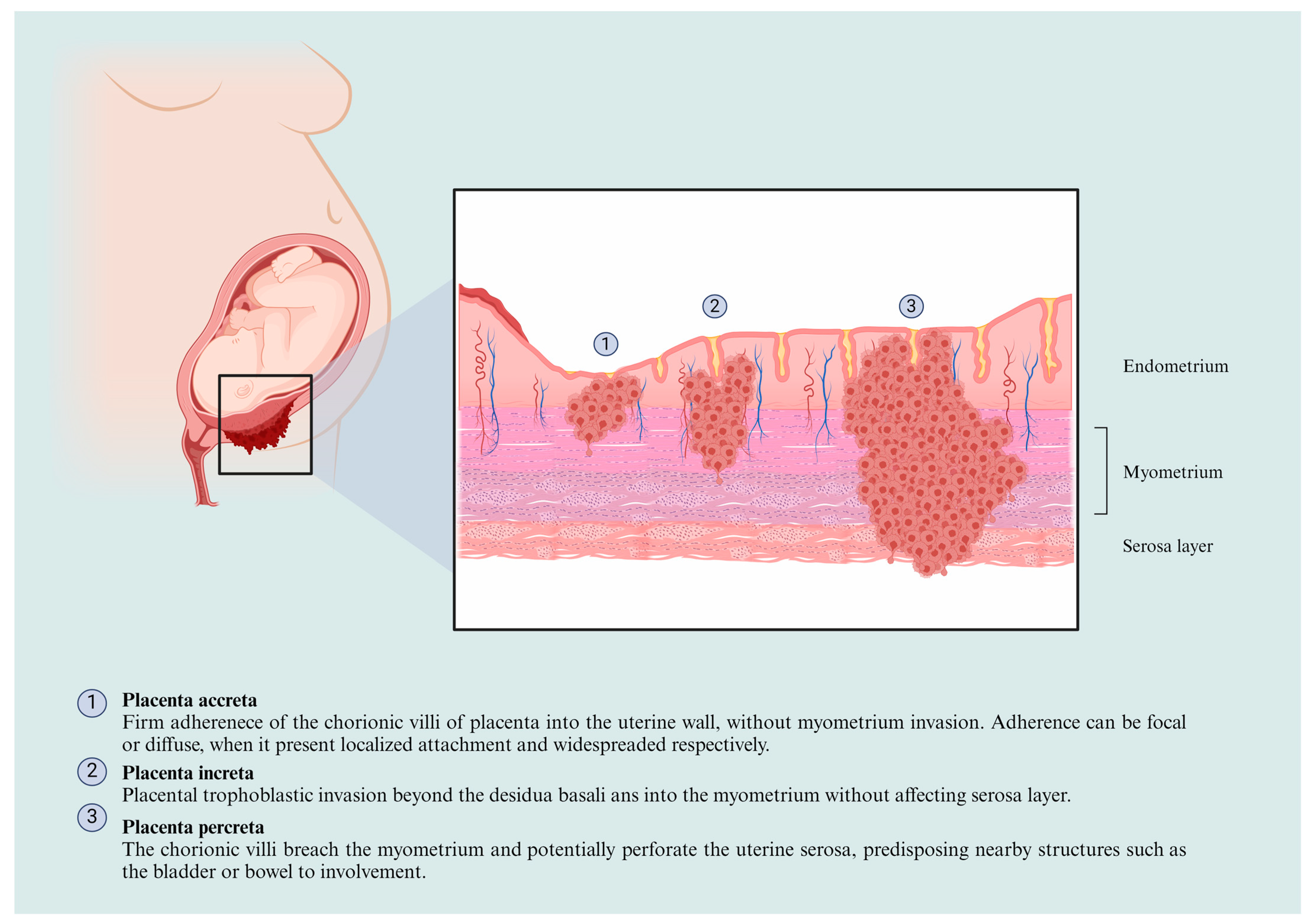
3. The PAS Classification and Physiopathological Features

The International Federation of Gynecology and Obstetrics (FIGOs) Committee first recognized the PAS classification for the Ethical Aspects of Human Reproduction and Women’s Health in 2011. The clinical distinction of morbid adherence of the placenta is associated with comprehensive prenatal diagnosis, a clinical approach, the correlation of treatment methods, and the frequency of severe obstetric complications [10]. Later, several retrospective and prospective studies were explicitly conducted to determine the classification influence on the results of the PAS treatment, which showed its significant impact on the choice of management [11].
In this regard, the classification of the PAS is based on the depth of placental invasion within the uterine wall and its extension of involvement. The Society for Maternal-Fetal Medicine (SMFM) developed a widely accepted classification system [12], dividing PAS into three categories: placenta accreta (PA), placenta increta (PI), and placenta percreta (PP), the features of which are presented in Figure 1.
The physiopathological features of the PAS involve a complex interplay of factors, including abnormal placentation, uterine scarring, and impaired decidualization. In normal pregnancies, the placenta separates easily from the uterine wall during childbirth due to the formation of a specialized layer known as decidua. Angiogenesis is a necessary cellular process for correct endometrial and embryonic growth and placentation. Invasion is pivotal for blastocyst differentiation into villous and extravillous trophoblasts (EVTs), acquiring pathological characteristics once they invade the decidua and myometrium [13]. Once implanted by migration, maternal uterine artery and vascular smooth muscle cells are replaced by trophoblasts through apoptosis [14].
However, in the PAS, the formation of the decidua is disrupted, leading to the abnormal adherence of the placenta [15]. The developmental behavior of severity in this spectrum comprises complex mechanisms, highlighting abnormal placentation, which allows trophoblast invasion to myometrium involving cellular differentiation, proliferation, and invasion in conjunction with growth factors and receptors [16].
Decidual deficiency is another factor that promotes placenta accreta development, and it has been linked with calcitonin and MAPK for trophoblast penetration into the endometrial epithelium [17]. Concerning trophoblastic invasion, molecules such as MMP-2 and 9, oxygen, and integrins are mainly involved [18]. In addition, it has previously been identified that PAS-associated proteins, including the vascular endothelial growth factor (VEGF), placenta growth factor (PlGF), along their respective receptors (VEGFR), epidermal growth factor receptor, c-erbB-2 oncoprotein, angiopoietin-1, angiopoietin-2, and Tie receptors [19,20,21]. Herein, cellular and molecular processes involved in the PAS are discussed.

4. Biomarkers Associated with Placenta Accreta Development

Clinical suspicion is based on the risk factors for the PAS and imaging findings [22]. However, the PAS is sometimes diagnosed only at delivery or by pathology since placental findings may not be visualized until the delivery time [23,24]. There is interest in identifying potential biomarkers for the PAS, particularly those that may be clinically useful and non-invasive.
Some researchers suggest combining serum biomarker values with imaging and clinical data to improve the diagnostic performance of ultrasonography and MRI-based methods for PAS disorders. The medical literature has shown that specific peripheral blood biomarkers, including those related to angiogenesis, the immune system, beta-human chorionic gonadotropin (β-hCG), and placental-derived cell-free DNA, may increase in patients with placental invasion disorders compared to normal pregnancies [25,26]. The most relevant biomarkers for the spectrum diagnosis are presented in Table 1.

5. Molecular Mechanisms Involved in the Placenta Acreta Spectrum

The PAS comprehends several mechanisms involving multifactorial processes, highlighting proliferation and invasion into local tissues, similar to a tumor. Other characteristics involved in PAS physiopathology include angiogenesis induction and cell death resistance, including the epithelial-to-mesenchymal transition (EMT) [40,41,42]. Despite several reports of placental pathologies, the precise molecular mechanisms of the PAS are still poorly understood. Here, we enlist the most relevant molecular mechanisms involved in the spectrum to date (Figure 2).

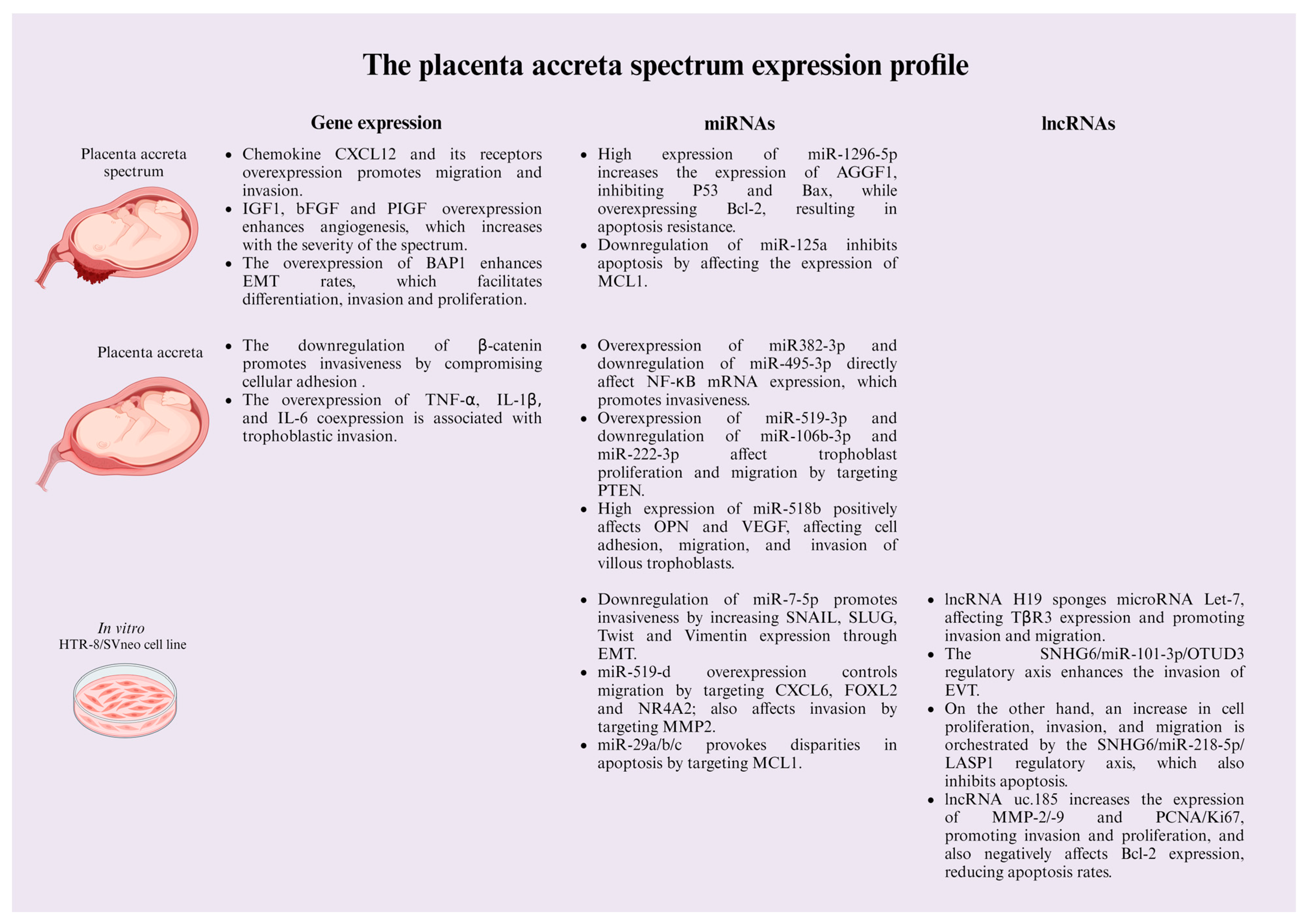
5.1. Gene Expression

In the process of invasion, proliferation, and migration, excessive trophoblast invasion and decidual deficiency are the main pathophysiological mechanisms of PA; these processes, in combination with other mechanisms such as endometrial invasion, migration through the myometrium, among others, trigger the spectrum [43].
In this regard, the chemokine CXCL12 and its receptors, CXCR4 and CXCR7, are known to play pivotal roles in the invasion process of trophoblast cells. Consequently, the mechanisms underlying the excessive invasion of trophoblasts in patients diagnosed with PAS present an upregulation of CXCL12 and CXCR4/CXCR7 in extravillous trophoblastic cells in a dose-dependent manner, increasing their proliferative capabilities at higher expressions, and contributing to the invasion into the uterine wall. Moreover, it was noted that the regulation of trophoblast migration and invasion are related to CXCL12 and CXCR4/CXCR7 expressions in the same manner, suggesting the participation of these chemokines in the PAS development [44]. On the other hand, a study conducted by Arakaza et al. indicated that the expression of insulin-like growth factor 1 (IGF-1), fibroblast growth factor 2 (bFGF), and PlGF are important for PAS development since their expression was found to be higher in PAS placental tissue in comparison to normal placental samples [19]. Interestingly, IGF-1 expression increased among them in relation to disease severity. This phenomenon might be associated with the function of IGF-1 in trophoblastic invasion because it acts as an angiogenic growth factor since it has been demonstrated to promote tubular formation through the activation of PI3 and _MAPK pathways in human endothelial cells. In addition, IGF-1 is overexpressed in women with endometriosis, favoring invasiveness, proliferation, decreased apoptosis, and angiogenesis on ectopic cells in conjunction with HGF and IGF-1 [45,46,47]. This last point might explain the behavior of IGF-1 expression in relation to severity since it is relative to invasion rates.
The role of β-catenin has been studied regarding placental disparities; it plays an important role in the maintenance of cellular homeostasis as well as intercellular connections, which consolidate cell adhesion [48]. Concerning placental affections, in women with placenta accreta, β-catenin expression is downregulated, leading to excessive trophoblastic invasion by losing interstitial connections [49]. In addition, EMT in trophoblasts has been linked with high BAP1 expression; this mechanism was elucidated by observing Bap1-null mouse trophoblast stem cells (mTSCs) using CRISPR/Cas9, resulting in augmented ETM rates, which, in turn, promotes differentiation, invasion, and proliferation capacities [50].
Inflammation is another mechanism involved in the PAS since an interaction exists between the trophoblast and the uterine tissue, resulting in the exacerbated release of proinflammatory mediators [51]. In this regard, in a recent study conducted by Abdel-Hamid, Mesbah, Soliman and Firgany [15], the expression of tumor necrosis factor-alpha (TNF-α), interleukin-1 beta (IL-1β), and IL6 were overexpressed in placentas of patients with placenta accreta; interestingly, their expression levels were associated with the number of EVT, which also were higher in comparison to normal placentation tissue samples. It was argued that it might be mainly comprised TNF-α due to its ability to inhibit trophoblastic invasion. In addition, the increased co-expression of TNF-α, IL-1β, and IL6 suggest their pivotal role as pro-inflammatory mediators in placenta accreta pathogenesis.

5.2. The Roles of Non-Coding RNAs in the PAS

Non-coding RNAs are pivotal in regulating biological processes, including pathologic development. ncRNAs comprise regulatory molecules, of which micro RNAs (miRNAs) and long non-coding RNAs (lncRNAs) are mainly addressed in the context of biological regulation [52]. Concerning placenta-related illnesses, there have been several ncRNAs identified for their roles in these pathologic disorders, mainly in intrauterine growth retardation, preeclampsia, and PAS stages [53]. Several studies have identified valuable information about how ncRNAs present differential expression levels and their roles in regulating the PAS [54]. Nonetheless, physiopathological regulation coffered by both miRNAs and lncRNAs is still limited. In this regard, we summarize ncRNA’s most relevant pathological roles based on PAS patients.

5.2.1. microRNAs

One of the key functions of miRNAs is regulating genes by mediating the degradation of mRNAs. miRNAs influence transcription and translation through two primary mechanisms: the canonical pathway, as briefly described earlier, involves the degradation of mRNAs based on the miRNA seed sequence [55]. Furthermore, circulating microRNAs have been demonstrated to play crucial roles in research and clinical settings, particularly in disease monitoring. Changes in circulating miRNAs have been linked to pathological processes, including chronic diseases, cancer, and the PAS [56,57].
miRNA behavior in PAS development is still poorly explored. Nonetheless, there is evidence that its role is related to cellular processes that accompany pathological development. In this regard, miR-7-5p plays an important role in trophoblast invasion since its overexpression converged in a significant diminution of cell invasion in HTR-8/SVneo cells; moreover, its downregulation results in an increase in SNAIL, SLUG, TWIST, and vimentin expression, promoting EMT and trophoblast invasion [58]. In extravillous trophoblast cells, miR-519d is highly expressed, and its main activity is to control migration by suppressing CXCL6, FOXL2, and NR4A2; in addition, MMP2 is a target gene of miR-519d, which suggests that it is involved in trophoblast invasiveness [59]. Murrieta-Coxca et al. [60] identified a plethora of deregulated expressed miRNAs in placenta accreta tissue; miR-24-3p, miR-193b-3p, miR-331-3p, miR-376c-3p, miR-382-3p, miR-495-3p, miR-519d-3p, and miR-3074-5p were overexpressed in PAS tissue while miR-106b-3p, miR-222-3p, miR-370-3p, miR-454-5p, and miR-3615-3p presented a downregulation in PAS tissue. Through biological pathway analysis, a significant reduction in NF-kB mRNA was identified, which was also confirmed in PAS samples, suggesting their influence of higher invasive capacities through the actions of miR-382-3p and miR-495-3p.
On the other hand, miR-106b-3p, miR-222-3p, and miR-519d-3p target PTEN, which controls the cell cycle, mainly in trophoblast proliferation and migration. The microRNA 1296-5p is overexpressed in the tissue of the PAS patient; its role is presumed to provide the regulation of apoptosis since its overexpression affected AGGF1, which, in turn, inhibits P53 and Bax expression while also increasing the expression of Bcl-2 protein [61]. In placenta accreta tissues, the overexpression of miR-518b was positively associated with OPN and VEGF, playing essential roles in regulating villous trophoblast cell migration, invasion, and adhesion [62]. In placenta accreta tissues, MCL1 expression is higher than in normal tissue, specifically in intermediate trophoblast cells, which inhibits apoptosis. The gene MCL1 has been reported as a target gene for both miR-29a/b/c and miR-125a; its cellular role results in the promotion of the apoptosis of trophoblast cells while downregulating MCL1 expression [63,64].

5.2.2. Long Non-Coding RNAs

Long non-coding RNAs (lncRNAs) are crucial because they play a vital role in maintaining homeostasis in biological processes. However, lncRNAs also contribute significantly to the development of diseases. Their biogenesis involves a comprehensive genomic system, including promoters, enhancers, and intergenic regions in eukaryotic genomes [65].
The roles exerted by lncRNAs are also pivotal in regulating cellular processes concerning placental invasive behaviors. In this regard, H19 downregulation can sponge miRNA let-7; as a consequence, TβR3 (type III TGF-β receptor) expression is negatively affected, promoting the invasion of EVT by increasing migration and invasion rates [66]. The high expression of lncRNA SNHG6 enhances the invasion of the human extravillous trophoblast HTR-8/SVneo cell line; this mechanism is argued to occur due to the SNHG6/miR-101-3p/OTUD3 regulatory axis [67]. Another regulatory axis in the PAS has been linked to lncRNA SNHG16 since its expression was diminished in placental affections, like preeclampsia. Nonetheless, its behavior has been evaluated in HTR-8/SVneo cells and is often overexpressed, resulting in the creation of cell proliferation, migration, and invasion as well as the inhibition of apoptosis, thus sponging miR-218-5p which, in turn, suppresses LASP1, which is a protein that facilitates cell invasion in diverse types of malignancies [68].
In addition, lncRNA uc.187 aberrant expression has been linked with higher proliferation rates, invasion, and lower apoptotic activities in HTR-8/SVneo cells. Regarding these mechanisms, they are activated by the increasing expression of MMP-2/-9 and PCNA/Ki-67 proteins, leading to enhanced invasion and proliferation, respectively; on the contrary, uc.187 overexpression affects the Bcl-2 protein, converging in a reduction in cell death in trophoblast cells [69].
Several reports indicate the cellular behavior of ncRNAs in placental pathologies. Since placenta accreta is an emerging pathology, and many aspects are still unknown. Comprehending molecular mechanisms can aid in developing early diagnostics and treatment in affected women.

6. Aberrant Signaling Pathways in the PAS

Several signaling pathways have been involved in the etiology of the PAS, such as the Notch signaling pathway, which is essential in regulating angiogenesis through the overexpression of periostin (POSTN) in the HUVEC cell line during the neovascularization process in conjunction with HES1 and Hey1 overexpression [70,71]. The above-mentioned pathway has been linked to an exacerbated hemorrhage in the PAS, mainly attributed to hypervascularity in the uteroplacental and utero–bladder interfaces [72].
Interestingly, AGGF1 is downregulated in PAS samples, and its deletion in human trophoblast HTR8/SVneo cells enhances mechanisms related to the invasive phenotype of PAS, such as proliferation, invasion, and migration. It also represses apoptosis by downregulating P53 and Bax and stimulating Bcl-2 overexpression, highlighting AGGF1 as a regulator of the P53 signaling axis [61].
Furthermore, the invasive trait of trophoblastic cells has been related to YKL-40 enhancer activity since it is overexpressed both in PAS samples and in vitro using HTR8/SVneo cells, promoting proliferation, migration, and invasion, but also inhibiting apoptosis through the activation of the Akt/MMP9 signaling pathway [73]. Additionally, STAT3, p38, and JNK pathways have been related to trophoblast invasion in the PAS since FYN stimulates the activation of STAT3, p38, and JNK through phosphorylation [74]. In addition, LAMC2 is overexpressed in placental cells, and in vitro has shown an increase in cell proliferation, invasion, and migration but inhibited apoptosis, accompanied by the elevated protein expression of MMP2, MMP9, and phosphorylated Akt (pAkt), which means that LAMC2 is implicated in the pathogenesis of PAS by activating the PI3K/Akt/MMP2/9 signaling pathway to stimulate trophoblast over-invasion [75].
Duan et al. [76] found that immunoblotting and qPCR analysis in abnormally invasive placentas (AIP), including PA, PI, or PP, showed that CCN3 overexpression is accompanied by high levels of p53, p16, p21, cyclin D1, Notch-1 cleaved, pFAK, pAkt, and pmTOR, as well as low levels of pRb, suggesting that CCN3 mediates senescence by cell cycle arrest through the activation of the FAK-Akt-mTOR pathway and cleaved Notch-1/p21, contributing to increasing the invasion properties of EVT.
Furthermore, growth factor signaling pathways, such as the macrophage-induced netrin-1/DCC/VEGF signaling pathway, have been implicated in trophoblastic angiogenesis in the PAS tissues through netrin-1, DCC receptors, VEGF overexpression, and the high recruitment of macrophages compared to normal placental tissue [77]. Likewise, using in vitro assays using the gestational choriocarcinoma cell line JEG-3 and the trophoblast cell line HTR-8/SVneo, it was found that the non-canonical TGF-β-UCHL5-Smad2 signaling pathway is essential for the invasion of EVTs, a critical step in placental development, in which Smad1/5/9 are the governing factors. In addition, the TGF-β-UCHL5-Smad2 pathway is also regulated by the ERK signaling pathway since it promotes angiogenesis and vascularization, and the alteration of these signaling pathways can cause abnormal placental invasion and angiogenesis, which leads to triggering PA [78]. Another novel mechanism described as involved in the development of the PAS consists of suppressing Wnt-β-catenin/VEGF signaling through the pigment epithelium-derived factor (PEDF), which is downregulated in PAS tissues [79]. In addition, it has been reported that PEDF overexpression inhibits EVTs proliferation, invasion, and angiogenesis and induces ferroptosis, a newly described form of regulated cell death creating a favorable scenario for adequate trophoblastic invasion; -

7. Perspectives and Conclusions

We provide an overview of available therapeutic strategies that could aid in the early diagnosis of the PAS. It is essential to comprehend the molecular mechanisms underlying the PAS, particularly the signaling pathways involved in the early stages accompanying this pathology, which is addressed by trophoblastic invasiveness and angiogenesis. Researchers aim to develop specific biomarkers that can effectively prevent the PAS. Moreover, exploring the signaling pathways involved could provide valuable tools to develop targeted therapies that treat the PAS, which is mainly attributed to TGF-β-UCHL5-Smad2, ERK, and Wnt-β-catenin/VEGF pathways, which play significant roles in the development and progression of the PAS. Therapeutic strategies targeting these pathways could potentially inhibit abnormal placental invasion and angiogenesis.
Additionally, implementing other molecules as novel biomarkers, such as PEDF, is pivotal to reducing the severity of the PAS since it is downregulated in pathological tissues. Otherwise, the overexpression of PEDF in vitro has been shown to inhibit the proliferation, invasion, and angiogenesis of EVTs and induce ferroptosis, suggesting a promising therapeutic strategy.
Physiopathology accompanying the spectrum is a multifactorial interplay of abnormal placentation, uterine scarring, and impaired decidualization, resulting in abnormal placental adherence [80]. Promising advances in identifying new biomarkers, such as AFP, sFlt-1, β-hCG, PlGF, and PAPP-A, could aid in the early detection and correct management of affected women [21,27,37]. In addition, the comprehension of molecular mechanisms, such as genetic expression and the roles of non-coding RNAs, are pivotal for unveiling the complexities of the PAS [43,53,54].
In addition, identifying aberrant signaling pathways, such as Notch, PI3K/Akt, STAT3, and TGF-β, offers potential targets for therapeutic interventions to modulate trophoblastic invasion and improve patient outcomes since they play pivotal roles in the invasiveness of trophoblastic cells contributing to the PAS pathogenesis [71,74]
Overall, this review highlights the importance of interdisciplinary care, early diagnosis, and a comprehensive understanding of the molecular underpinnings of PAS. Continued research into PAS’s biomarkers and molecular mechanisms is crucial for developing effective diagnostic and therapeutic strategies, ultimately improving affected women’s prognosis and quality of life.

Author Contributions

E.L.-V. and C.E.M.-P. conceived and designed the content of this review, E.L.-V., S.A.B.-O., E.P.G.-G., M.M.-M., P.Y.G.-A., M.A.-F., K.P.G.-C., D.E.C.-L., P.G.-Q. and C.E.M.-P. wrote the paper, E.L.-V., S.A.B.-O. and E.P.G.-G. conceived supervision and visualization of manuscript, C.E.M.-P., E.L.-V. and E.P.G.-G. contributed to revising and editing the paper critically for important intellectual content. All authors have read and agreed to the published version of the manuscript.

Funding

This research received no external funding.

Data Availability Statement

No new data were created or analyzed in this study. Data sharing is not applicable to this article.

Conflicts of Interest

The authors declare no conflicts of interest.

References

  1. Markfeld Erol, F.; Häußler, J.A.; Medl, M.; Juhasz-Boess, I.; Kunze, M. Placenta Accreta Spectrum (PAS): Diagnosis, Clinical Presentation, Therapeutic Approaches, and Clinical Outcomes. Medicina 2024, 60, 1180. [Google Scholar] [CrossRef] [PubMed]
  2. Piñas Carrillo, A.; Chandraharan, E. Placenta accreta spectrum: Risk factors, diagnosis and management with special reference to the Triple P procedure. Women’s Health 2019, 15, 1745506519878081. [Google Scholar] [CrossRef]
  3. Cahill, A.G.; Beigi, R.; Heine, P.; Silver, R.M.; Wax, J.R. Obstetric Care Consensus No. 7: Placenta Accreta Spectrum. Obstet. Gynecol. 2018, 132, e259–e275. [Google Scholar] [CrossRef]
  4. Bowman, Z.S.; Eller, A.G.; Bardsley, T.R.; Greene, T.; Varner, M.W.; Silver, R.M. Risk factors for placenta accreta: A large prospective cohort. Am. J. Perinatol. 2014, 31, 799–804. [Google Scholar] [CrossRef] [PubMed]
  5. Marshall, N.E.; Fu, R.; Guise, J.M. Impact of multiple cesarean deliveries on maternal morbidity: A systematic review. Am. J. Obstet. Gynecol. 2011, 205, 262.e1–262.e8. [Google Scholar] [CrossRef]
  6. Jauniaux, E.; Jurkovic, D. Placenta accreta: Pathogenesis of a 20th century iatrogenic uterine disease. Placenta 2012, 33, 244–251. [Google Scholar] [CrossRef] [PubMed]
  7. Jauniaux, E.; Chantraine, F.; Silver, R.M.; Langhoff-Roos, J. FIGO consensus guidelines on placenta accreta spectrum disorders: Epidemiology. Int. J. Gynecol. Obstet. 2018, 140, 265–273. [Google Scholar] [CrossRef] [PubMed]
  8. Betrán, A.P.; Ye, J.; Moller, A.B.; Zhang, J.; Gülmezoglu, A.M.; Torloni, M.R. The Increasing Trend in Caesarean Section Rates: Global, Regional and National Estimates: 1990–2014. PLoS ONE 2016, 11, e0148343. [Google Scholar] [CrossRef]
  9. Ye, J.; Zhang, J.; Mikolajczyk, R.; Torloni, M.R.; Gülmezoglu, A.M.; Betran, A.P. Association between rates of caesarean section and maternal and neonatal mortality in the 21st century: A worldwide population-based ecological study with longitudinal data. BJOG Int. J. Obstet. Gynaecol. 2016, 123, 745–753. [Google Scholar] [CrossRef]
  10. Gloria, C.; Salvatore, P.; Francesco, L.; Anna, G.R.; Francesca, M.; Chiara, B.; Gaspare, C.; Giuseppe, C. Placenta Accreta Spectrum Disorder in a Patient with Six Previous Caesarean Deliveries: Step by Step Management. Case Rep. Obstet. Gynecol. 2021, 2021, 2105248. [Google Scholar] [CrossRef]
  11. van Beekhuizen, H.J.; Stefanovic, V.; Schwickert, A.; Henrich, W.; Fox, K.A.; MHallem Gziri, M.; Sentilhes, L.; Gronbeck, L.; Chantraine, F.; Morel, O.; et al. A multicenter observational survey of management strategies in 442 pregnancies with suspected placenta accreta spectrum. Acta Obstet. Gynecol. Scand. 2021, 100 (Suppl. S1), 12–20. [Google Scholar] [CrossRef] [PubMed]
  12. Zheng, W.; Zhang, H.; Ma, J.; Dou, R.; Zhao, X.; Yan, J.; Yang, H. Validation of a scoring system for prediction of obstetric complications in placenta accreta spectrum disorders. J. Matern.-Fetal Neonatal Med. 2022, 35, 4149–4155. [Google Scholar] [CrossRef] [PubMed]
  13. Jansen, C.; Kastelein, A.W.; Kleinrouweler, C.E.; Van Leeuwen, E.; De Jong, K.H.; Pajkrt, E.; Van Noorden, C.J.F. Development of placental abnormalities in location and anatomy. Acta Obstet. Gynecol. Scand. 2020, 99, 983–993. [Google Scholar] [CrossRef] [PubMed]
  14. Li, Y.; Lorca, R.A.; Su, E.J. Molecular and cellular underpinnings of normal and abnormal human placental blood flows. J. Mol. Endocrinol. 2018, 60, R9–R22. [Google Scholar] [CrossRef]
  15. Abdel-Hamid, A.A.M.; Mesbah, Y.; Soliman, M.F.M.; Firgany, A.E.L. Dominance of Pro-Inflammatory Cytokines Over Anti-Inflammatory Ones in Placental Bed of Creta Cases. J. Microsc. Ultrastruct. 2024, 12, 14–20. [Google Scholar] [CrossRef]
  16. Tantbirojn, P.; Crum, C.P.; Parast, M.M. Pathophysiology of Placenta Creta: The Role of Decidua and Extravillous Trophoblast. Placenta 2008, 29, 639–645. [Google Scholar] [CrossRef]
  17. Wang, S.C.; Yu, M.; Li, Y.H.; Piao, H.L.; Tang, C.L.; Sun, C.; Zhu, R.; Li, M.Q.; Jin, L.P.; Li, D.J.; et al. Cyclosporin A promotes proliferating cell nuclear antigen expression and migration of human cytotrophoblast cells via the mitgen-activated protein kinase-3/1-mediated nuclear factor-κB signaling pathways. Int. J. Clin. Exp. Pathol. 2013, 6, 1999–2010. [Google Scholar]
  18. Gualdoni, G.; Gomez Castro, G.; Hernández, R.; Barbeito, C.; Cebral, E. Comparative matrix metalloproteinase-2 and -9 expression and activity during endotheliochorial and hemochorial trophoblastic invasiveness. Tissue and Cell 2022, 74, 101698. [Google Scholar] [CrossRef]
  19. Arakaza, A.; Liu, X.; Zhu, J.; Zou, L. Assessment of serum levels and placental bed tissue expression of IGF-1, bFGF, and PLGF in patients with placenta previa complicated with placenta accreta spectrum disorders. J. Matern.-Fetal Neonatal Med. 2024, 37, 2305264. [Google Scholar] [CrossRef]
  20. Schwickert, A.; Chantraine, F.; Ehrlich, L.; Henrich, W.; Muallem, M.Z.; Nonnenmacher, A.; Petit, P.; Weizsäcker, K.; Braun, T. Maternal Serum VEGF Predicts Abnormally Invasive Placenta Better than NT-proBNP: A Multicenter Case-Control Study. Reprod. Sci. 2021, 28, 361–370. [Google Scholar] [CrossRef]
  21. Zhang, F.; Gu, M.; Chen, P.; Wan, S.; Zhou, Q.; Lu, Y.; Li, L. Distinguishing placenta accreta from placenta previa via maternal plasma levels of sFlt-1 and PLGF and the sFlt-1/PLGF ratio. Placenta 2022, 124, 48–54. [Google Scholar] [CrossRef] [PubMed]
  22. Romeo, V.; Verde, F.; Sarno, L.; Migliorini, S.; Petretta, M.; Mainenti, P.P.; D’Armiento, M.; Guida, M.; Brunetti, A.; Maurea, S. Prediction of placenta accreta spectrum in patients with placenta previa using clinical risk factors, ultrasound and magnetic resonance imaging findings. La Radiol. Medica 2021, 126, 1216–1225. [Google Scholar] [CrossRef] [PubMed]
  23. Liu, X.; Wang, Y.; Wu, Y.; Zeng, J.; Yuan, X.; Tong, C.; Qi, H. What we know about placenta accreta spectrum (PAS). Eur. J. Obstet. Gynecol. Reprod. Biol. 2021, 259, 81–89. [Google Scholar] [CrossRef] [PubMed]
  24. Hecht, J.L.; Baergen, R.; Ernst, L.M.; Katzman, P.J.; Jacques, S.M.; Jauniaux, E.; Khong, T.Y.; Metlay, L.A.; Poder, L.; Qureshi, F.; et al. Classification and reporting guidelines for the pathology diagnosis of placenta accreta spectrum (PAS) disorders: Recommendations from an expert panel. Mod. Pathol. 2020, 33, 2382–2396. [Google Scholar] [CrossRef]
  25. Shainker, S.A.; Silver, R.M.; Modest, A.M.; Hacker, M.R.; Hecht, J.L.; Salahuddin, S.; Dillon, S.T.; Ciampa, E.J.; D’Alton, M.E.; Otu, H.H.; et al. Placenta accreta spectrum: Biomarker discovery using plasma proteomics. Am. J. Obstet. Gynecol. 2020, 223, 433.e1–433.e14. [Google Scholar] [CrossRef]
  26. Zhang, T.; Wang, S. Potential Serum Biomarkers in Prenatal Diagnosis of Placenta Accreta Spectrum. Front. Med. 2022, 9, 860186. [Google Scholar] [CrossRef]
  27. Berezowsky, A.; Pardo, J.; Ben-Zion, M.; Wiznitzer, A.; Aviram, A. Second Trimester Biochemical Markers as Possible Predictors of Pathological Placentation: A Retrospective Case-Control Study. Fetal Diagn. Ther. 2019, 46, 187–192. [Google Scholar] [CrossRef]
  28. Oztas, E.; Ozler, S.; Caglar, A.T.; Yucel, A. Analysis of first and second trimester maternal serum analytes for the prediction of morbidly adherent placenta requiring hysterectomy. Kaohsiung J. Med. Sci. 2016, 32, 579–585. [Google Scholar] [CrossRef]
  29. Lumbanraja, S.; Yaznil, M.R.; Siahaan, A.M.; Berry Eka Parda, B. Soluble FMS-Like Tyrosine Kinase-1: Role in placenta accreta spectrum disorder. F1000Research 2022, 10, 618. [Google Scholar] [CrossRef]
  30. Büke, B.; Akkaya, H.; Demir, S.; Sağol, S.; Şimşek, D.; Başol, G.; Barutçuoğlu, B. Relationship between first trimester aneuploidy screening test serum analytes and placenta accreta. J. Matern.-Fetal Neonatal Med. 2018, 31, 59–62. [Google Scholar] [CrossRef]
  31. Zhou, J.; Li, J.; Yan, P.; Ye, Y.H.; Peng, W.; Wang, S.; Wang, X.T. Maternal plasma levels of cell-free β-HCG mRNA as a prenatal diagnostic indicator of placenta accrete. Placenta 2014, 35, 691–695. [Google Scholar] [CrossRef] [PubMed]
  32. Faraji, A.; Akbarzadeh-Jahromi, M.; Bahrami, S.; Gharamani, S.; Raeisi Shahraki, H.; Kasraeian, M.; Vafaei, H.; Zare, M.; Asadi, N. Predictive value of vascular endothelial growth factor and placenta growth factor for placenta accreta spectrum. J. Obstet. Gynaecol. 2022, 42, 900–905. [Google Scholar] [CrossRef] [PubMed]
  33. Wang, F.; Chen, S.; Wang, J.; Wang, Y.; Ruan, F.; Shu, H.; Zhu, L.; Man, D. First trimester serum PAPP-A is associated with placenta accreta: A retrospective study. Arch. Gynecol. Obstet. 2021, 303, 645–652. [Google Scholar] [CrossRef]
  34. Desai, N.; Krantz, D.; Roman, A.; Fleischer, A.; Boulis, S.; Rochelson, B. Elevated first trimester PAPP-A is associated with increased risk of placenta accreta. Prenat. Diagn. 2014, 34, 159–162. [Google Scholar] [CrossRef]
  35. Thompson, O.; Otigbah, C.; Nnochiri, A.; Sumithran, E.; Spencer, K. First trimester maternal serum biochemical markers of aneuploidy in pregnancies with abnormally invasive placentation. BJOG Int. J. Obstet. Gynaecol. 2015, 122, 1370–1376. [Google Scholar] [CrossRef]
  36. Lyell, D.J.; Faucett, A.M.; Baer, R.J.; Blumenfeld, Y.J.; Druzin, M.L.; El-Sayed, Y.Y.; Shaw, G.M.; Currier, R.J.; Jelliffe-Pawlowski, L.L. Maternal serum markers, characteristics and morbidly adherent placenta in women with previa. J. Perinatol. 2015, 35, 570–574. [Google Scholar] [CrossRef] [PubMed]
  37. Penzhoyan, G.A.; Makukhina, T.B. Significance of the routine first-trimester antenatal screening program for aneuploidy in the assessment of the risk of placenta accreta spectrum disorders. J. Perinat. Med. 2019, 48, 21–26. [Google Scholar] [CrossRef]
  38. Kawashima, A.; Sekizawa, A.; Ventura, W.; Koide, K.; Hori, K.; Okai, T.; Masashi, Y.; Furuya, K.; Mizumoto, Y. Increased levels of cell-free human placental lactogen mRNA at 28-32 gestational weeks in plasma of pregnant women with placenta previa and invasive placenta. Reprod. Sci. 2014, 21, 215–220. [Google Scholar] [CrossRef]
  39. Li, J.; Zhang, N.; Zhang, Y.; Hu, X.; Gao, G.; Ye, Y.; Peng, W.; Zhou, J. Human placental lactogen mRNA in maternal plasma play a role in prenatal diagnosis of abnormally invasive placenta: Yes or no? Gynecol. Endocrinol. 2019, 35, 631–634. [Google Scholar] [CrossRef]
  40. Shainker, S.A.; Dannheim, K.; Gerson, K.D.; Neo, D.; Zsengeller, Z.K.; Pernicone, E.; Karumanchi, S.A.; Hacker, M.R.; Hecht, J.L. Down-regulation of soluble fms-like tyrosine kinase 1 expression in invasive placentation. Arch. Gynecol. Obstet. 2017, 296, 257–262. [Google Scholar] [CrossRef]
  41. Goh, W.; Yamamoto, S.Y.; Thompson, K.S.; Bryant-Greenwood, G.D. Relaxin, its receptor (RXFP1), and insulin-like peptide 4 expression through gestation and in placenta accreta. Reprod. Sci. 2013, 20, 968–980. [Google Scholar] [CrossRef] [PubMed]
  42. Bartels, H.C.; Postle, J.D.; Downey, P.; Brennan, D.J. Placenta Accreta Spectrum: A Review of Pathology, Molecular Biology, and Biomarkers. Dis. Markers 2018, 2018, 1507674. [Google Scholar] [CrossRef]
  43. Illsley, N.P.; DaSilva-Arnold, S.C.; Zamudio, S.; Alvarez, M.; Al-Khan, A. Trophoblast invasion: Lessons from abnormally invasive placenta (placenta accreta). Placenta 2020, 102, 61–66. [Google Scholar] [CrossRef]
  44. Long, Y.; Jiang, Y.; Zeng, J.; Dang, Y.; Chen, Y.; Lin, J.; Wei, H.; Xia, H.; Long, J.; Luo, C.; et al. The expression and biological function of chemokine CXCL12 and receptor CXCR4/CXCR7 in placenta accreta spectrum disorders. J. Cell. Mol. Med. 2020, 24, 3167–3182. [Google Scholar] [CrossRef]
  45. Heidari, S.; Kolahdouz-Mohammadi, R.; Khodaverdi, S.; Tajik, N.; Delbandi, A.-A. Expression levels of MCP-1, HGF, and IGF-1 in endometriotic patients compared with non-endometriotic controls. BMC Women’s Health 2021, 21, 422. [Google Scholar] [CrossRef]
  46. Shigematsu, S.; Yamauchi, K.; Nakajima, K.; Iijima, S.; Aizawa, T.; Hashizume, K. IGF-1 Regulates Migration and Angiogenesis of Human Endothelial Cells. Endocr. J. 1999, 46, S59–S62. [Google Scholar] [CrossRef]
  47. He, X.; Zhao, L.; Yue, L.; Zhang, W.; Wang, W.; Fu, Y.; Feng, Y.; Fu, F. The relationship between IGF1 and the expression spectrum of miRNA in the placenta of preeclampsia patients. Ginekol. Pol. 2019, 90, 596–603. [Google Scholar] [CrossRef] [PubMed]
  48. Pedone, E.; Marucci, L. Role of β-Catenin Activation Levels and Fluctuations in Controlling Cell Fate. Genes 2019, 10, 176. [Google Scholar] [CrossRef] [PubMed]
  49. Han, Q.; Zheng, L.; Liu, Z.; Luo, J.; Chen, R.; Yan, J. Expression of β-catenin in human trophoblast and its role in placenta accreta and placenta previa. J. Int. Med. Res. 2019, 47, 206–214. [Google Scholar] [CrossRef] [PubMed]
  50. Perez-Garcia, V.; Lea, G.; Lopez-Jimenez, P.; Okkenhaug, H.; Burton, G.J.; Moffett, A.; Turco, M.Y.; Hemberger, M. BAP1/ASXL complex modulation regulates epithelial-mesenchymal transition during trophoblast differentiation and invasion. eLife 2021, 10, e63254. [Google Scholar] [CrossRef]
  51. Cheng, S.B.; Nakashima, A.; Huber, W.J.; Davis, S.; Banerjee, S.; Huang, Z.; Saito, S.; Sadovsky, Y.; Sharma, S. Pyroptosis is a critical inflammatory pathway in the placenta from early onset preeclampsia and in human trophoblasts exposed to hypoxia and endoplasmic reticulum stressors. Cell Death Dis. 2019, 10, 927. [Google Scholar] [CrossRef]
  52. Mattick, J.S.; Amaral, P.P.; Carninci, P.; Carpenter, S.; Chang, H.Y.; Chen, L.-L.; Chen, R.; Dean, C.; Dinger, M.E.; Fitzgerald, K.A.; et al. Long non-coding RNAs: Definitions, functions, challenges and recommendations. Nat. Rev. Mol. Cell Biol. 2023, 24, 430–447. [Google Scholar] [CrossRef] [PubMed]
  53. Monteiro, L.J.; Peñailillo, R.; Sánchez, M.; Acuña-Gallardo, S.; Mönckeberg, M.; Ong, J.; Choolani, M.; Illanes, S.E.; Nardocci, G. The Role of Long Non-Coding RNAs in Trophoblast Regulation in Preeclampsia and Intrauterine Growth Restriction. Genes 2021, 12, 970. [Google Scholar] [CrossRef] [PubMed]
  54. Kannampuzha, S.; Ravichandran, M.; Mukherjee, A.G.; Wanjari, U.R.; Renu, K.; Vellingiri, B.; Iyer, M.; Dey, A.; George, A.; Gopalakrishnan, A.V. The mechanism of action of non-coding RNAs in placental disorders. Biomed. Pharmacother. 2022, 156, 113964. [Google Scholar] [CrossRef] [PubMed]
  55. He, L.; Hannon, G.J. MicroRNAs: Small RNAs with a big role in gene regulation. Nat. Rev. Genet. 2004, 5, 522–531. [Google Scholar] [CrossRef] [PubMed]
  56. Chen, P.-S.; Su, J.-L.; Hung, M.-C. Dysregulation of MicroRNAs in cancer. J. Biomed. Sci. 2012, 19, 90. [Google Scholar] [CrossRef]
  57. He, Y.; Ding, Y.; Liang, B.; Lin, J.; Kim, T.K.; Yu, H.; Hang, H.; Wang, K. A Systematic Study of Dysregulated MicroRNA in Type 2 Diabetes Mellitus. Int. J. Mol. Sci. 2017, 18, 456. [Google Scholar] [CrossRef]
  58. Shih, J.C.; Lin, H.H.; Hsiao, A.C.; Su, Y.T.; Tsai, S.; Chien, C.L.; Kung, H.N. Unveiling the role of microRNA-7 in linking TGF-β-Smad-mediated epithelial-mesenchymal transition with negative regulation of trophoblast invasion. FASEB J. 2019, 33, 6281–6295. [Google Scholar] [CrossRef]
  59. Xie, L.; Sadovsky, Y. The function of miR-519d in cell migration, invasion, and proliferation suggests a role in early placentation. Placenta 2016, 48, 34–37. [Google Scholar] [CrossRef]
  60. Murrieta-Coxca, J.M.; Barth, E.; Fuentes-Zacarias, P.; Gutiérrez-Samudio, R.N.; Groten, T.; Gellhaus, A.; Köninger, A.; Marz, M.; Markert, U.R.; Morales-Prieto, D.M. Identification of altered miRNAs and their targets in placenta accreta. Front. Endocrinol. 2023, 14, 1021640. [Google Scholar] [CrossRef]
  61. Wang, R.; Zhao, J.; Liu, C.; Li, S.; Liu, W.; Cao, Q. Decreased AGGF1 facilitates the progression of placenta accreta spectrum via mediating the P53 signaling pathway under the regulation of miR-1296-5p. Reprod. Biol. 2023, 23, 100735. [Google Scholar] [CrossRef] [PubMed]
  62. Long, Y.; Chen, Y.; Fu, X.Q.; Yang, F.; Chen, Z.W.; Mo, G.L.; Lao, D.Y.; Li, M.J. Research on the expression of MRNA-518b in the pathogenesis of placenta accreta. Eur. Rev. Med. Pharmacol. Sci. 2019, 23, 23–28. [Google Scholar] [CrossRef]
  63. Gu, Y.; Meng, J.; Zuo, C.; Wang, S.; Li, H.; Zhao, S.; Huang, T.; Wang, X.; Yan, J. Downregulation of MicroRNA-125a in Placenta Accreta Spectrum Disorders Contributes Antiapoptosis of Implantation Site Intermediate Trophoblasts by Targeting MCL1. Reprod. Sci. 2019, 26, 1582–1589. [Google Scholar] [CrossRef]
  64. Gu, Y.; Bian, Y.; Xu, X.; Wang, X.; Zuo, C.; Meng, J.; Li, H.; Zhao, S.; Ning, Y.; Cao, Y.; et al. Downregulation of miR-29a/b/c in placenta accreta inhibits apoptosis of implantation site intermediate trophoblast cells by targeting MCL1. Placenta 2016, 48, 13–19. [Google Scholar] [CrossRef] [PubMed]
  65. Ha, M.; Kim, V.N. Regulation of microRNA biogenesis. Nat. Rev. Mol. Cell Biol. 2014, 15, 509–524. [Google Scholar] [CrossRef] [PubMed]
  66. Zuckerwise, L.; Li, J.; Lu, L.; Men, Y.; Geng, T.; Buhimschi, C.S.; Buhimschi, I.A.; Bukowski, R.; Guller, S.; Paidas, M.; et al. H19 long noncoding RNA alters trophoblast cell migration and invasion by regulating TβR3 in placentae with fetal growth restriction. Oncotarget 2016, 7, 38398–38407. [Google Scholar] [CrossRef]
  67. Zhao, L.; Liu, X.; Ren, C.; Zhang, H.; Gao, L. Silencing of LncRNA SNHG6 protects trophoblast cells through regulating miR-101-3p/OTUD3 axis in unexplained recurrent spontaneous abortion. Histochem. J. 2022, 53, 871–882. [Google Scholar] [CrossRef]
  68. Yu, Z.; Zhang, Y.; Zheng, H.; Gao, Q.; Wang, H. LncRNA SNHG16 regulates trophoblast functions by the miR-218-5p/LASP1 axis. Histochem. J. 2021, 52, 1021–1033. [Google Scholar] [CrossRef]
  69. Cao, C.; Li, J.; Li, J.; Liu, L.; Cheng, X.; Jia, R. Long Non-Coding RNA Uc.187 Is Upregulated in Preeclampsia and Modulates Proliferation, Apoptosis, and Invasion of HTR-8/SVneo Trophoblast Cells. J. Cell. Biochem. 2017, 118, 1462–1470. [Google Scholar] [CrossRef]
  70. Zhou, B.; Lin, W.; Long, Y.; Yang, Y.; Zhang, H.; Wu, K.; Chu, Q. Notch signaling pathway: Architecture, disease, and therapeutics. Signal Transduct. Target. Ther. 2022, 7, 95. [Google Scholar] [CrossRef]
  71. Li, R.; Wang, W.; Qiu, X.; He, M.; Tang, X.; Zhong, M. Periostin promotes extensive neovascularization in placenta accreta spectrum disorders via Notch signaling. J. Matern.-Fetal Neonatal Med. 2023, 36, 2264447. [Google Scholar] [CrossRef] [PubMed]
  72. Calì, G.; D’Antonio, F.; Forlani, F.; Timor-Tritsch, I.E.; Palacios-Jaraquemada, J.M. Ultrasound Detection of Bladder-Uterovaginal Anastomoses in Morbidly Adherent Placenta. Fetal Diagn. Ther. 2017, 41, 239–240. [Google Scholar] [CrossRef] [PubMed]
  73. Liu, W.; Wang, R.; Liu, S.; Yin, X.; Huo, Y.; Zhang, R.; Li, J. YKL-40 promotes proliferation and invasion of HTR-8/SVneo cells by activating akt/MMP9 signalling in placenta accreta spectrum disorders. J. Obstet. Gynaecol. 2023, 43, 2211681. [Google Scholar] [CrossRef] [PubMed]
  74. Liu, M.; Su, C.; Zhu, L.; Dong, F.; Shu, H.; Zhang, H.; Wang, M.; Wang, F.; Man, D. Highly expressed FYN promotes the progression of placenta accreta by activating STAT3, p38, and JNK signaling pathways. Acta Histochem. 2023, 125, 151991. [Google Scholar] [CrossRef]
  75. Wang, R.; Liu, W.; Zhao, J.; Liu, L.; Li, S.; Duan, Y.; Huo, Y. Overexpressed LAMC2 promotes trophoblast over-invasion through the PI3K/Akt/MMP2/9 pathway in placenta accreta spectrum. J. Obstet. Gynaecol. Res. 2023, 49, 548–559. [Google Scholar] [CrossRef]
  76. Duan, L.; Schimmelmann, M.; Wu, Y.; Reisch, B.; Faas, M.; Kimmig, R.; Winterhager, E.; Köninger, A.; Gellhaus, A. CCN3 Signaling Is Differently Regulated in Placental Diseases Preeclampsia and Abnormally Invasive Placenta. Front. Endocrinol. 2020, 11, 597549. [Google Scholar] [CrossRef]
  77. Badary, D.M.; Elsaied, H.; Abdel-Fadeil, M.R.; Ali, M.K.; Abou-Taleb, H.; Iraqy, H.M. Possible Role of Netrin-1/Deleted in Colorectal Cancer/Vascular Endothelial Growth Factor Signaling Pathway in the Pathogenesis of Placenta Accreta Spectrum: A Case-control Study. Int. J. Gynecol. Pathol. 2024. [Google Scholar] [CrossRef]
  78. Hashimoto, K.; Miyagawa, Y.; Watanabe, S.; Takasaki, K.; Nishizawa, M.; Yatsuki, K.; Takahashi, Y.; Kamata, H.; Kihira, C.; Hiraike, H.; et al. The TGF-β/UCHL5/Smad2 Axis Contributes to the Pathogenesis of Placenta Accreta. Int. J. Mol. Sci. 2023, 24, 13706. [Google Scholar] [CrossRef]
  79. Li, R.; Weng, X.; Hu, X.; Wang, J.; Zheng, L. Pigment epithelium-derived factor inhibits proliferation, invasion and angiogenesis, and induces ferroptosis of extravillous trophoblasts by targeting Wnt-β-catenin/VEGF signaling in placenta accreta spectrum. Mol. Med. Rep. 2024, 29, 75. [Google Scholar] [CrossRef]
  80. Morlando, M.; Collins, S. Placenta accreta spectrum disorders: Challenges, risks, and management strategies. Int. J. Women’s Health 2020, 12, 1033–1045. [Google Scholar] [CrossRef]
Figure 1. The PAS classification. The invasive cells start at the uterine wall (1); the severity of the spectrum is relative to the invasive depth, affecting the myometrium layer (2) and subsequently invading the serosa layer (3). Figure created by https://www.biorender.com/. Accessed on 7 May 2024.
Ijms 25 09722 g001
Figure 2. The PAS expression profile. The placenta accreta spectrum is accompanied by changes in the expression of several genes and non-coding RNAs, which are crucial in the dysregulation of cellular processes, such as migration, invasiveness, proliferation, and resistance to cell death, which facilitates trophoblastic invasion into the uterine layers. Figure created by https://www.biorender.com/. Accessed on 7 May 2024.
Ijms 25 09722 g002
Table 1. Biomarkers related to the PAS.
BiomarkerSourceFindingsReferences
Alpha-fetoprotein
(AFP)
Maternal serumAPF showed sensitivity and specificity of 71 and 46%, respectively, to serve as a biomarker for pathological placentation, specifically in women with placenta previa and acreta in the second trimester. Thus, a high level of AFP can be used as a cause for suspicion in high-risk pathological placentation.[27]
Maternal serum AFP levels were associated with PAS patients; it was established as a predictor for PAS patients that require hysterectomy with 85.94% sensitivity and 71.43% specificity.[28]
Soluble fms-like tyrosine kinase-1
(sFlt-1)
Maternal serumThird trimester sFlt-1 serum levels were decreased in PAS-affected women, respectively, with pathological severity. [29]
Maternal plasmaConcentrations of sFlt-1 were lower in patients with PAS than those with normal placentation, with 90.0% sensitivity and 82.0% specificity. The lower concentrations were also associated with intraoperative blood loss.[21]
β human chorionic gonadotrophin
(β-hCG)
Maternal plasma or serumThe elevated concentration of β-HCG in serum may be appropriate for the prenatal diagnosis of placenta accreta, which suggests the relationship between the risk of PAS and the first trimester.[30]
Maternal serumhCG showed a sensitivity and specificity of 53 and 68%, respectively, to serve as a biomarker for pathological placentation. Higher levels of hCG can be used as a cause for suspicion in high-risk pathological placentation.[27]
Maternal plasma
cell-free β-hCG mRNA
Cell-free β-hCG mRNA concentrations were significantly elevated in women with placenta accreta. This suggests that β-hCG mRNA levels might be a marker for identifying women with placenta accreta likely to require hysterectomy.[31]
Placental growth factor
(PlGF)
Maternal plasmaConcentrations of PlGF were higher in patients with PAS than those with normal placentation, with 86.0% sensitivity and 93.0% specificity. Higher concentrations were also associated with intraoperative bleeding.[21]
Maternal serumPIGF serum levels were higher in PAS severity groups than in normal placentation patients, including placenta previa patients, suggesting these levels are a predictor criterion exclusive for PAS patients with 83% sensitivity and 82% specificity.[32]
Maternal serum and placental bed tissuesHigh serum levels and high placental bed expression in placenta previa patients with PAS disorders were explored. PlGF serum levels might predict PAS affection, excepting the severity grade based on FIGO.[19]
Pregnancy-associated plasma protein-A (PAPP-A)Maternal serumIncreased first-trimester serum was positively associated with placenta accreta, suggesting the potential role of PAPP-A as a biomarker in identifying pregnancies at high risk for placenta accreta.[30,33,34,35,36]
A significant correlation was found between PAPP-A levels and blood loss volume. This suggests that first-trimester PAPP-A levels may be useful for the early prediction of pathological blood loss at delivery in pregnant women with PAS and for recognizing a high-risk group for PAS.[37]
Human placental lactogen mRNA
(hPL mRNA)
Maternal plasmaThe expression of hPL mRNA is elevated in the plasma of women diagnosed with placenta previa and invasive placenta between 28 and 32 weeks of gestation.[38]
The multiple of the median (MoM) for hPL mRNA was significantly higher in the placenta accreta group compared to the control and placenta previa groups.[39]
Disclaimer/Publisher’s Note: The statements, opinions and data contained in all publications are solely those of the individual author(s) and contributor(s) and not of MDPI and/or the editor(s). MDPI and/or the editor(s) disclaim responsibility for any injury to people or property resulting from any ideas, methods, instructions or products referred to in the content.

