Article

Phylogenetic Lineages and Diseases Associated with Moraxella catarrhalis Isolates Recovered from Bulgarian Patients

by
Alexandra S. Alexandrova
1,*,
Vasil S. Boyanov
1,
Kalina Y. Mihova
2 and
Raina T. Gergova
1
1
Department of Medical Microbiology, Medical Faculty, Medical University of Sofia, 1431 Sofia, Bulgaria
2
Department of Medical Chemistry and Biochemistry, Molecular Medicine Center, Medical Faculty, Medical University of Sofia, 1431 Sofia, Bulgaria
*
Author to whom correspondence should be addressed.
Int. J. Mol. Sci. 2024, 25(18), 9769; https://doi.org/10.3390/ijms25189769
Submission received: 16 July 2024 / Revised: 1 September 2024 / Accepted: 7 September 2024 / Published: 10 September 2024
(This article belongs to the Section Molecular Microbiology)

Abstract

:
Moraxella catarrhalis has been recognized as an important cause of upper respiratory tract and middle ear infections in children, as well as chronic obstructive pulmonary disease and chronic bronchitis in adults. We aim to study the clonal structure, antimicrobial resistance, and serotypes of M. catarrhalis strains recovered from patients of different ages. Nasopharyngeal swabs, middle ear fluid, and sputum samples were collected. In vitro susceptibility testing was performed according to EUCAST criteria. The monoclonal Ab hybridoma technique was used for serotyping. All strains were subjected to MLST. The studied population demonstrated susceptibility to all tested antimicrobials M. catarrhalis strains, with the majority being serotype A (90.4%), followed by B (6.8%), and C (2.7%). We observed a predominant clonal complex CC224 (21.9%) along with other clusters including CC141 (8.2%), CC184 (8.2%), CC449 (6.8%), CC390 (5.5%), and CC67 (2.7%). Two primary founders, namely, ST224 and ST141, were identified. The analyzed genetic lineages displayed diversity but revealed the predominance of two main clusters, CC224 and CC141, encompassing multidrug-resistant sequence types distributed in other regions. These data underscore the need for ongoing epidemiological monitoring of successfully circulating clones and the implementation of adequate antibiotic policies to limit or delay the spread of multidrug-resistant strains in our region.

1. Background

Moraxella catarrhalis is a species in the genus Moraxella, Phylum Pseudomonadota, Class Gamma proteobacteria. It is an aerobic, nonmotile, nonspore-forming, Gram-negative diplococcus that is positive for oxidase, catalase, and indoxyl acetate esterase [1]. M. catarrhalis has been recognized as an important cause of otorhinolaryngological (ORL) infections in childhood, as well as chronic obstructive pulmonary disease (COPD) and chronic bronchitis in adults. The clinical interest in M. catarrhalis is relatively recent, and many laboratories in the past did not report M. catarrhalis as a pathogen, especially in cases when other well-known pathogens are present in the same specimens [1]. The carriage rate of M. catarrhalis in children is high, reaching up to 60% in some regions [2,3,4]. In childhood, nasopharyngeal colonization often precedes the development of M. catarrhalis-mediated disease, mostly after viral infection [5,6]. Authors have observed seasonal variations in M. catarrhalis infections, [7,8] with the highest incidence occurring in winter and spring [9,10,11]. Highly virulent M. catarrhalis strains can be the single cause of serious infections, such as sinusitis, otitis media, tracheitis, bronchitis, and pneumonia, and, less commonly, ocular infections in children [12,13,14,15,16,17,18,19,20,21]. Occasionally, the bacterium causes systemic disease, e.g., meningitis and sepsis [22,23,24,25]. Bacteremia due to M. catarrhalis should be considered, especially in febrile children with compromised immune function after a heavy ORL infection [26,27].
Different components in the outer membrane, such as proteins UspA2, OmpB1, OmpB2, OmpCD, OMPE, and lipooligosaccharides (LOSs), which mediate adhesion and complement resistance, are considered important virulence factors in the pathogenesis of M. catarrhalis; these factors enable the bacterium to surmount a major mechanism of human immune defense and have immunogenic properties [21,28,29]. The initial stages of bacterial colonization and infection involve interactions of the CD protein of M. catarrhalis, which has been shown to specifically attach to the mucin molecules from the nasopharynx and middle ear but not to mucin from saliva and tracheobronchial mucin [30,31]. OMPE is considered a major adhesion factor in human respiratory tract mucosal epithelium colonization by M. catarrhalis strains and is also involved in the transport of fatty acids [21]. There is no definite consensus on the presence or absence of fimbriae and its influence on the capacity of M. catarrhalis to adhere [32]. Lipid A from M. catarrhalis LOSs, also found in the cell walls of other species, has biological activity as an endotoxin. It can cause fever; block the complement system; stimulate the immune system by binding to Toll-like receptors; and lead to disseminated intravascular coagulopathy (DIC), shock, adrenal hemorrhage, and death [33,34].
According to the LOSs in the outer membrane, over 95% of M. catarrhalis strains can be classified into three serotypes: A, B, and C. The most common serotype is A, accounting for about 60% of the strains, while serotypes B and C are less prevalent, representing approximately 30% and 5% of the strains, respectively. Around 5% of M. catarrhalis strains cannot be serotyped [35].
M. catarrhalis is generally considered to have high sensitivity to antibiotics. The common antimicrobial resistance in M. catarrhalis is to penicillin due to the production of BRO β-lactamases. In M. catarrhalis, two types of β-lactamases can be found that are phenotypically identical: BRO-1 and BRO-2 [3,36]. Both are membrane-associated, and they differ by only a single amino acid. The enzymes are encoded by chromosomal genes, and these genes can be relatively easily transferred from cell to cell by conjugation [37,38].
Genotyping analysis and the establishment of genetic lineage relationships of M. catarrhalis are insufficient, particularly in Europe. In epidemiological studies, multilocus sequence typing (MLST) and whole genome sequencing (WGS) are employed to uncover the clonal structure and relatedness with internationally recognized clones [39,40].
We aim to study the clonal structure, antimicrobial resistance, and serotyping of M. catarrhalis strains isolated from patients of different ages with upper and lower respiratory tract infections collected during the 2024 winter–spring season in Sofia, Bulgaria.

2. Results

2.1. Studied Population

The 73 examined M. catarrhalis strains were collected from the Department of Medical Microbiology at the Medical University of Sofia during the 2023–2024 winter–spring season from patients with ages ranging from 1 to 74 years old (Table 1). They were categorized into two age groups: children from 1 to 11 years and adults from 50 to 74 years old. The most common age group was 1–5 years old, accounting for 41 (56.2%) cases. The young patients were diagnosed with respiratory tract infections, including rhinopharyngitis, rhinosinusitis, and adenoiditis (n = 34, 82.9%), as well as otitis media (n = 7, 17.1%). Among adult patients (50–70 years old), cases of chronic bronchitis and COPD totaled 20 (27.4%). Regarding gender, there were 46 (63%) male patients and 27 (37%) female patients.

2.2. Serotyping

Serotyping using MAbs revealed that 90.4% (n = 66) of M. catarrhalis strains were serotype A. Serotypes B and C were represented by n = 5 (6.8%) and n = 2 (2.7%) strains, respectively.

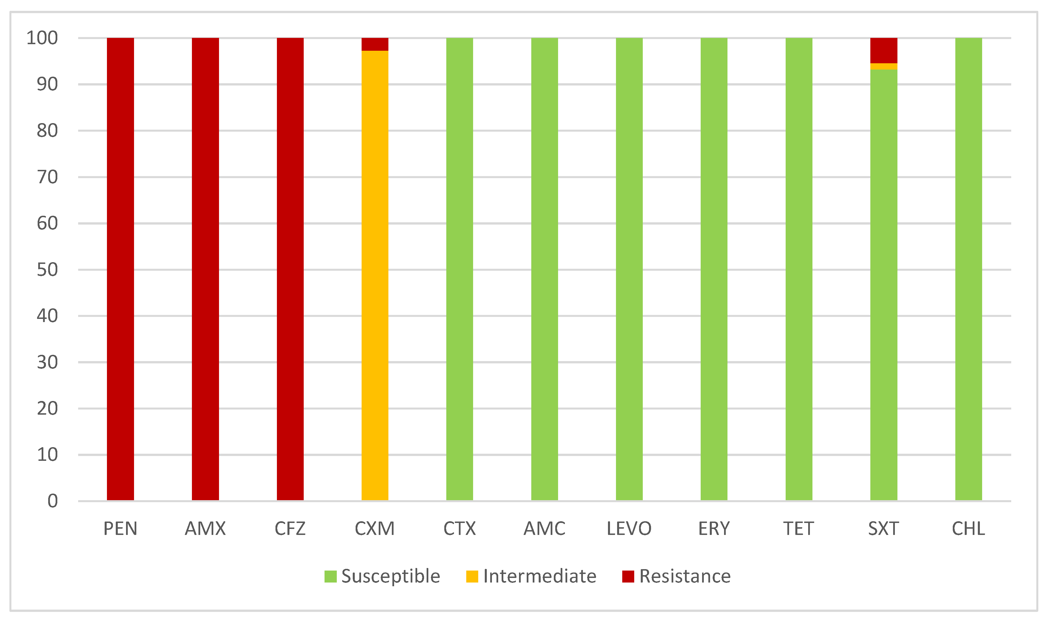
2.3. Antimicrobial Susceptibility
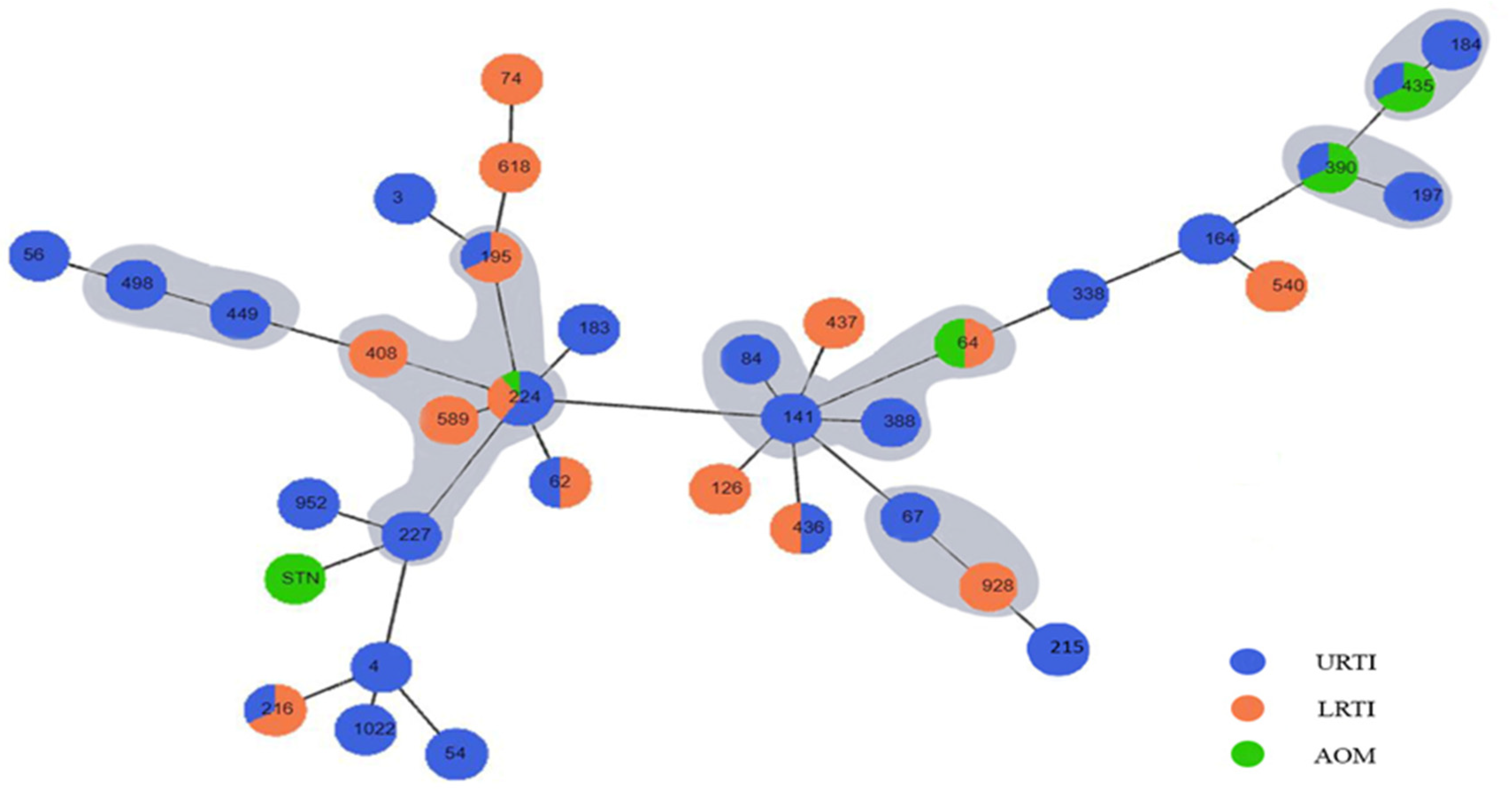
All the strains demonstrated resistance to penicillin, aminopenicillins, and the first generation of cephalosporins due to the presence of BRO β-lactamases detected using the Cefinase disc method. However, the strains exhibited very high susceptibility to almost all other tested antibiotics (Figure 1). The strains produced BRO-1 β-lactamase, and only one strain produced BRO-2 β-lactamase.
All M. catarrhalis strains exhibited intermediate susceptibility to oral cefuroxime, and two strains were resistant. A small number of strains (n = 4, 5.5%) were resistant to trimethoprim-sulfamethoxazole or exhibited intermediate susceptibility (n = 1, 1.4%). None of the strains (0.0%) were resistant to amoxicillin/clavulanic acid, cefotaxime, erythromycin, tetracycline, chloramphenicol, or levofloxacin (Figure 1).

2.4. Genotyping

The MLST analysis of the patterns of all 73 M. catarrhalis isolates revealed 36 STs, including one novel ST (STN).
In total, 39 strains (53.4%) exhibited similarity with different STs and formed a clonal complex in which every ST shares at least five of eight identical alleles with at least one other ST. They form the main clonal complexes in the studied population. The distribution among the STs showed that ST224 (n = 9, 12.3%) was predominant. In total, 36.9% of STs were represented by three strains each, including ST54, ST62, ST141, ST184, ST195, ST216, ST390, ST435, and ST449. In addition, 27.4% of the STs were found in two strains each, including ST3, ST183, ST215, ST227, ST436, ST498, ST540, ST952, ST1022, and STN.
We identified a predominant clonal complex CC224 (n = 16, 21.9%) and other small clusters, including CC141 (n = 6, 8.2%), CC184 (n = 6, 8.2%), CC449 (n = 5, 6.8%), CC390 (n = 4, 5.5%), and CC67 (n = 2, 2.7%). Two primary founders, namely ST224 and ST141, were disclosed. Within CC224, the ancestral ST224 revealed high similarity to ST408 and ST589 with a difference of one locus, and ST227 and ST195 were double and triple locus variants, respectively, displaying six and five identical alleles (Figure 2).
The other primary founder ST141 was clustered with ST64, ST84, and ST388, which shared seven or five identical alleles, indicating SLVs or TLVs. The ancestral types ST224 and ST141 were discovered both in children and adult patients with upper and lower respiratory tract infections.
CC184 represented six strains isolated from patients of different ages and diagnoses and clustered with ST184 and the SLVs ST435. CC449 is comprised of STs and its TLV ST498. ST390 and ST197 formed CC390. ST67 and ST928 displayed five identical house-keeping genes and comprised CC67.
We also observed that 32.9% of the strains formed small clusters of two or three isolates with the same sequence type (ST), but they did not share similarities with any other STs in the studied population (Figure 2). These individual STs were not common among the analyzed strains and did not show any relatedness to other STs. However, we anticipate successful spreading in the coming years of this type of clonal complex, which is exclusively composed of a single sequence type. These small groups were represented by ST216 (n = 3, 4.1%), ST54 (n = 3, 4.1%), ST3 (n = 2, 2.7%), ST62 (n = 2, 2.7%), ST183 (n = 2, 2.7%), ST215 (n = 2, 2.7%), ST436 (n = 2, 2.7%), ST540 (n = 2, 2.7%), ST952 (n = 2, 2.7%), ST1022 (n = 2, 2.7%), and STN (n = 2, 2.7%).
The STs that showed no relatedness to any other STs and were considered singletons accounted for 13.7%.
All clustered strains and singletons along with their main phenotypic and genotypic characteristics are presented in Table 2.

3. Discussion

It has become evident over the past few decades that M. catarrhalis has significant pathogenic potential. The bacterium is mainly involved in respiratory tract infections and rarely in systemic infections in immunocompromised patients. Many authors considered M. catarrhalis one of the most common bacterial organisms that causes otitis media, following the nontypeable Haemophilus influenzae (NTHi), Streptococcus pyogenes, and Streptococcus pneumoniae [41,42]. In our study, the number of cases of otitis media was lower compared to other upper respiratory tract infections, as we did not collect specimens only from a specific group of patients diagnosed with AOM, but in general, from patients with respiratory infections caused by M. catarrhalis.
The majority of the strains that were examined were found in young children. Many studies have reported that M. catarrhalis colonizes infants and children at a higher rate, especially in those under 5 years of age, with only 1–5% carriage in healthy adults. However, isolates from older patients were more likely to be pathogenically significant [18,43]. In our study population, all strains that caused chronic bronchitis and COPD were found in adults, while all cases of acute otitis media (AOM) were seen in children, mostly those under 5 years old.
Our results showed that all M. catarrhalis strains were resistant to penicillin, aminopenicillins, and first-generation cephalosporins and exhibited lower or no susceptibility to cefuroxime oral. The results demonstrated a progression of elevation of resistance to second-generation cephalosporins. According to our old results, susceptibility to cefuroxime at the start of 2000 was noted in about 97% strain, and around 65% before 2018 [44]. The predominant mechanism of resistance found in 98.6% of the strains involved BRO-1 β-lactamases; this mechanism was mostly associated with high resistance to penicillins without inhibitors, first-generation cephalosporins, and, to a lesser extent, second-generation cephalosporins. Remarkably high susceptibility to other tested antimicrobials, except a few strains demonstrating resistance to trimethoprim-sulfamethoxazole, was determined. Both BRO-1 and BRO-2 enzymes are readily inactivated by β-lactamase inhibitors, and the isolates are still susceptible to amoxicillin in combination with clavulanic acid. Many studies noted that routinely administered therapy consisting of a combination of aminopenicillins and β-lactamase inhibitors is usually sufficient [13,16,45,46]. M. catarrhalis is generally considered to have high sensitivity to many antibiotics. Authors from different regions reported that M. catarrhalis was full susceptibility to amoxicillin/clavulanic acid, cefotaxime, and moxifloxacin [39,40,47]. According to other reports, M. catarrhalis appeared extremely susceptible to macrolide antibiotics, erythromycin, and rokitamycin [39,40,48]. Susceptibility of all tested strains to azithromycin, doxycycline, co-trimoxazole was also reported [39,40,44,49].
In a study from Hungary, non-susceptibility to macrolides and trimethoprim-sulfamethoxazole was reported at less than 5%, with low MIC values, and single resistant strains to levofloxacin were reported [44]. Complicated therapy and antimicrobial resistance have been described in Asian countries, with strains resistant to trimethoprim/sulfamethoxazole, levofloxacin, ciprofloxacin, erythromycin, gentamicin, clarithromycin, telithromycin, cefotaxime, or chloramphenicol reported [40,50,51,52]. There are increasing reports of strains with low non-susceptibility or resistance to tetracycline, and tetracycline-resistant strains were isolated more frequently than strains resistant to other antibiotics [46,51].
The serotype distribution of M. catarrhalis confirmed the prevalence of serotype A over the years. In an earlier study of Bulgarian isolates of M. catarrhalis from patients with various respiratory infections and different ages, the Bulgarian strains mainly belonged to serotype A [21]. The results of this study confirmed that serotype A is still the leading pathogen in our country. We did not observe an association among the serotypes, diagnosis, and specific clonal complexes because most strains belong to the same serotype.
The examined M. catarrhalis population revealed various genetic lineages. Despite this diversity, one main clonal complex denoted CC224 and other small clusters, including CC141, CC184, and CC449, were predominant. Two likely primary founders were identified: ST224 and ST141. The major CC224 was also identified in studies from China and was reported to be susceptible to macrolides [53]. Our CC224 strains showed the same susceptibility, with even more susceptibility to all tested antibiotics. We did not observe a correlation for CC224 with the age of the patients or the diagnosis. Both strains recovered from children and adult specimens with upper and lower respiratory tract infections showed genetic relatedness with CC224.
CC141 comprised four different STs from susceptible strains. ST64 was a triple locus variant of ST141 reported in other studies as an ST resistant to azithromycin, erythromycin, and cefuroxime [50,54]. Apart from CC141, ST215 is found in our population and is also frequently identified in Asia with the same multidrug resistance reported in Chinese studies [50].
CC449 is successfully circulated in our country and has been described in reports from Asia as a purely macrolide-resistant clonal complex. In our country, all ST449 strains showed high susceptibility, but it is alarming that the same well-distributed cluster has been represented predominantly by resistance strains in other countries. We may expect strains from this cluster or CC141 to evolve in macrolide-resistant isolates and spread these genetic lineages to our geographic area. M. catarrhalis is a genetically heterogeneous species from which successful clones occasionally proliferate. The expansion of such competitive types has also been documented during distinct periods and in particular geographic regions [55,56,57]. Frequent horizontal gene transfer seems possible [58,59], and new phenotypic markers could be acquired through cross-species gene acquisition.
Other investigations cited the distribution of CC363, which comprised resistant M. catarrhalis strains to cefaclor and azithromycin; however, it is still not present in our area [54,55].
Both ST184 and ST435 from CC184 in our population were registered in the global database (https://enterobase.warwick.ac.uk/ (accessed on 31 August 2024)) in different regions in Europe, Australia, and the USA. ST67 was also a successful spreading type found in France, Australia, and the USA.
During our investigations, we discovered sequence types, such as ST 216 and ST 54, and other STs, each present in only a small number of two or three strains. Based on this, we anticipate the spread of these clonal complexes in the coming years. These clonal complexes consist exclusively of a single sequence type, which can happen when a specific strain undergoes clonal expansion within a population, leading to the dominance of that particular sequence type in the population’s structure.
The epidemiological studies of M. catarrhalis are still rare and insufficient in understanding how these lineages split and subsequently expanded in the human population.

4. Materials and Methods

4.1. Specimen Collection

Specimens were collected from patients of all age groups who had upper and lower respiratory tract infections between October and May of 2023–2024. Clinical strains of M. catarrhalis were isolated from nasal and nasopharyngeal swabs, punctures from ears and sinuses that were taken from children based on their clinical symptoms after a physical examination, and sputum samples collected from the adult patients only. The isolates were identified using conventional microbiological tests, including typical Gram morphology; positive tests for catalase, oxidase, and indoxyl acetate esterase reactions; the hockey puck sign; and assessments using the semiautomatic system Crystal NH BBL (Beckton Dickinson, Kelberg, Germany).

4.2. Antimicrobial Susceptibility Testing

Antimicrobial susceptibility testing was performed phenotypically with amoxicillin/clavulanic acid, 2/1 μg; cefotaxime 5 μg; cefuroxime 30 μg, erythromycin 15 μg; tetracycline 30 μg; chloramphenicol 30 μg; levofloxacin, 5 μg; and trimethoprim-sulfamethoxazole 30 μg (all from Mast Group Ltd., Reinfeld, Germany). European Committee on Antimicrobial Susceptibility Testing (EUCAST 2024) breakpoints were used to assess susceptibility and resistance. Production of β-lactamases was estimated using the Cefinase disc method (Beckton Dickinson, Kelberg, Germany). The primers and conditions for PCR detection of BRO-1 and BRO-2 genes were previously described [21].

4.3. Serotyping

Monoclonal antibodies (MAbs) against the three LOS chemotypes A, B, and C used for serotyping with an immunofluorescence method in this work were obtained in our laboratory by hybridoma technology as previously described [28]. The serotypes share a common core terminal unit, causing cross-reactivity among the three serotypes. More frequent cross-reactivity between A and C serotypes is possible potentially due to the presence of larger similar regions and common epitopes, which could provide a protective effect of antibodies against both serotypes A and C [28].

4.4. DNA Extraction and PCR Amplifications

Total DNA was extracted from pure cultures of M. catarrhalis strains using the extraction kit (DNA-Sorb-A DNA extraction kit, Sacace Biotechnologies, Como, Italy) in accordance with the manufacturer’s instructions. The extracted DNA was stored at −70 °C.
The PCR amplifications was done with 5 µL of DNA template in a 25 µL reaction mixture containing 1 µL of each primer and 10 µL 2 × Prime Taq Premix (GenetBio, Daejeon, Republic of Korea). Thermocycling was performed on the GTQ-cycler 96 (Hain lifesciences, Nehren, Germany) with PCR cycling conditions consisting of an initial denaturation at 94 °C for 4 min, followed by 35 cycles at 94 °C for 30 s, 52 °C for 30 s, and 72 °C for 30 s for all genes, except glyRS (58 °C) and adk (54 °C). The final extension was at 72 °C for 10 min. The primer sequences of the housekeeping genes are listed in Table 3.

4.5. Multilocus Sequence Typing (MLST) of M. catarrhalis

The PCR products were purified using the Exo-CIP™ Rapid PCR-Cleanup kit (New England Biolabs, Ipswich, MA, USA) before sequencing. Nucleotide sequencing of both strands of the PCR amplicon was performed using an ABI 3500 xl Genetic Analyzer (Applied Biosystems, Waltham, MA, USA). MLST was used for population analysis and monitoring the distribution of M. catarrhalis genetic lineages in our geographic area. We performed sequencing of eight “housekeeping genes”: ppa (pyrophosphate phospho-hydrolase), efp (elongation factor P), fumC (fumarate hydratase), trpE (anthranilate synthase component I), mutY (adenine glycosylase), adk (adenylate kinase), abcZ (ATP-binding protein), and glyRS (glycyl-tRNA synthetase beta subunit). We used the protocol for PCR amplification of the internal fragments of the housekeeping genes of M. catarrhalis described at https://enterobase.readthedocs.io/en/latest/mlst/mlst-legacy-info-mcatarrhalis.html (accessed on 31 August 2024).
After sequencing, we determined the allelic profile of each studied isolate and the sequence type (ST) on the MLST website using the Achtman scheme.
The obtained STs enabled us to analyze the genetic relationships between the isolates. A high number of identical locus variants indicates a higher degree of genetic relatedness. Within one clonal complex (CC), STs with single locus variants (SLVs), double locus variants (DLVs), or triple locus variants (TLVs) were identified. By identifying the clonal structure of the studied population, we uncovered the primary ancestral type and the organization of allelic variants in CCs based on their genetic similarity.
All clonal complexes were compared and analyzed for genetic relatedness to reference circulating clones in the international database (https://enterobase.warwick.ac.uk/species/index/mcatarrhalis (accessed on 31 August 2024)). The diagram of clonality was constructed using Phyloviz 2.0.

5. Conclusions

The studied population revealed that susceptible M. catarrhalis strains, predominantly serotype A, were isolated from children and adults with upper and lower respiratory tract infections. The analyzed genetic lineages were diverse but showed the predominance of two main clusters, CC224 and CC141, which encompassed sequence types distributed in other regions and showed multidrug resistance. These data give us reason to assume that resistant strains from the same genetic lines could also spread to our country. Therefore, ongoing epidemiological monitoring of successfully circulating clones and the implementation of adequate antibiotic policies should continue to be the subject of future studies and analyses.

Author Contributions

Conceptualization, A.S.A. and R.T.G.; Formal analysis, A.S.A.; Funding acquisition, A.S.A.; Investigation, A.S.A., V.S.B. and K.Y.M.; Methodology, A.S.A., V.S.B., K.Y.M. and R.T.G.; Project administration, A.S.A.; Software, A.S.A.; Supervision, A.S.A. and R.T.G.; Validation, A.S.A., V.S.B., K.Y.M. and R.T.G.; Visualization, A.S.A. and V.S.B.; Writing—original draft, A.S.A.; Writing—review & editing, A.S.A. and R.T.G. All authors have read and agreed to the published version of the manuscript.

Funding

This research was funded by Grant No. 180/03.08.2023 of Medical University-Sofia, Bulgaria.

Institutional Review Board Statement

Not applicable.

Informed Consent Statement

Not applicable.

Data Availability Statement

Data is contained within the article.

Acknowledgments

We thank Viktoria Atanasova for her involvement in the project and the implementation of some phenotypic methods.

Conflicts of Interest

There are no conflicts of interest.

References

  1. Karalus, R.; Campagnari, A. Moraxella catarrhalis: A review of an important human mucosal pathogen. Microbes Infect. 2000, 2, 547–559. [Google Scholar] [CrossRef]
  2. Deng, W.J.; Zhang, J.F.; Li, P.Y.; Zhou, J.L.; Yao, Z.J.; Ye, X.H. Co-carriage of Streptococcus pneumoniae and Moraxella catarrhalis among preschool children and its influencing factors. Zhongguo Dang Dai Er Ke Za Zhi 2022, 24, 874–880. [Google Scholar] [CrossRef]
  3. Verduin, C.M.; Hol, C.; Fleer, A.; van Dijk, H.; van Belkum, A. Moraxella catarrhalis: From emerging to established pathogen. Clin. Microbiol. Rev. 2002, 15, 125–144. [Google Scholar] [CrossRef]
  4. Morris, D.E.; Osman, K.L.; Cleary, D.W.; Clarke, S.C. The characterization of Moraxella catarrhalis carried in the general population. Microb. Genom. 2022, 8, 000820. [Google Scholar] [CrossRef]
  5. Aebi, C. Moraxella catarrhalis—Pathogen or commensal? In Hot Topics in Infection and Immunity in Children VII; Advances in Experimental Medicine and Biology; Springer: New York, NY, USA, 2011; Volume 697, pp. 107–116. [Google Scholar] [CrossRef]
  6. Goldstein, E.J.C.; Murphy, T.F.; Parameswaran, G.I. Moraxella catarrhalis, a human respiratory tract pathogen. Clin. Infect. Dis. 2009, 49, 124–131. [Google Scholar] [CrossRef]
  7. Gupta, N.; Arora, S.; Kundra, S. Moraxella catarrhalis as a respiratory pathogen. Indian J. Pathol. Microbiol. 2011, 54, 769–771. [Google Scholar] [CrossRef]
  8. Van Hare, G.F.; Shurin, P.A. The increasing importance of Branhamella catarrhalis in respiratory infections. Pediatr. Infect. Dis. J. 1987, 6, 92–94. [Google Scholar] [CrossRef]
  9. Mbaki, N.; Rikitomi, N.; Nagatake, T.; Matsumoto, K.; Tohoku, J. Correlation between Branhamella catarrhalis adherence to oropharyngeal cells and seasonal incidence of lower respiratory tract infections. Tohoku J. Exp. Med. 1987, 153, 111–121. [Google Scholar] [CrossRef]
  10. Wilkinson, T.M.A.; Aris, E.; Bourne, S.; Clarke, S.C.; Peeters, M.; Pascal, T.G.; Schoonbroodt, S.; Tuck, A.C.; Kim, V.; Ostridge, K.; et al. A prospective, observational cohort study of the seasonal dynamics of airway pathogens in the aetiology of exacerbations in COPD. Thorax 2017, 72, 919–927. [Google Scholar] [CrossRef]
  11. Marchisio, P.; Gironi, S.; Esposito, S.; Schito, G.C.; Mannelli, S.; Principi, N. Seasonal variations in nasopharyngeal carriage of respiratory pathogens in healthy Italian children attending day-care centres or schools. J. Med. Microbiol. 2001, 50, 1095–1099. [Google Scholar] [CrossRef]
  12. Venekamp, R.P.; Damoiseaux, R.A.; Schilder, A.G. Acute Otitis Media in Children. Am. Fam. Physician 2017, 95, 109–110. [Google Scholar]
  13. El Feghaly, R.E.; Nedved, A.; Katz, S.E.; Frost, H.M. New insights into the treatment of acute otitis media. Expert Rev. Anti-Infect. Ther. 2023, 21, 523–534. [Google Scholar] [CrossRef]
  14. Zhang, X.B.; Wu, X.; Nong, G.M. Update on protracted bacterial bronchitis in children. Ital. J. Pediatr. 2020, 46, 38. [Google Scholar] [CrossRef]
  15. Shaikh, N.; Hoberman, A.; Shope, T.R.; Jeong, J.H.; Kurs-Lasky, M.; Martin, J.M.; Bhatnagar, S.; Muniz, G.B.; Block, S.L.; Andrasko, M.; et al. Identifying Children Likely to Benefit from Antibiotics for Acute Sinusitis: A Randomized Clinical Trial. JAMA 2023, 330, 349–358. [Google Scholar] [CrossRef]
  16. Nawa, M.; Mwansa, J.; Mwaba, J.; Kaonga, P.; Mukubesa, A.N.; Simuyandi, M.; Chisenga, C.C.; Alabi, P.; Mwananyanda, L.; Thea, D.M.; et al. Microbiologic and virulence characteristics of Moraxella catarrhalis isolates from Zambian children presenting with acute pneumonia. Pediatr. Pulmonol. 2022, 57, 3084–3093. [Google Scholar] [CrossRef]
  17. Siegel, H.; Lang, S.; Maier, P.; Reinhard, T. Bacterial Conjunctivitis: Current Aspects of Diagnosis and Therapy. Klin. Monbl. Augenheilkd. 2024, 241, 231–246. [Google Scholar] [CrossRef]
  18. Perez, A.C.; Murphy, T.F. Potential impact of a Moraxella catarrhalis vaccine in COPD. Vaccine 2019, 37, 5551–5558. [Google Scholar] [CrossRef]
  19. Wiegers, H.M.G.; van Nijen, L.; van Woensel, J.B.M.; Bem, R.A.; de Jong, M.D.; Calis, J.C.J. Bacterial co-infection of the respiratory tract in ventilated children with bronchiolitis; a retrospective cohort study. BMC Infect. Dis. 2019, 19, 938. [Google Scholar] [CrossRef]
  20. Hirai, J.; Kinjo, T.; Koga, T.; Haranaga, S.; Motonaga, E.; Fujita, J. Clinical characteristics of community-acquired pneumonia due to Moraxella catarrhalis in adults: A retrospective single-centre study. BMC Infect. Dis. 2020, 20, 821. [Google Scholar] [CrossRef]
  21. Mitov, I.G.; Gergova, R.T.; Ouzounova-Raykova, V.V. Distribution of genes encoding virulence factors ompB2, ompCD, ompE, β-lactamase and serotype in pathogenic and colonizing strains of Moraxella catarrhalis. Arch. Med. Res. 2010, 41, 530–535. [Google Scholar] [CrossRef]
  22. Harb, H.; Al-Obaidi, H.; Irannejad, K.; Bagheri, F.A. Unique Case of Moraxella catarrhalis Meningitis Following Neurosurgical Intervention. Cureus 2024, 16, e59689. [Google Scholar] [CrossRef]
  23. Ioannou, P.; Alexakis, K.; Baliou, S.; Kofteridis, D.P. Infective Endocarditis by Moraxella Species: A Systematic Review. J. Clin. Med. 2022, 11, 1854. [Google Scholar] [CrossRef]
  24. Siwakoti, S.; Bajracharya, S.; Adhikaree, N.; Sah, R.; Rajbhandari, R.S.; Khanal, B. Early-Onset Neonatal Meningitis Caused by an Unusual Pathogen-Moraxella catarrhalis. Case Rep. Pediatr. 2019, 2019, 4740504. [Google Scholar] [CrossRef]
  25. Daoud, A.; Abuekteish, F.; Masaadeh, H. Neonatal meningitis due to Moraxella catarrhalis and review of the literature. Ann. Trop. Paediatr. 1996, 16, 199–201. [Google Scholar] [CrossRef]
  26. Kobayashi, Y. Bacteremic Moraxella catarrhalis pneumonia. J. Infect. Chemother. 2000, 6, 68. [Google Scholar] [CrossRef]
  27. Apisarnthanarak, A.; Mundy, L.M. Etiology of community-acquired pneumonia. Clin. Chest Med. 2005, 26, 47–55. [Google Scholar] [CrossRef]
  28. Gergova, R.T.; Iankov, I.D.; Haralambieva, I.H.; Mitov, I.G. Bactericidal monoclonal antibody against Moraxella catarrhalis lipooligosaccharide cross-reacts with Haemophilus spp. Curr. Microbiol. 2007, 54, 85–90. [Google Scholar] [CrossRef]
  29. de Vries, S.P.; Bootsma, H.J.; Hays, J.P.; Hermans, P.W. Molecular aspects of Moraxella catarrhalis pathogenesis. Microbiol. Mol. Biol. Rev. 2009, 73, 389–406. [Google Scholar] [CrossRef]
  30. Rikitomi, N.; Ahmed, K.; Nagatake, T. Moraxella (Branhamella) catarrhalis adherence to human bronchial and oropharyngeal cells: The role of adherence in lower respiratory tract infections. Microbiol. Immunol. 1997, 41, 487–494. [Google Scholar] [CrossRef]
  31. Reddy, M.S.; Murphy, T.F.; Faden, H.S.; Bernstein, J.M. Middle ear mucin glycoprotein: Purification and interaction with nontypable Haemophilus influenzae and Moraxella catarrhalis. Otolaryngol.—Head Neck Surg. 1997, 116, 175–180. [Google Scholar] [CrossRef]
  32. Luke, N.R.; Howlett, A.J.; Shao, J.; Campagnari, A.A. Expression of type IV pili by Moraxella catarrhalis is essential for natural competence and is affected by iron limitation. Infect. Immun. 2004, 72, 6262–6270. [Google Scholar] [CrossRef] [PubMed]
  33. Steimle, A.; Autenrieth, I.B.; Frick, J.S. Structure and function: Lipid A modifications in commensals and pathogens. Int. J. Med. Microbiol. 2016, 306, 290–301. [Google Scholar] [CrossRef] [PubMed]
  34. Xiao, X.; Sankaranarayanan, K.; Khosla, C. Biosynthesis and structure-activity relationships of the lipid a family of glycolipids. Curr. Opin. Chem. Biol. 2017, 40, 127–137. [Google Scholar] [CrossRef] [PubMed]
  35. Gao, Y.; Lee, J.; Widmalm, G.; Im, W. Preferred conformations of lipooligosaccharides and oligosaccharides of Moraxella catarrhalis. Glycobiology 2020, 30, 86–94. [Google Scholar] [CrossRef] [PubMed]
  36. McGregor, K.; Chang, B.J.; Mee, B.J.; Riley, T.V. Moraxella catarrhalis: Clinical significance, antimicrobial susceptibility and BRO beta-lactamases. Eur. J. Clin. Microbiol. Infect. Dis. 1998, 17, 219–234. [Google Scholar] [CrossRef]
  37. Khan, M.A.; Northwood, J.B.; Levy, F.; Verhaegh, S.J.; Farrell, D.J.; Van Belkum, A.; Hays, J.P. bro β-lactamase and antibiotic resistances in a global cross-sectional study of Moraxella catarrhalis from children and adults. J. Antimicrob. Chemother. 2010, 65, 91–97. [Google Scholar] [CrossRef]
  38. Bootsma, H.J.; van Dijk, H.; Vauterin, P.; Verhoef, J.; Mooi, F.R. Genesis of BRO β-lactamase-producing Moraxella catarrhalis: Evidence for transformation-mediated horizontal transfer. Mol. Microbiol. 2000, 36, 93–104. [Google Scholar] [CrossRef]
  39. Bristy, S.A.; Hossain, M.A.; Hasan, M.I.; Mahmud, S.M.H.; Moni, M.A.; Rahman, M.H. An integrated complete-genome sequencing and systems biology approach to predict antimicrobial resistance genes in the virulent bacterial strains of Moraxella catarrhalis. Brief. Funct. Genom. 2023, 22, 375–391. [Google Scholar] [CrossRef]
  40. Zhang, Z.; Yang, Z.; Xiang, X.; Liao, P.; Niu, C. Mutation of TonB-Dependent Receptor Encoding Gene MCR_0492 Potentially Associates with Macrolides Resistance in Moraxella catarrhalis Isolates. Infect. Drug Resist. 2022, 15, 2419–2426. [Google Scholar] [CrossRef]
  41. Sánchez Arlegui, A.; Del Arco Rodríguez, J.; De Velasco Vázquez, X.; Gallego Rodrigo, M.; Gangoiti, I.; Mintegi, S. Bacterial pathogens and antimicrobial resistance in acute otitis media. An. Pediatr. Engl. Ed. 2024, 100, 173–179. [Google Scholar] [CrossRef]
  42. Ngo, C.C.; Massa, H.M.; Thornton, R.B.; Cripps, A.W. Predominant Bacteria Detected from the Middle Ear Fluid of Children Experiencing Otitis Media: A Systematic Review. PLoS ONE 2016, 11, e0150949. [Google Scholar] [CrossRef] [PubMed]
  43. Wood, G.M.; Johnson, B.C.; McCormack, J.G. Moraxella catarrhalis: Pathogenic significance in respiratory tract infections treated by community practitioners. Clin. Infect. Dis. 1996, 22, 632–636. [Google Scholar] [CrossRef] [PubMed]
  44. Gergova, R.; Markovska, R. Antimicrobial resistance of Bulgarian isolates Moraxella catarrhalis during the period 1999–2018. J. IMAB 2020, 26, 3208–3212. [Google Scholar] [CrossRef]
  45. Hare, K.M.; Seib, K.L.; Chang, A.B.; Harris, T.M.; Spargo, J.C.; Smith-Vaughan, H.C. Antimicrobial susceptibility and impact of macrolide antibiotics on Moraxella catarrhalis in the upper and lower airways of children with chronic endobronchial suppuration. J. Med. Microbiol. 2019, 68, 1140–1147. [Google Scholar] [CrossRef]
  46. Król-Turmińska, K.; Olender, A.; Bogut, A. Tetracycline resistance in Moraxella catarrhalis clinical strains isolated in Poland. New Microbiol. 2020, 43, 103–106. [Google Scholar]
  47. Kovács, E.; Sahin-Tóth, J.; Tóthpál, A.; van der Linden, M.; Tirczka, T.; Dobay, O. Co-carriage of Staphylococcus aureus, Streptococcus pneumoniae, Haemophilus influenzae and Moraxella catarrhalis among three different age categories of children in Hungary. PLoS ONE 2020, 15, e0229021. [Google Scholar] [CrossRef]
  48. Chen, L.; Zhang, Y.H.; Wang, S.; Zhang, Y.; Huang, T.; Cai, Y. Prediction and analysis of essential genes using the enrichments of gene ontology and KEGG pathways. PLoS ONE 2017, 12, e0184129. [Google Scholar] [CrossRef]
  49. Bandet, T.; Whitehead, S.; Blondel-Hill, E.; Wagner, K.; Cheeptham, N. Susceptibility of clinical Moraxella catarrhalis isolates in British Columbia to six empirically prescribed antibiotic agents. Can. J. Infect. Dis. Med. Microbiol. 2014, 25, 155–158. [Google Scholar] [CrossRef]
  50. Zhao, N.; Ren, H.; Deng, J.; Du, Y.; Li, Q.; Zhou, P.; Zhou, H.; Jiang, X.; Qin, T. Genotypic and Phenotypic Characteristics of Moraxella catarrhalis from Patients and Healthy Asymptomatic Participants among Preschool Children. Pathogens 2022, 11, 984. [Google Scholar] [CrossRef]
  51. Flamm, R.K.; Sader, H.S.; Farrell, D.J.; Jones, R.N. Macrolide and tetracycline resistance among Moraxella catarrhalis isolates from 2009 to 2011. Diagn. Microbiol. Infect. Dis. 2012, 74, 198–200. [Google Scholar] [CrossRef]
  52. Raveendran, S.; Kumar, G.; Sivanandan, R.N.; Dias, M. Moraxella catarrhalis: A Cause of Concern with Emerging Resistance and Presence of BRO Beta-Lactamase Gene-Report from a Tertiary Care Hospital in South India. Int. J. Microbiol. 2020, 2020, 7316257. [Google Scholar] [CrossRef] [PubMed]
  53. Liu, Y.L.; Ding, R.; Jia, X.M.; Huang, J.J.; Yu, S.; Chan, H.T.; Li, W.; Mao, L.L.; Zhang, L.; Zhang, X.Y.; et al. Correlation of Moraxella catarrhalis macrolide susceptibility with the ability to adhere and invade human respiratory epithelial cells. Emerg. Microbes Infect. 2022, 11, 2055–2068. [Google Scholar] [CrossRef]
  54. Liu, Y.L.; Xiao, M.; Cheng, J.W.; Xu, H.P.; Xu, Z.P.; Ye, S.; Zhang, W.J.; Kudinha, T.; Kong, F.; Xu, Y.C. Moraxella catarrhalis Macrolide-Resistant isolates Are highly concentrated in Two MLST clonal complexes -CCN10 and CC363. Front. Microbiol. 2017, 8, 201. [Google Scholar] [CrossRef] [PubMed]
  55. Du, Y.; Zhou, H.; Wang, F.; Liang, S.; Cheng, L.; Du, X.; Pang, F.; Tian, J.; Kan, B.; Xu, J.; et al. Multilocus sequence typing-based analysis of Moraxella catarrhalis population structure reveals clonal spreading of drug-resistant strains isolated from childhood pneumonia. Infect. Genet. Evol. 2017, 56, 117–124. [Google Scholar] [CrossRef] [PubMed]
  56. Qin, L.; Masaki, H.; Gotoh, K.; Furumoto, A.; Terada, M.; Watanabe, K.; Watanabe, H. Molecular epidemiological study of Moraxella catarrhalis isolated from nosocomial respiratory infection patients in a community hospital in Japan. Intern. Med. 2009, 48, 797–803. [Google Scholar] [CrossRef] [PubMed]
  57. Hansen, K.; YambaYamba, L.; Wasserstrom, L.; Rünow, E.; Göransson, T.; Nilsson, A.; Ahl, J.; Riesbeck, K. Exploring the microbial landscape: Uncovering the pathogens associated with community-acquired pneumonia in hospitalized patients. Front. Public Health 2023, 11, 1258981. [Google Scholar] [CrossRef]
  58. Blakeway, L.V.; Tan, A.; Lappan, R.; Ariff, A.; Pickering, J.L.; Peacock, C.S.; Blyth, C.C.; Kahler, C.M.; Chang, B.J.; Lehmann, D.; et al. Moraxella catarrhalis Restriction-Modification Systems Are Associated with Phylogenetic Lineage and Disease. Genome Biol. Evol. 2018, 10, 2932–2946. [Google Scholar] [CrossRef]
  59. Earl, J.P.; de Vries, S.P.; Ahmed, A.; Powell, E.; Schultz, M.P.; Hermans, P.W.; Hill, D.J.; Zhou, Z.; Constantinidou, C.I.; Hu, F.Z.; et al. Comparative Genomic Analyses of the Moraxella catarrhalis Serosensitive and Seroresistant Lineages Demonstrate Their Independent Evolution. Genome Biol. Evol. 2016, 8, 955–974. [Google Scholar] [CrossRef]
Figure 1. Antimicrobial susceptibility of 73 M. catarrhalis strains isolated from Bulgarian patients in the winter–spring season of 2023–2024. PEN—Benzylpenicillin, AMX—Amoxicillin, CFZ—Cefazolin, CXM—Cefuroxime, CTX—Cefotaxime, AMC—Amoxicillin/Clavulanic acid, LEVO—Levofloxacin, ERY—Erythromycin, TET—tetracycline, Sxt—Trimethoprim-sulfamethoxazole, CHL—Chloramphenicol. The interpretation was performed according to the European Committee on Antimicrobial Susceptibility Testing (EUCAST 2024) breakpoints.
Ijms 25 09769 g001
Figure 2. Population snapshot of 73 M. catarrhalis strains recovered from children and adults with respiratory infections in Bulgaria. The sequence types (STs) are identified based on the allelic profiles on the MLST website and depicted inside the circles. The clonal complexes (CCs) were named after the predominant STs. If the STs are in an equal number, the CCs were named after the earlier STs found in the population. The clonal complexes are surrounded with a grey halo and comprise STs with relatedness at different levels: SLVs (single locus variants), DLVs (double locus variants), and TLVs (triple locus variants). The lines between the STs correspond to the relatedness. Specifically, the shortest lines correspond to SLVs. The longer the line is, the more genetically distant the representatives are. The diameter of the circles corresponds to the number of the STs. The colors in the circles correspond to the diagnosis of the patients: URTI—upper respiratory tract infections: rhinopharyngitis and rhinosinusitis, LRTI—lower respiratory tract infections: COPD (chronic obstructive pulmonary disease) and bronchitis. AOM—acute otitis media. The diagram was constructed using Phyloviz 2.0.
Ijms 25 09769 g002
Table 1. Distribution of patient age, diagnosis, and serotypes of M. catarrhalis strains isolated during the winter–spring season of 2024 in Bulgaria.
Age 1Diagnosis 2Serotype
Children0–11 y
n = 51 (69.9%)
URTIAOMLRTIABC
43 (58.9%)8 (11.0%)-46 (63.0%)4 (5.5%)1 (1.4%)
Adults50–74 y
n = 22 (30.1%)
3 (4.1%)-19 (26.0%)20(27.4%)1 (1.4%)1 (1.4%)
Total n (100%)73 (100%)46 (63.0%)8 (11.0%)19 (26.0%)66 (90.4%)5 (6.8%)2 (2.8%)
1 Age—All patients were categorized into two age groups. There were no collected samples from patients between 11 and 50 years old. 2 Diagnosis: URTI—upper respiratory tract infection, including rhinopharyngitis, rhinosinusitis, and adenoiditis; AOM—acute otitis media; LRTI— lower respiratory tract infection, including COPD (chronic obstructive pulmonary disease) and bronchitis.
Table 2. Phenotypic and genotypic characteristics of 73 M. catarrhalis isolates recovered from children and adults in Bulgaria during the winter–spring season of 2023–2024.
CC 1StrainAgeGender 2Diagnosis 3Sample 4SerotypeabcZadkefpfumCglyRSmutYppatrpEST 5
CC14157/137950fBronchitis chrSputumA217122333264
5572fRhinopharNphA262220332141
80802fRhinopharNphA262220332141
13534fAdenoiditisNphA262220332141
20/13213mAdenoiditisNphA29222033284
58/15581mRhinopharNphA8632202632388
CC224771fRhinopharNphA5332357332224
17/1795mAdenoiditisNphA5332357332224
613mAdenoiditisNphA5332357332224
54/8475mRhinopharNphA5332357332224
186fRhinopharNphA5332357332224
7116fRhinopharNphA5332357332224
55571mCOPDSputumA5332357332224
60861mBronchitis chrSputumA5332357332224
26/53K71mCOPDSputumA5332357332224
184363mCOPDSputumA53359357332589
109,31668mCOPDSputumA3323576732408
K978 5mRhinopharNphA332957332227
1664mAdenoiditisNphA332957332227
32702mRhinopharNphA28323313132195
6067mCOPDSputumA28323313132195
32702mRhinopharNphA28323313132195
CC184510210mRhinosinuitisNphA82564572222184
55/9802fRhinopharNphA82564572222184
22/621546fRhinosinuitisNphA82564572222184
4265fAOMMEFA825645722232435
184/2415mRhinopharNphA825645722232435
24/8144mAOMMEFB825645722232435
CC44936/34686mRhinopharNphA3322171582449
37/86572fRhinopharNphA3322171582449
31/1611mRhinopharNphB3322171582449
28261mRhinopharNphA318552151582498
112mRhinopharNphA318552151582498
CC39042/7856fAOMMEFA8256237932390
785/426fAOMMEFB8256237932390
42/7854fRhinopharNphA8256237932390
35223mAdenoiditisNphA8362179172197
CC6718411mRhinosinuitisNphA217232022267
57160mCOPDSputumA2211132222928
ST3753fRhinopharNphA222322223
12053mRhinopharNphA222322223
ST545922fRhinopharNphA8243730323254
35359mRhinosinuitisNphA8243730323254
3431fRhinosinuitisNphA8243730323254
ST62K83 3mAOMMEFC8262322225262
10964mAOMNphA8262322225262
ST18343/16802mAOMNphA9323371992183
14110mRhinosinuitisNphA9323371992183
ST215K1092 1fRhinopharNphA2229218172215
K1097 3mAOMNphA2229218172215
ST21610,83874mCOPDSputumA2518346932216
56/14972fRhinopharNphA2518346932216
39/463851fBronchitis chrSputumA2518346932216
ST43680833mAOMNphB27182210992436
32/16065mCOPDSputumA27182210992436
ST54022/47153mCOPDSputumB8172031283172540
28/322064mBronchitis chrSputumA8172031283172540
ST95240/3484fRhinopharynNphA15722921932952
22/5205M4fRhinopharNphA15722921932952
ST1022816fAOMNphA3334328321022
142mRhinopharNphA3334328321022
STNK511 4fAdenoiditisNphA3929129895STN
5123mOtitis mediaMEFA3929129892STN
Singletons69,2152mRhinopharNphA333433324
30344mAdenoiditisNphA31863311517256
6457mCOPDSputumA8262322225262
30/8674fOtitis mediaMEFA217122333264
34/390754fBronchitis chrSputumA29223342921274
72AI65mBronchitis chrSputumC366122504132126
175fAdenoiditisNphA8176327332164
31684mRhinopharNphA2171233332338
8055mCOPDSputumA217241078932437
25/91K74mBronchitis chrSputumA1083233131212618
1 CC—clonal complex. 2 Gender: f—female, m—male. 3 Diagnosis: Bronchitis chr—chronical bronchitis, Rhinophar—rhinopharyngitis, AOM—acute otitis media, COPD—chronic obstructive pulmonary disease. 4 Sample: Nph—nasopharynx, MEF—middle ear fluid. 5 ST—sequence type.
Table 3. Primer sets for PCR amplification of eight M. catarrhalis housekeeping genes.
GenePrimer SetBase Pairs
glyRSF 5′-GCACCGAAGAGTTGCCACCA-3′
R 5′-ACGCAACGGGCAAATCCACC-3′
762 bp
ppaF 5′-AATAAAATTCTAGATGCTGGC-3′
R 5′-ACTTATTGCTCTGTCCAGCG-3′
523 bp
efpF 5′-CTCTGATTGACAACTGGCAGG-3′
R 5′-GATATTCGCCAGTACGCG-3′
582 bp
fumCF 5′-GGGCGGTACAGCAGTCGGCAC-3′
R 5′-CTCATCAAATTCAGCTTCAG-3′
675 bp
trpEF 5′-TTATCCCGCATCGAAAATGG-3′
R 5′-GGTTTCATCCCATTCAGCC-3′
545 bp
mutYF 5′-GGCAATACCATCATCAGCCG-3′
R 5′-GGTAACTGACTTTGAACGCC-3′
609 bp
adkF 5′-GGCATTCCTCAAATCTCAAC-3′
R 5′-GATGGGCTTTATTGTCAAATG-3′
631 bp
abcZF 5′-ACATGCTGATGATGGTGAG-3′
R 5′-CACTGGCAAGTTCAAGCGC-3′
610 bp
The same primers were used for the following MLST procedure, except the primer sets for the following three genes: glyRS: F5′-GCACCGAAGAGTTGCCACCA-3′ and R5′-ATATCGGCTTGACGCTGATC-3′; fumC: F5′-GCTGTCAAAGTCGCTAAAG-3′ and R5′-CTCATCAAATTCAGCTTCAG-3′, and mutY: F5′-TATGCTGTGTGGGTATCTG-3′ and R5′-GGTAACTGACTTTGAACGCC-3′.
Disclaimer/Publisher’s Note: The statements, opinions and data contained in all publications are solely those of the individual author(s) and contributor(s) and not of MDPI and/or the editor(s). MDPI and/or the editor(s) disclaim responsibility for any injury to people or property resulting from any ideas, methods, instructions or products referred to in the content.

