Review

Prenatal Stress and Ethanol Exposure: Microbiota-Induced Immune Dysregulation and Psychiatric Risks

by
Rosana Camarini
1,*,
Priscila Marianno
1,
Maylin Hanampa-Maquera
1,
Samuel dos Santos Oliveira
2 and
Niels Olsen Saraiva Câmara
2,*
1
Department of Pharmacology, Institute of Biomedical Sciences, Universidade de São Paulo, São Paulo 05508-900, Brazil
2
Department of Immunology, Institute of Biomedical Sciences, Universidade de São Paulo, São Paulo 05508-900, Brazil
*
Authors to whom correspondence should be addressed.
Int. J. Mol. Sci. 2024, 25(18), 9776; https://doi.org/10.3390/ijms25189776
Submission received: 14 June 2024 / Revised: 22 August 2024 / Accepted: 25 August 2024 / Published: 10 September 2024
(This article belongs to the Special Issue New Insights into Gut Microbiota and Immunity)

Abstract

:
Changes in maternal gut microbiota due to stress and/or ethanol exposure can have lasting effects on offspring’s health, particularly regarding immunity, inflammation response, and susceptibility to psychiatric disorders. The literature search for this review was conducted using PubMed and Scopus, employing keywords and phrases related to maternal stress, ethanol exposure, gut microbiota, microbiome, gut–brain axis, diet, dysbiosis, progesterone, placenta, prenatal development, immunity, inflammation, and depression to identify relevant studies in both preclinical and human research. Only a limited number of reviews were included to support the arguments. The search encompassed studies from the 1990s to the present. This review begins by exploring the role of microbiota in modulating host health and disease. It then examines how disturbances in maternal microbiota can affect the offspring’s immune system. The analysis continues by investigating the interplay between stress and dysbiosis, focusing on how prenatal maternal stress influences both maternal and offspring microbiota and its implications for susceptibility to depression. The review also considers the impact of ethanol consumption on gut dysbiosis, with an emphasis on the effects of prenatal ethanol exposure on both maternal and offspring microbiota. Finally, it is suggested that maternal gut microbiota dysbiosis may be significantly exacerbated by the combined effects of stress and ethanol exposure, leading to immune system dysfunction and chronic inflammation, which could increase the risk of depression in the offspring. These interactions underscore the potential for novel mental health interventions that address the gut–brain axis, especially in relation to maternal and offspring health.

1. Introduction

The influence of maternal stress and alcohol exposure on the gut microbiome presents a compelling avenue of research with implications for the well-being of both mothers and their offspring. Although human studies in this field remain limited, emerging preclinical evidence suggests that changes in maternal gut microbiota due to stress and/or alcohol exposure can have lasting effects on offspring’s health. Understanding the dynamic interactions among stress, depression, and microbiota holds promise for developing innovative mental health interventions that consider the role of the gut–brain axis. Throughout this review, the terms “ethanol” and “alcohol” are used interchangeably to refer to the same substance—drinking alcohol.
The gut microbiota can communicate bidirectionally with the central nervous system (CNS) through the gut–brain axis [1], impacting the quality of life and the risk of depression [2]. Chronic stress alters the composition and diversity of the gut microbiota, potentially influencing mental health [3]. Dysregulation of this fine-tuned interaction may contribute to the onset or exacerbation of depression.
The early stages of life are crucial for immune system development, and the composition of the gut microbiota during this period plays a pivotal role in shaping the immune system itself and its responses. Stress affects maternal physiology by triggering the release of stress hormones like cortisol, which in turn impacts various systems, including the gut microbiota [4]. In parallel, prenatal ethanol exposure is recognized for its adverse effects on maternal health, and for the heightened risk of inducing Fetal Alcohol Spectrum Disorders [5]. Some of the negative effects of ethanol include disruption in physiological processes such as nutrient absorption, digestion, and metabolic pathways linked to gut microbiota composition [6].
Stress and ethanol consumption during pregnancy often interact in a reciprocal dynamic, wherein maternal stress can serve as a trigger for ethanol use as a coping mechanism [7], while ethanol, in turn, can exacerbate stress-related responses [8]. Studies have reported that some women consume alcohol during pregnancy because they believe it reduces stress, aids sleep, and acts as a relaxant to ease labor and delivery. They also perceive alcohol to be less harmful than other substances [9,10]. This mutual relationship underscores the rationale for this review due to its profound impact on maternal health and potential implications for offspring’s gut microbiota and immunity.
The amount of ethanol necessary to induce dysbiosis can vary depending on several factors, including the duration of ethanol exposure and the specific composition of the diet. Acute ethanol exposure can also lead to dysbiosis in mice [11], although the effects are generally more pronounced and sustained with chronic exposure in both rats and humans [12]. Similarly, both single [13] and repeated stress [14] can alter gut bacterial composition in rodents.
During pregnancy, exposure to ethanol and stress can have particularly severe consequences. The combination of ethanol and stress can disrupt the maternal gut microbiota balance, potentially leading to altered fetal development, impaired immune system function in the offspring, increased susceptibility to immune-related disorders, allergies, autoimmune diseases, and a heightened risk of neurodevelopmental disorders [15,16]. Furthermore, maternal microbiota changes may set the stage for long-term microbiota dysbiosis in the child, potentially linked to inflammation, a contributing factor in depression [17].
This review aims to synthesize preclinical and clinical research on the effects of stress and ethanol on maternal gut microbiota, elucidating their potential transgenerational impacts on offspring’s immunity, inflammation response, and susceptibility to depression. Moreover, this approach offers a more nuanced view that these elements may interact and influence maternal and fetal health.

2. Microbiota Host: From Passive Spectator to Essential Modulator in Health and Disease

2.1. Structural Components of Microbiota Host

Recent studies have revolutionized our understanding of microbiota–host interactions, revealing that the intestinal microbiota is not merely a passive observer but an important component in modulating the host organism’s homeostasis, directly influencing health and disease [18]. This interaction between commensal microorganisms and humans has occurred since the emergence of our species on the planet. It was evolutionarily selected in our distant ancestors due to the significant benefits it provided to both parties: the host offered shelter and nutrients, while the microorganisms aided in the degradation of dietary fibers and occupied niches formed by epithelial barriers, thereby preventing or hindering colonization by pathogenic microorganisms [19].
The biological entity constituted by the host and all the microorganisms that colonize it is called holobiont, and the intimate connection between its components allows for the expansion of individual physiological capabilities [19]. A classic example is the degradation and fermentation of food components, conducted by intestinal bacteria, to form short-chain fatty acids, which are molecules that are extremely important for human health [20]. A wide range of host biological processes can be modulated by the microbiota and its secreted components. The impact of the microbiota on the modulation of innate and adaptive immunity [21], metabolism [22], circadian cycle [23], and behavior [24] is well-established. Conversely, its dysregulation is associated with various disorders, such as autoimmunity [25], metabolic syndrome [26], neurodegenerative diseases [27], and psychiatric diseases [28].
The microorganisms that constitute the intestinal microbiota with a beneficial function for the host, in essence, are similar to pathogenic microorganisms. However, our immune system uses several devices to identify whether the presence of a microorganism represents a danger or not. The structure of the intestinal barrier is specifically designed to physically separate the microbial compartment (the intestinal lumen) from the immune cell compartment, primarily located in the lamina propria, Peyer’s patches, and mesenteric lymph nodes [19].
This intestinal barrier operates through various mechanisms: physical, chemical, immunological, and microbiological barriers. Regarding physical barriers, intestinal epithelial cells are organized in juxtaposition and tightly adhered to one another thanks to tight junction proteins such as occludin, claudins, and zonula occludens. This structure endows the intestine with highly selective permeability, allowing essential molecules to pass while restricting the translocation of microorganisms to the lamina propria, a process that commonly occurs after intestinal injuries [29] or changes in intestinal permeability, which will be discussed later in this review. Adjacent to the intestinal epithelium lies the mucus layer, a gel-like, viscous, elastic, and highly hydrated matrix with Mucin 2 (MUC2) as its fundamental structural component. MUC2 molecules are secreted by specialized intestinal epithelial cells called goblet cells and interact with each other via disulfide bridges, as well as with other gel-forming mucins such as MUC6, MU19, MUC5B, and MUC5AC [30]. This layer not only physically separates bacteria from the host’s tissues but also hydrates, lubricates, and protects the intestinal lumen [31].
The mucus layer also serves as a chemical barrier due to its ability to directly bind to bacterial peptidoglycans and control their growth [32]. However, another class of peptides known as antimicrobial peptides are the classic components of the chemical barrier. In mammals, C-lectins, defensins, histidines, and cathelicidins are key components in antibacterial defense, being prominently produced by neutrophils and epithelial cells, not only in the intestine but also in other barrier tissues [33]. In the intestine, Paneth cells are specialized epithelial cells responsible for the production of antimicrobial peptides, located in the crypts of the small intestine where they maintain close contact with intestinal stem cells [34].
Another class of specialized epithelial cells is M cells. They are cells devoid of glycocalyx components in their membrane, unlike what is observed in other cells of the intestinal epithelial monolayer. In these cells, the glycocalyx results in microfolds, which justifies its name. These cells connect the mucous and microbiological layer with the immunological layer, and their folds are essential in this process as they help in the capture of antigens and microorganisms present in the lumen and transport them through endocytic vesicles for delivery to dendritic cells (DCs) in the Peyer’s patches [35]. This antigenic sampling can also be conducted directly by the DCs underlying the intestinal epithelium. They are able to project their dendrites between the enterocytes and capture antigens, all without compromising the structure and permeability of the intestinal epithelium since these DCs also express tight junction proteins. Thus, there is a rapid loosening of these junctions between enterocytes, followed by re-stabilization with the junction proteins present in DCs [36,37].

2.2. Immunological Components of Microbiota Host

After capturing antigens, directly from the lumens or via M cells, DCs migrate to the mesenteric lymph nodes (mesLN) where they activate T cells specific to that antigen, promoting their proliferation and imprinting on these cells, which are important characteristics to guide their migratory profile. In this process, activated DCs in the gut release high levels of retinoic acid, a component formed from Vitamin A from food, and induce the expression of chemosign receptors α4β7 and C-C motif chemokine receptor type 9 (CCR9) in T cells, which are responsible for the migration of these cells to the lamina propria [38]. In an infectious context, these intestinal T cells have a lower rate of cytokine proliferation, but with longer activity, which ensures a local and direct response to the microorganism, the damage to the intestinal barrier is minimal [39,40].
Among helper T cells (Th), regulatory T cells (Treg) and Th17 are the most abundant and play a central role in maintaining the intestinal barrier, the first providing tolerance to microbiota and food antigens [41,42], and the second promoting resistance mainly to fungi and bacteria that may cause infection [43,44]. Both cell populations require the presence of transforming growth factor beta in their differentiation process, a cytokine widely present in the intestine. Even under homeostatic conditions, the presence of retinoic acid also helps activated T cells in the gut to differentiate into Treg while inhibiting their differentiation into Th17 cells [45]. However, the presence of interleukin (IL)-6, a pro-inflammatory cytokine rapidly produced in response to various infections, together with transforming growth factor beta, polarizes T cells towards the Th17 profile [46].
The immune system and commensal microorganisms directly interact, influencing each other in both health and disease. On one hand, the intestinal microbiota helps train immune cells in homeostasis, preparing them to detect pathogens [47]. Meanwhile, the immune system maintains the balance of the relationship by tolerating the microbiota while also “pruning” its excessive growth. In immunocompromised individuals, commensal microorganisms can proliferate uncontrollably, leading to infections. Conversely, overly aggressive immune responses against the microbiota can damage the host’s tissues, leaving their niche vulnerable to colonization by pathogenic microorganisms [18].
Extrinsic factors, such as eating habits, can also affect the host’s microbiota. Recent studies point to the ability of high-sugar diets to deplete segmented filamentous bacteria, a class of bacteria crucial for inducing Th17 cells in the intestine. Sugar, together with IL-22 produced by Type 3 innate lymphoid cells (ILC3), promotes the growth of Faecalibaculum rodentium and the elimination of segmented filamentous bacteria. In this context, Th1 cells that produce interferon-gama (IFNγ) begin to predominate in the lamina propria, promoting intestinal inflammation and increased permeability, which allows the translocation of bacteria to the liver, intensifying the inflammatory process and contributing to insulin resistance, a key factor in the development of metabolic syndrome [18]. The constitutional change of the microbiota in response to dietary changes occurs rapidly, with a switch from a fiber-rich diet to a sugar-rich diet for just 3 days being sufficient to alter the functional profile of the microbial community [48]. Moreover, alterations in the microbiota and sugar levels can “deprogram” T lymphocytes, rendering them temporarily unresponsive and leaving the host more susceptible to infections [49].
Dysbiosis is considered an altered state of the intestinal bacterial community. Although this definition appears simple, there are a series of complexities regarding what would be a normal microbiota pattern [50], especially when considering its great taxonomic diversity due to environmental, nutritional, and genetic factors. As a result, the definition of normality can vary over time and across different geographical locations [51,52].
Dysbiosis can result from factors like stress and alcohol exposure, as well as diet, antibiotics, age, and inflammation, among others, potentially leading to brain dysfunction due to the gut–brain connection. Bacterial extracellular vesicles have been identified as potential mediators of this communication by interacting with immune receptors and triggering neuroinflammatory responses. These extracellular vesicles, containing harmful substances such as lipopolysaccharides (LPS) and toxins, can cross barriers like the blood–brain barrier or placental barrier and activate immune receptors, such as toll-like receptors (TLRs) on glial cells, resulting in cytokine and inflammatory mediator production that can impair brain function and behavior [53].

3. Maternal Microbiota Disturbance: Implications for Offspring Immune System and Beyond

3.1. Maternal DIET and Microbiota

Abrupt changes in diet during pregnancy, combined with fluctuations in hormonal levels—especially increased production of progesterone and estrogens—are associated with weight gain, elevated blood glucose levels, even during fasting, and insulin resistance [54,55]. Adequate maternal intake of macronutrients and micronutrients is crucial for the proper development of the fetal immune system. Essential macronutrients include carbohydrates, lipids, and proteins, while critical micronutrients comprise minerals such as zinc, selenium, copper, iodine, and iron, along with vitamins like A, C, D, E, and folate. Inadequate maternal nutrition during pregnancy, particularly protein–energy malnutrition, can lead to dysfunction in the mother immune cells [56]. Moreover, maternal diet influences the development of both the adaptive and innate immune systems in the offspring. For example, maternal intake of certain fats can modulate the risk of developing conditions like eczema and allergies in children, which are related to the adaptive immune response. The quality and composition of maternal diet fats (saturated vs. polyunsaturated, N-3 vs. N-6 polyunsaturated fatty acids) are important for modulating the immune response. Particularly, the intake of N-3 can lead to lasting epigenetic changes in the offspring’s immune cells, affecting their immune responses [57,58].
Non-human studies have shown the effects of a high-fat diet on the offspring’s microbiome. Female macaques fed with a high-fat diet (36% fat) had distinct microbiota compared to the controls (13% fat). The offspring of mothers fed a high-fat diet exhibited an altered gut microbiome that persisted even after being weaned onto a control diet, showing a lasting reduction in Campylobacter [58]. A high-fat diet is associated with an increase in Gram-negative bacteria, a higher Firmicutes:Bacteroidetes ratio, colonic inflammation, and a decrease in regulatory T cells. These effects were passed to the offspring of mice fed with a Western diet (40–60% fat, primarily saturated) but reversed by altering the microbiota, suggesting that microbiota changes are the main drivers of diet-induced immune effects. Western diet pups had a higher abundance of the Lachnospiraceae and Clostridiales compared to the low-fat pups [59].

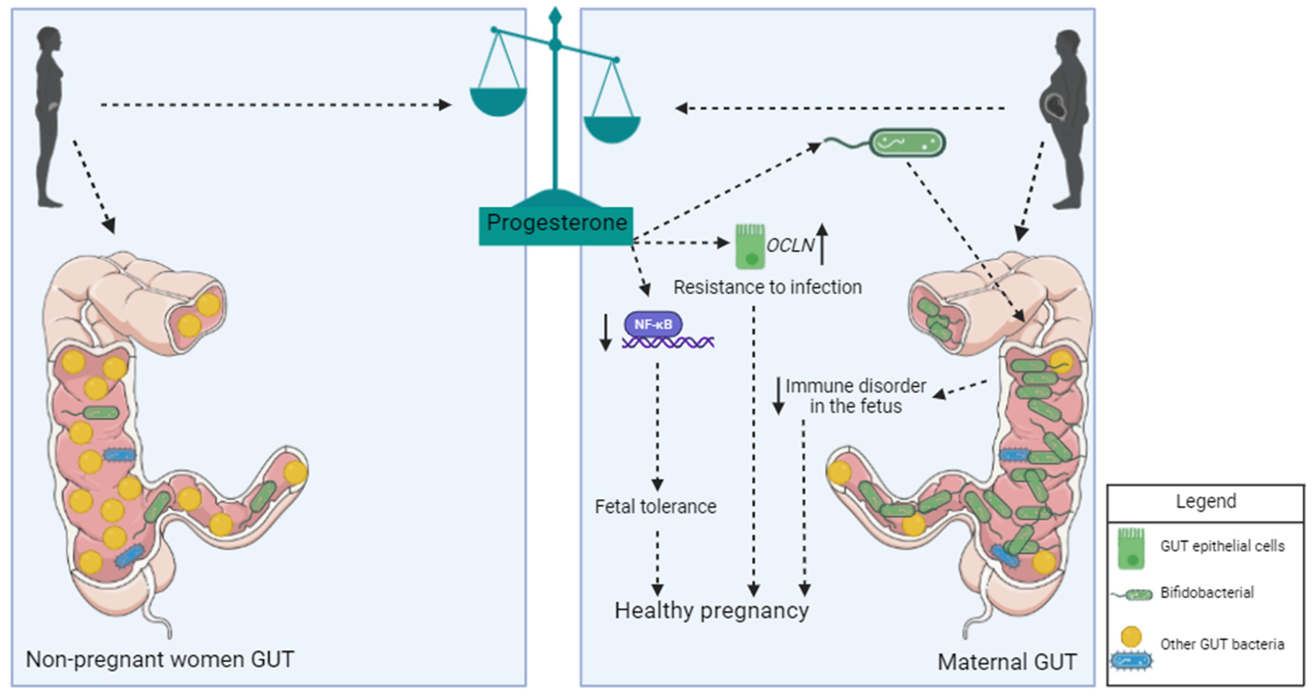
3.2. Influence of Progesterone on Microbiota during Pregnancy

Progesterone plays an important role in controlling the microbiota of pregnant women and the physical barriers of the intestine (Figure 1). Studies report that the hormone acts on the epithelial cells of the intestinal barrier, promoting increased expression of occludin and, therefore, strengthening tissue impermeability and reducing the likelihood of intestinal infections [60]. Additionally, the hormone acts directly on beneficial bacteria during pregnancy, promoting their expansion. Bacteria of the genus Bifdobacterium have the enzyme hydroxysteroid dehydrogenase in their membrane, which has the ability to metabolize progesterone [61]. Progesterone levels in feces negatively correlate with the concentration of LPS in the blood of pregnant women, a strong indication of the role of this hormone in regulating the intestinal microbiome and protecting the host against infections. The host response to bacterial LPS is also regulated by progesterone, which inhibits the activation of nuclear factor kappa B (NF-KB) and reduces the exacerbated inflammatory response, a risk factor for pre-eclampsia and premature birth [62].

3.3. Maternal Microbiota and Immunity: Implications for Fetal and Neonatal Health

The effector functions of the immune system that promote tolerance or resistance are spatially and temporally separated during pregnancy. Immunosuppression in the placental barrier, which is important to ensure that the fetus is not rejected, is not observed in the mucous membranes. In fact, mothers have very efficient immunological responses in these areas, which is extremely important due to the serious risks that infections during pregnancy pose to both mother and baby [62]. Furthermore, the regulatory immune responses in the placental barrier are particularly strong during the 3rd month of pregnancy and gradually decline as birth approaches [60]. The amniotic fluid also helps to regulate maternal immunity and influences immune responsiveness during the fetal and neonatal periods [63,64]. The immunomodulatory properties of amniotic fluid have been attributed to alpha-fetoprotein in regulating natural killer cells and T cell-mediated immune reactions [20]. However, Lang et al. have demonstrated that the primary mediators are not alpha-fetoprotein, but rather the transforming growth factors TGF-β1 and β2 [65].
The microbiota lives in close contact with individuals throughout their lives, but the existence of microbiota in amniotic fluid remains controversial. While it appears that the fluid is not a source of microorganisms for in utero colonization in mice, it may still contribute to fetal exposure to microbial components [66]. Studies have detected microbial DNA in human placental tissues and fluids [67]. However, it remains unclear whether this DNA indicates the presence of viable local organisms from past colonization or is the result of translocation from the bloodstream. Viable bacteria are scarce in the fetal intestine at mid-gestation, but specific strains with immunomodulatory potential, such as Micrococcus luteus, have been identified and may influence fetal immune development [68]. Evidence suggests that paternal microorganisms from semen can contribute to the uterine microbiome, potentially by transporting cervical bacteria upstream during fertilization [69,70]. It is well known that the majority of babies’ microbiota originates from the mother’s microbiota and the surrounding environment [71,72,73]. Thus, maintaining a balanced maternal microbiota is crucial for the initial constitution of newborns’ microbiota. Commensal microorganisms are especially important because they protect newborns, who are highly susceptible to infections due to their immature immune systems [74].
The fetal microbiota also plays a role in regulating immune responses. Pro-inflammatory cytokine production is reduced in human T cells stimulated with live Micrococcus luteus, the most prevalent bacterium in the fetal intestine, from viable fetuses. This suggests that the fetal microbiota helps generate immunological tolerance. M. luteus stimulates the development of dendritic cells and T cells in the fetal intestine [68]. While it is unclear whether these cells have a tolerogenic profile, they likely help control intestinal microbial populations and tolerate their presence, possibly through simpler mechanisms than those observed in adults, such as the production of IL-10 and TGF-β.
In addition to protecting against infections, the neonatal microbiota is crucial for protection against metabolic diseases like obesity. High levels of bifidobacteria in babies’ feces correlate with normal weight, while the abrupt presence of Staphylococcus aureus is associated with increased weight gain [75]. Bifidobacteria also protect against the development of allergies in neonates, while high levels of clostridia are linked to changes in the composition of short-chain fatty acids produced in the intestines of babies, making them more prone to allergies [76]. Colic, a common issue among babies, is also associated with microbiota imbalances. High levels of lactobacilli are found in babies without colic, while the presence of Gram-negative bacteria is more prevalent in babies with colic [77].
Hormonal, immunological, behavioral, and dietary changes during pregnancy affect the microbial composition of all barriers, starting from the 3rd month of pregnancy. In the intestine, there is an increase in Actinobacteria, Proteobacteria, and Bifidobacterium, in addition to a reduction in Faecalibacterium [78,79]. As previously discussed, the immune system shapes the diversity of the microbiome, and the microbiome modulates and trains the immune system. This is supported by observations showing that reduced intestinal colonization by bacteria from the Faecalibacterium genus is associated with increased local inflammation due to decreased production of butyrate, an important immunoregulatory agent produced by these bacteria [80].
The intimate interaction facilitated by the evolution of viviparity in mammals allows the transfer of various compounds produced by the mother and components of her microbiota to the fetus through the placenta. Viviparity emerged independently in different groups of living organisms with the common objectives of promoting nutrition, protection, and gas exchange to offspring [81]. In mammals, this relationship reaches its highest complexity, as evidenced by the existence of the placenta, an organ that allows the exchange of molecules between mother and fetus, while also functioning as a barrier to ensure a unique microenvironment for the fetus [82]. This process is essential for fetal development, and birth and plays a significant role in imprinting characteristics that affect the health of the offspring well beyond birth [83].
Using nonhuman primate models, Nash et al. (2023) [84] demonstrated that a maternal Western-style diet, rich in sugars and lipids, leads to the reprogramming of fetal hematopoietic stem and progenitor cells, causing a proinflammatory phenotype in the offspring. Their bone-marrow-derive macrophages are enriched in genes associated with NF-ΚB, as well as pathways associated with glycolytic metabolism, a profile that is maintained even after 3 years of birth. Similar results were found in macrophages differentiated from hematopoietic pluripotent cells, indicating that the mother’s diet induces epigenetic changes in hematopoietic progenitors that are maintained in terminally differentiated cells [84].
The immune system of newborns is strongly influenced by the intrauterine environment. Through the placenta, not only nutrients and oxygen are exchanged between mother and child, but also cells, microorganisms, and their metabolites are transferred from mother to baby [85]. This transfer, together with immunoglobulin (Ig) A molecules, also occurs during breastfeeding and is essential for the development of the intestinal barrier. Among all immunoglobulin isotypes in colostrum and milk, secretory IgA is the most important due to its high concentration and crucial role in defending mucous membranes, preventing microorganism entry into tissues and its anti-inflammatory properties [86]. Studying the impact of the maternal microbiota on the development of the fetal immune system is challenging, even in mice, because germ-free mothers birth germ-free babies, and colonized mothers deliver colonized babies. Therefore, determining whether changes in the development of children’s immune systems depend on their microbiota (or lack thereof) or on the maternal microbiota (or lack thereof) is a significant challenge. A model was developed to solve this equation, where pregnant germ-free females were colonized with a mutant strain of Escherichia coli, incapable of replicating in the human intestine due to a deficiency in synthesizing essential bacterial amino acids caused by mutations in genes associated with D-alanine biosynthesis. In this case, bacteria temporarily colonized the maternal intestine and were eliminated even before birth, resulting in germ-free offspring from mothers colonized only during pregnancy. This approach allowed for a direct demonstration of the positive impact of maternal microorganisms on the induction of Type 3 innate lymphoid cells and F4/80+CD11c+ mononuclear cells in the newborn intestine [87].
Direct microbial metabolites, or those resulting from the degradation of dietary fibers by intestinal commensals, are transferred to the offspring through the placenta and breastfeeding. These metabolites promote the expansion of immune cells in the intestine, contribute to the physical structuring of the intestinal barrier by stimulating the proliferation of epithelial cells, and induce the production and secretion of mucus, immunoglobulins, and defensins into the intestinal lumen [88]. Additionally, these compounds can act on hematopoietic progenitors present in the bone marrow of newborns, creating an inflammatory environment that imprints an inflammatory program on these cells, which may persist for years after birth in the offspring [89].
The relationship between the microbiota and the host extends beyond the gut, influencing local physiological processes and reaching the brain through the gut–brain axis [90]. This bidirectional communication system underscores the importance of maintaining a balanced microbiota for overall health [91], as dysregulation in the gut may contribute to neurological and psychiatric disorders [92,93].

4. Exploring the Interplay between Prenatal Maternal Stress, Microbiota, and Depression

4.1. Stress, Gut Microbiota, and Depression

This section explores the interaction among stress, gut microbiota, inflammation, and depression via the gut–brain axis. Chronic stress is a recognized risk factor for depression in humans [94] and non-human animals [95] and contributes to functional gastrointestinal disorders [96]. Drossman’s biopsychosocial model emphasizes the role of stress in gastrointestinal disorders, such as gastroesophageal reflux disease and Crohn’s disease [97]. In turn, alterations in the gut microbiota contribute to various physiological aspects, including inflammation and alterations in mood [2,98]. The bidirectional communication between the CNS and the gastrointestinal tract, where stressors affect the gut microbiota [99], may influence brain function and emotions. In fact, in the last decade, the “gut–brain axis” model has emerged as a promising therapeutic approach for stress-related disorders.
Chronic stress-induced gut dysbiosis is recognized as a significant factor in the development of depression, fostering inflammation and immune dysregulation through the induction of a “leaky gut syndrome” [98]. Inflammation plays a pivotal role in depression, evidenced by heightened levels of inflammatory cytokines and the activation of the nucleotide oligomerization domain (NOD)-like receptor family, pyrin domain-containing protein 3 (NLRP3) inflammasome in patients [100] and animal models [101]. The association between chronic stress and gastrointestinal diseases highlights the connection between depression and inflammatory conditions, such as inflammatory bowel diseases, involving mechanisms such as heightened levels of pro-inflammatory cytokines, alterations in vagal nerve signaling, gut dysbiosis, and changes in brain morphology [102].
Exposure to stress not only induces changes in gastrointestinal motility, secretion, and mucosal blood flow [103] but also disrupts the composition of intestinal microbiota [1] through mechanisms that involve stress hormones. Stress triggers the activation of the hypothalamic–pituitary–adrenal (HPA) axis, releasing a corticosterone-releasing factor from the hypothalamus, stimulating the release of adrenocorticotropic hormone and glucocorticoid hormones into the blood, such as cortisol in humans and corticosterone in rodents. These hormones bind to mineralocorticoid and glucocorticoid receptors of the epithelial cells of the gut, impacting intestinal barrier integrity and gut microbiota composition while increasing gut permeability [104,105]. Studies in germ-free mice have provided insights into the influence of microbiota on HPA axis development and stress responses, revealing that sterile environments lead to increased HPA axis activity, a phenomenon normalized by colonization with commensal bacteria [106].
As already mentioned in Section 2, the integrity of tight junction proteins, such as occludin and zonula occludens-1, is vital for maintaining intestinal and blood–brain barriers. Stress can compromise these barriers, by decreasing intestinal epithelial tight junction proteins (zonula occludens-1, occludin, claudin 1), increasing colon permeability and allowing harmful substances to enter the bloodstream and the brain, thereby increasing inflammation [29,107]. The elevated cortisol, a common outcome of stress, disrupts the integrity of the intestinal barrier, resulting in a “leaky gut” condition and an increase in LPS levels, known as endotoxemia, ultimately contributing to gastrointestinal distress [108]. The “leaky gut” phenomenon allows harmful substances like endotoxins to enter the bloodstream, causing chronic systemic and central inflammation, which hyperactivates the HPA axis, leading to heightened cortisol secretion. Depression, a stress-related mood disorder, is associated with imbalances in the HPA axis, with the gut microbiota playing a crucial role in modulating these disruptions [109,110].
Cortisol interacts with various immune cells through the widespread presence of glucocorticoid receptors, including those in the colonic epithelium [111]. This interaction has been implicated in shifts in colonic motility, potentially resulting in increased colonic transit and changes in the microbiota composition. Patients experiencing depression often exhibit elevated cortisol levels, accompanied by an increase in inflammatory responses, suggesting this can be one of the mechanisms by which depression may alter the gut microbiota [112].
Thus, the capacity of stress-induced alterations in microbiota to compromise the gut barrier function, potentially resulting in systemic inflammation and oxidative stress, introduces critical factors that contribute to susceptibility to a range of neuropsychiatric disorders, including depression [112]. It is essential to emphasize that the role of the microbiota–inflammasome axis in depression is highly intricate and should be considered as a multidirectional model. A definitive cause–effect relationship has not yet been clearly defined. A hypothesis proposes that inflammasome, a multiprotein complex that participates in the regulation of the immune system, is the central mediator through which stress contributes to depression [38], i.e., chronic inflammation can contribute to neural disruptions underlying depressive states. This hypothesis is supported by a number of studies. Elevated inflammation signals the brain to induce symptoms resembling depression, such as negative mood, fatigue, increased pain sensitivity, and cognitive deficits. Inflammatory mediators can also trigger clinical depression, particularly in vulnerable individuals. Antidepressant medications are less effective in patients with high levels of plasma inflammatory markers, inflammation-related gene variations, and proinflammatory gene-expression profiles [101]. In patients with major depression or in animal models of stress-induced depression, levels of pro-inflammatory cytokines in the blood, such as interleukin IL-1b, IL-6, IL-18, IL-1Ra, tumor necrosis factor-α (TNF-α), and inflammasome components in immune cells, such as NLRP3, are significantly upregulated [38,90,113]. Elevated levels of caspase-1, an apoptosis-associated speck-like protein containing a caspase recruitment domain (ASC) and a C-reactive protein have been observed in human patients with depression [113]. Furthermore, depression-like behaviors can be alleviated by inhibiting the NLRP3 gene or with antidepressant treatment. In addition, antidepressant drugs have been found to inhibit inflammasome activation, particularly the NLRP3, leading to a reduction in serum cytokine levels in mice [114]. NLRP3 is activated in response to cellular stress and infection, leading to the production of IL-1β, which is a pro-inflammatory cytokine responsible for the inflammation occurring in patients with inflammatory bowel disease [115]. In the intestines, the NLRP3 inflammasome modulates the intestinal microbiota [116], while the commensal microbiota stimulates NLRP3-dependent IL-1β release after intestinal injury, promoting inflammation [117]. The activation of intestinal inflammasome can affect the CNS through the vagus nerve, influencing feeding behaviors. Certain microbial metabolites can impact inflammasome activation in the gut, contributing to the balance between host-microbial mutualism and affecting the host’s metabolism and immune response. These data indicate that the activation of intestinal inflammasomes by the microbiota can lead to the production of molecules that affect the CNS via the vagus nerve, suggesting an essential innate immune pathway connecting the gut and the brain [90]. Collectively, these findings indicate that the gut microbiota plays a causal role in depression.
The reciprocal interaction between the NLRP3 inflammasome and the gut microbiota is substantiated by investigations with chronic unpredictable mild stress (CUMS). CUMS induced elevated levels of NLRP3, caspase-1, and the inflammasome adapter apoptosis-associated speck-like protein containing a caspase recruitment domain (ASC). Conversely, probiotics, known for their ability to shape the microbiota, mitigated the expression of the NLRP3 inflammasome. Particularly noteworthy, fecal transplantation from rats exposed to CUMS triggered NLRP3 inflammasome activation in recipient rats. The alterations in tight junction proteins and microbiota composition observed in the recipient rats closely mirrored those of the donor rats, providing compelling evidence for a link between the microbiota and NLRP3 inflammasome dynamics [118].
The absence of caspase-1, a component of the inflammasome complex, reduced depressive-like behaviors in mice under conditions of chronic restraint stress. This effect was paralleled by changes in gut microbiota composition induced by antibiotic treatment, resembling alterations observed in caspase-1-deficient mice [119].
In the context of diverse gut microbiota, it is plausible that specific pathobionts act synergistically with other factors to induce depression. Furthermore, Lowe et al. (2018) [120] demonstrated that changes in microbiota composition induced by antibiotics, which are known to decrease the gut microbiome load, mitigated inflammation in both the hippocampus and small intestine induced by alcohol exposure in mice, emphasizing the bidirectional relationship between the gut microbiota and brain.
Despite variations in methodological approaches, several studies conducted in both humans and animals have shown differences in the gut microbiota composition between healthy individuals and those expressing symptoms of depression. The most prominent link is associated with the Firmicutes:Bacteroidetes ratio. Rodents with higher levels of Bacteroidetes (e.g., families of Bacteroidaceae, Prevotellaceae, Rikenellaceae, and Porphyromonadaceae) and reduced Firmicutes (e.g., Roseburia, Eubacterium, Clostridium, Lactobacillus, and Ruminococcus) in their gut microbiota tended to exhibit depressive-like behavior [121]. Rodents with depressive-like behaviors and cognitive impairment due to antibiotic-induced microbiota depletion exhibit shifts in the abundance of beneficial bacteria, such as Bacteroides and Firmicutes, compared to their healthy counterparts [122]. Individuals with major depressive disorder (MDD) exhibit an imbalance in gut microbiota, such as increased Enterobacteriaceae and Alistipes and reduced Faecalibacterium [123], while probiotic administration (Lactobacillus acidophilus, Lactobacillus casei, and Bifidobacterium bifidum) significantly reduces depressive symptoms in MDD patients [124]. A meta-analysis revealed that patients with MDD have lower quantities of bacteria from the Veillonellaceae, Prevotellaceae, and Sutterellaceae families but higher levels of Actinomycetaceae when compared to healthy controls. Furthermore, MDD patients exhibit decreased levels of specific genera like Coprococcus, Faecalibacterium, Ruminococcus, Bifidobacterium, and Escherichia, along with increased levels of Paraprevotella [125].
Regarding bacterial α-diversity in MDD patients, some studies noted reduced bacterial diversity [92,126], while Jiang et al. found increased levels of fecal bacterial α-diversity in patients diagnosed with clinically significant depression, such as Bacteroidetes, Proteobacteria, and Actinobacteria [123]. Additionally, assessments of β-diversity, with the aim of discerning differences in the overall microbial community composition between healthy individuals and those with MDD, effectively distinguished patients from healthy controls [127]. It is crucial to note that although greater microbiome diversity is generally perceived as advantageous, its impact on CNS functions is still unknown territory and may not universally benefit individuals.
Fecal microbiota transplants from depressed individuals to healthy ones have also been explored in various studies, revealing that rats colonized with microbiota from depressed individuals reproduced depression-like features, demonstrating a link between dysbiotic microbiota and depression. Besides exhibiting depressive behaviors, they also displayed increased plasma kynurenine levels, a metabolite of amino acid tryptophan [126]. A study involving germ-free mice transplanted with fecal microbiota from MDD patients also demonstrated that depressive characteristics can be transmitted through the gut microbiome [128].
Alterations in gut microbiota composition can also exert an influence on serotonin levels, which is recognized for its role in the pathophysiology of depression. Serotonin is produced from its precursor, tryptophan, in the raphe nuclei, enterochromaffin cells, and the enteric nerves within the gut. In particular, around 95% of the body’s total serotonin is produced in the gut, with the majority found in plasma [129]. This neurotransmitter modulates the interaction between the gut and the CNS, mediating intrinsic gut reflexes such as motility and vasodilation [130]. It can also regulate interstitial cells of Cajal through serotonin (5-HT) 5-HT2B receptors, which are specialized mesenchymal cells responsible for electrical slow waves in gastrointestinal muscles [107]. The serotonergic system can also exert control on both innate and adaptive immune cells. Also, 5-HT receptors (5-HTRs) are expressed on various T cells, facilitating their activation and proliferation through 5-HTR3- and 5-HTR1-mediated signaling. Furthermore, serotonin can also serve as a pro-inflammatory mediator and incite gut inflammation by mechanisms that involve increased oxidative stress, activation of redox-sensitive transcription factors, and increased proinflammatory cytokines. For example, 5-HT signaling stimulates M2 macrophage polarization and affects cytokine secretion by macrophages and dendritic cells [130].
The metabolism of tryptophan follows two opposing paths, either leading to the formation of serotonin from 5-hydroxytryptophan or the generation of kynurenine or quinolinic acid. In response to inflammatory processes, the enzyme indoleamine 2,3-dioxygenase is activated, which converts tryptophan into kynurenine. The activation of the kynurenine pathway is part of the immune response to pathogens and injury. Altered kynurenine metabolism can disrupt the balance of serotonin, potentially contributing to depressive states. Indeed, there is a shift in tryptophan metabolism from serotonin to the kynurenine pathway in depressed patients [131]. The kynurenine pathway metabolism is regulated by inflammatory mediators and is influenced by the gut microbiota, which dynamically interacts with the host to regulate the immune system. Germ-free animals exhibit an immature immune system and reduced kynurenine pathway metabolism, which can be normalized by introducing gut microbiota [132].
The levels of serotonin, both in the peripheral system and the brain, can be influenced by the composition of the intestinal microbiota. Some specific genera, such as Candida, Streptococcus, Escherichia, and Enterococcus, possess the capability to directly synthesize serotonin. Bacteroides species, abundant in patients with MDD [133], can impact the tryptophan availability and in the synthesis of serotonin along the gut–brain axis since these bacteria convert tryptophan to indole and its derivatives [134]. As an example, B. uniformis and B. fragilis are microbial variants that alone are capable of enhancing susceptibility to stress-induced depression-like behaviors, causing metabolic serotonin disturbances and inhibiting hippocampal neurogenesis in mice [133]. In postpartum women with severe depression, it was described as a dysregulated kynurenine pathway, reduced serotonin levels, increased IL-6 and IL-8 levels, and reduced IL-2 and quinolinic acid concentrations in plasma [135], suggesting a role of the kynurenine pathway along with inflammatory cytokines in this condition. Although still inconclusive, the changes observed in the composition of gut microbiota in humans and rodents with postpartum depression may be an important factor behind the cause of the disease.

4.2. The Impact of Prenatal Maternal Stress on Maternal Offspring Microbiome Dynamics

During pregnancy, individuals may experience various stressors, encompassing adverse life events like natural disasters, wars, periods of famine, parental conflict, and financial or relationship stress, alongside experiences of depressive symptoms and/or anxiety. Depression is one of the most prevalent psychiatric disorders during pregnancy, posing serious adverse impacts on both maternal and infant health; it is relevant to emphasize that anxiety and depressive disorders are among the most prevalent psychiatric conditions and often occur together. An analysis of 173 studies on the occurrence of prenatal depression found that the prevalence of any antenatal depression is 20.7%, and the prevalence of major antenatal depression is 15% [136]. Experiencing depression, anxiety, or stress during pregnancy can expose both mother and infant to numerous risks. Psychologically, these include impaired bonding with the fetus and newborn, a higher likelihood of experiencing difficulties in coping with the emotional and psychological demands of the postnatal period, and postnatal depression [137]. Moreover, women suffering from depression during pregnancy are at higher risk of substance abuse [138]. Adverse outcomes in the offspring include premature birth, low birth weight, intrauterine growth restriction [139], and increased risk of psychiatric disorders such as depression and anxiety disorders [140,141], as well as changes in the intestinal microbiota and immune system [142]. In addition to major factors related to prenatal depression, such as a history of depression, marital status (single/separated/divorced), lack of social support, exposure to violence, unemployment, unplanned pregnancy, and history of smoking [136], gut dysbiosis can also play a role. Gut dysbiosis decreases the levels of estrogens and progesterone by altering their synthesis hormones [143], which can affect the mother’s mood.
Emerging studies in both animals and humans indicate a connection between maternal stress and depression with changes in the infant gut microbiota, which, in turn, affect gastrointestinal maturation and immune function. Specifically, maternal stress during pregnancy has been associated with reduced alpha diversity within the infant microbiome—a measure of the variety of species within a microbial community. This reduction in diversity indicates significant changes in the microbial composition of the offspring [144,145,146].
As aforementioned in Section 4.1, extended periods of stress may significantly affect gastrointestinal health by activating the HPA axis, resulting in “leaky gut”. The correlation between prenatal maternal stress and neuropsychiatric disorders in offspring is influenced by various factors, and potential mechanisms linking maternal prenatal cortisol concentrations to the infant intestinal microbiota have been described [147] since the intestinal microbiota is recognized for influencing the development of the HPA system.
Maternal stress can disrupt placental function, altering the fetal environment and increasing the levels of cortisol [148]. This disruption also poses challenges to the coordination between maternal glucocorticoids and the immune system, contributing to heightened maternal inflammation [149,150]. Notably, prenatal stress, even in cases where cortisol levels are not elevated, yielded a comparable impact on the infant microbiota as elevated cortisol in the absence of reported stress [147], suggesting that maternal stress (disturbed sleep or irregular diet) does not necessarily involve changes in the HPA axis, and vice-versa. Mothers experiencing stress or anxiety may transfer an altered microbiota to their infants or expose them to increased cortisol concentrations through placental transfer [147]. Reduced expression of the 11-beta-hydroxysteroid dehydrogenase type 2 gene in the placenta was associated with maternal anxiety in late pregnancy. This enzyme is involved in the conversion of the stress hormone cortisol to its inactive form, cortisone, which can explain alterations in placental function and increased fetal exposure to cortisol following prenatal stress [149].
Prenatal stress is related to alterations in the composition of the maternal gastrointestinal and vaginal microbiota, which have been associated with effects on the gut microbiota of the offspring [146]. Several studies have offered perspectives on how diverse maternal stressors may leave enduring imprints on the microbial landscapes of the next generation. In a study by Zijlmans [147] involving 56 healthy, vaginally born infants, it was found that maternal prenatal stress, measured through self-reports and/or salivary cortisol levels, resulted in lasting changes to the infant’s microbiota. These effects persisted up to 16 weeks of age, with adjustments made for breastfeeding and maternal postnatal stress having no significant impact. The altered microbiota showed a decrease in lactic acid bacteria and Actinobacteria, along with an increase in Proteobacteria like Escherichia, Serratia, and Enterobacter, a group of bacteria known for causing infection. This specific colonization pattern was strongly correlated with maternal reports of gastrointestinal symptoms and allergic reactions in the infant. The suggested mechanisms propose that maternal prenatal cortisol concentrations could cause dysregulation of the fetal HPA axis, resulting in higher cortisol concentration and alterations in the immune cells in the gut, ultimately affecting the composition of the infants’ microbiota.
A study conducted by Galley et al. (2023) [145] unveiled a correlation between maternal anxiety, depression, or perceived stress, and the abundance of various bifidobacterial species in infant stool samples within the first 13 months of life. Bifidobacteria, recognized as early colonizers of the infant gut, displayed potential dysfunction in the colonization process among offspring born to mothers experiencing stress, anxiety, or depression. In a noteworthy study conducted on pregnant women from South Africa, distinct psychological profiles during pregnancy were found to be associated with diverse effects on fecal bacteria. Specifically, exposure to lifelong intimate partner violence emerged as a significant factor linked to variations in both infant and maternal fecal bacteria. Infants born to mothers experiencing intimate partner violence exhibited a high proportion of the Enterobacteriaceae family (genus Citrobacter and three unclassified genera) at birth [151].
A recent study involving women who experienced stress during the third trimester of pregnancy found a correlation between prenatal stress and increased microbial diversity in their infants, as measured by stool samples collected at 1 month of age. It is noteworthy to observe that infants exposed to higher stress levels showed a greater abundance of Lactobacillus and Bifidobacterium, which are generally considered beneficial. Conversely, those exposed to lower stress levels exhibited a higher abundance of Bacteroides, Eggerthella, and Enterobacteriacea, indicating that differences in the abundance of microorganisms can be observed between infants exposed to higher versus lower levels of prenatal stress [152]. Generally, greater gut microbial diversity is seen as beneficial, which is linked to better health and resistance to stressors. For example, duration and frequency of physical activity, or eating more fruits and vegetables, are positively correlated with microbiome diversity [153]. However, while ecological competition generally improves microbiome stability, the high species diversity in the mammalian microbiome can actually destabilize it. According to the model by Coyte et al. (2015) [154], although increasing species numbers tends to destabilize the microbiome, the introduction of competition can create negative feedback loops that have a stabilizing effect. This means that a diverse and competitive microbial community may lead to a stable microbiome despite the destabilizing effects of increased species numbers.
Altered colonization patterns and gut dysbiosis are also evident in preclinical studies utilizing animal models of chronic prenatal stress. Pregnant rats subjected to restraint stress in late gestation had their male offspring assessed for bacterial composition of fecal content at 4 months of age. Notably, prenatal stress caused enduring alterations in the intestinal microbiota of the offspring. Those exposed to prenatal stress exhibited a higher abundance of three genera within the Clostridiales order: Oscillibacter, Anaerotruncus, and Peptococcus. Furthermore, there was a tendency toward reduced bacterial counts in the Lactobacillus genus [155]. Lactobacillus, a probiotic strain, is known to enhance mucosal barrier function, demonstrating antagonistic effects against intestinal pathogens and offering protection against inflammatory bowel disease [156,157]. Gur et al. (2019) [158] observed that maternal restraint stress may also reduce Bacteroides and Parabacteroides in male offspring of mice, genera which may be associated with abnormalities in social behaviors [159].
In another study involving rodents, offspring from dams exposed to prenatal stress exhibited a significant imbalance in bacterial abundance. Specifically, there was a reduction in anti-inflammatory probiotics, including Bifidobacteriales, Bifidobacteriaceae, and Bifidobacterium. Conversely, this group also displayed an increase in pro-inflammatory bacteria such as Desulfovibrionales, Desulfovibrionaceae, and Desulfovibrio. Moreover, the abundance of Desulfovibrio was correlated with the level of inflammation in offspring mice, suggesting that its excessive proliferation may contribute to the development of inflammatory diseases in adulthood, such as increasing susceptibility to colitis [160]. Gut dysbiosis was also observed in a study conducted with monkeys, revealing a reduction in both Lactobacilli and Bifidobacteria in infants born to females exposed to repeated stress during pregnancy [99].
Despite these discrepancies, evidence suggests that prenatal stress exposure can disrupt the diversity of the intestinal microbiome in both mothers and offspring, potentially affecting the neurodevelopment of the offspring [158]. The gut–brain connection emerges as a critical link between maternal prenatal stress and changes in behaviors, including social and depressive behaviors [158,161,162]. Notably, alterations in specific bacterial genera are correlated with changes in HPA axis reactivity [163]. Moreover, prenatal stress induces exaggerated HPA axis responses to stress, as evidenced by prolonged corticosterone release, elevated blood pressure, and impaired cognitive function in the offspring [155].
Following postnatal microbial colonization, a series of events influences the neural processing of sensory information related to the HPA axis [106], contributing to mood and mental health outcomes in the offspring. Jašarević and Bale’s (2019) [164] review suggests mechanisms through which maternal microbial signals impact prenatal and early postnatal development. During pregnancy, the maternal gut microbiota supplies essential metabolites for fetal growth, driving immune cell expansion, neural circuit formation, and metabolic provisioning. However, maternal exposure to stress may alter maternal gut microbiota composition, function, and availability of microbiota-derived metabolites, exerting effects on fetal growth, brain development, and psychiatric disorders. The diversity and composition of the infant stool microbiome correlate with maternal levels of anxiety, depression, and stress during pregnancy. For instance, bifidobacteria is a significant component of the infant gut microbiota [165]. Changes in infant bifidobacteria have been associated with maternal anxiety and depression. Moreover, inflammation during pregnancy affects the composition of bifidobacteria in their infants [145].
Bifidobacterium dentium, a species of bacteria found in the oral and intestinal microbioma, comprises about 0.7% of the microbiome in healthy adults. Mice colonized with B. dentium and its metabolites exhibited increased intestinal serotonin levels, higher expression of 5HT2AR, 5HT4R, and the serotonin transporter, and a greater number of serotonin–chromogranin–positive enterochromaffin cells. Moreover, B. dentium normalized anxiety-like behaviors of germ-free mice, emphasizing that modulation of the serotoninergic system by a particular microbe may provide new strategies to target depression/anxiety [166]. Thus, imbalances in specific microbial taxa for maintaining optimal immune function may impact offspring health, increasing the vulnerability to immune and psychiatric diseases.
The dysfunction in serotonin production and neurotransmission is proposed as a potential mechanism through which alterations in the microbiome contribute to mental health disorders, including MDD. In addition to the association between B. dentium and depressive disorders, it has been reported that the Enterobacteriaceae family, which includes enteric pathogens promoting an inflammatory response, is also linked to depression in an adult population [123]. Furthermore, the prevalence of IgA and IgM against LPS of these bacteria is greater in patients with MDD [167].
Table 1 summarizes the changes induced by prenatal stress exposure in the offspring microbiota and specific outcomes.

4.3. The Impact of Maternal Microbiota and Prenatal Stress on Offspring Immune System

Concerning the immune system, the vertical transmission of maternal microbiota during birth also influences postnatal immune development, eliciting a potent immunostimulatory effect in offspring [164]. Maternal microbial signals have the capacity to shape the offspring’s immune response, affecting factors such as innate leukocyte populations, transcriptional profiles, and overall functional outcomes. These effects are mediated by the transfer of microbial compounds and facilitated by maternal antibodies [87]. Moreover, microbial metabolites, particularly short-chain fatty acids (SCFA) produced by the maternal microbiota, play a regulatory role in intestinal immunity, T-cell development, and overall immune function [168]. Findings from mouse models of prenatal stress suggest that alterations in the maternal gut microbiome due to stress can affect the immune system of offspring. Chen et al. (2022) [169] observed a reduction in alpha diversity in fecal samples from stressed dams, and prenatal stress increased circulating levels of neutrophils and CD8 T-cells in the offspring. Another study analyzed mucosal immunity in pups exposed to prenatal stress, revealing a significant decline in the production of intestinal secretory IgA [160]—an antibody class that prevents the adhesion and penetration of pathogens through the intestinal barrier [170]. Additionally, prenatal stress exposure can increase levels of intestinal inflammation as evidenced by increased transcript levels of pro-inflammatory cytokines and the frequency of inflammatory monocytes [160,171]. Prenatal stress exposure reduced the expression of occludin, decreased the number of secretory immunoglobulin A, and increased the expression of IL-1β, IFN-γ, and TNF-α [160].
Understanding these mechanisms offers insights into how stress during pregnancy may affect the early development and programming of the offspring’s immune system, potentially increasing vulnerability to mental health disorders later in life.

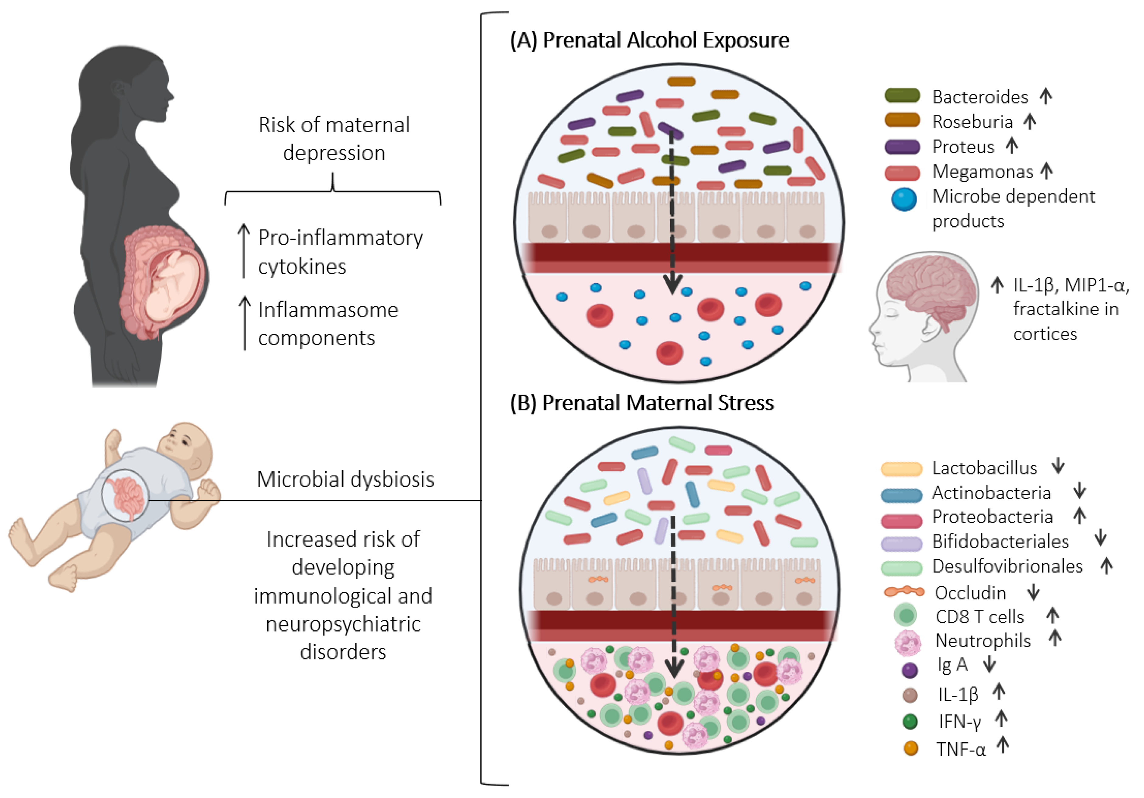
5. Ethanol-Induced Changes in Gut Microbiota: Understanding Consequences and Investigating Prenatal Exposure Effects

5.1. Consequences of Ethanol Consumption in Intestinal Dysbiosis

Alcohol is a widely accepted psychoactive substance in society, despite its addictive properties. Alcohol misuse globally stands as the seventh-leading risk factor for premature death and disabilities [172]. In 2018, the World Health Organization (WHO) reported that alcohol contributes to over 200 diseases and injury-related health conditions, encompassing liver diseases, cancer, cardiovascular diseases, tuberculosis, road injuries, violence, suicides, and HIV/AIDS [173].
Gastrointestinal (GI) diseases are common among those who consume alcohol excessively. The direct contact of alcohol with the mucosa that coats the upper gastrointestinal tract may induce various metabolic and functional changes, leading to significant mucosal damage that can result in gastritis, GI bleeding, and diarrhea. Chronic alcohol misuse is also linked to cancers of the digestive tract, including those of the oral cavity, esophagus, liver, colorectum, stomach, bowel, and pancreas [174].
Alcohol increases intestinal permeability, with decreased expression of occludins, and induces mucosal injuries in the gut, allowing the translocation of large molecules like endotoxins (i.e., LPS) and other bacterial toxins from the gut to the blood or lymph [175]. These toxic substances may cause harmful effects on multiple organs. The LPS binds to TLRs on immune cells, activating inflammatory cytokines. This process explains the elevated markers of monocyte activation and heightened immune response observed in excessive drinkers, particularly those with recent alcohol intake [176]. The ongoing communication between the gut microbiome and the brain facilitates the induction of neuroinflammation by cytokines, which is associated with the development of neuropsychiatric disorders [93].
Alcohol produces an imbalance in the intestinal bacterial population, generating intestinal dysbiosis [177,178], evidenced in both clinical data [179,180,181,182,183,184] and preclinical experiments [12,185,186,187,188]. Alcohol consumption correlates with reduced levels of commensal bacteria and probiotic species such as Lactobacillus and Bifidobacterium, along with important physicobiotics such as Faecalibacterium prausnitzii and Akkermansia muciniphila [178], and also Roseburia, Blautia, Bacteroides, Lachnospiraceae, and Enterococci [180]. On the other hand, Firmicutes is the phylum that has the greatest proliferation in the presence of alcohol, and genera Adlercreutzia, Allobaculum, and Turicibacter have been associated with anxiety- and depression-like disorders [188,189]. Proteobacteria and many species of Bacteroidetes also proliferate in the presence of alcohol [179]. Proteobacteria are a phylum that hosts many known bacteria such as Brucella, Rickettsia, Bordetella, Neisseria, Escherichia, Shigella, Salmonella, Yersina, and Helicobacter. Proteobacteria commonly exhibit Gram-negative staining, indicating the presence of LPS in their outer membrane. They are frequently elevated in disease conditions and have been suggested as a potential marker of microbiota instability [190]. Cuesta et al. (2021) [191] demonstrated a reduction in Gram-positive bacteria taxa, specifically a decrease in the phylum Firmicutes, possibly due to a decrease in Lachnospiraceae family, in mice treated with alcohol for 3 months. Additionally, the Muribaculaceae family and Alloprevotella genus increased in C57BL/6J mice after alcohol consumption. These findings are consistent with higher intestinal permeability induced by chronic ethanol, which may lead to inflammation.
The harmful effects of ethanol may arise directly from its toxicity or indirectly through its metabolites and the generation of reactive oxygen species (ROS) [192]. ROS are chemically reactive molecules containing oxygen, which can cause damage to cells and tissues. The production of ROS during ethanol metabolism can create an environment that is hostile to anaerobic bacteria, promoting the enrichment of aerotolerant bacteria [175,191,193].
Alcohol Use Disorder (AUD) is a chronic and relapsing condition characterized by an inability to control or reduce alcohol consumption [172,173]. Individuals with AUD who had high intestinal permeability showed alterations in their gut microbiota composition, such as a decrease in overall bacterial abundance and in specific bacterial families like Ruminococcaceae (Ruminococcus, Faecalibacterium, Subdoligranulum, Oscillibacter, and Anaerofilum), along with an increase in the abundance of Lachnospiraceae (genus Dorea) and the genus Blautia [181]. Bacteria from the Ruminococcaceae family, which increase during alcohol abstinence, are known to have a beneficial impact on intestinal barrier function [194]. Faecalibacterium prausnitzii is a bacterial species known for its anti-inflammatory properties, and its reduction is related to Crohn’s disease and ulcerative colitis [195,196]. The Lachnospiraceae family is a butyrate-producer. Butyrate, a SCFA produced by symbiotic bacteria in the GI tract through the fermentation of dietary fibers, has functions that include strengthening the gut barrier, reshaping the gut microenvironment, and suppressing inflammation [197,198]. This substance has a protective effect on the intestinal barrier as it leads to a reduction in intestinal permeability induced by alcohol [199]. The lower availability of butyrate is associated with increased intestinal permeability [200]. In the study by Singhal et al. (2021) [201], mice fed with a Lieber–DeCarli alcohol liquid diet exhibited a reduction in the abundance of the butyrate-producing family Lachnospiraceae, in contrast to the findings with AUD individuals found in Leclercq et al. (2020) [202].
Mice transplanted with feces from patients with AUD showed a decrease in F. prausnitzii and an increase in Lachnospiraceae abundance [201]. Since F. prausnitzii is positively correlated with butyric acid levels, which are protective against inflammation [203], this reduction suggests a potential link between gut microbiota alterations and inflammatory processes in AUD. Furthermore, these mice exhibited behavior similar to depression, including reduced sociability and decreased lipolysis [204].
Depressive behavior induced by chronic alcohol consumption was attenuated by dietary administration of sodium butyrate in mice. This compound reduced inflammation and suppressed microglia-mediated neuroinflammation through the GPR109A/PPAR-γ/TLR4-NF-κB-signaling pathway, a complex signaling cascade involved in regulating inflammation and immune responses. Moreover, sodium butyrate ameliorates gut dysbiosis caused by chronic alcohol exposure by increasing the abundance of beneficial bacteria such as Faecalibaculum and Alloprevotella [198].
Intestinal dysbiosis caused by alcohol has been associated with learning and memory dysfunction, depression, anxiety, and alcohol craving [161,202,204]. Additionally, it has been implicated in perpetuating and promoting drinking behavior [205]. In mice, exposure to alcohol for 3 weeks showed an increase in Adlercreutzia, Allobaculum, and Turicibacter, which are bacteria related to intestinal inflammation [206] and neuroimmune regulation [207]. It is noteworthy that Adlercreutzia was negatively correlated with anxiety-like behavior, positively with alcohol preference, and negatively with the expression of brain-derived neurotrophic factor (BDNF) and Gabra1 in the prefrontal cortex [188]. Since BDNF deficiency is associated with an increased preference for alcohol [208] and Gabra1 deficiency leads to alcohol-related cerebral hyperexcitability [209], these correlations suggest that Adlercreutzia may influence alcohol preference and brain excitability through its impact on BDNF and Gabra1 expression.
Probiotics administration, specifically Lactobacillus rhamnosus, Lactobacillus acidophilus, and Bifidobacterium, protected mice from chronic ethanol exposure-induced depressive-like behavior and hippocampal neuronal damage. Changes in the hippocampus are significant in depression, as reduced hippocampal volume is a clinical depression marker [210,211]. Bifidobacterium, the most widely used probiotic, seems to have positive effects in alleviating the symptoms of depression [212]. Bifidobacterium infantis has been linked to an increase in the serotonin precursor tryptophan [178]. Although tryptophan can generate serotonin, the main pathway of metabolism is the kynurenine pathway, where tryptophan is transformed into kynurenine. An increase in kynurenine levels after chronic intermittent alcohol consumption has been revealed, both in the periphery and in the brain. Moreover, this regimen of alcohol exposure induced anhedonia, which was prevented by antibiotic treatment [213].
Exposure to ethanol increases plasma LPS permeability and activates NF-kB, leading Kupffer cells to produce pro-inflammatory chemokines like TNF-α and cytokines [214]. It also raises levels of inflammasome components such as NLRP1, NLRP3, and ASC [215]. Using AAV transfection to downregulate hippocampal NLRP3 expression, Yao et al. (2023) [211] demonstrated that this downregulation decreased depressive behavior in animals exposed to chronic alcohol consumption, suggesting that gut microbiota can regulate depressive behavior through NLRP3-mediated hippocampal neuroinflammation. Additionally, they found that serum inflammatory cytokines mediate the link between gut microbiota and depressive behavior in these mice. Chronic alcohol consumption increased inflammatory markers (IL-1β, iNOS, TNF-α, COX-2, IL-10, and CXCL10) in the colons of wild-type mice but not in TLR4-KO mice. The TLR4-KO mice also exhibited a distinct microbiota, characterized by a reduced abundance of Firmicutes, potentially offering protection against ethanol-induced inflammation [191]. Moreover, TLR4 exerts an important role in alcohol-induced neuroinflammation [216] and depression [217], underscoring the significance of neuroinflammation in these disorders.
These findings highlight the critical role of the gut microbiota in mediating the relationship between alcohol abuse, immune system activation, and depressive behavior. The observed protective effect in TLR4-KO mice, with their distinct microbiota composition, suggests that targeting the microbiota–immune system axis could be a promising strategy for mitigating the negative effects of chronic alcohol consumption.
Table 2 summarizes the relationship between alcohol exposure, microbiota dysbiosis, and associated outcomes.
The exact mechanisms of alcohol-induced intestinal dysbiosis have not yet been elucidated. However, there are a series of hypotheses, such as slow peristalsis, increased fecal pH [175,218], and changes in bile salt concentrations [219]. Changes in luminal pH can potentially shift the competitive balance among different bacterial communities [175]. Dysbiosis and altered bile acid pools are linked to liver diseases like cirrhosis and metabolic syndrome. With advancing liver disease, dysbiosis occurs alongside decreased intestinal bile acid concentration, favoring Firmicutes and increasing deoxycholic acid production. Alcohol intake exacerbates these effects, leading to greater intestinal and systemic inflammation and worsening dysbiosis [219]. Due to the role of the gut microbiome in the gut–brain axis and its influence on AUD, manipulating the microbiota offers a promising therapeutic approach for AUD treatment.

5.2. Effects of Prenatal Ethanol Exposure on Maternal and Offspring Microbiota

Alcohol consumption during pregnancy may pose significant health risks for women and their offspring [220,221]. A household survey conducted by Tan et al. (2015) [222] found that 10.2% of pregnant women reported any alcohol use, with 3.1% engaging in binge drinking. Additionally, 1 in 10 women reported alcohol use in the past 30 days, and 1 in 33 reported binge drinking during pregnancy. Considering the aforementioned, there is no safe dose of alcohol during pregnancy [223] as prenatal alcohol exposure (PAE) has been linked to adverse behavioral effects [224], as well as learning and memory impairments [225,226]. PAE is also associated with neurological disorders [227], involving multiple mechanisms that contribute to developmental harm and adverse outcomes [5]. Although the impact of PAE on the intestinal microbiota has been relatively understudied, some studies have shown significant consequences in both offspring and mothers.
In a study involving pregnant women who consumed alcohol, fecal samples were collected at the end of pregnancy and from their newborns within 48 h after birth. These samples were compared with those from a non-alcohol consumption group. This comparison revealed a significant change in the maternal gut microbiota, characterized by a reduced abundance of the Faecalibacterium genus and an increased abundance of the Phascolarctobacterium and Blautia genera in the group of women who consumed alcohol. Additionally, the colonization of the offspring’s gut microbiota was impacted, indicating an overgrowth of Megamonas [15]. There is a predominance of certain bacterial genera, including Megamonas, in fecal samples from patients diagnosed with MDD, suggesting that an increased presence of this genus could be associated with this disorder [123]. Indeed, it is known that children exposed to alcohol in utero may be predisposed to exhibit higher levels of negative emotionality, thus presenting an increased risk for childhood depression [228].
PAE appears to have a lasting impact on the fecal microbiota of offspring, as revealed in a study involving rats that exhibited changes in bacterial diversity throughout adulthood. The genera Bacteroides, Roseburia, and Proteus are the most abundant in PAE animals. The authors also conducted analyses with stratification by sex and observed differences in the microbiota between males and females. Notably, males exhibited a higher α diversity, whereas no significant differences were discerned in females. Furthermore, variations in different genera were identified based on sex. Among males, the genus Akkermansia was detected in higher abundance, while Bifidobacterium displayed a lower abundance. Conversely, in females, there was an elevated abundance of the genera Proteus, Roseburia, and Faecalitalea. In both sexes, the genus Bacteroides exhibited higher abundance, while Ruminococcus displayed lower abundance in cases of PAE [229]. Some of these altered bacterial genera are associated with inflammatory conditions, such as irritable bowel syndrome. Studies have demonstrated that individuals with irritable bowel syndrome experience a reduction in Bifidobacterium and an increase in Bacteroides [230,231]. Furthermore, besides Bifidobacterium, bacteria such as Roseburia and Akkermansia also play a role in regulating the immune system by producing SCFA, which are important metabolites of the gut microbiome [210,232,233]. Levels of SCFAs, such as acetic acid, propionic acid, and pentanoic acid, have been shown to be reduced in animal models of depression [234].
Virdee et al. (2021) [235] revealed a distinct and significant biosignature of microbe-dependent products (MDPs), which distinguishes alcohol-exposed mothers from their controls. Maternal plasma was characterized by phenolic acids, which are associated with anti-inflammatory actions through the inhibition of pro-oxidant enzyme-signaling cascades [236]. Probably, their elevation in PAE may potentially mitigate some of the damage generated by alcohol in both the mother and the fetus. Many metabolites from maternal plasma are readily exchanged across the placenta and become bioavailable to the fetus [235]. Several MDPs elevated by PAE (catechol sulfate, 4-ethylphenylsulfate, erythritol, indolepropionate, oxindole, p-cresol, salicylate) are related to neuroinflammation, depression, anxiety, and autism [235].
Analysis of fecal samples from pregnant dams with ad libitum access to a liquid diet, containing alcohol with 36% of total calories derived from ethanol, showed lower bacterial diversity and uniformity, as well as a general increase in aerotolerant bacteria Desulfovibrionaceae and Akkermansia (Verrucomicrobiota) compared to the control group. The offspring of these dams exposed to ethanol showed an increased abundance of Parabacteroides, Alistipes, and the phylum Firmicutes, as well as a decrease in Ruminococcus [17].
Ethanol may lead to alterations in gut microbiota composition, influencing cytokine production and release. These cytokines, in response to changes in the gut microbiota, may contribute to inflammation and immune system dysregulation, potentially exacerbating the effects of ethanol exposure on various physiological processes, including depression, anxiety, and alcohol dependence [237].
Alterations in cytokine levels due to maternal alcohol exposure can influence neurocognitive and neuropsychiatric outcomes in children by affecting inflammatory components [230,238]. Increased levels of IL-15 and IL-10 were observed in the maternal blood samples collected during the second trimester in alcohol-consuming mothers, which were linked to neurodevelopmental delays in their offspring [228]. These findings suggest that maternal immune system dysregulation may contribute to adverse child outcomes and could serve as early biomarkers for neurodevelopmental delay, including alcohol-related conditions [228]. Pascual et al. (2017) [238] demonstrated maternal and fetal immune responses triggered by alcohol consumption during pregnancy, which was demonstrated by cytokines/chemokines’ production in fetal and postnatal brains, with upregulation of IL-1β, macrophage inflammatory proteins (MIP)-1α, and fractalkine in cortices of 15-day-old fetuses. Bake et al. (2021) [239] found sex-dependent effects on cytokine levels of prenatal alcohol exposure using pregnant rats exposed to ethanol vapor. They found that males exhibited suppressed cytokine levels and increased anxiety-like behavior, whereas females displayed elevated cytokine levels in adipose tissue and liver along with decreased social interaction. All these alterations could, at least in part, result from changes in intestinal barrier/permeability and microbiota.
Table 3 summarizes the changes induced by prenatal alcohol exposure in the mother and offspring microbiota.

6. Connecting the Dots: Exploring the Link between Microbiota Dysbiosis, Maternal Immunological Changes, and Depression

The microbiome encompasses the entire collection of genomes from all microorganisms within a particular environment, which includes the community of microorganisms, their products, and the environmental conditions they inhabit. The microbiome plays a crucial role in the overall function and effectiveness of both the innate and adaptive branches of the immune system, while the immune system helps maintain key aspects of its symbiotic relationship with microbes [18]. Imbalances in these interactions may contribute to the development of various immune-mediated disorders. High rates of depression have been observed in various conditions involving immune system activation [101].
The regulation of the maternal immune system throughout pregnancy is a complex and finely tuned process designed to protect the developing fetus. This regulation involves suppressing certain immune activities in order to create a favorable environment for fetal growth. Disruptions in the maternal immune system may cause significant consequences, particularly regarding the production of cytokines. Notably, studies indicate that cytokines such as IL-6, IL-1α, and TNF-α can move bidirectionally across the placenta, illustrating a dynamic transfer of these molecules during pregnancy [240]. Therefore, changes in the maternal microbiota may contribute to immune dysregulation, influencing subsequent effects on fetal development.
Cytokine transfer through the placenta is vital for proper fetal development, creating a direct communication link between the maternal immune system and the developing fetus. Disruptions in this delicate balance might result in imbalances in cytokine levels, potentially affecting the fetal immune environment with consequences extending beyond the prenatal period. Exposure to prenatal ethanol or stress has been shown to cause alterations in cytokine levels towards a proinflammatory state. Important exposure to inflammation in pregnant mothers led to lasting changes in both innate and adaptive immune functions in their newborns [241], potentially contributing to the development of depression later in life [242]. This fact has particular significance in the realm of neurodevelopmental disorders, where inappropriate activation of the maternal immune system has been identified as a contributing factor to psychosis, depressive- and anxiety-like behaviors, and increased substance use behavior [243,244,245] (Figure 2).
The microbiota–gut–brain axis emerges as a key system in regulating various CNS homeostatic processes, including neurotransmission, activation of stress responses, and neuroinflammation [246,247,248]. Alterations in immune function, such as an increased production of pro-inflammatory cytokines, have been observed in psychiatric conditions like depression and anxiety. The bidirectional communication between the immune system and the CNS is a complex interplay that influences both immune responses and brain function. Chronic inflammation, triggered by persistent immune activation, can affect neurotransmitter metabolism, neuroendocrine function, and neural circuits related to mood and behavior. This interaction highlights the intricate relationship between the immune system and psychiatric health, suggesting that immune dysregulation may contribute to the development and exacerbation of psychiatric disorders. This gains particular interest when the immune alterations are in the mother with consequences in the offspring.

7. Conclusions

Based on the reviewed studies, it can be concluded that maternal exposure to stress and ethanol significantly affects the microbiota of both the mother and her offspring through multiple interconnected mechanisms. Stress and ethanol activate the HPA axis, leading to increased levels of corticosterone in rodents and cortisol in humans. Elevated glucocorticoids disrupt gut permeability, resulting in a “leaky gut” and enhanced translocation of LPS into the bloodstream. The activation of inflammasome components contributes to systemic inflammation and alterations in gut microbiota composition. Dysbiosis during pregnancy can negatively impact the maternal–fetal interface, further influencing the immune system function. These disruptions can impair the offspring’s immune system and increase their susceptibility to psychiatric disorders later in life. Therefore, it is essential not to underestimate the effects of minor stressors or moderate alcohol intake during pregnancy, as both can significantly contribute to dysbiosis and immune dysregulation, which, when combined, may exacerbate adverse outcomes more than either factor alone. This review highlights the potential cumulative effects of prenatal alcohol and stress exposure on gut microbiota, the immune system, and overall offspring health.

Implications and Future Research Directions

The findings underscore the need for further research into the combined effects of stress and ethanol on the gut–brain axis. Future studies should explore pharmacological strategies targeting the gut–brain axis as potential interventions to mitigate the effects of maternal stress and ethanol exposure. Additionally, research should investigate the long-term consequences of these exposures on offspring mental health and explore potential therapeutic approaches to address microbiota and immune dysregulation.

Author Contributions

Conceptualization, R.C., P.M., M.H.-M., S.d.S.O. and N.O.S.C.; resources, R.C. and N.O.S.C.; writing—original draft preparation, R.C., P.M., M.H.-M., S.d.S.O. and N.O.S.C.; writing—review and editing, R.C., P.M., M.H.-M., S.d.S.O. and N.O.S.C.; visualization, R.C., P.M., M.H.-M., S.d.S.O. and N.O.S.C.; supervision, R.C. and N.O.S.C.; project administration, R.C. and N.O.S.C.; funding acquisition, R.C. and N.O.S.C. All authors have read and agreed to the published version of the manuscript.

Funding

This study was not funded by any particular grant. R.C., P.M, S.d.S.O., and N.O.S.C. research are funded by Sao Paulo Research Foundation (Fundação de Amparo à Pesquisa do Estado de São Paulo, grants #2023/13110-0; 2022/08743-1; 2022/16176-0, and 2023/07482-2, respectively). R.C. and NOSC are Research Fellows of the National Council for Scientific and Technological Development (Conselho Nacional de Desenvolvimento Científico e Tecnológico, CNPq).

Data Availability Statement

No new data were created or analyzed in this study. Data sharing is not applicable to this article.

Acknowledgments

Figures were created with BioRender.com.

Conflicts of Interest

The authors declare no conflicts of interest.

Abbreviations

AUD: Alcohol use disorder; BDNF: Brain-derived neurotrophic factor; CCR9: C-C motif chemokine receptor 9; CNS: Central nervous system; CUMS: Chronic unpredictable mild stress; DC: Dendritic cells; GI: Gastrointestinal; HPA: Hypothalamic-pituitary-adrenal; 5-HT: Serotonin; 5-HTRs: 5-HT receptors; IL: Interleukin; Ig: Immunoglobulin; LPS: Lipopolysaccharides; NF-KB: Nuclear factor kappa B; NLRP3: NOD-like receptor family, pyrin domain containing; MDPs: Microbe-dependent products; MDD: Major depressive disorder; MUC: Mucin; PAE: Prenatal alcohol exposure; ROS: Reactive oxygen species; SCFA: Short-chain fatty acids; Th: Helper T cells; Treg: Regulatory T cells; TGF: Transforming growth factor; TNF-α: Tumor necrosis factor-α; TLR4-KO: Toll-like receptor 4 knockout.

References

  1. Rhee, S.H.; Pothoulakis, C.; Mayer, E.A. Principles and clinical implications of the brain-gut-enteric microbiota axis. Nat. Rev. Gastroenterol. Hepatol. 2009, 6, 306–314. [Google Scholar] [CrossRef] [PubMed]
  2. Valles-Colomer, M.; Falony, G.; Darzi, Y.; Tigchelaar, E.F.; Wang, J.; Tito, R.Y.; Schiweck, C.; Kurilshikov, A.; Joossens, M.; Wijmenga, C.; et al. The neuroactive potential of the human gut microbiota in quality of life and depression. Nat. Microbiol. 2019, 4, 623–632. [Google Scholar] [CrossRef] [PubMed]
  3. Tanelian, A.; Nankova, B.; Miari, M.; Sabban, E.L. Microbial composition, functionality, and stress resilience or susceptibility: Unraveling sex-specific patterns. Biol. Sex Differ. 2024, 15, 20. [Google Scholar] [CrossRef] [PubMed]
  4. Foster, J.A.; Rinaman, L.; Cryan, J.F. Stress & the gut-brain axis: Regulation by the microbiome. Neurobiol. Stress 2017, 7, 124–136. [Google Scholar] [CrossRef]
  5. Popova, S.; Charness, M.E.; Burd, L.; Crawford, A.; Hoyme, H.E.; Mukherjee, R.A.S.; Riley, E.P.; Elliott, E.J. Fetal alcohol spectrum disorders. Nat. Rev. Dis. Primers 2023, 9, 11. [Google Scholar] [CrossRef]
  6. Butts, M.; Sundaram, V.L.; Murughiyan, U.; Borthakur, A.; Singh, S. The Influence of Alcohol Consumption on Intestinal Nutrient Absorption: A Comprehensive Review. Nutrients 2023, 15, 1571. [Google Scholar] [CrossRef]
  7. Esper, L.H.; Furtado, E.F. Stressful life events and alcohol consumption in pregnant women: A cross-sectional survey. Midwifery 2019, 71, 27–32. [Google Scholar] [CrossRef]
  8. Lovallo, W.R. Cortisol secretion patterns in addiction and addiction risk. Int. J. Psychophysiol. 2006, 59, 195–202. [Google Scholar] [CrossRef]
  9. Anderson, A.E.; Hure, A.J.; Kay-Lambkin, F.J.; Loxton, D.J. Women’s perceptions of information about alcohol use during pregnancy: A qualitative study. BMC Public Health 2014, 14, 1048. [Google Scholar] [CrossRef] [PubMed] [PubMed Central]
  10. Branco, E.I.; Kaskutas, L.A. “If it burns going down…”: How focus groups can shape fetal alcohol syndrome (FAS) prevention. Subst. Use Misuse 2001, 36, 333–345. [Google Scholar] [CrossRef] [PubMed]
  11. Ming, L.; Qiao, X.; Yi, L.; Siren, D.; He, J.; Hai, L.; Guo, F.; Xiao, Y.; Ji, R. Camel milk modulates ethanol-induced changes in the gut microbiome and transcriptome in a mouse model of acute alcoholic liver disease. J. Dairy Sci. 2020, 103, 3937–3949. [Google Scholar] [CrossRef] [PubMed]
  12. Wang, C.; Yan, J.; Du, K.; Liu, S.; Wang, J.; Wang, Q.; Zhao, H.; Li, M.; Yan, D.; Zhang, R.; et al. Intestinal microbiome dysbiosis in alcohol-dependent patients and its effect on rat behaviors. mBio 2023, 14, e0239223. [Google Scholar] [CrossRef] [PubMed]
  13. Galley, J.D.; Nelson, M.C.; Yu, Z.; Dowd, S.E.; Walter, J.; Kumar, P.S.; Lyte, M.; Bailey, M.T. Exposure to a social stressor disrupts the community structure of the colonic mucosa-associated microbiota. BMC Microbiol. 2014, 14, 189. [Google Scholar] [CrossRef] [PubMed]
  14. Partrick, K.A.; Chassaing, B.; Beach, L.Q.; McCann, K.E.; Gewirtz, A.T.; Huhman, K.L. Acute and repeated exposure to social stress reduces gut microbiota diversity in Syrian hamsters. Behav. Brain Res. 2018, 345, 39–48. [Google Scholar] [CrossRef]
  15. Wang, Y.; Xie, T.; Wu, Y.; Liu, Y.; Zou, Z.; Bai, J. Impacts of Maternal Diet and Alcohol Consumption during Pregnancy on Maternal and Infant Gut Microbiota. Biomolecules 2021, 11, 369. [Google Scholar] [CrossRef]
  16. Yeramilli, V.; Cheddadi, R.; Shah, J.; Brawner, K.; Martin, C. A Review of the Impact of Maternal Prenatal Stress on Offspring Microbiota and Metabolites. Metabolites 2023, 13, 535. [Google Scholar] [CrossRef]
  17. Bodnar, T.S.; Ainsworth-Cruickshank, G.; Billy, V.; Wegener Parfrey, L.; Weinberg, J.; Raineki, C. Alcohol consumption during pregnancy differentially affects the fecal microbiota of dams and offspring. Sci. Rep. 2024, 14, 16121. [Google Scholar] [CrossRef]
  18. Zheng, D.; Liwinski, T.; Elinav, E. Interaction between microbiota and immunity in health and disease. Cell Res. 2020, 30, 492–506. [Google Scholar] [CrossRef]
  19. Rosenberg, E.; Zilber-Rosenberg, I. Microbes Drive Evolution of Animals and Plants: The Hologenome Concept. mBio 2016, 7, e01395. [Google Scholar] [CrossRef]
  20. Cohen, Y.; Borenstein, E. The microbiome’s fiber degradation profile and its relationship with the host diet. BMC Biol. 2022, 20, 266. [Google Scholar] [CrossRef]
  21. Gensollen, T.; Iyer, S.S.; Kasper, D.L.; Blumberg, R.S. How colonization by microbiota in early life shapes the immune system. Science 2016, 352, 539–544. [Google Scholar] [CrossRef] [PubMed]
  22. Pedersen, H.K.; Gudmundsdottir, V.; Nielsen, H.B.; Hyotylainen, T.; Nielsen, T.; Jensen, B.A.; Forslund, K.; Hildebrand, F.; Prifti, E.; Falony, G.; et al. Human gut microbes impact host serum metabolome and insulin sensitivity. Nature 2016, 535, 376–381. [Google Scholar] [CrossRef] [PubMed]
  23. Zhang, Y.; Li, Y.; Barber, A.F.; Noya, S.B.; Williams, J.A.; Li, F.; Daniel, S.G.; Bittinger, K.; Fang, J.; Sehgal, A. The microbiome stabilizes circadian rhythms in the gut. Proc. Natl. Acad. Sci. USA 2023, 120, e2217532120. [Google Scholar] [CrossRef] [PubMed]
  24. Sherwin, E.; Bordenstein, S.R.; Quinn, J.L.; Dinan, T.G.; Cryan, J.F. Microbiota and the social brain. Science 2019, 366, eaar2016. [Google Scholar] [CrossRef]
  25. Kawano, Y.; Edwards, M.; Huang, Y.; Bilate, A.M.; Araujo, L.P.; Tanoue, T.; Atarashi, K.; Ladinsky, M.S.; Reiner, S.L.; Wang, H.H.; et al. Microbiota imbalance induced by dietary sugar disrupts immune-mediated protection from metabolic syndrome. Cell 2022, 185, 3501–3519.e20. [Google Scholar] [CrossRef]
  26. Cheney, A.M.; Costello, S.M.; Pinkham, N.V.; Waldum, A.; Broadaway, S.C.; Cotrina-Vidal, M.; Mergy, M.; Tripet, B.; Kominsky, D.J.; Grifka-Walk, H.M.; et al. Gut microbiome dysbiosis drives metabolic dysfunction in Familial dysautonomia. Nat. Commun. 2023, 14, 218. [Google Scholar] [CrossRef]
  27. Roy Sarkar, S.; Banerjee, S. Gut microbiota in neurodegenerative disorders. J. Neuroimmunol. 2019, 328, 98–104. [Google Scholar] [CrossRef]
  28. Afroz, K.F.; Reyes, N.; Young, K.; Parikh, K.; Misra, V.; Alviña, K. Altered gut microbiome and autism like behavior are associated with parental high salt diet in male mice. Sci. Rep. 2021, 11, 8364, Erratum in Sci. Rep. 2022, 12, 5686. [Google Scholar] [CrossRef]
  29. Chelakkot, C.; Ghim, J.; Ryu, S.H. Mechanisms regulating intestinal barrier integrity and its pathological implications. Exp. Mol. Med. 2018, 50, 1–9. [Google Scholar] [CrossRef]
  30. Corfield, A.P. The Interaction of the Gut Microbiota with the Mucus Barrier in Health and Disease in Human. Microorganisms 2018, 6, 78. [Google Scholar] [CrossRef]
  31. Schroeder, B.O. Fight them or feed them: How the intestinal mucus layer manages the gut microbiota. Gastroenterol. Rep. 2019, 7, 3–12. [Google Scholar] [CrossRef] [PubMed]
  32. Pelaseyed, T.; Bergström, J.H.; Gustafsson, J.K.; Ermund, A.; Birchenough, G.M.; Schütte, A.; van der Post, S.; Svensson, F.; Rodríguez-Piñeiro, A.M.; Nyström, E.E.; et al. The mucus and mucins of the goblet cells and enterocytes provide the first defense line of the gastrointestinal tract and interact with the immune system. Immunol. Rev. 2014, 260, 8–20. [Google Scholar] [CrossRef] [PubMed]
  33. Han, F.; Zhang, H.; Xia, X.; Xiong, H.; Song, D.; Zong, X.; Wang, Y. Porcine β-defensin 2 attenuates inflammation and mucosal lesions in dextran sodium sulfate-induced colitis. J. Immunol. 2015, 194, 1882–1893. [Google Scholar] [CrossRef]
  34. Bevins, C.L.; Salzman, N.H. Paneth cells, antimicrobial peptides and maintenance of intestinal homeostasis. Nat. Rev. Microbiol. 2011, 9, 356–368. [Google Scholar] [CrossRef] [PubMed]
  35. Neutra, M.R.; Pringault, E.; Kraehenbuhl, J.P. Antigen sampling across epithelial barriers and induction of mucosal immune responses. Annu. Rev. Immunol. 1996, 14, 275–300. [Google Scholar] [CrossRef]
  36. Rescigno, M.; Urbano, M.; Valzasina, B.; Francolini, M.; Rotta, G.; Bonasio, R.; Granucci, F.; Kraehenbuhl, J.P.; Ricciardi-Castagnoli, P. Dendritic cells express tight junction proteins and penetrate gut epithelial monolayers to sample bacteria. Nat. Immunol. 2001, 2, 361–367. [Google Scholar] [CrossRef]
  37. Nagler-Anderson, C. Man the barrier! Strategic defences in the intestinal mucosa. Nat. Rev. Immunol. 2001, 1, 59–67. [Google Scholar] [CrossRef]
  38. Iwata, M.; Hirakiyama, A.; Eshima, Y.; Kagechika, H.; Kato, C.; Song, S.Y. Retinoic acid imprints gut-homing specificity on T cells. Immunity 2004, 21, 527–538. [Google Scholar] [CrossRef]
  39. Masopust, D.; Vezys, V.; Wherry, E.J.; Barber, D.L.; Ahmed, R. Cutting edge: Gut microenvironment promotes differentiation of a unique memory CD8 T cell population. J. Immunol. 2006, 176, 2079–2083. [Google Scholar] [CrossRef]
  40. van Wijk, F.; Cheroutre, H. Mucosal T cells in gut homeostasis and inflammation. Expert Rev. Clin. Immunol. 2010, 6, 559–566. [Google Scholar] [CrossRef]
  41. Chen, W.; Jin, W.; Hardegen, N.; Lei, K.J.; Li, L.; Marinos, N.; McGrady, G.; Wahl, S.M. Conversion of peripheral CD4+CD25- naive T cells to CD4+CD25+ regulatory T cells by TGF-beta induction of transcription factor Foxp3. J. Exp. Med. 2003, 198, 1875–1886. [Google Scholar] [CrossRef] [PubMed]
  42. Izcue, A.; Coombes, J.L.; Powrie, F. Regulatory T cells suppress systemic and mucosal immune activation to control intestinal inflammation. Immunol. Rev. 2006, 212, 256–271. [Google Scholar] [CrossRef] [PubMed]
  43. Conti, H.R.; Shen, F.; Nayyar, N.; Stocum, E.; Sun, J.N.; Lindemann, M.J.; Ho, A.W.; Hai, J.H.; Yu, J.J.; Jung, J.W.; et al. Th17 cells and IL-17 receptor signaling are essential for mucosal host defense against oral candidiasis. J. Exp. Med. 2009, 206, 299–311. [Google Scholar] [CrossRef] [PubMed]
  44. Ishigame, H.; Kakuta, S.; Nagai, T.; Kadoki, M.; Nambu, A.; Komiyama, Y.; Fujikado, N.; Tanahashi, Y.; Akitsu, A.; Kotaki, H.; et al. Differential roles of interleukin-17A and -17F in host defense against mucoepithelial bacterial infection and allergic responses. Immunity 2009, 30, 108–119. [Google Scholar] [CrossRef]
  45. Mucida, D.; Park, Y.; Kim, G.; Turovskaya, O.; Scott, I.; Kronenberg, M.; Cheroutre, H. Reciprocal TH17 and regulatory T cell differentiation mediated by retinoic acid. Science 2007, 317, 256–260. [Google Scholar] [CrossRef]
  46. Veldhoen, M.; Hocking, R.J.; Atkins, C.J.; Locksley, R.M.; Stockinger, B. TGFbeta in the context of an inflammatory cytokine milieu supports de novo differentiation of IL-17-producing T cells. Immunity 2006, 24, 179–189. [Google Scholar] [CrossRef]
  47. Bousbaine, D.; Fisch, L.I.; London, M.; Bhagchandani, P.; Rezende de Castro, T.B.; Mimee, M.; Olesen, S.; Reis, B.S.; VanInsberghe, D.; Bortolatto, J.; et al. A conserved Bacteroidetes antigen induces anti-inflammatory intestinal T lymphocytes. Science 2022, 377, 660–666. [Google Scholar] [CrossRef]
  48. David, L.A.; Maurice, C.F.; Carmody, R.N.; Gootenberg, D.B.; Button, J.E.; Wolfe, B.E.; Ling, A.V.; Devlin, A.S.; Varma, Y.; Fischbach, M.A.; et al. Diet rapidly and reproducibly alters the human gut microbiome. Nature 2014, 505, 559–563. [Google Scholar] [CrossRef]
  49. Siracusa, F.; Schaltenberg, N.; Kumar, Y.; Lesker, T.R.; Steglich, B.; Liwinski, T.; Cortesi, F.; Frommann, L.; Diercks, B.P.; Bönisch, F.; et al. Short-term dietary changes can result in mucosal and systemic immune depression. Nat. Immunol. 2023, 24, 1473–1486. [Google Scholar] [CrossRef]
  50. Levy, M.; Kolodziejczyk, A.A.; Thaiss, C.A.; Elinav, E. Dysbiosis and the immune system. Nat. Rev. Immunol. 2017, 17, 219–232. [Google Scholar] [CrossRef]
  51. Siddiqui, R.; Makhlouf, Z.; Alharbi, A.M.; Alfahemi, H.; Khan, N.A. The Gut Microbiome and Female Health. Biology 2022, 11, 1683. [Google Scholar] [CrossRef] [PubMed]
  52. Siddiqui, R.; Mungroo, M.R.; Alharbi, A.M.; Alfahemi, H.; Khan, N.A. The Use of Gut Microbial Modulation Strategies as Interventional Strategies for Ageing. Microorganisms 2022, 10, 1869. [Google Scholar] [CrossRef] [PubMed]
  53. Cuesta, C.M.; Guerri, C.; Ureña, J.; Pascual, M. Role of Microbiota-Derived Extracellular Vesicles in Gut-Brain Communication. Int. J. Mol. Sci. 2021, 22, 4235. [Google Scholar] [CrossRef] [PubMed]
  54. Catalano, P.M.; Tyzbir, E.D.; Roman, N.M.; Amini, S.B.; Sims, E.A. Longitudinal changes in insulin release and insulin resistance in nonobese pregnant women. Am. J. Obstet. Gynecol. 1991, 165, 1667–1672. [Google Scholar] [CrossRef]
  55. Catalano, P.M.; Tyzbir, E.D.; Wolfe, R.R.; Roman, N.M.; Amini, S.B.; Sims, E.A. Longitudinal changes in basal hepatic glucose production and suppression during insulin infusion in normal pregnant women. Am. J. Obstet. Gynecol. 1992, 167, 913–919. [Google Scholar] [CrossRef]
  56. Connor, K.L.; Chehoud, C.; Altrichter, A.; Chan, L.; DeSantis, T.Z.; Lye, S.J. Maternal metabolic, immune, and microbial systems in late pregnancy vary with malnutrition in mice. Biol. Reprod. 2018, 98, 579–592. [Google Scholar] [CrossRef]
  57. Mirpuri, J. Evidence for maternal diet-mediated effects on the offspring microbiome and immunity: Implications for public health initiatives. Pediatr. Res. 2021, 89, 301–306. [Google Scholar] [CrossRef]
  58. Ma, J.; Prince, A.L.; Bader, D.; Hu, M.; Ganu, R.; Baquero, K.; Blundell, P.; Alan Harris, R.; Frias, A.E.; Grove, K.L.; et al. High-fat maternal diet during pregnancy persistently alters the offspring microbiome in a primate model. Nat. Commun. 2014, 5, 3889. [Google Scholar] [CrossRef]
  59. Myles, I.A.; Fontecilla, N.M.; Janelsins, B.M.; Vithayathil, P.J.; Segre, J.A.; Datta, S.K. Parental dietary fat intake alters offspring microbiome and immunity. J. Immunol. 2013, 191, 3200–3209. [Google Scholar] [CrossRef]
  60. Zhou, Z.; Bian, C.; Luo, Z.; Guille, C.; Ogunrinde, E.; Wu, J.; Zhao, M.; Fitting, S.; Kamen, D.L.; Oates, J.C.; et al. Progesterone decreases gut permeability through upregulating occludin expression in primary human gut tissues and Caco-2 cells. Sci. Rep. 2019, 9, 8367. [Google Scholar] [CrossRef]
  61. Nuriel-Ohayon, M.; Neuman, H.; Koren, O. Microbial Changes during Pregnancy, Birth, and Infancy. Front. Microbiol. 2016, 7, 1031. [Google Scholar] [CrossRef] [PubMed]
  62. Armistead, B.; Kadam, L.; Drewlo, S.; Kohan-Ghadr, H.R. The Role of NFκB in Healthy and Preeclamptic Placenta: Trophoblasts in the Spotlight. Int. J. Mol. Sci. 2020, 21, 1775. [Google Scholar] [CrossRef] [PubMed]
  63. Gleicher, N.; Cohen, C.J.; Kerenyi, T.D.; Gusberg, S.B. A blocking factor in amniotic fluid causing leukocyte migration enhancement. Am. J. Obstet. Gynecol. 1979, 133, 386–390. [Google Scholar] [CrossRef] [PubMed]
  64. Shohat, B.; Faktor, J.M. Immunosuppressive activity of human amniotic fluid of normal and abnormal pregnancies. Int. J. Fertil. 1988, 33, 273–277. [Google Scholar]
  65. Lang, A.K.; Searle, R.F. The immunomodulatory activity of human amniotic fluid can be correlated with transforming growth factor-beta 1 (TGF-beta 1) and beta 2 activity. Clin. Exp. Immunol. 1994, 97, 158–163. [Google Scholar] [CrossRef]
  66. Winters, A.D.; Romero, R.; Greenberg, J.M.; Galaz, J.; Shaffer, Z.D.; Garcia-Flores, V.; Kracht, D.J.; Gomez-Lopez, N.; Theis, K.R. Does the Amniotic Fluid of Mice Contain a Viable Microbiota? Front. Immunol. 2022, 13, 820366. [Google Scholar] [CrossRef]
  67. Aagaard, K.; Ma, J.; Antony, K.M.; Ganu, R.; Petrosino, J.; Versalovic, J. The placenta harbors a unique microbiome. Sci. Transl. Med. 2014, 6, 237ra65. [Google Scholar] [CrossRef]
  68. Rackaityte, E.; Halkias, J.; Fukui, E.M.; Mendoza, V.F.; Hayzelden, C.; Crawford, E.D.; Fujimura, K.E.; Burt, T.D.; Lynch, S.V. Viable bacterial colonization is highly limited in the human intestine in utero. Nat. Med. 2020, 26, 599–607. [Google Scholar] [CrossRef]
  69. Mändar, R.; Punab, M.; Borovkova, N.; Lapp, E.; Kiiker, R.; Korrovits, P.; Metspalu, A.; Krjutškov, K.; Nõlvak, H.; Preem, J.K.; et al. Complementary seminovaginal microbiome in couples. Res. Microbiol. 2015, 166, 440–447. [Google Scholar] [CrossRef]
  70. Verstraelen, H.; Vilchez-Vargas, R.; Desimpel, F.; Jauregui, R.; Vankeirsbilck, N.; Weyers, S.; Verhelst, R.; De Sutter, P.; Pieper, D.H.; Van De Wiele, T. Characterisation of the human uterine microbiome in non-pregnant women through deep sequencing of the V1-2 region of the 16S rRNA gene. PeerJ 2016, 4, e1602. [Google Scholar] [CrossRef]
  71. Makino, H.; Kushiro, A.; Ishikawa, E.; Muylaert, D.; Kubota, H.; Sakai, T.; Oishi, K.; Martin, R.; Ben Amor, K.; Oozeer, R.; et al. Transmission of intestinal Bifidobacterium longum subsp. longum strains from mother to infant, determined by multilocus sequencing typing and amplified fragment length polymorphism. Appl. Environ. Microbiol. 2011, 77, 6788–6793. [Google Scholar] [CrossRef] [PubMed]
  72. Matsumiya, Y.; Kato, N.; Watanabe, K.; Kato, H. Molecular epidemiological study of vertical transmission of vaginal Lactobacillus species from mothers to newborn infants in Japanese, by arbitrarily primed polymerase chain reaction. J. Infect. Chemother. 2002, 8, 43–49. [Google Scholar] [CrossRef] [PubMed]
  73. Matsuki, T.; Watanabe, K.; Tanaka, R.; Fukuda, M.; Oyaizu, H. Distribution of bifidobacterial species in human intestinal microflora examined with 16S rRNA-gene-targeted species-specific primers. Appl. Environ. Microbiol. 1999, 65, 4506–4512. [Google Scholar] [CrossRef] [PubMed]
  74. Sinha, T.; Brushett, S.; Prins, J.; Zhernakova, A. The maternal gut microbiome during pregnancy and its role in maternal and infant health. Curr. Opin. Microbiol. 2023, 74, 102309. [Google Scholar] [CrossRef]
  75. Kalliomäki, M.; Collado, M.C.; Salminen, S.; Isolauri, E. Early differences in fecal microbiota composition in children may predict overweight. Am. J. Clin. Nutr. 2008, 87, 534–538. [Google Scholar] [CrossRef]
  76. Kalliomäki, M.; Kirjavainen, P.; Eerola, E.; Kero, P.; Salminen, S.; Isolauri, E. Distinct patterns of neonatal gut microflora in infants in whom atopy was and was not developing. J. Allergy Clin. Immunol. 2001, 107, 129–134. [Google Scholar] [CrossRef]
  77. Savino, F.; Cresi, F.; Pautasso, S.; Palumeri, E.; Tullio, V.; Roana, J.; Silvestro, L.; Oggero, R. Intestinal microflora in breastfed colicky and non-colicky infants. Acta Paediatr. 2004, 93, 825–829. [Google Scholar] [CrossRef]
  78. Koren, O.; Goodrich, J.K.; Cullender, T.C.; Spor, A.; Laitinen, K.; Bäckhed, H.K.; Gonzalez, A.; Werner, J.J.; Angenent, L.T.; Knight, R.; et al. Host remodeling of the gut microbiome and metabolic changes during pregnancy. Cell 2012, 150, 470–480. [Google Scholar] [CrossRef]
  79. Nuriel-Ohayon, M.; Neuman, H.; Ziv, O.; Belogolovski, A.; Barsheshet, Y.; Bloch, N.; Uzan, A.; Lahav, R.; Peretz, A.; Frishman, S.; et al. Progesterone Increases Bifidobacterium Relative Abundance during Late Pregnancy. Cell Rep. 2019, 27, 730–736.e3. [Google Scholar] [CrossRef]
  80. Haro, C.; Garcia-Carpintero, S.; Alcala-Diaz, J.F.; Gomez-Delgado, F.; Delgado-Lista, J.; Perez-Martinez, P.; Rangel Zuñiga, O.A.; Quintana-Navarro, G.M.; Landa, B.B.; Clemente, J.C.; et al. The gut microbial community in metabolic syndrome patients is modified by diet. J. Nutr. Biochem. 2016, 27, 27–31. [Google Scholar] [CrossRef]
  81. Blackburn, D.G. Reconstructing the Evolution of Viviparity and Placentation. J. Theor. Biol. 1998, 192, 183–190. [Google Scholar] [CrossRef] [PubMed]
  82. Burton, G.J.; Fowden, A.L. The placenta: A multifaceted, transient organ. Philos. Trans. R. Soc. Lond. B Biol. Sci. 2015, 370, 20140066. [Google Scholar] [CrossRef] [PubMed]
  83. Renfree, M.B.; Hore, T.A.; Shaw, G.; Graves, J.A.; Pask, A.J. Evolution of genomic imprinting: Insights from marsupials and monotremes. Annu. Rev. Genom. Hum. Genet. 2009, 10, 241–262. [Google Scholar] [CrossRef] [PubMed]
  84. Nash, M.J.; Dobrinskikh, E.; Soderborg, T.K.; Janssen, R.C.; Takahashi, D.L.; Dean, T.A.; Varlamov, O.; Hennebold, J.D.; Gannon, M.; Aagaard, K.M.; et al. Maternal diet alters long-term innate immune cell memory in fetal and juvenile hematopoietic stem and progenitor cells in nonhuman primate offspring. Cell Rep. 2023, 42, 112393. [Google Scholar] [CrossRef]
  85. Ferretti, P.; Pasolli, E.; Tett, A.; Asnicar, F.; Gorfer, V.; Fedi, S.; Armanini, F.; Truong, D.T.; Manara, S.; Zolfo, M.; et al. Mother-to-Infant Microbial Transmission from Different Body Sites Shapes the Developing Infant Gut Microbiome. Cell Host Microbe 2018, 24, 133–145.e5. [Google Scholar] [CrossRef]
  86. Telemo, E.; Hanson, L.A. Antibodies in milk. J. Mammary Gland Biol. Neoplasia 1996, 1, 243–249. [Google Scholar] [CrossRef]
  87. Gomez de Agüero, M.; Ganal-Vonarburg, S.C.; Fuhrer, T.; Rupp, S.; Uchimura, Y.; Li, H.; Steinert, A.; Heikenwalder, M.; Hapfelmeier, S.; Sauer, U.; et al. The maternal microbiota drives early postnatal innate immune development. Science 2016, 351, 1296–1302. [Google Scholar] [CrossRef]
  88. Paone, P.; Cani, P.D. Mucus barrier, mucins and gut microbiota: The expected slimy partners? Gut 2020, 69, 2232–2243, Erratum in Gut 2023, 72, e7. [Google Scholar] [CrossRef] [PubMed] [PubMed Central]
  89. Liu, X.; Zhang, H.; Shi, G.; Zheng, X.; Chang, J.; Lin, Q.; Tian, Z.; Yang, H. The impact of gut microbial signals on hematopoietic stem cells and the bone marrow microenvironment. Front. Immunol. 2024, 15, 1338178. [Google Scholar] [CrossRef]
  90. Rutsch, A.; Kantsjö, J.B.; Ronchi, F. The Gut-Brain Axis: How Microbiota and Host Inflammasome Influence Brain Physiology and Pathology. Front. Immunol. 2020, 11, 604179. [Google Scholar] [CrossRef]
  91. Clemente, J.C.; Ursell, L.K.; Parfrey, L.W.; Knight, R. The impact of the gut microbiota on human health: An integrative view. Cell 2012, 148, 1258–1270. [Google Scholar] [CrossRef] [PubMed]
  92. Huang, Y.; Shi, X.; Li, Z.; Shen, Y.; Shi, X.; Wang, L.; Li, G.; Yuan, Y.; Wang, J.; Zhang, Y.; et al. Possible association of Firmicutes in the gut microbiota of patients with major depressive disorder. Neuropsychiatr. Dis. Treat. 2018, 14, 3329–3337. [Google Scholar] [CrossRef] [PubMed]
  93. Gorky, J.; Schwaber, J. The role of the gut-brain axis in alcohol use disorders. Prog. Neuropsychopharmacol. Biol. Psychiatry 2016, 65, 234–241. [Google Scholar] [CrossRef] [PubMed]
  94. Yang, L.; Zhao, Y.; Wang, Y.; Liu, L.; Zhang, X.; Li, B.; Cui, R. The Effects of Psychological Stress on Depression. Curr. Neuropharmacol. 2015, 13, 494–504. [Google Scholar] [CrossRef]
  95. Willner, P.; Muscat, R.; Papp, M. Chronic mild stress-induced anhedonia: A realistic animal model of depression. Neurosci. Biobehav. Rev. 1992, 16, 525–534. [Google Scholar] [CrossRef]
  96. Yoshioka, T.; Ohashi, M.; Matsumoto, K.; Omata, T.; Hamano, T.; Yamazaki, M.; Kimiki, S.; Okano, K.; Kobayashi, R.; Yamada, D.; et al. Repeated psychological stress, chronic vicarious social defeat stress, evokes irritable bowel syndrome-like symptoms in mice. Front. Neurosci. 2022, 16, 993132. [Google Scholar] [CrossRef]
  97. Drossman, D.A. Do psychosocial factors define symptom severity and patient status in irritable bowel syndrome? Am. J. Med. 1999, 107, 41S–50S. [Google Scholar] [CrossRef]
  98. Cruz-Pereira, J.S.; Rea, K.; Nolan, Y.M.; O’Leary, O.F.; Dinan, T.G.; Cryan, J.F. Depression’s Unholy Trinity: Dysregulated Stress, Immunity, and the Microbiome. Annu. Rev. Psychol. 2020, 71, 49–78. [Google Scholar] [CrossRef]
  99. Bailey, M.T.; Lubach, G.R.; Coe, C.L. Prenatal stress alters bacterial colonization of the gut in infant monkeys. J. Pediatr. Gastroenterol. Nutr. 2004, 38, 414–421. [Google Scholar] [CrossRef]
  100. Pandey, G.N.; Zhang, H.; Sharma, A.; Ren, X. Innate immunity receptors in depression and suicide: Upregulated NOD-like receptors containing pyrin (NLRPs) and hyperactive inflammasomes in the postmortem brains of people who were depressed and died by suicide. J. Psychiatry Neurosci. 2021, 46, E538–E547. [Google Scholar] [CrossRef]
  101. Kimura, L.F.; Novaes, L.S.; Picolo, G.; Munhoz, C.D.; Cheung, C.W.; Camarini, R. How environmental enrichment balances out neuroinflammation in chronic pain and comorbid depression and anxiety disorders. Br. J. Pharmacol. 2022, 179, 1640–1660. [Google Scholar] [CrossRef] [PubMed]
  102. Bisgaard, T.H.; Allin, K.H.; Keefer, L.; Ananthakrishnan, A.N.; Jess, T. Depression and anxiety in inflammatory bowel disease: Epidemiology, mechanisms and treatment. Nat. Rev. Gastroenterol. Hepatol. 2022, 19, 717–726. [Google Scholar] [CrossRef] [PubMed]
  103. Söderholm, J.D.; Perdue, M.H. Stress and gastrointestinal tract. II. Stress and intestinal barrier function. Am. J. Physiol. Gastrointest. Liver Physiol. 2001, 280, G7–G13. [Google Scholar] [CrossRef] [PubMed]
  104. Paton, S.E.J.; Solano, J.L.; Coulombe-Rozon, F.; Lebel, M.; Menard, C. Barrier-environment interactions along the gut-brain axis and their influence on cognition and behaviour throughout the lifespan. J. Psychiatry Neurosci. 2023, 48, E190–E208. [Google Scholar] [CrossRef] [PubMed]
  105. Salvador, E.; Shityakov, S.; Förster, C. Glucocorticoids and endothelial cell barrier function. Cell Tissue Res. 2014, 355, 597–605. [Google Scholar] [CrossRef]
  106. Sudo, N.; Chida, Y.; Aiba, Y.; Sonoda, J.; Oyama, N.; Yu, X.N.; Kubo, C.; Koga, Y. Postnatal microbial colonization programs the hypothalamic-pituitary-adrenal system for stress response in mice. J. Physiol. 2004, 558, 263–275. [Google Scholar] [CrossRef]
  107. Dodiya, H.B.; Forsyth, C.B.; Voigt, R.M.; Engen, P.A.; Patel, J.; Shaikh, M.; Green, S.J.; Naqib, A.; Roy, A.; Kordower, J.H.; et al. Chronic stress-induced gut dysfunction exacerbates Parkinson’s disease phenotype and pathology in a rotenone-induced mouse model of Parkinson’s disease. Neurobiol. Dis. 2020, 135, 104352. [Google Scholar] [CrossRef]
  108. Collins, S.M.; Bercik, P. The Relationship between Intestinal Microbiota and the Central Nervous System in Normal Gastrointestinal Function and Disease. Gastroenterology 2009, 136, 2003–2014. [Google Scholar] [CrossRef]
  109. Maes, M.; Kubera, M.; Leunis, J.C.; Berk, M.; Geffard, M.; Bosmans, E. In depression, bacterial translocation may drive inflammatory responses, oxidative and nitrosative stress (O&NS), and autoimmune responses directed against O&NS-damaged neoepitopes. Acta Psychiatr. Scand. 2013, 127, 344–354. [Google Scholar] [CrossRef]
  110. Madison, A.; Kiecolt-Glaser, J.K. Stress, depression, diet, and the gut microbiota: Human-bacteria interactions at the core of psychoneuroimmunology and nutrition. Curr. Opin. Behav. Sci. 2019, 28, 105–110. [Google Scholar] [CrossRef]
  111. Cima, I.; Corazza, N.; Dick, B.; Fuhrer, A.; Herren, S.; Jakob, S.; Ayuni, E.; Mueller, C.; Brunner, T. Intestinal epithelial cells synthesize glucocorticoids and regulate T cell activation. J. Exp. Med. 2004, 200, 1635–1646. [Google Scholar] [CrossRef] [PubMed]
  112. Amasi-Hartoonian, N.; Sforzini, L.; Cattaneo, A.; Pariante, C.M. Cause or consequence? Understanding the role of cortisol in the increased inflammation observed in depression. Curr. Opin. Endocr. Metab. Res. 2022, 24, 100356. [Google Scholar] [CrossRef] [PubMed]
  113. Alcocer-Gómez, E.; de Miguel, M.; Casas-Barquero, N.; Núñez-Vasco, J.; Sánchez-Alcazar, J.A.; Fernández-Rodríguez, A.; Cordero, M.D. NLRP3 inflammasome is activated in mononuclear blood cells from patients with major depressive disorder. Brain Behav. Immun. 2014, 36, 111–117. [Google Scholar] [CrossRef] [PubMed]
  114. Alcocer-Gómez, E.; Casas-Barquero, N.; Williams, M.R.; Romero-Guillena, S.L.; Cañadas-Lozano, D.; Bullón, P.; Sánchez-Alcazar, J.A.; Navarro-Pando, J.M.; Cordero, M.D. Antidepressants induce autophagy dependent-NLRP3-inflammasome inhibition in Major depressive disorder. Pharmacol. Res. 2017, 121, 114–121. [Google Scholar] [CrossRef]
  115. Ligumsky, M.; Simon, P.L.; Karmeli, F.; Rachmilewitz, D. Role of interleukin 1 in inflammatory bowel disease—Enhanced production during active disease. Gut 1990, 31, 686–689. [Google Scholar] [CrossRef]
  116. Yao, X.; Zhang, C.; Xing, Y.; Xue, G.; Zhang, Q.; Pan, F.; Wu, G.; Hu, Y.; Guo, Q.; Lu, A.; et al. Remodelling of the gut microbiota by hyperactive NLRP3 induces regulatory T cells to maintain homeostasis. Nat. Commun. 2017, 8, 1896. [Google Scholar] [CrossRef]
  117. Seo, S.U.; Kamada, N.; Muñoz-Planillo, R.; Kim, Y.G.; Kim, D.; Koizumi, Y.; Hasegawa, M.; Himpsl, S.D.; Browne, H.P.; Lawley, T.D.; et al. Distinct Commensals Induce Interleukin-1β via NLRP3 Inflammasome in Inflammatory Monocytes to Promote Intestinal Inflammation in Response to Injury. Immunity 2015, 42, 744–755. [Google Scholar] [CrossRef]
  118. Huang, L.S.; Anas, M.; Xu, J.; Zhou, B.; Toth, P.T.; Krishnan, Y.; Di, A.; Malik, A.B. Endosomal trafficking of two-pore K+ efflux channel TWIK2 to plasmalemma mediates NLRP3 inflammasome activation and inflammatory injury. eLife 2023, 12, e83842. [Google Scholar] [CrossRef]
  119. Wong, M.L.; Inserra, A.; Lewis, M.D.; Mastronardi, C.A.; Leong, L.; Choo, J.; Kentish, S.; Xie, P.; Morrison, M.; Wesselingh, S.L.; et al. Inflammasome signaling affects anxiety- and depressive-like behavior and gut microbiome composition. Mol. Psychiatry 2016, 21, 797–805. [Google Scholar] [CrossRef]
  120. Lowe, P.P.; Gyongyosi, B.; Satishchandran, A.; Iracheta-Vellve, A.; Cho, Y.; Ambade, A.; Szabo, G. Reduced gut microbiome protects from alcohol-induced neuroinflammation and alters intestinal and brain inflammasome expression. J. Neuroinflamm. 2018, 15, 298. [Google Scholar] [CrossRef]
  121. Ding, L.; Liu, J.; Yang, Y.; Cui, Z.; Du, G. Chronically socially isolated mice exhibit depressive-like behavior regulated by the gut microbiota. Heliyon 2024, 10, e29791. [Google Scholar] [CrossRef] [PubMed]
  122. Hoban, A.E.; Moloney, R.D.; Golubeva, A.V.; McVey Neufeld, K.A.; O’Sullivan, O.; Patterson, E.; Stanton, C.; Dinan, T.G.; Clarke, G.; Cryan, J.F. Behavioural and neurochemical consequences of chronic gut microbiota depletion during adulthood in the rat. Neuroscience 2016, 339, 463–477. [Google Scholar] [CrossRef] [PubMed]
  123. Jiang, H.; Ling, Z.; Zhang, Y.; Mao, H.; Ma, Z.; Yin, Y.; Wang, W.; Tang, W.; Tan, Z.; Shi, J.; et al. Altered fecal microbiota composition in patients with major depressive disorder. Brain Behav. Immun. 2015, 48, 186–194. [Google Scholar] [CrossRef] [PubMed]
  124. Akkasheh, G.; Kashani-Poor, Z.; Tajabadi-Ebrahimi, M.; Jafari, P.; Akbari, H.; Taghizadeh, M.; Memarzadeh, M.R.; Asemi, Z.; Esmaillzadeh, A. Clinical and metabolic response to probiotic administration in patients with major depressive disorder: A randomized, double-blind, placebo-controlled trial. Nutrition 2016, 32, 315–320. [Google Scholar] [CrossRef]
  125. Sanada, K.; Nakajima, S.; Kurokawa, S.; Barceló-Soler, A.; Ikuse, D.; Hirata, A.; Yoshizawa, A.; Tomizawa, Y.; Salas-Valero, M.; Noda, Y.; et al. Gut microbiota and major depressive disorder: A systematic review and meta-analysis. J. Affect. Disord. 2020, 266, 1–13. [Google Scholar] [CrossRef]
  126. Kelly, J.R.; Borre, Y.; O’Brien, C.; Patterson, E.; El Aidy, S.; Deane, J.; Kennedy, P.J.; Beers, S.; Scott, K.; Moloney, G.; et al. Transferring the blues: Depression-associated gut microbiota induces neurobehavioural changes in the rat. J. Psychiatr. Res. 2016, 82, 109–118. [Google Scholar] [CrossRef]
  127. Liu, P.; Gao, M.; Liu, Z.; Zhang, Y.; Tu, H.; Lei, L.; Wu, P.; Zhang, A.; Yang, C.; Li, G.; et al. Gut Microbiome Composition Linked to Inflammatory Factors and Cognitive Functions in First-Episode, Drug-Naive Major Depressive Disorder Patients. Front. Neurosci. 2022, 15, 800764. [Google Scholar] [CrossRef]
  128. Zheng, P.; Zeng, B.; Zhou, C.; Liu, M.; Fang, Z.; Xu, X.; Zeng, L.; Chen, J.; Fan, S.; Du, X.; et al. Gut microbiome remodeling induces depressive-like behaviors through a pathway mediated by the host’s metabolism. Mol. Psychiatry 2016, 21, 786–796. [Google Scholar] [CrossRef]
  129. Gershon, M.D. 5-Hydroxytryptamine (serotonin) in the gastrointestinal tract. Curr. Opin. Endocrinol. Diabetes Obes. 2013, 20, 14–21. [Google Scholar] [CrossRef]
  130. Mawe, G.M.; Hoffman, J.M. Serotonin signalling in the gut—Functions, dysfunctions and therapeutic targets. Nat. Rev. Gastroenterol. Hepatol. 2013, 10, 473–486, Erratum in Nat. Rev. Gastroenterol. Hepatol. 2013, 10, 564. [Google Scholar] [CrossRef]
  131. Maes, M.; Leonard, B.E.; Myint, A.M.; Kubera, M.; Verkerk, R. The new ‘5-HT’ hypothesis of depression: Cell-mediated immune activation induces indoleamine 2,3-dioxygenase, which leads to lower plasma tryptophan and an increased synthesis of detrimental tryptophan catabolites (TRYCATs), both of which contribute to the onset of depression. Prog. Neuropsychopharmacol. Biol. Psychiatry 2011, 35, 702–721. [Google Scholar] [CrossRef] [PubMed]
  132. Clarke, G.; Grenham, S.; Scully, P.; Fitzgerald, P.; Moloney, R.D.; Shanahan, F.; Dinan, T.G.; Cryan, J.F. The microbiome-gut-brain axis during early life regulates the hippocampal serotonergic system in a sex-dependent manner. Mol. Psychiatry 2013, 18, 666–673. [Google Scholar] [CrossRef] [PubMed]
  133. Zhang, Y.; Fan, Q.; Hou, Y.; Zhang, X.; Yin, Z.; Cai, X.; Wei, W.; Wang, J.; He, D.; Wang, G.; et al. Bacteroides species differentially modulate depression-like behavior via gut-brain metabolic signaling. Brain Behav. Immun. 2022, 102, 11–22. [Google Scholar] [CrossRef] [PubMed]
  134. Lukić, I.; Ivković, S.; Mitić, M.; Adžić, M. Tryptophan metabolites in depression: Modulation by gut microbiota. Front. Behav. Neurosci. 2022, 16, 987697. [Google Scholar] [CrossRef]
  135. Achtyes, E.; Keaton, S.A.; Smart, L.; Burmeister, A.R.; Heilman, P.L.; Krzyzanowski, S.; Nagalla, M.; Guillemin, G.J.; Escobar Galvis, M.L.; Lim, C.K.; et al. Inflammation and kynurenine pathway dysregulation in post-partum women with severe and suicidal depression. Brain Behav. Immun. 2020, 83, 239–247. [Google Scholar] [CrossRef]
  136. Yin, X.; Sun, N.; Jiang, N.; Xu, X.; Gan, Y.; Zhang, J.; Qiu, L.; Yang, C.; Shi, X.; Chang, J.; et al. Prevalence and associated factors of antenatal depression: Systematic reviews and meta-analyses. Clin. Psychol. Rev. 2021, 83, 101932. [Google Scholar] [CrossRef]
  137. Staneva, A.; Bogossian, F.; Pritchard, M.; Wittkowski, A. The effects of maternal depression, anxiety, and perceived stress during pregnancy on preterm birth: A systematic review. Women Birth 2015, 28, 179–193. [Google Scholar] [CrossRef]
  138. Horrigan, T.J.; Schroeder, A.V.; Schaffer, R.M. The triad of substance abuse, violence, and depression are interrelated in pregnancy. J. Subst. Abus. Treat. 2000, 18, 55–58. [Google Scholar] [CrossRef]
  139. Grote, N.K.; Bridge, J.A.; Gavin, A.R.; Melville, J.L.; Iyengar, S.; Katon, W.J. A meta-analysis of depression during pregnancy and the risk of preterm birth, low birth weight, and intrauterine growth restriction. Arch. Gen. Psychiatry 2010, 67, 1012–1024. [Google Scholar] [CrossRef]
  140. Halligan, S.L.; Murray, L.; Martins, C.; Cooper, P.J. Maternal depression and psychiatric outcomes in adolescent offspring: A 13-year longitudinal study. J. Affect. Disord. 2007, 97, 145–154. [Google Scholar] [CrossRef]
  141. Kingsbury, M.; Weeks, M.; MacKinnon, N.; Evans, J.; Mahedy, L.; Dykxhoorn, J.; Colman, I. Stressful Life Events During Pregnancy and Offspring Depression: Evidence From a Prospective Cohort Study. J. Am. Acad. Child. Adolesc. Psychiatry 2016, 55, 709–716.e2. [Google Scholar] [CrossRef] [PubMed]
  142. Yoo, J.Y.; Groer, M.; Dutra, S.V.O.; Sarkar, A.; McSkimming, D.I. Gut Microbiota and Immune System Interactions. Microorganisms 2020, 8, 1587, Erratum in Microorganisms 2020, 8, E2046. [Google Scholar] [CrossRef] [PubMed] [PubMed Central]
  143. Zhang, S.; Lu, B.; Wang, G. The role of gut microbiota in the pathogenesis and treatment of postpartum depression. Ann. Gen. Psychiatry 2023, 22, 36. [Google Scholar] [CrossRef] [PubMed]
  144. Armstrong, G.; Cantrell, K.; Huang, S.; McDonald, D.; Haiminen, N.; Carrieri, A.P.; Zhu, Q.; Gonzalez, A.; McGrath, I.; Beck, K.L.; et al. Efficient computation of Faith’s phylogenetic diversity with applications in characterizing microbiomes. Genome Res. 2021, 31, 2131–2137. [Google Scholar] [CrossRef]
  145. Galley, J.D.; Mashburn-Warren, L.; Blalock, L.C.; Lauber, C.L.; Carroll, J.E.; Ross, K.M.; Hobel, C.; Coussons-Read, M.; Dunkel Schetter, C.; Gur, T.L. Maternal anxiety, depression and stress affects offspring gut microbiome diversity and bifidobacterial abundances. Brain Behav. Immun. 2023, 107, 253–264. [Google Scholar] [CrossRef]
  146. Jašarević, E.; Howard, C.D.; Misic, A.M.; Beiting, D.P.; Bale, T.L. Stress during pregnancy alters temporal and spatial dynamics of the maternal and offspring microbiome in a sex-specific manner. Sci. Rep. 2017, 7, 44182. [Google Scholar] [CrossRef]
  147. Zijlmans, M.A.; Korpela, K.; Riksen-Walraven, J.M.; de Vos, W.M.; de Weerth, C. Maternal prenatal stress is associated with the infant intestinal microbiota. Psychoneuroendocrinology 2015, 53, 233–245. [Google Scholar] [CrossRef]
  148. O’Donnell, K.; O’Connor, T.G.; Glover, V. Prenatal stress and neurodevelopment of the child: Focus on the HPA axis and role of the placenta. Dev. Neurosci. 2009, 31, 285–292. [Google Scholar] [CrossRef]
  149. O’Donnell, K.J.; Bugge Jensen, A.; Freeman, L.; Khalife, N.; O’Connor, T.G.; Glover, V. Maternal prenatal anxiety and downregulation of placental 11beta-HSD2. Psychoneuroendocrinology 2012, 37, 818–826. [Google Scholar] [CrossRef]
  150. Hantsoo, L.; Kornfield, S.; Anguera, M.C.; Epperson, C.N. Inflammation: A Proposed Intermediary Between Maternal Stress and Offspring Neuropsychiatric Risk. Biol. Psychiatry 2019, 85, 97–106. [Google Scholar] [CrossRef]
  151. Naudé, P.J.W.; Claassen-Weitz, S.; Gardner-Lubbe, S.; Botha, G.; Kaba, M.; Zar, H.J.; Nicol, M.P.; Stein, D.J. Association of maternal prenatal psychological stressors and distress with maternal and early infant faecal bacterial profile. Acta Neuropsychiatr. 2020, 32, 32–42. [Google Scholar] [CrossRef] [PubMed]
  152. Weiss, S.J.; Hamidi, M. Maternal stress during the third trimester of pregnancy and the neonatal microbiome. J. Matern. Fetal Neonatal Med. 2023, 36, 2214835. [Google Scholar] [CrossRef] [PubMed]
  153. Manor, O.; Dai, C.L.; Kornilov, S.A.; Smith, B.; Price, N.D.; Lovejoy, J.C.; Gibbons, S.M.; Magis, A.T. Health and disease markers correlate with gut microbiome composition across thousands of people. Nat. Commun. 2020, 11, 5206. [Google Scholar] [CrossRef] [PubMed]
  154. Coyte, K.Z.; Schluter, J.; Foster, K.R. The ecology of the microbiome: Networks, competition, and stability. Science 2015, 350, 663–666. [Google Scholar] [CrossRef]
  155. Golubeva, A.V.; Crampton, S.; Desbonnet, L.; Edge, D.; O’Sullivan, O.; Lomasney, K.W.; Zhdanov, A.V.; Crispie, F.; Moloney, R.D.; Borre, Y.E.; et al. Prenatal stress-induced alterations in major physiological systems correlate with gut microbiota composition in adulthood. Psychoneuroendocrinology 2015, 60, 58–74. [Google Scholar] [CrossRef]
  156. Ait-Belgnaoui, A.; Durand, H.; Cartier, C.; Chaumaz, G.; Eutamene, H.; Ferrier, L.; Houdeau, E.; Fioramonti, J.; Bueno, L.; Theodorou, V. Prevention of gut leakiness by a probiotic treatment leads to attenuated HPA response to an acute psychological stress in rats. Psychoneuroendocrinology 2012, 37, 1885–1895. [Google Scholar] [CrossRef]
  157. Roselli, M.; Finamore, A.; Nuccitelli, S.; Carnevali, P.; Brigidi, P.; Vitali, B.; Nobili, F.; Rami, R.; Garaguso, I.; Mengheri, E. Prevention of TNBS-induced colitis by different Lactobacillus and Bifidobacterium strains is associated with an expansion of gammadeltaT and regulatory T cells of intestinal intraepithelial lymphocytes. Inflamm. Bowel Dis. 2009, 15, 1526–1536. [Google Scholar] [CrossRef]
  158. Gur, T.L.; Palkar, A.V.; Rajasekera, T.; Allen, J.; Niraula, A.; Godbout, J.; Bailey, M.T. Prenatal stress disrupts social behavior, cortical neurobiology and commensal microbes in adult male offspring. Behav. Brain Res. 2019, 359, 886–894. [Google Scholar] [CrossRef]
  159. Coretti, L.; Cristiano, C.; Florio, E.; Scala, G.; Lama, A.; Keller, S.; Cuomo, M.; Russo, R.; Pero, R.; Paciello, O.; et al. Sex-related alterations of gut microbiota composition in the BTBR mouse model of autism spectrum disorder. Sci. Rep. 2017, 7, 45356. [Google Scholar] [CrossRef]
  160. Sun, Y.; Xie, R.; Li, L.; Jin, G.; Zhou, B.; Huang, H.; Li, M.; Yang, Y.; Liu, X.; Cao, X.; et al. Prenatal Maternal Stress Exacerbates Experimental Colitis of Offspring in Adulthood. Front. Immunol. 2021, 12, 700995. [Google Scholar] [CrossRef]
  161. Hsiao, E.Y.; McBride, S.W.; Hsien, S.; Sharon, G.; Hyde, E.R.; McCue, T.; Codelli, J.A.; Chow, J.; Reisman, S.E.; Petrosino, J.F.; et al. Microbiota modulate behavioral and physiological abnormalities associated with neurodevelopmental disorders. Cell 2013, 155, 1451–1463. [Google Scholar] [CrossRef] [PubMed]
  162. Liu, L.; Wang, H.; Chen, X.; Zhang, Y.; Zhang, H.; Xie, P. Gut microbiota and its metabolites in depression: From pathogenesis to treatment. EBioMedicine 2023, 90, 104527. [Google Scholar] [CrossRef] [PubMed]
  163. Rosin, S.; Xia, K.; Azcarate-Peril, M.A.; Carlson, A.L.; Propper, C.B.; Thompson, A.L.; Grewen, K.; Knickmeyer, R.C. A preliminary study of gut microbiome variation and HPA axis reactivity in healthy infants. Psychoneuroendocrinology 2021, 124, 105046. [Google Scholar] [CrossRef] [PubMed]
  164. Jašarević, E.; Bale, T.L. Prenatal and postnatal contributions of the maternal microbiome on offspring programming. Front. Neuroendocrinol. 2019, 55, 100797. [Google Scholar] [CrossRef] [PubMed]
  165. Turroni, F.; Peano, C.; Pass, D.A.; Foroni, E.; Severgnini, M.; Claesson, M.J.; Kerr, C.; Hourihane, J.; Murray, D.; Fuligni, F.; et al. Diversity of bifidobacteria within the infant gut microbiota. PLoS ONE 2012, 7, e36957. [Google Scholar] [CrossRef]
  166. Engevik, M.A.; Luck, B.; Visuthranukul, C.; Ihekweazu, F.D.; Engevik, A.C.; Shi, Z.; Danhof, H.A.; Chang-Graham, A.L.; Hall, A.; Endres, B.T.; et al. Human-Derived Bifidobacterium dentium Modulates the Mammalian Serotonergic System and Gut-Brain Axis. Cell Mol. Gastroenterol. Hepatol. 2021, 11, 221–248. [Google Scholar] [CrossRef]
  167. Maes, M.; Vasupanrajit, A.; Jirakran, K.; Klomkliew, P.; Chanchaem, P.; Tunvirachaisakul, C.; Plaimas, K.; Suratanee, A.; Payungporn, S. Adverse childhood experiences and reoccurrence of illness impact the gut microbiome, which affects suicidal behaviours and the phenome of major depression: Towards enterotypic phenotypes. Acta Neuropsychiatr. 2023, 35, 328–345. [Google Scholar] [CrossRef]
  168. Nyangahu, D.D.; Jaspan, H.B. Influence of maternal microbiota during pregnancy on infant immunity. Clin. Exp. Immunol. 2019, 198, 47–56. [Google Scholar] [CrossRef]
  169. Chen, H.J.; Bischoff, A.; Galley, J.D.; Peck, L.; Bailey, M.T.; Gur, T.L. Discrete role for maternal stress and gut microbes in shaping maternal and offspring immunity. Neurobiol. Stress 2022, 21, 100480. [Google Scholar] [CrossRef]
  170. Pietrzak, B.; Tomela, K.; Olejnik-Schmidt, A.; Mackiewicz, A.; Schmidt, M. Secretory IgA in Intestinal Mucosal Secretions as an Adaptive Barrier against Microbial Cells. Int. J. Mol. Sci. 2020, 21, 9254. [Google Scholar] [CrossRef]
  171. Jašarević, E.; Howard, C.D.; Morrison, K.; Misic, A.; Weinkopff, T.; Scott, P.; Hunter, C.; Beiting, D.; Bale, T.L. The maternal vaginal microbiome partially mediates the effects of prenatal stress on offspring gut and hypothalamus. Nat. Neurosci. 2018, 21, 1061–1071. [Google Scholar] [CrossRef] [PubMed]
  172. GBD 2016 Alcohol Collaborators. Alcohol use and burden for 195 countries and territories, 1990–2016: A systematic analysis for the Global Burden of Disease Study 2016. Lancet 2018, 392, 1015–1035. [Google Scholar] [CrossRef] [PubMed]
  173. World Health Organization. Global Status Report on Alcohol and Health 2018; World Health Organization: Geneva, Switzerland, 2018. [Google Scholar]
  174. Maccioni, L.; Fu, Y.; Horsmans, Y.; Leclercq, I.; Stärkel, P.; Kunos, G.; Gao, B. Alcohol-associated bowel disease: New insights into pathogenesis. eGastroenterology 2023, 1, e100013. [Google Scholar] [CrossRef] [PubMed]
  175. Bull-Otterson, L.; Feng, W.; Kirpich, I.; Wang, Y.; Qin, X.; Liu, Y.; Gobejishvili, L.; Joshi-Barve, S.; Ayvaz, T.; Petrosino, J.; et al. Metagenomic analyses of alcohol induced pathogenic alterations in the intestinal microbiome and the effect of Lactobacillus rhamnosus GG treatment. PLoS ONE 2013, 8, e53028. [Google Scholar] [CrossRef]
  176. Liangpunsakul, S.; Toh, E.; Ross, R.A.; Heathers, L.E.; Chandler, K.; Oshodi, A.; McGee, B.; Modlik, E.; Linton, T.; Mangiacarne, D.; et al. Quantity of alcohol drinking positively correlates with serum levels of endotoxin and markers of monocyte activation. Sci. Rep. 2017, 7, 4462. [Google Scholar] [CrossRef]
  177. Peterson, V.L.; Jury, N.J.; Cabrera-Rubio, R.; Draper, L.A.; Crispie, F.; Cotter, P.D.; Dinan, T.G.; Holmes, A.; Cryan, J.F. Drunk bugs: Chronic vapour alcohol exposure induces marked changes in the gut microbiome in mice. Behav. Brain Res. 2017, 323, 172–176. [Google Scholar] [CrossRef]
  178. Qamar, N.; Castano, D.; Patt, C.; Chu, T.; Cottrell, J.; Chang, S.L. Meta-analysis of alcohol induced gut dysbiosis and the resulting behavioral impact. Behav. Brain Res. 2019, 376, 112196. [Google Scholar] [CrossRef]
  179. Carbia, C.; Bastiaanssen, T.F.S.; Iannone, L.F.; García-Cabrerizo, R.; Boscaini, S.; Berding, K.; Strain, C.R.; Clarke, G.; Stanton, C.; Dinan, T.G.; et al. The Microbiome-Gut-Brain axis regulates social cognition & craving in young binge drinkers. EBioMedicine 2023, 89, 104442. [Google Scholar] [CrossRef]
  180. Dubinkina, V.B.; Tyakht, A.V.; Odintsova, V.Y.; Yarygin, K.S.; Kovarsky, B.A.; Pavlenko, A.V.; Ischenko, D.S.; Popenko, A.S.; Alexeev, D.G.; Taraskina, A.Y.; et al. Links of gut microbiota composition with alcohol dependence syndrome and alcoholic liver disease. Microbiome 2017, 5, 141. [Google Scholar] [CrossRef]
  181. Leclercq, S.; Matamoros, S.; Cani, P.D.; Neyrinck, A.M.; Jamar, F.; Stärkel, P.; Windey, K.; Tremaroli, V.; Bäckhed, F.; Verbeke, K.; et al. Intestinal permeability, gut-bacterial dysbiosis, and behavioral markers of alcohol-dependence severity. Proc. Natl. Acad. Sci. USA 2014, 111, E4485–E4493. [Google Scholar] [CrossRef]
  182. Grander, C.; Adolph, T.E.; Wieser, V.; Lowe, P.; Wrzosek, L.; Gyongyosi, B.; Ward, D.V.; Grabherr, F.; Gerner, R.R.; Pfister, A.; et al. Recovery of ethanol-induced Akkermansia muciniphila depletion ameliorates alcoholic liver disease. Gut 2018, 67, 891–901. [Google Scholar] [CrossRef] [PubMed]
  183. Du, Y.; Li, L.; Gong, C.; Li, T.; Xia, Y. The diversity of the intestinal microbiota in patients with alcohol use disorder and its relationship to alcohol consumption and cognition. Front. Psychiatry 2022, 13, 1054685. [Google Scholar] [CrossRef] [PubMed]
  184. Baltazar-Díaz, T.A.; González-Hernández, L.A.; Aldana-Ledesma, J.M.; Peña-Rodríguez, M.; Vega-Magaña, A.N.; Zepeda-Morales, A.S.M.; López-Roa, R.I.; Del Toro-Arreola, S.; Martínez-López, E.; Salazar-Montes, A.M.; et al. Escherichia/Shigella, SCFAs, and Metabolic Pathways-The Triad That Orchestrates Intestinal Dysbiosis in Patients with Decompensated Alcoholic Cirrhosis from Western Mexico. Microorganisms 2022, 10, 1231. [Google Scholar] [CrossRef] [PubMed]
  185. Mutlu, E.; Keshavarzian, A.; Engen, P.; Forsyth, C.B.; Sikaroodi, M.; Gillevet, P. Intestinal dysbiosis: A possible mechanism of alcohol-induced endotoxemia and alcoholic steatohepatitis in rats. Alcohol. Clin. Exp. Res. 2009, 33, 1836–1846. [Google Scholar] [CrossRef] [PubMed]
  186. Yang, F.; Wei, J.; Shen, M.; Ding, Y.; Lu, Y.; Ishaq, H.M.; Li, D.; Yan, D.; Wang, Q.; Zhang, R. Integrated Analyses of the Gut Microbiota, Intestinal Permeability, and Serum Metabolome Phenotype in Rats with Alcohol Withdrawal Syndrome. Appl. Environ. Microbiol. 2021, 87, e0083421. [Google Scholar] [CrossRef]
  187. Wang, G.; Liu, Q.; Guo, L.; Zeng, H.; Ding, C.; Zhang, W.; Xu, D.; Wang, X.; Qiu, J.; Dong, Q.; et al. Gut Microbiota and Relevant Metabolites Analysis in Alcohol Dependent Mice. Front. Microbiol. 2018, 9, 1874. [Google Scholar] [CrossRef]
  188. Xu, Z.; Wang, C.; Dong, X.; Hu, T.; Wang, L.; Zhao, W.; Zhu, S.; Li, G.; Hu, Y.; Gao, Q.; et al. Chronic alcohol exposure induced gut microbiota dysbiosis and its correlations with neuropsychic behaviors and brain BDNF/Gabra1 changes in mice. Biofactors 2019, 4, 187–199. [Google Scholar] [CrossRef]
  189. Xiao, H.W.; Ge, C.; Feng, G.X.; Li, Y.; Luo, D.; Dong, J.L.; Li, H.; Wang, H.; Cui, M.; Fan, S.J. Gut microbiota modulates alcohol withdrawal-induced anxiety in mice. Toxicol. Lett. 2018, 287, 23–30. [Google Scholar] [CrossRef]
  190. Rizzatti, G.; Lopetuso, L.R.; Gibiino, G.; Binda, C.; Gasbarrini, A. Proteobacteria: A Common Factor in Human Diseases. BioMed Res. Int. 2017, 2017, 9351507. [Google Scholar] [CrossRef]
  191. Cuesta, C.M.; Pascual, M.; Pérez-Moraga, R.; Rodríguez-Navarro, I.; García-García, F.; Ureña-Peralta, J.R.; Guerri, C. TLR4 Deficiency Affects the Microbiome and Reduces Intestinal Dysfunctions and Inflammation in Chronic Alcohol-Fed Mice. Int. J. Mol. Sci. 2021, 22, 12830. [Google Scholar] [CrossRef]
  192. Bailey, S.M.; Pietsch, E.C.; Cunningham, C.C. Ethanol stimulates the production of reactive oxygen species at mitochondrial complexes I and III. Free Radic. Biol. Med. 1999, 27, 891–900. [Google Scholar] [CrossRef] [PubMed]
  193. Tsuruya, A.; Kuwahara, A.; Saito, Y.; Yamaguchi, H.; Tsubo, T.; Suga, S.; Inai, M.; Aoki, Y.; Takahashi, S.; Tsutsumi, E.; et al. Ecophysiological consequences of alcoholism on human gut microbiota: Implications for ethanol-related pathogenesis of colon cancer. Sci. Rep. 2016, 6, 27923. [Google Scholar] [CrossRef] [PubMed]
  194. Madsen, K.; Cornish, A.; Soper, P.; McKaigney, C.; Jijon, H.; Yachimec, C.; Doyle, J.; Jewell, L.; De Simone, C. Probiotic bacteria enhance murine and human intestinal epithelial barrier function. Gastroenterology 2001, 121, 580–591. [Google Scholar] [CrossRef] [PubMed]
  195. Machiels, K.; Joossens, M.; Sabino, J.; De Preter, V.; Arijs, I.; Eeckhaut, V.; Ballet, V.; Claes, K.; Van Immerseel, F.; Verbeke, K.; et al. A decrease of the butyrate-producing species Roseburia hominis and Faecalibacterium prausnitzii defines dysbiosis in patients with ulcerative colitis. Gut 2014, 63, 1275–1283. [Google Scholar] [CrossRef]
  196. Sokol, H.; Pigneur, B.; Watterlot, L.; Lakhdari, O.; Bermúdez-Humarán, L.G.; Gratadoux, J.J.; Blugeon, S.; Bridonneau, C.; Furet, J.P.; Corthier, G.; et al. Faecalibacterium prausnitzii is an anti-inflammatory commensal bacterium identified by gut microbiota analysis of Crohn disease patients. Proc. Natl. Acad. Sci. USA 2008, 105, 16731–16736. [Google Scholar] [CrossRef]
  197. Canani, R.B.; Costanzo, M.D.; Leone, L.; Pedata, M.; Meli, R.; Calignano, A. Potential beneficial effects of butyrate in intestinal and extraintestinal diseases. World J. Gastroenterol. 2011, 17, 1519–1528. [Google Scholar] [CrossRef]
  198. Wei, H.; Yu, C.; Zhang, C.; Ren, Y.; Guo, L.; Wang, T.; Chen, F.; Li, Y.; Zhang, X.; Wang, H.; et al. Butyrate ameliorates chronic alcoholic central nervous damage by suppressing microglia-mediated neuroinflammation and modulating the microbiome-gut-brain axis. Biomed. Pharmacother. 2023, 160, 114308. [Google Scholar] [CrossRef]
  199. Elamin, E.E.; Masclee, A.A.; Dekker, J.; Pieters, H.-J.; Jonkers, D.M. Short-chain fatty acids activate AMP-Activated protein kinase and ameliorate ethanol-induced intestinal barrier dysfunction in caco-2 cell monolayers. J. Nutr. 2013, 143, 1872–1881. [Google Scholar]
  200. Ghosh, S.S.; Wang, J.; Yannie, P.J.; Ghosh, S. Intestinal Barrier Dysfunction, LPS Translocation, and Disease Development. J. Endocr. Soc. 2020, 4, bvz039. [Google Scholar] [CrossRef]
  201. Singhal, R.; Donde, H.; Ghare, S.; Stocke, K.; Zhang, J.; Vadhanam, M.; Reddy, S.; Gobejishvili, L.; Chilton, P.; Joshi-Barve, S.; et al. Decrease in acetyl-CoA pathway utilizing butyrate-producing bacteria is a key pathogenic feature of alcohol-induced functional gut microbial dysbiosis and development of liver disease in mice. Gut Microbes 2021, 13, 1946367. [Google Scholar] [CrossRef]
  202. Leclercq, S.; Le Roy, T.; Furgiuele, S.; Coste, V.; Bindels, L.B.; Leyrolle, Q.; Neyrinck, A.M.; Quoilin, C.; Amadieu, C.; Petit, G.; et al. Gut Microbiota-Induced Changes in β-Hydroxybutyrate Metabolism Are Linked to Altered Sociability and Depression in Alcohol Use Disorder. Cell Rep. 2020, 33, 108238. [Google Scholar] [CrossRef] [PubMed]
  203. Bjørkhaug, S.T.; Aanes, H.; Neupane, S.P.; Bramness, J.G.; Malvik, S.; Henriksen, C.; Skar, V.; Medhus, A.W.; Valeur, J. Characterization of gut microbiota composition and functions in patients with chronic alcohol overconsumption. Gut Microbes 2019, 10, 663–675. [Google Scholar] [CrossRef] [PubMed]
  204. Hillemacher, T.; Bachmann, O.; Kahl, K.G.; Frieling, H. Alcohol, microbiome, and their effect on psychiatric disorders. Prog. Neuropsychopharmacol. Biol. Psychiatry 2018, 85, 105–115. [Google Scholar] [CrossRef] [PubMed]
  205. Day, A.W.; Kumamoto, C.A. Gut Microbiome Dysbiosis in Alcoholism: Consequences for Health and Recovery. Front. Cell Infect. Microbiol. 2022, 12, 840164. [Google Scholar] [CrossRef] [PubMed]
  206. Abudabos, A.M.; Al-Atiyat, R.M.; Albatshan, H.A.; Aljassim, R.; Aljumaah, M.R.; Alkhulaifi, M.M.; Stanley, D.M. Effects of concentration of corn distillers dried grains with solubles and enzyme supplementation on cecal microbiota and performance in broiler chickens. Appl. Microbiol. Biotechnol. 2017, 101, 7017–7026. [Google Scholar] [CrossRef]
  207. Chen, J.; Chia, N.; Kalari, K.R.; Yao, J.Z.; Novotna, M.; Paz Soldan, M.M.; Luckey, D.H.; Marietta, E.V.; Jeraldo, P.R.; Chen, X.; et al. Multiple sclerosis patients have a distinct gut microbiota compared to healthy controls. Sci. Rep. 2016, 6, 28484. [Google Scholar] [CrossRef]
  208. Hensler, J.G.; Ladenheim, E.E.; Lyons, W.E. Ethanol consumption and serotonin-1A (5-HT1A) receptor function in heterozygous BDNF (+/−) mice. J. Neurochem. 2003, 85, 1139–1147. [Google Scholar] [CrossRef]
  209. Charlton, M.E.; Sweetnam, P.M.; Fitzgerald, L.W.; Terwilliger, R.Z.; Nestler, E.J.; Duman, R.S. Chronic ethanol administration regulates the expression of GABAA receptor alpha 1 and alpha 5 subunits in the ventral tegmental area and hippocampus. J. Neurochem. 1997, 68, 121–127. [Google Scholar] [CrossRef]
  210. Yao, H.; Zhang, D.; Yu, H.; Shen, H.; Lan, X.; Liu, H.; Chen, X.; Wu, X.; Zhang, G.; Wang, X. AMPAkine CX516 alleviated chronic ethanol exposure-induced neurodegeneration and depressive-like behavior in mice. Toxicol. Appl. Pharmacol. 2022, 439, 115924. [Google Scholar] [CrossRef]
  211. Yao, H.; Zhang, D.; Yu, H.; Yuan, H.; Shen, H.; Lan, X.; Liu, H.; Chen, X.; Meng, F.; Wu, X.; et al. Gut microbiota regulates chronic ethanol exposure-induced depressive-like behavior through hippocampal NLRP3-mediated neuroinflammation. Mol. Psychiatry 2023, 28, 919–930. [Google Scholar] [CrossRef]
  212. Li, J.; Wang, J.; Wang, M.; Zheng, L.; Cen, Q.; Wang, F.; Zhu, L.; Pang, R.; Zhang, A. Bifidobacterium: A probiotic for the prevention and treatment of depression. Front. Microbiol. 2023, 14, 1174800. [Google Scholar] [CrossRef] [PubMed]
  213. Giménez-Gómez, P.; Pérez-Hernández, M.; O’Shea, E.; Caso, J.R.; Martín-Hernandez, D.; Cervera, L.A.; Centelles, M.L.G.; Gutiérrez-Lopez, M.D.; Colado, M.I. Changes in brain kynurenine levels via gut microbiota and gut-barrier disruption induced by chronic ethanol exposure in mice. FASEB J. 2019, 33, 12900–12914. [Google Scholar] [CrossRef] [PubMed]
  214. Li, X.; Chen, L.M.; Kumar, G.; Zhang, S.J.; Zhong, Q.H.; Zhang, H.Y.; Gui, G.; Wu, L.L.; Fan, H.Z.; Sheng, J.W. Therapeutic Interventions of Gut-Brain Axis as Novel Strategies for Treatment of Alcohol Use Disorder Associated Cognitive and Mood Dysfunction. Front. Neurosci. 2022, 16, 820106. [Google Scholar] [CrossRef] [PubMed]
  215. Lippai, D.; Bala, S.; Petrasek, J.; Csak, T.; Levin, I.; Kurt-Jones, E.A.; Szabo, G. Alcohol-induced IL-1β in the brain is mediated by NLRP3/ASC inflammasome activation that amplifies neuroinflammation. J. Leukoc. Biol. 2013, 94, 171–182. [Google Scholar] [CrossRef] [PubMed]
  216. Alfonso-Loeches, S.; Pascual-Lucas, M.; Blanco, A.M.; Sanchez-Vera, I.; Guerri, C. Pivotal role of TLR4 receptors in alcohol-induced neuroinflammation and brain damage. J. Neurosci. 2010, 30, 8285–8295. [Google Scholar] [CrossRef]
  217. Pandey, G.N.; Rizavi, H.S.; Ren, X.; Bhaumik, R.; Dwivedi, Y. Toll-like receptors in the depressed and suicide brain. J. Psychiatr. Res. 2014, 53, 62–68. [Google Scholar] [CrossRef]
  218. Bishehsari, F.; Magno, E.; Swanson, G.; Desai, V.; Voigt, R.M.; Forsyth, C.B.; Keshavarzian, A. Alcohol and Gut-Derived Inflammation. Alcohol. Res. 2017, 38, 163–171. [Google Scholar]
  219. Ridlon, J.M.; Kang, D.J.; Hylemon, P.B.; Bajaj, J.S. Gut microbiota, cirrhosis, and alcohol regulate bile acid metabolism in the gut. Dig. Dis. 2015, 33, 338–345. [Google Scholar] [CrossRef]
  220. Gabriel, K.; Hofmann, C.; Glavas, M.; Weinberg, J. The hormonal effects of alcohol use on the mother and fetus. Alcohol. Health Res. World 1998, 22, 170–177. [Google Scholar]
  221. Qiu, X.; Sun, X.; Li, H.O.; Wang, D.H.; Zhang, S.M. Maternal alcohol consumption and risk of postpartum depression: A meta-analysis of cohort studies. Public Health 2022, 213, 163–170. [Google Scholar] [CrossRef]
  222. Tan, C.H.; Denny, C.H.; Cheal, N.E.; Sniezek, J.E.; Kanny, D. Alcohol use and binge drinking among women of childbearing age—United States, 2011–2013. MMWR Morb. Mortal. Wkly. Rep. 2015, 64, 1042–1046. [Google Scholar] [CrossRef] [PubMed]
  223. Charness, M.E.; Riley, E.P.; Sowell, E.R. Drinking During Pregnancy and the Developing Brain: Is Any Amount Safe? Trends Cogn. Sci. 2016, 20, 80–82. [Google Scholar] [CrossRef] [PubMed]
  224. Kelly, S.J.; Goodlett, C.R.; Hannigan, J.H. Animal models of fetal alcohol spectrum disorders: Impact of the social environment. Dev. Disabil. Res. Rev. 2009, 15, 200–208. [Google Scholar] [CrossRef] [PubMed]
  225. Wagner, J.L.; Zhou, F.C.; Goodlett, C.R. Effects of one- and three-day binge alcohol exposure in neonatal C57BL/6 mice on spatial learning and memory in adolescence and adulthood. Alcohol 2014, 48, 99–111. [Google Scholar] [CrossRef]
  226. Cantacorps, L.; Alfonso-Loeches, S.; Moscoso-Castro, M.; Cuitavi, J.; Gracia-Rubio, I.; López-Arnau, R.; Escubedo, E.; Guerri, C.; Valverde, O. Maternal alcohol binge drinking induces persistent neuroinflammation associated with myelin damage and behavioural dysfunctions in offspring mice. Neuropharmacology 2017, 123, 368–384. [Google Scholar] [CrossRef]
  227. Brocardo, P.S.; Boehme, F.; Patten, A.; Cox, A.; Gil-Mohapel, J.; Christie, B.R. Anxiety- and depression-like behaviors are accompanied by an increase in oxidative stress in a rat model of fetal alcohol spectrum disorders: Protective effects of voluntary physical exercise. Neuropharmacology 2012, 62, 1607–1618. [Google Scholar] [CrossRef]
  228. O’Connor, M.J.; Kasari, C. Prenatal alcohol exposure and depressive features in children. Alcohol. Clin. Exp. Res. 2000, 24, 1084–1092. [Google Scholar] [CrossRef]
  229. Bodnar, T.S.; Lee, C.; Wong, A.; Rubin, I.; Wegener Parfrey, L.; Weinberg, J. Evidence for long-lasting alterations in the fecal microbiota following prenatal alcohol exposure. Alcohol. Clin. Exp. Res. 2022, 46, 542–555. [Google Scholar] [CrossRef] [PubMed]
  230. Parkes, G.C.; Rayment, N.B.; Hudspith, B.N.; Petrovska, L.; Lomer, M.C.; Brostoff, J.; Whelan, K.; Sanderson, J.D. Distinct microbial populations exist in the mucosa-associated microbiota of sub-groups of irritable bowel syndrome. Neurogastroenterol. Motil. 2012, 24, 31–39. [Google Scholar] [CrossRef]
  231. Zhuang, X.; Xiong, L.; Li, L.; Li, M.; Chen, M. Alterations of gut microbiota in patients with irritable bowel syndrome: A systematic review and meta-analysis. J. Gastroenterol. Hepatol. 2017, 32, 28–38. [Google Scholar] [CrossRef]
  232. Li, Z.; Hu, G.; Zhu, L.; Sun, Z.; Jiang, Y.; Gao, M.J.; Zhan, X. Study of growth, metabolism, and morphology of Akkermansia muciniphila with an in vitro advanced bionic intestinal reactor. BMC Microbiol. 2021, 21, 61. [Google Scholar] [CrossRef] [PubMed]
  233. Tamanai-Shacoori, Z.; Smida, I.; Bousarghin, L.; Loreal, O.; Meuric, V.; Fong, S.B.; Bonnaure-Mallet, M.; Jolivet-Gougeon, A. Roseburia spp.: A marker of health? Future Microbiol. 2017, 12, 157–170. [Google Scholar] [CrossRef] [PubMed]
  234. Wu, M.; Tian, T.; Mao, Q.; Zou, T.; Zhou, C.J.; Xie, J.; Chen, J.J. Associations between disordered gut microbiota and changes of neurotransmitters and short-chain fatty acids in depressed mice. Transl. Psychiatry 2020, 10, 350. [Google Scholar] [CrossRef] [PubMed]
  235. Virdee, M.S.; Saini, N.; Kay, C.D.; Neilson, A.P.; Kwan, S.T.C.; Helfrich, K.K.; Mooney, S.M.; Smith, S.M. An enriched biosignature of gut microbiota-dependent metabolites characterizes maternal plasma in a mouse model of fetal alcohol spectrum disorder. Sci. Rep. 2021, 11, 248. [Google Scholar] [CrossRef] [PubMed]
  236. Del Rio, D.; Rodriguez-Mateos, A.; Spencer, J.P.; Tognolini, M.; Borges, G.; Crozier, A. Dietary (poly)phenolics in human health: Structures, bioavailability, and evidence of protective effects against chronic diseases. Antioxid. Redox Signal. 2013, 18, 1818–1892. [Google Scholar] [CrossRef] [PubMed]
  237. Leclercq, S.; de Timary, P.; Delzenne, N.M.; Stärkel, P. The link between inflammation, bugs, the intestine and the brain in alcohol dependence. Transl. Psychiatry 2017, 7, e1048. [Google Scholar] [CrossRef]
  238. Pascual, M.; Montesinos, J.; Montagud-Romero, S.; Forteza, J.; Rodríguez-Arias, M.; Miñarro, J.; Guerri, C. TLR4 response mediates ethanol-induced neurodevelopment alterations in a model of fetal alcohol spectrum disorders. J. Neuroinflamm. 2017, 14, 145. [Google Scholar] [CrossRef]
  239. Bake, S.; Pinson, M.R.; Pandey, S.; Chambers, J.P.; Mota, R.; Fairchild, A.E.; Miranda, R.C.; Sohrabji, F. Prenatal alcohol-induced sex differences in immune, metabolic and neurobehavioral outcomes in adult rats. Brain Behav. Immun. 2021, 98, 86–100. [Google Scholar] [CrossRef]
  240. Zaretsky, M.V.; Alexander, J.M.; Byrd, W.; Bawdon, R.E. Transfer of inflammatory cytokines across the placenta. Obstet. Gynecol. 2004, 103, 546–550. [Google Scholar] [CrossRef]
  241. Gleditsch, D.D.; Shornick, L.P.; Van Steenwinckel, J.; Gressens, P.; Weisert, R.P.; Koenig, J.M. Maternal inflammation modulates infant immune response patterns to viral lung challenge in a murine model. Pediatr. Res. 2014, 76, 33–40. [Google Scholar] [CrossRef] [PubMed]
  242. Leonard, B.E. The concept of depression as a dysfunction of the immune system. Curr. Immunol. Rev. 2010, 6, 205–212. [Google Scholar] [CrossRef] [PubMed]
  243. Buka, S.L.; Tsuang, M.T.; Torrey, E.F.; Klebanoff, M.A.; Bernstein, D.; Yolken, R.H. Maternal infections and subsequent psychosis among offspring. Arch. Gen. Psychiatry 2001, 58, 1032–1037. [Google Scholar] [CrossRef] [PubMed]
  244. Enayati, M.; Solati, J.; Hosseini, M.H.; Shahi, H.R.; Saki, G.; Salari, A.A. Maternal infection during late pregnancy increases anxiety- and depression-like behaviors with increasing age in male offspring. Brain Res. Bull. 2012, 87, 295–302. [Google Scholar] [CrossRef] [PubMed]
  245. Henricks, A.M.; Sullivan, E.D.K.; Dwiel, L.L.; Li, J.Y.; Wallin, D.J.; Khokhar, J.Y.; Doucette, W.T. Maternal immune activation and adolescent alcohol exposure increase alcohol drinking and disrupt cortical-striatal-hippocampal oscillations in adult offspring. Transl. Psychiatry 2022, 12, 288. [Google Scholar] [CrossRef]
  246. Sampson, T.R.; Mazmanian, S.K. Control of brain development, function, and behavior by the microbiome. Cell Host Microbe 2015, 17, 565–576. [Google Scholar] [CrossRef]
  247. Bienenstock, J.; Kunze, W.; Forsythe, P. Microbiota and the Gut–Brain Axis. Nutr. Rev. 2015, 73, 28–31. [Google Scholar] [CrossRef]
  248. Młynarska, E.; Gadzinowska, J.; Tokarek, J.; Forycka, J.; Szuman, A.; Franczyk, B.; Rysz, J. The Role of the Microbiome-Brain-Gut Axis in the Pathogenesis of Depressive Disorder. Nutrients 2022, 14, 1921. [Google Scholar] [CrossRef]
Figure 1. Modulation of intestinal microbiota, immunity, and progesterone levels during pregnancy. During pregnancy, elevated progesterone levels act on intestinal epithelial cells, increasing the expression of occludins to enhance resistance to infections. These changes reduce NF-KB activation in immune cells to tolerate the fetus and promote the growth of Bacteroides, microorganisms that are passed to the fetus and provide protection against immunopathologies. ↑ = upregulated; ↓ = downregulated.
Ijms 25 09776 g001
Figure 2. Prenatal alcohol/stress alters the microbiota and immune function of the offspring. Exposure to prenatal ethanol or stress may induce alteration in cytokines levels, shifting them towards a proinflammatory state. Inflammation during pregnancy can lead to lasting changes in both innate and adaptive immune functions in newborns, increasing the risk of developing depression. ↑ = upregulated; ↓ = downregulated.
Ijms 25 09776 g002
Table 1. Prenatal maternal stress and offspring microbiota.
Prenatal Maternal Stress and Microbiota
AuthorPrenatal Stress ExposureSpecieMicrobiota (Offspring)Outcomes
Zijlmans et al. (2015)
doi: 10.1016/j.psyneuen.2015.01.006 [147]
Maternal prenatal stress (self-reports and/or salivary cortisol levels)Humans↓ Lactic acid bacteria (Lactobacillus, Lactoccus, Aerococcus);
↓ Actinobacteria;
↑ Proteobacteria (Escherichia, Serratia and Enterobacter).
Specific colonization patterns were correlated with maternal-reported gastrointestinal symptoms and allergic reactions in the infant.
Galley et al. (2023)
doi: 10.1016/j.bbi.2022.10.005 [145]
Maternal anxiety, depression, or perceived stressHumansBifidobacterium dentium in the offspring of mothers with higher anxiety, perceived stress, and depression.B. dentium was positively associated with elevated
IL-6 and IL-8 during pregnancy
Naudé et al. (2020)
doi: 10.1017/neu.2019.43 [151]
Prenatal distinct psychological profiles, including exposure to lifelong intimate partner violence (IPV)HumansInfants born to mothers that were exposed to high levels of IPV: ↑ Citrobacter and three unclassified genera (Enterobacteriaceae family) detected at birth. -
Weiss, Hamidi (2023)
doi: 10.1080/14767058.2023.2214835 [152]
Stress during the third trimester of pregnancyHumansLower stress levels: ↑ Bacteroides, Eggerthella, and Enterobacteriacea.
Higher stress levels: ↑ Lactobacillus and Bifidobacterium.
-
Golubeva et al. (2015)
doi: 10.1016/j.psyneuen.2015.06.002 [155]
Restraint stress (gestational day 14–20)RatsOscillibacter, Anaerotruncus, and Peptococcus;
Lactobacillus (tendency).
male offspring
-
Gur et al. (2019)
doi: 10.1016/j.bbr.2018.06.025 [158]
Restraint stress (gestational day 10–16)MiceBacteroides and Parabacteroides.
male offspring
-
Sun et al. (2021)
doi: 10.3389/fimmu.2021.700995 [160]
Variable stress beginning at gestational day 10Mice↓ anti-inflammatory probiotics: Bifidobacteriales, Bifidobacteriaceae, and Bifidobacterium.
↑ pro-inflammatory bacteria: Desulfovibrionales, Desulfovibrionaceae, and Desulfovibrio.
Prenatal maternal stress caused persistent overgrowth of Desulfovibrio resulting in exacerbated experimental colitis in adulthood.
Bailey, Lubach, Coe (2004)
doi: 10.1097/00005176-200404000-00009 [99]
Stressed during pregnancy using an acoustical startle paradigmMonkeysBifidobacteria and Lactobacilli.-
↑ = upregulated; ↓ = downregulated.
Table 2. Alcohol, microbiota dysbiosis, and outcomes.
Alcohol and Microbiota
AuthorAlcohol ExposureSpecieMicrobiotaOutcomes/Main Results
Mutlu et al. (2009) doi: 10.1111/j.1530-0277.2009.01022.x [185]Received alcohol or dextrose intragastrically by gavage twice daily for up to 10 weeksRats Alterations in the composition of mucosa-associated microbiota in the colonLittle or no dysbiosis after 4–6 weeks of alcohol feeding, but dysbiosis after 10 weeks of alcohol exposure
Bull-Otterson et al. (2013) doi: 10.1371/journal.
pone.0053028 [175]
Mice were fed liquid Lieber–DeCarli diet without or with alcohol (5% v/v) for 6 weeksMice↑ Proteobacteria, Actinobacteria
↓ Bacteriodetes, Firmicutes
↑ In plasma endotoxin
↑ Fecal pH
↑ Hepatic inflammation
Leclercq et al. (2014) doi: 10.1073/pnas. 1415174111 [181]Individuals with AUD Humans↑ Lachnospiraceae (Dorea) and Blautia
↓ Ruminococcaceae (Ruminococcus, Faecalibacterium, Subdoligranulum, Oscillibacter and Anaerofilum)
↑ Intestinal permeability (IP)
↑ Inflammatory markers (TNFα, IL-1β, IL-6, IL-8, and IL-10)
AUD individuals with high IP presented higher depression, anxiety, and alcohol-craving scores
Tsuruya et al. (2016) doi: 10.1038/srep27923 [193]Individuals with AUDHumansStreptococcus and Coprobacillus
Bacteroides and Ruminococcus (anaerobes)
Lower production of acetaldehyde from ethanol metabolism
Peterson et al. (2017) doi: 10.1016/j.bbr.2017.01.049 [177] C57BL/6J mice were exposed to 4 weeks of vaporized ethanolMiceAlistipes
Clostridium IV and XIVb, Dorea and Coprococcus
↓ α-diversity of microbiota
Dubinkina et al. (2017) doi: 10.1186/s40168-017-0359-2 [180]Individuals with AUD Humans↑ Enterobacteriaceae, ↓ Clostridiales—Individuals with AUD without liver cirrhosis.Strong negative influence of alcohol dependence and associated liver dysfunction on the intestinal microbiota
Grander et al. (2018) doi: 10.1136/gutjnl-2016-313432 [182]Individuals with acute alcoholic steatohepatitisHumansAkkermansia muciniphilaOral administration of A. muciniphila protected against ethanol-induced hepatic injuries
Wang et al. (2018) doi: 10.3389/fmicb. 2018.01874 [187]Consumption of increasing alcohol concentration 3%, 6%, 10% (v/v)Mice↑ Firmicutes, Clostridiales and Lachnospiraceae, Alistipes, OdoribacterGroups exposed to alcohol or with alcohol abstinence presented anxiety and depression compared
Xiao et al. (2018) doi: 10.1016/j.toxlet. 2018.01.021 [189]Chronically fed alcoholMice↑ Erysipelotriquia, Erythrobacter, Allobaculum and BlautiaIncreased gut microbiome diversity, with reduced commensal gut taxa. Anxiety behaviors associated with alcohol withdrawal
Xu et al. (2019) doi: 10.1002/biof.1469 [188]Exposure to 2–8% (v/v) ethanol was 21 daysMice↑ Actinobacteria and Cyanobacteria
Adlercreutzia spp., Allobaculum spp., Turicibacter spp.
Helicobacter spp.
Animals exhibited anxiety/depression-like behaviors
Leclercq et al. (2020) doi: 10.1016/j.celrep. 2020.108238 [202]Individuals with alcohol dependenceHumans↑ Lachnospiraceae ↓ Faecalibacterium praustnizii↑ Intestinal Permeability
↑ Introversion and social anxiety
Microbial transfer from human donors with alcohol dependence to miceMice↑ Firmicutes and Akkermansia muciniphila
Blautia, Faecalibacterium, and Bacteroidetes
↑ Depressive-like behavior
↑ Corticosterone level
↓ Social behavior
Cuesta et al. (2021) doi: 10.3390/ijms222312830 [191]TLR4 knockout mice treated with 10% (v/v) alcohol for 3 monthsMice↓ FirmicutesTLR4 is a key factor in determining the intestinal microbiota as TLR4 KO mice did not show the usual signs of ethanol-induced inflammation
Singhal et al. (2021) doi: 10.1080/19490976.2021.1946367 [201]C57BL/6 mice were fed a Lieber-DeCarli liquid diet containing ethanol 5%(v/v) for 7 weeksMice↓ Firmicutes, Lachnospiraceace, Clostridiaceae, RuminococccaceaeLoss of butyrate-producing bacteria and butyrate genes
Yang et al. (2021) doi: 10.1128/AEM.00834-21 [186]6% (v/v) alcohol was added to drinking water for 5 weeksRats ↑ Firmicutes and Bacteroidetes
↓ Prevotellaceae, Ruminococcus-1, Lachnospiraceae, Roseburia, Prevotellaceae and Lachnoclostridium
↓ Amino acids and lipids (serum metabolome disorders)
↓ Concentration of serotonin in the hippocampus
↓ Lipopolysaccharide
↑ Intestinal permeability biomarkers
Du et al. (2022) doi: 10.3389/fpsyt. 2022.1054685 [183]Individuals with alcohol dependenceHumansFaecalibacterium, Gemmiger and Lachnospiracea incertae sedis
Megamonas and Escherichia
The cognitive capacity of the participants was assessed using the Montreal Cognitive Assessment (MoCA) and the Mini-Mental State Examination (MMSE), revealing a negative correlation between the MoCA and MMSE scores with years of alcohol consumption and dependence on alcohol.
Baltazar-Díaz et al. (2022) doi: 10.3390/microorganisms10061231 [184]Individuals with alcoholic cirrhosisHumansEscherichia/Shigella e Prevotella
Blautia, Faecalibacterium
↓ α—diversity of microbiota
↓ Acetyl-CoA fermentation to butyrate
Wang et al. (2023) doi: 10.1128/mbio.02392-23 [12]Individuals with alcohol dependenceHumans↑ Saccharimonadaceae, Lachnospiraceae, and Fusobacterium
↓ Ruminococcaceae, Erysipelotrichaceae, and Roseburia
↑ Anxiety and depression behaviors
↑ Preference for alcohol
Microbial transfer from human donors with alcohol dependence to ratsRats
Wei et al. (2023) doi: 10.1016/j.biopha. 2023.114308 [198]C57BL/6 J mice were fed Lieber-DeCarli liquid diets (28% ethanol) for 6 weeksMiceStreptococcus, Parasutterella, Dubosiella, Muribaculum, Bifidobacterium, Bacteroides, Odoribacter, Alistipes, Lactobacillus and Parabacteroides
Ileibacterium, Lachnoclostridium, and Coriobacteriaceae
Neuronal necrosis in the hippocampus and prefrontal cortex
Exacerbate neurodegeneration and cognitive dysfunction
↑ TNF-α, IL-6, IL-1β and IL-18 in the hippocampus
Yao et al. (2023) doi: 10.1038/s41380-022-01841-y [211]C57BL/6N mice were exposed to ethanol-free drinking model for 90 days (Fecal microbiota transplantation model)Mice↑ Firmicutes, Actinobacteria, Erysipelotrichi, Erysipelotrichales, Bacillales, Erysipelotrichaceae, Staphylococcaceae, Allobaculum
↓ Bacteroidetes, Bacteroidia, Verrucomicrobiae
↑ Intestinal permeability
↑ Serum inflammatory factor levels
Activation of NLRP3 inflammasome in the hippocampus, leading to neuroinflammation and depressive-like behavior
Carbia et al. (2023)
doi: 10.1016/j.ebiom.2023.104442 [179]
Young people aged 18–25 years reported their alcohol use (at least 60 g or more of pure alcohol on at least one occasion in the last 30 days)HumansVeillonella dispar
Alistipes
Difficulties in emotional recognition
↑ = upregulated; ↓ = downregulated.
Table 3. Prenatal maternal alcohol and mother/offspring microbiota.
Prenatal Alcohol Exposure and Microbiota
AuthorAlcohol ExposureSpecieMicrobiota/Microbe-Dependent Products
Virdee et al. (2021) doi: 10.1038/s41598-020-80093-8 [235]Pregnant C57BL/6 mice were gavaged with 3 g/kg alcohol during GD8.5-17.5MiceSpecific microbe-dependent products (MDP) in the microbiome might serve as a biosignature for identifying PAE
Mothers: High levels of MDPs in plasma, most of them phenolic acids and indole derivatives
Offspring: High levels of MDPs implicated in neuroinflammation, depression, and anxiety (4-ethylphenylsulfate, oxindole, indolepropionate, p-cresol sulfate, and salicylate)
Wang et al. (2021)
doi: 10.3390/biom11030369 [15]
Maternal alcohol consumption during pregnancyHumansMothers: ↑ Phascolarctobacterium and Blautia
Faecalibacterium Offspring: ↑ Megamonas
Bodnar et al. (2022) doi: 10.1111/acer.14784 [229]Pregnant Sprague-Dawley rats received an ethanol diet during GD17-GD21Rats Offspring: ↑ Bacteroides, Roseburia (at the phylum Firmicutes), and Proteus
PAE generated sex-specific changes in the microbiota. In males, α-diversity was higher in PAE, and in females, this difference was not detected.
Significant alterations in Bacteroides, Bifidobacterium, Akkermansia, and Ruminiclostridium were found between PAE and control males.
In females, Bacteroides, Proteobacteria, Faecalitalea, Proteus, Roseburia, and Gastranaerophilales were identified to be altered by PAE.
Bodnar et al. (2024)
doi: 10.1038/s41598-024-64313-z [17]
Pregnant Sprague-Dawley rats with ad libitum access to an alcohol-containing liquid diet with 36% of total calories derived from ethanol during GD1-GD21RatsMothers: ↑ Desulfovibrionaceae, Akkermansia Offspring: ↑ Firmicutes, Parabacterteroides, Alistipes
Ruminococcus
↑ = upregulated; ↓ = downregulated.
Disclaimer/Publisher’s Note: The statements, opinions and data contained in all publications are solely those of the individual author(s) and contributor(s) and not of MDPI and/or the editor(s). MDPI and/or the editor(s) disclaim responsibility for any injury to people or property resulting from any ideas, methods, instructions or products referred to in the content.

