Article

Increased Motility in Campylobacter jejuni and Changes in Its Virulence, Fitness, and Morphology Following Protein Expression on Ribosomes with Altered RsmA Methylation

by
Agnieszka Sałamaszyńska-Guz
1,*,
Małgorzata Murawska
1,
Paweł Bącal
2,
Agnieszka Ostrowska
3,
Ewelina Kwiecień
1,
Ilona Stefańska
1 and
Stephen Douthwaite
4,*
1
Division of Microbiology, Department of Pre-Clinical Sciences, Institute of Veterinary Medicine, Warsaw University of Live Sciences—SGGW, Ciszewskiego 8, 02-786 Warsaw, Poland
2
Institute of Paleobiology, Polish Academy of Sciences, Twarda 51/55, 00-818 Warsaw, Poland
3
Department of Nanobiotechnology, Institute of Biology, Warsaw University of Live Sciences—SGGW, Ciszewskiego 8, 02-786 Warsaw, Poland
4
Department of Biochemistry and Molecular Biology, University of Southern Denmark, Campusvej 55, DK-5230 Odense, Denmark
*
Authors to whom correspondence should be addressed.
Int. J. Mol. Sci. 2024, 25(18), 9797; https://doi.org/10.3390/ijms25189797 (registering DOI)
Submission received: 1 August 2024 / Revised: 5 September 2024 / Accepted: 7 September 2024 / Published: 10 September 2024
(This article belongs to the Section Molecular Microbiology)

Abstract

:
Infection with Campylobacter jejuni is the major cause of human gastroenteritis in the United States and Europe, leading to debilitating autoimmune sequelae in many cases. While considerable progress has been made in detailing the infectious cycle of C. jejuni, a full understanding of the molecular mechanisms responsible for virulence remains to be elucidated. Here, we apply a novel approach by modulating protein expression on the pathogen’s ribosomes by inactivating a highly conserved rRNA methyltransferase. Loss of the RsmA methyltransferase results in a more motile strain with greater adhesive and cell-invasive properties. These phenotypical effects correlate with enhanced expression of specific proteins related to flagellar formation and function, together with enzymes involved in cell wall/membrane and amino acid synthesis. Despite the enhancement of certain virulent traits, the null strain grows poorly on minimal media and is rapidly out-competed by the wild-type strain. Complementation with an active copy of the rsmA gene rescues most of the traits changed in the mutant. However, the complemented strain overexpresses rsmA and displays new flaws, including loss of the spiral cell shape, which is distinctive for C. jejuni. Proteins linked with altered virulence and morphology are identified here by mass spectrometry proteomic analyses of the strains.

1. Introduction

Campylobacter infections are the leading cause of human bacterial gastroenteritis. Since 2005, campylobacteriosis has been the most commonly reported zoonosis in the European Union, representing more than 60% of all cases in 2022 (EFSA, ECDC 2023). An acute infection can have serious long-term sequelae, including peripheral neuropathies, Guillain–Barré syndrome (GBS), Miller Fisher syndrome (MFS), and irritable bowel syndrome [1]. The virulence mechanisms of Campylobacter jejuni, the most prevalent member of this genus of pathogens, are still not fully understood.
In one approach to gain insight into these mechanisms, we previously showed that a specific modification of the ribosomal RNA (rRNA) of C. jejuni modulates its virulence characteristics [2]. Modification of C. jejuni 23S rRNA on the 2′-O-position of nucleotide C1920 is catalyzed by the methyltransferase TlyA, and inactivation of this enzyme results in a wide range of effects, including defective ribosomal subunit association and reduced motility, biofilm formation, adherence/invasion into human epithelial cells, and survival within macrophages [2,3]. Loss of the TlyA methylation and the resultant phenotypic effects correlate with specific changes in the protein composition of C. jejuni [4]. The rRNAs of all bacteria contain numerous modifications, for the most part, methylations, that are added to specific nucleotides by individual enzymes [5,6,7]. Many of these nucleotide methylations are highly conserved phylogenetically. It is thus likely that TlyA would not be the only rRNA methyltransferase to affect the content of the bacterial proteome and that other rRNA methylations could modulate protein synthesis in subtly different ways. The effects of these changes in the proteome would, in turn, give further insight into how the levels of expression of specific proteins contribute to C. jejuni virulence.
In the present study, we focus on the methyltransferase RsmA (formerly KsgA), which is universally conserved amongst bacteria, Archaea, and Eukarya [8] and is thus more prevalent than TlyA. RsmA catalyzes N6,N6-dimethylation at the two invariant adenosines A1518 and A1519 (Escherichia coli numbering) within the loop of helix 45 at the 3΄-end of the small ribosomal subunit RNA [9]. E. coli rsmA-null mutants show resistance to the antibiotic kasugamycin [10] and are prone to decoding errors, exhibit a cold-sensitive phenotype, and display an overall loss of fitness [11,12]. Similar to both Gram-positive and Gram-negative bacterial pathogens, inactivation of RsmA is accompanied by a decrease in virulence potential. For instance, survival of a RsmA-deficient mutant of Yersinia pseudotuberculosis is significantly impaired in both a murine model of infection and in cultured HeLa cells [13,14], and deletion of RsmA in Salmonella enterica serovar Enteritidis reduces invasiveness in cultured human intestinal epithelial cells and chicken liver cells and reduces survival in egg albumen [15]. RsmA has also been shown to contribute to virulence in the Gram-positive pathogen Staphylococcus aureus [16]. Despite the obvious importance of RsmA in protein synthesis, no organism, to our knowledge, has previously been analyzed to determine how this enzyme affects the contents of a cell’s proteome.
Here, we have inactivated the rsmA gene of C. jejuni. As would be predicted from the studies on other bacterial pathogens, loss of the RsmA enzyme and resultant lack of methylation at rRNA nucleotides A1518 and A1519 confers increased tolerance to kasugamycin. Inactivation of rsmA in C. jejuni results in the attenuation of some virulence traits while enhancing several others, resulting in a phenotype that is distinctly different from that of the tlyA mutant. Mass spectrometric analyses of the proteomes of the C. jejuni mutant strain, before and after complementation with an active rsmA gene, give fresh insight into how biofilm formation, motility, epithelial cell invasion, and cell morphology are linked to the expression levels of specific C. jejuni proteins.

2. Results

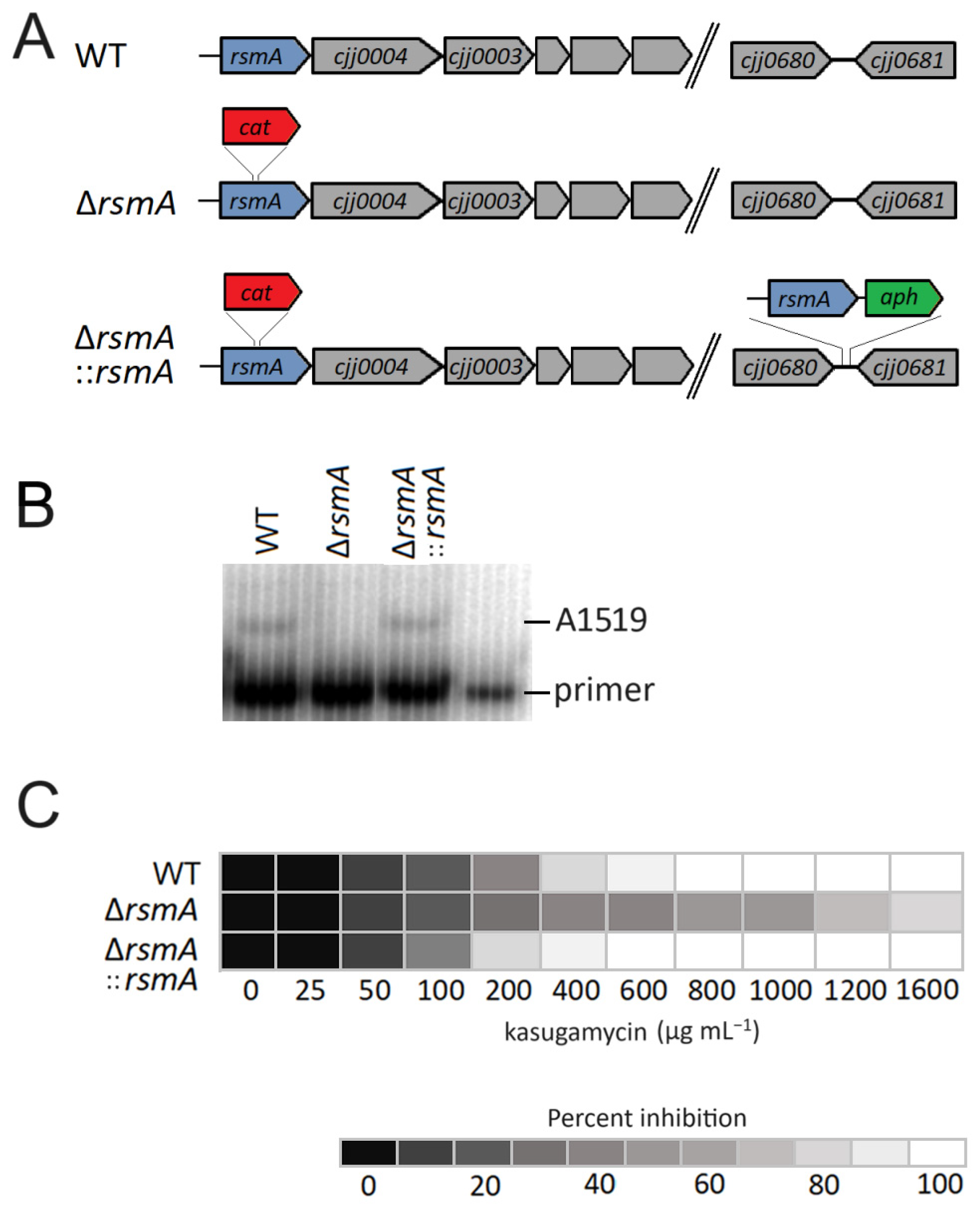
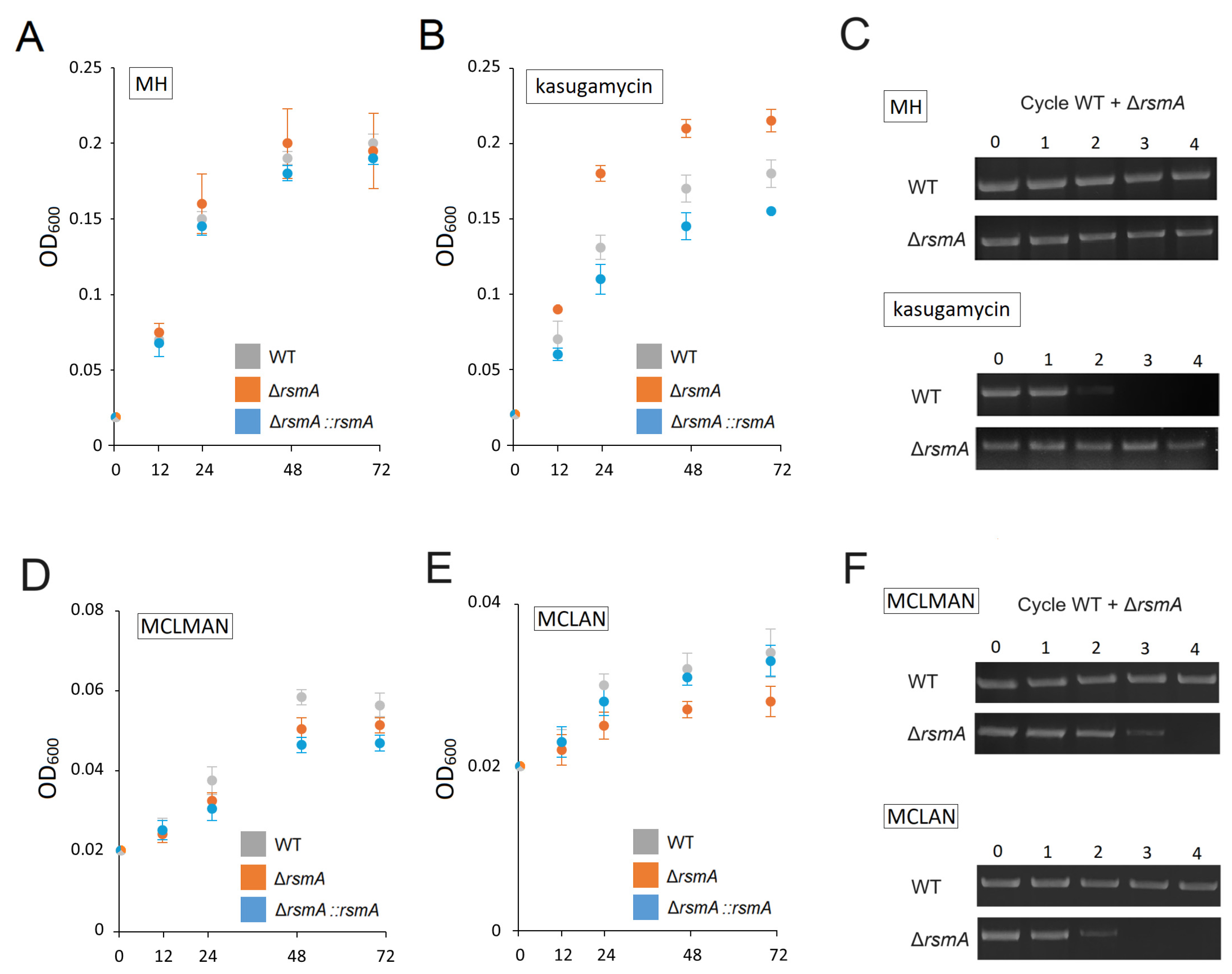
2.1. Changes in the C. jejuni Growth Phenotype after Loss of RsmA Methylation

Disruption of the C. jejuni rsmA gene by insertion of the cat cassette (Figure 1A) was confirmed by PCR sequencing of the chromosome region and by the acquisition of chloramphenicol resistance; the resultant lack of methylation at 16S rRNA nucleotides A1518 and A1519 was verified by primer extension (Figure 1B). Methylation of the rRNA was reestablished after complementing the null strain with an active copy of rsmA.
No difference was detected in the growth rates of the C. jejuni wild-type, null mutant, and complemented strains in rich medium (MH broth) at 37 °C (Figure 2A,C). However, as predicted from rsmA studies in other bacteria, inactivation of this gene conferred a growth advantage to the null mutant in the presence of the antibiotic kasugamycin. The rsmA null strain grew comfortably at 1600 µg kasugamycin mL−1 MH broth, whereas the wild-type and complemented strains did not grow at 800 µg kasugamycin mL−1 (Figure 1C). Consistent with this, growth of the null strain in a medium with sub-inhibitory concentration (400 µg mL−1) of kasugamycin was faster than the wild-type and complemented strains (Figure 2A), and in direct competition assays with this amount of drug, the wild-type cells were barely detectable after the second growth cycle (Figure 2C). Interestingly, the complemented strain was consistently more sensitive to kasugamycin than the original wild-type strain (Figure 1C).
Although no changes in the growth of the rsmA null strain were detected in a rich medium without a drug, defects became evident when grown in minimal media. In the MCLMAN minimum medium and particularly in the MCLAN medium (lacking methionine), the growth of the rsmA mutant was markedly weaker than the wild-type strain (Figure 2D,E). In competition assays, the proportion of mutant cells gradually declined and disappeared from the total population after three growth cycles in the MCLMAN medium and after two cycles in MCLAN (Figure 2F). The growth rate of the complemented strain was restored in the MCLAN medium but not in the MCLMAN medium, where growth remained at the level of the null mutant.

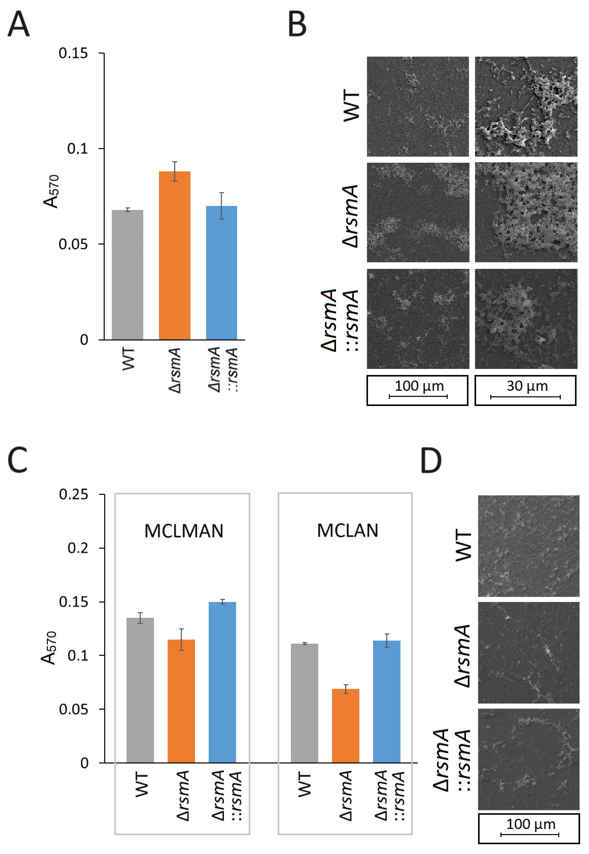
2.2. Changes in C. jejuni Biofilm Formation after Loss of RsmA Methylation

The ability of the rsmA-null strain to form biofilm on a polystyrene surface determined by the crystal violet assay was increased relative to the wild-type strain (Figure 3A,B). Complementation of the null strain with a functional copy of the rsmA gene reduced poly-styrene adherence to the level observed in the wild-type strain (Figure 3A,B). However, the reverse of these effects was observed in the MCLMAN and MCLAN minimal media, where the rsmA strain showed reduced biofilm-forming ability compared to the wild-type and the complemented strains (Figure 3C,D).

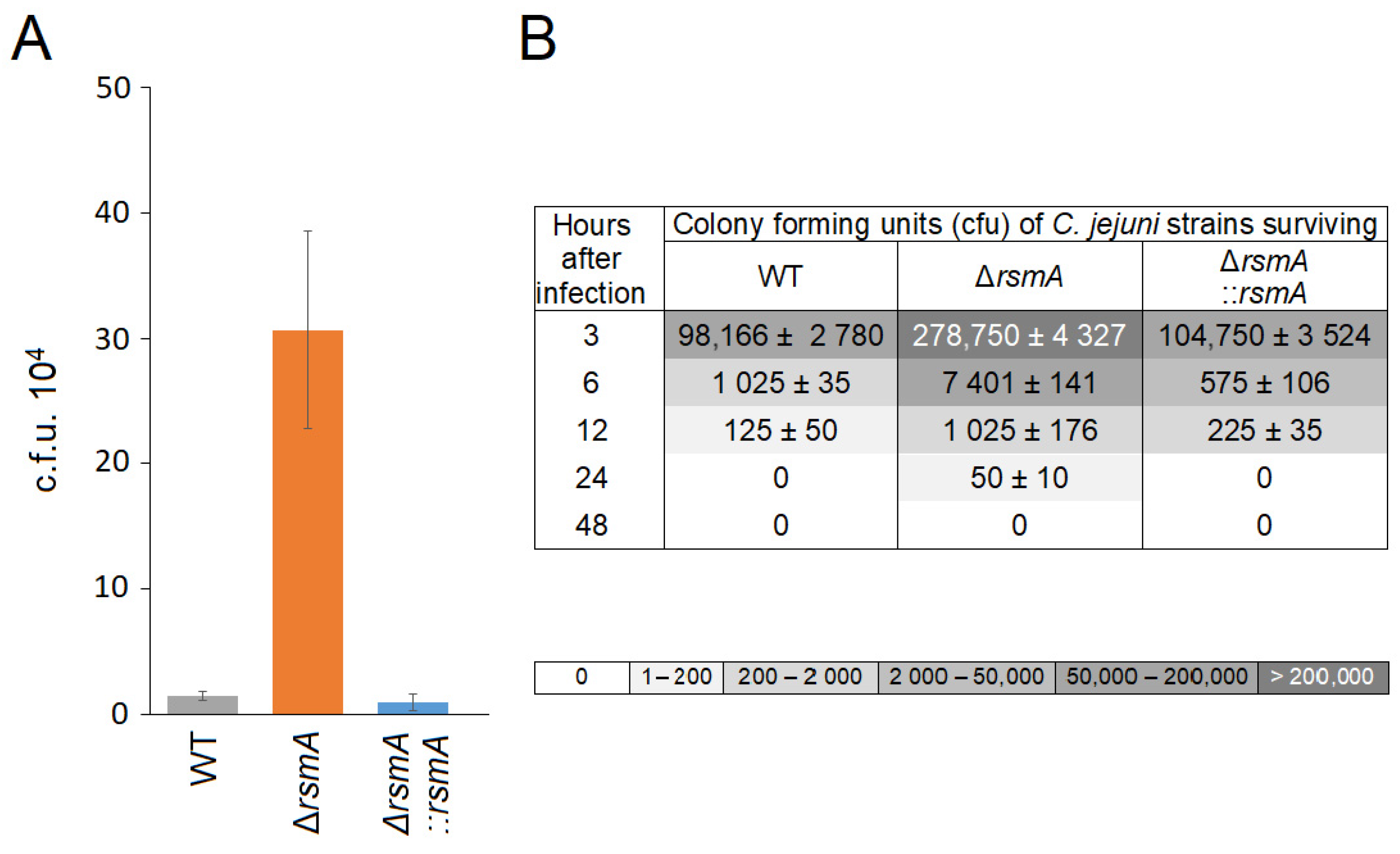
2.3. Changes in C. jejuni Virulence after Loss of RsmA Methylation

Surprisingly, the rsmA null mutant exhibited increased motility with a movement zone on agar plates that was almost twice as large as that of the wild-type. This effect was eliminated and restored to wild-type levels after complementation with an active copy of rsmA (Figure 4A). Scrutinizing the cells by electron microscopy, SEM showed that while almost all mutant cells possessed flagella, significantly fewer flagellated cells were observed for the wild-type and complemented strains (Figure 4B). The length of the flagella was similar in all strains. Observation of cells under phase contrast microscopy revealed that wild-type C. jejuni cells tended to tumble and travel for shorter distances in contrast to the rsmA mutant, where many cells exhibited a darting motility and traveled further with greater velocity (videos in the Supplementary Materials).
Coinciding with the increase in motility, the internalization ability of the rsmA mutant into epithelial cells was enhanced about twenty-fold in comparison with the wild-type strain. Complementation of the mutant with an active rsmA gene annulled this effect (Figure 5A). As a consequence of its greater internalization ability, a larger proportion of rsmA mutant bacteria were initially detected in RAW264.7 macrophages compared to the wild-type and complemented strains (Figure 5B). However, despite the entry of the larger number of the rsmA cells, combined with their longevity in the macrophages being marginally longer than that of the wild-type and complemented strains, none of the internalized strains were found to survive more than 48 h.

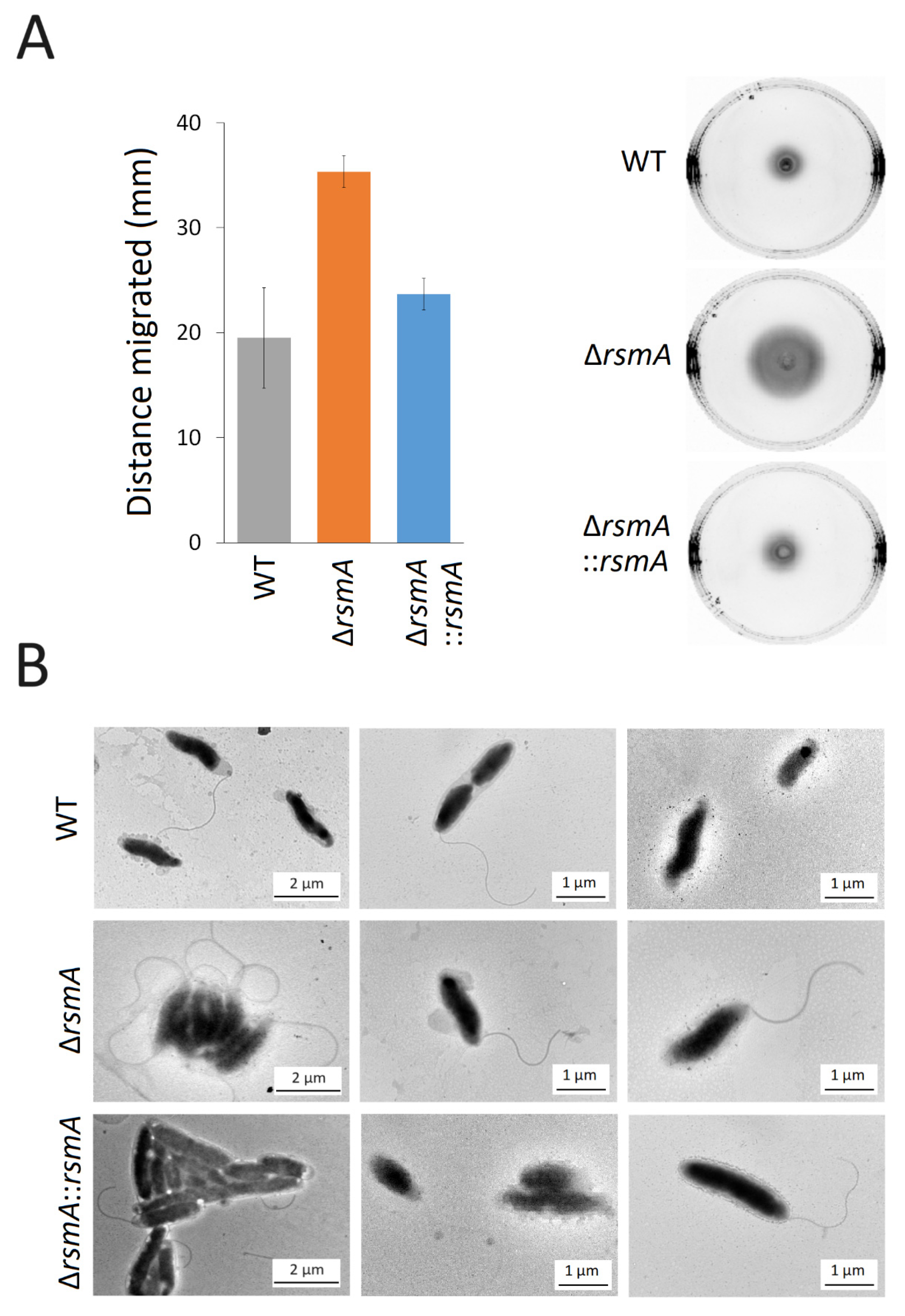
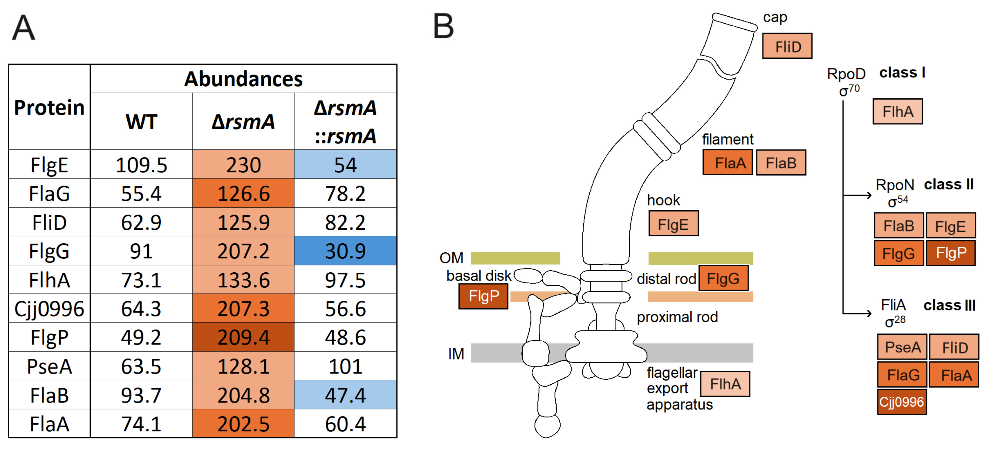
2.4. Upregulated Expression of Proteins Associated with Motility in the C. jejuni rsmA Mutant

The genome of C. jejuni 81-176, together with its two resident plasmids, encodes 1813 open reading frames (ORFs). Using a mass spectrometry-based quantitative proteomic approach, we detected 1307 of these protein products (1246 encoded by the chromosome, 29 by plasmid pVir, and 32 by plasmid pTet), representing 69% of the potential proteome.
Deletion of the rsmA gene in C. jejuni 81-176 altered the abundance of 147 proteins compared to the wild-type strain, 81 of which were upregulated and 66 downregulated (Table S1). As expected, a functional RsmA methyltransferase was absent in the mutant strain (although, because the rsmA gene was inactivated but not removed, peptides corresponding to RsmA were detected in the analyses (Table S1)). Complementing this strain with an active copy of rsmA reduced the number of significant changes to 30 proteins (Table 1 and Table S1). These changes included several major flagellar components, including FlaA, FlaB, and FliD; the main hook protein FlgE, the basal body proteins FlgG; the flagellar type III secretion system (T3SS) FlhA and FlaG; PseA (two-fold-higher levels than in the wild-type) and Cjj0996 (three-fold higher); and also FlgP (four-fold-higher levels) (Figure 6A and Table 1). Similar increases were seen in the abundances of the phosphate transporter PstS and the histidine synthesis enzymes HisF2 and HisH1. Other proteins classed as significantly changed by lack of RsmA methylation include the ribosomal protein S12, PepP, Cjj1105, Cjj1656, and three proteins encoded by pTet plasmid. These amounts of these latter proteins doubled upon rsmA inactivation, and although their levels were significantly rescued by complementation, they remain slightly higher than in the wild-type strain (Table 1 and Table S1).
Downregulated proteins included those involved in amino acid transport (GlnH), pseudouridine synthase RluB, and seven plasmid-encoded proteins. The abundances of all these proteins returned to almost wild-type levels after complementation (Table S1). However, several proteins were not rescued (partially or completely) to wild-type levels by complementation. This was possibly a consequence of secondary site mutations or polar effects associated with mutagenesis rather than being directly caused by the loss of the RsmA protein. This is clear, for instance, in the case of Cjj0003 (RluB) and Cjj0004, which are encoded immediately downstream of rsmA and were presumably affected by the upstream insertion of the cat cassette.

3. Discussion

Nucleotide modifications fine-tune the functional roles of rRNAs in ribosome biogenesis, in the translational process from initiation to termination, and in resistance to antibiotics [17,18]. Altering rRNA modifications can affect the translation process in a way that skews the relative proportions of certain proteins [4], which in turn results in physiological changes, such as the attenuation of specific virulence properties in bacterial pathogens [2,13,14,15,16,19]. Changes in protein expression might be expected to be most pronounced when a phylogenetically conserved modification enzyme is inactivated (or perhaps over-expressed), and here we show that carrying out a full proteomic analysis of a strain lacking such a modification can lead to the identification of protein factors that are important for pathogenicity.
In this study, we have inactivated the C. jejuni enzyme RsmA, which is a universally conserved dimethyltransferase responsible for the post-transcriptional modification of nucleotides A1518 and A1519 located near the 3′ end of 16S rRNA [20,21]. Dimethylation of the small ribosomal subunit at this location is highly conserved, with homologs of RsmA present in all free-living organisms and most organelles in the three domains of life [5]; only a few intracellular bacterial parasites and commensal Nanoarchaeota have been found to lack an RsmA homolog and the associated rRNA methylations [22]. Inactivation of RsmA in bacteria confers resistance to the antibiotic kasugamycin [10], and mutation of the rRNA indicates that nucleotide A1519 is the more important determinant of resistance [23]. In this context, we note that the kasugamycin resistance conferred by the inactivation of rsmA in C. jejuni was not only annulled by complementation with an active copy of rsmA, but the complemented strain became even more drug-sensitive than the wild-type strain (Figure 2A).
The primer extension assay clearly shows that nucleotide A1519 is fully dimethylated in the rRNAs from both the wild-type and complemented strains (Figure 1B), but (due to the reverse transcriptase stop at A1519) this method gives no indication of the methylation status at A1518. From the proteomics data (Table S1), expression of the RsmA methyltransferase in the complemented strain can be seen to be about three times the level in the wild-type strain, despite transcription of rsmA from its own promoter, and this effect must, therefore, be due to the displaced location of the gene in the chromosome (Figure 1A). Given the discrepancies in RsmA expression, it is plausible that A1518 is undermethylated in the wild-type strain and more completely dimethylated in the complemented strain, and this would account for the increased kasugamycin sensitivity (Figure 1C), and possibly also for some of the protein expression effects where complementation overcompensated their rescue (discussed below).
Aside from this well-documented role of RsmA in kasugamycin resistance, the main purpose of this study was to investigate how the lack of this enzyme alters the virulence potential of C. jejuni. One of the most noticeable physiological changes in the rsmA mutant is its increased motility in a rich medium, which is evident both on agar (Figure 4A) and in liquid culture (videos in the Supplementary Materials). This effect coincides with the expression of higher levels of the main functional and structural elements of the two polar flagella (Figure 6A), which, in combination with the helical cell body of C. jejuni, facilitate mobility through highly viscous environments [24]. FlaA is a major flagellin protein that, together with FlaB, is essential for the formation of the full-length flagellar filament [25], while FlaG controls the length of the flagella [26]. Each of these proteins is expressed at two to three times higher levels in the rsmA mutant (Figure 6A). FlgP (four-fold overexpressed in the rsmA strain) is mainly located in the outer membrane, and its absence results in flagellar paralysis [27,28].
From viewing electron micrographs of several hundred individual cells from each of the strains, we can conclude that whereas the rsmA mutant cells in nearly all cases displayed full-length flagella, no flagella could be detected in approximately half of the wild-type cells (Figure 4B). In the complemented strain, flagella were slightly more prevalent than in the wild-type population of cells, but their cell morphology was distinctly different. Typically, wild-type C. jejuni cells have a helical appearance, and this shape is retained in the rsmA strain. However, upon complementation, the cells lost curvature and became rod-shaped (Figure S1). More complete methylation of the 16S rRNA by more abundant copies of RsmA is presumably affecting the expression of factors influencing cell morphology, and likely candidates here are Cjj1105, the transpeptidase PbpC, and the peptidoglycan peptidase Pgp2, where the latter two proteins are markedly less plentiful in the complemented strain (Figure S1). Cjj1105 [29] and Pgp2 [30] have previously been associated with C. jejuni cell curvature, and PbpC has previously been associated with cell elongation [31].
The multitude of effects seen here in protein expression (147 significant changes, Table S1) are certainly not all a direct result of RsmA inactivation. This number was reduced to 30 significant changes by comparison of the rsmA strain with the complemented strain, but even so, some of these changes will be an indirect consequence of rsmA inactivation. For instance, the expression of flagellar genes is hierarchically controlled to facilitate the ordered secretion of flagellar proteins, such that their biosynthesis proceeds in a sequential manner. The increased abundance of FlhA in the rsmA mutant (Figure 6A) would enhance the expression of subsequent proteins in the flagellar biosynthesis hierarchy (Figure 6B), presumably through σ54 and σ28 regulation [32]. FlhA is an essential part of the T3SS secretion system and, together with the FlgSR two-component system, plays a key role in the flagellar export apparatus of C. jejuni [33]. In other bacterial pathogens, including Bacillus thuriengensis [34], Helicobacter pylori [35], and Pseudomonas aeruginosa [36], FlhA has been shown to play a major role in the regulation of flagellar expression and/or virulence.
In addition to motility, the flagella of C. jejuni are important for biofilm formation and for the invasion of epithelial cells [37,38]. Both of these traits are key aspects of C. jejuni virulence, and taking biofilm formation first, the rsmA mutant clearly has an enhanced ability to adhere to surfaces (Figure 3A,B). Consistent with this observation, the C. jejuni flagellar filaments FlaA, FlaB, and FlaG, the hook protein FlgE, and the T3SS protein FlhA have previously been shown to enhance biofilm formation [39,40,41,42].
In the case of epithelial cell invasion by C. jejuni, the disproportionally great increase in the ability of the rsmA mutant to be internalized was unexpected (Figure 5A) and thus at odds with observations made for S. Enteritidis [15]. The twenty-fold greater invasion of epithelial cells by the C. jejuni rsmA strain might not be readily explained merely by the increases in proteins involved in flagellar structure and function, such as FlaA, FlgP, and Cjj0996 (Figure 6A), all of which are needed for cell invasion [27,28,43] but are increased to a more modest extent here. Other factors are likely to play a role, and we note that the phosphate transporter PstS is also enhanced over three-fold in the rsmA mutant, and this protein has previously been linked to C. jejuni infection of humans [44]. Similarly, the PstS protein of enterohemorrhagic E. coli (EHEC) and enteropathogenic E. coli (EPEC) supports adherence and colonization of intestinal epithelial cells [45]. Furthermore, this protein is also evident in Gram-positive bacteria, where the PstS homolog of Mycobacterium tuberculosis affords protection from the antimicrobial stress response in macrophages [46,47].
After the complementation of the rsmA mutant, the increased levels of most of the motility-related proteins fell to wild-type levels. However, the abundances of FlgE, FlgG, and FlaB fell below the original wild-type levels (Figure 6A) and were thus inversely proportional to the level of RsmA, which was over-expressed in the complemented strain. We note that the expression of flgE, flgG, and flaB is regulated by σ54 dependent promoters (Figure 6B).
As touched upon above, the 147 significant changes in protein levels detected in the comparison of the rsmA mutant and the wild-type strain could be substantially reduced by comparing the rsmA strain to its complementary derivative (Table S1 and Figure 6A). While this emphasizes the need for complementation to rule out as many effects as possible that are unrelated to the methyltransferase under investigation, this still leaves over a dozen changes in protein content that might not be a direct effect of RsmA activity. For instance, expression of the Cjj0003 gene downstream is severely downregulated, presumably as a polar effect of rsmA inactivation immediately upstream, and thus might not be expected to be rescued by reinstating rsmA at another site in the chromosome (Figure 1A). Cjj0003 encodes the 23S rRNA pseudouridine synthase RluB [48], and loss of this modification could also affect protein synthesis on the ribosome, which is the premise here for investigating the loss of RsmA modification.
RsmA is important for the maturation of the small ribosomal subunit [11], and its methylations, at a location close to the anti-Shine Dalgarno mRNA binding sequence at the 3′-end of 16S rRNA, undoubtedly play a role in translational initiation, as does the interaction of kasugamycin [49] at the same location [50]. However, from the initiation sites of the mRNAs most affected here (Table S2), it is not immediately obvious what structural features might be responsible for altering their expression. Some changes in the proteome could result from collateral effects (such with Cjj0003) or as secondary effects from a regulator upstream in a biosynthetic pathway, such as FlhA (Figure 6B). The FlhA mRNA does, in fact, have an irregular Shine–Dalgarno structure (Table S2).
Considering the numerous unintentional and unexpected changes that occur when manipulating gene expression in pathogens, an important question that must be addressed is whether any of the resultant strains becomes a more effective pathogen. Needless to say, the goal of the present study is to define the determinants involved in virulence and certainly not engage in gain-of-function to produce a more dangerous C. jejuni strain. Reassuringly, we can conclude that despite the enhanced motility, improved attachment, and invasive properties of the rsmA strain, it would make a very poor pathogen. Intuitively, the loss of a phylogenetically highly conserved gene such as rsmA must have negative physiological consequences. This is evident in other species, for instance, with growth defects at lower than optimal temperatures in an E. coli rsmA strain [11], and here also for the C. jejuni rsmA strain in a medium that is anything less than nutrient-rich (Figure 2D,E). In the wild, optimal temperature and nutrient conditions for growth, in the absence of competing strains, are never experienced, and the rsmA strain would be rapidly out-competed by other bacteria present, as demonstrated in competition growth experiments here (Figure 2F).
In conclusion, we have engineered a C. jejuni strain lacking RsmA activity and linked the resultant phenotypical defects with changes in the expression of specific proteins. Complementing this strain with an active copy of rsmA rescues most of these defects and, in addition, inadvertently overexpresses RsmA to cause other physiological and morphological changes. This study validates the premise that making subtle adjustments to the bacterial ribosome (in this case, interfering with one set of rRNA methylations) causes changes in the way mRNAs are translated, which in turn can have profound effects on bacterial virulence. Combining this with high-resolution proteomic analyses using mass spectrometry facilitates the correlation of phenotypical changes with the defective expression of specific proteins. Each of the numerous rRNA modifications, particularly the highly conserved ones, plays a role in the fine-tuning of the translation process, and (as in the case of RsmA and TlyA) these roles will be different. With over thirty such rRNA modifications in E. coli [6] and almost as many in C. jejuni, there are ample prospects for perturbing the infective cycles of bacterial pathogens to identify factors essential for virulence.

4. Materials and Methods

4.1. Bacterial Strains and Growth Conditions

C. jejuni strains used in this study (Table 2) were grown under microaerobic conditions at 37 °C on brain–heart infusion (BHI) agar containing 5% (v/v) sheep blood or BHI agar containing 5% (v/v) sheep blood supplemented with chloramphenicol at 20 µg mL−1 and/or kanamycin at 30 µg mL−1.
For growth assays in minimal medium, we used MCLMAN limited medium (Medium with Cysteine, Leucine, Methionine, Aspartate, and Niacinamide) [51] and the same medium without methionine (MCLAN). Growth assays were initiated from an OD600 of 0.02 after resuspended cells in the corresponding medium. Each strain was assayed three times in biological triplicate, with plates incubated under microaerophilic conditions at 37 °C for 72 h.

4.2. Construction of rsmA Knockout Mutant

The null mutant of the C. jejuni 81176 was created by inserting a chloramphenicol cat resistance cassette (Cmr) into rsmA gene. Briefly, the rsmA gene was amplified by PCR using the rsmAF: ATGGTTAAAGCAAAAAAACAATACGGAC and rsmAR: TTATTTATCTCTTTGTTTTCGTCCATATTTATC primers and cloned into the pJET1.2 plasmid (Thermo Fisher Scientific, Waltham, MA, USA), prior to insertion of the cat cassette [52] using the primers rsmA_mutR: AAAAGAAATGGCTGAAAAATTCTGCGC and rsmA_mutF: CATTACAATAAGCCCTAGACAATTTTTATC to disrupt the coding sequence replacing 30 bp of its sequence. The plasmid containing the engineered rsmA gene was used for electro-transformation of C. jejuni 81-176 cells. The plasmid lacks a C. jejuni replicon; therefore, retention of Cmr required allelic exchange with the homologous chromosomal rsmA gene.
The resultant C. jejuni 81-176 ΔrsmA null mutant was complemented by inserting a wild-type copy of rsmA under control of its own promoter (with a total size of 2501 nucleotides) into the 121-bp intergenic region between cjj0680 and cjj0681 forming the C. jejuni strain 81-176 ΔrsmA::rsmA (Table 2; Figure 1A). All strains were verified by PCR and sequencing and frozen in aliquots immediately after construction to avoid any serial passaging before their use in the growth and virulence tests.
Table 2. Bacterial strains used in this study.
StrainsRelevant CharacteristicsSource/Reference
C. jejuni 81-176Wild-type[53]
C. jejuni 81-176 ΔrsmACmr, rsmA deletion mutantThis study
C. jejuni 81-176 ΔrsmA::rsmACmr, Kmr, rsmA deletion mutant complemented with C. jejuni rsmAThis study

4.3. Motility

The motility of C. jejuni cells was assessed by adding 3 µL of culture (OD600 = 0.5) onto BHI with 0.25% agar. Plates were left to dry and were incubated under microaerobic conditions for 48 h at 37 °C before measuring cell migration.
Direct real-time motility was observed and filmed using phase-contrast microscopy (Eclipse E400, Nikon, Tokyo, Japan). Bacteria were diluted to OD600 of 0.05–0.1 in BHI broth medium, 5 µL of culture were applied on a microscopy slide with coverslip, and motility was recorded immediately after preparation.

4.4. Virulence Phenotypes

Virulence characteristics of C. jejuni include its ability to form biofilms, its adhesion to and invasion of human epithelial cells, and its survival in macrophages as previously described [3]. Briefly, biofilm formation was measured spectroscopically after growing C. jejuni strains under microaerobic conditions in 24-well polystyrene plates. Biofilm formation was determined by crystal violet staining and measuring absorbance at 570 nm (A570) using a Bio-Mate spectrophotometer (Thermo Fisher Scientific, Waltham, MA, USA).
Invasion of C. jejuni was assayed using Caco-2 cells. Bacteria were added into the wells at an MOI of 100 bacteria to one epithelial cell. Infected monolayers were incubated for 2 h for invasion to occur. The monolayers were then washed three times with sterile phosphate-buffered saline (PBS) and incubated for 2 h in minimal essential medium containing 100 µg gentamicin mL−1 to kill extracellular bacteria. After this period, the monolayers were washed as described above and lysed with 0.1% Triton X-100 in PBS for 15 min at room temperature. Following serial dilution in PBS, invaded bacteria were enumerated by colony counting on BHI agar cultured under microaerophilic conditions.
For the survival assays, RAW 264.7 macrophages were incubated with C. jejuni at an approximate MOI of 100. Infected macrophage monolayers were incubated for 2 h, then medium containing 100 μg/mL of gentamicin was added to kill extracellular bacteria. The invasion period was monitored for 3, 6, 12, 24, and 48 h post-infection. Following each time point, the macrophages were lysed 0.1% Triton X-100 in PBS for 15 min at room temperature, and the live bacteria released were evaluated by plating serial dilutions on MH agar plates.
Statistical analysis was performed using IBM SPSS Statistics for Windows version 28 (IBM Corp., Armonk, NY, USA) software. The statistical significance of differences between tested groups was measured using the Mann–Whitney test.

4.5. MIC Determination

Overnight cultures of the C. jejuni strains were diluted to 0.5 McFarland standard and plated onto BHI agar containing sheep blood with kasugamycin concentrations increasing in two-fold steps. The MIC values are the lowest concentration at which no growth was observed after incubation under microaerobic conditions for 48 h at 37 °C.

4.6. Primer Extension

Total RNAs were extracted from C. jejuni strains by phenol extraction [54]. 5′-32P-end-labeled deoxynucleotide primers were hybridized to complementary regions of rRNA (16S nucleotides 1523-1540, E. coli numbering) and extended with AMV reverse transcriptase (Roche, Indianapolis, IN, USA). Extension products were run on denaturing polyacrylamide/urea gel and visualized by autoradiography using a Typhoon PhosphorImager (Amersham Bioscience, Amersham, UK) and identified by reference to dideoxynucleotide sequencing reactions run in parallel.

4.7. Competition Growth Assay and Florescence PCR

Approximate equal numbers of cells from two C. jejuni strains were mixed in either MH, MCLAN, or MCLMAN media with and without kasugamycin at 400 μg mL−1. This concentration of kasugamycin is defined here as a ‘sub-inhibitory’ level of the drug, which is less than that required to completely arrest growth (although growth will be slowed to different extents for the different strains). After a 24 h incubation cycle, cells were diluted 1000-fold, and the cycle was repeated. Aliquots of cells were harvested by centrifugation at the end of each cycle, and the cell pellets were resuspended in deionized water. The relative amounts of wild-type and mutated rsmA gene copies in the strain mixtures were assessed by Taq DNA Polymerase (Thermo Fisher Scientific, Waltham, MA, USA) PCR using the unlabeled forward primer (rsmAF) and the reverse primer (rsmAR) that was 5′-labeled with cyanine 3 (Cy3), both of which are complementary to regions of rsmA. The PCR products of 801 (for wild type) and 1184 base pairs (for the mutant with the cat insertion) were separated on agarose gels and visualized using the ChemiDoc Imaging System (Bio-Rad, Hercules, CA, USA). This approach has previously been used to follow competition between strains of Mycobacterium smegmatis in media with capreomycin [55].

4.8. Transmission Electron Microscopy (TEM)

The morphology of C. jejuni cells was evaluated by transmission electron microscope JEM-1220 (JEOL, Tokyo, Japan) after spreading droplets of bacterial culture onto TEM grids (Formvar on 3 mm 200 Mesh Cu Grids, Agar Scientific, Stansted, UK). Operating voltage was set to 80 keV.

4.9. Scanning Electron Microscopy (SEM)

Bacterial biofilms were grown on glass coverslips [2], and their morphology was examined using secondary electron (SE) and backscattered electron (BSE) images. SE images were generated using a Thermo Fisher Quattro S Environmental Electron Microscope (ESEM) at an accelerating voltage of 10 or 20 kV with working distance of approx. 10 mm; non-sputtered samples were investigated in low vacuum mode with an H2O chamber pressure of 50 Pa. Samples were either carbon-coated with Safematic CCU-010 or with a thin Pt layer with Bal-Tec SCD005 Sputter Coater.

4.10. Protein Extraction and Peptide Labeling

Overnight cultures of C. jejuni strains were harvested by centrifugation [2] and resuspended in lysis buffer (7 M urea, 2 M thiourea, 1% DTT, and 0.5% n-octyl-β-D-glucopyranoside) supplemented with protease inhibitors (Roche), and ultrasonicated for protein extraction. After measuring the protein concentrations (Bradford reagent, Merck, Rahway, NJ, USA), 200 µg samples were alkylated with 45 mM iodoacetamide (IAA), incubating for 30 min in the dark before adding 5 mM DTT and adjusting to pH 8.5 with 30 mM triethylammonium bicarbonate (TEAB) buffer. Proteins were predigested for 3 h at 37 °C with 0.005 AU lysyl endopeptidase (Lys-C, FUJIFILM Wako Pure Chemical Corporation, Hong Kong, China). Samples were then digested further at 37 °C with methylated trypsin at an enzyme/protein ratio of 1:50 [56] prior to quenching with 10% trifluoroacetic acid (TFA) and centrifuging at 20,000× g for 15 min. Peptides were desalted on R2C8 and R3C18 resin reversed-phase columns (Thermo Fisher Scientific), washed with 0.1% TFA, and eluted with 60% acetonitrile/0.1% TFA before drying under vacuum centrifugation and resuspending in 50 mM TEAB buffer.
The peptides were labeled with isobaric tags (TMTpro, Thermo Fisher Scientific, Waltham, MA, USA) according to the supplier’s recommendations. Based on total peptide intensity in the different samples, estimated in test runs, the samples were mixed in 1:1 (w/w) proportions and desalted on reverse-phase columns.

4.11. LC-MS3 Tandem Mass Spectrometry

The TMTpro-labeled peptides were fractionated into 12 concatenated fractions by reversed-phase chromatography in high pH buffers on a Dionex UltiMate 3000 (Thermo Fisher Scientific) equipped with a nanoEasy M/Z Peptide, CSH C18 130 Å, 1.7 µm, 300 µm × 100 mm analytical column (Waters, Milford, CT, USA). Peptides in the mobile phase of 20 mM ammonium formate in water, pH 9.3, were eluted at a flow rate of 5 µL/min over 71 min in a gradient of solvent B (80% acetonitrile and 20% solvent A) rising from 2% to 40%, followed by 50% solvent B to 122 min, before finally washing with 95% solvent B for 10 min. and then drying.
Fractions were dissolved in 5 μL solvent A (0.1% formic acid in water) and separated on an in-house prepared 20 cm analytical columns (100 μm ID, pulled tip fused silica capillary, containing 1.9 μm Reprosil-Pur 120 C18-AQ, Dr. Maisch GmbH, Ammerbuch, Germany). Peptides were separated at a flow rate of 300 μL/min with increasing concentrations of Solvent B (90% acetonitrile, 0.1% formic acid) beginning at 2% and raising to 8% over 2 min, then to 22% over the next 60 min, to 40% in 10 min, to 95% in 1 min and holding at 95% for 5 min, and finally to 100% over 1 min and holding at 100% for 3 min.
Mass spectrometry analyses were performed using an SPS-MS3 approach on an Orbitrap Eclipse Tribrid mass spectrometer (Thermo Fisher Scientific). Full scans over a mass range of 350–1600 m/z were performed within 3 s cycles at a resolution of 120 k in positive ion mode. Subsequently, precursor ions at 400–1600 m/z were selected for CID fragmentation and detection in Ion Trap (NCE 35, CID activation time 10 sec, Ion Trap Scan Rate 10 ms). MS2 spectral data were screened against the Campylobacter protein database (fasta file from UniProt-the Universal Protein Resource for protein sequence) with a search time limit of 35 ms. Static modifications were carbamidomethyl (C) and TMTpro 16plex (N-terminus, K), with a single missed cleavage being allowed. SPS-MS3 fragmentation was triggered after positive identification of precursor ions: 10 SPS precursor ions were accumulated and further fragmented with HCD (NCE 55, resolution 30k, scan range 100–500 m/z, maximum injection time “Auto”) and detected by Orbitrap for sample quantification.

4.12. Protein Identification and Quantification

Raw data files were processed by Thermo Proteome Discoverer software v 2.5 (Thermo Fisher Scientific). Protein identification was made from the C. jejuni 81-176 genome sequence (UniProt databases) using the Sequest search engine. For the search parameters, precursor mass tolerance was set to 10 ppm, with a fragment mass tolerance of 0.6 Da; carbamidomethylation of cysteine and TMTpro modification of N-termini and lysine residues were added to dynamic modifications, while acetylation was set to variable modifications; and two missed cleavage sites per protein were allowed. For reporter ion quantification, the co-isolation threshold was set to 50, and average reporter ion S/N threshold to 10. Data from Proteome Discoverer were processed using Excel 365 (Microsoft version 2408).
Quantification was based on two biological replicates of each strain and taking into consideration only the proteins that were identified from at least two unique peptides and with a false discovery rate of less than 1%. The abundances of individual proteins that differed with p-values below 0.05 in comparisons of the wild-type strain with the ΔrsmA strain and ΔrsmA with the complemented ΔrsmA::rsmA strain were considered significant.

Supplementary Materials

The following supporting information can be downloaded at https://www.mdpi.com/article/10.3390/ijms25189797/s1.

Author Contributions

Conceptualization: A.S.-G. and S.D.; data curation: A.S.-G.; funding acquisition: A.S.-G. and S.D.; investigation: A.S.-G., M.M., P.B., A.O., E.K. and I.S.; methodology: A.S.-G., P.B. and S.D.; project administration: A.S.-G. and S.D.; supervision: A.S.-G. and S.D.; validation: A.S.-G. and S.D.; visualization: A.S.-G., M.M., P.B. and A.O.; writing—original draft: S.D. and A.S.-G.; writing—review and editing: S.D. and A.S.-G. All authors have read and agreed to the published version of the manuscript.

Funding

Support from the National Science Centre [2018/30/M/NZ6/00429] to A.S.-G. and from the Danish Research Agency (FNU-rammebevilling 10-084554) to S.D. is gratefully acknowledged. Publication costs were covered by the Science Development Fund of the Warsaw University of Life Science—SGGW.

Data Availability Statement

The mass spectrometry proteomics data have been deposited to the ProteomeXchange Consortium via the PRIDE [57] partner repository with the dataset identifier PXD052567.

Acknowledgments

We thank Martin Røssel Larsen, Arkadiusz Nawrocki, and Lene Jakobsen, University of Southern Denmark, for essential advice and help with the proteomics experiments. The Institute of Paleobiology, Polish Academy of Sciences, Warsaw, is thanked for the use of their Field Emission SEM.

Conflicts of Interest

The authors declare no conflicts of interest.

References

  1. Sejvar, J.J.; Baughman, A.L.; Wise, M.; Morgan, O.W. Population incidence of Guillain-Barre syndrome: A systematic review and meta-analysis. Neuroepidemiology 2011, 36, 123–133. [Google Scholar] [CrossRef] [PubMed] [PubMed Central]
  2. Sałamaszyńska-Guz, A.; Rose, S.; Lykkebo, C.A.; Taciak, B.; Bącal, P.; Uśpieński, T.; Douthwaite, S. Biofilm formation and motility are promoted by Cj0588-directed methylation of rRNA in Campylobacter jejuni. Front. Cell. Infect. Microbiol. 2018, 7, 533. [Google Scholar] [CrossRef] [PubMed]
  3. Sałamaszyńska-Guz, A.; Serafińska, I.; Bącal, P.; Douthwaite, S. Virulence properties of Campylobacter jejuni are enhanced by displaying a mycobacterial TlyA methylation pattern in its rRNA. Cell. Microbiol. 2020, 22, e13199. [Google Scholar] [CrossRef] [PubMed]
  4. Sałamaszyńska-Guz, A.; Rasmussen, P.K.; Murawska, M.; Douthwaite, S. Campylobacter jejuni virulence factors identified by modulating their synthesis on ribosomes with altered rRNA methylation. Front. Cell. Infect. Microbiol. 2022, 11, 803730. [Google Scholar] [CrossRef] [PubMed]
  5. Boccaletto, P.; Stefaniak, F.; Ray, A.; Cappannini, A.; Mukherjee, S.; Purta, E.; Kurkowska, M.; Shirvanizadeh, N.; Destefanis, E.; Groza, P.; et al. MODOMICS: A database of RNA modification pathways. 2021 update. Nucleic Acids Res. 2022, 50, D231–D235. [Google Scholar] [CrossRef] [PubMed] [PubMed Central]
  6. Purta, E.; O’Connor, M.; Bujnicki, J.M.; Douthwaite, S. YgdE is the 2′-O-ribose methyltransferase RlmM specific for nucleotide C2498 in bacterial 23S rRNA. Mol. Microbiol. 2009, 72, 1147–1158. [Google Scholar] [CrossRef] [PubMed]
  7. Sergiev, P.V.; Aleksashin, N.A.; Chugunova, A.A.; Polikanov, Y.S.; Dontsova, O.A. Structural and evolutionary insights into ribosomal RNA methylation. Nat. Chem. Biol. 2018, 14, 226–235. [Google Scholar] [CrossRef] [PubMed]
  8. Harris, J.K.; Kelley, S.T.; Spiegelman, G.B.; Pace, N.R. The genetic core of the universal ancestor. Genome Res. 2003, 13, 407–412. [Google Scholar] [CrossRef] [PubMed] [PubMed Central]
  9. van Buul, C.P.; van Knippenberg, P.H. Nucleotide sequence of the ksgA gene of Escherichia coli: Comparison of methyltransferases effecting dimethylation of adenosine in ribosomal RNA. Gene 1985, 38, 65–72. [Google Scholar] [CrossRef] [PubMed]
  10. Helser, T.L.; Davies, J.E.; Dahlberg, J.E. Mechanism of kasugamycin resistance in Escherichia coli. Nat. New Biol. 1972, 235, 6–9. [Google Scholar] [CrossRef]
  11. Connolly, K.; Rife, J.P.; Culver, G. Mechanistic insight into the ribosome biogenesis functions of the ancient protein KsgA. Mol. Microbiol. 2008, 70, 1062–1075. [Google Scholar] [CrossRef] [PubMed] [PubMed Central]
  12. O’Connor, M.; Thomas, C.L.; Zimmermann, R.A.; Dahlberg, A.E. Decoding fidelity at the ribosomal A and P sites: Influence of mutations in three different regions of the decoding domain in 16S rRNA. Nucleic Acids Res. 1997, 25, 1185–1193. [Google Scholar] [CrossRef] [PubMed] [PubMed Central]
  13. Bergman, M.A.; Loomis, W.P.; Mecsas, J.; Starnbach, M.N.; Isberg, R.R. CD8(+) T cells restrict Yersinia pseudotuberculosis infection: Bypass of anti-phagocytosis by targeting antigen-presenting cells. PLoS Pathog. 2009, 5, e1000573. [Google Scholar] [CrossRef] [PubMed] [PubMed Central]
  14. Mecsas, J.; Bilis, I.; Falkow, S. Identification of attenuated Yersinia pseudotuberculosis strains and characterization of an orogastric infection in BALB/c mice on day 5 postinfection by signature-tagged mutagenesis. Infect. Immun. 2001, 69, 2779–2787. [Google Scholar] [CrossRef] [PubMed] [PubMed Central]
  15. Shah, D.H.; Zhou, X.; Kim, H.Y.; Call, D.R.; Guard, J. Transposon mutagenesis of Salmonella enterica serovar Enteritidis identifies genes that contribute to invasiveness in human and chicken cells and survival in egg albumen. Infect. Immun. 2012, 80, 4203–4215. [Google Scholar] [CrossRef] [PubMed] [PubMed Central]
  16. Kyuma, T.; Kizaki, H.; Ryuno, H.; Sekimizu, K.; Kaito, C. 16S rRNA methyltransferase KsgA contributes to oxidative stress resistance and virulence in Staphylococcus aureus. Biochimie 2015, 119, 166–174. [Google Scholar] [CrossRef] [PubMed]
  17. Douthwaite, S.; Fourmy, D.; Yoshizawa, S. Nucleotide methylations in rRNA that confer resistance to ribosome-targeting antibiotics. In Fine-Tuning of RNA Functions by Modification and Editing; Grosjean, H., Ed.; Topics in Current Genetics; Springer: New York, NY, USA, 2005; Volume 12, pp. 287–309. [Google Scholar]
  18. Laperyre, B. Conserved ribosomal RNA modifications and their putative roles in ribosomal biogenesis and translation. In Fine-Tuning of RNA Functions by Modification and Editing; Grosjean, H., Ed.; Topics in Current Genetics; Springer: New York, NY, USA, 2005; Volume 12, pp. 263–284. [Google Scholar]
  19. Bahena-Ceron, R.; Teixeira, C.; Ponce, J.R.J.; Wolff, P.; Couzon, F.; Francois, P.; Klaholz, B.P.; Vandenesch, F.; Romby, P.; Moreau, K.; et al. RlmQ: A newly discovered rRNA modification enzyme bridging RNA modification and virulence traits in Staphylococcus aureus. RNA 2024, 30, 200–212. [Google Scholar] [CrossRef] [PubMed] [PubMed Central]
  20. Helser, T.L.; Davies, J.E.; Dahlberg, J.E. Change in methylation of 16S ribosomal RNA associated with mutation to kasugamycin resistance in Escherichia coli. Nat. New Biol. 1971, 233, 12–14. [Google Scholar] [CrossRef] [PubMed]
  21. van Buul, C.P.; Hamersma, M.; Visser, W.; van Knippenberg, P.H. Partial methylation of two adjacent adenosines in ribosomes from Euglena gracilis chloroplasts suggests evolutionary loss of an intermediate stage in the methyl-transfer reaction. Nucleic Acids Res. 1984, 12, 9205–9208. [Google Scholar] [CrossRef] [PubMed] [PubMed Central]
  22. Seistrup, K.H.; Rose, S.; Birkedal, U.; Nielsen, H.; Huber, H.; Douthwaite, S. Bypassing rRNA methylation by RsmA/Dim1during ribosome maturation in the hyperthermophilic archaeon Nanoarchaeum equitans. Nucleic Acids Res. 2017, 45, 2007–2015. [Google Scholar] [CrossRef] [PubMed] [PubMed Central]
  23. Vila-Sanjurjo, A.; Squires, C.L.; Dahlberg, A.E. Isolation of kasugamycin resistant mutants in the 16S ribosomal RNA of Escherichia coli. J. Mol. Biol. 1999, 293, 1–8. [Google Scholar] [CrossRef] [PubMed]
  24. Ferrero, R.L.; Lee, A. Motility of Campylobacter jejuni in a viscous environment: Comparison with conventional rod-shaped bacteria. J. Gen. Microbiol. 1988, 134, 53–59. [Google Scholar] [CrossRef] [PubMed]
  25. Lis, L.; Connerton, I.F. The minor flagellin of Campylobacter jejuni (FlaB) confers defensive properties against bacteriophage infection. Front. Microbiol. 2016, 7, 1908. [Google Scholar] [CrossRef] [PubMed] [PubMed Central]
  26. Inoue, T.; Barker, C.S.; Matsunami, H.; Aizawa, S.I.; Samatey, F.A. The FlaG regulator is involved in length control of the polar flagella of Campylobacter jejuni. Microbiology 2018, 164, 740–750. [Google Scholar] [CrossRef] [PubMed]
  27. Sommerlad, S.M.; Hendrixson, D.R. Analysis of the roles of FlgP and FlgQ in flagellar motility of Campylobacter jejuni. J. Bacteriol. 2007, 189, 179–186. [Google Scholar] [CrossRef] [PubMed] [PubMed Central]
  28. Zhou, X.; Roujeinikova, A. The structure, composition, and role of periplasmic stator scaffolds in polar bacterial flagellar motors. Front. Microbiol. 2021, 12, 639490. [Google Scholar] [CrossRef] [PubMed] [PubMed Central]
  29. Frirdich, E.; Biboy, J.; Huynh, S.; Parker, C.T.; Vollmer, W.; Gaynor, E.C. Morphology heterogeneity within a Campylobacter jejuni helical population: The use of calcofluor white to generate rod-shaped C. jejuni 81-176 clones and the genetic determinants responsible for differences in morphology within 11168 strains. Mol. Microbiol. 2017, 104, 948–971. [Google Scholar] [CrossRef] [PubMed] [PubMed Central]
  30. Frirdich, E.; Vermeulen, J.; Biboy, J.; Soares, F.; Taveirne, M.E.; Johnson, J.G.; DiRita, V.J.; Girardin, S.E.; Vollmer, W.; Gaynor, E.C. Peptidoglycan LD-carboxypeptidase Pgp2 influences Campylobacter jejuni helical cell shape and pathogenic properties and provides the substrate for the DL-carboxypeptidase Pgp1. J. Biol. Chem. 2014, 289, 8007–8018. [Google Scholar] [CrossRef] [PubMed] [PubMed Central]
  31. Frirdich, E.; Vermeulen, J.; Biboy, J.; Vollmer, W.; Gaynor, E.C. Multiple Campylobacter jejuni proteins affecting the peptidoglycan structure and the degree of helical cell curvature. Front. Microbiol. 2023, 14, 1162806. [Google Scholar] [CrossRef] [PubMed] [PubMed Central]
  32. Lertsethtakarn, P.; Ottemann, K.M.; Hendrixson, D.R. Motility and chemotaxis in Campylobacter and Helicobacter. Annu. Rev. Microbiol. 2011, 65, 389–410. [Google Scholar] [CrossRef] [PubMed] [PubMed Central]
  33. Carrillo, C.D.; Taboada, E.; Nash, J.H.; Lanthier, P.; Kelly, J.; Lau, P.C.; Verhulp, R.; Mykytczuk, O.; Sy, J.; Findlay, W.A.; et al. Genome-wide expression analyses of Campylobacter jejuni NCTC11168 reveals coordinate regulation of motility and virulence by flhA. J. Biol. Chem. 2004, 279, 20327–20338. [Google Scholar] [CrossRef] [PubMed]
  34. Ghelardi, E.; Celandroni, F.; Salvett, S.; Beecher, D.J.; Gominet, M.; Lereclus, D.; Wong, A.C.; Senesi, S. Requirement of flhA for swarming differentiation, flagellin export, and secretion of virulence-associated proteins in Bacillus thuringiensis. J. Bacteriol. 2002, 184, 6424–6433. [Google Scholar] [CrossRef] [PubMed] [PubMed Central]
  35. McGee, D.J.; Coker, C.; Testerman, T.L.; Harro, J.M.; Gibson, S.V.; Mobley, H.L.T. The Helicobacter pylori flbA flagellar biosynthesis and regulatory gene is required for motility and virulence and modulates urease of H. pylori and Proteus mirabilis. J. Med. Microbiol. 2002, 51, 958–970. [Google Scholar] [CrossRef] [PubMed]
  36. Fleiszig, S.M.; Arora, S.K.; Van, R.; Ramphal, R. FlhA, a component of the flagellum assembly apparatus of Pseudomonas aeruginosa, plays a role in internalization by corneal epithelial cells. Infect. Immun. 2001, 69, 4931–4937. [Google Scholar] [CrossRef] [PubMed] [PubMed Central]
  37. Grant, C.C.; Konkel, M.E.; Cieplak, W., Jr.; Tompkins, L.S. Role of flagella in adherence, internalization, and translocation of Campylobacter jejuni in nonpolarized and polarized epithelial cell cultures. Infect. Immun. 1993, 61, 1764–1771. [Google Scholar] [CrossRef] [PubMed] [PubMed Central]
  38. Wassenaar, T.M.; van der Zeijst, B.A.; Ayling, R.; Newell, D.G. Colonization of chicks by motility mutants of Campylobacter jejuni demonstrates the importance of flagellin A expression. J. Gen. Microbiol. 1993, 139, 1171–1175. [Google Scholar] [CrossRef] [PubMed]
  39. Kalmokoff, M.; Lanthier, P.; Tremblay, T.L.; Foss, M.; Lau, P.C.; Sanders, G.; Austin, J.; Kelly, J.; Szymanski, C.M. Proteomic analysis of Campylobacter jejuni 11168 biofilms reveals a role for the motility complex in biofilm formation. J. Bacteriol. 2006, 188, 4312–4320. [Google Scholar] [CrossRef] [PubMed] [PubMed Central]
  40. Reeser, R.J.; Medler, R.T.; Billington, S.J.; Jost, B.H.; Joens, L.A. Characterization of Campylobacter jejuni biofilms under defined growth conditions. Appl. Environ. Microbiol. 2007, 73, 1908–1913. [Google Scholar] [CrossRef] [PubMed] [PubMed Central]
  41. Svensson, S.L.; Pryjma, M.; Gaynor, E.C. Flagella-mediated adhesion and extracellular DNA release contribute to biofilm formation and stress tolerance of Campylobacter jejuni. PLoS ONE 2014, 9, e106063. [Google Scholar] [CrossRef] [PubMed] [PubMed Central]
  42. Taniguchi, T.; Ohki, M.; Urata, A.; Ohshiro, S.; Tarigan, E.; Kiatsomphob, S.; Vetchapitak, T.; Sato, H.; Misawa, N. Detection and identification of adhesins involved in adhesion of Campylobacter jejuni to chicken skin. Int. J. Food Microbiol. 2021, 337, 108929. [Google Scholar] [CrossRef] [PubMed]
  43. Goon, S.; Ewing, C.P.; Lorenzo, M.; Pattarini, D.; Majam, G.; Guerry, P. A sigma28-regulated nonflagella gene contributes to virulence of Campylobacter jejuni 81-176. Infect. Immun. 2006, 74, 769–772. [Google Scholar] [CrossRef] [PubMed] [PubMed Central]
  44. Crofts, A.A.; Poly, F.M.; Ewing, C.P.; Kuroiwa, J.M.; Rimmer, J.E.; Harro, C.; Sack, D.; Talaat, K.R.; Porter, C.K.; Gutierrez, R.L.; et al. Campylobacter jejuni transcriptional and genetic adaptation during human infection. Nat. Microbiol. 2018, 3, 494–502. [Google Scholar] [CrossRef] [PubMed] [PubMed Central]
  45. Chekabab, S.M.; Harel, J.; Dozois, C.M. Interplay between genetic regulation of phosphate homeostasis and bacterial virulence. Virulence 2014, 5, 786–793. [Google Scholar] [CrossRef] [PubMed]
  46. Rengarajan, J.; Bloom, B.R.; Rubin, E.J. Genome-wide requirements for Mycobacterium tuberculosis adaptation and survival in macrophages. Proc. Natl. Acad. Sci. USA 2005, 102, 8327–8332. [Google Scholar] [CrossRef] [PubMed] [PubMed Central]
  47. Tischler, A.D.; Leistikow, R.L.; Kirksey, M.A.; Voskuil, M.I.; McKinney, J.D. Mycobacterium tuberculosis requires phosphate-responsive gene regulation to resist host immunity. Infect. Immun. 2013, 81, 317–328. [Google Scholar] [CrossRef] [PubMed] [PubMed Central]
  48. Del Campo, M.; Kaya, Y.; Ofengand, J. Identification and site of action of the remaining four putative pseudouridine synthases in Escherichia coli. RNA 2001, 7, 1603–1615. [Google Scholar] [PubMed]
  49. Zhang, Y.; Aleksashin, N.A.; Klepacki, D.; Anderson, C.; Vazquez-Laslop, N.; Gross, C.A.; Mankin, A.S. The context of the ribosome binding site in mRNAs defines specificity of action of kasugamycin, an inhibitor of translation initiation. Proc. Natl. Acad. Sci. USA 2022, 119, e2118553119. [Google Scholar] [CrossRef] [PubMed] [PubMed Central]
  50. Schuwirth, B.S.; Day, J.M.; Hau, C.W.; Janssen, G.R.; Dahlberg, A.E.; Cate, J.H.; Vila-Sanjurjo, A. Structural analysis of kasugamycin inhibition of translation. Nat. Struct. Mol. Biol. 2006, 13, 879–886. [Google Scholar] [CrossRef] [PubMed] [PubMed Central]
  51. Alazzam, B.; Bonnassie-Rouxin, S.; Dufour, V.; Ermel, G. MCLMAN, a new minimal medium for Campylobacter jejuni NCTC 11168. Res. Microbiol. 2011, 162, 173–179. [Google Scholar] [CrossRef] [PubMed]
  52. Yao, R.; Alm, R.A.; Trust, T.J.; Guerry, P. Construction of new Campylobacter cloning vectors and a new mutational cat cassette. Gene 1993, 130, 127–130. [Google Scholar] [CrossRef] [PubMed]
  53. Korlath, J.A.; Osterholm, M.T.; Judy, L.A.; Forfang, J.C.; Robinson, R.A. A point-source outbreak of campylobacteriosis associated with consumption of raw milk. J. Infect. Dis. 1985, 152, 592–596. [Google Scholar] [CrossRef] [PubMed]
  54. Douthwaite, S.; Powers, T.; Lee, J.Y.; Noller, H.F. Defining the structural requirements for a helix in 23S ribosomal RNA that confers erythromycin resistance. J. Mol. Biol. 1989, 209, 655–665. [Google Scholar] [CrossRef] [PubMed]
  55. Monshupanee, T.; Johansen, S.K.; Dahlberg, A.E.; Douthwaite, S. Capreomycin susceptibility is increased by TlyA-directed 2′-O-methylation on both ribosomal subunits. Mol. Microbiol. 2012, 85, 1194–1203. [Google Scholar] [CrossRef] [PubMed]
  56. Heissel, S.; Bunkenborg, J.; Kristiansen, M.P.; Holmbjerg, A.F.; Grimstrup, M.; Mørtz, E.; Kofoed, T.; Højrup, P. Evaluation of spectral libraries and sample preparation for DIA-LC-MS analysis of host cell proteins: A case study of a bacterially expressed recombinant biopharmaceutical protein. Protein Expr. Purif. 2018, 147, 69–77. [Google Scholar] [CrossRef] [PubMed]
  57. Perez-Riverol, Y.; Bai, J.; Bandla, C.; Garcia-Seisdedos, D.; Hewapathirana, S.; Kamatchinathan, S.; Kundu, D.J.; Prakash, A.; Frericks-Zipper, A.; Eisenacher, M.; et al. The PRIDE database resources in 2022: A hub for mass spectrometry-based proteomics evidences. Nucleic Acids Res. 2022, 50, D543–D552. [Google Scholar] [CrossRef] [PubMed] [PubMed Central]
Figure 1. Inactivation of rsmA in the C. jejuni chromosome. (A) C. jejuni 81-176 rsmA gene locus. The ΔrsmA mutant was constructed from the wild-type (WT) strain by deleting 10 bp of rsmA and inserting the cat (CmR) cassette at the same site. In the complemented strain, ΔrsmA::rsmA, an active copy of the rsmA gene under its own promoter, was inserted together with the aphA (KmR) cassette at the intergenic region between cjj0680 and cjj0681 (lower right). (B) Gel autoradiograms of primer extensions on 16S rRNA from WT (wild-type) strain, the ΔrsmA mutant strain, and the ΔrsmA::rsmA strain complemented with an active copy of rsmA. (C) Kasugamycin MIC determination in liquid cultures for WT, the rsmA mutant, and the complemented strains.
Ijms 25 09797 g001
Figure 2. Growth of C. jejuni strains in liquid medium. (A) Strains grown in MH broth at 37 °C. All strains started from an initial optical density (OD) of 0.02 and were measured after 12, 24, 48, and 72 h. There was no significant difference in the growth of strains in MH broth. (B) Strains grown in MH broth with a sub-inhibitory concentration of kasugamycin (400 µg mL−1): p < 0.005 for WT versus ΔrsmA and p < 0.05 for ΔrsmA versus ΔrsmA::rsmA. (C) Growth competition assay. The WT and ΔrsmA strains were grown together through four 24 h cycles in MH broth with and without kasugamycin at 400 µg mL−1. The proportion of each strain was determined by PCR with one of the primers fluorescently labeled. Upper row, no drug; lower row, with kasugamycin. Each data set is representative of a minimum of three growth assays. (D) Strains grown in the minimal medium MCLMAN: after 24, 48, and 72 h, p < 0.001 for WT versus ΔrsmA; no significant difference was observed for ΔrsmA versus ΔrsmA::rsmA. (E) Strains grown in the MCLAN medium: after 24, 48, and 72 h, p < 0.001 for WT versus ΔrsmA and p < 0.05 for ΔrsmA versus ΔrsmA::rsmA. (F) Cell growth competition assay with WT and ΔrsmA strains grown together through four cycles in MCLMAN and MCLAN. Each data set is representative of a minimum of three growth assays.
Ijms 25 09797 g002
Figure 3. Effects of rsmA inactivation on C. jejuni biofilm formation at 37 °C. (A) Biofilm formed on polystyrene surfaces after 48 h, quantified by crystal violet staining (color-coded as in Figure 2); p < 0.05 for wild-type (WT) versus ΔrsmA and p < 0.05 for ΔrsmA versus ΔrsmA::rsmA. (B) Biofilm produced by C. jejuni on cover glass after 48 h under microaerobic conditions visualized by Field-Emission Scanning Electron Microscopy. (C) Biofilms of C. jejuni strains formed in the minimal media MCLMAN and MCLAN after 48 h. In MCLMAN, p < 0.01 for WT versus ΔrsmA; p < 0.001 for ΔrsmA versus ΔrsmA::rsmA. In MCLAN, p < 0.001 for WT versus ΔrsmA and for ΔrsmA versus ΔrsmA::rsmA. Values represent means ± S.E.M. of three independent experiments. (D) Biofilm produced by C. jejuni in MCLMAN on cover glass after 48 h under microaerobic conditions visualized by Field-Emission Scanning Electron Microscopy; biofilms formed under the same conditions in MCLAN remained scanter for all three strains.
Ijms 25 09797 g003
Figure 4. Motility of the C. jejuni strains. (A) Motility of the C. jejuni strains on agar plates after 48 h growth. Strain motility is summarized in the histogram; values represent the means ± SEM of three independent experiments. Significant differences were observed for migration of the wild-type (WT) strain compared to the ΔrsmA null strain (p < 0.005) and for the ΔrsmA versus the complemented ΔrsmA::rsmA strain (p < 0.005). There was no significant difference between the wild-type and the complemented strains. (B) Representative Transmission Electron Micrographs of C. jejuni strains showing cell morphology and flagella.
Ijms 25 09797 g004
Figure 5. Invasive properties of the C. jejuni rsmA mutant. (A) Invasion of C. jejuni into Caco-2 epithelial cells was significantly increased by rsmA inactivation; p < 0.0001 for WT versus ΔrsmA, and also p < 0.0001 for ΔrsmA versus ΔrsmA::rsmA. (B) Survival of C. jejuni strains within macrophage RAW264.7 cells. The macrophages were infected with 107 cfu of the C. jejuni strains (time zero). Viable intracellular C. jejuni cells are tabulated with shading to indicate >200,000, 50,000 to 200,000, 2000 to 50,000, 200 to 2000, and 20 to 200 surviving cells over 24 h. No viable C. jejuni cells were detected at 48 h. p < 0.001 for the wild-type (WT) strain versus the ΔrsmA strain; p < 0.01 for ΔrsmA versus ΔrsmA::rsmA. The cfu values are means ± SEM of three independent experiments.
Ijms 25 09797 g005
Figure 6. Structure of the C. jejuni flagella. (A) Significant changes in the expression of C. jejuni flagellar assembly components after rsmA deletion. The abundances of upregulated and downregulated proteins, compared with the wild-type strain, are marked in shades of red and blue, respectively. Further details can be found in Table 1 and Table S1. (B) KEGG pathway for flagellar assembly in C. jejuni. Only proteins with significant changes (p < 0.05) are indicated. Schematic of the C. jejuni flagellum and its sequential assembly, adapted from the KEGG website (https://www.genome.jp/kegg/pathway.html (accessed on 25 November 2020)).
Ijms 25 09797 g006
Table 1. Significant differences in the proteome of the C. jejuni wild-type strain (WT) caused by inactivation of rsmA and those that are rescued by complementation with an active copy of rsmA.
DescriptionProteinGene IDAbundance Ratio
ΔrsmA/
WT
ΔrsmA/
ΔrsmA::rsmA
Flagellar hook proteinFlgECJJ81176_00252.14.2
Flagellar protein FlaGFlaGCJJ81176_05722.21.6
Flagellar hook-associated proteinFliDCJJ81176_05732.01.5
Flagellar basal-body rod proteinFlgGCJJ81176_07212.26.6
Flagellar biosynthesis proteinFlhACJJ81176_08901.81.3
PaaI family thioesteraseCjj0996CJJ81176_09963.23.6
Putative lipoprotein.FlgPCJJ81176_10454.24.3
Flagellin modification proteinPseACJJ81176_13332.01.2
FlagellinFlaBCJJ81176_13382.14.3
FlagellinFlaACJJ81176_13392.73.3
30S ribosomal protein S12RpsLCJJ81176_05112.21.5
Putative imidazole glycerol phosphate synthase subunitHisF2CJJ81176_13312.11.4
Imidazole glycerol phosphate synthase subunitHisH1CJJ81176_13322.91.4
Amino acid-binding proteinGlnHCJJ81176_08360.50.4
Phosphate ABC transporter. periplasmic phosphate-binding proteinPstSCJJ81176_06423.01.3
Thioredoxin family proteinCjj1656CJJ81176_16561.91.4
Peptidase. M24 familyPepPCJJ81176_06811.91.9
Peptidase. M23/M37 familyCjj1105CJJ81176_11051.81.3
Pseudouridine synthaseRluBCJJ81176_00030.40.7
pTet and pVir proteins
Uncharacterized proteinCpp33CJJ81176_pTet00302.61.4
Single-stranded DNA-binding proteinSsbCJJ81176_pTet00312.11.3
Uncharacterized proteinCpp35CJJ81176_pTet00322.11.3
VirB8VirB8CJJ81176_pVir00010.50.6
VirB9VirB9CJJ81176_pVir00020.50.6
Uncharacterized proteinCjp07CJJ81176_pVir00080.50.5
Uncharacterized proteinCjp08CJJ81176_pVir00090.40.6
VirB4VirB4CJJ81176_pVir00530.50.6
Uncharacterized proteinCpp20CJJ81176_pTet00150.40.6
Uncharacterized proteinCpp47CJJ81176_pTet00440.40.5
Ribosomal RNA small subunit methyltransferase ARsmACJJ81176_00050.20.08
Upregulated and downregulated proteins with significant changes of p > 0.05 are marked in red and blue, respectively.
Disclaimer/Publisher’s Note: The statements, opinions and data contained in all publications are solely those of the individual author(s) and contributor(s) and not of MDPI and/or the editor(s). MDPI and/or the editor(s) disclaim responsibility for any injury to people or property resulting from any ideas, methods, instructions or products referred to in the content.

