Case Report

A Korean Family Presenting with Renal Cysts and Maturity-Onset Diabetes of the Young Caused by a Novel In-Frame Deletion of HNF1B

1
Department of Pediatrics, College of Medicine, The Catholic University of Korea, Seoul 06591, Republic of Korea
2
Department of Preventive Medicine, Jeonbuk National University Medical School, Jeonju 54907, Republic of Korea
3
Research Institute of Clinical Medicine of Jeonbuk National University-Biomedical Research Institute of Jeonbuk National University Hospital, Jeonju 54907, Republic of Korea
4
Department of Thoracic and Cardiovascular Surgery, Jeonbuk National University Medical School and Hospital, Jeonju 54907, Republic of Korea
5
Department of Laboratory Medicine, Jeonbuk National University Medical School and Hospital, Jeonju 54907, Republic of Korea
*
Authors to whom correspondence should be addressed.
Int. J. Mol. Sci. 2024, 25(18), 9823; https://doi.org/10.3390/ijms25189823
Submission received: 19 August 2024 / Revised: 9 September 2024 / Accepted: 10 September 2024 / Published: 11 September 2024
(This article belongs to the Special Issue Molecular Research on Diabetes)

Abstract

:
Maturity-onset diabetes of the young (MODY; OMIM # 606391) comprises a cluster of inherited disorders within non-autoimmune diabetes mellitus (DM), typically emerging during adolescence or young adulthood. We report a novel in-frame deletion of HNF1B in a family with renal cysts and MODY, furthering our understanding of HNF1B-related phenotypes. We conducted sequential genetic testing to investigate the glucose intolerance, renal cysts, hepatic cysts, and agenesis of the dorsal pancreas observed in the proband. A comprehensive clinical exome sequencing approach using a Celemics G-Mendeliome Clinical Exome Sequencing Panel was employed. Considering the clinical manifestations observed in the proband, gene panel sequencing identified a heterozygous HNF1B variant, c.36_38delCCT/p.(Leu13del) (reference transcript ID: NM_000458.4), as the most likely cause of MODY in the proband. The patient’s clinical presentation was consistent with MODY caused by the HNF1B variant, showing signs of glucose intolerance, renal cysts, hepatic cysts, and agenesis of the dorsal pancreas. Sanger sequencing confirmed the same HNF1B variant and established the paternally inherited autosomal dominant status of the heterozygous variant in the patient, as well as in his father and sister. The presence of early-onset diabetes, renal cysts, a family history of the condition, and nephropathy appearing before or after the diagnosis of diabetes mellitus (DM) suggests a diagnosis of HNF1B-MODY5. Early diagnosis is crucial for preventing complications of DM, enabling family screening, providing pre-conceptional genetic counseling, and monitoring kidney function decline.

1. Introduction

Maturity-onset diabetes of the young (MODY; OMIM # 606391) comprises a cluster of inherited disorders within non-autoimmune diabetes mellitus (DM), typically emerging during adolescence or young adulthood. Thus far, 14 causative genes linked to MODY have been identified, including ABCC8, APPL1, BLK, CEL, GCK, HNF1A, HNF1B, HNF4A, INS, KCNJ11, KLF11, NEURO1, PAX4, and PDX1 [1]. HNF1B-associated MODY accounts for approximately 6% of all MODY cases, characterized by declining insulin secretion leading to progressive hyperglycemia [2]. Among the above genes, the hepatocyte nuclear factor 1β (HNF1B) gene, also known as transcription factor-2 (TCF2), is involved in the development of a specialized epithelium during both the early and late phases of embryogenesis, regulating the cell cycle. Mutations in the hepatocyte nuclear factor-1B (HNF1B) gene were first reported by Horikawa et al. in 1997 as a rare genetic cause of MODY associated with non-diabetic nephropathy [3]. Over 200 HNF1B mutations have since been identified, with the kidney and pancreas being the most commonly affected organs, leading to MODY and renal abnormalities. Renal cysts are the predominant manifestation among HNF1B-associated kidney diseases, though other renal anomalies, such as a single kidney, renal hypoplasia, and renal dysfunction, including hypomagnesemia or hyperuricemia, may also occur [4]. Extra-renal phenotypes include MODY, pancreatic hypoplasia or atrophy, genital tract anomalies, elevated liver enzymes, and early-onset gout. De novo HNF1B mutations account for approximately 50–60%, while the remainder are inherited in an autosomal dominant pattern [5,6]. Complete gene deletions on 17q12 are responsible for 50% of cases, while heterozygous mutations in the coding region or splice site of HNF1B constitute the remaining 50%, indicating haploinsufficiency as the disease mechanism. Here, we report a novel in-frame deletion of HNF1B in a family with renal cysts and MODY, furthering our understanding of HNF1B-related phenotypes.

2. Case Presentation

2.1. The Proband

A 16-year-old Korean boy (V-1 in Figure 1a) presented at the Department of Pediatrics, Daejeon St. Mary’s Hospital (Daejeon, Republic of Korea), with a history of polyuria and polydipsia over the past month. He was the first child of non-consanguineous parents, and the pregnancy was uneventful. His growth percentiles were normal at birth. There was a family history of DM suggestive of an autosomal dominant pattern. Specifically, his grandfather (III-1 in Figure 1a) was diagnosed with DM in his late 20s, began dialysis in his 60s, and passed away at 70 due to a cerebrovascular event. His great-grandaunt (II-2 in Figure 1a) was diagnosed with DM in her 20s and passed away in her 30s due to a traffic accident. His aunt (VI-1 in Figure 1a) was diagnosed with DM at the age of 20, started dialysis at 50, and passed away at 55 due to sepsis as a complication of DM. The patient was 167 cm tall, weighed 63 kg, and had a BMI of 22.6 kg/m2 (60th percentile). Physical and neurological examinations revealed no abnormalities. His fasting glucose level was 301 mg/dL, and glycated hemoglobin (HbA1c) was 15.4%. Serum ketone bodies were elevated, and urine sugar was strongly positive. Autoimmune antibodies, including anti-GAD, anti-IAA, and anti-ICA, used for differentiating type I DM, were all negative. C-peptide and insulin levels were 2.13 ng/mL (normal range, 1.1–4.4) and 8.50 μU/mL (normal range, 1.90–23), respectively. Abdominal computed tomography (CT) revealed bilateral renal cysts, a hepatic cyst, and agenesis of the dorsal pancreas, with no abnormalities detected in other abdominal organs (Figure 2b,e,f). Based on clinical, imaging, and biological variables, MODY5 was suspected due to young-onset DM, normal BMI, renal cysts, insulin secretion dysfunction, negative autoantibodies to IA-2, and an HNF1B score of 14, which is a pivotal tool for rational genetic testing [7]. He was treated with intensified insulin therapy (total 0.2 units/kg), leading to improved glycemic control. He showed no signs of diabetic retinopathy, neuropathy, or nephropathy.

2.2. The Proband’s Father
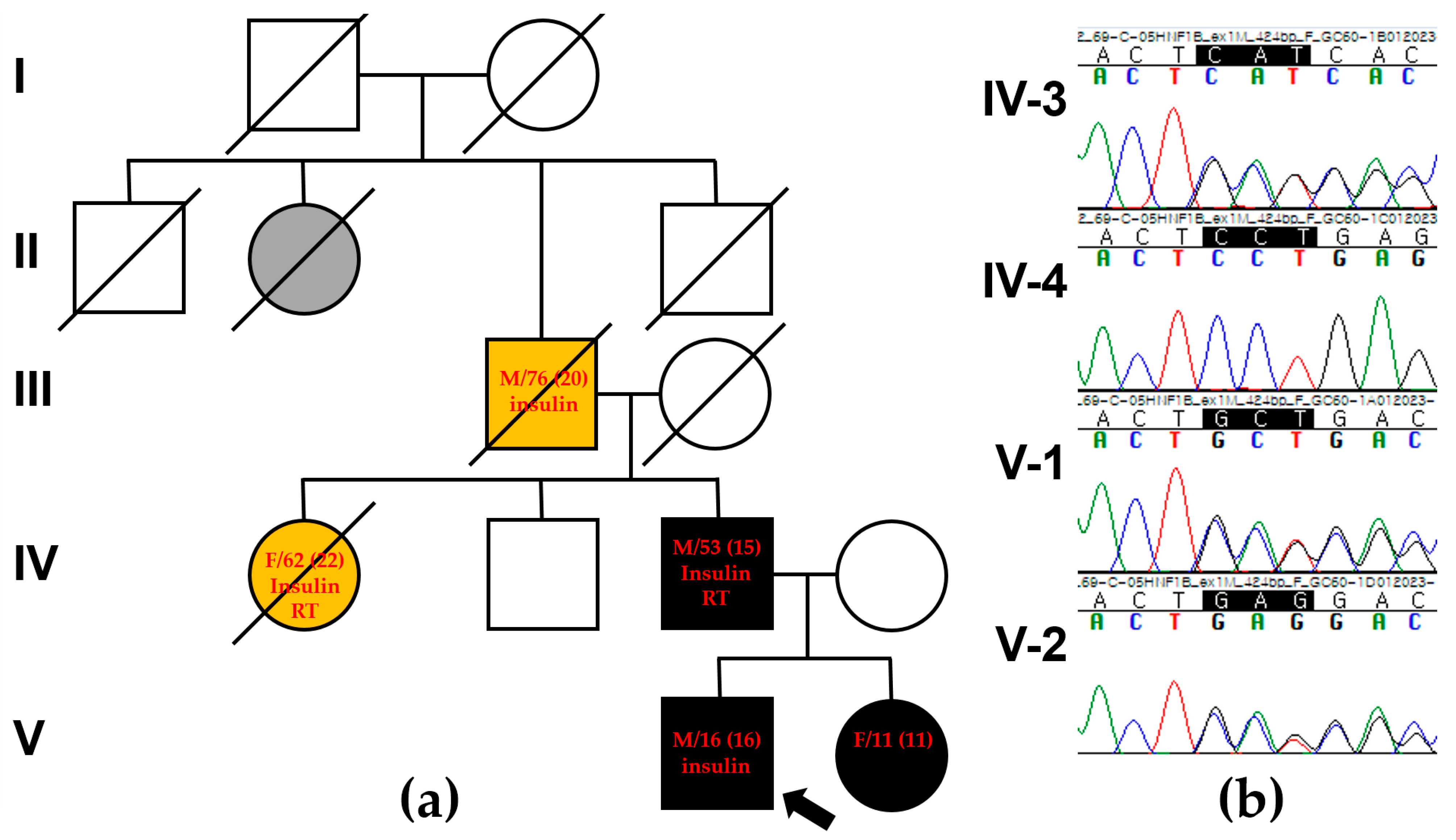
The proband’s father (IV-3 in Figure 1a), a 53-year-old man, was diagnosed with DM at the age of 15, requiring insulin injections thereafter. At the time of diagnosis, his fasting glucose level was 398 mg/dL, and his HbA1c was 17%. He was diagnosed with autoantibody-negative type I DM. Around the age of 35, he developed diabetic triopathy (retinopathy, nephropathy, and neuropathy), undergoing vitreoretinal surgery at 39. His renal function progressively declined, leading to end-stage renal disease in his late 40s, necessitating renal transplantation at 50. An abdominal CT scan revealed bilateral parenchymal thinning with cysts in both kidneys and pancreatic atrophy extending from the neck to the tail (Figure 2a,d). An ultrasound performed at age 39 showed a right renal cyst with a diameter of approximately 1 cm. However, a CT scan conducted at age 50 revealed that the cyst had enlarged to about 3 cm, accompanied by progressive renal atrophy.

3. Genetic Testing

Sequential genetic testing was conducted to explore the glucose intolerance, renal cysts, hepatic cysts, and agenesis of the dorsal pancreas observed in the proband. Initial evaluations, including conventional karyotyping and chromosomal microarray analysis, did not reveal any pathogenic structural or numerical chromosome changes or copy number variations. Next, we employed a comprehensive clinical exome sequencing (CES) approach using the Celemics G-Mendeliome Clinical Exome Sequencing Panel (Celemics, Inc., Seoul, Republic of Korea). This panel encompasses around 7000 genes associated with significant Mendelian genetic disorders, covering all pertinent regions. Massively parallel sequencing was performed with the DNBSEQ-G400RS High-throughput Sequencing Set and DNBSEQ-G400 sequencer (MGI Tech Co. Ltd., Shenzhen, China). Pathogenic variant interpretation adhered to the guidelines set by the American College of Medical Genetics and Genomics (ACMG) and the Association for Molecular Pathology (AMP). To identify potentially harmful variants, we used the following criteria: (1) variants located near or within exons of protein-coding genes linked to Mendelian diseases; (2) variants with allele frequencies less than 0.01; (3) variants causing nonsynonymous or nonsense changes in codons within exons, affecting highly conserved splice sites, or inducing frameshift mutations; (4) de novo or rare heterozygous, compound heterozygous, or homozygous variants in the same gene found in the proband; (5) genes included in the MODY Panel with clinical relevance, such as APPL1, BLK, CEL, GATA6, GCK, HNF1A, HNF1B, HNF4A, INS, KCNJ11, KLF11, NEURO1, PAX4, and PDX1; (6) the specific conditions of DM, glucose intolerance, renal cysts, hepatic cysts, and agenesis of the dorsal pancreas, which might be sporadic or inherited in an autosomal dominant manner, considering that the proband’s father and sister were affected. The allele frequencies of filtered variants were assessed using the Genome Aggregation Database (gnomAD, https://gnomad.broadinstitute.org/, accessed on 1 March 2024). In silico analyses were carried out to predict the pathogenicity of missense and insertion/deletion variants using MutationTaster (https://www.mutationtaster.org/, accessed on 1 March 2024) and VEST-4 (https://www.cravat.us/CRAVAT/, accessed on 1 March 2024).
As a result, gene panel sequencing identified a heterozygous HNF1B variant, c.36_38delCCT/p.(Leu13del), as the most probable cause of MODY in the proband (Reference transcript ID: NM_000458.4). This in-frame variant was not present in the gnomAD database and was predicted to be “disease-causing” by MutationTaster and “deleterious” by VEST-4, with a VEST score of 0.75 (p-value of 0.03915). VEST assigns scores from 0 to 1, where a score of 1 indicates a high-confidence prediction of a functional mutation [8]. A potential splice effect was not detected by SpliceAI (https://spliceailookup.broadinstitute.org/, accessed on 1 March 2024). The clinical presentation of the patient was consistent with MODY caused by the HNF1B variant, including glucose intolerance, renal cysts, hepatic cysts, and agenesis of the dorsal pancreas, which is aligned with renal cysts and diabetes syndrome (OMIM #137920). Sanger sequencing confirmed that the c.36_38delCCT/p.(Leu13del) variant of HNF1B was linked to the phenotype and confirmed its autosomal dominant inheritance pattern. The variant was present in the patient, his father (IV-3), and his sister (V-2), but not in his mother (IV-4) (Figure 1b). This variant was classified as likely pathogenic based on ACMG guidelines, with criteria including PM2 (absent from Exome Sequencing Project, 1000 Genomes, or ExAC), PM4 (protein length changes due to in-frame deletions/insertions or stop-loss variants), PP1 (co-segregation with disease in multiple affected family members), and PP4 (the patient’s phenotype or family history specific to a single genetic cause). Protein structure analysis using AlphaFold (https://alphafold.ebi.ac.uk/, accessed on 1 March 2024) showed high per-residue confidence scores (90 > pLDDT > 70) of 86.87 for the HNF1B p.Leu13 residue in the dimerization domain (Figure 3a,b). Additionally, sequence alignment of the conserved dimerization domain of the HNF1B protein across various vertebrate species demonstrated that the p.Leu13 residue is highly conserved between humans and Takifugu (Figure 3c). Deletions and duplications were assessed using the multiplex ligation-dependent probe amplification assay with SALSA MLPA Probemix P241 MODY Mix 1 (MRC-Holland, Amsterdam, The Netherlands), and no abnormalities were detected except for c.36_38delCCT/p.(Leu13del).

4. Discussion

The HNF1B gene, located on chromosome 17q12, comprises an N-terminal dimerization domain, a homeobox and POU domain involved in DNA binding, and a transactivation domain at the C-terminus [9,10]. Belonging to the homeobox-containing family of transcription factors, HNF1B plays a crucial role in the development and function of epithelial tissues in the kidney, pancreas, liver, and genitourinary tracts [11]. Over 200 different variants have been reported within the HNF1B gene, with mutations primarily occurring in the first four exons encoding the dimerization and DNA-binding domains [12]. To date, the likely pathogenic or pathogenic variants registered in ClinVar comprise 80 frameshift, 78 missense, 38 nonsense, and 28 splice-site mutations (https://www.ncbi.nlm.nih.gov/clinvar/, accessed on 7 August 2024). The variable phenotypes observed among individuals or families, along with incomplete penetrance, pose challenges in understanding the condition, particularly due to the 50–60% rate of de novo mutations identified in index cases [13,14]. The majority of (likely) pathogenic variants in HNF1B are truncating (59.4%; 114/194), whereas missense variants, which cluster in important protein domains, constitute the second-largest group (39.1%; 75/194) [15]. In general, an out-of-frame mutation caused by deleting one or two nucleotides from a defined reading frame creates an entirely new open reading frame with completely different nucleotide triplets or codons. This usually results in a completely altered amino acid sequence, leading to a non-functional protein. In contrast to out-of-frame deletions (or insertions) of three nucleotides, in-frame deletions (or insertions) do not create a premature stop codon. This results in a protein with only a few amino acids added or missing, making it more likely that the protein remains functional. However, the prenatal and postnatal phenotypes of seven individuals with renal cysts caused by a novel in-frame deletion, p.(Gly239del), within the HNF1B DNA-binding domain were previously reported [15]. In our MODY5 family, an in-frame deletion of the HNF1B gene results in a protein lacking one amino acid, potentially affecting the tertiary structure of the protein. The deletion of the CCT removes the leucine amino acid but does not modify the reading frame, leaving the other amino acids unaltered. The in-frame deletion of leucine at position 13 identified in our case is highly conserved among different species. This deletion is located within the HNF1B dimerization domain, which mediates DNA dimerization. Even though in silico analysis predicted our in-frame deletion to have a harmful effect, additional functional studies are needed to demonstrate the functional impairment of the in-frame deletion. In particular, AlphaFold does not account for interactions with other molecules, such as nucleic acids, small-molecule co-factors, ions, and other non-protein components. Additionally, AlphaFold is not designed to model post-translational modifications or the structures of free nucleic acids. However, it may still predict a protein’s conformation as if it were bound to a ligand or ion, even if the actual ligand or ion is absent.
On the other hand, HNF1B is broadly expressed in various fetal tissues and is essential for visceral endoderm specification [16]. In animal models, including adult mice and rats, HNF1B is expressed in the liver, kidneys, pancreatic islets, stomach, and intestine [17]. Congenital anomalies of the female reproductive organs, such as bicornuate uterus, uterus didelphys, rudimentary uterus, and vaginal atresia, have also been reported [18]. Its expression during early embryonic development is critical for kidney and pancreas formation [19]. HNF1B plays a critical role in the growth of the collecting ducts, renal pelvis, and ureter, as well as in the differentiation of the metanephric mesenchyme, which are essential for nephron and collecting system development [20]. Regarding clinical findings, about 66% of patients with MODY5 have a family history of DM, and the median age at diabetes diagnosis is 16 years old. Renal cysts are present in about 72% of patients, hypomagnesemia in about 92%, and pancreatic hypoplasia in about 72% [21]. Various types of HNF1B variants have been reported, including 117 missense variants, 98 nonsense variants, 58 small deletions or insertions, and 24 splicing variants, according to the Human Gene Mutation Database. Most of these variants are located in the first four exons of the gene, with exons 2 and 4 and the intron 2 splicing site being hotspots. The highly conserved DNA-binding domain of HNF1B is crucial for transcription regulation [22]. Up to 50% of MODY5 cases are caused by a 17q12 deletion encompassing 15 genes, including HNF1B, and following deletions, missense or nonsense mutations are the most frequent [4,23]. Compared to deletions and mutations, HNF1B gene deletion is associated with significantly lower magnesium and serum creatinine levels and higher eGFR [24,25].
Pancreatic atrophy is reported in approximately 30% of patients, and DM in about 50% [26]. Imaging studies have shown dysgenesis of the pancreas, particularly in the body or tail, in approximately 70% of cases [27]. MODY typically develops before 25 years of age, and in this family, DM was diagnosed from adolescence to early adulthood. HbA1c levels ranged from 6% to 10%, and fasting blood glucose levels were around 500 mg/dL at diagnosis. Autoimmune antibodies were negative in all patients, and BMI was within the normal range, without obesity, in a previous study [28]. Renal dysfunction predominantly manifests as a chronic tubulointerstitial pattern and is often found in advanced chronic kidney disease (CKD, stages III–IV) [29]. Hypomagnesemia and hypokalemia are commonly observed, occurring in 10–62% and 46% of patients, respectively [30]. Liver dysfunction, characterized by an asymptomatic elevation in liver enzyme levels, is common in patients with HNF1B mutations [31]. However, structural liver abnormalities have been observed in around 30% of patients with HNF1B variants [24]. In this family, renal cysts were present, and renal dysfunction progressed slowly. Hypomagnesemia, hyperuricemia, liver dysfunction, and genital abnormalities were not observed. Dorsal pancreatic agenesis is extremely rare but may be present in a significant portion of DM cases, potentially up to 50%. The proband was diagnosed with DM and revealed pancreatic dysgenesis at 16 years old. Other acquired causes are not suspected, and HNF1B is known to be involved in pancreatic organogenesis. However, in his 53-year-old father, diffuse atrophic changes in the pancreas may be a secondary finding of DM or a manifestation of an HNF1B mutation. Since the proband and his sister differ in sex and age, variations in symptoms, such as BMI and the presence or absence of pancreatic abnormalities, are expected. Long-term follow-up is essential to evaluate whether their clinical symptoms diverge over time.
Among patients with congenital abnormalities of the kidney and urinary tract, approximately 10% had HNF1B mutations. Predictive factors for detecting HNF1B mutations included bilateral renal anomalies, renal cysts of unknown origin, a combination of two major renal anomalies, and hypomagnesemia. In this family, clinical manifestations suggested a moderate predictive value for HNF1B mutations prior to other genetic tests. However, the patients’ main concern was early-onset diabetes mellitus with an autosomal inherited pattern, which led us to conduct broader genetic testing, including genes associated with MODY. Most patients with HNF1B-related nephropathy typically present with simple renal cysts without a significant decrease in kidney function. Renal dysfunction usually manifests in the 40s, and approximately 3-50% of patients progress to chronic kidney disease (CKD), ultimately requiring renal transplantation, particularly in cases without diabetic nephropathy [32,33,34,35]. Generally, HNF1B-related nephropathy exhibits a slow progressive course in childhood, except for very early onset cases. Patients with HNF1B haploinsufficiency may present with renal hypoplasia and dysgenesis, familial juvenile hyperuricemic nephropathy, glomerulocystic kidney disease, and renal interstitial fibrosis, ultimately leading to CKD [26]. Less than 1% of affected patients with HNF1B mutations exhibit DM alone [36]. Studies involving reverse-transcription PCR and in situ hybridization have shown that HNF-1beta mRNA is observed in normal human metanephrons, with the highest transcript levels observed in fetal medullary and cortical collecting ducts and lower levels in nephrogenic cortex mesenchyme, primitive nephron tubules, and immature glomeruli [37]. In animal models, the inactivation of HNF1B from postnatal day 10 onward does not result in cystic dilations in tubules after their proliferative morphogenetic extension is completed [38]. HNF1B directly regulates the transcription of Pkhd1, and inhibition of PKHD1 gene expression may affect the development of renal cysts in humans with HNF1B mutations [39].
DM, the second most common feature of HNF1B defects, has been observed in 5–50% of HNF1B mutation carriers in previous studies, accounting for approximately 5% of cases of MODY [40,41,42,43]. HNF1B is a tissue-specific transcription factor that forms homodimers or heterodimers with HNF1A and transactivates various genes, including albumin, alpha-fetoprotein, and glucose transporter 2 [44,45]. Defects in HNF1B lead to alterations in pancreatic morphology, ranging from pancreatic dysgenesis to diffuse pancreatic atrophy [46]. Pancreatic exocrine deficit has been reported in 20–75% of patients, and MODY is caused by beta-cell dysfunction and insulin resistance [47]. Evidence of pancreatic exocrine dysfunction has been observed in patients, as evidenced by fecal elastase deficiency [48]. Patients with HNF1B-MODY often respond poorly to oral hypoglycemic agents such as sulfonylureas, with early insulin therapy required in up to 80% of cases [46,49,50]. In patients with HNF1B-MODY, renal involvement is not associated with diabetic nephropathy but rather with aberrant embryonic development [51]. Long-term DM complications, including retinopathy and nephropathy, occur in patients with the longest duration of DM (median 21 years) [52]. In animal models, a precursor cellular stage of the embryonic pancreas and HNF1B in a genetic hierarchy control the formation of pancreatic endocrine cells [53]. HNF1B is targeted by miR-802-dependent silencing, and it has been revealed that a short hairpin RNA (shRNA)-mediated reduction in HNF1B in the liver leads to glucose intolerance, damage to insulin signaling, and increased hepatic gluconeogenesis. Conversely, hepatic overexpression of HNF1B enhances insulin sensitivity in Lepr(db/db) mice [54]. Our familial cases were diagnosed between ages 15 and the 20s, presenting with blood glucose levels around 300 mg/dL at diagnosis, accompanied by markedly reduced insulin secretion. Uric acid and magnesium levels were initially within normal ranges but increased around the time of dialysis initiation. Other studies similarly reported comparable blood glucose and insulin secretion patterns but varied in their findings for uric acid and magnesium levels [55]. Pancreatic developmental abnormalities were observed only in the proband, consistent with the known influence of the HNF1B gene on pancreatic development.
On the other hand, patients with HNF1B mutations often exhibit prevalent liver enzyme dysfunction, particularly in association with MODY5, affecting around 40% of adults [13]. Histopathological findings typically reveal bile ductopenia, steatosis, and periportal fibrosis, which may result in neonatal or adult cholestatic hepatopathy [56]. While other liver abnormalities have not been commonly reported, the proband in this family presented with a single hepatic cyst but normal liver function. Liver involvement in HNF1B-related conditions has not been extensively studied, with reported cases mainly presenting as an asymptomatic elevation in transaminase levels or, less commonly, as cholestatic liver diseases. Unusually, the proband in our family had a simple hepatic cyst without abnormal liver enzymes or related symptoms. These findings contribute to expanding our understanding of HNF1B-MODY5. Further studies are necessary to establish the underlying mechanisms and genotype–phenotype correlations. Clinical manifestations in our family presenting with renal cysts and MODY caused by the HNF1B c.36_38delCCT /p.(Leu13del) variant are summarized in Table 1.

5. Conclusions

In conclusion, we have described a family with MODY5 characterized by a novel in-frame deletion mutation affecting the dimerization domain of the HNF1B gene. Notably, contrary to previous reports, the proband exhibited a single hepatic cyst, with liver enzyme levels within normal ranges. The presence of early-onset diabetes, renal cysts, a family history, and nephropathy appearing before or after the diagnosis of DM suggests a diagnosis of HNF1B-MODY5. Early diagnosis is crucial for preventing complications of DM and allows for family screening and pre-conceptional genetic counseling. Patients with HNF1B-MODY should receive comprehensive medical care from endocrinology, nephrology, urology, and gynecology departments. To date, there is no well-established genotype–phenotype correlation indicating which types of variants are associated with particular clinical manifestations. The factors contributing to phenotypic variation remain inadequately understood and could potentially include the functional consequences of distinct gene variants, stochastic fluctuations in HNF1B gene expression during the early developmental period, or additional genetic and/or environmental influences. The current findings highlight the need for further investigation through prospective studies.

Author Contributions

Conceptualization, J.Y.H.; methodology, J.Y.H. and J.P.; software, J.G.; validation, T.Y.K.; formal analysis, J.Y.H. and J.P.; investigation, J.G. and T.Y.K.; resources, J.Y.H.; data curation, J.Y.H.; writing—original draft preparation, J.Y.H.; writing—review and editing, J.Y.H., T.Y.K., and J.P.; visualization, J.Y.H. and J.P.; supervision, J.P.; project administration, J.Y.H.; and funding acquisition, T.Y.K. and J.P. All authors have read and agreed to the published version of the manuscript.

Funding

This paper was supported by the Fund of Biomedical Research Institute, Jeonbuk National University Hospital.

Institutional Review Board Statement

This study was conducted according to the guidelines of the Declaration of Helsinki and approved by the Institutional Review Board of the Catholic University of Korea (Approval number: DC24ZASI0023; Date of approval: 19 March 2023).

Informed Consent Statement

Written informed consent was obtained from the subjects for participation in the clinical and molecular analyses and the publication of the data included in this study.

Data Availability Statement

Data are contained within the article.

Conflicts of Interest

The authors declare no conflicts of interest.

References

  1. Gardner, D.S.; Tai, E.S. Clinical features and treatment of maturity onset diabetes of the young (MODY). Diabetes Metab. Syndr. Obes. Targets Ther. 2012, 5, 101–108. [Google Scholar] [CrossRef] [PubMed]
  2. Colclough, K.; Ellard, S.; Hattersley, A.; Patel, K. Syndromic monogenic diabetes genes should be tested in patients with a clinical suspicion of maturity-onset diabetes of the young. Diabetes 2022, 71, 530–537. [Google Scholar] [CrossRef] [PubMed]
  3. Horikawa, Y.; Iwasaki, N.; Hara, M.; Furuta, H.; Hinokio, Y.; Cockburn, B.N.; Lindner, T.; Yamagata, K.; Ogata, M.; Tomonaga, O.; et al. Mutation in hepatocyte nuclear factor-1 beta gene (TCF2) associated with MODY. Nat. Genet. 1997, 17, 384–385. [Google Scholar] [CrossRef] [PubMed]
  4. Clissold, R.L.; Hamilton, A.J.; Hattersley, A.T.; Ellard, S.; Bingham, C. HNF1B-associated renal and extra-renal disease-an expanding clinical spectrum. Nat. Rev. Nephrol. 2015, 11, 102–112. [Google Scholar] [CrossRef] [PubMed]
  5. Nkonge, K.M.; Nkonge, D.K.; Nkonge, T.N. The epidemiology, molecular pathogenesis, diagnosis, and treatment of maturity-onset diabetes of the young (MODY). In Clinical Diabetes and Endocrinology; Springer: Cham, Switzerland, 2020; Volume 6, pp. 1–10. [Google Scholar]
  6. Ulinski, T.; Lescure, S.; Beaufils, S.; Guigonis, V.; Decramer, S.; Morin, D.; Clauin, S.; Desche, G.; Bensman, A.; Bellanné-Chantelot, C. Renal phenotypes related to hepatocyte nuclear factor-1β (TCF2) mutations in a pediatric cohort. J. Am. Soc. Nephrol. 2006, 17, 497–503. [Google Scholar] [CrossRef] [PubMed]
  7. Faguer, S.; Chassaing, N.; Bandin, F.; Prouheze, C.; Garnier, A.; Casemayou, A.; Huart, A.; Schanstra, J.P.; Calvas, P.; Decramer, S.; et al. The HNF1B score is a simple tool to select patients for HNF1B gene analysis. Kidney Int. 2014, 86, 1007–1015. [Google Scholar] [CrossRef]
  8. Douville, C.; Masica, D.L.; Stenson, P.D.; Cooper, D.N.; Gygax, D.M.; Kim, R.; Ryan, M.; Karchin, R. Assessing the Pathogenicity of Insertion and Deletion Variants with the Variant Effect Scoring Tool (VEST-Indel). Hum. Mutat. 2016, 37, 28–35. [Google Scholar] [CrossRef]
  9. Bohn, S.; Thomas, H.; Turan, G.; Ellard, S.; Bingham, C.; Hattersley, A.T.; Ryffel, G.U. Distinct molecular and morphogenetic properties of mutations in the human HNF1β gene that lead to defective kidney development. J. Am. Soc. Nephrol. 2003, 14, 2033–2041. [Google Scholar] [CrossRef]
  10. Igarashi, P.; Shao, X.; Mcnally, B.T.; Hiesberger, T. Roles of HNF-1β in kidney development and congenital cystic diseases. Kidney Int. 2005, 68, 1944–1947. [Google Scholar] [CrossRef]
  11. Ferrè, S.; Igarashi, P. New insights into the role of HNF-1β in kidney (patho)physiology. Pediatr. Nephrol. 2019, 34, 1325–1335. [Google Scholar] [CrossRef]
  12. Çubuk, H.; Yalçın Çapan, Ö. A Review of Functional Characterization of Single Amino Acid Change Mutations in HNF Transcription Factors in MODY Pathogenesis. Protein J. 2021, 40, 348–360. [Google Scholar] [CrossRef] [PubMed]
  13. Bellanné-Chantelot, C.; Chauveau, D.; Gautier, J.-F.; Dubois-Laforgue, D.; Clauin, S.; Beaufils, S.; Wilhelm, J.-M.; Boitard, C.; Noël, L.-H.; Velho, G. Clinical spectrum associated with hepatocyte nuclear factor-1β mutations. Ann. Intern. Med. 2004, 140, 510–517. [Google Scholar] [CrossRef] [PubMed]
  14. Edghill, E.L.; Bingham, C.; Ellard, S.; Hattersley, A.T. Mutations in hepatocyte nuclear factor-1beta and their related phenotypes. J. Med. Genet. 2006, 43, 84–90. [Google Scholar] [CrossRef] [PubMed]
  15. Vasileiou, G.; Hoyer, J.; Thiel, C.T.; Schaefer, J.; Zapke, M.; Krumbiegel, M.; Kraus, C.; Zweier, M.; Uebe, S.; Ekici, A.B.; et al. Prenatal diagnosis of HNF1B-associated renal cysts: Is there a need to differentiate intragenic variants from 17q12 microdeletion syndrome? Prenat. Diagn. 2019, 39, 1136–1147. [Google Scholar] [CrossRef]
  16. Barbacci, E.; Reber, M.; Ott, M.-O.; Breillat, C.; Huetz, F.; Cereghini, S. Variant hepatocyte nuclear factor 1 is required for visceral endoderm specification. Development 1999, 126, 4795–4805. [Google Scholar] [CrossRef]
  17. Cereghini, S.; Ott, M.-O.; Power, S.; Maury, M. Expression patterns of vHNF1 and HNF1 homeoproteins in early postimplantation embryos suggest distinct and sequential developmental roles. Development 1992, 116, 783–797. [Google Scholar] [CrossRef]
  18. Amed, S.; Oram, R. Maturity-onset diabetes of the young (MODY): Making the right diagnosis to optimize treatment. Can. J. Diabetes 2016, 40, 449–454. [Google Scholar] [CrossRef]
  19. Tshivhase, A.; Matsha, T.; Raghubeer, S. Diagnosis and treatment of MODY: An updated mini review. Appl. Sci. 2021, 11, 9436. [Google Scholar] [CrossRef]
  20. Mateus, J.C.; Rivera, C.; O’Meara, M.; Valenzuela, A.; Lizcano, F. Maturity-onset diabetes of the young type 5 a MULTISYSTEMIC disease: A CASE report of a novel mutation in the HNF1B gene and literature review. Clin. Diabetes Endocrinol. 2020, 6, 16. [Google Scholar] [CrossRef]
  21. Ge, S.; Yang, M.; Cui, Y.; Wu, J.; Xu, L.; Dong, J.; Liao, L. The Clinical Characteristics and Gene Mutations of Maturity-Onset Diabetes of the Young Type 5 in Sixty-One Patients. Front. Endocrinol. 2022, 13, 911526. [Google Scholar] [CrossRef]
  22. Chen, Y.-Z.; Gao, Q.; Zhao, X.-Z.; Chen, Y.-Z.; Craig, L.B.; Xiong, X.-S.; Mei, C.-L.; Shi, Y.-Q.; Chen, X.-M. Systematic review of TCF2 anomalies in renal cysts and diabetes syndrome/maturity onset diabetes of the young type 5. Chin. Med. J. 2010, 123, 3326–3333. [Google Scholar] [PubMed]
  23. Bellanné-Chantelot, C.; Clauin, S.; Chauveau, D.; Collin, P.; Daumont, M.; Douillard, C.; Dubois-Laforgue, D.; Dusselier, L.; Gautier, J.-F.; Jadoul, M. Large genomic rearrangements in the hepatocyte nuclear factor-1β (TCF2) gene are the most frequent cause of maturity-onset diabetes of the young type 5. Diabetes 2005, 54, 3126–3132. [Google Scholar] [CrossRef] [PubMed]
  24. Dubois-Laforgue, D.; Cornu, E.; Saint-Martin, C.; Coste, J.; Bellanné-Chantelot, C.; Timsit, J. Diabetes, Associated Clinical Spectrum, Long-term Prognosis, and Genotype/Phenotype Correlations in 201 Adult Patients with Hepatocyte Nuclear Factor 1B (HNF1B) Molecular Defects. Diabetes Care 2017, 40, 1436–1443. [Google Scholar] [CrossRef]
  25. Decramer, S.; Parant, O.; Beaufils, S.; Clauin, S.; Guillou, C.; Kessler, S.; Aziza, J.; Bandin, F.; Schanstra, J.P.; Bellanné-Chantelot, C. Anomalies of the TCF2 gene are the main cause of fetal bilateral hyperechogenic kidneys. J. Am. Soc. Nephrol. 2007, 18, 923–933. [Google Scholar] [CrossRef] [PubMed]
  26. Gambella, A.; Kalantari, S.; Cadamuro, M.; Quaglia, M.; Delvecchio, M.; Fabris, L.; Pinon, M. The Landscape of HNF1B Deficiency: A Syndrome Not Yet Fully Explored. Cells 2023, 12, 307. [Google Scholar] [CrossRef] [PubMed]
  27. Türkvatan, A.; Erden, A.; Türkoğlu, M.A.; Yener, Ö. Congenital variants and anomalies of the pancreas and pancreatic duct: Imaging by magnetic resonance cholangiopancreaticography and multidetector computed tomography. Korean J. Radiol. 2013, 14, 905–913. [Google Scholar] [CrossRef]
  28. Bockenhauer, D.; Jaureguiberry, G. HNF1B-associated clinical phenotypes: The kidney and beyond. Pediatr. Nephrol. 2016, 31, 707–714. [Google Scholar] [CrossRef]
  29. Sagen, J.V.; Bostad, L.; Njølstad, P.R.; Søvik, O. Enlarged nephrons and severe nondiabetic nephropathy in hepatocyte nuclear factor-1beta (HNF-1beta) mutation carriers. Kidney Int. 2003, 64, 793–800. [Google Scholar] [CrossRef]
  30. Faguer, S.; Decramer, S.; Chassaing, N.; Bellanné-Chantelot, C.; Calvas, P.; Beaufils, S.; Bessenay, L.; Lengelé, J.P.; Dahan, K.; Ronco, P.; et al. Diagnosis, management, and prognosis of HNF1B nephropathy in adulthood. Kidney Int. 2011, 80, 768–776. [Google Scholar] [CrossRef]
  31. Kotalova, R.; Dusatkova, P.; Cinek, O.; Dusatkova, L.; Dedic, T.; Seeman, T.; Lebl, J.; Pruhova, S. Hepatic phenotypes of HNF1B gene mutations: A case of neonatal cholestasis requiring portoenterostomy and literature review. World J. Gastroenterol. 2015, 21, 2550–2557. [Google Scholar] [CrossRef]
  32. Okorn, C.; Goertz, A.; Vester, U.; Beck, B.B.; Bergmann, C.; Habbig, S.; König, J.; Konrad, M.; Müller, D.; Oh, J. HNF1B nephropathy has a slow-progressive phenotype in childhood—With the exception of very early onset cases: Results of the German Multicenter HNF1B Childhood Registry. Pediatr. Nephrol. 2019, 34, 1065–1075. [Google Scholar] [CrossRef] [PubMed]
  33. Bingham, C.; Bulman, M.P.; Ellard, S.; Allen, L.I.; Lipkin, G.W.; van’t Hoff, W.G.; Woolf, A.S.; Rizzoni, G.; Novelli, G.; Nicholls, A.J. Mutations in the hepatocyte nuclear factor-1β gene are associated with familial hypoplastic glomerulocystic kidney disease. Am. J. Hum. Genet. 2001, 68, 219–224. [Google Scholar] [CrossRef] [PubMed]
  34. Nakayama, M.; Nozu, K.; Goto, Y.; Kamei, K.; Ito, S.; Sato, H.; Emi, M.; Nakanishi, K.; Tsuchiya, S.; Iijima, K. HNF1B alterations associated with congenital anomalies of the kidney and urinary tract. Pediatr. Nephrol. 2010, 25, 1073–1079. [Google Scholar] [CrossRef] [PubMed]
  35. Thomas, R.; Sanna-Cherchi, S.; Warady, B.A.; Furth, S.L.; Kaskel, F.J.; Gharavi, A.G. HNF1B and PAX2 mutations are a common cause of renal hypodysplasia in the CKiD cohort. Pediatr. Nephrol. 2011, 26, 897–903. [Google Scholar] [CrossRef] [PubMed]
  36. Edghill, E.L.; Stals, K.; Oram, R.A.; Shepherd, M.H.; Hattersley, A.T.; Ellard, S. HNF1B deletions in patients with young-onset diabetes but no known renal disease. Diabet. Med. 2013, 30, 114–117. [Google Scholar] [CrossRef]
  37. Kolatsi-Joannou, M.; Bingham, C.; Ellard, S.; Bulman, M.P.; Allen, L.I.S.; Hattersley, A.T.; Woolf, A.S. Hepatocyte nuclear factor-1beta: A new kindred with renal cysts and diabetes and gene expression in normal human development. J. Am. Soc. Nephrol. 2001, 12, 2175–2180. [Google Scholar] [CrossRef]
  38. Verdeguer, F.; Le Corre, S.; Fischer, E.; Callens, C.; Garbay, S.; Doyen, A.; Igarashi, P.; Terzi, F.; Pontoglio, M. A mitotic transcriptional switch in polycystic kidney disease. Nat. Med. 2010, 16, 106–110. [Google Scholar] [CrossRef]
  39. Hiesberger, T.; Bai, Y.; Shao, X.; McNally, B.T.; Sinclair, A.M.; Tian, X.; Somlo, S.; Igarashi, P. Mutation of hepatocyte nuclear factor-1beta inhibits Pkhd1 gene expression and produces renal cysts in mice. J. Clin. Investig. 2004, 113, 814–825. [Google Scholar] [CrossRef]
  40. Heidet, L.; Decramer, S.; Pawtowski, A.; Moriniere, V.; Bandin, F.; Knebelmann, B.; Lebre, A.-S.; Faguer, S.; Guigonis, V.; Antignac, C. Spectrum of HNF1B mutations in a large cohort of patients who harbor renal diseases. Clin. J. Am. Soc. Nephrol. 2010, 5, 1079. [Google Scholar] [CrossRef]
  41. Madariaga, L.; Moriniere, V.; Jeanpierre, C.; Bouvier, R.; Loget, P.; Martinovic, J.; Dechelotte, P.; Leporrier, N.; Thauvin-Robinet, C.; Jensen, U.B. Severe prenatal renal anomalies associated with mutations in HNF1B or PAX2 genes. Clin. J. Am. Soc. Nephrol. 2013, 8, 1179. [Google Scholar] [CrossRef]
  42. Adalat, S.; Woolf, A.S.; Johnstone, K.A.; Wirsing, A.; Harries, L.W.; Long, D.A.; Hennekam, R.C.; Ledermann, S.E.; Rees, L.; Van’t Hoff, W. HNF1B mutations associate with hypomagnesemia and renal magnesium wasting. J. Am. Soc. Nephrol. 2009, 20, 1123. [Google Scholar] [CrossRef] [PubMed]
  43. Shepherd, M.; Shields, B.; Hammersley, S.; Hudson, M.; McDonald, T.J.; Colclough, K.; Oram, R.A.; Knight, B.; Hyde, C.; Cox, J. Systematic population screening, using biomarkers and genetic testing, identifies 2.5% of the UK pediatric diabetes population with monogenic diabetes. Diabetes Care 2016, 39, 1879–1888. [Google Scholar] [CrossRef] [PubMed]
  44. Mendel, D.B.; Hansen, L.P.; Graves, M.K.; Conley, P.B.; Crabtree, G.R. HNF-1 alpha and HNF-1 beta (vHNF-1) share dimerization and homeo domains, but not activation domains, and form heterodimers in vitro. Genes. Dev. 1991, 5, 1042–1056. [Google Scholar] [CrossRef] [PubMed]
  45. Cereghini, S. Liver-enriched transcription factors and hepatocyte differentiation. FASEB J. 1996, 10, 267–282. [Google Scholar] [CrossRef] [PubMed]
  46. El-Khairi, R.; Vallier, L. The role of hepatocyte nuclear factor 1β in disease and development. Diabetes Obes. Metab. 2016, 18, 23–32. [Google Scholar] [CrossRef]
  47. Motyka, R.; Kołbuc, M.; Wierzchołowski, W.; Beck, B.B.; Towpik, I.E.; Zaniew, M. Four cases of maturity onset diabetes of the young (MODY) type 5 associated with mutations in the hepatocyte nuclear factor 1 beta (HNF1B) gene presenting in a 13-year-old boy and in adult men aged 33, 34, and 35 years in Poland. Am. J. Case Rep. 2021, 22, e928994. [Google Scholar] [CrossRef]
  48. Haldorsen, I.; Vesterhus, M.; Raeder, H.; Jensen, D.; Søvik, O.; Molven, A.; Njølstad, P. Lack of pancreatic body and tail in HNF1B mutation carriers. Diabet. Med. 2008, 25, 782–787. [Google Scholar] [CrossRef]
  49. Pearson, E.R.; Badman, M.K.; Lockwood, C.R.; Clark, P.M.; Ellard, S.; Bingham, C.; Hattersley, A.T. Contrasting diabetes phenotypes associated with hepatocyte nuclear factor-1α and-1β mutations. Diabetes Care 2004, 27, 1102–1107. [Google Scholar] [CrossRef]
  50. Hattersley, A.T.; Greeley, S.A.; Polak, M.; Rubio-Cabezas, O.; Njølstad, P.R.; Mlynarski, W.; Castano, L.; Carlsson, A.; Raile, K.; Chi, D.V. ISPAD Clinical Practice Consensus Guidelines 2018: The Diagnosis and Management of Monogenic Diabetes in Children and Adolescents. Pediatr. Diabetes 2018, 19 (Suppl. S27), 47–63. [Google Scholar] [CrossRef]
  51. Bingham, C.; Ellard, S.; Allen, L.; Bulman, M.; Shepherd, M.; Frayling, T.; Berry, P.J.; Clark, P.M.; Lindner, T.; Bell, G.I. Abnormal nephron development associated with a frameshift mutation in the transcription factor hepatocyte nuclear factor-1β1: 1See Editorial by Woolf, p. 1202. Kidney Int. 2000, 57, 898–907. [Google Scholar] [CrossRef]
  52. Amaral, S.; Palha, A.; Bogalho, P.; Silva-Nunes, J. Maturity-onset diabetes of the young secondary to HNF1B variants (HNF1B-MODY): A series of 10 patients from a single diabetes center. Diabetol. Metab. Syndr. 2023, 15, 21. [Google Scholar] [CrossRef] [PubMed]
  53. Maestro, M.A.; Boj, S.F.; Luco, R.F.; Pierreux, C.E.; Cabedo, J.; Servitja, J.M.; German, M.S.; Rousseau, G.G.; Lemaigre, F.P.; Ferrer, J. Hnf6 and Tcf2 (MODY5) are linked in a gene network operating in a precursor cell domain of the embryonic pancreas. Hum. Mol. Genet. 2003, 12, 3307–3314. [Google Scholar] [CrossRef] [PubMed]
  54. Kornfeld, J.W.; Baitzel, C.; Könner, A.C.; Nicholls, H.T.; Vogt, M.C.; Herrmanns, K.; Scheja, L.; Haumaitre, C.; Wolf, A.M.; Knippschild, U.; et al. Obesity-induced overexpression of miR-802 impairs glucose metabolism through silencing of HNF1B. Nature 2013, 494, 111–115. [Google Scholar] [CrossRef] [PubMed]
  55. Horikawa, Y. Maturity-onset diabetes of the young as a model for elucidating the multifactorial origin of type 2 diabetes mellitus. J. Diabetes Investig. 2018, 9, 704–712. [Google Scholar] [CrossRef]
  56. Oba, Y.; Sawa, N.; Mizuno, H.; Hoshino, J.; Kinowaki, K.; Ohashi, K.; Morisada, N.; Iijima, K.; Yamaguchi, Y.; Ubara, Y. Autosomal Dominant Tubulointerstitial Kidney Disease HNF1B With Maturity-Onset Diabetes of the Young: A Case Report With Kidney Biopsy. Kidney Med. 2021, 3, 278–281. [Google Scholar] [CrossRef]
Figure 1. Pedigree analysis and segregation analysis. (a) The family pedigree shows autosomal dominant maturity-onset diabetes of the young (MODY). The proband is indicated by a black arrow. Numbers in parentheses indicate the age (year) at diagnosis. The gray symbol indicates that the family member was clinically suspected to have hereditary MODY, but this was not confirmed genetically. The yellow symbol indicates that the family member was clinically suspected to have diabetic triopathy, but this was not confirmed genetically. Black symbols indicate family members who were clinically suspected and confirmed by clinical exome sequencing. Treatments, including insulin and renal transplantation (RT), are described in each symbol. (b) Sanger sequencing confirmed a heterozygous HNF1B variant, c.36_38delCCT /p.(Leu13del), occurring as a consequence of autosomal dominant paternal origin in the proband (V-1) (Reference transcript ID: NM_000458.4).
Ijms 25 09823 g001
Figure 2. Radiologic findings in the proband and his family. (ac) Abdominal CT findings show renal cysts in the proband’s father (a), the proband (b), and the proband’s sister (c). (df) The cortical thinning of both kidneys and atrophic changes in the pancreas (d) were found in the proband’s father, while a hepatic cyst (e) and agenesis of the dorsal pancreas (f) were found in the proband. The tail of the pancreas was not visible on consecutive axial slices, showing only the head and body of the pancreas (indicated by red arrow).
Ijms 25 09823 g002
Figure 3. The 3D protein structure and conservation analysis of HNF1B. (a) The 3D protein modeling of the structure of human HNF1B using de novo protein modeling. The HNF1B protein structure was built through the DMPfold 1.0 Fast Mode algorithm on the PSIPRED server. RSCB PDB database; DNA-binding domain: 2DA6, 2H8R, and 5K9S. (b) Protein structure analysis using AlphaFold showed a very high per-residue confidence score (pLDDT) of 86.87 for the HNF1B p.Leu13 residue, highlighted in pink and indicated by a red arrow. (c) Sequence alignment of the conserved dimerization domain of the HNF1B protein in multiple vertebrate species. The protein sequence of the p.Leu13 residue is highly conserved between Homo sapiens and Takifugu. It is highlighted by the empty red box.
Ijms 25 09823 g003
Table 1. Clinical manifestations in a family presenting with renal cysts and maturity-onset diabetes of the young caused by the HNF1B p.Leu13del variant.
PatientsProband’s Father
(VI-3)
Proband
(V-1)
Proband’s Sister (V-2)Reference Ranges
Sex/age (year)M/53M/16F/11
BMI (kg/m2)22.522.628.418.5–22.9
HNF1B score141410<8
Renal cystbilateralbilateralbilateral
Pancreatic abnormalitiesDiffuse atrophic changesAgenesis of dorsal pancreasnone
Genitourinary tract defectnonenonenone
Laboratory findings at ageat 15 y oldat 50 y oldat 16 y
old
at 11 y
old
FBS (mg/dL)3982283011151–120
HbA1c (%)187.515.46.14.5–5.6
BUN (mg/dL)1528.913.913.96–20
Creatinine (mg/dL)15.521.030.580.5–1.2
eGFR (mL/min per 1.73 m2)11510.810714190–120
AST/ALT (IU/L)15/2116/1425/1217/158–40/5–41
Uric acid (mg/dL)2.43.9262.4–7
Mg (mg/dL)1.52.31.61.81.6–2.6
Ketone body (mg/L)380.1027.1371.301200–120
Ketonenegativenegativenegativenegativenegative
Autoantibodiesnegativen/anegativenegativenegative
Insulin (AC) (uU/mL)0.90n/a0.801.51.9–23
Insulin (PC) (uU/mL)8n/a8.5101.9–23
C-peptide (AC) (ng/mL)0.81.650.751.41.1–4.4
C-peptide (PC) (ng/mL)21.932.133.21.1–4.4
Urine sugar (mg/100mL)3+2+3+negativenegative
Urine protein (mg/100mL)negative2+negativenegativenegative
Urine albumin/Cr ratio (ug/mg)1020062<30
BMI, body mass index; y, year; FBS, fasting blood sugar; eGFR, estimated glomerular filtration rate; +, positive urine dipstick testing; Cr, creatinine.
Disclaimer/Publisher’s Note: The statements, opinions and data contained in all publications are solely those of the individual author(s) and contributor(s) and not of MDPI and/or the editor(s). MDPI and/or the editor(s) disclaim responsibility for any injury to people or property resulting from any ideas, methods, instructions or products referred to in the content.

