Article

Membrane-Bound Redox Enzyme Cytochrome bd-I Promotes Carbon Monoxide-Resistant Escherichia coli Growth and Respiration

by
Martina R. Nastasi
1,
Vitaliy B. Borisov
2,* and
Elena Forte
1,*
1
Department of Biochemical Sciences, Sapienza University of Rome, 00185 Rome, Italy
2
Belozersky Institute of Physico-Chemical Biology, Lomonosov Moscow State University, 119991 Moscow, Russia
*
Authors to whom correspondence should be addressed.
Int. J. Mol. Sci. 2024, 25(2), 1277; https://doi.org/10.3390/ijms25021277
Submission received: 16 November 2023 / Revised: 23 December 2023 / Accepted: 18 January 2024 / Published: 20 January 2024

Abstract

:
The terminal oxidases of bacterial aerobic respiratory chains are redox-active electrogenic enzymes that catalyze the four-electron reduction of O2 to 2H2O taking out electrons from quinol or cytochrome c. Living bacteria often deal with carbon monoxide (CO) which can act as both a signaling molecule and a poison. Bacterial terminal oxidases contain hemes; therefore, they are potential targets for CO. However, our knowledge of this issue is limited and contradictory. Here, we investigated the effect of CO on the cell growth and aerobic respiration of three different Escherichia coli mutants, each expressing only one terminal quinol oxidase: cytochrome bd-I, cytochrome bd-II, or cytochrome bo3. We found that following the addition of CO to bd-I-only cells, a minimal effect on growth was observed, whereas the growth of both bd-II-only and bo3-only strains was severely impaired. Consistently, the degree of resistance of aerobic respiration of bd-I-only cells to CO is high, as opposed to high CO sensitivity displayed by bd-II-only and bo3-only cells consuming O2. Such a difference between the oxidases in sensitivity to CO was also observed with isolated membranes of the mutants. Accordingly, O2 consumption of wild-type cells showed relatively low CO sensitivity under conditions favoring the expression of a bd-type oxidase.

Graphical Abstract

1. Introduction

Carbon monoxide (CO) is a well-known gaseous molecule that has long been recognized to mediate important physiological processes when produced in low amounts [1,2,3,4,5,6,7,8,9,10,11,12,13,14,15,16,17,18,19,20,21,22]. In eukaryotes, CO is formed endogenously as a byproduct upon the degradation of heme to biliverdin and iron catalyzed by heme oxygenase [23]. In bacteria, this gaseous molecule is generated by homologs of eukaryotic heme oxygenases and via alternative CO-producing mechanisms [24]. Interestingly, CO is considered as a probable signaling molecule between the host and the gut microbiome [24]. Some bacteria can also use CO as a source of energy and carbon [25]. High concentrations of CO are toxic, and some pathogenic bacteria were reported to be susceptible either to CO produced by the host heme oxygenases or to transition-metal-based CO-releasing molecules (CORMs) [26,27]. CORMs developed to deliver physiologically relevant levels of CO experimentally or therapeutically [28,29] showed an additive effect when combined with other antibiotics in certain microbes [26,27]. However, care should be taken as one of the most widely used CORMs, the water-soluble Ru-containing CORM-3, was reported to exert cytotoxic effects due to a thiol-reactive Ru(II) ion and releases little CO [30]. Thus, the development of novel effective CO-releasing drugs is an urgent problem as the therapeutic use of CO has emerged as an antimicrobial strategy in medicine. Bacterial proteins, which contain a pentacoordinate high-spin heme in the reduced state, should bind CO as a strong exogenous ligand, and this, in turn, should affect their function. The terminal oxidases of bacterial respiratory chains have such a heme in their active sites and therefore are among these proteins. These redox-active enzymes belong to class EC 7 translocases. Terminal oxidases catalyze four-electron reduction of O2 to 2H2O at the expense of oxidation of the respiratory substrate, either quinol or ferrocytochrome c. This redox reaction is coupled to the generation of proton-motive force which serves as the driving force for ATP synthesis and other useful work. There are two different groups of these enzymes found in bacteria: heme-copper oxidases, including aa3-type cytochrome c oxidase and bo3-type quinol oxidase, and copper-lacking bd-type quinol oxidases, also called cytochromes bd [31,32,33,34,35,36,37,38,39,40,41,42,43,44,45,46,47,48,49,50,51]. Heme-copper oxidases are true proton pumps, whereas cytochromes bd generate proton-motive force solely due to the vector transfer of protons along the intraprotein proton-conducting pathway, without a mechanism of proton pumping [47,52,53,54]. Escherichia coli is a ubiquitous member of the gut microbiome of humans and warm-blooded animals. Like many other aerobic bacteria, it contains the branched and flexible O2-dependent respiratory chain. In the chain, type I and type II NADH dehydrogenases transfer electrons from NADH to ubiquinone-8 and/or menaquinone-8. Ubiquinone-8 can also accept electrons from succinate via succinate dehydrogenase. Then, electrons from ubiquinol-8 and/or menaquinol-8 are transferred by three terminal oxidases, cytochromes bo3, bd-I, and bd-II, to the final electron acceptor, O2, to produce 2H2O [55,56,57,58,59].
In E. coli, the bo3, bd-I, and bd-II oxidases are encoded by the cyoABCDE, cydABX, and appCBX operons, respectively. With the use of a rigorous chemostat methodology and powerful modeling tools, it was shown that the cyoABCDE operon was maximally induced under fully aerobic conditions [60]. The cydABX operon was maximally expressed at 56% aerobiosis [61]. The appCBX operon was maximally expressed at 0% aerobiosis. Spectroscopic assays of oxidase levels confirmed these conclusions [60,62]. Thus, at high aeration, cytochrome bo3 is preferentially expressed, whereas a shift from aerobic to microaerobic conditions activates the expression of the bd-type cytochromes [55]. The atomic structures of the three cytochromes were reported [43,44,63,64,65,66,67]. Cytochrome bo3, composed of four subunits, carries the ubiquinol-binding site and three metal redox groups, a low-spin heme b, a high-spin heme o3, and CuB. Heme b accepts electrons from ubiquinol; heme o3 and CuB compose a binuclear site for O2 reduction [55]. Cytochromes bd-I and bd-II consist of four and three subunits, respectively. Both bd enzymes contain the ubiquinol/menaquinol-binding site (called the Q-loop) and three hemes, a low-spin b558, a high-spin b595, and a high-spin d, but no copper ion. Heme b558 is the immediate electron acceptor for ubiquinol and/or menaquinol. Heme d serves as the O2-reducing site. The functional role of heme b595 is not clear yet, but at least apparently it transfers electrons from heme b558 to heme d [55].
It is worth mentioning that cytochrome c and chlorophyll a were reported to be suitable redox mediators for the development of enzymatic biofuel cell systems [68]. In these systems, electron transfer from glucose oxidase to the electrode was facilitated through cytochrome c and chlorophyll a adsorbed on the electrode [68]. In this regard, the applicability of cytochrome d and cytochrome o3 for charge transfer from/to bacteria and in biofuel cells could also be evaluated.
A protective role of cytochrome bd-I against nitric oxide [69,70,71], peroxynitrite [72], and ammonia [73] was reported. Cytochrome bd-I likely provides NO resistance in E. coli for two reasons. The first reason is the extraordinarily high rate of NO dissociation from heme d2+: the koff values were reported to be 0.133 s−1 [69] and 0.163 s−1 [70]. This is about 30 times higher than that for NO dissociation from heme a32+ in the mitochondrial cytochrome c oxidase [74] and can explain why following inhibition by NO the oxygen reductase activity of the bd-I enzyme is restored much faster than that of the mitochondrial enzyme. The second plausible reason is the ability of a bd oxidase to rapidly convert NO into NO2 in turnover, although that was reported only for cytochrome bd from Azotobacter vinelandii so far [75]. The fact that the bd-I oxidase is not inhibited by peroxynitrite can be due to its ability to catalytically scavenge peroxynitrite [72]. The reaction likely occurs on the heme d active site, and at least four possible reaction mechanisms have been suggested [76]. Cytochrome bd-I is not only resistant to but also activated by ammonia under alkaline conditions [73]. In this case, ammonia is suggested to react with a few catalytic intermediates of the enzyme. In particular, NH3 can promote the formation of the peroxy state (P) from the oxidized state (O). NH3 also possibly reacts with the one-electron-reduced state (O1) to produce the ferryl state (F). In these reactions, NH3 presumably serves as a two-electron donor being oxidized to NH2OH [73].
Both bd-type oxidases also contribute to E. coli resistance to cyanide [77], hydrogen peroxide [78,79,80], and sulfide [77,81]. On the contrary, the activity of the bo3 oxidase was shown to be highly sensitive to inhibition by cyanide, sulfide, nitric oxide, and ammonia [70,73,77,81]. Thus, a bd-type terminal oxidase endows E. coli and possibly other bacteria with resistance to the above-mentioned stressors and, being absent in eukaryotic cells, can serve as a good therapeutic target [82].
Increased expression of cytochrome bd is in fact a likely mechanism for survival used by the pathogenic microorganisms in the presence of reactive oxygen and nitrogen species generated by the host immune system to fight infection [59]. In view of the fact that cytochrome bd-I exhibits tolerance to such reactive species, it is relevant to examine its potential resistance also to CO, which has been reported to be important in host–pathogen relationships [83,84,85,86] and is a heme ligand like NO. It has to be noted that data on the effect of CO on the function of bacterial terminal oxidases are limited and contradictory. Of the three E. coli oxidases, cytochrome bo3 was shown to be the least sensitive to inhibition by CO if the enzymes were purified and detergent-solubilized [87]. In contrast, according to a recent short report [88], cytochrome bd-I is more resistant to inhibition by CO than cytochrome bd-II and cytochrome bo3 if CO is added to E. coli cell suspensions, at [O2] = 150 μM. Bayly et al. also studied the physiological response of Mycobacterium smegmatis to CO [83]. The respiratory chain of mycobacteria is known to contain two different terminal oxidases: cytochrome bcc-aa3 supercomplex and cytochrome bd [89]. Bayly et al. reported that in M. smegmatis cell cultures, the activity of cytochrome bd is resistant to CO while cytochrome bcc-aa3 supercomplex is strongly inhibited by CO [83]. Furthermore, M. smegmatis lacking the bd oxidase shows a significant growth defect in the presence of this gas [83].
In this work, we studied the effect of CO on aerobic respiration sustained by bo3, bd-I, or bd-II oxidases in cell cultures of E. coli respiratory mutants by varying the O2 concentration at which CO was added, and we examined the ability of these cytochromes to sustain bacterial growth in the presence of CO. We also tested the CO inhibition of O2 consumption of both isolated membranes of the mutants and wild-type cells grown under conditions favoring the expression of either cytochrome bo3 or a bd-type oxidase.

2. Results

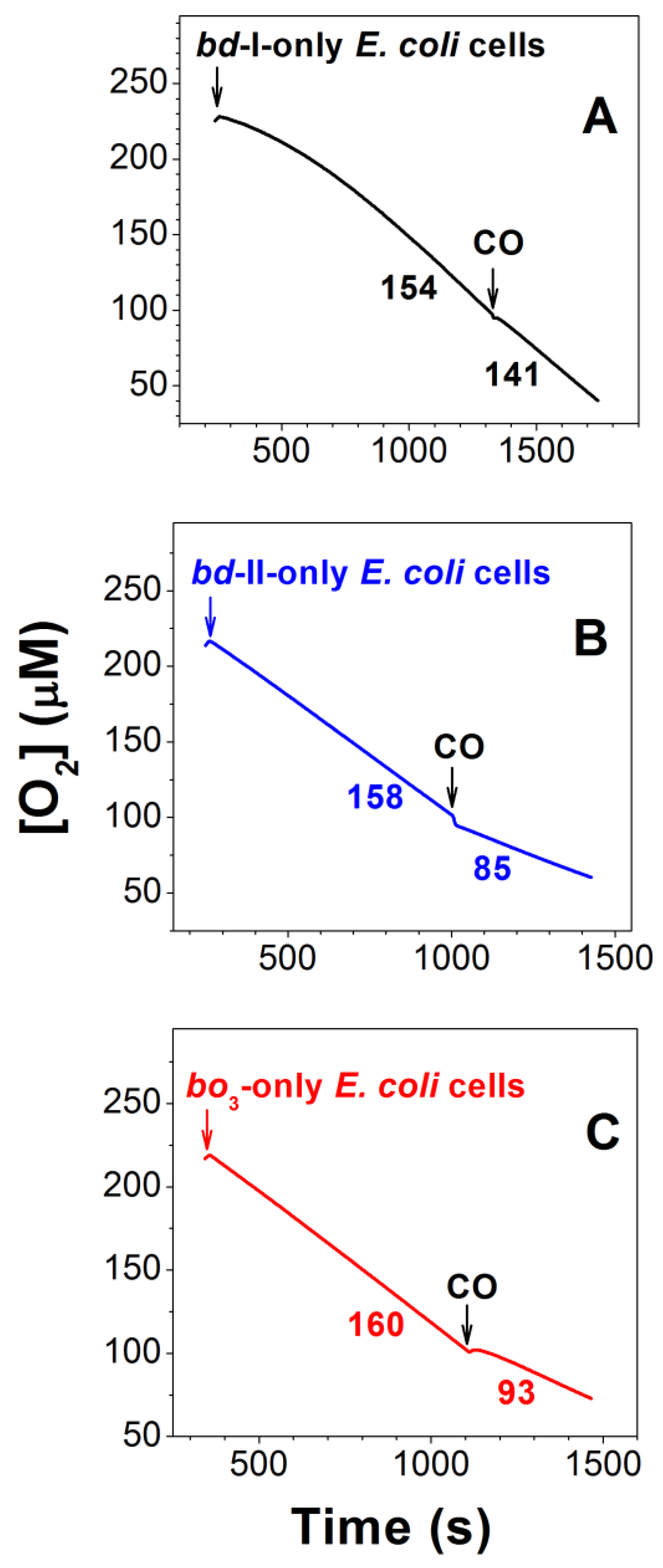
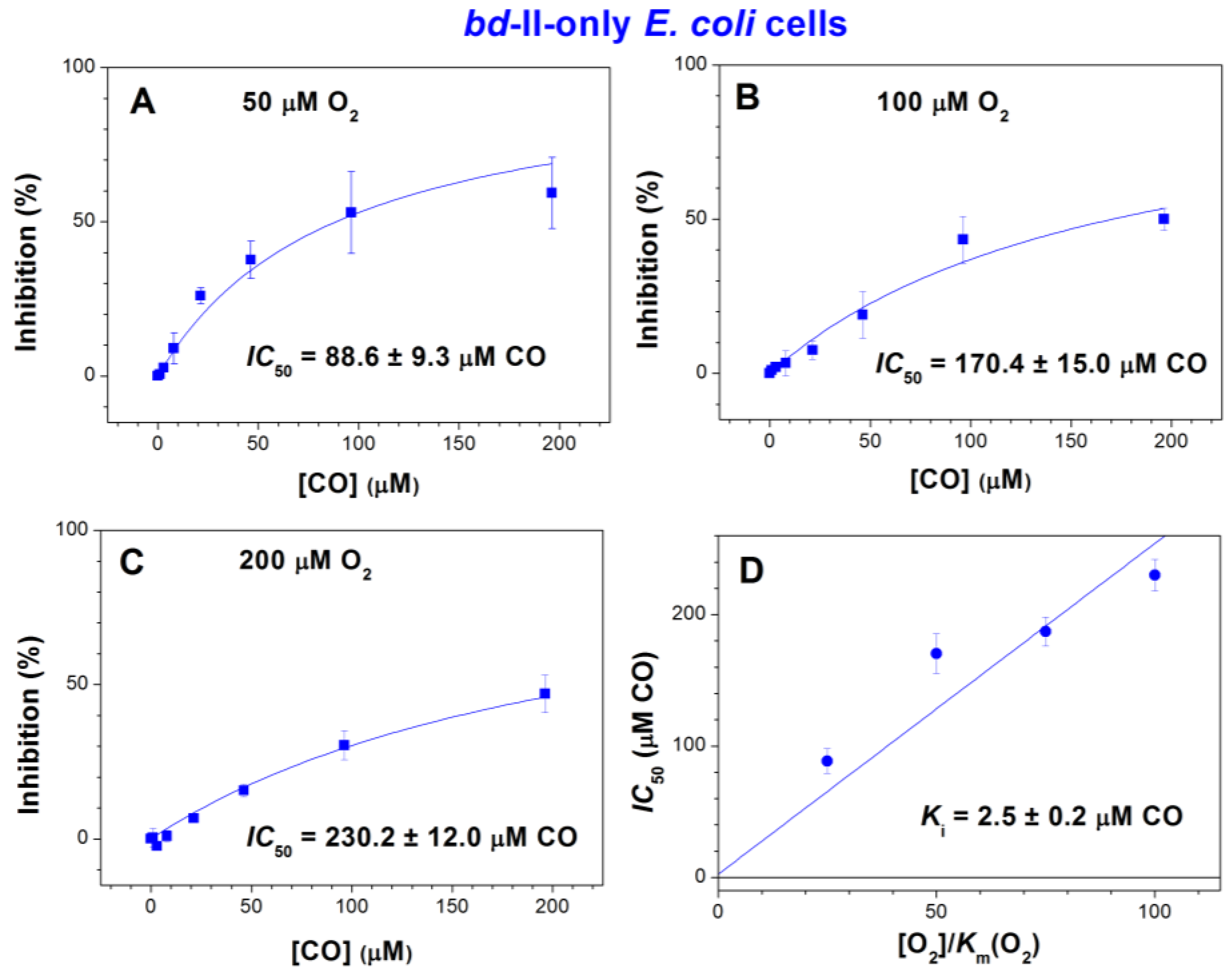
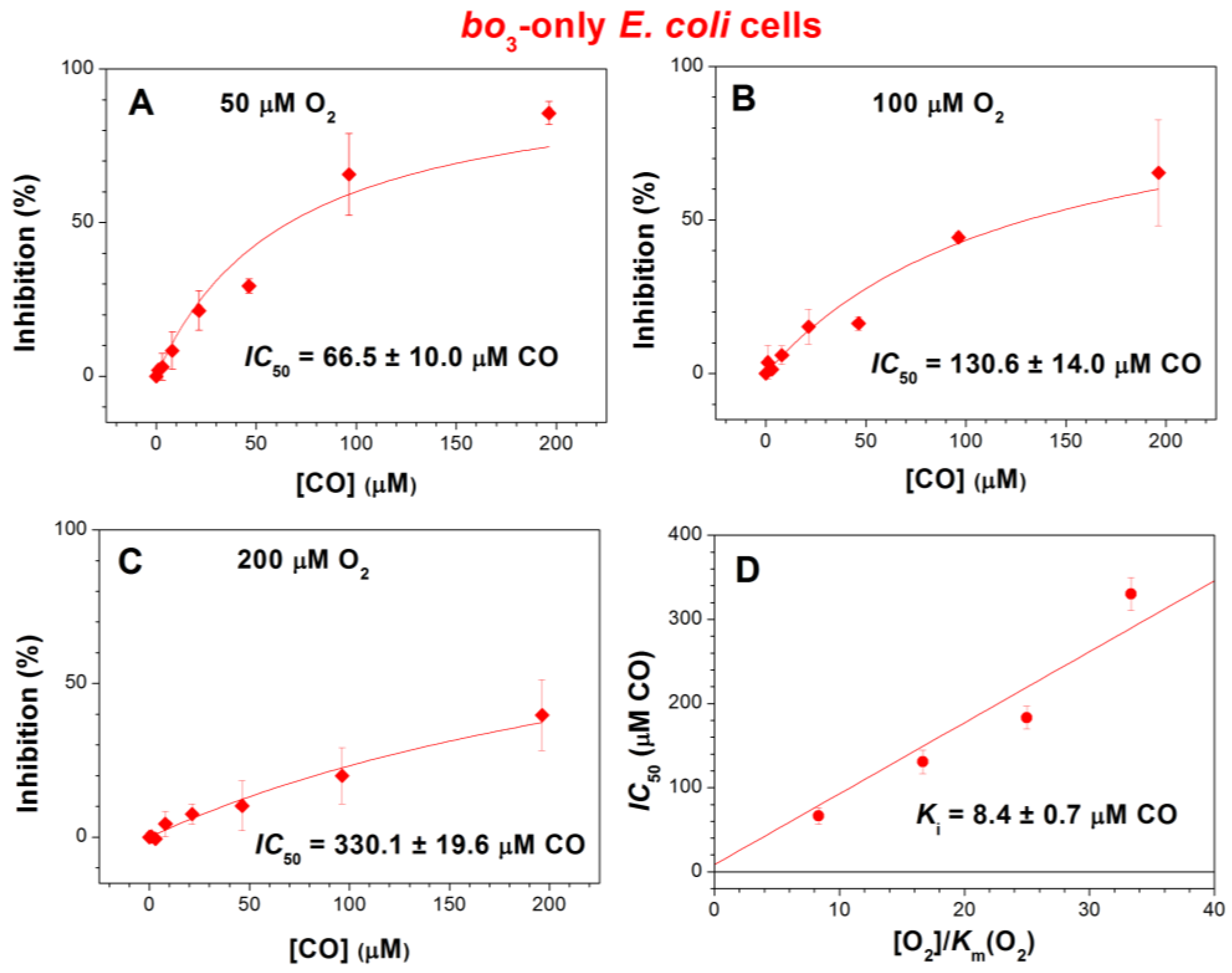
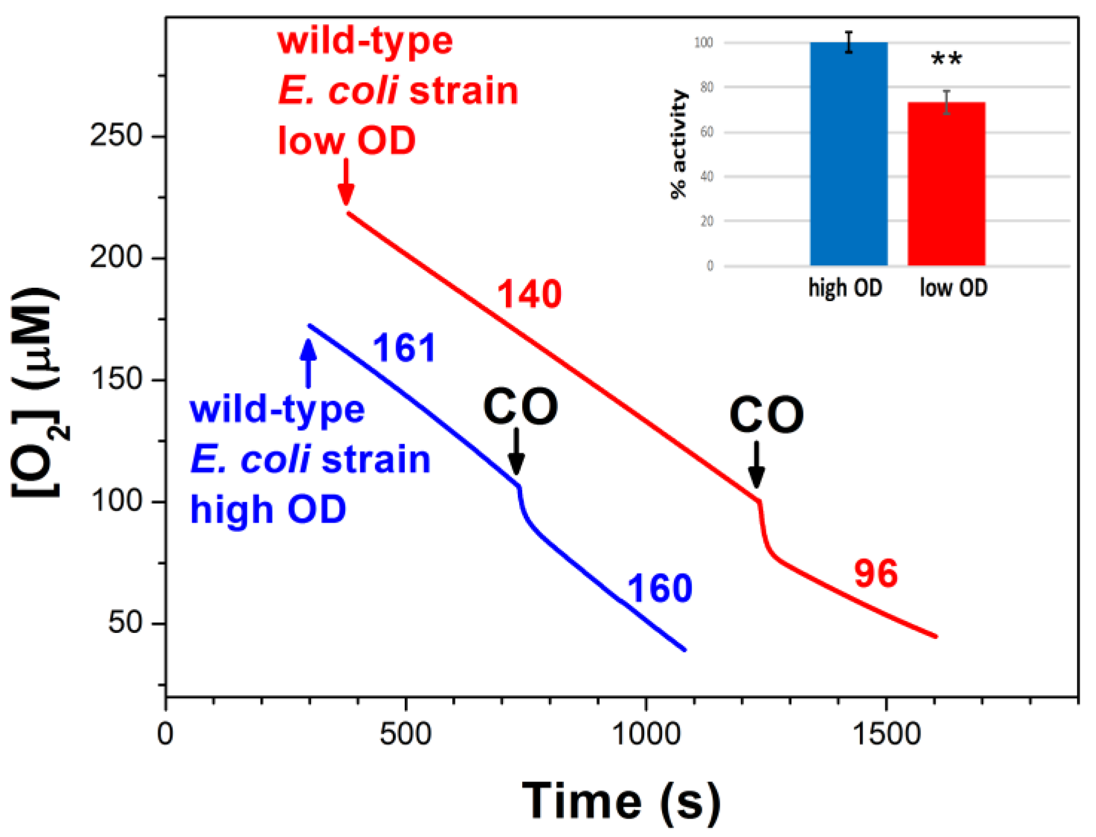
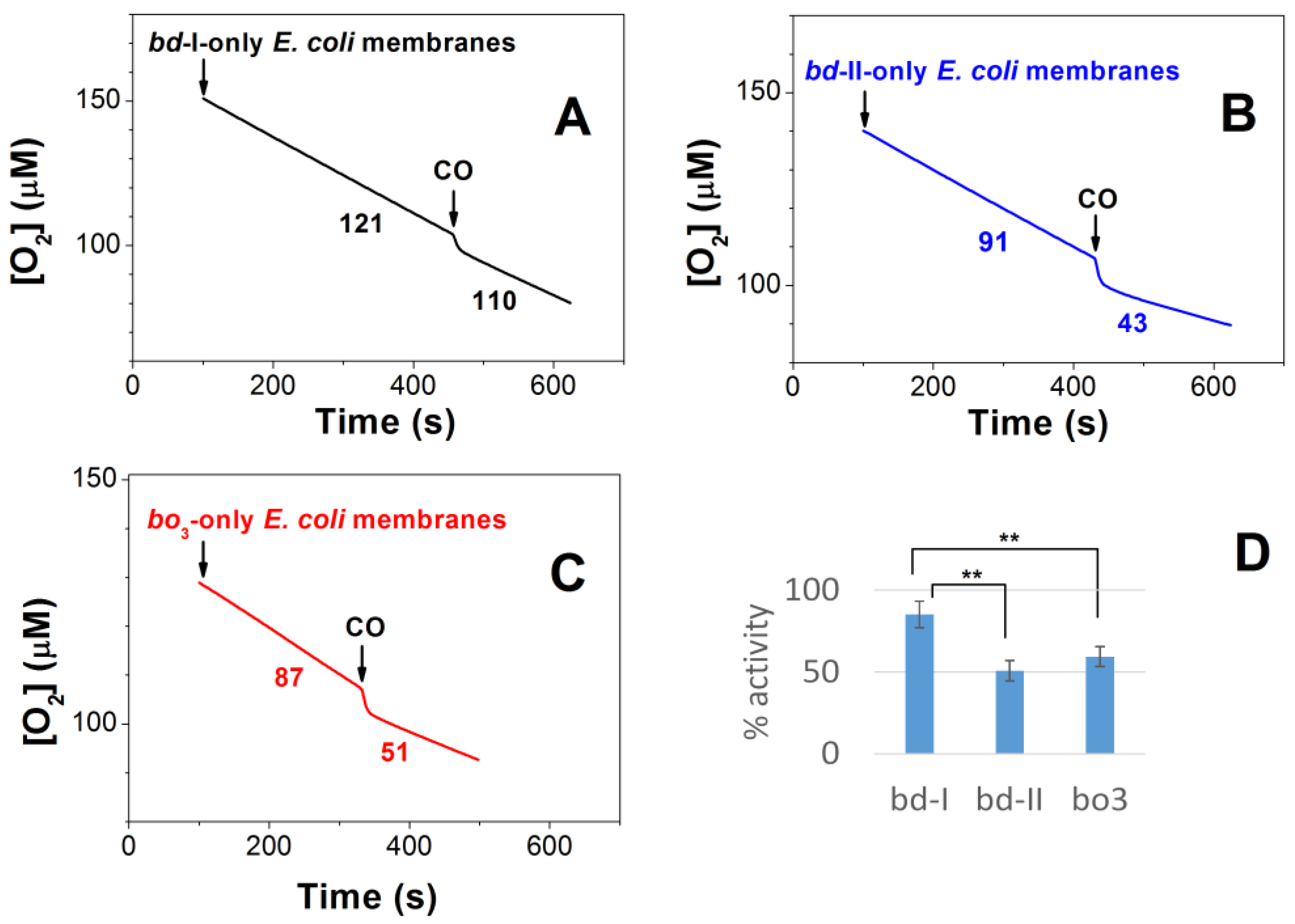
2.1. Effect of CO on E. coli Aerobic Respiration

We examined the effect of CO on the aerobic respiration of E. coli cells of the three different mutant strains. The aerobic respiratory chain of each mutant contains only one terminal quinol oxidase: cytochrome bd-I, cytochrome bd-II, or cytochrome bo3. In each mutant, aerobic cellular respiration was supplied by endogenous electron-donor respiratory substrates; therefore, the use of an exogenous reducing system was not required. Figure 1A shows that 96.3 μM CO added at [O2] = 100 μM inhibits O2 consumption by bd-I-only E. coli cells to a small extent of 11.6 ± 1.1%. Under these experimental conditions, the CO inhibitory effect on the aerobic respiration of bd-II-only and bo3-only E. coli cells is much greater, 43.3 ± 7.6% and 44.3 ± 1.5%, respectively (Figure 1B,C).
We compared the inhibition of respiration of the three mutants by increasing [CO] added at three different O2 concentrations, 50, 100, and 200 μM (Figure 2, Figure 3 and Figure 4). In the case of bd-I-only cells, at [O2] = 50, 100, and 200 μM, the maximum inhibition percentage at a maximum concentration of added CO, 196.3 μM, appeared to be 39.6 ± 4.5%, 18.0 ± 7.8%, and 9.7 ± 4.9%, respectively (Figure 2). The respective values were 59.3 ± 11.5%, 50.0 ± 3.6%, and 47.0 ± 6.0% for bd-II-only cells (Figure 3A–C) and 85.6 ± 3.7%, 65.3 ± 17.2%, and 39.7 ± 11.5% for bo3-only cells (Figure 4A–C). Thus, at all O2 concentrations studied, in E. coli cell suspensions, cytochrome bd-I turned out to be much more resistant to inhibition by CO than cytochrome bd-II or cytochrome bo3. One can see that in each mutant, the degree of inhibition decreases with increasing [O2]. This suggests competitive enzyme inhibition; i.e., in all three terminal oxidases, CO competes with the substrate, O2, for binding to the enzyme’s active site under turnover conditions.
As the inhibition of respiration of bd-II-only and bo3-only E. coli mutants by CO was significant, we were able to obtain the apparent half-maximal inhibitory concentration values, IC50, for CO added at different O2 concentrations. At [O2] = 50, 100, and 200 μM, the respective IC50 values were 88.6 ± 9.3, 170.4 ± 15.0, and 230.2 ± 12.0 μM CO for bd-II-only cells (Figure 3A–C) and 66.5 ± 10.0, 130.6 ± 14.0, and 330.1 ± 19.6 μM CO for bo3-only cells (Figure 4A–C). In view of the insignificant inhibition, it was not possible to obtain IC50 for bd-I-only cells.
With the IC50 values at different O2 concentrations, we were in a position to estimate the inhibition constants (Ki) for CO in the case of bd-II-only and bo3-only E. coli cells. To achieve this goal, the IC50 values acquired at different [O2] values were plotted as a function of [O2]/Km(O2). The data were fitted to the appropriate equation assuming a competitive mode of inhibition [91]. For analysis purposes, the previously reported Km(O2) values, 2 µM (for cytochrome bd-II [90]) and 6 µM (for cytochrome bo3 [70]), were used. As a result of this analysis, we obtained the following Ki values for CO: 2.5 ± 0.2 µM for bd-II-only cells (Figure 3D) and 8.4 ± 0.7 µM for bo3-only cells (Figure 4D).
We also investigated the effect of CO on the aerobic respiration of wild-type E. coli cells. Transcriptomic studies on wild-type E. coli cultured at different O2 availabilities, coupled with biochemical determination of respiratory oxidase expression [60,61], showed that both the transcript abundance of cyoA and cydA and the expression of the corresponding bo3 and bd cytochromes are observed under fully aerobic and microaerobic conditions, respectively. Consistently, a change in oxidase expression from cytochrome bo3 to the bd-type cytochromes was reported to occur with cell growth, following a progressive reduction in O2 availability in the medium [77]. In agreement with Forte et al. [77], when studying cells in an early growth phase of the culture (OD600 < 0.8), most of the respiration (50–70%) is sensitive to 50 μM sulfide, pointing to a prevalent expression of cytochrome bo3. Conversely, in a late growth phase of the culture (OD600 > 2.5), when O2 is limiting, sulfide has little effect on respiration, indicating a prevalent expression of the bd-type cytochromes. Accordingly, O2 consumption of wild-type cells that were harvested at high OD600 (the prevalent expression of a bd-type oxidase) and low OD600 (the prevalent expression of cytochrome bo3) showed low and high sensitivity to CO, respectively (Figure 5).
In addition, we tested the CO inhibition of O2 consumption of membranes isolated from mutants. As with cell cultures, CO effectively inhibits the O2 reductase activity of both bd-II- and bo3-containing membranes, whereas the activity of bd-I-containing membranes is relatively resistant to inhibition by CO (Figure 6).
The addition of N2 to respiring cells and membranes did not significantly alter the O2 consumption rate, indicating that the observed inhibitory effect on wild-type cells at low OD values as well as on bd-II and bo3 mutant strains is due to CO (Supplementary Figures S1 and S2).

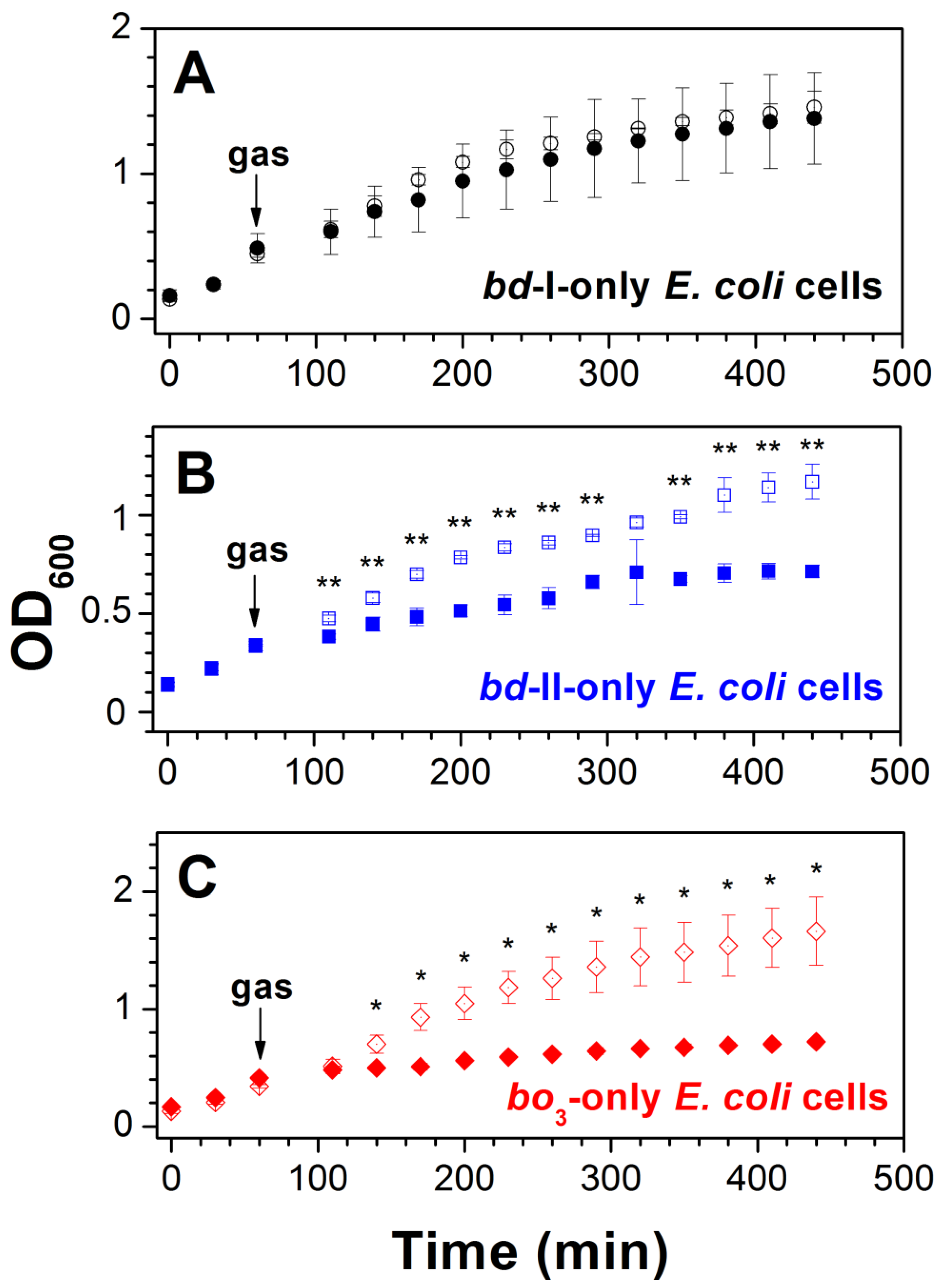
2.2. Effect of CO on E. coli Cell Growth
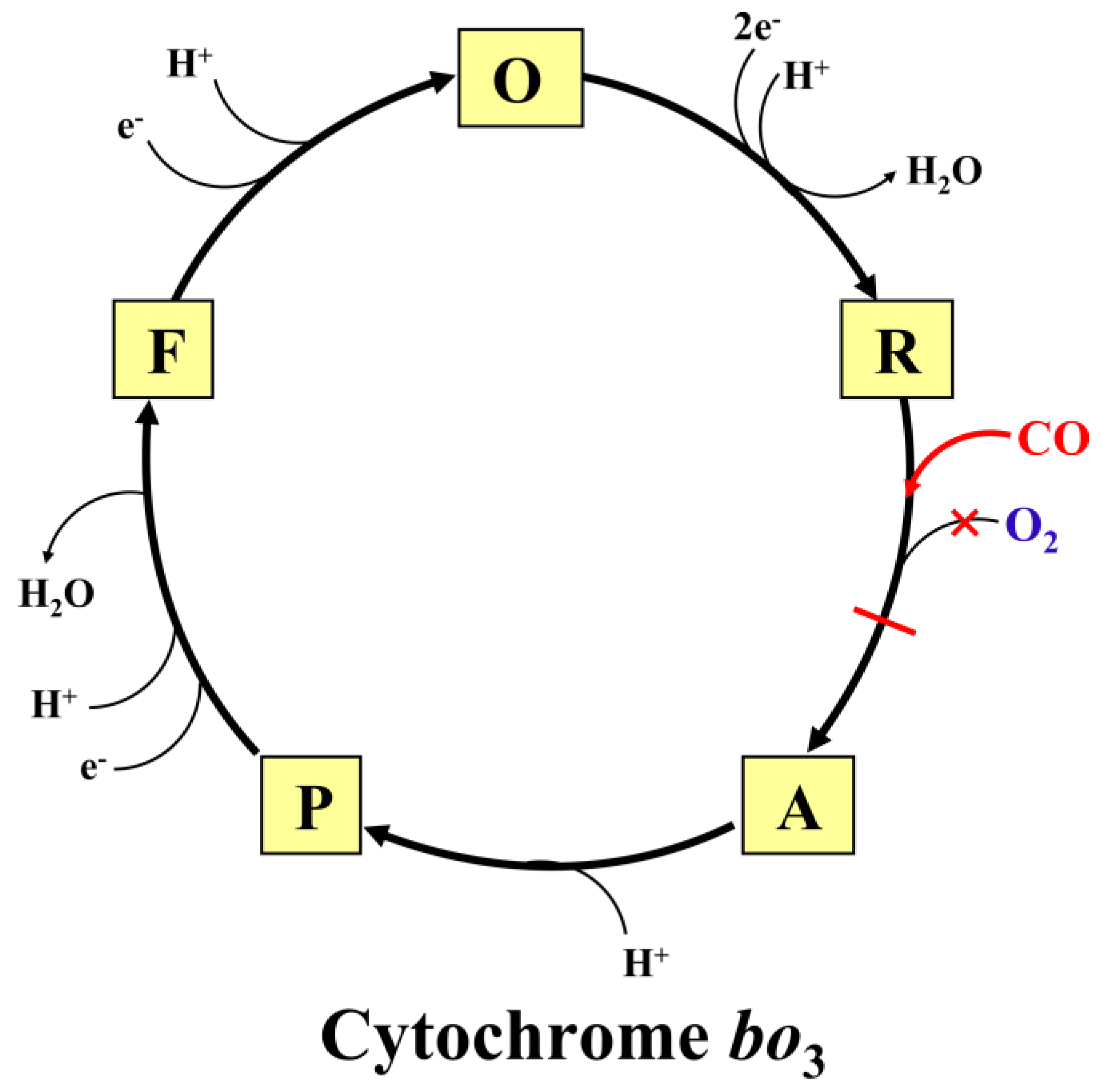
The observation that the degree of resistance of the O2 consumption process by bd-I-only E. coli cells to CO is quite high, as opposed to the high sensitivity for the gas displayed by bd-II-only and bo3-only cells, prompted us to examine whether cytochrome bd-I, beyond enabling aerobic respiration, promotes E. coli cell growth in the presence of CO. In order to determine the effect of CO on E. coli cell growth, we studied the growth of the three different respiratory mutant strains in the presence of either ~20% CO or ~20% N2 as a control. Following the addition of ~20% CO to the E. coli strain expressing cytochrome bd-I as the only terminal oxidase at 60 min after the start of growth in air, a minimal effect on cell growth was observed (Figure 7A). On the contrary, the growth of both bd-II-only and bo3-only strains was severely impaired over the same time window after the addition of ~20% CO, compared to the control with ~20% N2 (Figure 7B,C). Thus, these data suggest that, unlike cytochromes bd-II and bo3, the bd-I oxidase promotes E. coli growth in the presence of CO.

3. Discussion

We examined the effect of CO on the O2-dependent respiration of the E. coli respiratory mutants containing one of the three terminal oxidases. The experiments were performed at different concentrations of the enzyme substrate, O2. In all mutants tested, the inhibitory action of CO decreased with increasing [O2]. The fact that the degree of inhibition decreases as the substrate concentration increases clearly suggests that CO acts as a competitive inhibitor for the enzyme-catalyzed O2 reduction reaction under steady-state conditions. CO apparently competes with O2 for binding to a high-spin pentacoordinate ferrous heme in the enzyme’s active site. Previous spectroscopic studies showed that the redox-active group that, in the reduced state, is able to bind both O2 and CO is heme d in the bd-type oxidases and heme o3 in the bo3 oxidase [87,92,93]. For this reason, we think that in the present enzyme-inhibition experiments with E. coli cells, heme d and heme o3 are targets for CO in cytochromes bd-I/bd-II and cytochrome bo3, respectively.
We found that the bd-I oxidase is much less sensitive to CO than both the bd-II and bo3 oxidases, as confirmed by measuring the effect of CO on O2 consumption by both whole cells and isolated membranes of the respiratory mutants. Accordingly, the O2 consumption of wild-type cells displayed low CO sensitivity under conditions favoring the expression of a bd-type oxidase and high CO sensitivity when cytochrome bo3 was predominantly expressed. Consistently, cell growth proved to be almost unaffected by CO in an E. coli mutant strain expressing solely cytochrome bd-I, but was drastically inhibited after the addition of the gas to mutant strains expressing either cytochrome bo3 or cytochrome bd-II as the only terminal oxidase. These results are in disagreement with an earlier study according to which O2 consumption by the isolated cytochromes bd-I and bd-II is more sensitive to inhibition by CO than that by the isolated bo3 oxidase [87]. The measurement conditions, such as buffer, pH, and temperature, in [87] and in this work were the same, but the difference was the environment for the enzyme (detergent in [87] versus natural lipid bilayer in this study). Therefore, we assume that this inconsistency can be explained by differences in the protein environment which affect enzyme sensitivity towards CO. In this work, the oxidases were in vivo conditions, i.e., integrated into native bacterial membranes, whereas in [87], the enzymes were isolated and incorporated into detergent micelles. Indeed, more and more data are accumulating on how the lipid membrane environment can significantly affect the structure, function, and dynamics of various membrane proteins [94,95]. For instance, regarding terminal oxidases, it was shown that the membrane environment modulates the ligand-binding characteristics of the E. coli cytochrome bd-I [96]. According to another study, solubilization of the membrane-bound bovine cytochrome c oxidase leads to an increase in the binding affinity of the enzyme for cyanide by 100–1000 times [97]. In addition, it was reported that in E. coli membranes, the bd-I oxidase is apparently in a supercomplex with other membrane-bound respiratory enzymes [56]. Such protein–protein interactions could also modulate the sensitivity of cytochrome bd-I to CO.
We hypothesize that when the E. coli cytochromes are integrated into the native lipid bilayer, the binding affinities of the enzymes for CO and O2 are such that CO binding to heme d in the bd-I oxidase is outcompeted by O2 while the binding of the inhibitor to heme d in the bd-II oxidase or to heme o3 in the bo3 oxidase is not. The resistance of cytochrome bd-I to CO is in agreement with the extremely fast dissociation of CO from the oxidase (koff = 6.0 ± 0.2 s−1 for the fully reduced form of the CO-bound enzyme [69]). Such a high enzyme–ligand dissociation rate constant would indeed lead to the prompt restoration of respiration. The variation between the terminal oxidases in the affinity for the ligands may arise from differences in the structural organization of the O2-binding sites and their specific environment. Consistently, among the factors that can regulate the binding affinity of heme proteins for gaseous ligands are the chemical structure and geometry of the proximal axial ligand of the heme and its distal amino acid residues [98]. Distal amino acid residues may either stabilize the bound diatomic gaseous molecule via weak interactions, including electrostatic effects, hydrogen bonds, van der Waals interactions, and the hydrophobic effect, or, contrariwise, destabilize ligand binding by providing steric constraints [98,99].
It is of interest to mention global E. coli responses to CO. Transcription factor measurements and modeling showed that gene expression is significantly perturbed by CO, with major consequences for energy metabolism, iron homeostasis, and amino acid metabolism [86]. Genes encoding energy-transducing proteins are highly affected by CO via the global regulators, ArcA and FNR. CO inhibition of respiration results in over-reduction of the quinone pool; accumulation of the fermentation product, pyruvate; and enhanced expression of iron acquisition genes. Regarding the respiratory terminal oxidases, the transcriptomic analysis of wild-type E. coli exposed to CO gas revealed that under aerobic conditions there is a 5- to 10-fold decrease in the expression of the cyoABCDE operon, encoding cytochrome bo3. In contrast, there is a 4-fold increase in the expression of the cydABX operon, encoding cytochrome bd-I. Changes in the expression of the appCBX operon, encoding cytochrome bd-II oxidase, are slight [86]. The CO inhibition data reported here are consistent with the transcriptional response to CO in wild-type cells reported by Wareham et al. [86]. Indeed, it seems reasonable that following CO gas addition, the CO-sensitive bo3 oxidase is downregulated and the CO-insensitive bd-I oxidase is upregulated. The bd-II oxidase normally is not expressed under aerobic conditions, and there is no need for its upregulation after CO treatment since the enzyme is sensitive to CO.
The difference in susceptibility to CO inhibition between cytochromes bd-I and bo3 is indeed not unexpected since they belong to different superfamilies of terminal oxidases and, as reported before, also differ in resistance to other stressors, such as cyanide, sulfide, nitric oxide, and ammonia [70,73,77,81]. However, the fact that cytochrome bd-I is much more resistant to CO than cytochrome bd-II in respiring E. coli cells is surprising. In order to explain this phenomenon, the following structural differences between the two bd enzymes have to be noted. First, the two proteins differ in the number of subunits. The bd-I oxidase is composed of four subunits, namely CydA, CydB, CydX, and CydY, whereas the bd-II oxidase contains one fewer subunit, being composed of AppC, AppB, and AppX. Notably, the extra subunit in cytochrome bd-I, CydY, shields the high-spin pentacoordinate heme b595 from the lipid bilayer interface. This shielding prevents external ligands from accessing this potential ligand-binding site [64,65]. Consistently, heme b595 resistance to external ligand binding in cytochrome bd-I was previously identified in an MCD study [92]. Since cytochrome bd-II has no CydY, direct access of gaseous molecules from the membrane lipid environment is possible [66,67]. Therefore, in the bd-II oxidase, CO could possibly bind to the reduced heme b595 under turnover conditions. This binding would, in turn, inhibit the electron transfer from heme b558 to heme d that occurs through heme b595, leading to the inhibition of the enzyme-catalyzed O2 consumption. Second, it was reported that in the bd-II oxidase, the axial heme d ligand is His19 of the AppC subunit [66,67]. However, there is no consensus on the nature of the axial ligand of heme d in the bd-I oxidase. It is either His19 (of the CydA subunit, homolog to AppC) [64] or Glu99 of CydA [65]. Notably, His19 and Glu99 are located on opposite sides of the plane of the porphyrin macrocycle. If the axial heme d ligand in the bd-I and bd-II enzymes in bacterial cells is indeed different, this may also underlie the difference in sensitivity of the two proteins to CO. The fact that heme d in the bd-II oxidase has a much higher midpoint redox potential than that in the bd-I oxidase (+440 vs. +258 mV) [28]) would be consistent with the different nature of its axial ligand. Third, the bd-II protein incorporated into amphipols was shown to be mainly in the form of a dimer, while the bd-I enzyme exists only as a monomer [66]. This difference might also contribute to the observed difference in the sensitivity of these two bd oxidases to CO. In this regard, it is also worth noting that Cupriavidus necator H16, like E. coli and some other bacteria, has two different operons encoding cytochrome bd, cydA1B1 and cydA2B2. Interestingly, the expression of only one of the two (cydA2B2) seems to enable cell growth in the presence of CO under heterotrophic conditions [100]. The deletion of cydA2B2 had a detrimental effect on CO resistance, and plasmid-based expression of cydA1B1 did not improve CO tolerance [64]. These data are consistent with our observation that, of the two bd oxidases in E. coli cells, only cytochrome bd-I promotes aerobic respiration and growth in the presence of CO.
The possible molecular mechanisms of the inhibition of the catalytic activity of the E. coli cytochromes bo3, bd-II, and bd-I by CO are shown in Figure 8, Figure 9 and Figure 10, respectively (for more detail, see the legends to the figures).
In this work, we have used mutant strains, and this may present limitations, as mutations can potentially influence other processes in the cell that might go unnoticed and remain unaccounted for. However, the data on the wild-type cells and membranes are consistent with those obtained with the mutant strains. This gives us reason to believe that our conclusions are correct. As we mentioned above, the addition of CO triggers short-term alterations in the E. coli transcriptome [86], but a complete picture of the impact of CO on bacterial bioenergetics is lacking. Further studies are needed to shed light on long-term adaptation effects and fully uncover the extent of CO resistance in vivo.

4. Materials and Methods

4.1. Materials, E. coli Mutant Strains and Growth Conditions

The CO and N2 gases were purchased from Linde (Danbury, CT, USA) and Air Liquide (Air Liquide Italia Spa, Milano, Italy), respectively. Other chemicals were purchased from Merck KGaA (Darmstadt, Germany). A stock solution of CO or N2 was prepared by equilibrating degassed water with the pure gas at 1 atm and room temperature, yielding 1 mM CO and 0.7 mM N2 in solution. E. coli respiratory mutant strains TBE025 (MG1655 ΔcydB nuoB appB::kan), TBE026 (MG1655 ΔcydB nuoB cyoB::kan), and TBE037 (MG1655 ΔappB nuoB cyoB::kan), which respectively express cytochrome bo3, cytochrome bd-II, or cytochrome bd-I as the sole terminal oxidase, were used [77]. E. coli cultures were grown in Luria–Bertani (LB) medium supplemented with 30 μg/mL kanamycin, at 37 °C and 200 rpm. In the case of growth studies, after inoculation at an OD600 of 0.15 ± 0.03, the cells were grown in 50 mL rubber-stoppered flasks in 16 mL of air-equilibrated LB at 37 °C for 60 min; when OD600 reached 0.4 ± 0.08, they were bubbled for 30 s with either CO (pure gas at 1 atm and room temperature) or N2 (pure gas at 1 atm and room temperature) as a control to yield a final concentration of both gases of ~20%. The gas percentage was assessed by taking into account the different O2 concentrations, present in equal volumes of LB equilibrated with air or flushed with CO/N2 for 30 s, and calculated according to the following equation: Gas % = ([O2] air equilibrated − [O2] gas flushed)/[O2] air equilibrated × 100. [O2] air equilibrated and [O2] gas flushed are the O2 concentration values measured in the degassed chamber of a high-resolution respirometer (Oxygraph-2k, Oroboros Instruments GmbH, Innsbruck, Austria) of non-fluxed and fluxed E. coli cultures with CO or N2, respectively. The growth of the E. coli cultures was then monitored via a standard method using optical density measurements in an Eppendorf BioSpectrometer basic at 600 nm every 30 min. When the OD600 was above 1, cultures were diluted before reading.

4.2. Investigation of the Effect of CO on Respiration of Wild-Type E. coli Cells

To assess the effect of CO on the respiration of wild-type E. coli cells (strain MG1655), we investigated aerobic cultures in which a change in oxidase expression from cytochrome bo3 to the bd-type cytochromes is expected to occur during cell growth, following a progressive reduction in the availability of O2 in the medium, taking into account the striking difference between the oxidases in sensitivity to sodium sulfide, according to Forte et al. [77]. When cells grown in Luria–Bertani (LB) medium are assayed in an early growth phase of the culture (OD600 < 0.8), most of the respiration (50–70%) is sensitive to 50 μM sodium sulfide, indicating a prevalent expression of cytochrome bo3. In contrast, in a late growth phase of the culture (OD600 > 2.5), when oxygen is limiting, sodium sulfide causes only marginal effects on respiration, indicating a prevalent expression of the bd-type cytochromes.

4.3. Isolation of Membranes from E. coli Respiratory Mutants

To isolate membranes from E. coli respiratory mutants expressing only one terminal quinol oxidase (cytochrome bd-I or cytochrome bd-II or cytochrome bo3), cell cultures were grown in LB medium until an OD600 of about 2 was reached. The cells were then pelleted by centrifugation at 10,000 rpm for 10 min and washed twice in 20 mM TRIS pH 8.3 containing 0.5 mM EDTA and 5 mM MgSO4. The cells were resuspended in the same buffer containing 1 mg/mL lysozyme and incubated for two hours on ice. A spatula tip of DNAse and RNase was then added, and the cells were lysed by sonication. Cell debris was removed by centrifugation at 15,000 rpm for 10 min. Membrane fractions were collected and stored at −80 °C. The protein content was determined using the Bradford method, using the Bradford reagent (Sigma) with bovine serum albumin as standard.

4.4. Oxygraphic Measurements

Oxygraphic measurements were performed at 25 °C in 50 mM K/phosphate pH 7.0, using a high-resolution respirometer (Oxygraph-2k, Oroboros Instruments GmbH, Innsbruck, Austria) equipped with two 1.5-mL chambers. The O2 consumption of E. coli cells was followed with endogenous reductants. In the case of isolated membranes, O2 consumption was measured in the presence of 2.5 mM dithiothreitol (DTT) and 2.5 μM 2,3-dimethoxy-5-methyl-6-(3-methyl-2-butenyl)-1,4-benzoquinone (Q1), an artificial reducing couple specific for quinol oxidases. The catalytic O2-consuming activity was obtained by subtracting the non-enzymatic O2 consumption (Q1/DTT autoxidation in the absence of the membranes) from the O2 consumption rate measured after the addition of membranes.

4.5. Spectroscopic Measurements

UV–visible absorption spectra were recorded to estimate the amount of each terminal oxidase present in cell suspensions. For this purpose, an Agilent Cary 60 UV-Vis or a Varian Cary 300 Bio UV-Visible spectrophotometer was used. The amount of oxidase present in each strain was estimated from the dithionite-reduced-minus-ferricyanyde-oxidized difference absorption spectrum of sonicated cells using Δε561–580 of 21 mM−1 cm−1 (bd-I and bd-II) [92] and 16.3 mM−1 cm−1 (bo3) [88].

4.6. Data Analysis

Data analysis was carried out using software packages Origin v7.0 (OriginLab Corporation, Northampton, MA, USA) and GIM (Scientific Graphic Interactive Management System developed by A.L. Drachev in Lomonosov Moscow State University). The percentage inhibition of O2 consumption of cell suspensions (i%) was calculated using the equation i% = ((V0 − V0,i)/V0)·100, where V0 and V0,i are the initial rates in the absence and in the presence of the inhibitor (CO), respectively. The apparent IC50 values of cytochromes bd-II and bo3 for CO were estimated by plotting i% as a function of CO concentration added ([CO]0). The data were fitted to the standard hyperbolic equation i% = imax%·[CO]0/(IC50 + [CO]0) using a built-in approximation function (‘Hyperbola function’) in ‘Advanced Fitting tool’ in the Origin program. The imax% parameter is a theoretical maximum percent inhibition. The inhibition constants (Ki) of cytochromes bd-II and bo3 for CO were estimated by plotting, as a function of [O2]/Km(O2), the IC50 values measured at different O2 concentrations and fitting the data to the equation IC50 = (Ki·[O2]/Km(O2)) + Ki [91], assuming O2 competitive inhibition. The apparent Km(O2) values 2 µM (for cytochrome bd-II) and 6 µM (for cytochrome bo3) were taken from [90] and [70], respectively.
Statistical analyses were performed using an unpaired t-test for comparisons between two strains or conditions.

5. Conclusions

In this study, we show that of the two E. coli bd-type oxidases, only bd-I promotes growth in the presence of toxic concentrations of CO, giving the bacterium resistance to the gas. Our results are relevant for clarifying the discrepancies present in the literature on the effect of CO on the O2 consumption of E. coli oxidases and expanding the knowledge of cytochromes bd, a protein family of increasing interest due to their unique functional and structural features and their importance to pathogens. These findings are also important in the field of microbial physiology as they contribute to a better understanding of how different terminal complexes participate in the respiratory chain under various growth conditions. Moreover, they may provide a basis for biotechnological applications in which an increased bacterial resistance to CO is needed [100]. Finally, we believe that these findings could have a biomedical significance. These data should be taken into account when next-generation antimicrobials, whose mechanism of action is based on the release of CO that blocks the aerobic respiration and growth of pathogenic bacteria, are studied for potential use in medicine. Therapeutic CO delivery in humans and animals by such CO-releasing molecules would most likely be less efficient if the target pathogen possesses a terminal oxidase similar to the E. coli bd-I enzyme.

Supplementary Materials

The supporting information can be downloaded at: https://www.mdpi.com/article/10.3390/ijms25021277/s1.

Author Contributions

Conceptualization, V.B.B. and E.F.; methodology, M.R.N. and E.F.; formal analysis, V.B.B.; investigation, M.R.N. and E.F.; data curation, V.B.B., M.R.N. and E.F.; writing—original draft preparation, V.B.B., E.F. and M.R.N.; funding acquisition, E.F. All authors have read and agreed to the published version of the manuscript.

Funding

This work was funded by Sapienza grant number RP12117A8AA5B0E7 (to E.F.).

Institutional Review Board Statement

Not applicable.

Informed Consent Statement

Not applicable.

Data Availability Statement

The data presented in this study are contained in the article and the Supplementary Materials.

Acknowledgments

We thank M. Bekker (Amsterdam, the Netherlands) for the E. coli mutant strains used in this study.

Conflicts of Interest

The authors declare no conflicts of interest. The funders had no role in the design of the study; in the collection, analyses, or interpretation of data; in the writing of the manuscript; or in the decision to publish the results.

References

  1. Ingi, T.; Ronnett, G.V. Direct demonstration of a physiological role for carbon monoxide in olfactory receptor neurons. J. Neurosci. 1995, 15, 8214–8222. [Google Scholar] [CrossRef] [PubMed]
  2. Friebe, A.; Schultz, G.; Koesling, D. Sensitizing soluble guanylyl cyclase to become a highly CO-sensitive enzyme. Embo J. 1996, 15, 6863–6868. [Google Scholar] [CrossRef] [PubMed]
  3. Siow, R.C.; Sato, H.; Mann, G.E. Heme oxygenase-carbon monoxide signalling pathway in atherosclerosis: Anti-atherogenic actions of bilirubin and carbon monoxide? Cardiovasc. Res. 1999, 41, 385–394. [Google Scholar] [CrossRef] [PubMed]
  4. Boehning, D.; Snyder, S.H. Circadian rhythms. Carbon monoxide and clocks. Science 2002, 298, 2339–2340. [Google Scholar] [CrossRef] [PubMed]
  5. Morse, D.; Sethi, J.; Choi, A.M. Carbon monoxide-dependent signaling. Crit. Care Med. 2002, 30, S12–S17. [Google Scholar] [CrossRef] [PubMed]
  6. Ryter, S.W.; Otterbein, L.E.; Morse, D.; Choi, A.M. Heme oxygenase/carbon monoxide signaling pathways: Regulation and functional significance. Mol. Cell Biochem. 2002, 234-235, 249–263. [Google Scholar] [CrossRef]
  7. Bilban, M.; Haschemi, A.; Wegiel, B.; Chin, B.Y.; Wagner, O.; Otterbein, L.E. Heme oxygenase and carbon monoxide initiate homeostatic signaling. J. Mol. Med. (Berl.) 2008, 86, 267–279. [Google Scholar] [CrossRef]
  8. Piantadosi, C.A. Carbon monoxide, reactive oxygen signaling, and oxidative stress. Free Radic. Biol. Med. 2008, 45, 562–569. [Google Scholar] [CrossRef]
  9. Olson, K.R.; Donald, J.A. Nervous control of circulation-the role of gasotransmitters, NO, CO, and H2S. Acta Histochem. 2009, 111, 244–256. [Google Scholar] [CrossRef]
  10. Wang, X.M.; Kim, H.P.; Nakahira, K.; Ryter, S.W.; Choi, A.M. The heme oxygenase-1/carbon monoxide pathway suppresses TLR4 signaling by regulating the interaction of TLR4 with caveolin-1. J. Immunol. 2009, 182, 3809–3818. [Google Scholar] [CrossRef]
  11. Farrugia, G.; Szurszewski, J.H. Carbon monoxide, hydrogen sulfide, and nitric oxide as signaling molecules in the gastrointestinal tract. Gastroenterology 2014, 147, 303–313. [Google Scholar] [CrossRef] [PubMed]
  12. Choi, Y.K.; Maki, T.; Mandeville, E.T.; Koh, S.H.; Hayakawa, K.; Arai, K.; Kim, Y.M.; Whalen, M.J.; Xing, C.; Wang, X.; et al. Dual effects of carbon monoxide on pericytes and neurogenesis in traumatic brain injury. Nat. Med. 2016, 22, 1335–1341. [Google Scholar] [CrossRef] [PubMed]
  13. Wood, H. Traumatic brain injury: Carbon monoxide-a potential therapy for traumatic brain injury? Nat. Rev. Neurol. 2016, 12, 615. [Google Scholar] [CrossRef] [PubMed]
  14. Kim, H.J.; Joe, Y.; Kim, S.K.; Park, S.U.; Park, J.; Chen, Y.; Kim, J.; Ryu, J.; Cho, G.J.; Surh, Y.J.; et al. Carbon monoxide protects against hepatic steatosis in mice by inducing sestrin-2 via the PERK-eIF2alpha-ATF4 pathway. Free Radic. Biol. Med. 2017, 110, 81–91. [Google Scholar] [CrossRef] [PubMed]
  15. Klemz, R.; Reischl, S.; Wallach, T.; Witte, N.; Jurchott, K.; Klemz, S.; Lang, V.; Lorenzen, S.; Knauer, M.; Heidenreich, S.; et al. Reciprocal regulation of carbon monoxide metabolism and the circadian clock. Nat. Struct. Mol. Biol. 2017, 24, 15–22. [Google Scholar] [CrossRef] [PubMed]
  16. Correa-Costa, M.; Gallo, D.; Csizmadia, E.; Gomperts, E.; Lieberum, J.L.; Hauser, C.J.; Ji, X.; Wang, B.; Camara, N.O.S.; Robson, S.C.; et al. Carbon monoxide protects the kidney through the central circadian clock and CD39. Proc. Natl. Acad. Sci. USA 2018, 115, E2302–E2310. [Google Scholar] [CrossRef]
  17. Joe, Y.; Kim, S.; Kim, H.J.; Park, J.; Chen, Y.; Park, H.J.; Jekal, S.J.; Ryter, S.W.; Kim, U.H.; Chung, H.T. FGF21 induced by carbon monoxide mediates metabolic homeostasis via the PERK/ATF4 pathway. Faseb J. 2018, 32, 2630–2643. [Google Scholar] [CrossRef]
  18. Minegishi, S.; Sagami, I.; Negi, S.; Kano, K.; Kitagishi, H. Circadian clock disruption by selective removal of endogenous carbon monoxide. Sci. Rep. 2018, 8, 11996. [Google Scholar] [CrossRef]
  19. Chen, Y.; Joe, Y.; Park, J.; Song, H.C.; Kim, U.H.; Chung, H.T. Carbon monoxide induces the assembly of stress granule through the integrated stress response. Biochem. Biophys. Res. Commun. 2019, 512, 289–294. [Google Scholar] [CrossRef]
  20. Rahman, F.U.; Park, D.R.; Joe, Y.; Jang, K.Y.; Chung, H.T.; Kim, U.H. Critical Roles of Carbon Monoxide and Nitric Oxide in Ca(2+) Signaling for Insulin Secretion in Pancreatic Islets. Antioxid. Redox Signal. 2019, 30, 560–576. [Google Scholar] [CrossRef]
  21. Stucki, D.; Steinhausen, J.; Westhoff, P.; Krahl, H.; Brilhaus, D.; Massenberg, A.; Weber, A.P.M.; Reichert, A.S.; Brenneisen, P.; Stahl, W. Endogenous Carbon Monoxide Signaling Modulates Mitochondrial Function and Intracellular Glucose Utilization: Impact of the Heme Oxygenase Substrate Hemin. Antioxidants 2020, 9, 652. [Google Scholar] [CrossRef] [PubMed]
  22. Park, J.; Zeng, J.S.; Sahasrabudhe, A.; Jin, K.; Fink, Y.; Manthiram, K.; Anikeeva, P. Electrochemical Modulation of Carbon Monoxide-Mediated Cell Signaling. Angew. Chem. Int. Ed. Engl. 2021, 60, 20325–20330. [Google Scholar] [CrossRef] [PubMed]
  23. Yuan, Z.; De La Cruz, L.K.; Yang, X.; Wang, B. Carbon monoxide signaling: Examining its engagement with various molecular targets in the context of binding affinity, concentration, and biologic response. Pharmacol. Rev. 2022, 74, 823–873. [Google Scholar] [CrossRef] [PubMed]
  24. Hopper, C.P.; De La Cruz, L.K.; Lyles, K.V.; Wareham, L.K.; Gilbert, J.A.; Eichenbaum, Z.; Magierowski, M.; Poole, R.K.; Wollborn, J.; Wang, B. Role of carbon monoxide in host-gut microbiome communication. Chem. Rev. 2020, 120, 13273–13311. [Google Scholar] [CrossRef] [PubMed]
  25. Dent, M.R.; Weaver, B.R.; Roberts, M.G.; Burstyn, J.N. Carbon monoxide-sensing transcription factors: Regulators of microbial carbon monoxide oxidation pathway gene expression. J. Bacteriol. 2023, 205, e0033222. [Google Scholar] [CrossRef] [PubMed]
  26. Tavares, A.F.; Parente, M.R.; Justino, M.C.; Oleastro, M.; Nobre, L.S.; Saraiva, L.M. The bactericidal activity of carbon monoxide-releasing molecules against Helicobacter pylori. PLoS ONE 2013, 8, e83157. [Google Scholar] [CrossRef]
  27. Desmard, M.; Davidge, K.S.; Bouvet, O.; Morin, D.; Roux, D.; Foresti, R.; Ricard, J.D.; Denamur, E.; Poole, R.K.; Montravers, P.; et al. A carbon monoxide-releasing molecule (CORM-3) exerts bactericidal activity against Pseudomonas aeruginosa and improves survival in an animal model of bacteraemia. Faseb J. 2009, 23, 1023–1031. [Google Scholar] [CrossRef]
  28. Davidge, K.S.; Sanguinetti, G.; Yee, C.H.; Cox, A.G.; McLeod, C.W.; Monk, C.E.; Mann, B.E.; Motterlini, R.; Poole, R.K. Carbon monoxide-releasing antibacterial molecules target respiration and global transcriptional regulators. J. Biol. Chem. 2009, 284, 4516–4524. [Google Scholar] [CrossRef]
  29. Wareham, L.K.; McLean, S.; Begg, R.; Rana, N.; Ali, S.; Kendall, J.J.; Sanguinetti, G.; Mann, B.E.; Poole, R.K. The broad-spectrum antimicrobial potential of [Mn(CO)4(S2CNMe(CH2CO2H))], a water-soluble CO-releasing molecule (CORM-401): Intracellular accumulation, transcriptomic and statistical analyses, and membrane polarization. Antioxid. Redox. Signal. 2018, 28, 1286–1308. [Google Scholar] [CrossRef]
  30. Southam, H.M.; Smith, T.W.; Lyon, R.L.; Liao, C.; Trevitt, C.R.; Middlemiss, L.A.; Cox, F.L.; Chapman, J.A.; El-Khamisy, S.F.; Hippler, M.; et al. A thiol-reactive Ru(II) ion, not CO release, underlies the potent antimicrobial and cytotoxic properties of CO-releasing molecule-3. Redox Biol. 2018, 18, 114–123. [Google Scholar] [CrossRef]
  31. Sousa, F.L.; Alves, R.J.; Ribeiro, M.A.; Pereira-Leal, J.B.; Teixeira, M.; Pereira, M.M. The superfamily of heme-copper oxygen reductases: Types and evolutionary considerations. Biochim. Biophys. Acta 2012, 1817, 629–637. [Google Scholar] [CrossRef] [PubMed]
  32. Murali, R.; Gennis, R.B.; Hemp, J. Evolution of the cytochrome bd oxygen reductase superfamily and the function of CydAA’ in Archaea. ISME J. 2021, 15, 3534–3548. [Google Scholar] [CrossRef] [PubMed]
  33. Siletsky, S.A. Investigation of the Mechanism of Membrane Potential Generation by Heme-Copper Respiratory Oxidases in a Real Time Mode. Biochemistry 2023, 88, 1513–1527. [Google Scholar] [CrossRef] [PubMed]
  34. Safari, C.; Ghosh, S.; Andersson, R.; Johannesson, J.; Bath, P.; Uwangue, O.; Dahl, P.; Zoric, D.; Sandelin, E.; Vallejos, A.; et al. Time-resolved serial crystallography to track the dynamics of carbon monoxide in the active site of cytochrome c oxidase. Sci. Adv. 2023, 9, eadh4179. [Google Scholar] [CrossRef] [PubMed]
  35. Moe, A.; Dimogkioka, A.R.; Rapaport, D.; Ojemyr, L.N.; Brzezinski, P. Structure and function of the S. pombe III-IV-cyt c supercomplex. Proc. Natl. Acad. Sci. USA 2023, 120, e2307697120. [Google Scholar] [CrossRef] [PubMed]
  36. Khalfaoui-Hassani, B.; Blaby-Haas, C.E.; Verissimo, A.; Daldal, F. The Escherichia coli MFS-type transporter genes yhjE, ydiM, and yfcJ are required to produce an active bo3 quinol oxidase. PLoS ONE 2023, 18, e0293015. [Google Scholar] [CrossRef]
  37. Shimada, A.; Etoh, Y.; Kitoh-Fujisawa, R.; Sasaki, A.; Shinzawa-Itoh, K.; Hiromoto, T.; Yamashita, E.; Muramoto, K.; Tsukihara, T.; Yoshikawa, S. X-ray structures of catalytic intermediates of cytochrome c oxidase provide insights into its O2 activation and unidirectional proton-pump mechanisms. J. Biol. Chem. 2020, 295, 5818–5833. [Google Scholar] [CrossRef]
  38. Shimada, A.; Hara, F.; Shinzawa-Itoh, K.; Kanehisa, N.; Yamashita, E.; Muramoto, K.; Tsukihara, T.; Yoshikawa, S. Critical roles of the CuB site in efficient proton pumping as revealed by crystal structures of mammalian cytochrome c oxidase catalytic intermediates. J. Biol. Chem. 2021, 100967. [Google Scholar] [CrossRef]
  39. Shimada, A.; Baba, J.; Nagao, S.; Shinzawa-Itoh, K.; Yamashita, E.; Muramoto, K.; Tsukihara, T.; Yoshikawa, S. Crystallographic cyanide-probing for cytochrome c oxidase reveals structural bases suggesting that a putative proton transfer H-pathway pumps protons. J. Biol. Chem. 2023, 299, 105277. [Google Scholar] [CrossRef]
  40. Panda, S.; Phan, H.; Karlin, K.D. Heme-copper and Heme O2-derived synthetic (bioinorganic) chemistry toward an understanding of cytochrome c oxidase dioxygen chemistry. J. Inorg. Biochem. 2023, 249, 112367. [Google Scholar] [CrossRef]
  41. Ishigami, I.; Sierra, R.G.; Su, Z.; Peck, A.; Wang, C.; Poitevin, F.; Lisova, S.; Hayes, B.; Moss, F.R., 3rd; Boutet, S.; et al. Structural insights into functional properties of the oxidized form of cytochrome c oxidase. Nat. Commun. 2023, 14, 5752. [Google Scholar] [CrossRef] [PubMed]
  42. Verameyenka, K.G.; Naumouskaya, V.A.; Maximova, N.P. Cytochrome c oxidase is one of the key enzymes providing the ability to produce phenazines in Pseudomonas chlororaphis subsp. aurantiaca. World J. Microbiol. Biotechnol. 2023, 39, 279. [Google Scholar] [CrossRef] [PubMed]
  43. Guo, Y.; Karimullina, E.; Emde, T.; Otwinowski, Z.; Borek, D.; Savchenko, A. Monomer and dimer structures of cytochrome bo3 ubiquinol oxidase from Escherichia coli. Protein Sci. 2023, e4616. [Google Scholar] [CrossRef] [PubMed]
  44. Li, J.; Han, L.; Vallese, F.; Ding, Z.; Choi, S.K.; Hong, S.; Luo, Y.; Liu, B.; Chan, C.K.; Tajkhorshid, E.; et al. Cryo-EM structures of Escherichia coli cytochrome bo3 reveal bound phospholipids and ubiquinone-8 in a dynamic substrate binding site. Proc. Natl. Acad. Sci. USA 2021, 118. [Google Scholar] [CrossRef]
  45. Jose, A.; Schaefer, A.W.; Roveda, A.C., Jr.; Transue, W.J.; Choi, S.K.; Ding, Z.; Gennis, R.B.; Solomon, E.I. The three-spin intermediate at the O-O cleavage and proton-pumping junction in heme-Cu oxidases. Science 2021, 373, 1225–1229. [Google Scholar] [CrossRef] [PubMed]
  46. Asseri, A.H.; Godoy-Hernandez, A.; Goojani, H.G.; Lill, H.; Sakamoto, J.; McMillan, D.G.G.; Bald, D. Cardiolipin enhances the enzymatic activity of cytochrome bd and cytochrome bo3 solubilized in dodecyl-maltoside. Sci. Rep. 2021, 11, 8006. [Google Scholar] [CrossRef]
  47. Kaila, V.R.I.; Wikstrom, M. Architecture of bacterial respiratory chains. Nat. Rev. Microbiol. 2021, 19, 319–330. [Google Scholar] [CrossRef]
  48. Wikstrom, M.; Springett, R. Thermodynamic efficiency, reversibility, and degree of coupling in energy conservation by the mitochondrial respiratory chain. Commun. Biol. 2020, 3, 451. [Google Scholar] [CrossRef]
  49. Di Trani, J.M.; Gheorghita, A.A.; Turner, M.; Brzezinski, P.; Adelroth, P.; Vahidi, S.; Howell, P.L.; Rubinstein, J.L. Structure of the bc1-cbb3 respiratory supercomplex from Pseudomonas aeruginosa. Proc. Natl. Acad. Sci. USA 2023, 120, e2307093120. [Google Scholar] [CrossRef]
  50. Zhang, L.; Dong, T.; Yang, J.; Hao, S.; Sun, Z.; Peng, Y. Anammox Coupled with Photocatalyst for Enhanced Nitrogen Removal and the Activated Aerobic Respiration of Anammox Bacteria Based on cbb3-Type Cytochrome c Oxidase. Environ. Sci. Technol. 2023, 57, 17910–17919. [Google Scholar] [CrossRef]
  51. Yang, X.; Liu, S.; Yin, Z.; Chen, M.; Song, J.; Li, P.; Yang, L. New insights into the proton pumping mechanism of ba3 cytochrome c oxidase: The functions of key residues and water. Phys. Chem. Chem. Phys. 2023, 25, 25105–25115. [Google Scholar] [CrossRef] [PubMed]
  52. Noodleman, L.; Gotz, A.W.; Han Du, W.G.; Hunsicker-Wang, L. Reaction pathways, proton transfer, and proton pumping in ba3 class cytochrome c oxidase: Perspectives from DFT quantum chemistry and molecular dynamics. Front. Chem. 2023, 11, 1186022. [Google Scholar] [CrossRef] [PubMed]
  53. Wikstrom, M.; Pecorilla, C.; Sharma, V. The mitochondrial respiratory chain. Enzymes 2023, 54, 15–36. [Google Scholar] [CrossRef] [PubMed]
  54. Wikstrom, M.; Gennis, R.B.; Rich, P.R. Structures of the intermediates in the catalytic cycle of mitochondrial cytochrome c oxidase. Biochim. Biophys. Acta. Bioenerg. 2023, 1864, 148933. [Google Scholar] [CrossRef] [PubMed]
  55. Poole, R.K.; Cook, G.M. Redundancy of aerobic respiratory chains in bacteria? Routes, reasons and regulation. Adv. Microb. Physiol. 2000, 43, 165–224. [Google Scholar] [CrossRef] [PubMed]
  56. Melo, A.M.; Teixeira, M. Supramolecular organization of bacterial aerobic respiratory chains: From cells and back. Biochim. Biophys. Acta 2016, 1857, 190–197. [Google Scholar] [CrossRef] [PubMed]
  57. Melin, F.; Sabuncu, S.; Choi, S.K.; Leprince, A.; Gennis, R.B.; Hellwig, P. Role of the tightly bound quinone for the oxygen reaction of cytochrome bo3 oxidase from Escherichia coli. FEBS Lett. 2018, 592, 3380–3387. [Google Scholar] [CrossRef]
  58. Borisov, V.B.; Forte, E. Impact of hydrogen sulfide on mitochondrial and bacterial bioenergetics. Int. J. Mol. Sci. 2021, 22, 12688. [Google Scholar] [CrossRef]
  59. Borisov, V.B.; Siletsky, S.A.; Paiardini, A.; Hoogewijs, D.; Forte, E.; Giuffre, A.; Poole, R.K. Bacterial oxidases of the cytochrome bd family: Redox enzymes of unique structure, function and utility as drug targets. Antioxid. Redox Signal. 2021, 34, 1280–1318. [Google Scholar] [CrossRef]
  60. Rolfe, M.D.; Ter Beek, A.; Graham, A.I.; Trotter, E.W.; Asif, H.M.; Sanguinetti, G.; de Mattos, J.T.; Poole, R.K.; Green, J. Transcript profiling and inference of Escherichia coli K-12 ArcA activity across the range of physiologically relevant oxygen concentrations. J. Biol. Chem. 2011, 286, 10147–10154. [Google Scholar] [CrossRef]
  61. Alexeeva, S.; Hellingwerf, K.; Teixeira de Mattos, M.J. Quantitative assessment of oxygen availability: Perceived aerobiosis and its effect on flux distribution in the respiratory chain of Escherichia coli. J. Bacteriol. 2002, 184, 1402–1406. [Google Scholar] [CrossRef] [PubMed]
  62. Trotter, E.W.; Rolfe, M.D.; Hounslow, A.M.; Craven, C.J.; Williamson, M.P.; Sanguinetti, G.; Poole, R.K.; Green, J. Reprogramming of Escherichia coli K-12 metabolism during the initial phase of transition from an anaerobic to a micro-aerobic environment. PLoS ONE 2011, 6, e25501. [Google Scholar] [CrossRef] [PubMed]
  63. Abramson, J.; Riistama, S.; Larsson, G.; Jasaitis, A.; Svensson-Ek, M.; Laakkonen, L.; Puustinen, A.; Iwata, S.; Wikstrom, M. The structure of the ubiquinol oxidase from Escherichia coli and its ubiquinone binding site. Nat. Struct. Biol. 2000, 7, 910–917. [Google Scholar] [CrossRef] [PubMed]
  64. Safarian, S.; Hahn, A.; Mills, D.J.; Radloff, M.; Eisinger, M.L.; Nikolaev, A.; Meier-Credo, J.; Melin, F.; Miyoshi, H.; Gennis, R.B.; et al. Active site rearrangement and structural divergence in prokaryotic respiratory oxidases. Science 2019, 366, 100–104. [Google Scholar] [CrossRef] [PubMed]
  65. Thesseling, A.; Rasmussen, T.; Burschel, S.; Wohlwend, D.; Kagi, J.; Muller, R.; Bottcher, B.; Friedrich, T. Homologous bd oxidases share the same architecture but differ in mechanism. Nat. Commun. 2019, 10, 5138. [Google Scholar] [CrossRef] [PubMed]
  66. Grauel, A.; Kagi, J.; Rasmussen, T.; Makarchuk, I.; Oppermann, S.; Moumbock, A.F.A.; Wohlwend, D.; Muller, R.; Melin, F.; Gunther, S.; et al. Structure of Escherichia coli cytochrome bd-II type oxidase with bound aurachin D. Nat. Commun. 2021, 12, 6498. [Google Scholar] [CrossRef] [PubMed]
  67. Grund, T.N.; Radloff, M.; Wu, D.; Goojani, H.G.; Witte, L.F.; Josting, W.; Buschmann, S.; Muller, H.; Elamri, I.; Welsch, S.; et al. Mechanistic and structural diversity between cytochrome bd isoforms of Escherichia coli. Proc. Natl. Acad. Sci. USA 2021, 118, e2114013118. [Google Scholar] [CrossRef]
  68. Samukaite Bubniene, U.; Zukauskas, S.; Ratautaite, V.; Vilkiene, M.; Mockeviciene, I.; Liustrovaite, V.; Drobysh, M.; Lisauskas, A.; Ramanavicius, S.; Ramanavicius, A. Assessment of cytochrome c and chlorophyll a as natural redox mediators for enzymatic biofuel cells powered by glucose. Energies 2022, 15, 6838. [Google Scholar] [CrossRef]
  69. Borisov, V.B.; Forte, E.; Sarti, P.; Brunori, M.; Konstantinov, A.A.; Giuffre, A. Redox control of fast ligand dissociation from Escherichia coli cytochrome bd. Biochem. Biophys. Res. Commun. 2007, 355, 97–102. [Google Scholar] [CrossRef]
  70. Mason, M.G.; Shepherd, M.; Nicholls, P.; Dobbin, P.S.; Dodsworth, K.S.; Poole, R.K.; Cooper, C.E. Cytochrome bd confers nitric oxide resistance to Escherichia coli. Nat. Chem. Biol. 2009, 5, 94–96. [Google Scholar] [CrossRef]
  71. Shepherd, M.; Achard, M.E.; Idris, A.; Totsika, M.; Phan, M.D.; Peters, K.M.; Sarkar, S.; Ribeiro, C.A.; Holyoake, L.V.; Ladakis, D.; et al. The cytochrome bd-I respiratory oxidase augments survival of multidrug-resistant Escherichia coli during infection. Sci. Rep. 2016, 6, 35285. [Google Scholar] [CrossRef] [PubMed]
  72. Borisov, V.B.; Forte, E.; Siletsky, S.A.; Sarti, P.; Giuffre, A. Cytochrome bd from Escherichia coli catalyzes peroxynitrite decomposition. Biochim. Biophys. Acta 2015, 1847, 182–188. [Google Scholar] [CrossRef] [PubMed]
  73. Forte, E.; Siletsky, S.A.; Borisov, V.B. In Escherichia coli ammonia inhibits cytochrome bo3 but activates cytochrome bd-I. Antioxidants 2021, 10, 13. [Google Scholar] [CrossRef] [PubMed]
  74. Sarti, P.; Giuffre, A.; Forte, E.; Mastronicola, D.; Barone, M.C.; Brunori, M. Nitric oxide and cytochrome c oxidase: Mechanisms of inhibition and NO degradation. Biochem. Biophys. Res. Commun. 2000, 274, 183–187. [Google Scholar] [CrossRef] [PubMed]
  75. Borisov, V.B.; Forte, E.; Sarti, P.; Brunori, M.; Konstantinov, A.A.; Giuffre, A. Nitric oxide reacts with the ferryl-oxo catalytic intermediate of the CuB-lacking cytochrome bd terminal oxidase. FEBS Lett. 2006, 580, 4823–4826. [Google Scholar] [CrossRef] [PubMed]
  76. Borisov, V.B.; Forte, E. Bioenergetics and reactive nitrogen species in bacteria. Int. J. Mol. Sci. 2022, 23, 7321. [Google Scholar] [CrossRef] [PubMed]
  77. Forte, E.; Borisov, V.B.; Falabella, M.; Colaco, H.G.; Tinajero-Trejo, M.; Poole, R.K.; Vicente, J.B.; Sarti, P.; Giuffre, A. The terminal oxidase cytochrome bd promotes sulfide-resistant bacterial respiration and growth. Sci. Rep. 2016, 6, 23788. [Google Scholar] [CrossRef]
  78. Borisov, V.B.; Forte, E.; Davletshin, A.; Mastronicola, D.; Sarti, P.; Giuffre, A. Cytochrome bd oxidase from Escherichia coli displays high catalase activity: An additional defense against oxidative stress. FEBS Lett. 2013, 587, 2214–2218. [Google Scholar] [CrossRef]
  79. Al-Attar, S.; Yu, Y.; Pinkse, M.; Hoeser, J.; Friedrich, T.; Bald, D.; de Vries, S. Cytochrome bd displays significant quinol peroxidase activity. Sci. Rep. 2016, 6, 27631. [Google Scholar] [CrossRef]
  80. Forte, E.; Nastasi, M.R.; Borisov, V.B. Preparations of terminal oxidase cytochrome bd-II isolated from Escherichia coli reveal significant hydrogen peroxide scavenging activity. Biochemistry 2022, 87, 720–730. [Google Scholar] [CrossRef]
  81. Korshunov, S.; Imlay, K.R.; Imlay, J.A. The cytochrome bd oxidase of Escherichia coli prevents respiratory inhibition by endogenous and exogenous hydrogen sulfide. Mol. Microbiol. 2016, 101, 62–77. [Google Scholar] [CrossRef] [PubMed]
  82. Harikishore, A.; Mathiyazakan, V.; Pethe, K.; Gruber, G. Novel targets and inhibitors of the Mycobacterium tuberculosis cytochrome bd oxidase to foster anti-tuberculosis drug discovery. Expert Opin. Drug Discov. 2023, 18, 917–927. [Google Scholar] [CrossRef] [PubMed]
  83. Bayly, K.; Cordero, P.R.F.; Kropp, A.; Huang, C.; Schittenhelm, R.B.; Grinter, R.; Greening, C. Mycobacteria tolerate carbon monoxide by remodeling their respiratory chain. mSystems 2021, 6, e01292-20. [Google Scholar] [CrossRef] [PubMed]
  84. Scharn, C.R.; Collins, A.C.; Nair, V.R.; Stamm, C.E.; Marciano, D.K.; Graviss, E.A.; Shiloh, M.U. Heme oxygenase-1 regulates inflammation and mycobacterial survival in human macrophages during Mycobacterium tuberculosis infection. J. Immunol. 2016, 196, 4641–4649. [Google Scholar] [CrossRef] [PubMed]
  85. Wegiel, B.; Larsen, R.; Gallo, D.; Chin, B.Y.; Harris, C.; Mannam, P.; Kaczmarek, E.; Lee, P.J.; Zuckerbraun, B.S.; Flavell, R.; et al. Macrophages sense and kill bacteria through carbon monoxide-dependent inflammasome activation. J. Clin. Invest. 2014, 124, 4926–4940. [Google Scholar] [CrossRef] [PubMed]
  86. Wareham, L.K.; Begg, R.; Jesse, H.E.; Van Beilen, J.W.; Ali, S.; Svistunenko, D.; McLean, S.; Hellingwerf, K.J.; Sanguinetti, G.; Poole, R.K. Carbon monoxide gas is not inert, but global, in its consequences for bacterial gene expression, iron acquisition, and antibiotic resistance. Antioxid. Redox. Signal. 2016, 24, 1013–1028. [Google Scholar] [CrossRef] [PubMed]
  87. Forte, E.; Borisov, V.B.; Siletsky, S.A.; Petrosino, M.; Giuffre, A. In the respiratory chain of Escherichia coli cytochromes bd-I and bd-II are more sensitive to carbon monoxide inhibition than cytochrome bo3. Biochim. Biophys. Acta Bioenerg. 2019, 1860, 148088. [Google Scholar] [CrossRef]
  88. Nastasi, M.R.; Borisov, V.B.; Forte, E. The terminal oxidase cytochrome bd-I confers carbon monoxide resistance to Escherichia coli cells. J. Inorg. Biochem. 2023, 247, 112341. [Google Scholar] [CrossRef]
  89. Kalia, N.P.; Singh, S.; Hards, K.; Cheung, C.Y.; Sviriaeva, E.; Banaei-Esfahani, A.; Aebersold, R.; Berney, M.; Cook, G.M.; Pethe, K.M. Tuberculosis relies on trace oxygen to maintain energy homeostasis and survive in hypoxic environments. Cell Rep. 2023, 42, 112444. [Google Scholar] [CrossRef]
  90. Bekker, M.; de Vries, S.; Ter Beek, A.; Hellingwerf, K.J.; de Mattos, M.J. Respiration of Escherichia coli can be fully uncoupled via the nonelectrogenic terminal cytochrome bd-II oxidase. J. Bacteriol. 2009, 191, 5510–5517. [Google Scholar] [CrossRef]
  91. Cheng, Y.; Prusoff, W.H. Relationship between the inhibition constant (KI) and the concentration of inhibitor which causes 50 per cent inhibition (I50) of an enzymatic reaction. Biochem. Pharmacol. 1973, 22, 3099–3108. [Google Scholar] [CrossRef] [PubMed]
  92. Borisov, V.; Arutyunyan, A.M.; Osborne, J.P.; Gennis, R.B.; Konstantinov, A.A. Magnetic circular dichroism used to examine the interaction of Escherichia coli cytochrome bd with ligands. Biochemistry 1999, 38, 740–750. [Google Scholar] [CrossRef] [PubMed]
  93. Puustinen, A.; Wikstrom, M. The heme groups of cytochrome o from Escherichia coli. Proc. Natl. Acad. Sci. USA 1991, 88, 6122–6126. [Google Scholar] [CrossRef] [PubMed]
  94. Corradi, V.; Sejdiu, B.I.; Mesa-Galloso, H.; Abdizadeh, H.; Noskov, S.Y.; Marrink, S.J.; Tieleman, D.P. Emerging diversity in lipid-protein interactions. Chem. Rev. 2019, 119, 5775–5848. [Google Scholar] [CrossRef] [PubMed]
  95. Cournia, Z.; Allen, T.W.; Andricioaei, I.; Antonny, B.; Baum, D.; Brannigan, G.; Buchete, N.V.; Deckman, J.T.; Delemotte, L.; Del Val, C.; et al. Membrane protein structure, function, and dynamics: A perspective from experiments and theory. J. Membr. Biol. 2015, 248, 611–640. [Google Scholar] [CrossRef]
  96. Borisov, V.B. Effect of membrane environment on ligand-binding properties of the terminal oxidase cytochrome bd-I from Escherichia coli. Biochemistry 2020, 85, 1603–1612. [Google Scholar] [CrossRef]
  97. Andreev, I.M.; Konstantinov, A.A. Reaction of oxidized cytochrome oxidase with cyanide. Effects of pH, cytochrome c and membrane environment. Bioorg. Khim. (in Russian) 1983, 9, 216–227. [Google Scholar]
  98. Tsai, A.L.; Berka, V.; Martin, E.; Olson, J.S. A "sliding scale rule" for selectivity among NO, CO, and O2 by heme protein sensors. Biochemistry 2012, 51, 172–186. [Google Scholar] [CrossRef]
  99. Bartlett, G.J.; Newberry, R.W.; VanVeller, B.; Raines, R.T.; Woolfson, D.N. Interplay of hydrogen bonds and n-->pi* interactions in proteins. J. Am. Chem. Soc. 2013, 135, 18682–18688. [Google Scholar] [CrossRef]
  100. Wickham-Smith, C.; Malys, N.; Winzer, K. Improving carbon monoxide tolerance of Cupriavidus necator H16 through adaptive laboratory evolution. Front. Bioeng. Biotechnol. 2023, 11, 1178536. [Google Scholar] [CrossRef]
  101. Proshlyakov, D.A.; Pressler, M.A.; DeMaso, C.; Leykam, J.F.; DeWitt, D.L.; Babcock, G.T. Oxygen activation and reduction in respiration: Involvement of redox-active tyrosine 244. Science 2000, 290, 1588–1591. [Google Scholar] [CrossRef] [PubMed]
  102. Paulus, A.; Rossius, S.G.; Dijk, M.; de Vries, S. Oxoferryl-porphyrin radical catalytic intermediate in cytochrome bd oxidases protects cells from formation of reactive oxygen species. J. Biol. Chem. 2012, 287, 8830–8838. [Google Scholar] [CrossRef] [PubMed]
  103. Belevich, I.; Borisov, V.B.; Verkhovsky, M.I. Discovery of the true peroxy intermediate in the catalytic cycle of terminal oxidases by real-time measurement. J. Biol. Chem. 2007, 282, 28514–28519. [Google Scholar] [CrossRef] [PubMed]
Figure 1. Typical O2 consumption traces showing the effect of CO on E. coli aerobic respiration in cell suspensions of mutant strains expressing cytochrome bd-I (A), cytochrome bd-II (B), or cytochrome bo3 (C) as the sole terminal oxidase. Here, 96.3 μM CO was added at [O2] = 100 μM. The O2 consumption rates (nM O2/s) measured prior to and following the addition of CO are shown adjacent to each trace. Concentrations of bd-I, bd-II, and bo3 oxidases were 75, 52, and 47 nM, respectively.
Ijms 25 01277 g001
Figure 2. CO inhibition of aerobic respiration of cell suspensions of E. coli mutant strain expressing cytochrome bd-I as the sole terminal oxidase at different O2 concentrations. Measurements were performed at increasing [CO] added at [O2] = 50 μM (A), 100 μM (B), and 200 μM (C). Concentrations of bd-I oxidase in experiments in which CO was added at 50, 100, and 200 μM O2 were 66 ± 15, 64 ± 9, and 65 ± 10 nM, respectively. Values represent the mean (n = 3) ± standard deviation.
Ijms 25 01277 g002
Figure 3. CO inhibition of aerobic respiration of cell suspensions of E. coli mutant strain expressing cytochrome bd-II as the sole terminal oxidase at different O2 concentrations. (AC) Determination of apparent IC50. Square symbols are experimental data points. Measurements were performed at increasing [CO] added at [O2] = 50 μM (A), 100 μM (B), and 200 μM (C). (D) Determination of Ki. It was estimated using the depicted IC50 values (circle symbols), assuming competitive inhibition. The IC50 value obtained at [O2] = 150 μM (187.1 ± 11.1 μM CO) was taken from [88]. The apparent Km(O2) value (2 µM) was taken from [90]. Concentrations of bd-II oxidase in experiments in which CO was added at 50, 100, and 200 μM O2 were 53 ± 11, 51 ± 8, and 51 ± 11 nM, respectively. Values represent the mean (n = 3) ± standard deviation.
Ijms 25 01277 g003
Figure 4. CO inhibition of aerobic respiration of cell suspensions of E. coli mutant strain expressing cytochrome bo3 as the sole terminal oxidase at different O2 concentrations. (AC) Determination of apparent IC50. Rhombus symbols are experimental data points. Measurements were performed at increasing [CO] added at [O2] = 50 μM (A), 100 μM (B), and 200 μM (C). (D) Determination of Ki. It was estimated using the depicted IC50 values (circle symbols), assuming competitive inhibition. The IC50 value obtained at [O2] = 150 μM (183.3 ± 13.5 μM CO) was taken from [52]. The apparent Km(O2) value (6 µM) was taken from [70]. Concentrations of bo3 oxidase in experiments in which CO was added at 50, 100, and 200 μM O2 were 58 ± 17, 56 ± 9, and 57 ± 9 nM, respectively. Values represent the mean (n = 3) ± standard deviation.
Ijms 25 01277 g004
Figure 5. Traces of O2 consumption of cell suspensions showing the effect of CO on aerobic respiration of the E. coli wild-type (wt) strain MG1655 at high OD (blue line) and low OD (red line), indicating the prevalent expression of the bd oxidases and cytochrome bo3, respectively. Here, 96.3 μM CO was added at [O2] = 100 μM. The O2 consumption rates (nM O2/s) measured prior to and following the addition of CO are shown adjacent to each trace. Additions: 1 mL of cells at 0.48 OD (red line), 0.2 mL of cells at 3 OD (blue line). Inset: Percent activity after CO addition to respiring wild-type cells. Asterisks denote statistically significant differences between wild-type cells grown at high and low OD (**, p < 0,01; t-test).
Ijms 25 01277 g005
Figure 6. Traces of O2 consumption showing the effect of CO on aerobic respiration of isolated membranes from E. coli mutant strains expressing cytochrome bd-I (A), cytochrome bd-II (B), or cytochrome bo3 (C) as the sole terminal oxidase. (D) Percent of O2 reductase activity after CO additions to respiring membranes. Asterisks denote statistically significant differences with respect to bd-I activity (**, p < 0,01; t-test). Here, 96.3 μM CO was added at [O2] = 100 μM. The O2 consumption rates (nM O2/s) measured prior to and following the addition of CO are shown adjacent to each trace. Additions: 0.3 mg/mL of bd-I-containing isolated membranes (black line), 0.5 mg/mL of bd-II-containing isolated membranes (blue line), 0.6 mg/mL of bo3-containing isolated membranes (red line).
Ijms 25 01277 g006
Figure 7. Effect of CO on E. coli cell growth. Cell growth of E. coli mutant strains expressing cytochrome bd-I (A), cytochrome bd-II (B), or cytochrome bo3 (C) as the sole terminal oxidase assayed in the presence of either ~20% CO (‘closed symbols’) or ~20% N2 (‘open symbols’). The arrow shows the time (60 min) at which cells were subjected to the gas flushing treatment for 30 s. Values represent the mean (n = 3) ± standard deviation. Asterisks denote statistically significant differences between CO- and N2-treated cells (*, p < 0,05; **, p < 0,01; t-test).
Ijms 25 01277 g007
Figure 8. Proposed mechanism for the inhibitory effects of CO on the catalytic cycle of cytochrome bo3. Catalytic intermediates O (o33+–OH CuB2+), R (o32+ CuB+), A (o32+–O2 CuB+), P (o34+=O2− CuB2+–OH), and F (o34+=O2− CuB2+) are shown. Only chemical protons are shown. Pumped protons are not shown for clarity. The two ferryl intermediates P and F probably differ in the presence of an aromatic amino acid radical in P, as in the PM species of aa3-type cytochrome c oxidase [101]. CO binds to heme o32+ in the R species, forming the o32+–CO complex that prevents the binding of O2 to heme o32+, and, as a consequence, leads to the inhibition of the enzyme activity.
Ijms 25 01277 g008
Figure 9. Proposed mechanism for the inhibitory effects of CO on the catalytic cycle of cytochrome bd-II. Proposed catalytic intermediates O1 (b5582+ b5953+ d3+–OH), A1 (b5583+ b5953+ d2+–O2), A3 (b5582+ b5952+ d2+–O2), P (b5582+ b5953+ d*4+=O2 where d*4+=O2 is a ferryl porphyrin π-cation radical [102,103]), and F (b5583+ b5953+ d4+=O2−) are shown. In the O1 species, an electron is probably distributed between the three hemes. The reaction of CO with O1 stabilizes the electron on heme d, and the d2+–CO complex is generated. This, in turn, prevents the binding of O2 to heme d2+ and, as a consequence, leads to the inhibition of the enzyme activity. Furthermore, CO could also bind to heme b5952+ in the A3 species, producing the b5952+–CO complex. This would stabilize heme b595 in the reduced state and would not allow heme b5952+ to rapidly donate an electron to O2 bound to heme d2+ to perform concerted four-electron reduction of O2 to 2H2O.
Ijms 25 01277 g009
Figure 10. Proposed mechanism for the inhibitory effects of CO on the catalytic cycle of cytochrome bd-I. Proposed catalytic intermediates O1 (b5582+ b5953+ d3+–OH), A1 (b5583+ b5953+ d2+–O2), A3 (b5582+ b5952+ d2+–O2), P (b5582+ b5953+ d*4+=O2 where d*4+=O2 is a ferryl porphyrin π-cation radical [66,67]), and F (b5583+ b5953+ d4+=O2−) are shown. CO reacts with the O1 species, yielding the d2+–CO complex, as suggested for cytochrome bd-II. However, at variance with cytochrome bd-II, in the case of cytochrome bd-I, due to the unusually high off-rate [69], CO does not bind with high affinity to heme d2+ and is rapidly ejected from the enzyme. As a result, CO does not affect its catalytic activity much. Also, in cytochrome bd-I, heme b5952+ does not bind CO to any significant extent [92]. These features thus presumably make cytochrome bd-I relatively insensitive to CO.
Ijms 25 01277 g010
Disclaimer/Publisher’s Note: The statements, opinions and data contained in all publications are solely those of the individual author(s) and contributor(s) and not of MDPI and/or the editor(s). MDPI and/or the editor(s) disclaim responsibility for any injury to people or property resulting from any ideas, methods, instructions or products referred to in the content.

