Review

Therapeutic Applications of Extracellular Vesicles in Inflammatory Bowel Disease

1
Department of Internal Medicine, Korea University College of Medicine, Seoul 02841, Republic of Korea
2
Department of Otorhinolaryngology—Head & Neck Surgery, Korea University College of Medicine, Seoul 02841, Republic of Korea
3
Mucosal Immunology Institute, Korea University College of Medicine, Seoul 02841, Republic of Korea
*
Author to whom correspondence should be addressed.
Int. J. Mol. Sci. 2024, 25(2), 745; https://doi.org/10.3390/ijms25020745
Submission received: 29 November 2023 / Revised: 28 December 2023 / Accepted: 2 January 2024 / Published: 6 January 2024
(This article belongs to the Special Issue Molecular Advances in Inflammatory Bowel Diseases 2.0)

Abstract

:
The treatment landscape for inflammatory bowel disease (IBD) has undergone substantial advancements with the introduction of biologics. However, a considerable number of patients either show an immediate lack of response or lose responsiveness over time, necessitating the development of innovative and effective treatment approaches. Extracellular vesicles (EVs) are small lipid bilayer-enclosed structures that facilitate cell-to-cell molecular transfer and are integral to the pathogenesis of IBD. They play pivotal roles in maintaining the integrity of the intestinal epithelial barrier and the expulsion of cellular metabolites. The potential use of EVs as drug carriers or therapeutic agents has opened up a plethora of clinical applications. This review investigates the creation and content of EVs, their role in IBD development, and advances in their isolation and analytical techniques. Furthermore, the therapeutic promise they hold for IBD is explored, along with the latest research on their roles as IBD drug delivery systems.

1. Introduction

Inflammatory bowel disease (IBD), encompassing both ulcerative colitis (UC) and Crohn’s disease (CD), is a chronic condition that causes inflammation within the digestive tract [1]. Prolonged inflammation, coupled with immune dysregulation, damages the gastrointestinal tract. Over the past decade, IBD has become a global disease with increasing incidence, especially among young adolescents [2,3]. Because the etiology and pathogenesis of IBD have not been accurately identified, it has polygenic and multifactorial properties [4]. IBD is characterised by impaired intestinal barrier integrity, microbial dysfunction, and a reduced ability to repair inflammatory damage in the gut [5]. A key element of immune regulation is the balance between immune suppression and stimulation, and a correct response to environmental triggers is the key element of immune regulation, which is disrupted in patients with IBD, leading to inflammation and impaired intestinal barrier integrity. Currently, the treatment for inflammatory bowel disease focuses on suppressing inflammation and healing the intestinal mucosa [6,7]. The advent of biological drugs that administer antibodies that inhibit various inflammatory mediators has improved the treatment environment for IBD [8]. However, many patients are initially unresponsive or gradually lose their response to biologics, with 10–20% of UC patients and 50% of CD patients requiring surgery within 10 years of diagnosis [9,10]. Moreover, patients treated with long-term anti-TNF-α agents have an increased risk of side effects such as opportunistic infections and malignancy [11]. Therefore, developing new treatments with suitable efficacy and safety for IBD patients is imperative.
Extracellular vesicles (EVs) are small spheres surrounded by a lipid layer that cells use to deliver small molecules to other cells. EVs are also involved in the pathogenesis of IBD [12] and play key roles in the regulation of intestinal epithelial barrier integrity and the export of cellular metabolites [13,14]. EVs can be found in numerous biological fluids, making them easily accessible for use in diagnosis, prognosis, and treatment. Various biomaterials derived from cells, from which vesicles are formed, are contained in EVs. In addition to the phospholipids that make up the cell membrane, EVs contain various types of surface and intracellular proteins and genetic materials [15]. Cells play an important role in molecular interactions via genetic materials transmitted through EVs. Moreover, EVs show potential as drug delivery substances or therapeutic agents, making their clinical applications wide. Compared to synthetic drug carriers, EVs have considerable advantages in terms of biocompatibility, immune tolerance, and non-toxicity [14].
In this review, we discussed the biogenesis and content of EVs and summarized their roles in IBD pathogenesis. Furthermore, we reviewed the latest advances in methodologies for EV isolation and analysis and summarized the therapeutic potential of EVs in IBD. Herein, we introduced recent research on the role of EVs as a drug delivery system for IBD.

2. Extracellular Vesicles

2.1. EV Biogenesis

EVs are classified into three groups based on their biogenesis process and size: exosomes (40–160 nm), microvesicles (100–1000 nm), and apoptotic bodies (>1000 nm) [16]. Microvesicles originate directly from the plasma membrane, whereas apoptotic bodies are generated by blebbing the plasma membrane during apoptosis [17]. Exosomes are EVs generated by intraluminal budding of the multivesicular body membrane (Figure 1) [16]. The plasma membrane’s inward folding forms cup-shaped structures, encompassing proteins that are soluble in the extracellular environment. This process is a precursor to the creation of early-sorting endosomes [18]. Early-sorting endosomes can mature into late-sorting endosomes and eventually produce intracellular multivesicular bodies (MVBs) [19,20]. These MVBs, which combine with the cell’s plasma membrane to expel their internal vesicles, are termed ‘exosomes’ [18].

2.2. EV Membrane Composition and Molecular Cargo

The membrane and cargo of EVs consist of proteins, nucleic acids, lipids, and metabolites (Figure 1). Proteins can be classified into intraluminal and transmembrane proteins [21,22]. EVs are typically rich in transmembrane proteins such as syndecans and tetraspanins. Syndecans and tetraspanins play important roles in exosome biogenesis. Moreover, tetraspanins are involved in cargo sorting and antigen presentation. Cell-specific proteins include MHC-I, MHC-II, and integrin expressed on the surfaces of EVs. Specific integrins vary depending on the cell type from which EVs are secreted. Common intraluminal proteins include the ESCRT machinery with related proteins, heat shock proteins, and cytoskeletal proteins [21]. The RNA species in EVs are diverse and mainly include mRNA and small regulatory RNA. When RNA present in EVs is delivered to target cells, it participates in gene expression to alter protein synthesis in the recipient cells [18,23]. For example, non-coding RNA miR-29a is transported by EVs and contributes to intestinal barrier integrity by downregulating aquaporin, NF-κB repressing factor, glutamine synthetase, and claudin-1 [24]. Lipids, including cholesterol, phospholipids, glycerophospholipids, diglycerides, arachidonic acid, sphingomyelin, and sphingolipids such as ceramide, are also important components of EVs [25]. Lipid bilayer membranes impart lipophilic properties to EVs and protect them from extracellular enzymes, allowing them to migrate easily within the intestinal wall without losing their biological activity [25].

3. Functional Roles of EVs in IBD Pathogenesis

EVs contain molecular particles from their parental cells, including RNAs and proteins, which facilitate their transport over long distances inside the body. EVs are involved in various immune responses in the body and play critical roles in the immune responses related to IBD. These include the modulation of immune responses, maintenance of intestinal barrier integrity, and shaping of the gut microbiota in IBD.

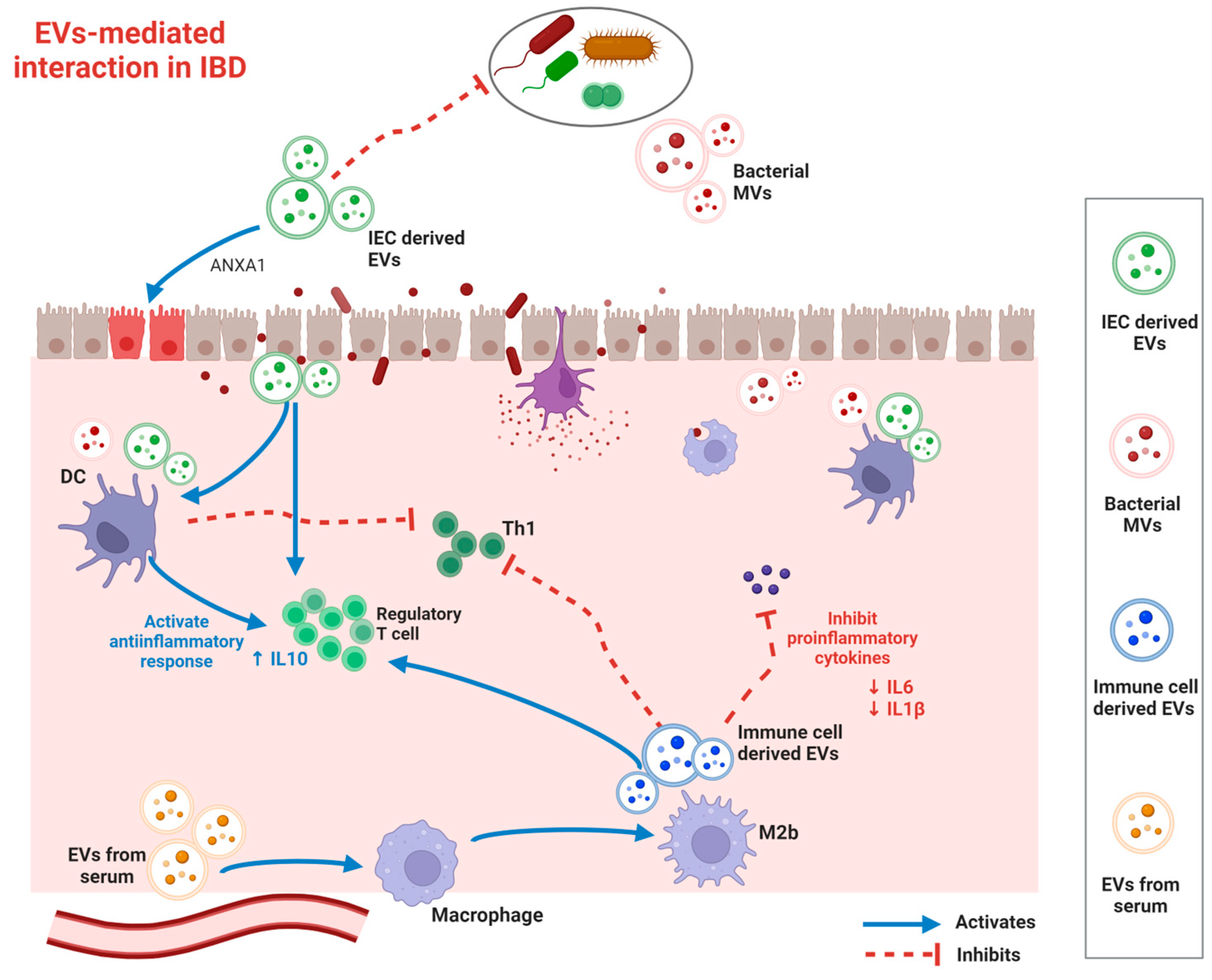
3.1. Immunomodulation

EVs are involved in immune regulation through the functional transfer of proteins, nucleotides, and other cargo between immune cells (Figure 2). An increase in innate immune cells, such as dendritic cells (DCs), macrophages, neutrophils, and T cells, has been observed in the active IBD intestinal environment. Macrophage regulation is crucial for the pathogenesis of IBD, and exosomes are involved in this process. Macrophages are classified into two types based on their function and activation: classically activated M1 macrophages and alternatively activated M2 macrophages. An imbalance between M1 and M2 macrophages is closely associated with inflammatory diseases, including IBD. EVs that polarize macrophages to the M1 type can worsen IBD [12,26]. Exosomes in visceral adipose tissue promote M1 macrophage polarization, leading to intestinal inflammation [27]. Systemically administered exosomes from human bone marrow mesenchymal stem cells (MSCs) considerably attenuate inflammation in IBD colitis models by polarizing M2b macrophages without causing intestinal fibrosis. Exosomes from polarized M2b macrophages increase the number of regulatory Treg cells and suppress proinflammatory cytokines, including IL-1β, IL-6, and IL-17A (Table 1) [26]. Regulating macrophage pyroptosis through exosomes can regulate inflammation in IBD [28]. Human umbilical cord mesenchymal stem cell (hucMSC)-derived exosomes carrying miR-203a-3p.2 attenuate colitis by inhibiting CASP11/4-induced macrophage pyroptosis [29].
T-cells are key members of the adaptive immune response and play crucial roles in IBD pathogenesis. Regulatory T cells (Tregs) and T-helper 17 (Th17) cells are critical for immune homeostasis and play crucial roles in IBD [30]. An imbalance between Th17 and Treg cells is a major cause of intestinal tolerance failure and contributes to IBD [31]. MSC-Exos alleviate IBD colitis by normalizing the Th17/Treg cell balance. Olfactory ecto-mesenchymal stem cell-derived exosomes exerted immunosuppressive effects by inhibiting Th1 and Th17 cell differentiation and promoting Treg cell induction in a murine colitis model [32]. EVs derived from Tregs induce the transformation of other T cells into the Treg phenotype (Table 1). In addition, EVs isolated from breast milk stimulated the development and proliferation of Tregs in a dose-dependent manner [33].
DCs are antigen-presenting cells (APCs) capable of activating naive T cells, which facilitate IBD pathogenesis. DC-derived EVs influence IBD progress via immunomodulation. Exosomes from DCs (DC-Exos) treated with IL-10 upregulate the levels of Tregs in the colonic lamina propria and attenuate colitis in a mouse model [34]. Moreover, exosomal miRNAs modulate the immune system in IBD by regulating intestinal immune cell responses. Exosomal miR146a and miR155 enter recipient DCs and mediate the suppression of their target genes [35].

3.2. Intestinal Barrier Modulation

The impairment of the intestinal barrier is a significant contributor to the development and progression of inflammatory bowel disease (IBD). EVs help repair damaged intestinal epithelial barriers and recover gastrointestinal function in patients with IBD. Extracellular vesicles (EVs) have the ability to influence the function of different types of intestinal epithelial cells (IECs) and control the stability of junctional complexes among IECs (Table 1) [36]. DC exosomes recover intestinal barrier function in DSS-induced colitis via exosomal miR-146b, activating the NF-kB signaling pathway [37]. The overexpression of miR-146b activated the NF-κB pathway, improved epithelial barrier function, and relieved intestinal inflammation in DSS-induced colitis mice. miR-146b improves intestinal inflammation by upregulating NF-κB as a result of the decreased expression of siah2, which ubiquitinates TRAF proteins [37]. LncRNA NEAT1 regulates the intestinal epithelial barrier and inhibits inflammatory responses in IBD [38]. Exosomal miR-223 destroys intestinal barrier function and increases intestinal epithelial permeability by inhibiting the TJ-related protein claudin-8 in IECs [39]. IEC accelerates the recovery of intestinal wall function by secreting EVs containing annexin A1 (ANXA1), a protein that activates wound recovery in patients [40]. IEC also releases EVs containing high levels of TGF-β1 to restore immune balance in the inflamed bowel.

3.3. Gut Microbiome Modulation

EVs in host cells participate in the network between the host and intestinal microbes, serving a crucial function in microbial reconstruction. Recent studies have found pathways by which altered gut microbiota may affect the development of IBD [41]. For instance, the activity of TLR 2/6 in probiotics is significantly reduced by EVs from TLR 2-deficient mouse sera, leading to microbial dysbiosis [42]. Akkermansia muciniphila-derived EVs can enhance epithelial barrier function and decrease the production of IL-6 by IECs [43]. Lactobacillus plantarum Q7-derived EVs reduce the pro-inflammatory bacteria Proteobacteria and increase the anti-inflammatory bacteria Bifidobacteria and Muribaculaceae, attenuating colitis [44]. Clostridium butyricum-derived EVs also play a role in restoring gut barrier function, normalizing bacterial dysbiosis, and restoring miR-199a-3p expression [45]. Exosomal nucleic acids participate in microbial modulation. miRNAs such as miR-515-5p and miR-1226-5p can enter Fusobacterium nucleatum and Escherichia coli to regulate bacterial gene transcripts and affect bacterial growth [46].

4. Current Methodologies for EV Isolation and Analysis

Developing sensitive, accurate, and standardized bioassays that can determine the composition and molecular profile of EVs in clinical samples is imperative for the rapid clinical application of EVs [47]. EVs are novel targets that are different from current analysis targets and have unique size characteristics; they are much smaller than cells but larger than proteins [20]. Technical problems often occur when conventional analysis methods are used and inaccurate results are obtained. Therefore, the development of new isolation and analysis techniques that consider the characteristics of EVs is an important goal. Recently, various techniques for exosome isolation and analysis have been developed. Therefore, understanding the characteristics, strengths, and weaknesses of each technology type is essential. Adopting and using appropriate technologies for each environment is important in research and clinical applications. Because various separation technology methods have distinct advantages and disadvantages, understanding their characteristics and selecting an optimal separation technology that matches the given sample characteristics are important for obtaining highly reproducible results.

4.1. EV Isolation

For clinical applications in diagnosis and treatment, it is necessary to isolate EVs first. The introduction of standard methods with the highest purification quality and lowest impact on EV components remains a potential challenge. Exosome isolation is usually based on physicochemical properties such as density, size, and mass, as well as affinity interactions with specific proteins.
Ultracentrifugation (UC) has been introduced as the gold standard for EV isolation [48]. However, some obstacles limit the clinical application of UC, including expensive equipment, training, large input samples, and labor-intensive and long run times [49]. The EV structure may change because of the high centrifugal force, which may affect downstream analysis. EVs were isolated using density gradient ultracentrifugation (DGC) based on their size, mass, and density using a pre-constructed density gradient medium. Although this method maintains the structural integrity of EVs, it is time-consuming and laborious [47].
Various methods, such as ultrafiltration and size-exclusion chromatography, have been applied to separate exosomes using size-based separation techniques. Owing to their fast separation speed and high efficiency, these two techniques are the most widely used, and several commercial products that use these methods are being released. However, separating exosomes of different sizes is difficult because of their low resolution. To overcome this, symmetric flow field-flow fractionation (AF4) was recently developed [50]. Using this method, large exosome vesicles (90–120 nm) can be separated from small exosome vesicles (60–80 nm) [51].
The precipitation separation method was commercialized early owing to its simplicity and high separation efficiency. Because aqueous polymers reduce the hydration of EVs and cause precipitation, the precipitated EV products can be easily isolated with low centrifugal forces. However, co-precipitation protein interference is caused by nonspecific interactions between the polymers and proteins.
The immunoaffinity capture-based technique is a separation method in which antibodies are fixed to surfaces such as microfluidic chips, nanostructures, and beads, and specific proteins present on the exosome surface are targeted and captured [47]. This method can be used to selectively separate and concentrate a very small group of exosomes from specific clinical samples, such as those for cancer diagnosis.

4.2. EV Analyzation

Exosome characterization technologies are currently being developed to comprehensively understand the morphological and physicochemical characteristics of exosomes. Therefore, developing various characterization techniques to analyze different subsets of EVs is necessary. Dynamic light scattering (DLS) and nanoparticle tracking analysis (NTA) are used as analytical methods to determine the size distribution of exosomes [52]. Western blotting (WB), enzyme-linked immunosorbent assays (ELISA), and flow cytometry are capable of determining the surface proteins of exosomes [52]. Protein analysis using WB or ELISA methods has limitations in that it requires a large number of samples and is time-consuming. Current protein analysis in EVs without strong amplification strategies has technical limitations. For instance, applying flow cytometry to extracellular vesicle analysis is challenging and requires careful consideration [52]. This is because the rapid movement of EVs, which are much smaller than the cell size, produces a weak signal in flow cytometry.
Therefore, new analytical technologies have been developed and proposed. Among these, technologies that are close to commercialization are based on nanoplasmonics [53]. Plasmonic sensors are optical sensors that use metal films or nanostructures. This technique has been widely used to detect analytes and characterize their molecular interactions over the past 30 years. The detection range (10–300 nm) of the plasmonic sensor is similar to the size of the exosomes (50–200 nm) and has a very high sensitivity for exosome detection.

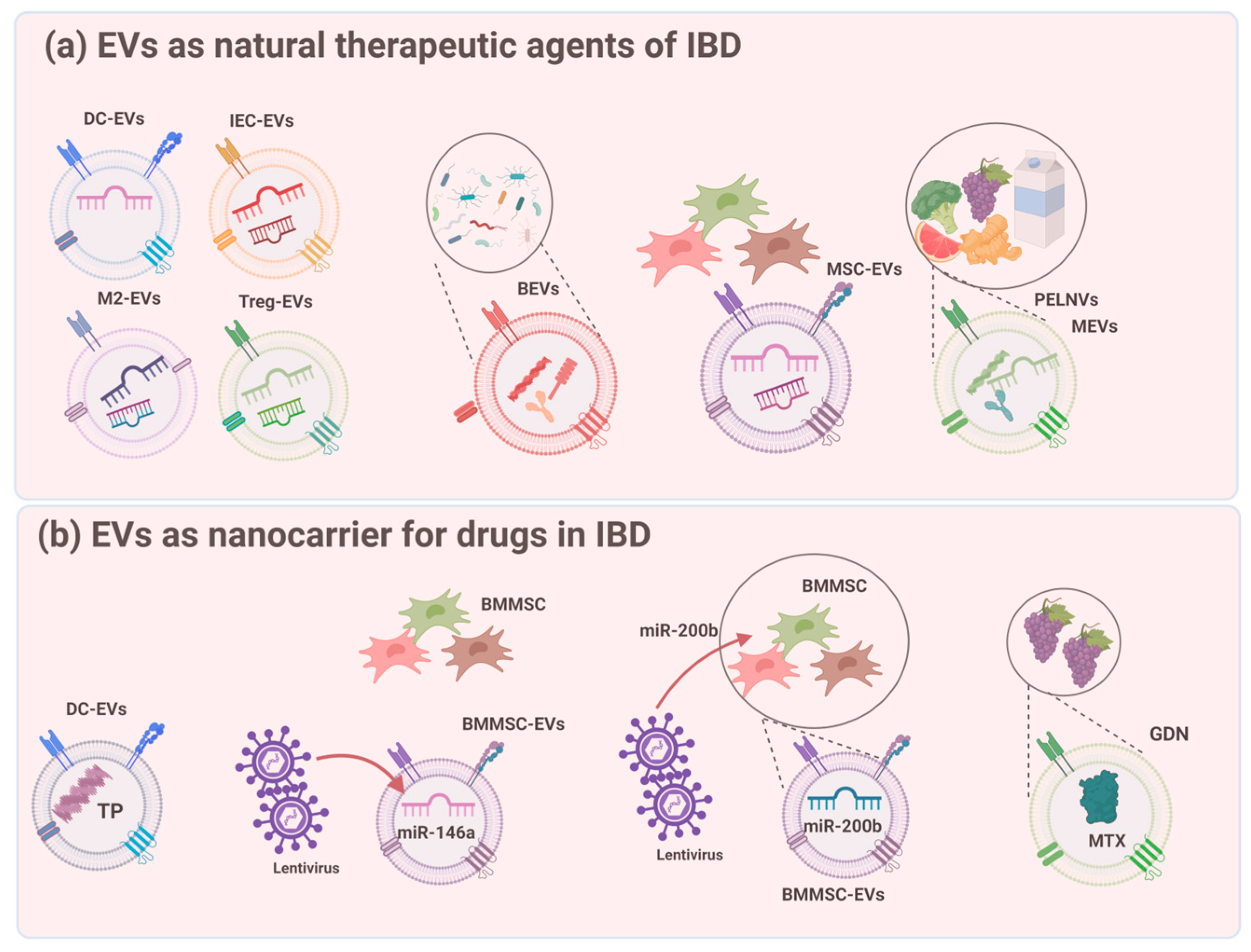
5. EVs as Natural Therapeutic Agents for IBD

Natural EVs are considered new potential treatments for IBD. EVs originate from various sources, such as immune cells, IECs, the gut microbiota, various MSCs, and ingesta (Figure 3). The advantages of using EVs as direct therapy for IBD include safety, effectiveness, and cost-efficiency. Given that research in the field of EVs is still in its early stages, there are a limited number of studies that have conducted direct one-to-one comparisons of their efficacy in animal models of IBD [54].

5.1. Stem Cell-Derived EVs

EVs from stem cells play important roles in immune regulation, self-renewal, expansion, and damage repair (Table 1) [55]. Treatment with SC-EVs is more convenient in terms of transportation and storage because it prevents various side effects that may be caused by stem cell transplantation. Diverse MSCs are the fundamental sources of EVs in humans, including bone marrow MSCs (BMMSCs) [56], human adipose MSCs (hADSCs) [57], olfactory ecto-MSCs (OE-MSCs) [32], and human placental MSCs (hP-MSCs) [58]. MSC-EVs exert therapeutic effects against IBD mainly through modulation of the intestinal barrier, immune response, and intestinal microbiota (Figure 3). MSC-EVs can maintain barrier integrity by reducing IEC apoptosis, reversing goblet cell loss and microvilli and TJ function, and activating IEC and ISC proliferation [57]. MSC-EVs are capable of adjusting pro-inflammatory cytokines (e.g., TNF-α, IL-1β, IL-6, IL-12, IL-17, and IFN-γ) and anti-inflammatory cytokines (e.g., IL-4, IL-13, IL-10, and TGF-β) [32,58,59]. Moreover, MSC-EVs mediate immune cells such as Treg cells, M1/M2 macrophages, and Th1/Th17 subpopulations [32]. MSC-EVs inhibits colonic macrophages, downregulates inflammatory cytokine levels, and inhibits the NF-kB signaling pathway for treating colitis. MSC-EVs can improve the gut microbiota composition by significantly restoring the structure of OTUs (operational taxonomic units) and colitis-induced reduction in α-diversity, increasing the abundance of ‘healthy’ bacteria, decreasing disease-associated bacteria, decreasing detrimental functions, and enhancing other vital cellular functions [60,61]. The inflammation-modulating effects of hucMSC-EVs in IBD have also attracted attention. HucMSC exosomes modulate inflammation in DSS-induced colitis by regulating ubiquitin modification levels, neddylation inhibition by exosomal miR-326, and IL-7 expression in macrophages [62,63]. HucMSC-EVs are also involved in intestinal fibrosis prevention in IBD colitis models.

5.2. EVs from IECs and Immune Cells

The protective effect of IEC-EVs on the intestinal mucosa is well known (Table 1). Endogenous aANXA1 is released as a component of EVs derived from the intestinal epithelial cells. Leoni et al. showed that ANXA1-containing EVs accelerate the recovery of epithelial barrier dysfunction in IBD [40]. Exosomes extracted from various M2 macrophage subtypes can be used for treatment. Exosomes from M2b macrophages may exert a protective effect by increasing the number of Treg cells and IL-4 levels in the spleen [26]. DC-Exos treated with Schistosoma japonicum-soluble egg antigen also significantly improved DSS-induced acute colitis in mice [64]. These EVs reduced pro-inflammatory cytokine expression and improved all the analyzed colitis parameters.

5.3. EVs from the Microbiota

FMT is a popular treatment that uses gut flora reconstitution to restore the normal microbiome; however, people with IBD have higher risks [65]. Therefore, Bacterial EVs (BEVs) have been considered as potential treatments (Figure 3). EVs derived from Clostridium butyricum attenuated murine DSS-induced colitis by remodeling the composition of the gut microbiota and repolarizing M2 macrophages [60]. EVs derived from the probiotic Lactobacillus plantarum Q7 also improved the dysregulation of the gut microbiota and promoted the diversity of the gut microbiota in IBD mice [44]. EVs originating from Akkermansia muciniphila augment the expression and functionality of tight junctions, thereby reinstating the intestinal permeability of Caco-2 cells (Table 1) [43].

5.4. EVs from the Ingesta

Recently, EVs derived from the ingesta have received considerable attention because of their availability, safety, and favorable cost [66]. Milk is a primary source of ingested EVs for IBD treatment. Milk-derived EVs (MEVs) play an essential role in the maturation of the gastrointestinal tract. In a study by Tong et al. [67], milk-derived extracellular vesicles (mEVs) derived from bovine and human breast milk exerted similar protective effects on epithelial tight junction functionality in vitro. The oral administration of mEVs restores gut barrier integrity at multiple levels, including the mucus, epithelial, and immune barriers. Incubation with mEVs upregulated the expression of tight junction proteins, ZO-1, and Occludin in Caco-2 cells. They can also restore disturbed gut microbiota and enhance the intestinal barrier by decreasing matrix metallopeptidase 9 (MMP9) and increasing mucoprotein 2 (MUC2) [68,69]. Daily consumed food items like fruits and vegetables, as well as exosome-like nanoparticles derived from them, also engage with the intestinal mucosa through osmotic processes (Figure 3). Wang et al. showed that grapefruit-derived exosome-like vesicles (GDNs) were selectively absorbed by macrophages in the intestine, which led to an increase in heme oxygenase-1 levels and a decrease in IL-1 and TNF-α (Table 1) [70]. Additionally, both grapes (GELNs) and broccoli (BDNs) release exosome-like nanoparticles that serve as therapeutic aids in alleviating DSS-induced colitis (Table 1). These nanoparticles function by triggering intestinal tissue remodeling mediated by stem cells and stimulating DC amp-activated protein kinase [71,72]. Nanoparticles derived from ginger can be assimilated by IECs and macrophages, leading to fortification of the intestinal barrier and restoration of the gut microbiota [73]. The oral administration of turmeric-derived nanovesicles exhibited excellent anti-inflammatory efficacy in IBD by regulating the intestinal microbiota, repairing the damaged intestinal barrier, and transforming M1 macrophages into M2 macrophages [74].

6. EVs as Nanocarriers for Drugs in IBD

Efforts to develop nanoparticle-based drug delivery technologies have been ongoing over the past decade. However, nanoparticles are removed by the mononuclear phagocyte system during the blood cycle; eventually, less than 1% of the drug dose reaches the target tumor. In contrast, exosomes have the potential to act as highly efficient drug carriers. In particular, exosomes can be successful platforms for various drug delivery systems because of their ability to mediate cellular communication. Exosomes can be modified to exhibit the desired targeting ability through parent cell modification and can be loaded with various therapeutic agents. Exosomes can combine the advantages of existing synthetic nanoparticle-based drug carriers and cell-mediated delivery methods while complementing the shortcomings of each method (Figure 3).

6.1. Cargo Loading Techniques

The primary methods through which therapeutic agents are integrated into exosomes are pre-loading (indirect loading) and post-loading (direct loading) (Figure 4) [75]. In the pre-loading technique, drugs are loaded into parental cells before exosome isolation, allowing them to be encapsulated within exosomes during their natural biogenesis [76]. This method involves transfecting the target gene to produce targeted exosomes or modifying parental cells by co-incubating them with the cargo. A major consideration of this method is the use of parent cells to obtain exosomes. To obtain a large number of exosomes, a large number of parental cells are required, and the method for increasing the production yield of exosomes is also important. The pre-loading method allows for the continuous generation of exosomes loaded with cargo while preserving membrane integrity [77] and has high stability and low immunogenicity. However, the parental cells may be potentially toxic because of the transfection agents. Another disadvantage is its low efficiency compared to that of electroporation and ultrasonic loading technologies [75].
The post-loading method incorporates the drug into pre-isolated natural exosomes. The post-loading method provides improved efficiency compared to the pre-loading method, as it allows for more effective control of the loading capacity. The post-loading method includes two distinct strategies: passive loading and active loading (Table 2). Passive loading using the incubation method allows the drug to passively diffuse into exosomes owing to its physical properties. This method is simple and nondestructive for maintaining exosome integrity. However, this method is mainly useful for hydrophobic drugs that interact with the lipid surfaces of exosomes, which is a potential limitation [78]. The lipid bilayer structure of the vesicle membrane facilitates the loading of hydrophobic drugs such as curcumin and paclitaxel into exosomes through incubation. Recently, considerable progress has been made in the development of active cargo-loading strategies. Various methods have been proposed, including electroporation, sonication, freeze–thawing, and extrusion, which can efficiently load cargo molecules by inducing temporary pores or mechanical stress in exosome membranes [75]. These methods enable precise control of the loading process to optimize the loading conditions and achieve the desired level of cargo loading. Table 2 summarizes the advantages and disadvantages of these methods.
Nevertheless, depending on the employed methodology, certain limitations and risks need to be taken into account. These may include exosome aggregation and the possibility of membrane damage arising from variations in pH, physical forces, and surfactants. Selecting the optimal cargo-loading method according to the cargo content and treatment purpose is crucial.

6.2. Application of Exosomes as Nanocarriers in IBD

Recently, MSCs have gained considerable attention as promising nanocarriers for IBD treatment. MSC-derived exosomes deliver cytokines, miRNAs, and external cargo to the immune microenvironment to regulate immune effector cells. miR-146a significantly attenuated drug-induced colitis in a mouse model after lentiviral transfection into BMMSC-EVs. miR-200b, after being transfected into BMMSCs with lentivirus, could be transferred to IEC-6 cells by being packaged in EVs, improving colon fibrosis. Moreover, DCs can regulate intestinal immunity through exosomes containing various antigenic peptides. Various anti-inflammatory factors (including IL-10, IL4, and TGF-β1) interacted with DCs to generate various immunomodulatory exosome subtypes. Exosomes derived from the TGF-β1 gene-modified bone marrow DC (TGF-β1-EXO) have inhibited Th17 responses and upregulated regulatory T cells, playing a protective role during IBD development in a murine model. Exosomes derived from IL-10-treated bone marrow-derived DCs reduced the severity of drug-induced colitis in mice. Alexander et al. found that endogenous miR-155 and miR-146a, two critical miRNAs that regulate inflammation, are released from dendritic cells within exosomes and are subsequently taken up by recipient dendritic cells. Exogenous miRNAs mediate target gene repression and can reprogram the cellular response to endotoxins, where exosome-delivered miR-155 is enhanced, whereas miR-146a reduces inflammatory gene expression.
Exosomes secreted by M2 macrophages are involved in the resolution of inflammation and intestinal repair. M2 macrophage-derived exosomal miR-590-3p reduces inflammatory signals and promotes epithelial regeneration by targeting LATS1 and subsequently activating YAP/β-catenin-regulated transcription. The role of M2 macrophage-derived EVs (M2-EVs) in UC inflammation has also been investigated. M2 macrophages were transfected with lentiviral vectors overexpressing LncRNA maternally expressed 3 (MEG3). High expression of MEG3 in EV was observed after isolation. M2-EVs carrying MEG3 enhanced UC cell viability and reduced inflammatory responses via the miR-20b-5p/CREB1 axis, thereby attenuating inflammation in a murine IBD model.

7. Conclusions and Future Perspectives

In the current review, we summarized the roles of EVs of different origins in the pathogenesis and progression of IBD, as well as their potential in clinical diagnosis and therapy. In addition, we introduced recent studies on the role of EVs as drug delivery systems in IBD. Exosomes are being actively researched in various fields, including isolation and analysis technologies and the development of diagnostic and treatment methods. However, because the history of exosome research is limited, a standardized method has not yet been developed. Exosome-mediated treatment technologies have received attention in various fields, and several early clinical trials have been conducted.
However, compared to current drug delivery systems that use nanoparticles or liposomes, this is in its early stages, and its strengths and weaknesses have been identified. More research is needed on the in vivo distribution of exosomes, their mechanisms, and their potential toxicity when administered in vivo.
The production yield of exosomes depends on the secretion capacity of the cell, the complexity and cost of large-scale cell culture, and the time required for exosome production. Industrial production is difficult because of low yields, which is an obstacle that cannot be ignored. As previously mentioned, selecting and modifying donor cells for exosome production and developing new methods to maximize the uniform production of exosomes are the needs of the hour.
Additionally, exosomes have limited cargo transport efficiency. Exosomes inherently carry large amounts of natural components that significantly complicate and limit their cargo loads. Therefore, developing exosome engineering techniques for improved loading capacities is necessary.
Another disadvantage is that the quality control of exosomes is difficult. Exosomes are highly heterogeneous, even when produced by a single cell type. Because of the lack of sensitive, high-efficiency exosome analysis methods, separating homogeneous exosomes from heterogeneous exosome groups is technically challenging.
Currently, strategies to precisely control the content of exosomes are lacking, which hinders the development of treatment technologies using exosomes. However, if new creative approaches are developed to overcome these shortcomings, a new era of IBD treatment will emerge. The high biocompatibility of exosomes provides greater opportunities for clinicians to develop safe and targeted therapies for IBD treatment and management.

Author Contributions

Conceptualization, S.H.K. and T.H.K.; formal analysis, S.H.K., J.B., J.M.S. and T.H.K.; funding acquisition, B.K. and T.H.K.; investigation, J.B., S.K. and B.K.; methodology, S.H.K. and B.K.; project administration, J.M.S.; supervision, J.M.S.; validation, J.M.S.; visualization, S.H.K. and T.H.K.; writing—original draft, S.H.K.; writing—review and editing, S.H.K. and T.H.K. All authors have read and agreed to the published version of the manuscript.

Funding

This research was supported by a grant from Korea University, Seoul, Republic of Korea (Grant No. K2314111) and the Basic Science Research Program of the National Research Foundation of Korea, funded by the Ministry of Science and Technology and the Ministry of Science, ICT and Future Planning (2017R1A2B2003575 and NRF-2020R1A2C1006398); the Ministry of Science and ICT, Korea, under the ICT Creative Consilience program (IITP-2023-2020-0-01819), supervised by the Institute for Information and Communications Technology Planning and Evaluation (IITP); and the Korea Health Technology R&D Project (HI17C0387, HR22C1302) through the Korea Health Industry Development Institute (KHIDI), funded by the Ministry of Health and Welfare.

Institutional Review Board Statement

Not applicable.

Informed Consent Statement

Not applicable.

Data Availability Statement

The datasets in this study are available from the corresponding author on reasonable request.

Conflicts of Interest

The authors declare no conflicts of interest.

References

  1. Ng, S.C.; Shi, H.Y.; Hamidi, N.; Underwood, F.E.; Tang, W.; Benchimol, E.I.; Panaccione, R.; Ghosh, S.; Wu, J.C.Y.; Chan, F.K.L.; et al. Worldwide incidence and prevalence of inflammatory bowel disease in the 21st century: A systematic review of population-based studies. Lancet 2017, 390, 2769–2778. [Google Scholar] [CrossRef] [PubMed]
  2. Ray, K. IBD: The changing epidemiology of IBD. Nat. Rev. Gastroenterol. Hepatol. 2017, 14, 690. [Google Scholar] [PubMed]
  3. Kuenzig, M.E.; Fung, S.G.; Marderfeld, L.; Mak, J.W.Y.; Kaplan, G.G.; Ng, S.C.; Wilson, D.C.; Cameron, F.; Henderson, P.; Kotze, P.G.; et al. Twenty-first Century Trends in the Global Epidemiology of Pediatric-Onset Inflammatory Bowel Disease: Systematic Review. Gastroenterology 2022, 162, 1147–1159.e4. [Google Scholar] [CrossRef]
  4. Ray, G.; Longworth, M.S. Epigenetics, DNA Organization, and Inflammatory Bowel Disease. Inflamm. Bowel Dis. 2019, 25, 235–247. [Google Scholar] [CrossRef] [PubMed]
  5. Ocansey, D.K.W.; Wang, L.; Wang, J.; Yan, Y.; Qian, H.; Zhang, X.; Xu, W.; Mao, F. Mesenchymal stem cell-gut microbiota interaction in the repair of inflammatory bowel disease: An enhanced therapeutic effect. Clin. Transl. Med. 2019, 8, 31. [Google Scholar] [CrossRef] [PubMed]
  6. Villablanca, E.J.; Selin, K.; Hedin, C.R.H. Mechanisms of mucosal healing: Treating inflammatory bowel disease without immunosuppression? Nat. Rev. Gastroenterol. Hepatol. 2022, 19, 493–507. [Google Scholar] [CrossRef] [PubMed]
  7. Otte, M.L.; Lama Tamang, R.; Papapanagiotou, J.; Ahmad, R.; Dhawan, P.; Singh, A.B. Mucosal healing and inflammatory bowel disease: Therapeutic implications and new targets. World J. Gastroenterol. 2023, 29, 1157–1172. [Google Scholar] [CrossRef]
  8. Neurath, M.F. Current and emerging therapeutic targets for IBD. Nat. Rev. Gastroenterol. Hepatol. 2017, 14, 269–278. [Google Scholar] [CrossRef]
  9. Frolkis, A.D.; Dykeman, J.; Negron, M.E.; Debruyn, J.; Jette, N.; Fiest, K.M.; Frolkis, T.; Barkema, H.W.; Rioux, K.P.; Panaccione, R.; et al. Risk of surgery for inflammatory bowel diseases has decreased over time: A systematic review and meta-analysis of population-based studies. Gastroenterology 2013, 145, 996–1006. [Google Scholar] [CrossRef]
  10. Pouillon, L.; Bossuyt, P.; Peyrin-Biroulet, L. Considerations, challenges and future of anti-TNF therapy in treating inflammatory bowel disease. Expert. Opin. Biol. Ther. 2016, 16, 1277–1290. [Google Scholar] [CrossRef]
  11. Papamichael, K.; Van Stappen, T.; Jairath, V.; Gecse, K.; Khanna, R.; D’Haens, G.; Vermeire, S.; Gils, A.; Feagan, B.G.; Levesque, B.G.; et al. Review article: Pharmacological aspects of anti-TNF biosimilars in inflammatory bowel diseases. Aliment. Pharmacol. Ther. 2015, 42, 1158–1169. [Google Scholar] [CrossRef] [PubMed]
  12. Shen, Q.; Huang, Z.; Yao, J.; Jin, Y. Extracellular vesicles-mediated interaction within intestinal microenvironment in inflammatory bowel disease. J. Adv. Res. 2022, 37, 221–233. [Google Scholar] [CrossRef] [PubMed]
  13. Chang, X.; Wang, S.L.; Zhao, S.B.; Shi, Y.H.; Pan, P.; Gu, L.; Yao, J.; Li, Z.S.; Bai, Y. Extracellular Vesicles with Possible Roles in Gut Intestinal Tract Homeostasis and IBD. Mediat. Inflamm. 2020, 2020, 1945832. [Google Scholar] [CrossRef] [PubMed]
  14. Pegtel, D.M.; Gould, S.J. Exosomes. Annu. Rev. Biochem. 2019, 88, 487–514. [Google Scholar] [CrossRef] [PubMed]
  15. Kalluri, R.; LeBleu, V.S. The biology, function, and biomedical applications of exosomes. Science 2020, 367, eaau6977. [Google Scholar] [CrossRef]
  16. Abels, E.R.; Breakefield, X.O. Introduction to Extracellular Vesicles: Biogenesis, RNA Cargo Selection, Content, Release, and Uptake. Cell Mol. Neurobiol. 2016, 36, 301–312. [Google Scholar] [CrossRef] [PubMed]
  17. Sedgwick, A.E.; D’Souza-Schorey, C. The biology of extracellular microvesicles. Traffic 2018, 19, 319–327. [Google Scholar] [CrossRef] [PubMed]
  18. Tancini, B.; Buratta, S.; Sagini, K.; Costanzi, E.; Delo, F.; Urbanelli, L.; Emiliani, C. Insight into the Role of Extracellular Vesicles in Lysosomal Storage Disorders. Genes 2019, 10, 510. [Google Scholar] [CrossRef]
  19. Gurunathan, S.; Kang, M.H.; Jeyaraj, M.; Qasim, M.; Kim, J.H. Review of the Isolation, Characterization, Biological Function, and Multifarious Therapeutic Approaches of Exosomes. Cells 2019, 8, 307. [Google Scholar] [CrossRef]
  20. Akers, J.C.; Gonda, D.; Kim, R.; Carter, B.S.; Chen, C.C. Biogenesis of extracellular vesicles (EV): Exosomes, microvesicles, retrovirus-like vesicles, and apoptotic bodies. J. Neurooncol. 2013, 113, 1–11. [Google Scholar] [CrossRef]
  21. Lepage, P.; Colombet, J.; Marteau, P.; Sime-Ngando, T.; Dore, J.; Leclerc, M. Dysbiosis in inflammatory bowel disease: A role for bacteriophages? Gut 2008, 57, 424–425. [Google Scholar] [CrossRef] [PubMed]
  22. Wagner, J.; Maksimovic, J.; Farries, G.; Sim, W.H.; Bishop, R.F.; Cameron, D.J.; Catto-Smith, A.G.; Kirkwood, C.D. Bacteriophages in gut samples from pediatric Crohn’s disease patients: Metagenomic analysis using 454 pyrosequencing. Inflamm. Bowel Dis. 2013, 19, 1598–1608. [Google Scholar] [CrossRef] [PubMed]
  23. Ng, S.C.; Ananthakrishnan, A.N. New approaches along the IBD course: Diet, tight control and stem cells. Nat. Rev. Gastroenterol. Hepatol. 2019, 16, 82–84. [Google Scholar] [CrossRef]
  24. Ahmed, I.; Roy, B.C.; Khan, S.A.; Septer, S.; Umar, S. Microbiome, Metabolome and Inflammatory Bowel Disease. Microorganisms 2016, 4, 20. [Google Scholar] [CrossRef] [PubMed]
  25. Skotland, T.; Sandvig, K.; Llorente, A. Lipids in exosomes: Current knowledge and the way forward. Prog. Lipid Res. 2017, 66, 30–41. [Google Scholar] [CrossRef] [PubMed]
  26. Yang, R.; Liao, Y.; Wang, L.; He, P.; Hu, Y.; Yuan, D.; Wu, Z.; Sun, X. Exosomes Derived From M2b Macrophages Attenuate DSS-Induced Colitis. Front. Immunol. 2019, 10, 2346. [Google Scholar] [CrossRef] [PubMed]
  27. Wei, M.; Gao, X.; Liu, L.; Li, Z.; Wan, Z.; Dong, Y.; Chen, X.; Niu, Y.; Zhang, J.; Yang, G. Visceral Adipose Tissue Derived Exosomes Exacerbate Colitis Severity via Pro-inflammatory MiRNAs in High Fat Diet Fed Mice. ACS Nano 2020, 14, 5099–5110. [Google Scholar] [CrossRef] [PubMed]
  28. Cai, X.; Zhang, Z.Y.; Yuan, J.T.; Ocansey, D.K.W.; Tu, Q.; Zhang, X.; Qian, H.; Xu, W.R.; Qiu, W.; Mao, F. hucMSC-derived exosomes attenuate colitis by regulating macrophage pyroptosis via the miR-378a-5p/NLRP3 axis. Stem Cell Res. Ther. 2021, 12, 416. [Google Scholar] [CrossRef]
  29. Xu, Y.; Tang, X.; Fang, A.; Yan, J.; Kofi Wiredu Ocansey, D.; Zhang, X.; Mao, F. HucMSC-Ex carrying miR-203a-3p.2 ameliorates colitis through the suppression of caspase11/4-induced macrophage pyroptosis. Int. Immunopharmacol. 2022, 110, 108925. [Google Scholar] [CrossRef]
  30. Harrison, O.J.; Powrie, F.M. Regulatory T cells and immune tolerance in the intestine. Cold Spring Harb. Perspect. Biol. 2013, 5, a018341. [Google Scholar] [CrossRef]
  31. Jiang, P.; Zheng, C.; Xiang, Y.; Malik, S.; Su, D.; Xu, G.; Zhang, M. The involvement of TH17 cells in the pathogenesis of IBD. Cytokine Growth Factor. Rev. 2023, 69, 28–42. [Google Scholar] [CrossRef] [PubMed]
  32. Tian, J.; Zhu, Q.; Zhang, Y.; Bian, Q.; Hong, Y.; Shen, Z.; Xu, H.; Rui, K.; Yin, K.; Wang, S. Olfactory Ecto-Mesenchymal Stem Cell-Derived Exosomes Ameliorate Experimental Colitis via Modulating Th1/Th17 and Treg Cell Responses. Front. Immunol. 2020, 11, 598322. [Google Scholar] [CrossRef] [PubMed]
  33. Admyre, C.; Johansson, S.M.; Qazi, K.R.; Filen, J.J.; Lahesmaa, R.; Norman, M.; Neve, E.P.; Scheynius, A.; Gabrielsson, S. Exosomes with immune modulatory features are present in human breast milk. J. Immunol. 2007, 179, 1969–1978. [Google Scholar] [CrossRef] [PubMed]
  34. Durant, L.; Stentz, R.; Noble, A.; Brooks, J.; Gicheva, N.; Reddi, D.; O’Connor, M.J.; Hoyles, L.; McCartney, A.L.; Man, R.; et al. Bacteroides thetaiotaomicron-derived outer membrane vesicles promote regulatory dendritic cell responses in health but not in inflammatory bowel disease. Microbiome 2020, 8, 88. [Google Scholar] [CrossRef] [PubMed]
  35. Alexander, M.; Hu, R.; Runtsch, M.C.; Kagele, D.A.; Mosbruger, T.L.; Tolmachova, T.; Seabra, M.C.; Round, J.L.; Ward, D.M.; O’Connell, R.M. Exosome-delivered microRNAs modulate the inflammatory response to endotoxin. Nat. Commun. 2015, 6, 7321. [Google Scholar] [CrossRef] [PubMed]
  36. Ramos, G.P.; Papadakis, K.A. Mechanisms of Disease: Inflammatory Bowel Diseases. Mayo Clin. Proc. 2019, 94, 155–165. [Google Scholar] [CrossRef]
  37. Nata, T.; Fujiya, M.; Ueno, N.; Moriichi, K.; Konishi, H.; Tanabe, H.; Ohtake, T.; Ikuta, K.; Kohgo, Y. MicroRNA-146b improves intestinal injury in mouse colitis by activating nuclear factor-kappaB and improving epithelial barrier function. J. Gene Med. 2013, 15, 249–260. [Google Scholar] [CrossRef] [PubMed]
  38. Liu, R.; Tang, A.; Wang, X.; Chen, X.; Zhao, L.; Xiao, Z.; Shen, S. Inhibition of lncRNA NEAT1 suppresses the inflammatory response in IBD by modulating the intestinal epithelial barrier and by exosome-mediated polarization of macrophages. Int. J. Mol. Med. 2018, 42, 2903–2913. [Google Scholar] [CrossRef]
  39. Wang, H.; Chao, K.; Ng, S.C.; Bai, A.H.; Yu, Q.; Yu, J.; Li, M.; Cui, Y.; Chen, M.; Hu, J.F.; et al. Pro-inflammatory miR-223 mediates the cross-talk between the IL23 pathway and the intestinal barrier in inflammatory bowel disease. Genome Biol. 2016, 17, 58. [Google Scholar] [CrossRef]
  40. Leoni, G.; Neumann, P.A.; Kamaly, N.; Quiros, M.; Nishio, H.; Jones, H.R.; Sumagin, R.; Hilgarth, R.S.; Alam, A.; Fredman, G.; et al. Annexin A1-containing extracellular vesicles and polymeric nanoparticles promote epithelial wound repair. J. Clin. Investig. 2015, 125, 1215–1227. [Google Scholar] [CrossRef]
  41. Tun, H.M.; Bridgman, S.L.; Chari, R.; Field, C.J.; Guttman, D.S.; Becker, A.B.; Mandhane, P.J.; Turvey, S.E.; Subbarao, P.; Sears, M.R.; et al. Roles of Birth Mode and Infant Gut Microbiota in Intergenerational Transmission of Overweight and Obesity From Mother to Offspring. JAMA Pediatr. 2018, 172, 368–377. [Google Scholar] [CrossRef] [PubMed]
  42. van Bergenhenegouwen, J.; Kraneveld, A.D.; Rutten, L.; Kettelarij, N.; Garssen, J.; Vos, A.P. Extracellular vesicles modulate host-microbe responses by altering TLR2 activity and phagocytosis. PLoS ONE 2014, 9, e89121. [Google Scholar] [CrossRef] [PubMed]
  43. Chelakkot, C.; Choi, Y.; Kim, D.K.; Park, H.T.; Ghim, J.; Kwon, Y.; Jeon, J.; Kim, M.S.; Jee, Y.K.; Gho, Y.S.; et al. Akkermansia muciniphila-derived extracellular vesicles influence gut permeability through the regulation of tight junctions. Exp. Mol. Med. 2018, 50, e450. [Google Scholar] [CrossRef] [PubMed]
  44. Hao, H.; Zhang, X.; Tong, L.; Liu, Q.; Liang, X.; Bu, Y.; Gong, P.; Liu, T.; Zhang, L.; Xia, Y.; et al. Effect of Extracellular Vesicles Derived From Lactobacillus plantarum Q7 on Gut Microbiota and Ulcerative Colitis in Mice. Front. Immunol. 2021, 12, 777147. [Google Scholar] [CrossRef] [PubMed]
  45. Ma, L.; Lyu, W.; Song, Y.; Chen, K.; Lv, L.; Yang, H.; Wang, W.; Xiao, Y. Anti-Inflammatory Effect of Clostridium butyricum-Derived Extracellular Vesicles in Ulcerative Colitis: Impact on Host microRNAs Expressions and Gut Microbiome Profiles. Mol. Nutr. Food Res. 2023, 67, e2200884. [Google Scholar] [CrossRef] [PubMed]
  46. Liu, S.; da Cunha, A.P.; Rezende, R.M.; Cialic, R.; Wei, Z.; Bry, L.; Comstock, L.E.; Gandhi, R.; Weiner, H.L. The Host Shapes the Gut Microbiota via Fecal MicroRNA. Cell Host Microbe 2016, 19, 32–43. [Google Scholar] [CrossRef] [PubMed]
  47. Hou, R.; Li, Y.; Sui, Z.; Yuan, H.; Yang, K.; Liang, Z.; Zhang, L.; Zhang, Y. Advances in exosome isolation methods and their applications in proteomic analysis of biological samples. Anal. Bioanal. Chem. 2019, 411, 5351–5361. [Google Scholar] [CrossRef]
  48. Thery, C.; Amigorena, S.; Raposo, G.; Clayton, A. Isolation and characterization of exosomes from cell culture supernatants and biological fluids. Curr. Protoc. Cell Biol. 2006, 30, 3–22. [Google Scholar] [CrossRef]
  49. Konoshenko, M.Y.; Lekchnov, E.A.; Vlassov, A.V.; Laktionov, P.P. Isolation of Extracellular Vesicles: General Methodologies and Latest Trends. BioMed Res. Int. 2018, 2018, 8545347. [Google Scholar] [CrossRef]
  50. Kang, D.; Oh, S.; Ahn, S.M.; Lee, B.H.; Moon, M.H. Proteomic analysis of exosomes from human neural stem cells by flow field-flow fractionation and nanoflow liquid chromatography-tandem mass spectrometry. J. Proteome Res. 2008, 7, 3475–3480. [Google Scholar] [CrossRef]
  51. Sitar, S.; Kejzar, A.; Pahovnik, D.; Kogej, K.; Tusek-Znidaric, M.; Lenassi, M.; Zagar, E. Size characterization and quantification of exosomes by asymmetrical-flow field-flow fractionation. Anal. Chem. 2015, 87, 9225–9233. [Google Scholar] [CrossRef] [PubMed]
  52. Wang, X.; Xia, J.; Yang, L.; Dai, J.; He, L. Recent progress in exosome research: Isolation, characterization and clinical applications. Cancer Gene Ther. 2023, 30, 1051–1065. [Google Scholar] [CrossRef] [PubMed]
  53. Im, H.; Shao, H.; Park, Y.I.; Peterson, V.M.; Castro, C.M.; Weissleder, R.; Lee, H. Label-free detection and molecular profiling of exosomes with a nano-plasmonic sensor. Nat. Biotechnol. 2014, 32, 490–495. [Google Scholar] [CrossRef] [PubMed]
  54. Cai, J.; Wu, J.; Wang, J.; Li, Y.; Hu, X.; Luo, S.; Xiang, D. Extracellular vesicles derived from different sources of mesenchymal stem cells: Therapeutic effects and translational potential. Cell Biosci. 2020, 10, 69. [Google Scholar] [CrossRef] [PubMed]
  55. Li, Y.; Altemus, J.; Lightner, A.L. Mesenchymal stem cells and acellular products attenuate murine induced colitis. Stem Cell Res. Ther. 2020, 11, 515. [Google Scholar] [CrossRef] [PubMed]
  56. Liu, H.; Liang, Z.; Wang, F.; Zhou, C.; Zheng, X.; Hu, T.; He, X.; Wu, X.; Lan, P. Exosomes from mesenchymal stromal cells reduce murine colonic inflammation via a macrophage-dependent mechanism. JCI Insight 2019, 4, e131273. [Google Scholar] [CrossRef] [PubMed]
  57. Yu, H.; Yang, X.; Xiao, X.; Xu, M.; Yang, Y.; Xue, C.; Li, X.; Wang, S.; Zhao, R.C. Human Adipose Mesenchymal Stem Cell-derived Exosomes Protect Mice from DSS-Induced Inflammatory Bowel Disease by Promoting Intestinal-stem-cell and Epithelial Regeneration. Aging Dis. 2021, 12, 1423–1437. [Google Scholar] [CrossRef] [PubMed]
  58. Duan, L.; Huang, H.; Zhao, X.; Zhou, M.; Chen, S.; Wang, C.; Han, Z.; Han, Z.C.; Guo, Z.; Li, Z.; et al. Extracellular vesicles derived from human placental mesenchymal stem cells alleviate experimental colitis in mice by inhibiting inflammation and oxidative stress. Int. J. Mol. Med. 2020, 46, 1551–1561. [Google Scholar] [CrossRef]
  59. Heidari, N.; Abbasi-Kenarsari, H.; Namaki, S.; Baghaei, K.; Zali, M.R.; Ghaffari Khaligh, S.; Hashemi, S.M. Adipose-derived mesenchymal stem cell-secreted exosome alleviates dextran sulfate sodium-induced acute colitis by Treg cell induction and inflammatory cytokine reduction. J. Cell Physiol. 2021, 236, 5906–5920. [Google Scholar] [CrossRef]
  60. Wang, Y.; Xie, Q.; Zhang, Y.; Ma, W.; Ning, K.; Xiang, J.Y.; Cui, J.; Xiang, H. Combination of probiotics with different functions alleviate DSS-induced colitis by regulating intestinal microbiota, IL-10, and barrier function. Appl. Microbiol. Biotechnol. 2020, 104, 335–349. [Google Scholar] [CrossRef]
  61. Ocansey, D.K.W.; Zhang, Z.; Xu, X.; Liu, L.; Amoah, S.; Chen, X.; Wang, B.; Zhang, X.; Mao, F. Mesenchymal stem cell-derived exosome mitigates colitis via the modulation of the gut metagenomics-metabolomics-farnesoid X receptor axis. Biomater. Sci. 2022, 10, 4822–4836. [Google Scholar] [CrossRef] [PubMed]
  62. Wu, Y.; Qiu, W.; Xu, X.; Kang, J.; Wang, J.; Wen, Y.; Tang, X.; Yan, Y.; Qian, H.; Zhang, X.; et al. Exosomes derived from human umbilical cord mesenchymal stem cells alleviate inflammatory bowel disease in mice through ubiquitination. Am. J. Transl. Res. 2018, 10, 2026–2036. [Google Scholar] [PubMed]
  63. Wang, G.; Yuan, J.; Cai, X.; Xu, Z.; Wang, J.; Ocansey, D.K.W.; Yan, Y.; Qian, H.; Zhang, X.; Xu, W.; et al. HucMSC-exosomes carrying miR-326 inhibit neddylation to relieve inflammatory bowel disease in mice. Clin. Transl. Med. 2020, 10, e113. [Google Scholar] [CrossRef] [PubMed]
  64. Wang, L.; Yu, Z.; Wan, S.; Wu, F.; Chen, W.; Zhang, B.; Lin, D.; Liu, J.; Xie, H.; Sun, X.; et al. Exosomes Derived from Dendritic Cells Treated with Schistosoma japonicum Soluble Egg Antigen Attenuate DSS-Induced Colitis. Front. Pharmacol. 2017, 8, 651. [Google Scholar] [CrossRef] [PubMed]
  65. Dailey, F.E.; Turse, E.P.; Daglilar, E.; Tahan, V. The dirty aspects of fecal microbiota transplantation: A review of its adverse effects and complications. Curr. Opin. Pharmacol. 2019, 49, 29–33. [Google Scholar] [CrossRef] [PubMed]
  66. Chen, L.; Ou, Q.; Kou, X. Extracellular vesicles and their indispensable roles in pathogenesis and treatment of inflammatory bowel disease: A comprehensive review. Life Sci. 2023, 327, 121830. [Google Scholar] [CrossRef] [PubMed]
  67. Tong, L.; Zhang, S.; Liu, Q.; Huang, C.; Hao, H.; Tan, M.S.; Yu, X.; Lou, C.K.L.; Huang, R.; Zhang, Z.; et al. Milk-derived extracellular vesicles protect intestinal barrier integrity in the gut-liver axis. Sci. Adv. 2023, 9, eade5041. [Google Scholar] [CrossRef] [PubMed]
  68. Lin, Y.; Lu, Y.; Huang, Z.; Wang, Y.; Song, S.; Luo, Y.; Ren, F.; Guo, H. Milk-Derived Small Extracellular Vesicles Promote Recovery of Intestinal Damage by Accelerating Intestinal Stem Cell-Mediated Epithelial Regeneration. Mol. Nutr. Food Res. 2022, 66, e2100551. [Google Scholar] [CrossRef]
  69. Mecocci, S.; Ottaviani, A.; Razzuoli, E.; Fiorani, P.; Pietrucci, D.; De Ciucis, C.G.; Dei Giudici, S.; Franzoni, G.; Chillemi, G.; Cappelli, K. Cow Milk Extracellular Vesicle Effects on an In Vitro Model of Intestinal Inflammation. Biomedicines 2022, 10, 570. [Google Scholar] [CrossRef]
  70. Wang, B.; Zhuang, X.; Deng, Z.B.; Jiang, H.; Mu, J.; Wang, Q.; Xiang, X.; Guo, H.; Zhang, L.; Dryden, G.; et al. Targeted drug delivery to intestinal macrophages by bioactive nanovesicles released from grapefruit. Mol. Ther. 2014, 22, 522–534. [Google Scholar] [CrossRef]
  71. Ju, S.; Mu, J.; Dokland, T.; Zhuang, X.; Wang, Q.; Jiang, H.; Xiang, X.; Deng, Z.B.; Wang, B.; Zhang, L.; et al. Grape exosome-like nanoparticles induce intestinal stem cells and protect mice from DSS-induced colitis. Mol. Ther. 2013, 21, 1345–1357. [Google Scholar] [CrossRef] [PubMed]
  72. Deng, Z.; Rong, Y.; Teng, Y.; Mu, J.; Zhuang, X.; Tseng, M.; Samykutty, A.; Zhang, L.; Yan, J.; Miller, D.; et al. Broccoli-Derived Nanoparticle Inhibits Mouse Colitis by Activating Dendritic Cell AMP-Activated Protein Kinase. Mol. Ther. 2017, 25, 1641–1654. [Google Scholar] [CrossRef] [PubMed]
  73. Zhang, M.; Viennois, E.; Prasad, M.; Zhang, Y.; Wang, L.; Zhang, Z.; Han, M.K.; Xiao, B.; Xu, C.; Srinivasan, S.; et al. Edible ginger-derived nanoparticles: A novel therapeutic approach for the prevention and treatment of inflammatory bowel disease and colitis-associated cancer. Biomaterials 2016, 101, 321–340. [Google Scholar] [CrossRef]
  74. Liu, C.; Yan, X.; Zhang, Y.; Yang, M.; Ma, Y.; Zhang, Y.; Xu, Q.; Tu, K.; Zhang, M. Oral administration of turmeric-derived exosome-like nanovesicles with anti-inflammatory and pro-resolving bioactions for murine colitis therapy. J. Nanobiotechnology 2022, 20, 206. [Google Scholar] [CrossRef] [PubMed]
  75. Ferreira, D.; Moreira, J.N.; Rodrigues, L.R. New advances in exosome-based targeted drug delivery systems. Crit. Rev. Oncol. Hematol. 2022, 172, 103628. [Google Scholar] [CrossRef] [PubMed]
  76. Koh, H.B.; Kim, H.J.; Kang, S.W.; Yoo, T.H. Exosome-Based Drug Delivery: Translation from Bench to Clinic. Pharmaceutics 2023, 15, 2042. [Google Scholar] [CrossRef]
  77. Pascucci, L.; Cocce, V.; Bonomi, A.; Ami, D.; Ceccarelli, P.; Ciusani, E.; Vigano, L.; Locatelli, A.; Sisto, F.; Doglia, S.M.; et al. Paclitaxel is incorporated by mesenchymal stromal cells and released in exosomes that inhibit in vitro tumor growth: A new approach for drug delivery. J. Control Release 2014, 192, 262–270. [Google Scholar] [CrossRef]
  78. Bellavia, D.; Raimondo, S.; Calabrese, G.; Forte, S.; Cristaldi, M.; Patinella, A.; Memeo, L.; Manno, M.; Raccosta, S.; Diana, P.; et al. Interleukin 3-receptor targeted exosomes inhibit in vitro and in vivo Chronic Myelogenous Leukemia cell growth. Theranostics 2017, 7, 1333–1345. [Google Scholar] [CrossRef]
  79. Agrawal, A.K.; Aqil, F.; Jeyabalan, J.; Spencer, W.A.; Beck, J.; Gachuki, B.W.; Alhakeem, S.S.; Oben, K.; Munagala, R.; Bondada, S.; et al. Milk-derived exosomes for oral delivery of paclitaxel. Nanomedicine 2017, 13, 1627–1636. [Google Scholar] [CrossRef]
  80. Smyth, T.; Kullberg, M.; Malik, N.; Smith-Jones, P.; Graner, M.W.; Anchordoquy, T.J. Biodistribution and delivery efficiency of unmodified tumor-derived exosomes. J. Control Release 2015, 199, 145–155. [Google Scholar] [CrossRef]
  81. Sun, D.; Zhuang, X.; Xiang, X.; Liu, Y.; Zhang, S.; Liu, C.; Barnes, S.; Grizzle, W.; Miller, D.; Zhang, H.G. A novel nanoparticle drug delivery system: The anti-inflammatory activity of curcumin is enhanced when encapsulated in exosomes. Mol. Ther. 2010, 18, 1606–1614. [Google Scholar] [CrossRef] [PubMed]
  82. Aqil, F.; Kausar, H.; Agrawal, A.K.; Jeyabalan, J.; Kyakulaga, A.H.; Munagala, R.; Gupta, R. Exosomal formulation enhances therapeutic response of celastrol against lung cancer. Exp. Mol. Pathol. 2016, 101, 12–21. [Google Scholar] [CrossRef] [PubMed]
  83. Fuhrmann, G.; Serio, A.; Mazo, M.; Nair, R.; Stevens, M.M. Active loading into extracellular vesicles significantly improves the cellular uptake and photodynamic effect of porphyrins. J. Control Release 2015, 205, 35–44. [Google Scholar] [CrossRef] [PubMed]
  84. Kim, M.S.; Haney, M.J.; Zhao, Y.; Mahajan, V.; Deygen, I.; Klyachko, N.L.; Inskoe, E.; Piroyan, A.; Sokolsky, M.; Okolie, O.; et al. Development of exosome-encapsulated paclitaxel to overcome MDR in cancer cells. Nanomedicine 2016, 12, 655–664. [Google Scholar] [CrossRef] [PubMed]
  85. Hadla, M.; Palazzolo, S.; Corona, G.; Caligiuri, I.; Canzonieri, V.; Toffoli, G.; Rizzolio, F. Exosomes increase the therapeutic index of doxorubicin in breast and ovarian cancer mouse models. Nanomedicine 2016, 11, 2431–2441. [Google Scholar] [CrossRef] [PubMed]
  86. Kamerkar, S.; LeBleu, V.S.; Sugimoto, H.; Yang, S.; Ruivo, C.F.; Melo, S.A.; Lee, J.J.; Kalluri, R. Exosomes facilitate therapeutic targeting of oncogenic KRAS in pancreatic cancer. Nature 2017, 546, 498–503. [Google Scholar] [CrossRef] [PubMed]
  87. Yang, Z.; Shi, J.; Xie, J.; Wang, Y.; Sun, J.; Liu, T.; Zhao, Y.; Zhao, X.; Wang, X.; Ma, Y.; et al. Large-scale generation of functional mRNA-encapsulating exosomes via cellular nanoporation. Nat. Biomed. Eng. 2020, 4, 69–83. [Google Scholar] [CrossRef] [PubMed]
  88. Pan, S.; Pei, L.; Zhang, A.; Zhang, Y.; Zhang, C.; Huang, M.; Huang, Z.; Liu, B.; Wang, L.; Ma, L.; et al. Passion fruit-like exosome-PMA/Au-BSA@Ce6 nanovehicles for real-time fluorescence imaging and enhanced targeted photodynamic therapy with deep penetration and superior retention behavior in tumor. Biomaterials 2020, 230, 119606. [Google Scholar] [CrossRef]
  89. Sancho-Albero, M.; Encabo-Berzosa, M.D.M.; Beltran-Visiedo, M.; Fernandez-Messina, L.; Sebastian, V.; Sanchez-Madrid, F.; Arruebo, M.; Santamaria, J.; Martin-Duque, P. Efficient encapsulation of theranostic nanoparticles in cell-derived exosomes: Leveraging the exosomal biogenesis pathway to obtain hollow gold nanoparticle-hybrids. Nanoscale 2019, 11, 18825–18836. [Google Scholar] [CrossRef]
  90. Haney, M.J.; Zhao, Y.; Jin, Y.S.; Li, S.M.; Bago, J.R.; Klyachko, N.L.; Kabanov, A.V.; Batrakova, E.V. Macrophage-Derived Extracellular Vesicles as Drug Delivery Systems for Triple Negative Breast Cancer (TNBC) Therapy. J. Neuroimmune Pharmacol. 2020, 15, 487–500. [Google Scholar] [CrossRef]
  91. Haney, M.J.; Klyachko, N.L.; Zhao, Y.; Gupta, R.; Plotnikova, E.G.; He, Z.; Patel, T.; Piroyan, A.; Sokolsky, M.; Kabanov, A.V.; et al. Exosomes as drug delivery vehicles for Parkinson’s disease therapy. J. Control Release 2015, 207, 18–30. [Google Scholar] [CrossRef] [PubMed]
  92. Jang, S.C.; Kim, O.Y.; Yoon, C.M.; Choi, D.S.; Roh, T.Y.; Park, J.; Nilsson, J.; Lotvall, J.; Kim, Y.K.; Gho, Y.S. Bioinspired exosome-mimetic nanovesicles for targeted delivery of chemotherapeutics to malignant tumors. ACS Nano 2013, 7, 7698–7710. [Google Scholar] [CrossRef] [PubMed]
Figure 1. Biogenesis and molecular composition of exosomes. (a) Multivesicular bodies (MVBs) are created through the inward invagination of the endosomal-limiting membrane, resulting in an MVB composed of numerous intraluminal vesicles (ILVs). MVBs merge with either autophagosomes or lysosomes for degradation. However, some MVBs deviate from this course and are instead transported to the plasma membrane, where they dock. Subsequent exocytosis releases exosomes, which possess similar lipid bilayer orientation to the plasma membrane. (b) Diagrammatic representation of the membrane structure and contents of EVs. The membrane is composed of a lipid bilayer and incorporates various transmembrane proteins. Possible intravesicular proteins, lipids, and metabolites within the vesicle are also illustrated. EVs can transport nucleic acids like mRNAs of short length, and several non-coding RNAs. Partially created with BioRender.com, accessed on 13 December 2023.
Ijms 25 00745 g001
Figure 2. Role of extracellular vesicles (EVs) in the regulation of the intestinal microenvironment. EVs, derived from both the host and its commensal organisms, are instrumental in preserving intestinal homeostasis, maturing immune cells, and managing metabolic functions. However, in conditions like inflammatory bowel disease, the equilibrium between the host and its commensal organisms is disrupted. This imbalance is characterized by microbiota dysbiosis, an aberrant immune response, and a compromised intestinal barrier. Partially created with BioRender.com, accessed on 13 December 2023.
Ijms 25 00745 g002
Figure 3. Therapeutic potential of EVs in IBD. (a) Natural exosomes without any genetic/chemical modification. (b) Loading therapeutic agent into exosomes through exosome engineering. Partially created with BioRender.com, accessed on 13 December 2023.
Ijms 25 00745 g003
Figure 4. Drug loading methods for exosomes. Partially created with BioRender.com, accessed on 13 December 2023.
Ijms 25 00745 g004
Table 1. Overview of the EVs suppressing IBD pathogenesis.
EV SourcesEV TypesEffects on Suppressing IBD Pathogenesis
Stem Cells Derived EVs
hucMSCsExosomesProtect intestinal barrier
-
Increase tight junction (TJ) expression
-
Restore goblet cells
-
Improve permeability of the intestinal mucosa
Suppress
inflammation
-
Downregulate proinflammatory/anti-inflammatory cytokines
-
Regulate the reaction of Th2 and Th17 cells
Improve gut
microbiota
composition
-
Enhance the presence of beneficial microbes
-
Reduce the prevalence of bacteria associated with diseases.
hP-MSCsExosomesSuppress inflammation
-
Balance anti-inflammatory cytokines and pro-inflammatory cytokines
Suppress oxidative
stress
-
Reduce the function of myeloperoxidase (MPO) and reactive oxygen species (ROS)
Protect the intestinal
barrier
-
Minimize apoptosis in intestinal epithelial cells (IECs)
-
Stimulate the proliferation of both IECs and intestinal stem cells (ISCs).
-
Support the preservation of TJs
BMMSCsExosomesSuppress inflammation
-
Enable M2b polarization and enhance resistance to inflammation in macrophages within the colon
EVs from IECs and immune cells
Treg cellsExosomesProtect the intestinal
barrier
-
Stimulate the growth of IECs and suppress their programmed cell death through miR-195a-3p
M2 macrophagesExosomesProtect the intestinal
barrier
Suppress
inflammation
-
Enhance IEC proliferation via epithelial YAP
-
Promote wound healing of IECs by exosomal miR-590-3p
-
Inhibit the induction of pro-inflammatory cytokines
-
Increase Treg cells in spleens
IECsExosomesRecover the
epithelial barrier
-
Exosomal ANXA1 initiates the activation of wound healing pathways through FPR signaling
IECsExosomesSuppress
inflammation
-
Induce immunosuppressive DCs and Treg cells
EVs from microbiota
Lactobacillus plantarum, Clostridium butyricumCMVs
Exosomes
Suppress
Inflammation
Improve gut
microbiota
-
Promote the infiltration of M2 macrophages into the colon and inhibit M1 macrophage polarization
-
Diminish neutrophil infiltration
-
Balance pro-inflammatory and immunoregulatory cytokines
-
Reduce Th1 and Th17, and increase Th2 and Treg cells in colon
-
Reduce pro-inflammatory bacteria and increase anti-inflammatory bacteria
Akkermansia muciniphilaOMVsRecover the
epithelial barrier
-
Increase TJ expression
-
Increase the gut permeability of Caco-2 cells
EVs from ingesta
GELNs, GDNPs-2PELNVsEnhance intestinal
barrier
-
Remodulate Lgr5+ ISCs via β-catenin-mediated signaling pathways
-
Protect IECs
GDNsPELNVsSuppress
inflammation
-
Upregulate heme oxygenase-1 (HO-1) expression
-
Inhibit IL-1β production and TNF-α in intestinal macrophages
BDNsPELNVsSuppress
inflammation
-
Prevent DC activation and induce tolerant DCs via AMPK activation in DCs
EVs from parasites
Trichinella spiralisExosomesSuppress
inflammation
-
Reduce neutrophil infiltration
-
Balance pro-inflammatory and immunoregulatory cytokines
Abbreviations: OMVs, outer membrane vesicles; CMVs, cytoplasmic membrane vesicles; PELNVs, plant exosome-like nanovesicles; GELNs, grape exosome-like nanoparticles; GDNPs-2, ginger-derived nanoparticles; GDNs, grapefruit-derived nanovesicles; BDNs, broccoli-derived nanoparticles.
Table 2. Outline of various exosome cargo loading techniques.
Engineering
Strategy
Type of StrategyTypes of CargosAdvantagesDisadvantages
Passive cargo
loading
IncubationDrugs: paclitaxel [79], doxorubicin [80], curcumin [81], celastrol [82], porphyrins [83]
Nucleic acids: siRNA, microRNA, Nanomaterials (MIL-88A), Fe3O4.
Quick and simple, no effects on exosome integrityLow efficiency, more effective for
hydrophobic compounds
Active cargo
loading
ElectroporationDrugs: paclitaxel [84], doxorubicin [85]
Nucleic acids: siRNA, shRNA [86], mRNA [87]
Nanomaterials: PMA/Au-BSA@Ce6 [88], hollow Au nanoparticles [89]
Simple and quickDestroying the integrity of the membrane structure, reducing the loading efficiency, low loading rate
SonicationDrugs: paclitaxel [84], doxorubicin [90], gemcitabine
Enzymes: catalase [91]
Nanomaterials: hollow Au nanoparticles [89]
Loading of hydrophobic
and hydrophilic compounds
Exosome aggregation, compromised membrane
integrity, changes in
size
Freeze–thaw
cycles
Enzymes: catalase [91]Simple and quickLow drug loading capacity, exosome
aggregation, changes in size
ExtrusionDrugs: doxorubicin [83], 5-fu, gemcitabine, carboplatin [92]
Enzymes: catalase [91]
Higher drug loading capacityDamage to the plasma membrane structure
Membrane
permeabilizers
Drugs: porphyrins [83]Relatively effective drug loading
strategy
Immunogenicity, additional purification steps
Abbreviations: Fe3O4, ferrosoferric oxide; miRNA, microRNA; mRNA, messenger RNA; PMA/Au-BSA@Ce6, gold nanoparticles coated with an amphiphilic polymer and coupled with chlorin 6 containing bovine serum albumin; shRNA, short hairpin RNA; siRNA, short interfering RNA.
Disclaimer/Publisher’s Note: The statements, opinions and data contained in all publications are solely those of the individual author(s) and contributor(s) and not of MDPI and/or the editor(s). MDPI and/or the editor(s) disclaim responsibility for any injury to people or property resulting from any ideas, methods, instructions or products referred to in the content.

